Uncategorized

Diastrophic dysplasia

diastrophic dysplasia

Diastrophic dysplasia

Diastrophic dysplasia also called diastrophic dwarfism, is a type of short limb skeletal dysplasia (micromelic dwarfism) that is present at birth (congenital). The word “dysplasia” refers to abnormal growth. Diastrophic dysplasia is a disorder of cartilage and bone development that leads to an onset of joint pain and deformity. Diastrophic dysplasia is a rare genetic condition that causes short stature and unusually short arms and legs (short-limbed dwarfism), where a child’s legs and arms do not grow and develop to the typical adult length. Adult patients have a stature between 100 and 140 cm. Other characteristics of diastrophic dysplasia are abnormal development of bones (skeletal dysplasia) and joints (joint dysplasia) in many areas of the body; progressive abnormal curvature of the spine (scoliosis and/or kyphosis); abnormal tissue changes of the outer, visible portions of the ears (pinnae); and/or, in some cases, malformations of the head and facial (craniofacial) area. Typically there is limb shortening, hitchhiker thumbs, spinal deformity, and large joint contractures with associated deformities and premature degenerative disease. Other classic findings include ulnar deviation of the fingers, a large sandal gap (space between first and second pedal digits), and clubfoot. However, the range and severity of associated symptoms and physical findings may vary greatly from case to case.

Diastrophic dysplasia is characterized by limb shortening, normal-sized skull, hitchhiker thumbs, spinal deformities (scoliosis, exaggerated lumbar lordosis, cervical kyphosis), and contractures of the large joints with deformities and early-onset osteoarthritis. Joint contractures and spinal deformity tend to worsen with age 1). Other typical findings are ulnar deviation of the fingers, gap between the first and second toes, and clubfoot 2). On occasion diastrophic dysplasia can be lethal at birth, but most affected individuals survive the neonatal period and develop physical limitations with normal intelligence 3). Occasionally, children with diastrophic dysplasia die in infancy due to respiratory complications such as pneumonia.

At birth, babies with diastrophic dysplasia tend to be shorter, usually about 16.5 inches long. On average, most newborns are between 19 and 21 inches long.

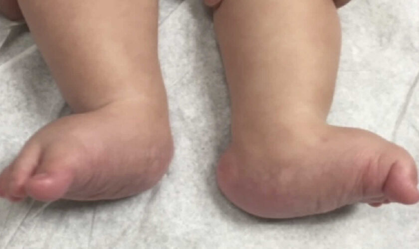
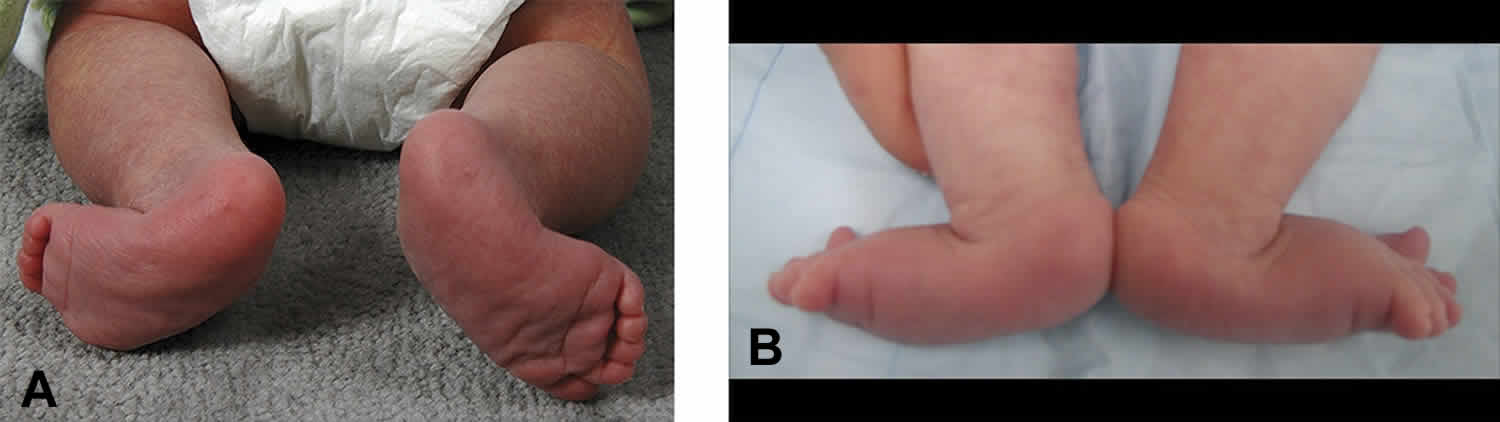
In most infants with diastrophic dysplasia, the first bone within the body of each hand (first metacarpals) may be unusually small and “oval shaped,” causing the thumbs to deviate away (abduction) from the body (“hitchhiker thumbs”). Other fingers may also be abnormally short (brachydactyly) and joints between certain bones of the fingers (proximal interphalangeal joints) may become fused (symphalangism), causing limited flexion and restricted movement of the finger joints. Affected infants also typically have severe foot deformities (talipes or “clubfeet”) due to abnormal deviation and fusion of certain bones within the body of each foot (metatarsals). In addition, many children with the disorder experience limited extension, partial (subluxation) or complete dislocation, and/or permanent flexion and immobilization (contractures) of certain joints.

In most infants with diastrophic dysplasia, there is also incomplete closure of bones of the spinal column (spina bifida occulta) within the neck area and the upper portion of the back (lower cervical and upper thoracic vertebrae). In addition, during the first year of life, some affected children may begin to develop progressive abnormal sideways curvature of the spine (scoliosis). During adolescence, individuals with the disorder may also develop abnormal front-to-back curvature of the spine (kyphosis), particularly affecting vertebrae within the neck area (cervical vertebrae). In severe cases, progressive kyphosis may lead to difficulties breathing (respiratory distress). Some individuals may also be prone to experiencing partial dislocation (subluxation) of joints between the central areas (bodies) of cervical vertebrae, potentially resulting in spinal cord injury. Such injury may cause muscle weakness (paresis) or paralysis and/or life-threatening complications.

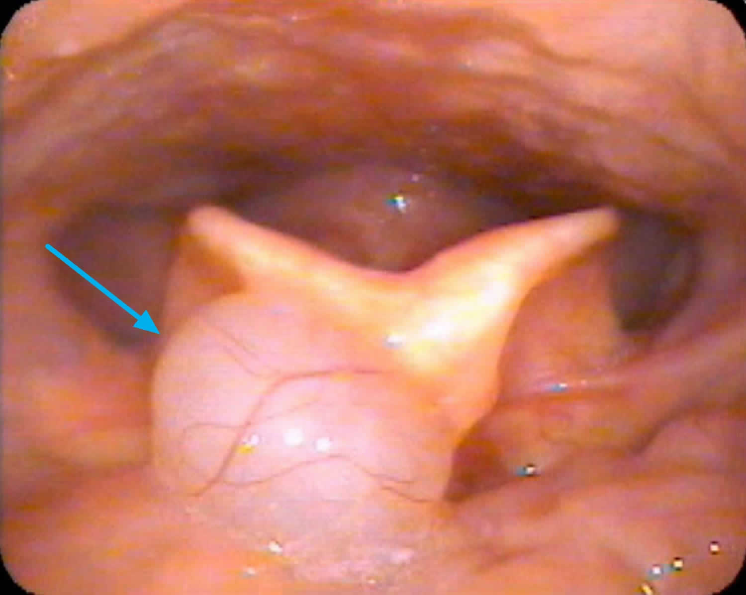
In addition, most newborns with diastrophic dysplasia have or develop abnormal fluid-filled sacs (cysts) within the outer, visible portions of the ears (pinnae). Within the first weeks of life, the pinnae become swollen and inflamed and unusually firm, thick, and abnormal in shape. Over time, the abnormal areas of tissue (lesions) may accumulate deposits of calcium salts (calcification) and eventually develop into bone (ossification). Some affected infants may also have abnormalities of the head and facial (craniofacial) area including incomplete closure of the roof of the mouth (cleft palate) and/or abnormal smallness of the jaws (micrognathia). In addition, in some affected infants, abnormalities of supportive connective tissue (cartilage) within the windpipe (trachea), voice box (larynx), and certain air passages in the lungs (bronchi) may result in collapse of these airways, causing life-threatening complications such as respiratory obstruction and difficulties breathing. In some individuals with the disorder, additional symptoms and physical findings may also be present.

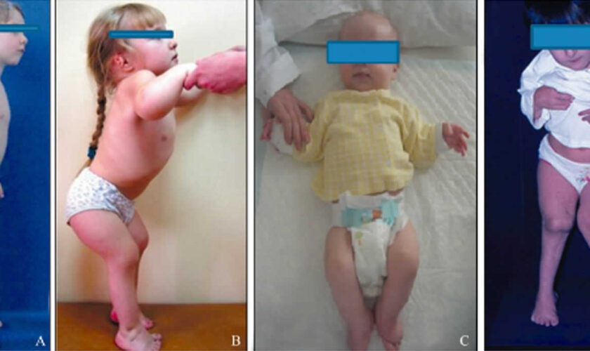
Children with diastrophic dysplasia have characteristic craniofacial features (Figure 1).

Children born with diastrophic dysplasia may do physical things — like rolling over, sitting up and walking — later than other children.

Diastrophic dysplasia is caused by mutations in the SLC26A2 gene and is inherited in an autosomal recessive manner 4). Diastrophic dysplasia occurs equally in males and females and most often in whites. Although the exact prevalence of diastrophic dysplasia is unknown, researchers estimate that it affects about 1 in 500,000 newborns in the United States. Diastrophic dysplasia is more common in Finland, where it affects about 1 in 33,000 newborns 5).

Management of diastrophic dysplasia consists of maintaining joint position and mobility through physical therapy and casting. Surgical correction of clubfoot may be necessary. Arthroplasty of the hips and knees to decrease pain and increase motility may also be indicated. Indications for surgical correction of scoliosis have not yet been established 6).

Figure 1. Diastrophic dysplasia

diastrophic dysplasia

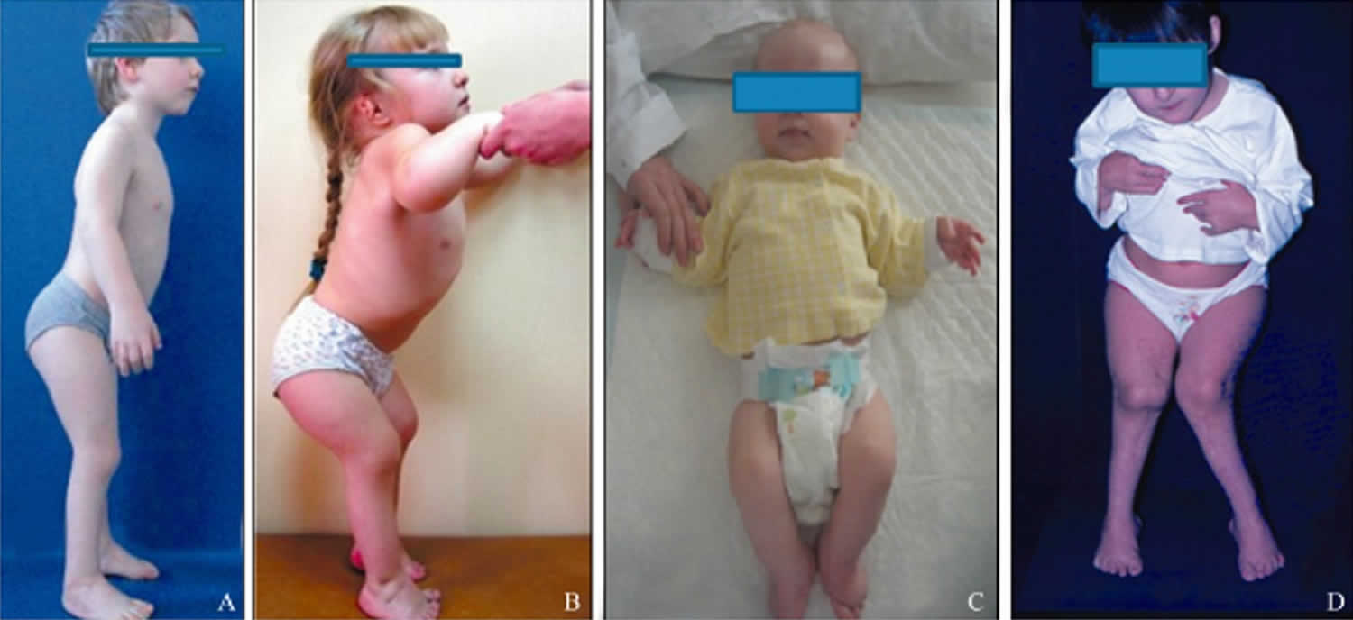
Footnote: The clinical appearances of children with diastrophic dysplasia showing the characteristic craniofacial features (A–C). Figure 1A shows a 10‐year‐old boy with severe short stature, thickened ear lobes and multiple contractures (elbows, pelvis, and knees). Figure 1B shows a 2‐year‐old girl with severe short stature, thickened ear lobes and swellings of the pinnae associated with narrow external auditory canals. Figure 1C shows a 6‐month‐old boy with micromelic dwarfism, a characteristic craniofacial contour and contractures of the elbows and knees. Figure 1D shows a 12‐year‐old girl whose knees had been operated on to relieve the contractures; however, the contractures had recurred.

[Source 7) ]

Diastrophic dysplasia causes

Diastrophic dysplasia is one of several skeletal disorders caused by mutations in the SLC26A2 gene also called DTDST gene (diastrophic dysplasia sulfate transporter gene) that has been located on the long arm (q) of chromosome 5 (5q32-q33.1). The SLC26A2 gene or DTDST gene provides instructions for making a protein that transports charged molecules (ions), particularly sulfate ions, across cell membranes. This protein appears to be active in many of the body’s tissues, including developing cartilage. Cartilage is a tough, flexible tissue that makes up much of the skeleton during early development. Most cartilage is later converted to bone, except for the cartilage that continues to cover and protect the ends of bones and is present in the nose and external ears.

Cartilage cells use sulfate ions transported by the SLC26A2 protein to build molecules called proteoglycans. These molecules, which each consist of several sugars attached to a protein, help give cartilage its rubbery, gel-like structure. Because sulfate ions are required to make proteoglycans, the transport activity of the SLC26A2 protein is essential for normal cartilage formation.

Mutations in the SLC26A2 gene or DTDST gene impede cellular incorporation of sulfate and result in production of undersulfated cartilage proteoglycans, disrupting the assembly of cartilage matrix, preventing bones from forming properly and resulting in the skeletal problems characteristic of diastrophic dysplasia 8).

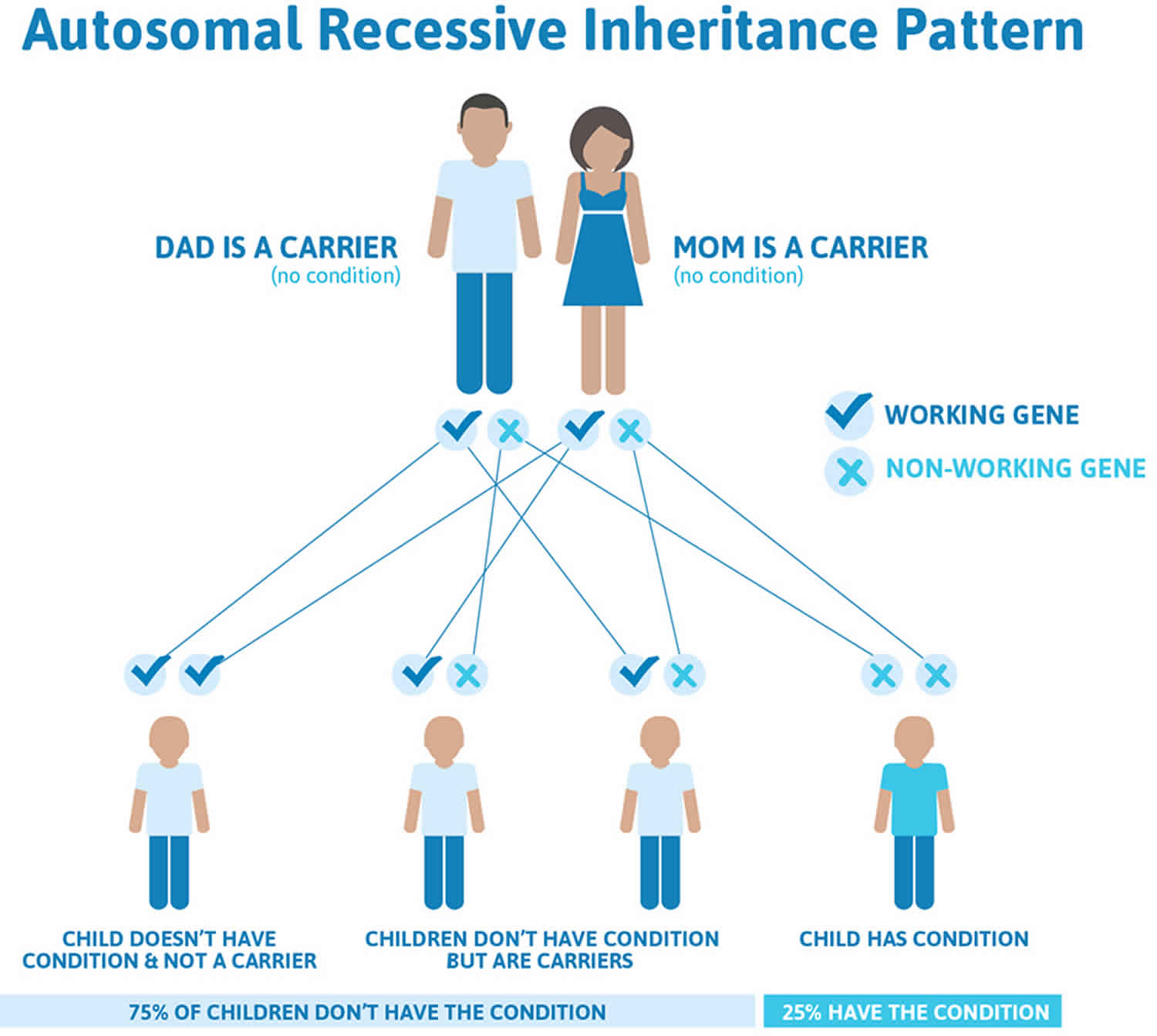
Diastrophic dysplasia inheritance pattern

Diastrophic dysplasia is inherited in an autosomal recessive pattern, which means both copies of the gene in each cell have mutations. The parents of an individual with an autosomal recessive condition each carry one copy of the mutated gene, but they typically do not show signs and symptoms of the condition.

It is rare to see any history of autosomal recessive conditions within a family because if someone is a carrier for one of these conditions, they would have to have a child with someone who is also a carrier for the same condition. Autosomal recessive conditions are individually pretty rare, so the chance that you and your partner are carriers for the same recessive genetic condition are likely low. Even if both partners are a carrier for the same condition, there is only a 25% chance that they will both pass down the non-working copy of the gene to the baby, thus causing a genetic condition. This chance is the same with each pregnancy, no matter how many children they have with or without the condition.

  • If both partners are carriers of the same abnormal gene, they may pass on either their normal gene or their abnormal gene to their child. This occurs randomly.
  • Each child of parents who both carry the same abnormal gene therefore has a 25% (1 in 4) chance of inheriting a abnormal gene from both parents and being affected by the condition.
  • This also means that there is a 75% ( 3 in 4) chance that a child will not be affected by the condition. This chance remains the same in every pregnancy and is the same for boys or girls.
  • There is also a 50% (2 in 4) chance that the child will inherit just one copy of the abnormal gene from a parent. If this happens, then they will be healthy carriers like their parents.
  • Lastly, there is a 25% (1 in 4) chance that the child will inherit both normal copies of the gene. In this case the child will not have the condition, and will not be a carrier.

These possible outcomes occur randomly. The chance remains the same in every pregnancy and is the same for boys and girls.

Figure 3 illustrates autosomal recessive inheritance. The example below shows what happens when both dad and mum is a carrier of the abnormal gene, there is only a 25% chance that they will both pass down the abnormal gene to the baby, thus causing a genetic condition.

Figure 2. Diastrophic dysplasia autosomal recessive inheritance pattern

Diastrophic dysplasia autosomal recessive inheritance pattern

People with specific questions about genetic risks or genetic testing for themselves or family members should speak with a genetics professional.

Resources for locating a genetics professional in your community are available online:

Diastrophic dysplasia symptoms

The symptoms and physical findings associated with diastrophic dysplasia may be extremely variable, differing in range and severity even among affected family members (kindreds). However, in all individuals with the disorder, there is abnormal development of bones and joints of the body (skeletal and joint dysplasia).

Symptoms of diastrophic dysplasia can include:

  • Shortening of the limbs
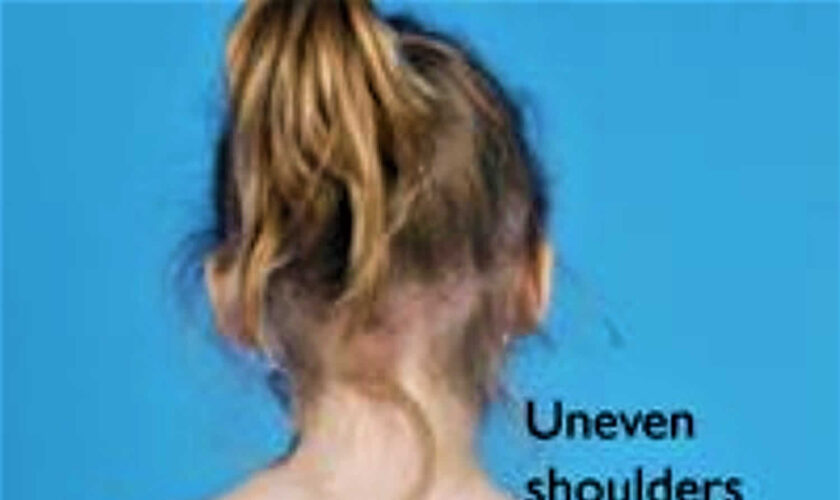
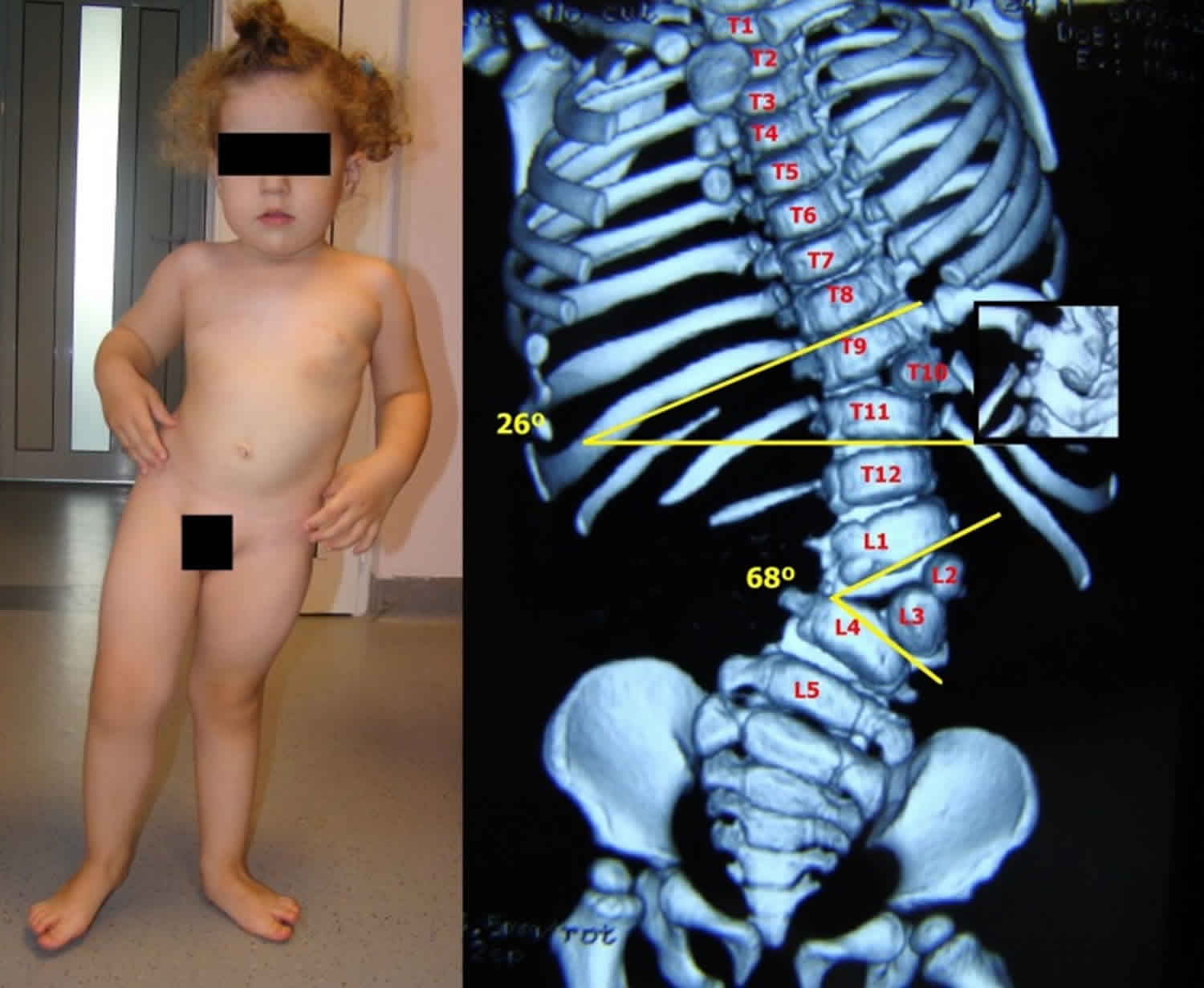
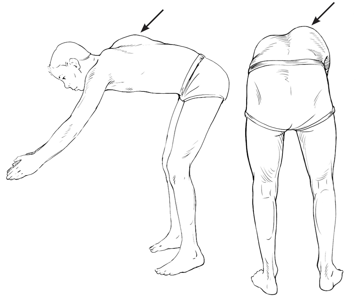
  • Scoliosis and increased lumbar lordosis with a prominent abdomen
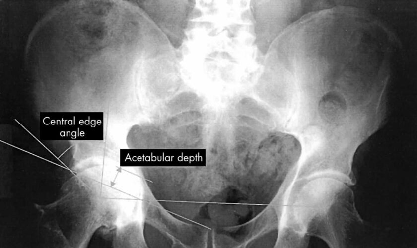
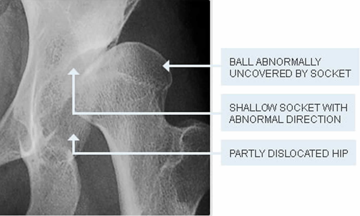
  • Hip dysplasia, in which the two hip joints are misaligned or crooked
  • Deformities of the joints
  • Foot deformities
  • Hernia
  • Cauliflower ear, a deformity of the cartilage of the ear
  • Cleft palate, an opening or gap in the roof of the mouth
  • Hitchhiker’s thumb, where the thumb sticks out at a right angle to the hand, looking like the child is trying to hitch a ride
  • Brachydactyly, in which one or more of the fingers is abnormally short

During normal development before birth (embryonic and fetal development) as well as development during early childhood, cartilage in many areas of the body is gradually replaced by bone (ossification). In addition, a layer of cartilage (epiphyseal cartilage [growth plate]) separates the shafts (diaphyses) of long bones (e.g., bones of the arms and legs) from their ends (epiphyses), allowing long bones to grow until the cartilage is no longer present. In those affected by diastrophic dysplasia, however, there is delayed growth before and after birth (prenatal and postnatal growth retardation), the development of the ends of the long bones (epiphyses) is irregular, and the ossification of the epiphyses is delayed. Thus, affected newborns and children typically have markedly short, bowed arms and legs and short stature (short-limbed dwarfism). In addition, in such cases, growth failure is typically progressive, in part due to absence of the “growth spurt” that usually occurs during puberty. The severity of such growth failure may vary greatly from case to case, including among affected siblings.

Due to abnormalities of skeletal development, infants and children with diastrophic dysplasia also have additional distinctive malformations of bones of the hands, feet, and other areas of the body. For example, the first bone within the body of each hand (first metacarpals) may be unusually small, short, and “oval shaped.” As a result, the thumbs deviate away (abduction) from the body (“hitchhiker thumbs”). In addition, other fingers may be abnormally short (brachydactyly) and joints between particular bones of the fingers (proximal interphalangeal joints) may become fused (symphalangism), causing limited flexion and restricted movement (reduced mobility) of the finger joints. In some cases, bones of the wrists may also be malformed due to premature ossification.

Infants with the disorder also typically have severe foot deformities (talipes or “clubfeet”) due to abnormal fusion and deviation of bones within the body of each foot (metatarsals). In most cases, the heels turn outward (talipes valgus) while the fore part of each foot deviates inward (metatarsus adductus). In other infants, the soles of the feet may be flexed (talipes equinus) and, in some cases, the heels may also turn inward (talipes equinovarus). The great toes, like the thumbs, may also deviate away (abduction) from the body.

In addition to having limited flexion of finger joints, many affected infants and children also experience partial dislocation (subluxation) and/or complete dislocation of particular joints of the body. For example, in many cases, dislocations of the knees and hips occur upon weightbearing. Affected individuals may also have abnormally loose and/or stiff joints; experience limited extension of joints at the elbows and/or knees; and/or develop permanent flexion and immobilization (contracture) of certain joints (e.g., knees). Due to joint and bone abnormalities such as those affecting the feet, many individuals with diastrophic dysplasia have a tendency to walk on tiptoe. In addition, affected individuals may be predisposed to degenerative changes (osteoarthrosis) of particular joints (e.g. of the hips), resulting in pain with use of the joint, tenderness, stiffness, and, in some cases, deformity.

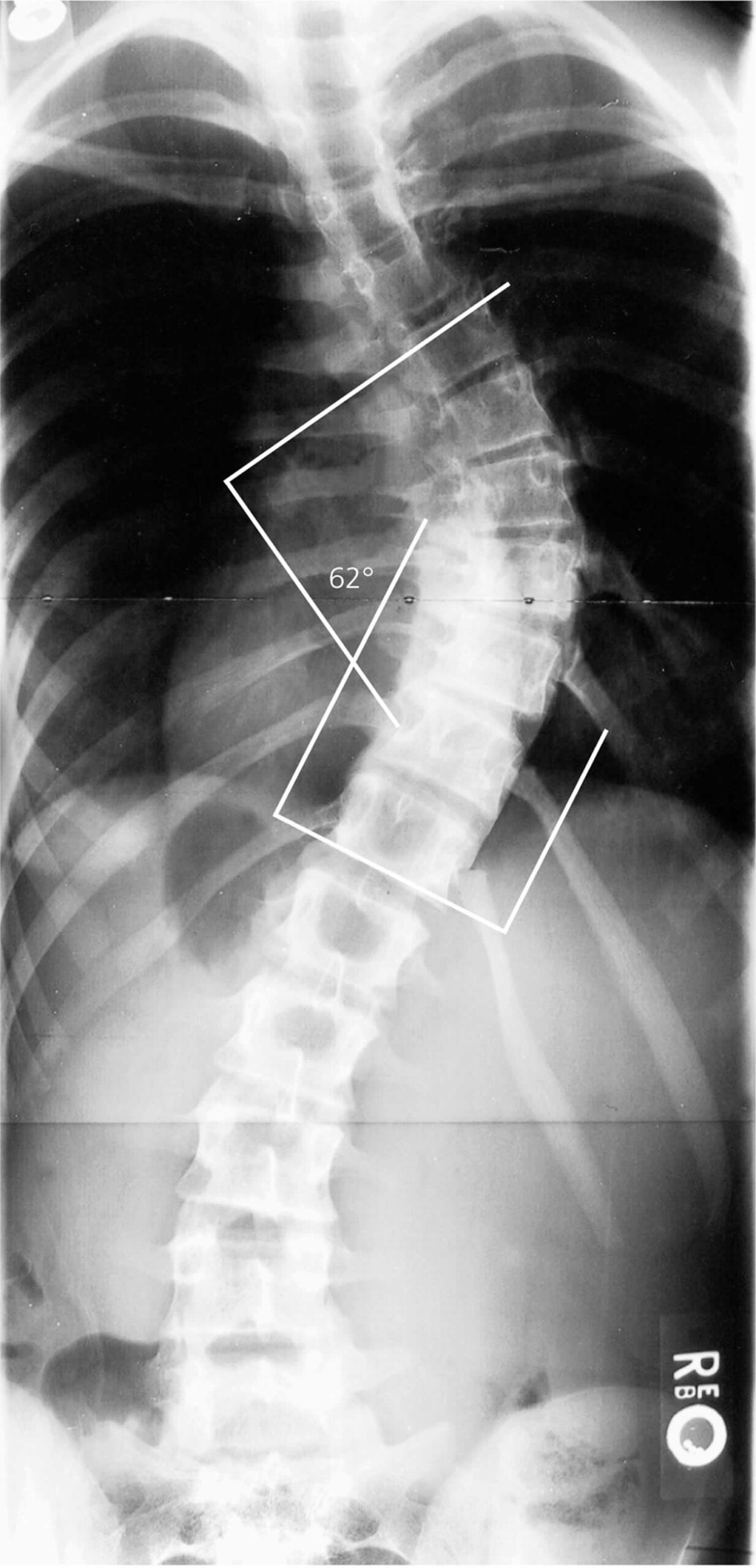
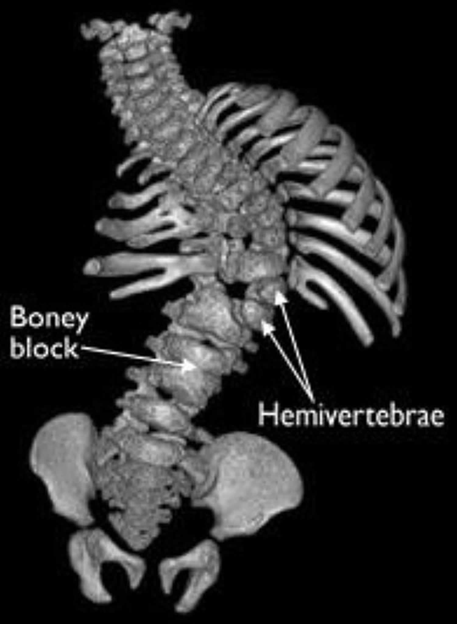
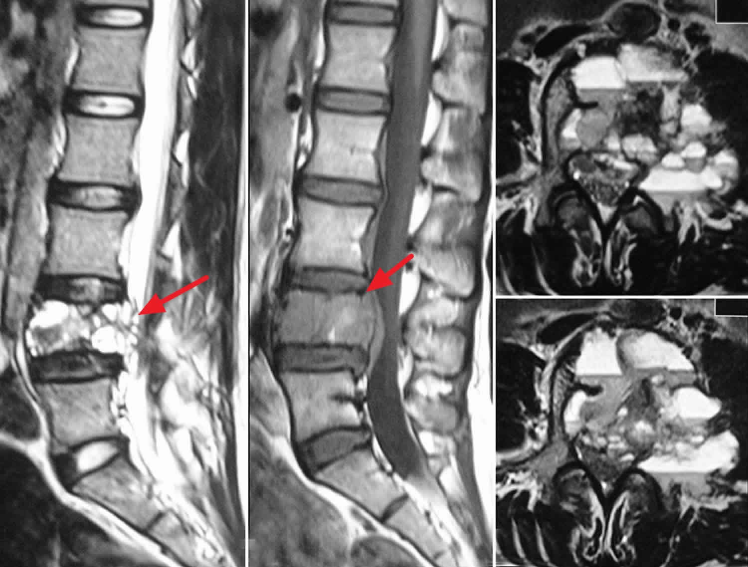
Many infants with diastrophic dysplasia also have abnormalities of bones within the spinal column (vertebrae). For example, in most affected infants, there may be incomplete closure of vertebrae (spina bifida occulta) within the neck area and the upper portion of the back (lower cervical and upper thoracic vertebrae) and/or abnormal narrowing of portions of the vertebrae of the lower back (interpedicular narrowing in lumbar vertebrae). During the first year of life, some infants may begin to develop progressive abnormal sideways curvature of the spine (scoliosis). In addition, during adolescence, individuals with diastrophic dysplasia may also develop abnormal front-to-back curvature of the spine (kyphosis), particularly affecting vertebrae of the neck region (cervical vertebrae). In severe cases, progressive kyphosis may result in difficulties breathing (respiratory distress). Some individuals with the disorder may also be prone to experiencing partial dislocation of joints between the central areas (bodies) of cervical vertebrae (cervical subluxation), potentially resulting in compression of the spinal cord. (This cylindrical structure of nerve tissue extends from the lower portion of the brain and is located inside the central canal within the spinal column [spinal cavity].) Such spinal cord injury may result in muscle weakness (paresis) or paralysis and/or life-threatening complications.

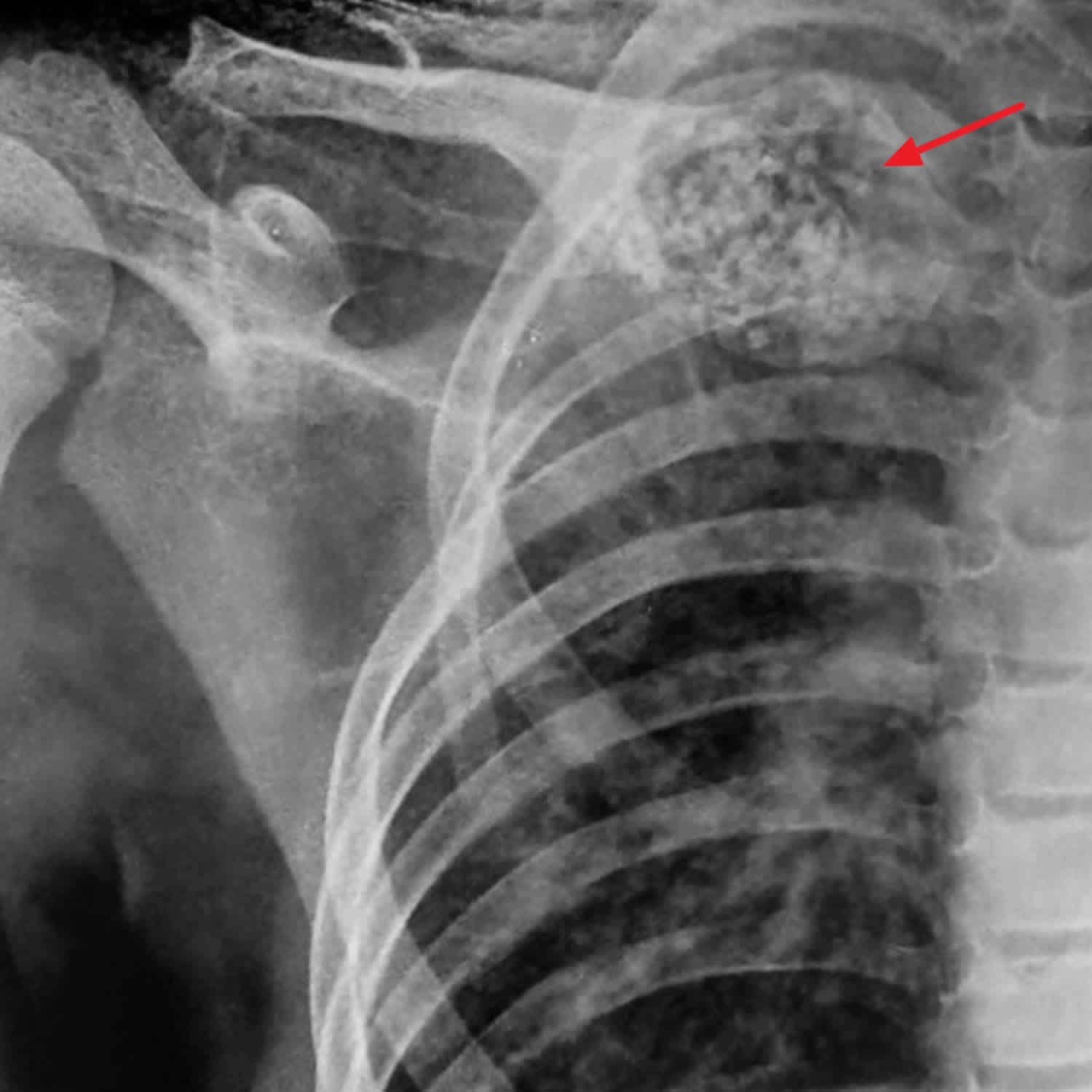
Most newborns with diastrophic dysplasia also have or develop fluid-filled sacs (cysts) within the outer, visible portions of the ears (pinnae). Within approximately two to five weeks after birth, the pinnae become swollen and inflamed. When such swelling and inflammation subside, the pinnae remain unusually thick, hard, and abnormal in shape. The abnormal areas of tissue (lesions) may gradually accumulate deposits of calcium salts (calcification) and eventually be replaced by bone (ossification). Although affected infants may experience associated abnormal narrowing (stenosis) of the external ear canal (external auditory canal), hearing is usually normal. However, according to reports in the literature, other affected infants and children may experience hearing impairment due to such auditory canal stenosis or abnormal fusion or absence of the three tiny bones (auditory ossicles) in the middle ear that conduct sound to the inner ear.

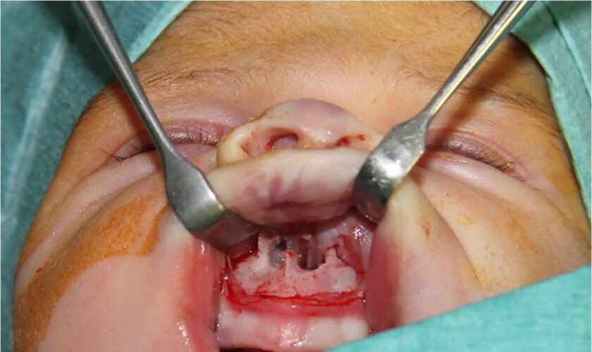
Some infants with diastrophic dysplasia also have characteristic malformations of the head and facial (craniofacial) area, such as an unusually high, prominent forehead; abnormal smallness of the jaws (micrognathia); and/or a broad, highly arched roof of the mouth (palate) or incomplete closure of the palate (cleft palate). Cleft palate has been reported to occur in anywhere from 25 to 60% of affected infants, and may cause difficulties with feeding and/or breathing. In addition, in some infants with diastrophic dysplasia, abnormalities of supportive connective tissue (cartilage) within the windpipe (trachea), voice box (larynx), and air passages in the lungs (bronchi) may cause abnormal narrowing (e.g., laryngotracheal stenosis) and collapse of such airways. In such cases, life-threatening complications such as respiratory obstruction and difficulties breathing (respiratory distress) may result. However, in many cases nasal speech (hyponasality) occurs as a result of the abnormally shaped vocal tract.

Approximately one third of infants and children with diastrophic dysplasia also have dental abnormalities, such as abnormally small teeth and dental crowding. In addition, in some cases, affected infants may have benign, reddish purple growths in the midportion of the face (midline frontal hemangioma) due to an abnormal distribution of tiny blood vessels (capillaries). Some individuals with the disorder may also have additional symptoms and physical findings.

Diastrophic dysplasia diagnosis

In some families with a previous history of diastrophic dysplasia, it is possible that the disorder may be detected before birth (prenatally) during early pregnancy (e.g., first trimester) based upon the results of specialized genetic (i.e., DNA marker) testing. In addition, in some cases, the disorder may be detected during mid pregnancy (e.g., second trimester) through fetal ultrasonography, a specialized imaging technique in which sound waves are used to create an image of the developing fetus. In such cases, diagnosis is most easily established when a clear family history is present. During fetal ultrasonography, a diagnosis of diastrophic dysplasia may be considered due to detection of certain characteristic findings, such as marked shortening of bones of the fingers (phalanges), arms, and legs; abnormal deviation (abduction) of the thumbs (“hitchhiker thumbs”) and great toes; severe deformities of both feet (talipes or “clubfeet”); and/or other findings.

In most cases, diastrophic dysplasia is diagnosed and/or confirmed at birth based upon a thorough clinical evaluation, identification of characteristic physical findings, and a variety of specializing tests, such as advanced imaging techniques.

The diagnosis of diastrophic dysplasia rests on a combination of clinical, radiologic, and histopathologic features. Diagnostic evaluation for diastrophic dysplasia begins with a thorough medical history and physical examination of your child. The diagnosis is confirmed by molecular genetic testing of SLC26A2, the only gene in which pathogenic variants are known to cause diastrophic dysplasia 9). Biochemical studies of fibroblasts and/or chondrocytes may be used in the rare instances in which molecular genetic testing fails to identify SLC26A2 pathogenic variants.

Doctors use a variety of diagnostic tests to diagnose diastrophic dysplasia and possible complications, including:

  • X-rays, which produce images of bones.
  • Magnetic resonance imaging (MRI), which uses a combination of large magnets, radiofrequencies and a computer to produce detailed images of organs and structures within the body.
  • Computed tomography (CT) scan, which uses a combination of X-rays and computer technology to produce cross-sectional images (“slices”) of the body.
  • EOS imaging, an imaging technology which creates 3-dimensional models from two planar images. Unlike a CT scan, EOS images are taken while the child is in an upright or standing position, enabling improved diagnosis due to weight-bearing positioning.
  • Blood tests, which can help determine drug usage and effectiveness, biochemical diseases and organ function.
  • Genetic testing, in which a sample of your child’s saliva is used to identify your child’s DNA.
  • Radioisotope bone scan, which can help locate areas of abnormal growth.
  • Arthrography, which uses colored dye injected into a joint — most commonly the shoulder, hip, knee, elbow or wrist — and X-ray images are taken to identify any problems.

Diastrophic dysplasia treatment

Every child’s condition is different, so treatment is determined on a case-by-case basis. The treatment of diastrophic dysplasia is directed toward the specific symptoms that are apparent in each individual. Treatment for diastrophic dysplasia is multi-pronged because the condition affects several body systems. Treatment may require the coordinated efforts of a team of specialists who may need to work together to systematically and comprehensively plan an affected child’s treatment. Such specialists may include pediatricians; physicians who diagnose and treat abnormalities of the skeleton, joints, muscles, and related tissues (orthopedists); surgeons; physical therapists; dental specialists (orthodontists); specialists who assess and treat hearing problems (audiologists); and/or other health care professionals.

In some cases, careful monitoring may be all that is required. Physicians may carefully monitor affected infants to ensure prompt detection and appropriate preventive or corrective treatment of respiratory obstruction and distress that may result due to certain abnormalities potentially associated with the disorder (e.g., laryngotracheal stenosis). In addition, special supportive measures may be used to help ensure an appropriate intake of nutrients in infants who experience feeding difficulties due to cleft palate. In some cases, surgical procedures may be performed to correct malformations resulting in breathing and/or feeding difficulties. The specific procedures performed will depend upon the location, severity, and combination of such anatomical abnormalities.

In other cases, nonsurgical or surgical treatments may be needed to address specific aspects of the condition.

For example, if your child has scoliosis, a team of orthopaedic surgeons will consider the severity of the curve, where it occurs in the spine, and your child’s age and stage of growth, before determining the best course of action. Treatment may include nonsurgical options such as bracing and physical therapy, or surgical options such as spinal fusion or implanting growing rods to stabilize your child’s spine as she continues to grow.

In affected children with dental abnormalities, braces (orthodontics), dental surgery, and/or other corrective procedures may be undertaken to correct such malformations. Steroid injections and/or other measures may also be used to help decrease the ear deformity that often affects infants with the disorder.

Many children with diastrophic dysplasia are also diagnosed with a variety of orthopaedic conditions including: hip dysplasia, and hand and foot anomalies. In some cases, these conditions are present at birth and can be treated when the child is young.

For example, a child with hitchhiker’s thumb will likely need hand surgery; while a child with a bowed leg may only need a long-leg cast.

In other cases the complication from diastrophic dysplasia may only become evident — or problematic — as your child grows. This is often true for spinal deformities such as scoliosis and hip conditions.

In some cases, physical therapy in combination with surgical and supportive measures may be helpful in improving an affected individual’s ability to walk and perform other movements (mobility). According to the medical literature, although the foot deformities (i.e., talipes or clubfeet) associated with the disorder may be resistant to treatment, early, persistent therapy may be helpful in achieving beneficial results. In addition, because particular skeletal changes associated with diastrophic dysplasia are progressive (e.g., kyphosis) and, in some cases, may lead to severe complications (e.g., respiratory distress, compression of the spine, potential paresis or paralysis), physicians may perform ongoing monitoring to ensure prompt detection of and appropriate preventive and/or corrective measures for such abnormalities.

Genetic counseling will be of benefit for affected individuals and their families. Other treatment for this disorder is symptomatic and supportive.

Follow-up care

Your child with diastrophic dysplasia should continue to be monitored by an orthopaedic physician into adulthood. Annual visits to follow the development of the spine and hips is appropriate.

If your child had spine surgery, he or she will need to see the orthopaedic surgeon about one to two weeks after surgery, then again at three and six months post-surgery. After that, annual monitoring by trained clinicians is strongly encouraged to ensure any problems are spotted and treated as soon as possible.

Additionally, physicians may recommend your child see several different specialists because so many body systems are involved in a diagnosis of diastrophic dysplasia.

For example, your child may see:

  • An otolaryngologist or plastic surgeon for cleft palate
  • A geneticist for individual or family counseling
  • An orthopaedist for any bone- and muscle-related issues
  • A pulmonologist for any breathing issues
  • Physical therapists and occupational therapists to expand your child’s physical dexterity and skill
  • Nutritionists to improve your child’s dietary health

During follow-up visits, X-rays and other diagnostic testing may be done. The goal of continued monitoring is to help spot any irregularities in growth or development and to address health issues as they develop.

Diastrophic dysplasia life expectancy

Children with diastrophic dysplasia can lead normal lives. They can hold good jobs, get married, have children and more.

If you have questions about how your child’s condition and any related health issues may affect your child’s prognosis or long-term goals, talk to your child’s healthcare provider.

References   [ + ]

read more

Congenital vertical talus

congenital vertical talus

Congenital vertical talus

Congenital vertical talus also called rocker-bottom foot is a rare congenital foot deformity in which the sole of a child’s foot flexes abnormally in a convex position giving the foot a rocker-bottom appearance. For this reason, congenital vertical talus is often called rocker-bottom foot. Congenital vertical talus is characterized by a prominent calcaneus/heel and a convexly rounded sole. Congenital vertical talus is usually a rigid deformity, unlike the more common calcaneovalgus foot (flexible deformity), and rarely improves with stretching or bracing. In most cases, surgery is required.

When a child is born, their feet usually appear flat because of the extra fat pads on the bottom. As the child grows, a concave arch in their foot normally develops. In a child with rocker-bottom foot, the bottom of the foot flexes in the opposite direction, making the middle of the foot touch the floor, while the toes and heel curve upward, touching the shin.

Congenital vertical talus is an uncommon disorder. Jacobsen and Crawford 1) reported only 273 cases. Congenital vertical talus (rocker-bottom foot) affects about 1 in 10,000 births and occurs equally in boys and girls. In about half of the cases, both feet are affected. Some have estimated the incidence of congenital vertical talus to be one tenth that of congenital clubfoot.

Congenital vertical talus is often associated with an underlying musculoskeletal or neurological condition such as:

  • Spina bifida
  • Trisomy 13, 14, 15 or 18
  • Arthrogryposis multiplex congenita

In a minority of cases, the cause of rocker-bottom foot is unknown, in an otherwise healthy child.

Although the cause of congenital vertical talus is likely heterogeneous, recent evidence strongly supports a genetic cause linking it to genes expressed during early limb development 2). Traditional management for vertical talus involves extensive surgeries that are associated with significant short- and long-term complications. A minimally invasive approach that relies on serial manipulation and casting to achieve most of the correction has been shown to produce excellent short-term results with regard to clinical and radiographic correction in both isolated and nonisolated cases of vertical talus. Although long-term studies are needed, achieving correction without extensive surgery may lead to more flexible and functional feet, much as Ponseti method has done for clubfeet 3).

Autopsy and surgical findings have contributed to the understanding of the pathologic anatomy of the vertical talus 4). The hindfoot is in marked equinus and valgus caused by contracture of the Achilles tendon and the posterolateral ankle and subtalar joint capsules. The midfoot and forefoot are dorsiflexed and abducted relative to the hindfoot secondary to contractures of the tibialis anterior, extensor digitorum longus, extensor hallucis brevis, peroneus tertius, and extensor hallucis longus tendons and the dorsal aspect of the talonavicular capsule. The navicular is dorsally and laterally dislocated on the head of the talus, resulting in the development of a hypoplastic and wedge-shaped navicular. Both the talar head and neck are abnormal in shape and orientation, resulting in a flat appearance that is angled medially from the midline. The position of the talus stretches vertically and weakens the plantar soft tissues, including the calcaneonavicular, or spring ligament, which gives the foot a rocker-bottom appearance. The plantar surface of the foot is convex, whereas the dorsal aspect of the midfoot has deep creases (see Figure 1). The calcaneus is in extreme equinus, which often causes either dorsolateral subluxation or frank dorsal dislocation of the calcaneocuboid joint. The posterior tibial tendon and the peroneus longus and brevis are commonly subluxated anteriorly over the medial and lateral malleolus, respectively; the subluxated tendons may then function as ankle dorsiflexors rather than plantar flexors 5).

Primary surgical treatment of a congenital vertical talus in a child younger than 2 years can be done with either a one-stage or two-stage extensive soft-tissue release 6). The first stage of the two-stage approach consists of lengthening the contracted dorsolateral tendons, releasing the associated dorsolateral capsular contractures, and reducing the talonavicular and subtalar joint complex. The second stage consists of lengthening the Achilles and peroneal tendons as well as performing a posterolateral capsular release 7). Historically, the one-stage approach was simply a combination of the two stages into a one-stage procedure. Seimon 8) modified the one-stage approach, emphasizing that, by carefully addressing the dorsolateral soft-tissue contractures, less extensive soft-tissue release was needed posteriorly. Mazzocca et al 9) compared Seimon’s dorsal approach with more extensive staged approaches and found that it required less surgical time, had fewer complications, and resulted in improved clinical outcomes. Today, most authors use some form of the single-stage approach 10) and report better results than those achieved using a two-stage approach 11). However, the complications associated with both approaches (eg, wound necrosis, osteonecrosis, undercorrection and overcorrection of deformities) are concerning 12). Longer-term problems include stiffness of the ankle and subtalar joints and the development of degenerative arthritis, leading to the need for salvage procedures, such as subtalar and triple arthrodeses. These problems are similar to the poor long-term outcomes and functional disability reported with extensive soft-tissue release surgery for clubfoot 13).

Figure 1. Vertical talus deformity

vertical talus deformity

Footnote: Clinical photographs of a newborn’s feet demonstrating the features of vertical talus. The plantar aspect of the feet (A) show forefoot abduction deformities, and the dorsolateral aspect of the feet (B) demonstrate deep creases on presentation secondary to forefoot and midfoot dorsiflexion.

[Source 14) ]

Congenital vertical talus classification

Current classification systems for vertical talus focus on either a description of the anatomic abnormalities present or the presence or absence of associated diagnoses. The most widely used anatomic classification system was proposed by Coleman et al 15). They described two types of vertical talus; type 1 deformity is characterized by a rigid dorsal dislocation of the talonavicular joint. In addition to a rigid dislocation of the talonavicular joint, a type 2 deformity has a dislocation or subluxation of the calcaneocuboid joint (ie, the long axis of the calcaneus lies plantar to the long axis of the cuboid). Other classification systems have focused on whether the vertical talus was an isolated deformity or was present in addition to other abnormalities 16). The problem with these classification systems is that they do not directly take into account the motor function of the lower legs. Weak or absent motor function in the lower leg musculature is predictive of not only poor response to initial treatment but also a risk of relapse 17). The child’s ability to dorsiflex and plantarflex the toes can be evaluated by lightly stimulating the dorsal and plantar aspects of the foot. Movement can be graded as definitive, slight, or absent. This simple examination can be repeated at each clinical visit to improve accuracy. A new classification system that takes this into account is needed because the ability to better predict the response to treatment will allow for the development of an individualized treatment program for patients.

It should be noted that current classification systems have attempted to define oblique talus as a milder form of vertical talus based on radiographic and clinical examination criteria 18). However, these attempts at classification have not translated into consistent treatment recommendations because some oblique tali do require treatment despite being milder in nature 19). Oblique tali that have an associated Achilles tendon contracture are at risk of becoming symptomatic with time. For this reason, some experts consider oblique tali and vertical tali to be related entities that occur along a spectrum of severity. Similar to clubfeet, not all vertical tali have the same rigidity. If oblique talus is diagnosed on radiography, but an equinus contracture (defined as the inability to achieve 10° of passive ankle dorsiflexion with the knee extended and flexed) is present, some experts treat it as a vertical talus. Treatment decisions, should be based on the rigidity of the talonavicular joint as well as of the hindfoot.

Vertical talus causes

In most cases, the cause of vertical talus deformity remains unknown. Approximately one half of cases of vertical talus occur in conjunction with neurologic disorders (neuromuscular and central nervous system) 20) or known genetic defects and/or syndromes 21). The other half occur in children without other congenital anomalies and are considered idiopathic or isolated cases 22). Ogata et al 23) proposed a congenital vertical talus classification system that divides patients into the following three groups:

  • Idiopathic
  • Genetic/syndromic
  • Neuromuscular

The most common neurologic disorders associated with vertical talus are distal arthrogryposis and myelomeningocele 24) and the most common genetic defects include aneuploidy of chromosomes 13, 15, and 18 25). Vertical talus is also commonly associated with a variety of syndromes, including De Barsy, Costello, and Rasmussen syndromes22 and split hand and split foot limb malformation disorders. Of the 50% of cases of vertical talus that are isolated, almost 20% have a positive family history of vertical talus. In most of these cases, congenital vertical talus is inherited in an autosomal dominant fashion, supporting the theory that a significant number of isolated cases have a genetic origin, as well 26). Specific gene mutations in the homeobox transcription factor and cartilage-derived morphogenetic protein-1 genes have been found to be causative in some patients with familial, autosomal dominant isolated vertical talus and in some families with congenital hand and foot anomalies of which vertical talus is a feature 27). Growth differentiation factor 5 is closely related to the bone morphogenetic proteins associated with neurologic and limb development.

No single gene defect is responsible for all cases of vertical talus; therefore, it is likely that the pathophysiologic basis for the development of vertical talus is heterogeneous in nature. One hypothesis to explain vertical talus associated with neuromuscular disorders is an imbalance in muscle strength. In patients with myelomeningocele with vertical talus, a weak posterior tibialis and relatively strong ankle dorsiflexors could be contributing factors, whereas weakness of the foot intrinsic muscles may play a contributing role in other neuromuscular disorders. These mechanisms and congenital muscle abnormalities, which are also seen in the setting of distal arthrogryposis, may play a role in some cases of isolated vertical talus, as well. This is supported by the high percentage of abnormal skeletal muscle biopsies performed in this patient population 28). Congenital vascular deficiency of the lower extremities has also been proposed as a potential cause of vertical talus based on magnetic resonance angiography findings that demonstrated congenital arterial deficiencies of the lower extremity in a group of patients with isolated vertical talus 29).

Vertical talus associations

  • Aneuploidic syndromic
    • trisomy 13
    • trisomy 18
    • 18q deletion syndrome
  • Non-aneuploidic non-syndromic
    • spina bifida
    • arthrogryposis

Congenital vertical talus symptoms

The most common symptom of congenital vertical talus is a rocker-bottom appearance of the foot, which is usually obvious at birth or seen when a child begins to walk.

Other symptoms include:

  • An upward flex of the mid- and forefoot
  • The hindfoot is elevated due to an abnormal flex in the ankle
  • The midfoot cannot be properly aligned with the hindfoot
  • Abnormal positioning of the foot; child may walk on the inside of their foot, while the outside edge is elevated, leading to improper balance and weight distribution

Clinically, congenital vertical talus presents as a rigid flatfoot with a rocker-bottom appearance of the foot. The calcaneus is in fixed equinus, and the Achilles tendon is very tight. The hindfoot is in valgus, and the head of the talus is found medially in the sole, creating the rocker-bottom appearance. The forefoot is abducted and dorsiflexed.

The foot is stiff. In ambulatory children, calluses can develop under the head of the talus, which is very prominent along the plantar-medial foot.

Associated genetic syndromes must be excluded; therefore, a consultation with a pediatric geneticist may be indicated.

Congenital vertical talus diagnosis

Early detection of congenital vertical talus is important for successful treatment. Trained pediatric orthopaedic surgeon will perform a complete medical history, a physical examination and a visual evaluation of your child.

During the physical exam, the doctor will examine your entire child — not just their foot. The doctor will be looking for other abnormalities such as multiple joint contractures or evidence suggesting your child may have a larger multisystem genetic disorder.

Doctors will also closely examine your child’s foot — while standing and in motion — to determine if your child has rocker-bottom foot, or a more common and benign conditions such as calcaneovalgus foot or flat foot. Though symptoms of these conditions may mimic each other in young children, treatments are very different.

Physical examination

Hindfoot equinus, hindfoot valgus, forefoot abduction, and forefoot dorsiflexion are present in all newborns with vertical talus. The rigidity of the deformity is the key to distinguishing between vertical talus and more common conditions, such as calcaneovalgus foot, posteromedial bowing of the tibia, and oblique talus. If hindfoot equinus is not a clinical feature, then the deformity is not vertical talus and is likely positional in nature. Because of the frequency of neuromuscular and genetic abnormalities associated with vertical talus, it is important to perform a comprehensive physical examination. The clinician should look for facial dysmorphic features that require a referral to a geneticist or abnormalities suggestive of a neuromuscular etiology, which would require MRI evaluation of the neuroaxis and referral to a pediatric neuromuscular specialist. The presence of a sacral dimple, in particular, should alert the examiner to possible central nervous system anomalies.

It is equally important for the examiner to document motor function of the foot and ankle with special attention to the toe flexors and extensors. This is done by stimulating the plantar and dorsal aspects of the foot separately to elicit plantar flexion and dorsiflexion of the toes. This should be done serially during treatment sessions because the examination can be difficult, and results from serial examinations are more telling. The presence of dorsiflexion and plantar flexion of the toes is recorded as absent, slight, or definitive. This should be recorded for the great toe alone as well as the lesser toes as a separate group. In our experience, slight or absent ability to move the toes with stimulation correlates with a vertical talus deformity that is more rigid and less responsive to treatment. It may also be indicative of a subtle congenital neurologic or muscular anomaly.

Clinically, a congenital vertical talus foot has a convex plantar surface that results in a rocker-bottom appearance (Figure 1A). The dorsum of the foot has deep creases secondary to forefoot and midfoot dorsiflexion (Figure 1B). The extreme dorsiflexion of the forefoot creates a distinct palpable gap dorsally where the navicular and talar head would articulate in a normal foot. Characteristics of this gap can help the examiner assess rigidity. If the gap reduces with plantar flexion of the forefoot, then the deformity has a degree of flexibility; this may help predict responsiveness to treatment. Left untreated, a rigid vertical talus deformity may worsen with weight bearing because secondary adaptive changes occur in the tarsal bones 30). Painful callosities can develop along the plantar medial border of the foot around the prominent and unreduced talar head. Heel strike does not occur, shoe wear becomes difficult, and pain develops 31).

Imaging studies

To confirm the diagnosis or better understand the anatomy of your child’s foot and leg, doctors may also order imaging tests such as:

  • X-rays, which produce images of bones. Weightbearing anteroposterior (AP) and lateral views of the foot are the first radiographs that must be obtained. A lateral radiograph with the foot in maximum plantarflexion is mandatory to confirm congenital vertical talus. The hallmark of congenital vertical talus deformity is an abnormally positioned talus bone (this is the bone that connects the foot to the ankle). Because the navicular may not be ossified, the alignment of the first metatarsal to the talus must be evaluated. In a vertical talus, the metatarsal does not line up with the talus. Lines drawn through the long axis of the first metatarsal and the talus converge on the plantar aspect of the foot. Hamanishi 32) described two radiographic angles: the talar axis–first metatarsal base angle (TAMBA) and the calcaneal axis–first metatarsal base angle (CAMBA). The changing point from a flexible oblique talus to rigid CVT is a TAMBA of approximately 60° and a CAMBA of 20°.
  • EOS imaging, an imaging technology that creates 3-dimensional models from two flat images. Unlike a CT scan, EOS images are taken while the child is in an upright or standing position, enabling improved diagnosis due to weight-bearing positioning.
  • Computed tomography (CT) scan, which uses a combination of X-rays and computer technology to examine bones and produces cross-sectional images (“slices”) of the body.
  • Magnetic resonance imaging (MRI), which uses a combination of large magnets, radiofrequencies and a computer to produce detailed images of organs, soft tissues, muscles, ligaments and other structures within the body. Your child is exposed to no radiation during an MRI. Magnetic resonance imaging (MRI) of the spine may be indicated if an occult spinal dysraphism, such as lipomeningocele, is suspected 33). Posterior and lateral lumbar spine radiographs also may be useful to exclude occult spinal dysraphism. Thometz et al 34) evaluated nine patients with congenital vertical talus using MRI to evaluate the three-dimensional morphologic changes and pathoanatomy. They concluded that there is significant pathology at the level of the subtalar joint.
  • Ultrasonography. Ultrasonography has been reported to be helpful in distinguishing between congenital vertical talus (irreducible talonavicular dorsal dislocation) and oblique talus (reducible talonavicular dorsal dislocation) 35). Lateral radiographs of the foot in maximal plantarflexion can reveal if the navicular is reducible. However, radiographs of an infant’s foot can be difficult to interpret. The use of dynamic ultrasonography has been reported to be helpful in the evaluation of infants with vertical or oblique talus 36).

If your child appears to have a neurological condition, the orthopedic physician may refer your child to a neurologist for a complete neurological exam.

Congenital vertical talus treatment

All children with congenital vertical talus will require some form of treatment. While some children may be helped with non-surgical treatment, most will require surgery.

Non-surgical treatment

Doctors may recommend a variety of non-surgical treatments to prevent your child’s condition from getting worse. These include:

  • Stretching exercises for the forefoot and hindfoot
  • Serial manipulation and casting of the midfoot and forefoot in a flexed position to reduce the upward curve of the foot

Improvements from these treatments do occur, but are often temporary.

Congenital vertical talus surgery

Surgery for congenital vertical talus is complicated because it involves correcting foot movement in three directions — side-to-side, up-and-down and front-and-back. A specialist in pediatric foot deformities should perform it. Surgery can dramatically improve the long-term outcomes for your child with congenital vertical talus, but it can also be a stressful experience for you and your child. With adequate serial casting, need for extensive soft-tissue release surgery can be minimized to minimally invasive tendon procedures which leave smaller scars and shorter recovery time. Other procedures can include bone work in older children.

Controversy exists over the choice of surgical approaches. However, some experts believe that the choice of structures to be released is a more important factor in determining outcomes than is the choice of incisions to be used. Special attention must be paid to the dorsal and dorsolateral contracted tissues. Controversy also exists over the need for an anterior tibialis tendon transfer.

Several authors, beginning with Osmond-Clarke 37), Herndon and Heyman 38) and Coleman and associates 39), described staged two-incision reconstructive surgery. The first stage of the Coleman procedure consisted of lengthening the extensor digitorum longus, the extensor hallucis longus, and the tibialis anterior, with capsulotomies of the talonavicular and calcaneocuboid joints and release of the talocalcaneal interosseous ligament. The second stage consisted of Achilles tendon lengthening and a posterior capsulotomy of the ankle and subtalar joints.

After noting a high incidence of complications with the two-stage technique, Ogata et al 40) recommended a single-stage procedure with a medial approach. Kodros and Dias 41) published results they derived using a single-stage approach with a Cincinnati incision.

Seimon described a single-stage dorsal approach in which the extensor digitorum longus and the peroneus tertius were tenotomized and the talonavicular joint was opened 42). The talonavicular joint was reduced and held with a K-wire. The Achilles tendon was lengthened percutaneously. Stricker and Rosen 43) published their experience with this technique, as did Mazzocca et al 44); both groups noted excellent results with few complications.

The trend toward less surgery for congenital vertical talus continued with Dobbs et al 45), who published their technique of casting, percutaneous K-wire pinning of the talonavicular joint, and percutaneous heel-cord tenotomy. No patients had extensive soft-tissue releases, though some required lengthening of the tibialis anterior or the peroneus brevis tendon. Casting without pinning of the talonavicular joint was associated with recurrence of deformity.

Saini et al 46) reported on their surgical experience with 20 cases of congenital vertical talus using a dorsal approach. According to the authors, talonavicular reduction was achieved in all 20 feet, and postoperative talocalcaneal and talo-first metatarsal angles were significantly improved. The results were retained at 4-year follow-up 47).

Bhaskar 48) described a surgical technique used for idiopathic congenital vertical talus in four feet; this technique was similar to the Ponseti technique for clubfoot, except that the forces applied were in a reverse direction. The four feet were treated by serial manipulation and casting, tendo Achillis tenotomy, and percutaneous pinning of the talonavicular joint.

To correct the forefoot deformity, four to six plaster cast applications were required 49). Once the talus and navicular were aligned, percutaneous fixation of the talonavicular joint with a K-wire and percutaneous tendo Achillis tenotomy under anesthesia were performed, followed by application of a cast with the foot in slight dorsiflexion. After treatment, the mean talocalcaneal angle decreased from 70º to 31º, and the mean talar axis–first metatarsal base angle (TAMBA) decreased from 60º to 10.5º.

Wright et al 50) reported on 12 children (21 feet) with idiopathic and teratologic causes. They noted 10 recurrences, a rate higher than those cited in other reports. The authors felt that a limited capsulotomy of the talonavicular joint might reduce the risk of recurrence. They did not find a difference in results between the two groups of patients.

In 2012, Chalayon et al 51) reported on 15 consecutive patients (25 feet) with nonisolated congenital vertical talus who were followed for a minimum of 2 years after reverse Ponseti casting, percutaneous Achilles tendon lengthening and pin fixation of the talonavicular joint. Five feet required a small medial incision to ensure joint reduction and accurate pin placement, and 20 feet had selective capsulotomies of the talonavicular joint and the anterior aspect of the subtalar joint. Initial correction was obtained in all cases, but recurrence was noted in three patients (five feet).

Yang and Dobbs 52) published a comparison of the minimally invasive method versus extensive soft-tissue release with a minimum follow-up of 5 years (Dobbs technique). They documented that the minimally invasive method resulted in better results in terms of range of motion and patient-reported outcomes.

Chan et al 53) evaluated the Dobbs method for correction of idiopathic congenital vertical talus versus correction of teratologic congenital vertical talus. The results were comparable, but the recurrence rate was slightly higher for teratologic congenital vertical talus.

Complications

Complications can occur around the time of surgery (perioperatively) or can manifest early or late in the postoperative period.

Common complications in the perioperative period include infection, wound-healing problems, and skin slough; however, these complications are not unique to congenital vertical talus.

In the first 1-2 years after surgery, the deformity can recur, usually secondary to undercorrection. Undercorrection can occur because of incomplete talonavicular reduction, insufficient posterior ankle release, or residual forefoot abduction. [16] Recurrence of the deformity can also be attributable to neurologic causes, especially in patients with spina bifida. Kodros and Dias reported a high recurrence rate in patients with spina bifida and believed that in these cases the recurrences might be secondary to a tethered spinal cord or other neurologic abnormality.

avascular necrosis (AVN) of the talus is a unique complication of congenital vertical talus surgery. It was more often reported in the older literature and was associated with the two-stage release and extensive surgery. Subsequent articles by Kodros and Dias 54), Seimon 55), Stricker and Rosen 56), and Mazzocca et al 57) did not report occurrences of avascular necrosis (AVN) of the talus.

Late complications include restricted range of motion of the foot and ankle, which can contribute to calf muscle atrophy. This in turn can lead to easy fatigue of the affected limb.

Congenital vertical talus prognosis

Most children who are surgically treated for congenital vertical talus have good outcomes. Some children may need an orthotic to ensure proper foot alignment during critical growth and development periods.

Children who have congenital vertical talus as part of a larger neurological or musculoskeletal syndrome will likely need lifelong follow-up care.

In general, the outcome and prognosis are good 58). Some minor calf atrophy and foot size asymmetry occur and are more noticeable in unilateral cases. Ankle range of motion is about 75% of normal. If avascular necrosis (AVN) of the talus occurs, the results are less optimal because of ankle pain, stiffness, and weakness.

Patients with congenital vertical talus have a more favorable prognosis when treated with the Dobbs technique than they do when treated with extensive soft-tissue release. Idiopathic congenital vertical talus tends to have a more favorable outcome than teratologic congenital vertical talus does 59).

References   [ + ]

read more

Congenital scoliosis

congenital scoliosis

Congenital scoliosis

Congenital scoliosis is a sideways curvature of the spine that is caused by a defect that was present at birth. The term “congenital” means that you are born with the condition. Congenital scoliosis occurs in only 1 in 10,000 newborns and is much less common than the type of scoliosis that begins in adolescence.

Congenital scoliosis starts as the spine forms before birth. Part of one vertebra (or more) does not form completely or the vertebrae do not separate properly. Some types of congenital scoliosis can change quickly with growth while others remain unchanged.

Children with congenital scoliosis sometimes have other health issues, such as kidney, heart or bladder problems.

Even though congenital scoliosis is present at birth, it is sometimes impossible to see any spine problems until a child reaches adolescence.

No certain cause of congenital scoliosis has been identified until today 1). Congenital scoliosis is a failure of vertebral formation, segmentation, or a combination of the 2 arising from abnormal vertebral development during weeks 4 to 6 of gestation. The associated spinal deformity can be of varying severity and result in a stable or progressive deformity based on the type and location of the anomalous vertebrae 2). The ultimate cause is probably multifactorial, involving some combination of inherited genetic susceptibility and de-novo alteration in molecular mechanisms, possibly from exposure to teratogens such as cigarette smoking, organophosphate pesticides, or carbon monoxide 3).

The diagnosis requires a thorough clinical and imaging examination in order to establish an individualized therapeutic strategy.

The treatment of congenital scoliosis is different from the adolescent idiopathic one. Therapeutic criteria are significantly different. It is essential to assess the difference in growth of the concavity related to the convexity when choosing a particular procedure. The magnitude of the curve and the progressive rate are fundamental issues to the surgeon.

Defects of segmentation usually induce a severe deformity. If the segmentation defect is associated to 2-3 fully segmented hemivertebrae, a maximal progression rate is present and significant curves are present at early ages (Figure 1). A severe curve in older children is very difficult to correct and a result is never obtained as in idiopathic scoliosis where correction may be up to 50-60%. In congenital scoliosis corrections of such kind of curves require laborious interventions, osteotomy or segmental resection, with high neurological risks 4) and low rates of success, not exceeding 20% as presented in different statistical data.

As a strategic aspect, the surgeon has to know that the preoperative planning has to identify the presence or absence of a dysraphic status 5) or syringomyelia. Always, the first aim is to stop the progression of the deformity.

Figure 1. Congenital scoliosis

congenital scoliosis

Footnote: Congenital scoliosis with a high progression potential due to 3 hemivertebrae situated on the same side of the spine. Trunk shifting and shoulder imbalance are noticed at an early age

[Source 6) ]

Congenital scoliosis types

Congenital scoliosis represents a wide range of pathology from the simple, stable hemivertebra to the complex, progressive spinal deformity with chest wall abnormalities and associated cardiac, renal, and neural axis anomalies 7).

The first classification of congenital scoliosis based on X-rays imaging was described by Winter in 1968 8). Kawakami 9) reclassified the vertebral malformations depending on the presence or absence of normal formation based on a 3D-CT study. The purpose of these classifications is to understand the embryology, etiology, prognostic and to choose the right therapeutic strategy.

Congenital scoliosis is a malformation characterized by a longitudinal and rotational imbalance.

  1. Congenital scoliosis with imbalance in the longitudinal growth is produced by defects of formation, segmentation or mixed ones.
  2. Congenital scoliosis with rotational imbalance is mainly characterized by the vertebral rotation related to the curve in the coronal plan and they may be due to an effect of:
    • spinal traction – osseous bridges with congenital transverso-sacrate synostosis;
    • spinal pushing – mega-apophysis of the L5 transverse process;
    • mixed (traction and pushing) – sacral agenesis with pelvic malposition.
    • These types of scoliosis are secondary to a congenital malformation, either vertebral or pelvic, which induce as the main manifestation the vertebral rotation by means of traction, pushing or mixed action. Usually, the scoliosis and the vertebral rotation are not present at birth. During growth and development, the first presence is the vertebral rotation accompanied by walking impairment and next by the scoliotic curve.

Congenital scoliosis with longitudinal imbalance, mainly with deviation in the coronal plan in comparison with vertebral rotation, may be due to defects of formation with the presence of: trapezoidal vertebra, hemivertebra or vertebral hemibody. Hemivertebra depending on segmentation may be fully segmented, hemisegmented (partially segmented) or unsegmented. There may be congenital malformations characterized by the presence of more than one hemivertebra disposed in the following manner: adjacent (successive) – 2-3 hemivertebrae disposed unilaterally inducing a short arch scoliosis, being noticed at birth and having a high rate of evolution, unilateral alternant (intermittent) – 2-3 hemivertebrae placed unilaterally leading to a long arch scoliosis and a unique curve and bilateral alternant which themselves may be:

  • compensated – 2 symmetric hemivertebrae in a 4-5 vertebral segment inducing an equilibrated spinal deformity not requiring surgery;
  • uncompensated – if the hemivertebrae are disposed on a distance of more than 6 vertebrae leading to a double congenital scoliosis.

Due to defects of segmentation, congenital scolioses are characterized by a unilateral defect: longitudinal bar or a bilateral defect: vertebral block.

The third possibility is represented by mixed anomalies where we may find the next malformations:

  • hemivertebra and a bar on the opposite spinal side;
  • hemivertebra, vertebral block and longitudinal bar.

Incomplete formation of vertebrae

As the spine forms before birth, part of one vertebra (or more) may not form completely. When this occurs, the abnormality is called a hemivertebra and can produce a sharp angle in the spine. The angle can get worse as the child grows.

This abnormality can happen in just one vertebra or in many throughout the spine. When there is more than one hemivertebra, they will sometimes balance each other out and make the spine more stable.

Failure of separation of vertebrae

During fetal development, the spine forms first as a single column of tissue that later separates into segments that become the bony vertebrae. If this separation is not complete, the result may be a partial fusion (boney bar) joining two or more vertebrae together.

Such a bar prevents the spine from growing on one side after a child is born. This results in a spinal curve that increases as a child grows.

Combination of bars and hemivertebrae

The combination of a bar on one side of the spine and a hemivertebra on the other causes the most severe growth problem. These cases can require surgery at an early age to stop the increased curvature of the spine.

Compensatory curves

In addition to scoliosis curves, a child’s spine may also develop compensatory curves in order to maintain an upright posture. This occurs when the spine tries to make up for a scoliosis curve by creating other curves in the opposite direction above, or below, the affected area. The vertebrae are shaped normally in compensatory curves.

Congenital scoliosis causes

Congenital scoliosis is a failure of vertebral formation, segmentation, or a combination of the 2 arising from abnormal vertebral development during weeks 4 to 6 of gestation. The associated spinal deformity can be of varying severity and result in a stable or progressive deformity based on the type and location of the anomalous vertebrae 10).

The ultimate cause of a congenital spinal abnormality is an irrecoverable difference in spine development at the embryonic level. Much research has been performed to understand the mechanisms of embryonic segmentation, and a variety of genetic defects have been suggested as a cause for congenital vertebral malformations.

Single-nucleotide polymorphisms in glucose-metabolizing genes, including GLUT1, HK1, and LEP, have been linked to the occurrence of malformations observed in diabetic embryopathy 11). Hox-mediated gene expression has been thought to be the target for spine abnormalities related to carbon monoxide 12). In addition, an increased risk of congenital vertebral malformations is noted in monozygotic and dizygotic twins 13).

Despite the isolation of genetic mechanisms, no convincing familial linkage exists to explain the majority of congenital scoliosis cases. Winter et al 14) found that only 13 of 1250 patients had a positive family history of such deformity. Furthermore, hereditary congenital scoliosis is relegated to sporadic case reports and is described mostly in the setting of an overlying syndrome, such as Jarcho-Levin (extensive defects of segmentation in association with spondylocostal, costovertebral, or spondylothoracic dysplasia).

The ultimate cause is probably multifactorial, involving some combination of inherited genetic susceptibility and de-novo alteration in molecular mechanisms, possibly from exposure to teratogens such as cigarette smoking, organophosphate pesticides, or carbon monoxide 15).

Congenital scoliosis symptoms

Congenital scoliosis is often detected during the pediatrician’s examination at birth because of a slight abnormality of the back.

Scoliosis is not painful, so if the curvature is not detected at birth, it can go undetected until there are obvious signs — which could be as late as adolescence. A child may suspect that something is wrong when clothes do not fit properly. Parents can discover the problem in early summer when they see their child in a bathing suit.

The physical signs of scoliosis include:

  • Tilted, uneven shoulders, with one shoulder blade protruding more than the other
  • Prominence of the ribs on one side
  • Uneven waistline
  • One hip higher than the other
  • Overall appearance of leaning to the side
  • In rare cases there may be a problem with the spinal cord or nerves that produces weakness, numbness, or a loss of coordination.

Associated anomalies

Letts et al 16) found that 82% of patients with congenital scoliosis had associated malformation in four different organ systems. Beals et al found that 61% of patients with congenital scoliosis had abnormalities in seven different organ systems. Highest on the list were anomalies of the genitourinary tract.

Research on patients with congenital scoliosis by MacEwen et al 17) revealed a 20% incidence of urinary tract anomalies detected on routine intravenous pyelography (IVP), whereas a study by Hensinger et al 18) on cervical spine anomalies found a rate of 33%. Many of these anomalies, such as the presence of a single kidney, duplicate ureters, or crossed renal ectopia, while being of interest, were not potentially dangerous. However, in about 5% of the patients, obstructive uropathy, most commonly urethrovesicular obstruction, was present.

Renal ultrasonography (US) and magnetic resonance imaging (MRI) may be used to diagnose renal anomalies accurately 19).

A second area of great concern is cardiac anomalies. As many as 10-15% of patients with congenital scoliosis have been noted to have congenital heart defects. Murmurs should never be attributed to the scoliosis and must be evaluated thoroughly.

The frequency of spinal dysraphism is high in patients with congenital scoliosis. The prevalence of a dysraphic lesion was approximately 40% in three independent studies. McMaster 20) reported that about 20% of these patients with congenital scoliosis had some form of dysraphism (eg, diastematomyelia, tethered spinal cord, fibrous dural band, syringomyelia, or intradural lipoma). Many other anomalies can occur in addition to the above problems, such as Sprengel deformity, Klippel-Feil deformity, Goldenhar syndrome (oculoauriculovertebral dysplasia), and anal atresia.

Neurologic malformations

Congenital scolioses are associated in 35% of the patients with other neurologic malformations related to the nervous system and its coating. The most frequently encountered are diastematomyelia, Chiari’s malformation, intradural lipoma and tethered cord 21).

Congenital heart malformations

Congenital heart malformations are present in up to 25% of the patients with congenital scoliosis. Severe anomalies like Fallot tetralogy or the transposition of the great vessels require surgery prior to a spinal surgical approach 22).

Urologic anomalies

Urologic anomalies are encountered in 20% of the cases. These anomalies associated to congenital scoliosis are horseshoe kidney, vesicoureteral reflux (VUR) or hypospadias. Some kids may also have inguinal hernia, which is usually of great size, needing surgery 23).

Musculoskeletal anomalies

These malformations, clinically and imagistically detected, are usually treated after scoliosis surgery. They include Sprengel’s disease, Klippel-Feil syndrome 24), congenital femoral hypoplasia or acetabular dysplasia.

Congenital scoliosis diagnosis

The doctor will initially take a detailed medical history and may ask questions about recent growth.

Taking a full, detailed history and performing a full physical examination are mandatory because associated anomalies of many organs are common. [18] Maternal perinatal history, family history, and developmental milestones must be explored fully. A comprehensive review of systems includes evaluation for the following:

  • Hearing, visual, and dental problems
  • Cleft palate and cleft lip
  • Hernias, anorectal abnormalities, and genitourinary problems
  • Cardiac murmurs
  • Respiratory complaints
  • Neurologic disorders

In the physical examination, the physician must not only explore the spinal deformity but also focus particular attention on chest deformities and cutaneous lesions (especially dimples and hair patches overlying the spine). A detailed neurologic examination should be performed.

The genitalia should be examined for maturity, epispadias, hypospadias, and the presence of undescended testicles. The hand must be examined for clubhand, thenar hypoplasia, or other, more subtle, anomalies. The feet must be studied for clubfeet, cavus or varus deformities, vertical tali, clawing of the toes, or other signs of motor weakness.

During the physical exam, your doctor may have your child stand and then bend forward from the waist, with arms hanging loosely, to see if one side of the rib cage is more prominent than the other.

Your doctor may also perform a neurological exam to check for:

  • Muscle weakness
  • Numbness
  • Abnormal reflexes

Physical examination

Physical examination for scoliosis mainly consists of the Adam’s forward bend test or the forward bending test (Figure 2) 25). A spinal deformity will be most noticeable when your child is in this position. Your child stands and bends forward at the waist, your doctor will observe your child from the back assessing for symmetry of the back from behind and beside your child, looking for a difference in the shape of the ribs on each side 26). A child with possible scoliosis will have a lateral bending of the spine, but the curve will cause spinal rotation and eventually a rib hump, which is visible on examination 27).

The standard screening test for scoliosis is the forward bending test. Your child will bend forward and your doctor will observe your child from the back.

With your child standing upright, your doctor will check to see if the hips are level, the shoulders are level, and that the position of the head is centered over the hips. He or she will check the movement of the spine in all directions.

To rule out the presence of a spinal cord or nerve problem, your doctor may check the strength in your child’s legs and the reflexes in the abdomen and legs.

Figure 2. Forward bending test (Adam’s forward bend test)

Adam's forward bend test

[Source 28) ]

Tests

Although the forward bending test can detect scoliosis, it cannot detect the presence of congenital abnormalities. Imaging tests can provide more information.

X-rays. Images of your child’s spine are taken from the back and the side. The x-rays will show the abnormal vertebrae and how severe the curve is. If a doctor suspects that an underlying condition — such as a tumor — is causing the scoliosis, he or she may recommend additional imaging tests, such as an MRI.

Once your doctor makes the diagnosis of congenital scoliosis, your child will be referred to a pediatric orthopaedic surgeon for specialized care and further tests.

Computed tomography (CT) scan and 3D-CT. A CT scan can provide a detailed image of your child’s spine, showing the size, shape, and position of the vertebrae. To see the vertebrae better, your doctor may have a 3-D image made from the CT scan. This looks like a photograph of the bones (Figure 4).

Ultrasound. Your doctor will do an ultrasound of your child’s kidneys to detect any problems.

Magnetic resonance imaging (MRI) scan. Because an MRI can evaluate soft tissues better than a CT scan, an MRI will be done to check for abnormalities of the spinal cord at least once for every patient.

Figure 3. Cobb angle

Cobb angle of scoliosis

Figure 4. CT scan of hemivertebrae scoliosis

CT scan of hemivertebrae scoliosis

Footnote: This 3-D image from a CT scan shows hemivertebrae, as well as a fused, boney block.

Congenital scoliosis treatment

The presence of a scoliotic deformity at birth is a sign of worse prognosis and it requires treatment starting with the first days of life 29). Not all scolioses need bracing or surgery. 25% of them present a low progression rate or compensating defects of formation. These deformities have to be periodically evaluated and usually do not require surgery.

There are several treatment options for congenital scoliosis. In planning your child’s treatment, your doctor will take into account the type of vertebral abnormality, the severity of the curve, and any other health problems your child has.

Your doctor will determine how likely it is that your child’s curve will get worse, and then suggest treatment options to meet your child’s specific needs.

About 75% of congenital scoliosis require surgery. Surgery is indicated at the age of 1-4 years.

The essential criteria to choose the right moment of surgery is the magnitude of the scoliotic curve. Evaluation is performed by measuring the Cobb’s angle (Figure 3). Up to 40° of Cobb’s angle, the patient is periodically carefully monitored, at every 4-6 months. Above 40° Cobb’s angle, surgery is required. The presence of a respiratory disorder associated to some congenital malformations endangers the patient and imposes a more careful supervision and surgery as soon as possible. Congenital scoliosis with more than one fully segmented, successive hemivertebrae and severe deformities of the rib cage with thoracic insufficiency syndrome may be operated at the age of 8-12 months, even if Cobb’s angle is less than 40°.

Nonsurgical treatment

Observation

A child with a small curve that seems to be unchanging will be monitored to make sure the curve is not getting worse. Although it does not happen in every patient, congenital scoliosis curves can get bigger as the spine grows and the deformity of the back becomes more noticeable. It is likely that a curve in a young child will get worse because younger children still have a lot of growing to do.

Your doctor will follow the changes of your child’s curve using x-rays taken at 6- to 12- month intervals during the growing years.

Physical activity does not increase the risk for curve progression. Children with congenital scoliosis can participate in most sports and hobbies.

Bracing or casting

Braces or casts are not effective in treating the curvature caused by the congenitally abnormal vertebrae, but they are sometimes used to control compensatory curves where the vertebrae are normally shaped 30). This is because the primary deformity in congenital scoliosis is in the vertebrae rather than in the soft tissues, and the curves tend to be rigid. In addition, in cases where the natural history indicates a poor prognosis, orthotic treatment is contraindicated. Thus, contraindications for orthotic treatment are as follows:

  • Short, stiff curves
  • Unsegmented bar
  • Congenital lordosis
  • Congenital kyphosis

Congenital scoliosis responds well to bracing only when the scoliosis features long curves with good flexibility—best determined by bending or by traction radiographs—or when the scoliosis is unbalanced secondary to an unbalanced hemivertebra at the T12 or L5 level.

The brace of choice is the Milwaukee brace for high thoracic curves (apex T6 or above), because it avoids the constriction of the thorax that may occur with an underarm brace, and the thoracolumbosacral orthosis (TLSO) for lower curves. Winter et al indicated that certain patients did well in the Milwaukee brace for many years and that a few could even be treated permanently with an orthosis and avoid surgery 31).

The best results were in patients with mixed anomalies that were flexible and in patients with a progressive secondary curve. Braces are unlikely to be effective if the scoliosis is more than 40° or if less than 50% flexibility is established using side bending or distraction radiographs 32).

A significant shoulder elevation is best treated with a shoulder ring that is attached to the Milwaukee brace, and head support pads can be added to create a neutral head position if the patient has a head tilt. Only the Milwaukee brace and its modifications can control high curves.

Congenital scoliosis surgery

Surgical treatment is reserved for patients who:

  • have curves that have significantly worsened during the course of x-ray monitoring
  • have severe curves
  • have a large deformity of the spine or trunk
  • are developing a neurological problem due to an abnormality in the spinal cord

An important goal of surgery is to allow the spine and chest to grow as much as possible. There are several surgical options.

  • Spinal fusion. In this procedure, the abnormal curved vertebrae are fused together so that they heal into a single, solid bone. This will stop growth completely in the abnormal segment of the spine and prevent the curve from getting worse.
  • Hemivertebra removal. A single hemivertebra can be surgically removed. The partial correction of the curve that is achieved by doing this can then be maintained using metal implants. This procedure will only fuse two to three vertebrae together.
  • Growing rod. Growing rods do not actually grow but can be lengthened with minor surgery that is repeated every 6 to 8 months. The goal of a growing rod is to allow continued growth while correcting the curve. One or two rods are attached to the spine above and below the curve. Every 6 to 8 months, the child returns to the doctor and the rod is lengthened to keep up with the child’s growth. When the child is full grown, the rod(s) are replaced and a spinal fusion is performed.

In situ spinal fusion

This procedure, even if being a safe technique, presents certain indications because correction possibilities are limited. It is indicated in progressive scoliosis presenting with a minimal deformity at surgery time, no more than 25°, with a limited area of no more than 5 vertebrae. It may be regarded as a prophylactic act in cases of high rate progression scoliosis with a fully segmented hemivertebra. This kind of arthrodesis insignificantly limits the growth in length of the spinal column and it may be used as an elective procedure in children with age ranging 1 to 4 years.

Present deformities correct slowly and the result is efficient if the growth potential is properly assessed by a CT-scan or MRI. In situ fusion may be performed by an open anterior approach, by thoracoscopy or by an open posterior approach through the pedicles. Usually, the surgeon chooses one of these options depending on its experience and the deformity’s location.

Convex hemiepiphysiodesis

The elective indication is congenital scoliosis due to defect of formation with the presence of a hemivertebra 33). During surgery, the correction of the curve is partially obtained, the remaining correction being achieved slowly in time because of the ablation of the intervertebral disks on the convex side.

Best results are achieved if the procedure is performed in a child with age ranging 1 to 4 years. All over, in the long term, same as for in situ fusion, the correction limit is up to 20-25°. Argues about its indication are present in literature due to the fact that expected results were not obtained. Good results may be obtained if the curve is less than 30°, if it is associated to posterior fusion, the progressive rate before surgery being constantly of 8-10° per year and the malformation being a fully segmented vertebra 34). The approach is via a thoracotomy or an abdomino-thoracotomy on the side of the convexity depending on the level of the malformation.

Excision of the hemivertebra

The excision of the hemivertebra is recommended if the curve progresses rapidly. It becomes an emergency in the presence of spinal canal stenosis or disk hernia, as a measure of decompression.

Excision of the hemivertebra is the best treatment method in comparison to in situ fusion and hemiepiphysiodesis. Maximal efficiency is obtained if performed at the age of 1-4 years, when the hemivertebra has a thoracic, lumbar or lumbo-sacral position and there is an imbalance of the trunk. Excision of the hemivertebra may be performed by an anterior or a posterior approach 35). A posterior approach is indicated in case of an isolated resection. A posterior and anterior simultaneous approach allows a complete excision of the adjacent disks of the hemivertebra by a circumferential exposure. This allows total visibility when excising the hemibody and the pedicle. This kind of approach requires the reposition of the patient during surgery.

The approach is variable depending on the site of the hemivertebra: Transthoracic for T4-T11, Hodgson (transpleural, retroperitoneal for T9-L5), Burnei (transthoracic, retropleural for T2-T11) and Mirbaha (extrapleural, retroperitoneal for T11-L5).

Some surgeons prefer a successive approach during the same surgical procedure by rotating the patient in the same sterile field, next instrumenting the spine after the hemivertebral excision 36). During 1998 and 2006 Burnei et al 37) practiced 23 procedures with a medium correction of 64% (an average of 41° preoperatively to an average of 16° postoperatively). The evolution in time of the deformity has been of about 3-4° per year, requiring a conversion of the anterior instrumentation to a posterior one at puberty in order to stabilize the spine.

Posterior excision of the hemivertebra ensures very good results. This method is best for a hemivertebra located in the thoracolumbar junction and is accompanied by kyphosis 38).

Growing rods

The first to initiate this concept was Harrington in 1960. The promoter of this method is Akbarnia 39) who improved the distraction device by using tandem connectors on 2 rods with distraction possibilities. He succeeded in correcting the angle from 82° to 38° in cases of early onset scoliosis and ensured a 1.2 cm/ year growth of the spine 40).

This method is a fusionless curve correcting one. Growing rods have become implants suitable in congenital scoliosis with large curves with normal disks above and below the malformation or the curve’s apex and with flexibility of the upper and lower segments of the spine. Growing rods are more suitable due to the fact that children younger than 5 years treated by thoracic fusion developed important respiratory problems finally leading to respiratory insufficiency. Data regarding physiopathology, growth and development of the spine and thoracic organs up to 5 years showed that the height and volume of the vertebra are of about 70% of an adult. That is why congenital scoliosis is associated with a diminished trunk height and, as a consequence, a shorter stature. Arthrodesis in these children will lead to a more reduced trunk height and thoracic volume. Nowadays, fusion is avoided in children of less than 10 years of age, just as Harrington predicted.

Growing rods are distracted at every 6 months. Transpedicular screws have to be used with caution in the upper thoracic part in very young children, but if required at least 4 should be used in order to spread the local tension 41). If the established spinal anchoring points prove to be anomalous not allowing the placement of implants, a VEPTR (Vertical Expandable Prosthetic Titanium Rib) should be used.

Halo traction

If the spinal column in congenital scoliosis is very stiff halo traction should be used before surgery. Traction is indicated even in some cases with neurological problems. It is a gravitational traction allowing the patient to sit in bed or walk with a wheelchair or with any other walking device. Gravity will ensure a partial reduction of the curve up to 70% before surgery without any neurological issues. If required, the traction weight will be diminished or even suppressed.

VEPTR (Vertical Expandable Prosthetic Titanium Rib)

This device ensures a progressive correction of the curve and the expansion of the thorax by a thoracotomy. The elective indication is in cases of scoliosis associated to rib synostoses and thin thorax that induce a defective lung development and evolve to thoracic insufficiency if not treated 42). Thoracic volume may be increased by the use of VEPTR, fixed rib-to-rib or more frequently rib-to-spine. If the deformity is a lumbar one and the pelvis is unbalanced, a device rib-to-ilium is indicated. Expansion thoracoplasty resides in the axial sectioning of the bony synostotic rib plate followed by intra-operative distraction maintained next by the aid of VEPTR (Vertical Expandable Prosthetic Titanium Rib).

Contraindications of VEPTR consist in inadequate strength of bone for the attachment of the device, absence of ribs for attachment, inadequate soft tissue for coverage, age of less than 6 months, absent diaphragmatic function, allergy to material, infection at the operative site and age beyond skeletal maturity or spinal canal stenosis.

In our series, we met a case of congenital scoliosis with spinal stenosis due to the protrusion of the 11th and 12th rib into the canal, which were removed before scoliosis correction 43).

This method allows an important correction of the Cobb angle up to 60% and the vital capacity of the lung remains the same or even it increases in some cases. As a rule, the spine will grow and the volume of the hemithorax increases without an improvement of the functional pulmonary volume. The current results showed a benefic result especially in congenital scoliosis associated to chest wall deformities 44). If needed, the device may be repositioned or converted. The complications in the use of VEPTR are bone erosion, skin breakthrough, infection, post-operative pain, device fracture due to stress fatigue, scapular elevation, brachial plexus palsy 45) and medullar lesion, as an exception.

The thoracoplasty is adequate to the simultaneous treatment of the scoliotic curve, thoracic expansion and chest wall lesions 46). A proper correction of the thoracic deformity may require the use of 2 or more such devices.

Guided-growth implants

Growing rods and VEPTR (Vertical Expandable Prosthetic Titanium Rib) require periodic minimal invasive procedures for distraction. The use of guided-growth implants like Shilla or a modified Luque trolley presents the advantage of an in situ correction and arthrodesis of the apical site of the deformity. Spinal growth is ensured and guided by the implant below and above the apex of the curve 47). The guided-growth implants are indicated in early onset, neuro-muscular and syndromic scoliosis in children less than 10 years of age.

Vertebrectomy

Vertebrectomy is the most radical of all procedures for congenital scoliosis. It consists of the removal of two or more vertebrae in their entirety, including the pedicles from both sides as well as the laminae and bodies. Vertebrectomy is performed to create mobility, but it is carried out at the price of instability. The procedure is neurologically risky and must be accompanied by appropriate spinal cord monitoring and wakeup tests. It should be reserved for the most severe deformities and performed only by highly skilled spinal surgeons 48).

Congenital scoliosis surgery recovery

Rehabilitation. Young children usually recover quickly from surgery and are discharged from the hospital within 1 week. Depending on the operation, a child may need to wear a cast or brace for 3 to 4 months.

Once they are healed, children are allowed to participate in most of the activities that they had previously participated in.

Congenital scoliosis prognosis

Congenital scoliosis detected at an early age is one of the most challenging types of scoliosis to treat. The curves can be large to begin with and because children have so much growth ahead of them, the chance of severe curve is high.

Although fusion of vertebrae at an early age results in the spine and trunk being shorter than they would have been, children can have outstanding results and achieve normal, or near-normal, function.

References   [ + ]

read more

Acetabular dysplasia

Acetabular dysplasia

Acetabular dysplasia

Acetabular dysplasia also called hip dysplasia, is a disorder that occurs when the acetabulum (hip socket) is shallow and doesn’t provide sufficient coverage of the femoral head (ball), causing instability of the hip joint. Over time, this instability causes damage to the labrum and cartilage lining of the joint, which can result in pain and development of early hip osteoarthritis 1). Hip joints which are abnormally shallow are predisposed to progressive damage to the cartilage, leading to osteoarthritis. Acetabular dysplasia is one of the leading causes for the development of hip joint osteoarthritis, particularly in women. Many patients worldwide have undergone hip arthroplasty due to secondary osteoarthritis caused by acetabular dysplasia, and the incidence has been known to be more frequent in Asia 2). Classic descriptions of acetabular dysplasia include anterosuperior acetabular deficiency 3). However, acetabular retroversion has been reported in approximately one in seven hips with acetabular dysplasia 4).

Acetabular dysplasia can be a result of developmental dislocation of the hip (DDH) that was treated in infancy or childhood. Therefore, children treated for hip dysplasia should be closely followed by a physician until their bones are fully grown. In babies and children with developmental dysplasia (dislocation) of the hip (DDH), the hip joint has not formed normally. The ball is loose in the socket and may be easy to dislocate. Although developmental dislocation of the hip (DDH) is most often present at birth, it may also develop during a child’s first year of life. Recent research shows that babies whose legs are swaddled tightly with the hips and knees straight are at a notably higher risk for developing developmental dislocation of the hip (DDH) after birth. As swaddling becomes increasingly popular, it is important for parents to learn how to swaddle their infants safely, and to understand that when done improperly, swaddling may lead to problems like developmental dislocation of the hip (DDH).

Sometimes acetabular dysplasia can develop as the result of other childhood hip conditions such as infection, trauma or Perthes disease.

Acetabular dysplasia can exist as a mild issue that can take years to decades for symptoms to develop. Patients who have been diagnosed with acetabular dysplasia often have a family history of early hip osteoarthritis or hip dysplasia.

The preferred treatment for adolescents and young adults with acetabular dysplasia is a periacetabular osteotomy (PAO), which is a surgical procedure that repositions the acetabulum into a more stable position with the acetabulum covering the femoral head properly. The surgery improves hip function, reduces pain, and stops the damage occurring inside of the hip joint.

During a periacetabular osteotomy (PAO), the acetabulum is repositioned to cover more of the femoral head in order to improve the stability of the hip joint. The PAO surgery improves hip function, decreases hip pain, and stops the damage occurring inside of the joint that can lead to hip arthritis over time.

The periacetabular osteotomy (PAO) preserves the integrity of the pelvic ring, but allows precise and full correction of even severe hip dysplasia. It involves cutting the pelvis around the entire acetabulum, which is then repositioned into a position that better covers the femoral head. Usually, 3 or 4 screws are used to hold the acetabulum in its new position. Over time, new bone will grow where the cuts are made, fusing the acetabulum to the rest of the pelvis.

Occasionally, the hip joint may need to be opened or a hip arthroscopy may need to be performed at the same time as a periacetabular osteotomy (PAO) in order to repair damage inside of the joint, such as a labral tear.

The recovery and expectation for patients who have a periacetabular osteotomy (PAO) performed are:

  • In the hospital for 3-5 days after surgery
  • Walking and using crutches 1-2 days after surgery
  • Using crutches for 6-8 weeks after surgery
  • Most patients are completely healed and back to sports 3-6 months after surgery

Figure 1. Acetabular hip dysplasia

acetabular hip dysplasia

acetabular dysplasia

Footnote: Acetabular hip dysplasia. The acetabulum is not providing sufficient coverage of the femoral head, causing instability of the hip joint.

Hip joint anatomy

The hip is one of the body’s largest joints. It is a “ball-and-socket” joint. The socket is formed by the acetabulum, which is a part of the large pelvis bone. The ball is the femoral head, which is the upper end of the femur (thighbone).

The bone surfaces of the ball and socket are covered with articular cartilage, a smooth, slippery substance that protects and cushions the bones and enables them to move easily.

The acetabulum is ringed by strong fibrocartilage called the labrum. The labrum forms a gasket around the socket, creating a tight seal and helping to hold the femoral head in place.

In patients with acetabular hip dysplasia, the acetabulum is shallow, meaning that the ball, or femoral head, cannot firmly fit into the socket. As a result of this abnormality, the way that force is normally transmitted between the bone surfaces is altered. The labrum can end up bearing the forces that should normally be distributed evenly throughout the hip joint. In addition, more force is placed on a smaller surface of the hip cartilage and labrum. Over time, the smooth articular cartilage becomes frayed and wears away and the labrum becomes torn or damaged. These degenerative changes can progress to early osteoarthritis.

The severity of hip dysplasia can vary from patient to patient. In mild cases, the head of the femur may simply be loose in the socket. In more severe cases, there may be complete instability in the joint and/or the femoral head may be completely dislocated out of the socket.

Figure 2. Hip joint anatomy

Hip joint anatomy

Acetabular dysplasia causes

An abnormally shallow acetabulum can result from several developmental diseases of the hip joint. Invariably, acetabular dysplasia usually results from developmental dysplasia of the hip (DDH), which is the most common developmental hip disease, that is undiscovered or untreated during infancy or early childhood. In addition, other childhood bony diseases, such as slipped capital femoral epiphysis and Legg-Calves-Perthes disease, can also produce residual acetabular dysplasia 5).

Developmental dislocation (dysplasia) of the hip (DDH) can occur in families, passed on from one generation to the next. It can be present in either hip and in any individual. It usually affects the left hip and occurs more often in:

  • Girls
  • First-born children
  • Babies born in the breech position

Acetabular dysplasia can also arise secondary to certain neuromuscular conditions, such as cerebral palsy (CP) 6), Charcot-Marie-Tooth disease, myelomeningocele, and arthrogryposis 7). Acetabular dysplasia may be seen as an associated finding in syndromes such as Ehlers-Danlos syndrome and Larson syndrome. Essentially, any condition that interferes with the interdependent relation between the femoral head and the acetabulum during the growth period can lead to acetabular dysplasia.

Acetabular dysplasia symptoms

The symptoms of acetabular dysplasia are directly related to its severity. Acetabular hip dysplasia in younger children is not a painful condition. However, over time, pain results when the altered forces in the hip cause degenerative changes to occur in the articular cartilage and the labrum. Patients with mild acetabular dysplasia may remain pain-free until the fourth or fifth decade of life, or they may experience only vague discomfort with strenuous weightbearing activities, particularly during the most productive years of their life. In the mild forms of pain-free acetabular dysplasia, abductor lurch or a limp is the only presenting symptom. Patients with severe acetabular dysplasia begin to experience pain in the second decade of life.

In most cases, symptoms of acetabular dysplasia may include:

  • Pain located in the groin area, although it may sometimes be more toward the outside of the hip
  • Pain in the front of the hip
  • Pain in the muscles around the hip
  • Pain occasional and mild initially, but may increase in frequency and intensity over time
  • Pain that is worse with activity or near the end of the day
  • Feelings of instability or the hip/leg “giving way”

Some patients may also experience the feeling of locking, catching, or popping within the groin.

The patient’s activity level, functional status, and expectations are also contributing factors in the genesis of the symptoms. For instance, a severely dysplastic acetabulum in a nonambulatory cerebral palsy (CP) patient may be asymptomatic, whereas mild acetabular hip dysplasia in an adolescent athlete may be painful and may limit the activity level significantly. Thus, characterization of the patient’s symptoms should be individualized and should be correlated with the pathology underlying the residual acetabular dysplasia.

Lateral abductor-fatigue pain should be differentiated from anterior groin pain that is intra-articular in origin and indicates joint overload, with possible labral pathology or cartilage damage; the onset of degenerative changes is expected in such cases.

Acetabular dysplasia diagnosis

During the physical examination, your doctor will discuss your child’s medical history and symptoms. He or she will move your child’s hip in different directions to assess the range of motion and duplicate the pain or discomfort he or she is feeling.

Acetabular dysplasia is carefully diagnosed through several different tests:

  • Overall range of motion of the hip in flexion, extension, internal and external rotation
  • Observation of gait
  • Muscle strength and reflexes
  • Positive anterior apprehension test: Tests for instability of the hip when it is extended and turned out.
  • Positive anterior impingement test: Tests for irritation of the acetabular labrum when the hip is flexed and turned inwards.
  • Imaging: X-rays and MRIs show different views of the hip, the degree of dysplasia, and any damage to the cartilage and labrum.

Acetabular dysplasia treatment

Treatment for acetabular hip dysplasia focuses on delaying or preventing the onset of osteoarthritis while preserving the natural hip joint for as many years as possible.

Treatment of acetabular dysplasia is fundamentally surgical (ie, pelvic osteotomy); little in the way of nonsurgical treatment can be offered 8). Symptomatic medical therapy, muscle-strengthening exercises, and weight-relieving exercises can be provided initially until the execution of the pelvic osteotomy. The goals are to eliminate hip irritability and instability and minimize the chances of further hip-joint degeneration.

In deciding whether to reduce the hips, a distinction should be made between a patient who presents later with a unilateral dislocation and one who presents with bilateral hip dislocations. There are differences in the long-term health of the hip: Bilateral dislocated hips tend to have better function without symptoms into adulthood, whereas a unilateral dislocation is more likely to have significant disability. In general, reduction of a unilateral hip dislocation is recommended up to 6-8 years of age; bilateral dislocations are more likely to be left alone at that age.

Nonsurgical treatment

Your doctor may recommend nonsurgical treatment if your child has mild hip dysplasia and no damage to the labrum or articular cartilage. Nonsurgical treatment may also be tried initially for patients who have such extensive joint damage that the only surgical option would be a total hip replacement.

Common nonsurgical treatments for acetabular dysplasia include:

  • Observation. If your child has minimal symptoms and mild acetabular dysplasia, your doctor may recommend simply monitoring the condition to make sure it does not get worse. Your child will have follow-up visits every 6 to 12 months so that the doctor can check for any progression that may warrant treatment.
  • Lifestyle modification. Your doctor may also recommend that your child avoid the activities that cause the pain and discomfort. For a child who is overweight, losing weight will also help to reduce pressure on the hip joint.
  • Physical therapy. Specific exercises can improve the range of motion in the hip and strengthen the muscles that support the joint. This can relieve some stress on the injured labrum or cartilage.
  • Medications. Nonsteroidal anti-inflammatory drugs (NSAIDs), such as ibuprofen and naproxen, can help relieve pain and reduce swelling in an arthritic joint. In addition, cortisone is an anti-inflammatory agent that can be injected directly into a joint. Although an injection of cortisone can provide pain relief and reduce inflammation, the effects are temporary.

Surgical treatment

Your doctor may recommend surgery if your child is experiencing pain and has limited damage to the articular cartilage in his or her hip. The surgical procedure most commonly used to treat acetabular hip dysplasia is an osteotomy or “cutting of the bone.” In an osteotomy, the doctor reshapes and reorients the acetabulum and/or femur so that the two joint surfaces are in a more normal position.

There are different types of osteotomies that can be performed to treat acetabular hip dysplasia. The specific procedure your doctor recommends will depend on a number of factors, including:

  • Your child’s age
  • The severity of the dysplasia
  • The extent of damage to the labrum
  • Whether osteoarthritis is present
  • The number of remaining growing years

Periacetabular osteotomy (PAO)

Currently, the osteotomy procedure most commonly used to treat adolescent hip dysplasia is a periacetabular osteotomy (PAO). “Periacetabular” means “around the acetabulum.”

In most cases, periacetabular osteotomy (PAO) takes from 2 to 3 hours to perform. During the surgery, the doctor makes four cuts in the pelvic bone around the hip joint to loosen the acetabulum. He or she then rotates the acetabulum, repositioning it into a more normal position over the femoral head. The doctor will use x-rays to direct the bony cuts and to ensure that the acetabulum is repositioned correctly. Once the bone is repositioned, the doctor inserts several small screws to hold it in place until it heals.

  • Arthroscopy. In conjunction with periacetabular osteotomy (PAO), your doctor may use hip arthroscopy to repair a torn labrum. During arthroscopy, the doctor inserts a small camera, called an arthroscope, into the joint. The camera displays pictures on a television screen, and your doctor uses these images to guide miniature surgical instruments. Arthroscopic procedures may include:
  • Labral refixation. In this procedure, the doctor trims the torn and frayed tissue around the acetabular rim and reattaches the torn labrum to the bone of the rim.
    Debridement. In some cases, simply removing the torn or weakened labral tissue can provide pain relief.

Figure 3. Periacetabular osteotomy (PAO)

periacetabular osteotomy for acetabular dysplasia

Figure 4. Periacetabular osteotomy (PAO) combined with a femoral osteotomy

Periacetabular osteotomy combined with a femoral osteotomy for acetabular dysplasia

Footnote: The red arrow points to the right hip after a periacetabular osteotomy (PAO) combined with a femoral osteotomy has been performed. The femoral head is now properly covered and the femoral head is pointing towards the center of the socket. The right hip appears more symmetric with the normal left hip.

Surgical somplications

As with any surgical procedure, there are risks involved with periacetabular osteotomy (PAO). Your doctor will discuss each of the risks with you and will take specific measures to help avoid potential complications.

Although the risks are low, the most common complications include:

  • Infection
  • Blood clots
  • Injuries to blood vessels and nerves
  • Persistent hip pain
  • Failure of the osteotomy to heal

Recovery

Your child will remain in the hospital for 2 to 4 days after surgery. During this time, he or she will be monitored and given pain medication.

In most cases, full weight-bearing will not be allowed on the operated leg for 6 to 12 weeks while the bones heal in their new position. During this time, your child will need to use crutches.

About 6 weeks after surgery, your child will have a follow-up visit with the doctor. X-rays will be taken so that the doctor can see how well the periacetabular osteotomy (PAO) has healed. During your visit, the doctor will determine when it is safe to put weight on the leg and when physical therapy can begin. The physical therapist will show your child specific exercises to help maintain range of motion and restore strength and flexibility in the hip joint.

Acetabular dysplasia prognosis

It is useful to consider acetabular dysplasia as a disorder with graded severity, ranging from a very mild (borderline) deformity through to very severe joint irregularity. The outcome for dysplasia is significantly influenced by the amount of dysplasia present. Some patients with mild forms of dysplasia may indeed not develop arthritis into the future, or alternatively have outcomes very similar to patients who do not have dysplasia. For this reason, not all patients with acetabular hip dysplasia require corrective surgical treatment, particularly if only mild deformity is present. In this respect, rather than asking “do I have have dysplasia?” a much more practical question is to to ask “how much dysplasia is present?”.

Periacetabular osteotomy is usually successful in delaying the need for an artificial hip joint and relieving pain. Whether or not a total hip replacement will be needed in the future depends on a number of factors, including the degree of osteoarthritis that was present in the joint when the periacetabular osteotomy (PAO) was performed.

How long will my hip joint last?

This is sometimes a difficult question to answer. Once the hip becomes painful, acetabular dysplasia predictably causes progressive damage to the joint, but the progression can be very slow. Most patients experience ongoing discomfort which gradually worsens over many years, even decades. Patients with very shallow hip joint sockets who have developed symptoms around the age of 20 rarely get beyond their early to mid- thirties without requiring an artificial joint replacement.

Why not just wait until I develop severe arthritis, then get an artificial joint replacement?

This is certainly an option. Artificial joint replacements are a reliable and safe method of treating established osteoarthritis. As joint replacement technology improves we are seeing far more wear resistant bearing surfaces more suitable for use in younger people with hip osteoarthritis. Younger patients managed with artificial joint replacements however do have a much higher likelihood (over their whole lifespan) of requiring increasingly complex re-operations to revise failed artificial joint replacements. In selected patients with acetabular dysplasia, early corrective surgical intervention can slow or prevent the progression of joint damage and improve symptoms.

References   [ + ]

read more

Megaureter

megaureter

Megaureter

Megaureter is a descriptive term for dilated ureter or “large ureter”, which is an expanded or widening of one or both of the ureters of a child that does not function normally. Ureters are the two funnel-shaped tubes that carry urine from the kidneys to the bladder (Figure 1). Whereas a normal ureter is about 3-5 mm, the size of a megaureter is usually greater than 10 millimeters (three-eighths of an inch) in diameter. This can result from an abnormality of the ureter itself (primary megaureter) or from the bladder being blocked (secondary megaureter). The different types of megaureters are described below.

The two important questions about megaureter are whether there is reflux (backwash) of urine causing the megaureter or whether there is blockage at the ureterovesical junction causing megaureter (Figure 2). If there is reflux, the diagnosis is “refluxing megaureter” or “megaureter from reflux”. If there is obstruction, the diagnosis is “obstructed megaureter” or “primary obstructed megaureter”. If there is neither reflux or obstruction, then the diagnosis is “primary non-obstructed megaureter”. In very rare circumstances, there can be reflux and obstruction and the diagnosis is “refluxing obstructed megaureter”. The two most common types of megaureter by far are the “primary non-obstructed megaureter” and the “refluxing megaureter”.

Complications associated with megaureter include reverse flow (or reflux) of urine into the kidneys and pooling of urine inside the ureter that does not drain due to blockage. The pooling can cause a child to develop a urinary tract infection. In some children, complications from megaureter can cause kidney damage and failure.

Despite worldwide availability of prenatal ultrasound, many patients with a congenital megaureter are not diagnosed until adulthood 1). Adult primary obstructive megaureter is usually a symptomatic condition and is related to high complication rates, including infections, stone formation, and renal failure 2). Spontaneous resolution is rare and prompt surgical management is advocated 3).

Megaureters are treated differently based on their cause. If there is reflux causing the megaureter, then it is treated with prophylactic antibiotics and occasionally endoscopic or open surgery as appropriate (please see section on vesicoureteral reflux).

If there is obstruction or partial obstruction causing the megaureter at the ureterovesical junction then several factors are considered when recommending treatment. If the kidney with the obstructed or partially obstructed megaureter has decreased function, or there are recurrent urinary tract infections, then a ureteral reimplant (see section on ureteral reimplantation) with removal of the blockage (Figure 1) is recommended. However, if the obstructed or partially obstructed megaureter is not causing symptoms and the kidney is functioning fine, then surgery can often be avoided since the obstruction may resolve over time and the megaureter may go away.

If there is no obstruction and no reflux, the vast majority of these types of megaureters resolve with time and only periodic ultrasounds are performed to assess kidney growth.

Most patients with megaureter will receive prophylactic antibiotics until the megaureter goes away on its own, potty training is completed, or in the cases where surgery is performed, after the surgery is completed.

Figure 1. Ureter

Ureter

Figure 2. Megaureter

megaureter

Normal urinary system

The urinary tract is like a plumbing system, with special ‘pipes’ that allow water and salts to flow through them. The urinary tract is made up of 2 kidneys, 2 ureters, the bladder, and the urethra.

The kidneys act as a filter system for the blood. They remove toxins and keep the useful sugar, salts, and minerals. Urine, the waste product, is made in the kidneys and flows down 2, 10 to 12-inch-long tubes called ureters into the bladder. The ureters are about a quarter inch wide and have muscled walls which push the urine into the bladder. The bladder stretches or expands to store the urine until you’re ready to drain it by peeing. It also closes the pathways into the ureters so urine can’t flow back into the kidneys. The tube that carries the urine from the bladder out of the body is called the urethra.

How is megaureter treated?

  • Most patients require no intervention and patients can outgrow this over time.
  • Surgery may be needed for megaureters that do not resolve with time, that have had a worsening in dilation, or have developed infections. In such cases, ultrasound testing is done on a regular schedule to make sure the kidneys keep working normally.
  • Initial treatment of megaureters of all types usually includes the use of antibiotics to treat and to reduce the chance of urinary tract infections.
  • When kidney function is affected, open surgery (requiring an incision) is the preferred treatment method. Surgery is used to reconnect ureters to the bladder, to narrow down widened ureters, or to remove blockages that may be present.
  • If it is possible to do so without risking kidney damage or infection, surgery can be delayed until a child is at least one year old in order to reduce some of the risks of operating on a very young child.
  • Another treatment option in children over two years old is balloon dilation. A long, thin telescope with a light at the end is placed into the bladder and up the ureter. A balloon is inflated to stretch the narrowed part of the ureter and then a silicone tube is put in place for four to six weeks to expand the tissue.

Left untreated, megaureter can lead to infections, blocked flow of urine, and possibly serious damage to the kidneys.

Is surgery always needed to fix a megaureter?

No. Most megaureters found before birth get better over time without needing surgery. Megaureters found in older patients with pain or infection are more likely to need surgery. Antibiotics are often given to prevent urinary tract infection.

Is minimally-invasive surgery an option?

It may be possible to place a stent or catheter through the blocked part of the megaureter as a short-term fix to help the kidney drain. Laparoscopic techniques to fix megaureters are being explored.

Why are megaureters treated?

Regardless of the cause of megaureter (reflux or obstruction), megaureters are treated to prevent urinary tract infection and possible kidney damage. Both reflux and obstruction can lead to kidney damage, especially in the setting of urinary tract infections. Prophylactic antibiotics may be given because of the increased risk of urinary tract infection. Surgery may be required as well (see section on surgery below).

Are there long-term problems to megaureter if I don’t do anything?

Possibly yes. They include:

  • ureteral stones
  • urinary tract infection
  • kidney function getting worse
  • back pain.

Megaureter types

There are two main types of megaureter:

  1. Refluxing megaureter: In this type, the urine flows back up the ureter from the bladder. This backflow, known as vesicoureteral reflux, expands the ureter.
  2. Primary obstructed megaureter: The ureter is too narrow where it enters the bladder, causing a blockage of urine flow at that point.

There are also combinations of the two main types:

  • Primary non-obstructed, non-refluxing megaureter: This occurs when there is neither reflux nor obstruction.
  • Refluxing obstructed megaureter: This rare condition occurs when there is both reflux and obstruction.

The 4 categories of megaureters are refluxing, obstructing, refluxing/obstructing, and nonrefluxing/nonobstructing. Each category is further divided into primary or secondary, based on either intrinsic or extrinsic causes for their appearance, as follows:

  1. Primary obstructed megaureter is most commonly caused by an adynamic juxtavesical segment of the ureter that fails to effectively propagate urine flow.
  2. Secondary obstructed megaureter occurs usually when ureteral dilatation is the result of a functional ureteral obstruction associated with elevated bladder pressures secondary to posterior urethral valves (PUV) or a neurogenic bladder that impedes ureteral emptying.
  3. Primary refluxing megaureter is associated with severe vesicoureteral reflux (VUR) that alters ureteral efficiency by ureteral distention. The megaureter-megacystis syndrome is an extreme form of the primary refluxing megaureters in which massive reflux prevents effective bladder emptying because urine is passed back and forth between the ureters and bladder.
  4. Secondary refluxing megaureter occurs secondary to posterior urethral valves (PUV) or neurogenic bladder when elevated bladder pressures cause decompensation of the ureterovesicular junction (UVJ) (also known as vesicoureteric junction [VUJ]).
  5. Primary nonrefluxing/nonobstructed megaureter is diagnosed when no evidence of obstruction or reflux can be demonstrated (diagnosis of exclusion).
  6. Secondary nonrefluxing/nonobstructed megaureter occurs secondary to diabetes insipidus, in which high urinary flow rates may overwhelm the maximum transport capacity of the ureter by peristalsis, or as the result of ureteral atony accompanying a gram-negative urinary tract infection (UTI).
  7. Primary refluxing obstructed megaureter occurs in the presence of an incompetent vesicoureteric junction (VUJ) that allows reflux through an adynamic distal segment.

The two most common types are refluxing megaureter and primary non-obstructed, non-refluxing megaureter.

Another class of megaureters is known as secondary megaureters. These are caused by health problems that include:

  • A blockage in the male urethra
  • Prune belly syndrome (the absence of abdominal muscles at birth)
  • Neurogenic bladder (a poorly functioning bladder due to damage to the nerves that control the bladder)

Megaureter causes

Megaureter can occur alone, but usually it occurs in combination with other disorders, such as prune belly syndrome.

A megaureter can be associated with the reverse flow of urine (vesicoureteral reflux, VUR). A megaureter can also be associated with an obstruction. The obstruction can either be the result of a ureterocele, or narrowing where the ureter meets the bladder (ureteral vesical junction obstruction).

A megaureter that is not associated with other problems occurs during fetal development. It occurs when a section of the ureter, which is normally a muscular layer of tissue, is replaced by stiff, fibrous tissue. In the absence of a muscular layer, normal peristalsis (worm-like movement of the ureter that propels urine toward the bladder) cannot occur.

The goal with megaureters is to determine which are obstructed, which have the reverse flow of urine, called reflux, and which have both.

Is megaureter genetic?

At this time, scientists don’t know if there are genetic links.

Congenital megaureter

Congenital megaureter also called primary megaureter is an enlarged ureter which are intrinsic to the ureter, rather than as a result of a more distal abnormality; e.g. bladder, urethra (also called secondary megaureter).

Congenital megaureter or primary megaureter includes:

  1. Obstructed primary megaureter: This type is when the ureter is too thin where it enters the bladder. This block causes the ureter to get wider further up. The narrowing can damage the kidney over time. Surgery may be needed to fix the problem and remove the block. It’s important to follow up with your health care provider even if the symptoms improve.
  2. Refluxing primary megaureter: Refluxing primary megaureter is a result of an abnormal vesicoureteric junction, which impedes the normal anti-reflux mechanisms. This can be due to a short vertical intramural segment, congenital paraureteric diverticulum, ureterocele with or without associated duplicated collecting system, etc.

    • although vesicoureteric reflux (VUR) is a cause of primary congenital megaureter it is usually considered separately
  3. Non-refluxing unobstructed primary megaureter: These are wide ureters that aren’t caused by blockage or urine backflow. Many of these get better with time. Your health care provider will check carefully to rule out a block or reflux.

In all three types of megaureter, patients are often asymptomatic. Symptoms, when present, are usually arise from complications due to urinary stasis (e.g. urinary sepsis and nephrolithiasis).

Congenital primary megaureter is sometimes associated with:

  • congenital megacalyces 4)
  • ipsilateral renal dysplasia 5)

In all three types the ureter is enlarged (>7 mm) sometimes markedly so 6). On all modalities able to visualize the ureter (CT, US, MRI, IVP) it appears as a tubular structure usually posterior to the bladder 7).

Primary megaureter is usually asymptomatic and requiring no treatment. If complications occur or the degree of obstruction is marked then, reimplantation following resection of the aganglionic segment may be performed.

Obstructive primary megaureter

In obstructive primary megaureter the ureter tapers to a short segment of normal caliber or narrowed distal ureter, usually just above the vesicoureteric junction (VUJ). The distal ureter above this narrowed segment is most dilated (similar to achalasia). There is associated hydronephrosis, and active peristaltic waves can be seen on ultrasound.

Obstructive primary megaureter is related to a distal adynamic segment with proximal dilatation and is a common cause of obstructive uropathy in children 8). It is analogous to esophageal achalasia or colonic Hirschsprung disease although a lack of ganglion cells within the wall of the ureter has not been proven to be the cause 9).

Refluxing primary megaureter

In refluxing primary megaureter, vesicoureteric reflux (VUR) is demonstrated. It is relatively common and usually considered separately.

In refluxing primary megaureter, the ureters are wider because of urine flowing back up the ureters from the bladder (“vesicoureteral reflux” [VUR]). Normally, once urine is in the bladder, it shouldn’t go back up the ureters. A refluxing megaureter is a sign of vesicoureteral reflux. This is more common in newborn males. Sometimes the reflux and stretched ureters gets better over the first year of life. But if the problem doesn’t go away, surgery may be needed. Refluxing megaureters may be linked to a health issue where the bladder doesn’t drain all the way. Instead, it sends urine back up the ureters, and the bladder swells. This condition is called “megacystis megaureter syndrome.”

Non-refluxing unobstructed primary megaureter

In non-refluxing unobstructed primary megaureter, there is absent or only a minor degree of hydronephrosis. Although rare, a congenital megaureter may co-exist with congenital megacalyces 1, making the assessment of hydronephrosis more difficult.

This is thought to be the most common cause of primary megaureter in neonates, and even though the vesicoureteric junction is normal, with no evidence of reflux or obstruction the ureter is enlarged. The reason for this is unknown.

Secondary megaureters

These are megaureters that show up as a result of other health problems. Some of these health problems that cause megaureters are:

  • posterior urethral valves (a block in the male urethra)
  • prune belly syndrome
  • neurogenic bladder (spina bifida, spinal cord injury, etc.)

Obstructed refluxing megaureters

This type is caused by a ureter that’s blocked and also suffers from reflux. This is dangerous, as the ureters get bigger and more blocked with time. People with this problem are more likely to get urinary tract infections.

Megaureter symptoms

Each child may experience megaureter symptoms differently. The symptoms of a megaureter may resemble other conditions or medical problems. Always consult your child’s doctor for a diagnosis.

Doctors used to find most megaureters when checking a child with a urinary tract infection (UTI). These patients often have fever, back pain, and vomiting. But today, because of the widespread use of checking with ultrasound before birth, most megaureters are discovered as hydronephrosis or a stretched (“dilated”) urinary tract in the fetus.

Because megaureters can cause severe infection or blocks that lead to kidney damage, this health issue can be serious. Urinary tract stretching may suggest a blockage, but that’s not always the case. In some cases, a dilated ureter may not affect the kidney at all. Also, most patients with megaureters found before birth don’t get symptoms. Also, occasionally flank pain can be seen or there may be blood in the urine. It’s important to have it checked to make sure it won’t affect the way the kidney works and cause problems later.

Megaureter possible complications

Most megaureters found before birth will improve over time without surgery. In older patients with pain or infection, surgery is more likely to be needed.

If nothing is done to correct megaureter, complications can include:

  • Formation of stones in the ureter
  • Urinary tract infections
  • Kidney function that continues to get worse
  • Back pain

Megaureter diagnosis

The severity of the megaureter often determines how a diagnosis is made. Often a megaureter is diagnosed by ultrasound while a woman is still pregnant. After birth, some children may have other problems that may suggest the presence of megaureter. Children who are diagnosed later often have developed urinary tract infections that require evaluation by a doctor. If your child gets a urinary tract infection or other symptoms that could be signs of a megaureter, check with your child’s doctor. A urologist will likely do tests to check how his/her urinary tract is working. This may prompt your child’s doctor to perform further diagnostic tests, which may include the following:

  • Intravenous pyelogram (IVP). A diagnostic imaging technique that uses an X-ray to view the structures of the urinary tract. An intravenous contrast of dye is given so that the structures can be seen on film. An IVP also reveals the rate and path of urine flow through the urinary tract.
  • Voiding cystourethrogram (VCUG). A voiding cystourethrogram (VCUG) is an x-ray test done to look for vesicoureteral reflux. A catheter (hollow tube) is placed in the urethra (tube that drains urine from the bladder to the outside of the body) and the bladder is filled with a liquid dye. X-ray images will be taken as the bladder fills and empties. The images will show if there is any reverse flow of urine into the ureters and kidneys.
  • Abdominal ultrasound. Ultrasound, also known as sonography, uses sound waves bouncing off organs in the body to make a picture of what’s inside. This painless imaging test is often done to check how the kidney, ureters, and bladder look. Ultrasound is very good at finding widened ureters. In fact, while sonography rarely picks up normal ureters because of their narrowed size, it makes excellent images of dilated ones.
  • Diuretic renal scan (MAG III renal scan). A diagnostic nuclear imaging technique that is conducted by injecting a radioactive fluid into the vein. The radioactive material is then carried to the kidneys where it gives off signals that can be picked up by cameras. Midway during the procedure a diuretic medication is given to speed up urine flow through the kidneys. This helps detect any area of blockage in the urinary tract.
  • Magnetic resonance of the urinary tract (MR-U). Magnetic resonance of the urinary tract (MR-U) uses magnetic fields to make pictures of what’s inside the body. This test shows the urinary tract even better than ultrasound or diuretic renal scans. MR-U involves injecting dye and getting pictures of the urinary tract using magnetic fields. This test isn’t often used for small children because it calls for sedation or general anesthesia.
  • Blood tests. Tests to assess your child’s electrolytes and to determine kidney function.

Megaureter treatment

Experts have shown that as long as kidney function is not significantly affected and urinary tract infections do not become an issue, some megaureters can be managed medically 10). This may involve the use of antibiotic prophylaxis and radiology observation with repeated ultrasounds. When the dilation is severe without showing signs of improvement, or kidney function is affected, surgical repair may be necessary. Left untreated, megaureter can lead to infections, blocked flow of urine, and possibly serious damage to the kidneys.

Specific treatment for megaureter will be determined by your child’s doctor based on:

  • Your child’s age, overall health, and medical history
  • The extent of the disease
  • Your child’s tolerance for specific medications, procedures, or therapies
  • Expectations for the course of the disease
  • Your opinion or preference

Your child may require antibiotic therapy as a precaution to prevent future urinary tract infections.

In some cases, medical intervention is not required because the megaureter will resolve on its own over time. If there is a blockage of the urinary tract, however, a megaureter may require surgical intervention. The surgical procedure involves removing the section of the ureter that is abnormal, reducing it, and reconnecting the ureter.

Megaureter surgery

If tests show a block or impaired kidney function, your child may need surgery to fix it. The typical surgery for megaureters involves putting the ureters back into the bladder (“ureteral reimplantation”) and trimming the widened ureter (“ureteral tapering”). If your child doesn’t have a urinary tract infection or decrease in kidney function, the surgery can be delayed until he/she is 12 months old. Surgery in infants isn’t easy and should be done by surgeons skilled at neonatal surgery. Many babies are kept on antibiotics until surgery to help protect them from infections.

During the procedure, the surgeon makes a cut in the lower belly. Depending on the child’s anatomy, the surgeon will get to the ureter either through the bladder (transvesical) or from outside the bladder (extravesical). The ureter is removed from the bladder. If the ureter is very wide, it may need to be trimmed (tapered). Any blocks will be removed. The ureter is then replaced in the bladder. Your child may have a catheter for a few days to help healing. He/she will often stay in the hospital for between 2 and 4 days.

Most megaureters with symptoms are best treated by this open type of surgery. For obstructed megaureters, the block is removed. For refluxing megaureters, the reflux (urine back-up) is corrected. And for very wide ureters, the ureters can be trimmed.

Surgical options

  • Cutaneous distal ureterostomy: This may be necessary in a newborn with massive ureteral dilation or poor renal function. The ureter is surgically brought to the surface of the skin to allow it to drain urine freely into the diaper. This allows the affected kidney and ureter to decompress. Around 18 months of age, the ureter is then reimplanted into the bladder.
  • Ureterovesical junction obstruction: This surgical procedure involves removing the section of the ureter that is abnormal, reducing it and reconnecting the ureter. The segments of most megaureters regain tone once they are unobstructed.

Other options

In children over 2 years old, balloon dilation of the narrowed part may be possible. The surgeon looks into the bladder with a long, thin telescope with a light at the end (cystoscope). A small wire is passed through the bladder opening and up the ureter. A balloon is used to stretch the narrowed part of the ureter. A silicone tube is left in the ureter for 4 to 6 weeks. Studies show this can clear the block and help most cases of reflux.

Minimally invasive methods, like injecting substances to fix reflux, don’t work well because of the abnormal connection to the bladder.

Laparoscopy is surgery done through thin tubes put into the body through a small cut. The surgeon uses a special camera to see inside the body and miniaturized tools. Laparoscopy for ureteral reimplantation is hard and requires a highly skilled laparoscopic surgeon.

After treatment

For megaureters that require surgery, generally prophylactic antibiotics are continued for some time after surgery, and serial ultrasounds are performed to monitor the kidney. The surgery is very successful at relieving the obstruction.

For megaureters that do not have reflux or obstruction, they generally resolve on their own during childhood. Prophylactic antibiotics are often continued until after potty training and periodic ultrasounds are performed to monitor kidney growth and the megaureter. Once the megaureter resolves, there is no need for further follow up or prophylactic antibiotics.

Even if surgery is needed, the vast majority of children with megaureters go on to live normal lives.

After surgery the size of the ureter may not be corrected immediately, so tests will be repeated several weeks later to show how well the surgery worked. Some of the tests that were done before surgery may need to be repeated several weeks later. The size of the ureter may not improve right away after surgery, so it’ll need to be checked over time.

Some problems that can arise from the surgery are:

  • bleeding
  • blocked ureter
  • vesicoureteral reflux (new or ongoing)

A blockage of urine may occur soon after the operation or after a longer period of time. This problem is seen in only 5 out of 100 of cases, but it may require more surgery. Vesicoureteral reflux problems are seen after surgery in 5 out of 100 of cases as well. This may go away on its own. Most patients are followed for a number of years after surgery. Ultrasound is used to make sure the appearance of the kidney and ureter continues to improve. A renal scan is often done to make sure the kidney is working properly and that the block is fixed. A voiding cystourethrogram (VCUG) is often done a few months after surgery to check for reflux.

Megaureter prognosis

The outcome of ureteral anomalies chiefly depends on the presence or absence of obstruction and/or infection, and associated kidney injury. In the absence of these, no treatment may be necessary, especially in the case of isolated ureteral duplication anomalies, low-grade vesicoureteral reflux (VUR), a small orthotopic ureterocele, or a nonobstructed, nonrefluxing primary megaureter. With respect to primary megaureters, as in the case of vesicoureteral reflux (VUR), spontaneous resolution is common. In the case of the obstructed primary megaureter, spontaneous resolution is less likely to occur; however, one study reported a 70% spontaneous regression 11).

Cases of high-grade vesicoureteral reflux (VUR) are less likely to spontaneously resolve and more likely to put the kidney at risk of scarring due to pyelonephritis. Prevention of infection is essential to minimize the risk of renal damage; therefore, continuous antibiotic prophylaxis is usually used in children with high-grade vesicoureteral reflux (VUR) while awaiting spontaneous resolution. In the case of obstructive ureteroceles, treatment to relieve obstruction optimizes preservation of renal function, as chronic obstruction can lead to renal deterioration.

References   [ + ]

read more

Aneurysmal bone cyst

aneurysmal bone cyst

Aneurysmal bone cyst

Aneurysmal bone cyst is a benign, blood-filled lesion in the bone that tends to expand or grow and mostly diagnosed in children and adolescents. While it is referred to as a cyst, it is a true benign bone tumor surrounded by a thin wall of bone. Aneurysmal bone cyst is a type of osseous lesion characterized by a benign pseudocyst with fibrous connective tissue stroma and large spaces filled with blood and no endothelial lining 1). The World Health Organization (WHO) defines aneurysmal bone cyst as a benign tumorlike lesion that is described as “an expanding osteolytic lesion consisting of blood-filled spaces of variable size separated by connective tissue septa containing trabeculae or osteoid tissue and osteoclast giant cells” 2). Even though aneurysmal bone cysts are not cancerous, aneurysmal bone cysts tend to grow quickly, and treatment is recommended. If an aneurysmal bone cyst is not treated it can cause pain, fractures, disrupt growth and cause neurological symptoms.

Aneurysmal bone cysts can occur in any bones, but they are more common in the long bones, most commonly found around the knee, pelvis or spine with prevalence of only 2% in the jaws 3).

Aneurysmal bone cysts are typically eccentrically located in the metaphysis of long bones, adjacent to an unfused growth plate. Although they have been described in most bones, the most common locations are 4):

  • Long bones: 50-60%, typically the metaphysis
    • lower limb: 40%
      • tibia and fibula: 24%, especially proximal tibia
      • femur: 13%, especially proximally
    • upper limb: 20%
  • Spine and sacrum: 20-30%
    • especially posterior elements, with extension into the vertebral body in 40% of cases 5)
  • Craniofacial: jaw, basisphenoid, and paranasal sinuses
  • Epiphysis, epiphyseal equivalent, or apophysis: rare but important

Almost all aneurysmal bone cysts of the spine involve the posterior elements, and a high incidence of neurologic symptoms is observed, as well as more local aggressive behavior.

The pelvis accounts for approximately 50% of lesions occurring in the flat bones 6). Secondary lesions tend to have a predilection for the areas of the body in which the primary lesion typically arises.

Although the aneurysmal bone cyst can appear in persons of any age, it is generally a disease of the young (albeit a rare one in the very young). About 50-80% of aneurysmal bone cysts occur before age 20 7), with 70-86% occurring in patients between 10 and 20 years of age 8). The mean patient age at onset is 13-17.7 years and 13 years the median age of patients reported by many studies 9). Most studies have also found aneurysmal bone cysts occur slightly more frequently in females than males.

Aneurysmal bone cysts are generally considered rare, accounting for only 1-6% of all primary bony tumors 10). A group from Austria reported an annual incidence of 0.14 aneurysmal bone cysts per 100,000 people 11); however, the true incidence is difficult to calculate because of the existence of spontaneous regression and clinically silent cases.

A biopsy-proven incidence study from the Netherlands showed that aneurysmal bone cysts were the second most common tumor or tumorlike lesion found in children 12).

Aneurysmal bone cysts may occur spontaneously, or be a secondary reaction to another bony growth elsewhere in the body. Research has revealed a high incidence of accompanying tumors specifically chondroblastoma and giant cell tumors in 23 to 32 percent of patients with an aneurysmal bone cyst.

There is no consensus in the literature regarding the best therapeutic method for treating aneurysmal bone cysts. The most commonly used treatment methods are resection, curettage, embolization and intracystic injections. The choice of treatment method varies greatly, especially in children 13). But, despite of the method chosen, the goal is always the success of the treatment, which consists of complete reossification, with new bone formation with normal volume and mineralization characteristics 14) and without recurrence of the cystic lesion 15). There is a 10-15 percent recurrence rate with treatment 16).

Figure 1. Aneurysmal bone cyst calcaneus

Aneurysmal bone cyst calcaneus

Figure 2. Aneurysmal bone cyst jaw

aneurysmal bone cyst mandible

[Source 17) ]

Figure 3. Aneurysmal bone cyst of the clavicle

aneurysmal bone cyst collarbone
Figure 4. Aneurysmal bone cyst spine

Aneurysmal bone cyst spine

Footnote: MRI scan of the lumbar spine sagittal T1 (a) and T2 (b) Weighted images and axial sections; (c and d) of T2 weighted images showing characteristic findings of aneurysmal bone cyst with multiple fluid-fluid levels.

[Source 18) ]

Aneurysmal bone cyst causes

While the cause of aneurysmal bone cysts is currently unknown. Most investigators believe that aneurysmal bone cysts are the result of a vascular malformation within the bone; however, the ultimate cause of the malformation is a topic of controversy. Different theories about several vascular malformations exist; these include arteriovenous fistulas and venous blockage. The vascular lesions then cause increased pressure, expansion, erosion, and resorption of the surrounding bone. The malformation is also believed to cause local hemorrhage that initiates the formation of reactive osteolytic tissue. Findings from a study in which manometric pressures within the aneurysmal bone cysts were measured support the theory of altered hemodynamics.

Three commonly proposed theories are as follows:

  • Aneurysmal bone cysts may be caused by a reaction secondary to another bony lesion – This theory has been proposed because of the high incidence of accompanying tumors in 23-32% of aneurysmal bone cysts; although giant cell tumors of bone are most commonly present, many other benign and malignant tumors are found, including fibrous dysplasia, osteoblastoma, chondromyxoid fibroma, nonossifying fibroma, chondroblastoma, osteosarcoma, chondrosarcoma, unicameral or solitary bone cyst, hemangioendothelioma, and metastatic carcinoma; aneurysmal bone cysts in the presence of another lesion are called secondary aneurysmal bone cysts, and treatment of these aneurysmal bone cysts is based on what is appropriate for the underlying tumor
  • Aneurysmal bone cysts may arise de novo; those that arise without evidence of another lesion are classified as primary aneurysmal bone cysts
  • Aneurysmal bone cysts may arise in an area of previous trauma

Recently aneurysmal bone cysts have been linked to a mutation of the ubiquitin specific peptidase 6 (USP6) gene on chromosome 17. Researchers are currently working to better understand the genetic mutation, learn when it develops, and discover how it may affect a child’s development.

A certain percentage of primary aneurysmal bone cysts may be truly neoplastic—as opposed to vascular, developmental, or reactive—phenomena. It has been shown that as many as 69% of primary aneurysmal bone cysts demonstrate a characteristic clonal t(16;17) genetic translocation 19) leading to upregulation of the TRE17/USP6 oncogene 20), whereas no secondary aneurysmal bone cysts demonstrate this cytogenetic aberration.

Most primary aneurysmal bone cysts demonstrate a t(16;17)(q22;p13) fusion of the TRE17/CDH11-USP6 oncogene. This fusion leads to increased cellular cadherin-11 activity that seems to arrest osteoblastic maturation in a more primitive state 21). This process may be the neoplastic driving force behind primary aneurysmal bone cysts as opposed to secondary aneurysmal bone cysts, which seem to occur reactively as a result of another underlying disease process.

Aneurysmal bone cysts consist of blood-filled spaces of variable size that are separated by connective tissue containing trabeculae of bone or osteoid tissue and osteoclast giant cells. They are not lined by endothelium. A fine needle aspiration cytology is usually nondiagnostic, often dominated by fresh blood 22).

Although often primary, up to a third of aneurysmal bone cysts are secondary to an underlying lesion (e.g. fibrous dysplasia, osteosarcoma, giant cell tumor, chondromyxoid fibroma6, non-ossifying fibroma, chondroblastoma) 23), 24).

A variant of aneurysmal bone cysts is the giant cell reparative granuloma which is usually seen in the tubular bones of the hands and feet as well as in the craniofacial skeleton. Occasionally they are also seen in appendicular long bones where they are known as solid aneurysmal bone cysts. Histologically these two entities are identical 25).

Aneurysmal bone cyst symptoms

Patients with aneurysmal bone cyst may present with pain, which may be of insidious onset or abrupt due to pathological fracture, with a palpable lump or with restricted movement or a combination of these symptoms in the affected area. The symptoms are usually present for several weeks to months before the diagnosis is made, and the patient may also have a history of a rapidly enlarging mass. Neurologic symptoms associated with aneurysmal bone cysts may develop secondary to pressure or tenting of the nerve over the lesion, typically in the spine.

Pathologic fracture occurs in about 8% of aneurysmal bone cysts, but the incidence may be as high as 21% in aneurysmal bone cysts that have spinal involvement.

The symptoms of an aneurysmal bone cyst can include:

  • Pain
  • Swelling
  • Stiffness
  • Deformity in the area of the growth
  • The feeling of warmth over the affected area
  • Decreased range of motion, weakness or stiffness
  • Reactive torticollis
  • Occasionally, bruit over the affected area
  • Warmth over the affected area

Aneurysmal bone cyst diagnosis

Diagnostic tests to diagnose aneurysmal bone cysts, including:

  • X-rays, which produce images of bones.
  • Magnetic resonance imaging (MRI), which uses a combination of large magnets, radiofrequencies and a computer to produce detailed images of organs, soft tissues, muscles, ligaments and other structures within the body. Your child is exposed to no radiation during an MRI.
  • Computed tomography (CT) scan, which uses a combination of X-rays and computer technology to examine bones and produces cross-sectional images (“slices”) of the body.
  • EOS imaging, an imaging technology that creates 3-dimensional models from two flat images. Unlike a CT scan, EOS images are taken while the child is in an upright or standing position, enabling improved diagnosis due to weight-bearing positioning.
  • Angiography, a radiograph-type X-ray test which reveals the inside of blood vessels and organs.
  • Needle biopsy, which is a procedure where a doctor places a small needle through the skin and into the lesion to withdraw a small sample of the abnormal tissue. The tissue is analyzed to confirm any findings.

In addition to diagnosing the specific type of growth your child may have, these tests will also help determine the size and location of the tumor. All of this information is crucial in determining the best treatment options for your child.

Aneurysmal bone cyst staging

The staging of benign musculoskeletal neoplasms was described by Enneking in 1986 26), who classified benign lesions into the following three broad categories:

  • Stage 1: Latent (inactive)
  • Stage 2: Active
  • Stage 3: Aggressive

This system has been adopted by the Musculoskeletal Tumor Society (MSTS). Part of the Enneking classification contains the Lodwick radiographic grading system (see below) 27).

Lodwick radiographic grading with bone destruction

Lodwick grade 1A is characterized as follows 28):

  • Mandatory geographic destruction
  • Characteristic regular, lobulated, or multicentric edge
  • No or partial cortex penetration
  • Mandatory sclerotic rim
  • Expanded shell optional, 1 cm or less

Lodwick grade 1B is characterized as follows:

  • Mandatory geographic destruction
  • Characteristic regular, lobulated, multicentric, or ragged or poorly defined edge
  • No or partial cortex penetration
  • Optional sclerotic rim
  • If sclerotic rim present, expanded shell must be larger than 1 cm

Lodwick grade 1C is characterized as follows:

  • Mandatory geographic destruction
  • Edge characteristic is regular, lobulated, multicentric, ragged or poorly defined, or moth-eaten, 1 cm or smaller
  • Mandatory total penetration of the cortex
  • Optional sclerotic rim
  • Optional expanded shell

Lodwick grade 2 is characterized as follows:

  • Moth-eaten or geographic destruction – If geographic destruction, mandatory moth-eaten edge is larger than 1 cm
  • By definition, total penetration of cortex
  • Optional sclerotic rim, but unlikely
  • Optional expanded shell, but unlikely

Lodwick grade 3 is characterized as follows:

  • Mandatory permeated destruction
  • Any edge
  • By definition, total penetration of cortex
  • Optional sclerotic rim, but unlikely

Latent or inactive musculoskeletal neoplasms

Latent (inactive) musculoskeletal neoplasms have the following characteristics:

  • Asymptomatic
  • Usually incidental findings
  • Rare to have a pathologic fracture or other dysfunction
  • May grow slowly, but almost always reach a steady state where they no longer grow
  • Remain intracompartmental
  • Do not deform the compartment
  • If palpable, are small, movable, and nontender
  • Radiography – Well marginated, with a mature shell of cortical-like reactive bone without deformation or expansion of the encasing bone; Lodwick 1A
  • Isotope scan – Little or no increased uptake
  • Angiography – No significant neoangiogenesis
  • CT – Homogeneous density, good margination, no cortical broaching or cross-facial extension
  • Histology – Low cell-to-matrix ratio; mature, well-differentiated matrices; benign cytologic characteristics; encapsulation by mature fibrous tissue or cortical bone; little or no reactive mesenchymal proliferation, inflammatory infiltrate, or neoangiogenesis about the lesions

Active musculoskeletal neoplasms

Active musculoskeletal neoplasms have the following characteristics:

  • Mildly symptomatic
  • Discovered because of patient discomfort or the presence of a pathologic fracture or mechanical dysfunction
  • Grow steadily, continue to enlarge during observation
  • Appear responsive to contact inhibition but not at normal levels
  • Can expand by deformation of the overlying cortical bone, articular cartilage, or fascial septa
  • Remain encapsulated
  • Only a thin layer of filmy areolar tissue separates the reactive zone between the lesions and the surrounding normal tissue.
  • If palpable, are small with moderate tenderness and movable (The increase in size can be felt on serial examinations.)
  • Radiography – Well-defined, yet irregular margination; a mature cancellous ring margin, rather than a cortical shell; irregular or corrugated inner aspect, resulting in a septated appearance; expansion, bulging, deformation, or the combination of overlying cortex/reactive bone is frequently observed; Lodwick 1B
  • Isotope scan – Increased isotope uptake only around the limits of the defect
  • Angiography – Often, a reactive angiogenesis is observed around the lesion, almost never within.
  • CT – Homogeneous density; irregular but intact reactive bone, expansion of the overlying cortex, and intracompartmental containment by bone or fascia
  • Histology – Relatively balanced cell-to-matrix ratio; well-defined matrices; benign cytologic characteristics; intact capsule of mature fibrous tissue and/or cancellous bone; narrow zone of mesenchymal, inflammatory, and vascular reactive tissue between the capsule and the surrounding normal tissue; resorption of the preexisting bone by osteoclasts, rather than by neoplastic cells, as the mechanism of expansion; may have areas of intermittent resorption that produce an irregular, serrated, and sometimes corrugated interface between the capsule and the adjacent reactive bone

Aggressive musculoskeletal neoplasms

Aggressive musculoskeletal neoplasms have the following characteristics:

  • Despite being benign, may act more like a low-grade malignancy
  • Often symptomatic
  • Discovered because of patient discomfort, a growing mass, or a pathologic fracture
  • If palpable, are often large and tender; may feel rapid enlargement on serial physical examinations; may feel more fixed
  • May have an inflammatory appearance
  • Little contact inhibition
  • Penetrate or permeate the natural barriers to tumor growth, which are cortical bone, fascial septa, and articular cartilage
  • Penetrate the capsule with fingerlike projections directly into the surrounding zone
  • Destroy or resorb the surrounding bone or fascia and permeate into adjacent tissues or compartments rather than expanding by concomitant endosteal resorption and subperiosteal apposition
  • In unrestrained areas, may expand rapidly and may be preceded by a pseudocapsule
  • Radiography – Ragged, permeative interface with adjacent bone; incomplete attempts at containment by reactive bone; cortical destruction; endosteal buttresses; periosteal Codman triangles; rapid soft-tissue expansion; Lodwick 1C
  • Isotope scan – Increased uptake in the early vascular phase and the late bone phase, often beyond radiographic limits
  • Angiography – Distinct reactive zone of neovasculature on the early arterial phase and an intralesional hypervascular blush on the late venous phase
  • CT – Nonhomogeneous, mottled, attenuating areas with defects in attempts at reactive containment; early extracompartmental extension from bone; indistinct margins in soft tissues; possible neurovascular bundle involvement
  • Histology – High cell-to-matrix ratio; clearly differentiated matrices of varying maturity; predominantly benign cytologic characteristics without anaplasia or pleomorphism, but with frequent hyperchromatic nuclei; mitosis occasionally encountered; possible vascular invasion; extensions are usually still continuous with the main mass but may have some satellite lesions; thick, succulent zone of reactive tissue between the penetrated capsule and the more peripheral normal tissue (zone or pseudocapsule encircles but does not inhibit growth of the aggressive tumor; however, it does inhibit tumor nodules from extending directly into normal tissue); destruction of surrounding bone via reactive osteoclasts, not by tumor cells; tumor fingers that may grow into the reactive bone

Aneurysmal bone cyst treatment

There are many treatment options available for bone and soft tissue tumors, and some children will need a combination of these therapies. Orthopaedic, oncology and other specialists collaborate to provide your child with individualized care and the best possible outcomes. Your child’s clinical team will recommend the best treatment for your child’s individual situation.

Treatment for aneurysmal bone cysts may include:

  • Intralesional curettage, which involves scraping out the bone to completely remove the tumor and all cyst lining
  • Intraoperative adjuvants — such as cryotherapy (liquid nitrogen), phenol (a chemical) or cauterization (burning the tumor bed) — which are used to remove microscopic tumor cells
  • Bone grafting, a surgical procedure to replace missing bone with artificial graft material or cadaver bone

Depending on the size and location of aneurysmal bone cyst removed, your child may be able to return home that day or may spend one night in the hospital.

Aneurysmal bone cysts generally are treated surgically usually with curettage and resection 29). The extent of the treatment depends on the localization of the cyst, its size, its clinical characteristics and the age of the patient 30). With the vascular type, bleeding may be intense 31), especially when the lesion is reached. Accordingly, preoperative embolization is commonly performed to minimize excessive bleeding during the curettage procedure.

Some anatomic locations may be difficult to access surgically. If this situation is encountered, other methods of treatment, such as intralesional injection, selective serial arterial embolization and sclerotherapy performed by an interventional radiologist, may be successful 32). Percutaneous embolization, or sclerotherapy, has been considered a treatment option for aneurysmal bone cysts as reported by some authors 33) primarily because, if performed by an experienced professional, it is an easy, safe, cost-effective and minimally invasive procedure compared to surgery. In addition, a success rate of over 90% with the use of sclerotherapy for treating aneurysmal bone cysts is reported 34).

It is possible to find different fibrosing agents in the literature, including Ethibloc®, Absolute Alcohol and Histoacryl® 35). However, the most commonly used agents are Ethibloc and Absolute Alcohol.

Histoacryl® consists of an acrylic resine (n-butyl-2-cyanoacrylate) that acts as a tissue glue, and in contact with blood, it is quickly polymerized, preventing bleeding 36). In order to prevent n-butyl-2-cyanoacrylate solidification from occurring too fast, mixing with lipiodol is required 37). After injection, the cystic lesion is filled with this solidifying mixture that is visualized as an opacified image 38). The Histoacryl® will then be reabsorbed, and the reossification process will take place in the region of the lesion 39). Many authors had demonstrated new bone formation occurring in different areas of the human body when sclerosis of bone lesions are performed with fibrosing agents 40).

The first use of n-butyl-2-cyanoacrylate in sclerotherapy was reported by Soehendra et al. in 1986 41), who used this tissue adhesive agent to treat bleeding gastric varices, reporting the success of the therapy in 3 patients. Since then, many authors have begun to use this fibrosing agent in sclerotherapy.

However, since aneurysmal bone cysts are most frequently found in the long bones, the therapeutic procedures most commonly reported also involve these skeletal regions. According to the literature, percutaneous embolization with Histoacryl® is generally used to treat aneurysmal bone cysts in long bones 42). In addition, this fibrosing agent is the only recommended to treat aneurysmal bone cyst in the skull and spine, due to the considerable inflammatory reaction caused by Ethibloc 43). Rossi et al. 44) reported 36 aneurysmal bone cysts (4 in the thoracic cage, 6 in the spine, 9 in the long bones and 17 in the pelvis) treated with n-butyl-2-cyanoacrylate injection. The authors reported a success rate of 94%, and in most lesions (61%) only one embolization was required. In a total of 55 procedures performed, complications were observed only in 3 of them (5%) 45).

The literature also reports the use of Histoacryl® in other facial lesions. Alaraj et al. 46) reported a series of 20 patients with cranial, facial, and neck tumors treated with n-butyl-2-cyanoacrylate embolization, either preoperatively or palliatively in cases of uncontrollable bleeding.

In the future, advances in osteoinductive materials (eg, genetically engineered bone morphogenic protein) may offer a less invasive treatment of aneurysmal bone cyst.

Impending pathologic fracture, especially a fracture of the hip, is a challenging problem and an indication for intervention, which often includes curettage, adjuvant treatment, and internal fixation.

Rarely, asymptomatic aneurysmal bone cysts may be seen in which there is clinically insignificant destruction of bone. In such cases, close monitoring alone of the lesion may be indicated because of the evidence that some aneurysmal bone cysts spontaneously resolve. When a patient is monitored in this manner, the diagnosis must be certain, and the lesion should not be increasing in size.

Surgical therapy

Extensive preoperative planning should be completed with the use of cross-sectional imaging. Embolization as a treatment or preoperative technique should be considered. When possible, a tourniquet should be used. Thought should also be given to what possible methods and materials may be needed to provide stability after aneurysmal bone cyst excision or resection.

Depending on the size and nature of the lesion, the patient’s fluid volume and blood loss may have to be monitored closely.

Curettage and excision

The unusual stage 1 aneurysmal bone cyst can be treated with intralesional curettage 47); the more common stage 2 aneurysmal bone cyst is treated by intralesional excision. The difference between curettage and excision is that excision involves wide unroofing of the lesion through a cortical window by careful abrasion of all the surfaces with a high-speed burr and, possibly, local adjuvants such as phenol, methylmethacrylate (MMA), or liquid nitrogen. These adjuvants are controversial because firm evidence that they are effective is lacking, and their use entails considerable risk.

En-bloc or wide excision is typically reserved for stage 3 aneurysmal bone cysts that are not amenable to intralesional excision (eg, extensive bony destruction); the recurrence rate after en-bloc excision is about 7%. Reconstructive options after wide excision include structural allografting and reconstruction with either endoprostheses or allograft-prosthetic composites.

In the past, intralesional excision was the mainstay of treatment. The aneurysmal bone cyst is accessed, a window is opened in the bony wall, and then the contents of the aneurysmal bone cyst are removed. Excision of the walls with curettes, rongeurs, or high-speed burrs has been described. The intralesional method leaves more bony structure intact than en-bloc or regional resection.

Intralesional excision may also be used around joints and other vital areas to try to preserve function. The defect may then be filled with bone chips, bone strut, or other supporting material to add strength and to enhance healing of the excised area.

Concerns for local resection include the following:

  • The region must be expendable and not affect function (eg, spinous process, rib, clavicle, or fibula)
  • Some investigators believe that elective arterial embolization should be tried first if it is not contraindicated

Concerns for en-bloc excision of a deep lesion include the following:

  • Resection destabilizes the area; some surgeons use more than one third of the bone width
  • Loss of function (eg, joint loss) is possible
  • Some investigators believe that elective arterial embolization should be tried first if it is not contraindicated

Concerns for intralesional removal include the following:

  • The area may be surgically inaccessible
  • Some investigators believe that elective arterial embolization should be tried first if it is not contraindicated

Adjuvant therapy

The surgeon may also use adjuvant therapy, which extends the area of treatment beyond that which can be physically excised. The use of liquid nitrogen, phenol, argon beam gas plasma photocoagulation, and polymethylmethacrylate (PMMA) may achieve an extended area of treatment.

The adjuvants involve the use of chemical, freezing, or thermal means to cause bone necrosis and microvascular damage to the walls of the physically excised cyst, disrupting the possible etiology. Compared with en-bloc and regional resection, the use of adjuvants leaves more bone intact, and an increased area is treated compared with the area treated with intralesional resection alone.

Liquid nitrogen is the most popular adjuvant, and it is often described in the literature. After the aneurysmal bone cyst is exposed and a window is opened, liquid nitrogen may be applied by pouring it into the cyst through a funnel or by using a machine that is designed to spray the liquid onto the walls of the lesion. The surgeon should be sure to leave the window open, allowing the gas to escape.

A total of two or three cycles of freezing and thawing should be used to obtain maximum bone necrosis. The surrounding tissue, especially the neurovascular bundles, must be protected to ensure these structures are not damaged. Avoiding the use of a tourniquet with cryotherapy is suggested to keep the surrounding tissue vascularized, making it more resistant to freezing.

Phenol is much less often used as an adjuvant. Some authors have questioned the effectiveness of phenol because of its poor penetration of bony tissue compared with that of liquid nitrogen. However, phenol has had some success in certain studies, and it has the benefit of being easy to use. Phenol is simply applied to the mechanically removed walls by using soaked swabs. Any remaining phenol is removed with suction, and the cavity is filled with absolute alcohol. Finally, the cavity is irrigated with isotonic sodium chloride solution.

Polymethylmethacrylate (PMMA) may also be used, though the effectiveness of its thermal properties in causing bone necrosis has been questioned in the literature. However, polymethylmethacrylate (PMMA) does have the benefit of rendering a large lesion mechanically sound and making it easier to recognize a local recurrence. If polymethylmethacrylate (PMMA) is used in a subchondral location, the joint surface should be protected by placing cancellous grafts or Gelfoam (Pharmacia & Upjohn Co, Kalamazoo, MI) before cementation. It is not clear that removing the cement and replacing it with a bone graft is necessary.

Argon beam coagulation has also been used in several studies, with some promising results 48). One study noted that surgical treatment with curettage and adjuvant argon beam coagulation is an effective treatment option for aneurysmal bone cyst; the primary complication was postoperative fracture 49).

An additional study found that argon beam photocoagulation, while avoiding the toxic effects of phenol, yielded statistically equivalent recurrence rates, functional outcomes, and complication rates in the treatment of benign-aggressive bone tumors 50). However, the authors also noted an increased fracture rate in the argon beam photocoagulation cohort as compared with the phenol cohort.

Concerns for adjuvant intralesional therapy include the following:

  • Substances such as liquid nitrogen and phenol could penetrate tissues and damage the surrounding structures, with neural and vascular tissues being at particularly high risk; for this reason, some investigators discourage the use of intralesional therapy in the spine
  • Caution should be used in areas prone to fracture; liquid nitrogen and argon beam photocoagulation can make the surrounding bone stock more brittle and thus increase the likelihood of fracture

Additional considerations

NOTE: Special consideration is necessary in dealing with aneurysmal bone cysts that are near open physes. The reader is referred to the literature for general considerations when operating around physes. The reported rate of physeal injury is significant, and patients and their families must be made aware of this possibility. Furthermore, it has been shown that attempts to spare the adjacent physes by performing a less-than-aggressive curettage of aneurysmal bone cysts have resulted in increased risk of local recurrence in patients with open growth plates 51).

Spinal aneurysmal bone cysts usually cause neurologic symptoms and pose treatment challenges. The details of surgical excision can be found elsewhere. There is evidence to support an attempt at one or two trials of selective arterial embolization before surgical excision.

A group in Japan developed an endoscopic approach to the treatment of aneurysmal bone cyst 52). They successfully treated four patients with aneurysmal bone cysts that lacked the aneurysmal component. The technique was completed with a variety of curettes, ball forceps, Kirschner wires (K-wires), an arthroscope, and a drill. The method may leave a more stable structure and is minimally invasive.

Treatment for a secondary aneurysmal bone cyst is that which is appropriate for the underlying lesion.

Selective arterial embolization

Selective arterial embolization has shown much promise for aneurysmal bone cysts in small studies. However, the number of cases treated with this therapy is not large, both because aneurysmal bone cysts are rare and because selective arterial embolization has been available only since the 1980s 53).

With the use of angiography, an embolic agent is placed at a feeding artery to the aneurysmal bone cyst, cutting off the nutrient supply and altering the hemodynamics of the lesion. Various materials, such as springs and foam, have been used to create the emboli.

Selective arterial embolization has the advantage of being able to reach difficult locations, being able to save joint function when subchondral bone destruction is present, and making the complications that are associated with invasive surgery (eg, bleeding) less likely to occur. Selective arterial embolization may be performed within 48 hours before surgery to reduce the amount of intraoperative hemorrhage.

Some of the literature suggests that selective arterial embolization can be a primary treatment for aneurysmal bone cyst if the following conditions are met:

  • Histologically confirmed tissue diagnosis of aneurysmal bone cyst
  • Technical feasibility and safety
  • Stability; no evidence of pathologic fracture or impeding fracture
  • No neurologic involvement

Contraindications for selective arterial embolization include the following:

  • Uncertain diagnosis; need to perform an open biopsy
  • Structural instability; pathologic or impending fracture
  • Neurologic symptoms
  • Mechanical disruption
  • Unsafe location to embolize with angiography or anatomically (eg, segmental arteries, certain cervical and thoracic areas that may lead to spinal cord ischemia, or subcutaneous bones [such as the clavicle or iliac crest] that may lead to adjacent skin necrosis and need for flap or skin graft coverage)

Intralesional injection

Only case evidence exists for intralesional injection, but the injection may be attempted for cases in which surgical access is difficult and for those in which other modalities are contraindicated 54).

Note: Do not use this approach if the patient has allergies to the injection components, a pathologic or impeding fracture, neurologic symptoms, or unbearable symptoms such as pain. Do not use intralesional injection if a more proven treatment is indicated.

There has also been case evidence for the use of calcitonin 55) and methylprednisolone injections in the regression of aneurysmal bone cysts. This is thought to combine the inhibitory angiostatic and fibroblastic effects of methylprednisolone with the osteoclastic inhibitory effect and the trabecular bone-stimulating properties of calcitonin. The injections are performed under computed tomography (CT) guidance and anesthesia. Growth of the aneurysmal bone cyst must be closely monitored, and the treatment may need to be repeated several times. Years may pass before the aneurysmal bone cyst resolves.

ETHIBLOC (Ethicon, Norderstedt, Germany) injection is also performed under CT guidance and anesthesia 56). The injected solution is a mixture of zein, oleum papaveris, and propylene glycol and acts as a fibrosing agent, and an inflammatory reaction may occur after its administration. Bony healing may take months to years. Side effects (eg, localized thrombosis, pulmonary embolus, osseocutaneous fistula formation, and severe surrounding tissue necrosis) make it a poor first-line choice in the absence of an obvious surgical contraindication 57).

Some case evidence also suggests healing improvement when systemic calcitonin treatment is used as an adjuvant to other treatment modalities.

An Australian study by Clayer 58) in 15 patients with pathologically confirmed aneurysmal bone cyst suggests that percutaneous aspiration and injection of aneurysmal bone cysts using an aqueous solution of calcium sulfate may have value. All patients except one who have reported pain before the procedure were completely without symptoms at 4 weeks post injection. The calcium sulfate was reabsorbed within 8 weeks.

During the minimum 2-year follow-up period, two patients developed local recurrence of the lesion, one of whom later developed a pathologic fracture. Two other patients sustained pathologic fractures at 12 and 22 months post injection, respectively. Clayer concluded that this procedure has “early clinical and radiological responses and a low complication rate in a consecutive group of patients with aneurysmal bone cyst” 59).

In several series, intralesional percutaneous injection of doxycycline has been reported to be beneficial in inducing stromal cell necrosis, reversing bone destruction, and preserving neighboring anatomy including physes and subchondral bone 60). The principal proposed mechanisms of action for the success seen include the following 61):

  • Matrix metalloproteinase (MMP) and angiogenesis inhibition
  • Osteoclast inhibition and apoptosis
  • Enhanced osteoblastic bone healing

Contraindications for intralesional injection are as follows:

  • Uncertain diagnosis; need to obtain an open biopsy
  • Structural instability; pathologic or impending fracture
  • Neurologic symptoms
  • Mechanical disruption
  • Allergy to injected substance
  • Unbearable symptoms; lengthy time to resolution.

Treatment complications

Complications can vary with the location in which the aneurysmal bone cyst arises. Many of these are related to the proximity of the surrounding tissues.

Universal complications that have been described with surgery include the following:

  • Recurrence
  • Blood loss
  • Wound infection
  • Wound slough
  • Wound hematoma
  • Osteomyelitis
  • Damage to the surrounding tissue
  • Possible physis damage
  • Pulmonary embolism

Additional complications that have been shown with spinal locations include the following:

  • Tear of the dura
  • Transient spastic paralysis from hematomas
  • Tear in the vena cava
  • Persistent back ache
  • Deformity
  • Neurologic symptoms

Complications that are associated with liquid nitrogen include the following:

  • Rare gas embolism
  • Rare late fracture
  • Wound necrosis
  • Damage to the surrounding tissue (eg, neurovascular bundles, physis)

A complication that is associated with phenol is necrosis of the surrounding tissue exposed to the phenol (eg, neurovascular bundles, physis).

A complication that is associated with selective arterial embolization is unintentional embolization of a vital area.

Finally, fracture risk may be elevated in those adjuvantly treated with argon beam photocoagulation, particularly in weightbearing bones.

Aneurysmal bone cyst recurrence

Aneurysmal bone cysts can recur in 10-15 percent of patients, so it is important for your child to continue to see your child’s surgeon after treatment. Recurrence usually happens within the first year after surgery, and almost all episodes occur within 2 years 62). However, patients should still be monitored on a regular basis for 5 years. It is beneficial to detect recurrence early when the lesion is still small and easier to treat. Children should be monitored until they have reached maturity to ensure that any possible recurrence does not cause deformity or interfere with their growth. Any patients who have received radiation should be monitored for life because of the risk of secondary sarcoma.

Your child will see the orthopaedic surgeon about one to two weeks after surgery, then again every three to four months for two years to monitor for possible recurrence of the growth.

During follow-up visits, X-rays and other diagnostic testing of the tumor site are recommended to closely monitor your child’s health, check the reconstruction, and make sure there is no recurrence.

If the aneurysmal bone cyst returns, surgeons will treat the recurrence with intralesional curettage, intraoperative adjuvants, and bone grafting.

In most cases, an aneurysmal bone cyst tumor will not recur more than two years after surgery.

In a published review of 897 cases of aneurysmal bone cyst, the following rates of occurrence were reported 63):

  • Tibia – 17.5%
  • Femur – 15.9%
  • Vertebra – 11.2%
  • Pelvis – 11.6%
  • Humerus – 9.1%
  • Fibula – 7.3%
  • Foot – 6.3%
  • Hand – 4.7%
  • Ulna – 3.8%
  • Radius – 3.1%
  • Other – 9.2%

Spontaneous regression may occur, including following partial removal, but this is not the typical natural history 64).

Aneurysmal bone cyst prognosis

The prognosis for an aneurysmal bone cyst is generally excellent, though some patients need repeated treatments because of recurrence, which is the most common problem encountered when treating an aneurysmal bone cyst.

The overall cure rate is 90-95% 65). A younger age, open growth plates, and a metaphyseal location all have been associated with an increased risk of recurrence 66). The stage of the aneurysmal bone cyst has not been shown to influence the rate of recurrence; however, most clinicians believe that Enneking/Musculoskeletal Tumor Society (MSTS) stage 3 lesions have the highest recurrence rate, other factors being equal. Capanna morphologic types I and II recur more often than types III, IV, and V.

Reported primary recurrence rates have varied greatly. Small studies have shown a benefit to using selective arterial embolization, and some authors advocate it as a first-line treatment. Other authors argue that not enough data on selective embolization exist and that surgery is the first-line treatment. Intralesional excision has the most data to suggest that it is a safe and effective method.

Recurrence rates for different techniques have varied. Some studies have reported recurrence rates as high as 59% with intralesional excision 67) and as low as 0% with resection 68). In a summary of studies of different treatment methods, the following rates of recurrence were reported 69):

  • Irradiation – 34 performed with 4 recurrences (11.8% recurrence rate)
  • Irradiation and curettage – 35 performed with 5 recurrences (14.3% recurrence rate)
  • Curettage and bone graft – 484 performed with 149 recurrences (30.8% recurrence rate)
  • Curettage and cryobiopsy – 78 performed with 10 recurrences (12.8% recurrence rate)
  • Marginal resection – 81 performed with 6 recurrences (7.4% recurrence rate)
  • Wide resection – 59 performed with 0 recurrences (0% recurrence rate).

References   [ + ]

read more

Nasal pyriform aperture stenosis

congenital pyriform aperture stenosis

Pyriform aperture stenosis

Congenital nasal pyriform aperture stenosis is a very rare and potentially lethal form of upper airway obstruction in newborns where the front opening of the nose is narrow due to an overgrowth in the upper jaw bone that can lead to severe respiratory distress in newborns 1). Congenital nasal pyriform aperture stenosis is a rare condition and its prevalence is unknown, that may present as an isolated malformation or may be associated with other craniofacial anomalies 2). Pyriform aperture stenosis is sometimes associated with other abnormalities, including the presence of a single central incisor tooth and pituitary abnormalities. Immediate recognition and appropriate therapy are mandatory for this potentially life-threating condition.

Because infants are obligatory nasal breathers until 6–8 weeks of age, any form of nasal airway obstruction could cause severe distress symptoms; thus, early diagnosis and treatment are mandatory 3).

Pyriform aperture stenosis refers to narrowing of the nasal pyriform aperture and results from early fusion and hypertrophy of the medial nasal processes. The nasal pyriform aperture or nasal aperture is the pear-shaped most anterior and narrow opening of the bony nasal airways formed by the nasal and maxillary bones (Figure 1). Nasal pyriform aperture forms the boundary between the anterior nasal vestibule (of the nasal cavity) and the posterior nasal cavity proper 4). The maxillary bone forms the inferior and lateral boarders and the nasal bone forms the superior boarder, of this pear shaped aperture. The maxillary spines mark the inferior margin of the pyriform aperture. The medial boarder is formed by the rounded edge from the premaxilla bone and the sharper edge of the maxilla. These two bones fuse anteriorly, to form the anterior nasal spine. The limen nasi (a mucous ridge between the nasal cavity proper and the nasal vestibule) is a landmark for the location of the pyriform aperture.

Clinically, congenital nasal pyriform aperture stenosis shows unspecific symptoms of nasal airway obstruction such as apnoic crisis, episodic cyanosis and inability to nurse 5). Congenital pyriform aperture stenosis should be suspected in newborns with clinical signs of severe nasal obstruction associated with a difficulty to pass a small catheter though the anterior nasal valve.

Treatment depends on the severity of obstruction, symptoms, and prognosis of the child. Establishing a secure airway is the priority 6). Conservative treatment may be prioritized, with close follow up until growing expansion of the nasal cavity 7). This will prevent surgical distress and long-term effects on maxillofacial growth, e.g., asymmetry. Nasal humidification and irrigation, topical steroids, decongestants, and aspiration are used. This conservative management was recommended in those patients with multiple anomalies and a poor prognosis 8). Difficulty feeding and respiratory distress are important factors to be considered when indicating surgical treatment, and this may not be related to the actual size of the pyriform aperture 9). Success is related to with associated comorbidities, and the need for reinterventions is usually seen in cases of craniofacial dysmorphism 10).

Surgical access can be transnasal or sublabial 11). The transnasal access does not apply to newborns due to the small dimensions and poor exposure that increases the risk of trauma to soft tissue 12). Surgical correction of the congenital pyriform aperture stenosis though a sublabial approach followed by a nasal stenting revealed to be most effective treatment for these patients.

In the sublabial approach, a subperiosteal flap is bluntly dissected to expose the pyriform aperture. A diamond burr is used to enlarge the lateral and inferior walls of the pyriform aperture anterior to the inferior turbinate to prevent lesion to the nasolacrimal duct. A satisfactory opening is achieved when a 3.5-mm diameter endotracheal cuffed tube can be pushed through the nose 13). Usually stents are positioned and fixed in both nostrils to avoid granulation tissue and restenosis, and can be left in for 3 to 14 days 14).

Surgical complications are related to bone dissection and granulation tissue formation. Trauma to the periosteum, nasal mucosa, dental roots, and nasolacrimal duct should be avoided. Patients with stents should be closely followed up due to a risk of restenosis when removed early or tissue necrosis when removed too late 15). Merea et al. 16) published a series of six patients who underwent the classic sublabial procedure associated with bilateral inferior turbinectomy without stenting. These researchers reported successful follow-up, with no cases of restenosis or septal perforations. Interference with facial growth may be a complication but is not frequently reported 17).

A different treatment modality, which is less invasive and has a lower risk of complications, has been proposed by Gungor and Reiersen 18). The researchers reported a case of congenital pyriform aperture stenosis dilation with a 7-mm balloon and stenting for 12 days. In this report, the researchers justified the use of stents due to the natural plasticity of the bone and cartilages of the maxillofacial structure secondary to the effects of estrogen. They reported 1 year of follow-up with no restenosis. Dysphagia and swallowing disorders should be evaluated after surgery even in those cases with isolated congenital pyriform aperture stenosis because some symptoms may persist. Sultan et al. 19) reported an elevated risk of malnutrition and failure to thrive even after surgical correction.

Figure 1. Nasal pyriform aperture

nasal pyriform aperture

nasal pyriform aperture

Figure 2. Nasal cavity

Nasal cavity

Figure 3. Pre-operative CT scan showing the nasal pyriform aperture stenosis

nasal pyriform aperture stenosis pre-operative CT scan

Pyriform aperture stenosis causes

The cause of nasal pyriform aperture stenosis is unknown, but it arises in the fourth month of fetal development because of an overgrowth of the nasal process of the maxilla and may present as an isolated condition or in association with other congenital disorders. In congenital nasal pyriform aperture stenosis, an overgrowth of the nasal process of the maxilla is the cause of narrowing of the pyriform aperture 20). Several studies demonstrated a relationship between congenital pyriform aperture stenosis and craniofacial abnormalities such as holoprosencephaly, cleft palate, and the early presence of maxillary central incisors 21). Congenital nasal pyriform aperture stenosis is often associated with the more common choanal atresia, which is characterized by narrowing of the posterior airways by membranous and bony tissue.

Pyriform aperture stenosis symptoms

Clinically, congenital nasal pyriform aperture stenosis manifests as nonspecific symptoms of nasal airway obstruction, such as apneic crisis, episodes of cyanosis, inspiratory stridor, sternal retraction, thoracic asymmetry, hypoxemia, acidosis and inability to nurse 22). These symptoms appear early in the neonatal period and can increase with exposure to upper respiratory infection. Neonatal respiratory distress is commonly related to meconium aspiration, hyaline membrane syndrome, infection, craniofacial malformation, and other congenital diseases. Thus, the exclusion of these pathologies in differential diagnosis should lead to the diagnosis of congenital nasal pyriform aperture stenosis.

Children with mild congenital nasal pyriform aperture stenosis will often have noisy breathing but able to maintain their airway and feed appropriately. Children with severe congenital nasal pyriform aperture stenosis will often have significant distress, necessitating airway support and occasionally intubation.

Pyriform aperture stenosis symptoms include:

  • Cyclic respiratory distress relieved with crying
  • Noisy breathing
  • Feeding difficulties
  • Inability to pass a suction or scope into the anterior nasal passage (congenital nasal pyriform aperture stenosis)
  • Nasal drainage

Pyriform aperture stenosis diagnosis

Newborns with congenital nasal pyriform aperture stenosis often have difficulty moving air through their nose shortly after birth. Since infants breathe only through their nose until they are about 4 months old, they are unable to compensate by breathing through their mouths. The degree of respiratory distress depends on the degree of nasal narrowing. Feeding and weight gain are often impaired as well.

The diagnosis of congenital nasal pyriform aperture stenosis is based on clinical evaluation, including nasal endoscopy and especially CT scans. The inability to pass a 5F catheter and a radiographically measured pyriform opening < 8–10 mm in a full-term infant are considered diagnostic. If holoprosencephaly (an abnormality of brain development in which the brain doesn’t properly divide into the right and left hemispheres) is suspected by the presence of a central maxillary incisor, encephalic CT scan or magnetic resonance imaging (MRI) should also be performed 23). Once the diagnosis has been confirmed, the treatment approach must take into account the severity of the clinical condition, any associated comorbidities, and the neonate’s global prognosis.

Aero-digestive evaluation

If your baby has congenital nasal pyriform aperture stenosis, she will need to be evaluated for both airway and feeding issues. Management of the airway often requires a combination of supportive, medical and surgical care. Feeding and swallowing issues are very common in children with these conditions and often need to be addressed by speech pathologists and gastrointestinal specialists. Children born with syndromes often need other subspecialty evaluations (cardiology, ophthalmology, etc.) and benefit from the coordinated care provided in the multidisciplinary-setting of the Center for Pediatric Airway Disorders.

Pyriform aperture stenosis treatment

Because neonates are obligatory nasal breathers, any condition that prevents normal nasal airflow must be diagnosed and treated correctly. Treatment depends on the severity of pyriform aperture stenosis (unilateral or bilateral), how much it affects the child’s breathing and eating as well as what other medical conditions the patient has.

Initial management of congenital nasal pyriform aperture stenosis involves the establishment of a secure airway by McGovern nipple placement or endotracheal intubation, with appropriate monitoring in the intensive care unit until the exact cause and severity of nasal obstruction have been established. In cases of mild congenital nasal pyriform aperture stenosis, a nonsurgical approach involving the positioning of silastic stents in the nasal cavity and the use of local decongestants is preferable 24). However, the small dimensions of the nasal stents may lead to their occlusion and make daily cleaning very difficult, thus increasing the risk of obstruction and soft-tissue injury during cleaning and repositioning.

In cases of moderate or severe nasal pyriform aperture stenosis, the approach is surgical and involves pyriform aperture enlargement through an endo-oral sublabial approach to reshape the stenotic area with burs. This method is safe and enables good field exposure, prevents damage to the nasolabial soft tissues, and does not cause visible scarring. Morbidity is irrelevant and results are achieved immediately after surgery 25).

A transnasal approach has also been described, but it is not advisable in neonates because of the reduced dimensions of anatomic structures, which increase the risk of soft-tissue trauma 26).

The surgical procedure begins with bilateral exposure of the pyriform aperture to free its bony margin, leaving the mucoperiosteum intact along the nasal floor and pyriform aperture. Drilling of the nasal floor must be avoided to prevent damage to the tooth buds 27). The aperture is considered satisfactory when it allows for the passage of a 3.5-mm endotracheal tube stent. The bony procedures should be performed anterior to the inferior turbinate to avoid nasolacrimal duct injury 28).

In cases of associated choanal atresia, excess membrane and bony tissue should also be removed. The use of endoscopy is currently recommended to more safely control the posterior nasal fossa and the positioning of nasal stents.

To reduce recurrence and scar-related stenosis, the use of nasal stents is recommended. The choice of device should be based on the presence of choanal atresia. In isolated congenital nasal pyriform aperture stenosis, the use of short nasal conformers, such as those routinely applied in cleft patients, is preferable because of their easier management: they are more comfortable for the patient, cleaning and replacement are easier and safer, and damage related to aspiration and displacement are avoided. On the other hand, the use of longer soft silastic nasal stents (o.d. 3.96 mm, i.d. 3.0 mm) is mandatory in the presence of choanal atresia to prevent obstructive scarring in the posterior nasal area and to ensure the stability of the surgical enlargement. We typically retain the stents for about 3 weeks if choanal atresia is present and for only 6–7 days in cases of isolated congenital nasal pyriform aperture stenosis 29).

Figure 3. Nasal pyriform aperture stenosis with nasal silastic stents

nasal pyriform aperture stenosis with nasal silastic stents

Mild nasal pyriform aperture stenosis treatment

A child with a mild case of congenital nasal pyriform aperture stenosis may not have respiratory distress or significant feeding issues. These patients may initially be managed with close observation and occasionally supplemental oxygen. Your doctor may also recommend using a nasal saline to keep the nasal linings healthy and free of discharge. Most often patients with mild congenital nasal pyriform aperture stenosis will improve over time with the growth of the airway.

Severe nasal pyriform aperture stenosis treatment

Infants with severe congenital nasal pyriform aperture stenosis should have surgery as soon as they are stable and have been evaluated for other anomalies. The major goal of the surgical repair is to open the nasal airway sufficiently, allowing the infant to breathe through her nose without difficulty. There are several approaches available to accomplish this goal.

Surgical management of congenital nasal pyriform aperture stenosis is often performed using a sublabial approach, where an incision is made inside the upper lip where it meets the gums to access the bony openings into the nose. The encroaching bone is then removed using a curette or drill. Temporary stents are often necessary in order to maintain the repair site for a few weeks as the area heals. Once the openings are enlarged, the infant’s breathing and feeding problems are resolved.

Further follow-up is only necessary if the child redevelops congenital nasal pyriform aperture stenosis symptoms.

References   [ + ]

read more

Bronchopulmonary sequestration

bronchopulmonary sequestration

Bronchopulmonary sequestration

Bronchopulmonary sequestration also called accessory lung or pulmonary sequestration, is a rare congenital cystic piece of abnormal lung tissue that doesn’t function like normal lung tissue. It can form outside (extralobar) or inside (intralobar) the lungs, but is not connected directly to the airways. Bronchopulmonary sequestration consists of a nonfunctioning mass of lung tissue that lacks normal communication with the tracheobronchial tree and receives its arterial blood supply from the systemic circulation 1). The blood supply to bronchopulmonary sequestration is through aberrant vessels from systemic circulation, most commonly the descending thoracic aorta 2). The abnormal tissue can be microcystic, containing many small cysts, or macrocystic, containing several large cysts. The term sequestration is derived from the Latin verb sequestare, which means ‘to separate’ and it was first introduced as a medical term by Pryce in 1964 3). Bronchopulmonary sequestration is rare, representing about 1 to 6% of all congenital lung anomalies and may go undetected during the prenatal period and early childhood years 4). Some authors propose a greater male prevalence (this may be the case for the extralobar type) 5). The age of presentation is dependent on the type of bronchopulmonary sequestration. Nearly one-half of the adult patients diagnosed with bronchopulmonary sequestration manifested no relevant symptoms.

Bronchopulmonary sequestration is divided into two types:

  1. Intralobar bronchopulmonary sequestration in which the mass forms inside the lungs. These lesions account for about 75% of cases of bronchopulmonary sequestration, affect males and females equally, and are generally isolated birth defects. All intralobar lesions require surgical removal (resection) after birth.
  2. Extralobar bronchopulmonary sequestration in which the abnormal mass forms outside but nearby the lungs. In some instances, extralobar bronchopulmonary sequestration may be located in the abdomen. These lesions account for only about 25% of bronchopulmonary sequestration cases, and are more likely to affect males than females. Small extralobar bronchopulmonary sequestration can frequently be managed without surgery after birth, while large lesions will require surgery.

    • Extralobar intrathoracic
    • Extralobar subdiaphragmatic

Intralobar bronchopulmonary sequestration which is the more common type, where the lesion lies within pleural layer surrounding the lobar lung and extralobar bronchopulmonary sequestration which has its own pleural covering, maintaining complete anatomic separation from adjacent normal lung 6).

Most patients with intralobar bronchopulmonary sequestration present in adolescence or early adulthood with recurrent pneumonias in the affected lobe 7). Patients with bronchopulmonary sequestration can be asymptomatic and the diagnosis achieved incidentally. Other presenting symptoms may include cough, hemoptysis, chest pain and dyspnea 8). Extralobar bronchopulmonary sequestration is often identified on prenatal ultrasound and becomes symptomatic early in life, whereas intralobar bronchopulmonary sequestration is more commonly identified later in life secondary to recurrent infection. Extralobar bronchopulmonary sequestration rarely becomes infected because it is separated from the tracheobronchial tree by its own pleural investment 9).

Bronchopulmonary sequestration is one of several types of congenital lung lesions and may be confused with congenital cystic adenomatoid malformation (CCAM). While similar in some ways, bronchopulmonary sequestration and CCAM are unique conditions that require individualized treatment. A child can also develop a hybrid lesion, which has characteristics of both a bronchopulmonary sequestration and CCAM. This unusual condition makes diagnosis challenging.

Treatment for bronchopulmonary sequestration depends on the type and size of lung lesion, as well as whether the condition is causing any serious health complications for mother or baby.

While some cases of small extralobar bronchopulmonary sequestrations will not require surgery, large extralobar bronchopulmonary sequestrations and all intralobar bronchopulmonary sequestrations can lead to breathing problems, infection, and life-threatening complications like heart failure. Surgery is needed to remove the abnormal tissue.

Most children with bronchopulmonary sequestration can be safely treated with surgery after birth. In rare cases — when the lesion has grown abnormally large, is restricting lung growth or impairing blood flow, putting your baby at risk for heart failure — fetal intervention may be necessary.

It has been generally believed that most patients should have their bronchopulmonary sequestration resected even if they are asymptomatic due to concerns regarding eventual complication, mainly infection of bronchopulmonary sequestration. However, this issue remains debatable since data regarding the long-term clinical course and outcome of those with unresected bronchopulmonary sequestration are sparse, particularly in the adult population 10). A cohort study included adults in their third to seventh decades of life without symptoms referable to the presence of bronchopulmonary sequestration and no relevant symptoms or events occurred during follow-up of patients with unresected bronchopulmonary sequestration 11).

Petersen et al. 12) reviewed the literature for patients above the age of 40 with intralobar bronchopulmonary sequestration and found 15 cases including two patients from their own medical center. Most of these adult patients underwent surgical resection of their intralobar bronchopulmonary sequestration. The largest study in the literature on bronchopulmonary sequestration is from China where Wei et al. 13) reported 2625 cases of bronchopulmonary sequestration including 132 adult patients. However, their report does not describe how many of their adult bronchopulmonary sequestration patients underwent surgical resection, associated surgical outcome, nor clinical course of patients who did not undergo surgical resection 14). In a study by Makhija et al. 15), 102 older patients (age 4 to 80 years) with congenital cystic lung disease undergoing surgical management were reported and included 20 with bronchopulmonary sequestration (20%); postsurgical complication rate of 9.8% for the entire cohort was reported.

Berna et al. 16) studied 26 adult patients with intralobar bronchopulmonary sequestration all of whom underwent surgical resection. Hemoptysis or recurrent infection was present in 54%. All 26 patients underwent surgical resection of their bronchopulmonary sequestration including 20 patients (77%) who underwent lobectomies. Postoperative complication rate was 25% and included pleural empyema, hemoptysis, prolonged air leak, arrhythmia, and fistulae. All patients were alive and well at long-term follow-up (mean 36.5 months).

The surgical resection of sequestration carries the risk of complications; the surgical complication rate in a cohort was 28% which included chylous leak, intraoperative mild bleeding, chronic chest pain, arm numbness and pneumonia. No surgical mortality occurred. These results are similar to those reported by Berna et al 17).

Figure 1. Congenital pulmonary sequestration

Bronchopulmonary sequestration

fetal pulmonary sequestration

Intralobar bronchopulmonary sequestration

Intralobar bronchopulmonary sequestration is a subtype of pulmonary sequestration. Intralobar bronchopulmonary sequestration is the commoner type of pulmonary sequestration (four times commoner than extralobar bronchopulmonary sequestration), accounting for 75% of all sequestrations and is characterized by the sequestration surrounded by normal lung tissue without its own pleural covering. Patients usually present before the third decade with recurrent infection. There is strong predilection for intralobar bronchopulmonary sequestration towards the lower lobes (predominantly left lower lobe).

There is increasing data to support the concept of sequestrations stemming from recurrent infections that produce aberrant arterial vessels arising from the aorta 18). Feeding vessels include branches from the thoracic aorta (75%), abdominal aorta, intercostal artery or multiple arteries.

Extralobar bronchopulmonary sequestration

Extralobar bronchopulmonary sequestration is a subtype of bronchopulmonary sequestration. Extralobar pulmonary sequestration is usually encountered in infants, most being diagnosed before six months. Extralobar pulmonary sequestration is the less common type of pulmonary sequestration, accounting only for 15-25%. It is more common in male (M:F 4:1).

Extralobar bronchopulmonary sequestration is covered by its own pleura and this is what differentiates extralobar bronchopulmonary sequestration from intralobar bronchopulmonary sequestration. There is strong predilection for extralobar bronchopulmonary sequestration towards the left lower lobe (65-90%). Of these, 75% are found in the costophrenic sulcus on the left side. They may also be found in the mediastinum, pericardium, and within or below the diaphragm.

Extralobar bronchopulmonary sequestration receives vascular supply mainly from the aorta (thoracic or abdominal) or from other arterial vessels (splenic, subclavian, gastric, intercostal or multiple vessels) and venous drainage can be either systemic or pulmonary.

Extralobar bronchopulmonary sequestrations are associated with other congenital malformations in more than 50% of cases, such as congenital diaphragmatic hernias, congenital pulmonary airway malformation (CPAM) type II (hybrid lesions), and congenital heart disease 19).

Bronchopulmonary sequestration causes

Scientists do not know what causes bronchopulmonary sequestration. Bronchopulmonary sequestration is believed to result from abnormal diverticulation of foregut and aberrant lung buds  20). Most clinicians believe the condition begins during prenatal development when an extra lung bud forms and migrates with the esophagus. Depending on when the extra lung bud forms, it may become part of one of the lungs (intralobar), or grow separately (extralobar).

Bronchopulmonary sequestration has not been linked to a genetic or chromosomal anomaly, and does not appear to run in families (is not hereditary).

The most frequently supported theory of pulmonary sequestration formation involves an accessory lung bud that develops from the ventral aspect of the primitive foregut. The pluripotential tissue from this additional lung bud migrates in a caudal direction with the normally developing lung. It receives its blood supply from vessels that connect to the aorta and cover the primitive foregut. These attachments to the aorta remain to form the systemic arterial supply of the sequestration 21)

Early embryologic development of the accessory lung bud results in formation of the sequestration within normal lung tissue. The sequestration is encased within the same pleural covering. This is the intrapulmonary variant. In contrast, later development of the accessory lung bud results in the extrapulmonary type that may give rise to communication with the gastrointestinal tract. Both types of sequestration usually have arterial supply from the thoracic or abdominal aorta. Rarely, the celiac axis, internal mammary, subclavian, or renal artery may be involved 22).

Intrapulmonary sequestration occurs within the visceral pleura of normal lung tissue. Usually, no communication with the tracheobronchial tree occurs. The most common location is in the posterior basal segment, and nearly two thirds of pulmonary sequestrations appear in the left lung. Venous drainage is usually via the pulmonary veins 23). Foregut communication is very rare, and associated anomalies are uncommon.

Extrapulmonary sequestration is completely enclosed in its own pleural sac. It may occur above, within, or below the diaphragm, and nearly all appear on the left side. No communication with the tracheobronchial tree occurs. Venous drainage is usually via the systemic venous system. Foregut communication and associated anomalies, such as diaphragmatic hernia, are more common.

Bronchopulmonary sequestration symptoms

Symptoms of bronchopulmonary sequestration can vary, and depend on the size of the lesion.

After birth, children with bronchopulmonary sequestration may experience:

  • No symptoms
  • Trouble breathing
  • Wheezing or shortness of breath
  • Frequent lung infections like pneumonia
  • Upper respiratory infections that take longer than usual to resolve
  • Feeding difficulties and trouble gaining weight as infants

All suspected lung lesions, whether found before or after birth, require careful imaging. Determining the type, size and location of the lesion will guide treatment recommendations.

Intrapulmonary sequestration

Although an intrapulmonary sequestration is usually diagnosed later in childhood or adolescence, symptoms may begin early in childhood with multiple episodes of pneumonia. A chronic or recurrent cough is common. Intrapulmonary sequestration shares the visceral pleura that covers the adjacent lung tissue and is usually located in the posterobasal segment of the lower lobes. The thoracic or abdominal aorta often provides the arterial blood supply. Venous drainage is commonly provided to the left atrium via the pulmonary veins.

An elemental communication with other bronchi or lung parenchyma may be present, allowing infection to occur. Rarely, an esophageal bronchus may be present. Resolution of infection is usually slow and incomplete because of inadequate bronchial drainage.

Overdistension of the cystic mass with air can result in compression of normal lung tissue with impairment of cardiorespiratory function. Aeration probably occurs through the pores of Kohn.

Other congenital anomalies may appear in 10% of cases.

Extrapulmonary sequestration

Many patients present in infancy with respiratory distress and chronic cough. Lesions are commonly diagnosed coincidentally during investigation of, or surgery for, an associated congenital anomaly. Therefore, clinical symptoms may be absent or minor.

Extrapulmonary sequestration may manifest as gastrointestinal symptoms if communication with the gastrointestinal tract is present. As a result, infants may have feeding difficulties. In addition, extrapulmonary sequestration may manifest as recurrent lung infection, similar to the intrapulmonary form. This type of sequestration does not contain air unless communication with the foregut is present.

Bronchopulmonary sequestration diagnosis

Thanks to improvements in prenatal imaging, most cases of bronchopulmonary sequestration are discovered during routine ultrasounds between 18 to 20 weeks’ gestation. Pulmonary sequestrations are diagnosed with a prenatal ultrasound showing a mass in the chest of the fetus. A solid mass will typically appear on the ultrasound as a bright spot in the fetus’s chest cavity. The mass may displace the heart from its normal position or push the diaphragm downward, but the key feature of a sequestration is the artery leading from the cystic mass directly to the aorta. This is what distinguishes a pulmonary sequestration from a congenital cystic adenomatoid malformation (CCAM). Expert fetal imaging specialists experienced in evaluating fetal lung lesions can detect the source of the blood flow to the lung lesion as well how blood is drained from the lesion. This is an important step to confirm an accurate diagnosis and distinguish between an intralobar and extralobar bronchopulmonary sequestration, hybrid lesion, CCAM or other type of fetal lung lesions.

If you are carrying a baby suspected to have bronchopulmonary sequestration, you should be seen by a center with expertise in lung lesions for a more thorough examination.

Bronchopulmonary sequestration treatment

Nearly one-half of adult patients with pulmonary sequestration present with no relevant symptoms. The decision regarding surgical resection needs to weigh various factors including clinical manifestations related to bronchopulmonary sequestration, risk of surgical complications, comorbidities, and individual patient preferences.

Small or moderate-sized bronchopulmonary sequestrations that don’t change much during the pregnancy can be successfully managed after birth, usually with surgery to remove the abnormal lung tissue. These babies typically do not have any difficulty during pregnancy or after birth.

Management of an asymptomatic pulmonary sequestration with no connection to the surrounding lung is controversial; however, most experts advocate resection of bronchopulmonary sequestrations because of the likelihood of recurrent lung infection, high blood flow through the tissue can cause heart failure, the need for larger resection if the sequestration becomes chronically infected, and the possibility of hemorrhage from arteriovenous anastomoses 24). This surgery is quite safe even in the first year of life, and does not compromise lung function or development. These children will grow up normally and have normal lung function.

Surgical resection is the treatment of choice for patients who present with infection or symptoms resulting from compression of normal lung tissue.

Extrapulmonary lesions can usually be excised without loss of normal lung tissue.

Intrapulmonary lesions often require lobectomy because the margins of the sequestration may not be clearly defined. Complete thoracoscopic resection of pulmonary lobes in infants and children has been described with low mortality and morbidity 25).

Fetuses who do not have hydrops when bronchopulmonary sequestration is first detected must be followed closely with ultrasounds at least every week to look for the development of hydrops. Fetal hydrops is the build-up of excess fluid, which can be seen in the fetal abdomen, lungs, skin or scalp.

If the baby doesn’t develop hydrops, the medical team will continue to follow a “wait and see” approach with close follow-up. Many bronchopulmonary sequestrations begin to decrease in size before 26 weeks of pregnancy, and almost all can be safely dealt with after birth at a tertiary perinatal center. Some lesions even take care of themselves entirely.

A few fetuses develop fluid collection in the chest cavity, which may be treated by placing a catheter shunt to drain the chest fluid into the amniotic fluid.

If the fetus has a very large bronchopulmonary sequestration that will make resuscitation after delivery dangerous, a specialized delivery can be planned, called the ex utero intrapartum treatment (EXIT) procedure.

Management of pregnancy with bronchopulmonary sequestration

Depending on the gestational age of your baby and the size of the mass, you will continue to have regular ultrasounds to closely monitor the growth of the lung lesion.

Rarely, these masses can grow quite large, taking up valuable space in the chest. This can restrict normal lung growth and can lead to underdeveloped lungs which will not function adequately at birth. Large masses can also shift the heart and impair blood flow. This can lead to fetal heart failure (fetal hydrops) and cause the buildup of fluid in the fetus and placenta.

Some of these masses are associated with a large pleural effusion, or fluid collection in the chest cavity. This fluid collection can also compromise the ability of the fetal heart to function normally.

Over several visits, clinicians will determine how quickly your child’s bronchopulmonary sequestration is growing.

Fetal intervention for bronchopulmonary sequestration

Treatment for bronchopulmonary sequestration depends on the type and size of lung lesion, as well as whether the condition is causing any serious health complications.

Some babies with bronchopulmonary sequestration cannot wait for treatment after birth because the lesion is too large, growing too rapidly, or causing life-threatening complications in utero such as fetal heart failure.

Fetal interventions to treat bronchopulmonary sequestration include:

Draining fluid from the chest

A small number of bronchopulmonary sequestrations can develop a large pleural effusion, or accumulation of fluid in the chest, outside of the lung, which can compress the lungs and heart. This fluid can be drained prenatally and a shunt can be left in place to provide continued drainage of the fluid.

The shunting procedure itself is performed under ultrasound guidance. A large trocar (hollow needle) is guided through the mother’s abdomen and uterus, and into the fetal chest. The shunt is passed through the trocar to divert the accumulated fluid from the fetal chest to the amniotic sac. The shunt will remain until delivery. The goal of these procedures is to decrease the accumulation of fluid to ward off heart failure (fetal hydrops).

C-section to resection

Babies with large lung lesions can be safely delivered by C-section and be carried immediately to the adjacent operating room where expert fetal surgeons will remove the mass. After the mass is removed, a dedicated Neonatal Surgical Team will provide further specialized care for your baby.

Ex utero intrapartum treatment (EXIT) procedure

Rarely, a large bronchopulmonary sequestration may require a specialized delivery technique, such as the ex utero intrapartum treatment (EXIT) procedure. The EXIT procedure is performed in a Special Delivery Unit (SDU).

In an EXIT procedure, your surgical team will partially deliver the baby so that they are still attached to the placenta and receiving oxygen through the umbilical cord. This procedure allows time for fetal surgeons to establish an airway and remove the mass while the baby is still attached and supported by the mother. After removal, your baby will be delivered and our Neonatal Surgical Team will provide further specialized care.

Delivery of babies with bronchopulmonary sequestration

Mothers carrying babies with small lung lesions — without other associated anomalies — may be able to deliver at their local hospital, without the need for high-risk neonatal care. Babies with larger lesions, or those with complications or associated disorders, should be delivered in a center that offers expert care for both mother and baby in one location.

Babies with prenatally diagnosed lung lesions who will require treatment immediately or soon after birth are delivered in the Special Delivery Unit (SDU), specifically designed to keep mother and baby together and avoid transport of fragile infants.

Your baby will need immediate access to the Newborn/Infant Intensive Care Unit (NICU) and a dedicated Neonatal Surgical Team. Where a healthcare team experienced in performing complex, delicate procedures needed to establish an airway while delivering babies who may not be able to breathe on their own at birth, as well as any immediate surgeries that your baby might need is needed.

Surgery for bronchopulmonary sequestration after birth

Bronchopulmonary sequestration lesions can be successfully treated with surgery after birth.

  • All intralobar bronchopulmonary sequestration lesions should be surgically removed because of an increased risk of infection as well as the potential for high blood flow through the tissue that can lead to heart failure later in life.
  • Large extralobar bronchopulmonary sequestration, especially those with high blood flow, may compromise your baby’s ability to breathe or put too much stress on your baby’s heart, and should be surgically removed.
  • Small extralobar bronchopulmonary sequestration may not require surgery to remove the lesion.

Removing the bronchopulmonary sequestration mass when your child is young has multiple benefits, including promoting compensatory lung growth (ability of lungs to grow and fill the space in the chest) and avoiding potential complications such as lung infections.

First, you will come in for an appointment for your child to be evaluated by the surgical team. A CT scan with contrast will be performed to confirm the diagnosis and determine the exact location of your child’s lung lesion.

The average length of stay after lung lesion surgery is two to three days.

Follow-up care

Follow-up care for children with bronchopulmonary sequestration will depend on the treatment the child received.

Most children treated for small lesions after birth will only need monitoring for the first year after surgery to ensure normal lung growth and lung function. The majority will require no additional long-term follow-up care.

Children treated for more severe or complex lung lesions may require ongoing monitoring through childhood. For those who experience limited lung growth resulting in pulmonary hypoplasia, comprehensive long-term care with a focus on improving your child’s pulmonary health, evaluating neurodevelopmental growth, meeting nutritional needs, monitoring for late onset hearing loss or any surgical issues, and more.

Bronchopulmonary sequestration prognosis

Most babies with bronchopulmonary sequestration have a very good outcome and have normal lung function after their lesions are removed. This is due to rapid compensatory lung growth that occurs during childhood. Having surgery early maximizes this compensatory growth.

The pulmonary sequestrations remain the same size or grow with the fetus, but usually do not cause severe problems, probably because there is enough room for the normal part of the lung to grow. The mass may shrink in size before birth. In all these cases, the outlook for a normal life is excellent. These fetuses should be followed closely, delivered near term, and the pulmonary sequestration should be removed surgically after birth. Often the removal is an elective procedure in early childhood.

A small number of fetuses with pulmonary sequestrations may develop large pleural effusions — excess fluid in the chest cavity — and even signs of heart failure. Unlike congenital cystic adenomatoid malformations, which cause trouble because of their size, bronchopulmonary sequestrations may cause trouble because of the high blood flow through the tumor. These are the only cases that require treatment before birth.

Children with moderate to large lesions can also do extremely well, but their outlook depends on expert treatment to avoid potential complications. These babies require highly specialized expert care from time of diagnosis to delivery and surgery to ensure the best possible long-term outcomes.

References   [ + ]

read more

Vallecular cysts

vallecular cyst

Vallecular cysts

Vallecular cysts also called epiglottic mucus retention cysts or base of tongue cysts, are benign retention cysts of the minor salivary glands that are typically present at birth in the tongue base of affected infants 1). The commonest site for vallecular cysts is the lingual surface of epiglottis accounting for 10.5% to 20.1% of all laryngeal cysts 2). Vallecular cysts distort the epiglottis when they increase in size and eventually fill the vallecula. The vallecula is the depression behind the root of the tongue between the median and lateral epiglottic folds on each side 3).

The incidence of vallecular cysts on laryngoscopy has been reported as 1 in 1,250 to 1 in 4,200, but the true incidence is difficult to estimate 4).

Vallecular cyst is a rare cause of upper airway obstruction in infants and children and are typically not associated with other anomalies or syndromes. Presentation like acute stridor with near fatal respiratory distress is extremely rare. In infants and children, vallecular cysts present most commonly with stridor and feeding difficulty but may cause life-threatening airway obstruction 5). In adults, most vallecular cysts are asymptomatic but may present with sensation of a lump in the throat (globus), voice change, difficulty swallowing (dysphagia), painful swallowing (odynophagia) or shortness of breath (dyspnea) 6). Vallecular cysts may also be discovered during administration of anesthesia, where they may obscure the view of the glottis and cause difficult endotracheal intubation 7). In adults, vallecular cysts are more common but less dangerous. The peak incidence is in the fifth decade of life, and the majority of cysts occur in men 8).

Some believe that the vallecular cyst develops because of an obstruction of a minor salivary gland when the duct of a mucous gland or lingual tonsillar crypt becomes obstructed and dilates 9), while others believe that the vallecular cyst is a variant of a thyroglossal duct cyst. Vallecular cysts have therefore been classified as ductal cysts, retention cysts, and lymphoepithelial cysts and are caused by inflammation, irritation, or trauma 10).

Infants with vallecular cysts are considered to be at risk of airway obstruction and death 11). Therefore, all such cysts in infants and children should be removed surgically, with marsupialization via carbon dioxide laser (CO2 laser) or electrocautery being the most commonly used method 12). Vallecular cyst is commonly managed using microlaryngoscope and specialized instruments.

Figure 1. Vallecular cyst

vallecular cyst

Figure 2. Pharynx and larynx anatomy

pharynx and larynx anatomy

larynx

Vallecular cyst causes

Vallecular cysts are thought to be secondary cysts formed from either ductal obstruction of mucous glands or cystic tongue lesions developed from misplaced embryonic remnants of the foregut 13).

Vallecular cysts commonly arise from the lingual surface of the epiglottis and are unilocular cysts containing clear sterile fluid arising from the lingular surface of the epiglottis 14).

Histologically vallecular cysts are lined by non-keratinizing squamous or respiratory epithelium with mucous glands with an external lining of squamous epithelium 15).

Vallecular cyst symptoms

Vallecular cysts may present with diverse symptoms affecting the voice, airway, and swallowing. Patients with vallecular cysts often have similar symptoms/signs as those with laryngomalacia.

  • Inspiratory stridor is usually present at birth (noisy inhale)
  • Feeding difficulties
  • Minimal, moderate or severe respiratory distress

Vallecular cysts can cause feeding difficulties due to upper airway obstruction and pressure at the laryngeal inlet 16).

Nearly two-thirds of vallecular cysts are asymptomatic and are diagnosed incidentally on routine laryngeal examination 17).

Vallecular cyst diagnosis

If the vallecular cysts are very small, diagnosis may be delayed until the child is older and begins to complain of swallowing difficulties. In the majority of patients, vallecular cyst is large enough to bring the patient to the attention of the otolaryngologist (ear, nose and throat specialist) who can then confirm the diagnosis using flexible laryngoscopy. Imaging (CT scans, X-rays, etc.) is not required for patients with vallecular cysts.

Antenatal diagnosis of vallecular cyst has been reported by Cuillier et al. 18) in a 28 week gestation fetus with polyhydramnios using MRI following a suspicion on ultrasound imaging. In this case, polyhydramnios was secondary to partial obstruction of the esophagus due to mass effect. Vallecular cyst was noted at birth to be filling the oral cavity and needed cyst aspiration followed by endotracheal intubation due to airway obstruction. Prenatal diagnosis of a significant vallecular cyst gives the window of opportunity for parental counseling and multidisciplinary planning for intervention after birth. In suspected cases with severe airway obstruction diagnosed prenatally, an ex-utero intrapartum treatment (EXIT) procedure may be planned 19).

Aero-digestive evaluation

Infants with vallecular cysts need to be evaluated for both airway and feeding issues. Management of the airway often requires a combination of supportive, medical and surgical care. Feeding and swallowing issues are common in children with vallecular cysts and often need to be addressed by speech pathologists and gastroenterology specialists.

Vallecular cyst treatment

Surgical removal is the treatment of choice for vallecular cyst 20). Surgery is performed endoscopically. Once the airway is secured with an endotracheal tube, this may be performed either by aspiration, marsupialization  (deroofing) or excision using either microlaryngeal instruments or a laser 21). Marsupialization via coblation has been used to treat vallecular cyst. Coblation involves the use of radiofrequency and normal saline to create an isoelectric field of sodium and chloride ions moving at high speeds that have sufficient energy to breakdown tissues 22). This modality has the advantages of a minimally invasive technique with reduced thermal damage, bleeding, tissue damage and postoperative recovery time 23). In general, simple aspiration of vallecular cyst is avoided due to the high risk of recurrence 24). Most reports show low recurrence rate of vallecular cyst after marsupialization. Li Y et al. 25) reported recurrence of vallecular cyst in 15% of cases in their center following marsupialization.

Patients generally do very well after surgery and most often resume normal diet with no breathing issues. Occasionally, patients may require some support for secondary laryngomalacia or reflux until the airway grows sufficiently. Recurrence of the cyst is very rare following treatment.

Vallecular cyst treatment options

Surgical treatment for vallecular cysts in infants includes aspiration, marsupialization (deroofing) and excision. The surgical approach is transoral under direct vision with or without a microlaryngoscope or using a microlaryngoscope with a camera assembly 26). The various tools used for this purpose include direct electrocautery, carbon dioxide laser (CO2 laser) or microlarygngoscopic instruments.

The use of the carbon dioxide laser (CO2 laser) for surgery of the vocal fold is a subject of controversy. Many prefer to avoid it, for although the cutting beam is reasonably precise, it is hypothesized that the tissue reaction is somewhat unpredictable, probably because of the emitted heat. The alternative is microscopic instruments. Although they are more technically difficult to use, they offer equivalent accuracy and perhaps less potential for inadvertent damage and scarring.

Suzuki et al. 27) reported a negligible recurrence rate after marsupialization of vallecular cysts as compared to complete excision. Complete excision is more invasive and there is the possibility of bleeding and postoperative residual scarring. Hence, marsupialization is the preferred treatment of vallecular cysts.

The same study recommends that aspiration should be attempted only as an initial maneuver in cases of difficult intubation and not as definitive treatment due to high rates of recurrence.

Da Vinci robot-assisted excision of a vallecular cyst was reported recently by McLeod et al. 28) and further research is needed to explore this modality of treatment.

Excision of cyst using a tonsillar snare has also been reported as an easy and cost-effective method of treatment 29).

Conventional laparoscopic instruments used were a 4 mm 0-degree telescope; 3 mm hook electrocautery and Maryland forceps. These give very good vision during surgery. The use of long instruments (approximately 33 cm) permits easy accessibility and maneuverability to the deep oral cavity and avoids undue overlapping and fighting between instruments. The instruments are insulated along their length which prevents thermal injury to other structures in the oral cavity. Pediatric surgeons are used to operating with conventional laparoscopic instruments as against specialized instruments like microdebrider, microscissors, etc.

References   [ + ]

read more

Vocal cord cysts

vocal cord cyst

Vocal cord cysts

Vocal cord cysts sometimes referred to as vocal fold cysts are benign growths that have a sac around a fluid-filled or semisolid center. Vocal cord cysts are less common than vocal cord nodules and polyps. Vocal cord cysts are not typically associated with overuse of the voice or vocal fold trauma. Vocal cord cysts can also be caused by using the voice while one is sick with an upper respiratory infection or laryngitis.

There are basically two types of cyst that can occur on the vocal folds or ‘vocal cords’. Probably the most common is the mucus retention cyst, which develops when the duct of a small mucus producing gland becomes blocked forming a closed sac that fills with fluid or a semi-solid material. The origins of other vocal cord cysts, known as epidermoid (or sebaceous) cysts, that appear inside the layers of the vocal fold are less well understood. They tend to be more solid in nature than retention cysts and they are usually long standing, possibly even congenital (present at birth). Often they are more difficult to spot without specialist equipment as they are not necessarily visible with routine examination techniques. Patients with epidermoid cysts often report that they have always had a deeper than average voice, even as a child. However, they may not be aware of any real vocal difficulties until the vocal load is increased should they, for example, become a singer, actor or teacher.

Occasionally a vocal cord cyst will burst, discharging its contents but leaving a small strip of remnant mucous membrane (vocal fold lining) that was once part of the cyst roof. These are referred to as mucosal bridges. Unfortunately, beneath them usually lies the scarred floor of the cyst, which is deeply adherent to the underlying ligament, leaving an area of vocal fold that cannot vibrate.

Patients with identifiable vocal cord cysts or extensive scarring will usually require surgery with follow up speech therapy to try and reduce the chance of post operative scarring.

Recent advances in surgical technique (phonosurgery) mean that vocal cord cysts can be removed and scars can be modified, either resolving or improving the vocal symptoms. It is important to choose a voice specialist laryngologist who is familiar with phonosurgical techniques. Vocal cord cysts frequently respond well to surgical removal and the results are generally good. Occasionally they can recur, so it is important to attend a review appointment with your laryngologist and to return and see them again should your symptoms recur.

Your surgeon will explain your diagnosis and discuss, as honestly as they can, what they plan to do at surgery and the possible outcomes. Unfortunately, examination in the voice clinic, even with specialist equipment, may only tell the surgeon that there is an area of vocal fold stiffness. It will not necessarily determine what is causing it. The nature of the problem and its full extent may only be discovered at the time of surgery, making it very difficult for the surgeon to be accurate in either the outpatient diagnosis or outcome prognosis.

Many surgeons will refer you to speech therapy at the time you are placed on the waiting list for surgery. It is important that you make contact with the therapist as soon as you know your surgery date so that they can arrange a timely appointment for you. Usually, the sooner you receive therapy the faster you can begin to develop good vocal technique again. The aim of post operative voice therapy is to resolve any remaining compensatory muscle tension and to shake loose stiffness that is a normal part of post operative healing.

Figure 1. Vocal cord cyst

vocal cord cyst

Figure 2. Vocal fold cyst (epidermoid cyst)

vocal fold cyst

Vocal cord anatomy

The vocal fold is composed of a muscle covered by a free mucosal edge that vibrates and can be separated into discrete layers in which various types of pathology may develop. Each layer has distinct mechanical properties and can be differentiated by the concentration of elastin and collagen fibers in a 3-dimensional layered structure parallel to the leading edge.

Histologically, the vocal fold is a complex structure. The delicate arrangement of the extracellular matrix proteins within the lamina propria permits passive movement of the epithelium, or vocal cover, over the body, resulting in the formation of the mucosal wave as air is passed through the glottis as a release of building subglottic pressure. Most benign lesions occur in the superficial layer of the lamina propria; therefore, surgical approaches to benign lesions should ideally be confined to this layer. Benign lesions are usually superficial to the vocal ligament and the thyroarytenoid muscle.

Figure 3. Larynx and pharynx anatomy

Larynx and pharynx anatomy

larynx

Figure 4. Vocal cord anatomy (vocal fold anatomy)

Vocal cord anatomy

Vocal cord cyst causes

Vocal cord cysts can develop in several ways:

  • In utero, while a baby’s vocal cords are developing
  • From abuse or misuse of the voice (including straining, yelling and frequent singing)
  • Mucous becomes trapped in the glands in the voice box (similar to a pimple)

Repeated trauma from vocal misuse or overuse may lead to the development of vocal fold nodules, polyps, or cysts. Mucus retention cysts may occur secondary to ductal obstruction, and epidermoid cysts may occur from congenital cell rests or from healing injured mucosa. A focal thickening may also form as a reaction to trauma caused by the cyst on the contralateral cord. Benign lesions are found within the lamina propria and cause dysphonia by disrupting the vibratory pattern and close approximation of the true vocal folds.

Epidermoid cysts may occur secondary to vocal abuse and overuse or may be secondary to a remnant of epithelium trapped within the lamina propria 1). Mucus retention cysts may occur spontaneously or may be associated with poor vocal hygiene. They are presumed to arise from an obstructed mucus-producing gland. As the cyst enlarges, it can start to significantly affect the vibratory region of the vocal fold.

A study by Hanshew et al 2) suggested that Streptococcus pseudopneumoniae and, possibly, Pseudomonas, may play a role in the etiology of benign vocal fold lesions, such as cysts, nodules, polyps, and Reinke edema. The investigators found the bacterial communities of 31 out of 44 such lesions to be dominated by Streptococcus pseudopneumoniae, unlike the microbiota found in healthy saliva and throat samples. Twelve of the remaining 13 lesions contained Pseudomonas, which was not seen in the healthy samples.

Vocal cord cyst symptoms

The symptoms of a vocal cord cyst include hoarseness, straining, breathiness, pitch limitations, multiple tones, loss of vocal range, vocal fatigue or loss of voice and sometimes pain when trying to speak or sing. Gastroesophageal reflux disease (GERD) can worsen symptoms.

Singers commonly report abrupt loss of voice or break at a certain pitch. Generally, patients with intracordal lesions have dysphonia that becomes more severe with use. They may also describe periods of aphonia following vocal overuse. Sometimes a vocal fold cyst can affect only the singing voice and not the speaking voice or have little or no effect on voice quality. In the latter situation, no indication exists for treatment. However, for a patient to have a normal speaking and singing voice is not unusual, and a patient may be able to perform. When a patient reports complete aphonia, a significant functional component can be expected. Cysts rarely cause symptoms of stridor, aspiration, globus sensation, or dysphagia.

Vocal cord cyst diagnosis

The diagnosis of vocal cord cysts is made by laryngoscopy or stroboscopy, tests that examine the voice box.

  • Laryngoscopy: A doctor will place a spaghetti-like camera in your child’s nose and down the throat. This allows our team to look at your child’s voice box, or larynx.
  • Stroboscopy: A small, thin, flexible endoscope with a camera is gently inserted through the nose to the area in the back of the throat above the vocal cords. The study evaluates the motion of your child’s vocal cords when there are concerns regarding the strength, pitch and quality of his voice. If there is decreased vibration (stiffness) and a lesion is seen on the vocal cord, it is suggestive of a cyst. Despite the excellent visualization provided by videostroboscopy, it cannot replace direct laryngoscopy and palpation of the lesion as the criterion standard in diagnosis and should not be considered a substitute, especially in patients in whom a neoplastic process is possible.

Most of the time, these exams can be done while your child is awake and in an office setting.

Occasionally, when not obviously noted in the office, cysts may be diagnosed by microlaryngoscopy and/or a vocal cord palpating tool while under anesthesia in the operating room.

Vocal cord cyst treatment

Vocal cord cysts are usually treated by complete surgical excision. Unlike vocal cord nodules, vocal cord cysts will not go away with conservative methods (such as voice therapy) but the voice can be improved somewhat using these methods, delaying the need for surgery.

Medical management of possible reflux and allergies, as well as proper use of the voice are important as they can reduce reactive damage to the opposite vocal fold, and may help prevent future lesions by minimizing voice abuse.

In singers, surgery is indicated when the accustomed performance style or required schedule cannot be maintained, for recurrent disabling periods of dysphonia, or for intolerable vocal strain and fatigue. These requirements must be assessed on an individual basis since some performers are able to sing infrequently enough to prevent significant problems.

Vocal cord cyst surgery

Although nodules and polyps may respond to conservative management, vocal cysts typically do not. Delay in surgical treatment and continued trauma can potentially lead to progression of cyst formation and intracordal scarring.

The goal of surgical excision is preservation of the mucosal cover with minimal disruption of the underlying tissue. In addition, the deep layers of the lamina propria harbor fibroblasts that produce extracellular proteins. Avoid this layer to prevent scarring along the vocal ligament and tethering of the mucosal cover. The microflap approach to the excision of benign laryngeal lesions was developed with these goals in mind.

A study by Jensen and Rasmussen 3) indicated that microscopic phonosurgery is an effective treatment for benign vocal fold lesions, including cysts. The study included 97 patients who underwent the surgery for vocal fold polyps, cysts, nodules, or edema, with data from postoperative clinical evaluation available for 89 of these individuals. In 85% of the patients, postoperative voice quality was reported to be unaffected, while in 13% of patients, voice quality was improved but moderately affected, and in one patient, with a cyst and sulcus vocalis, voice quality was severely affected.

Diagnostic direct microlaryngoscopy should be considered when the diagnosis of vocal fold cyst is uncertain or when a neoplastic process cannot be excluded.

A retrospective study by Tibbetts et al 4) found that in patients who underwent microflap excision of vocal fold cysts—including 19 with mucus retention cysts and two with epidermal inclusion cysts—improvement in Voice Handicap Index–10 scores did not differ significantly between individuals who were treated with postoperative voice therapy and patients who were not.

Intraoperative details

The lateral microflap is used when the lesion is adherent to the vocal ligament and the overlying mucosa is normal. The advantage of the lateral microflap is that the incision and the subsequent scar are lateral to the medial surface of the vocal fold. In addition, the uninvolved portion of the vocal ligament may be used to orient the flap, and dissection may proceed from known to unknown. The medial microflap is indicated for lesions that involve a discrete portion of the vocal fold and appear to separate easily from the underlying vocal ligament on palpation. This approach allows for a shorter flap and can be used to treat redundant or adherent mucosa overlying a lesion. At the conclusion of the procedure, a solution of triamcinolone acetate may be injected into the flap. This is thought to further minimize scar formation. With both techniques, most patients experience return of mucosal wave and are satisfied with voice quality.

Results with the microflap have been excellent, with return of good-to-excellent voice and mucosal wave in most patients. Use of the laser in the surgical treatment of benign nodules, polyps, or cysts to minimize scar formation is minimal.

Postoperative details

Place the patient on strict voice rest for 2 weeks after microflap surgery. Patients with more extensive dissections may be placed on a short course of corticosteroids. Administer a 7-day course of antibiotics and a mild narcotic for pain relief to all patients. Treat patients with symptoms or findings of laryngopharyngeal reflux with a proton-pump–inhibiting agent.

Complications

Complications are related either to laryngoscopy or to vocal fold mucosal injury. Pressure effects from suspension laryngoscopy may result in tongue numbness, altered taste, and oropharyngeal, mucosal, and dental injuries. Deep-plane dissection or exposure of the vocal ligament can result in scarring and fibrosis of the mucosa with loss of mucosal wave and glottal insufficiency. Injudicious use of the laser can result in a wide zone of thermal damage with mucosal scarring and fibrosis, unintended burn injuries, and endotracheal tube fires. The best way to treat scarring is to prevent it. Use of microflap techniques avoids a raw mucosal surface that heals by secondary intention. Avoidance of the deeper layers of the lamina propria and vocal ligament minimizes the fibroblastic response.

Follow-up care

If voice therapy is recommended, the initial follow-up will be about three months after beginning the therapy in order to assess progress and response. If reflux management is recommended, then a three-month follow-up in clinic may be recommended as well. If there is no response to voice therapy and/or medical therapy, then a surgical intervention may be recommended. The follow-up after surgery will include voice therapy with your child’s speech pathologist, return to clinic about one month post-surgery and then about three months, six months and 12 months later.

At the 2-week postoperative visit, perform videostroboscopy and have the patient resume therapy with the speech pathologist. A gradual return to voice use occurs over the first few weeks, increasing by 5-minute intervals twice daily. Singers may begin to work with the vocal pedagogue (ie, singing teacher) at 1 month, but they are cautioned to decrease vocal work if they feel any discomfort or strain. Most patients can expect 90% of their functional surgical result at approximately 3 months.

Vocal cord cyst prognosis

When a vocal cord cyst is completely removed, the outlook is good, as long as the voice is cared for with voice therapy and proper use of the voice. Scarring from surgical removal of the cyst is a risk, and while unusual, it can cause persistent voice problems.

There is a risk of cysts rupturing on their own if they are not removed, which can also lead to scarring.

Courey et al 5) found that 85% of patients with an absent wave preoperatively regained their mucosal wave, while 97% percent of patients with an intact preoperative wave retained this important parameter. Blinded comparison of preoperative and postoperative voice samples from this series showed that the postoperative voice was rated as better in 100% (48 of 48) of patients. Although long-term results in these patients remain excellent, continued emphasis should be placed on the prevention of pathology (eg, voice training, good vocal hygiene, maintenance of systemic health).

References   [ + ]

read more