Uncategorized

Pediatric lupus

pediatric lupus

Pediatric lupus

Pediatric lupus also known as pediatric systemic lupus erythematosus (pSLE) is a disease in which the immune system is overactive and does not function properly. The immune system attacks the body and creates inflammation in the skin, joints, kidneys, lungs, nervous system, and other organs of the body. People with lupus can have times of very active disease, called a flare, and times where the disease is mostly quiet, called remission.

When SLE occurs in children doctors tend to call it pediatric systemic lupus erythematosus (pSLE) because it typically hits kids harder than adults and carries extra health risks, since children have more years to accrue organ damage compared with adults.

About 20 percent of people with lupus or SLE developed the disease before 20 years of age. It is rare to get lupus before age 5 years. In the U.S., lupus affects an estimated 5,000 to 10,000 youngsters. Lupus is more common in females and in certain ethnic groups, including African-American, Hispanic, South and Southeast Asian and North American First Nations populations.

Pediatric lupus key facts

  • Lupus is a chronic disease, with flares and remissions.
  • Lupus is not contagious and it cannot be prevented.
  • Lupus can affect many different areas of the body.
  • Children with lupus are more likely to have problems with vital organ systems — most critically, the kidneys and the central nervous system (brain and spinal cord)
  • Children with lupus develop damage from their disease more quickly
  • Children with lupus have a higher “burden of disease” over their lifetime (meaning that the earlier their lupus begins, the more years they spend living with it)
  • Treatment is different for each child; each child is unique, as is each treatment plan.
  • Lupus and several medications used for lupus suppress the immune system. Work with your rheumatology team to learn about lupus and find the best treatment plan to control it.
  • Becoming more involved in your care will help as you grow with this illness to make choices and transition into adulthood.

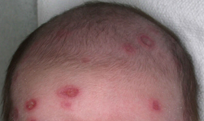
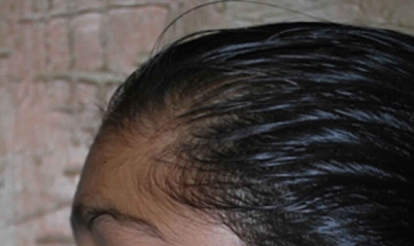
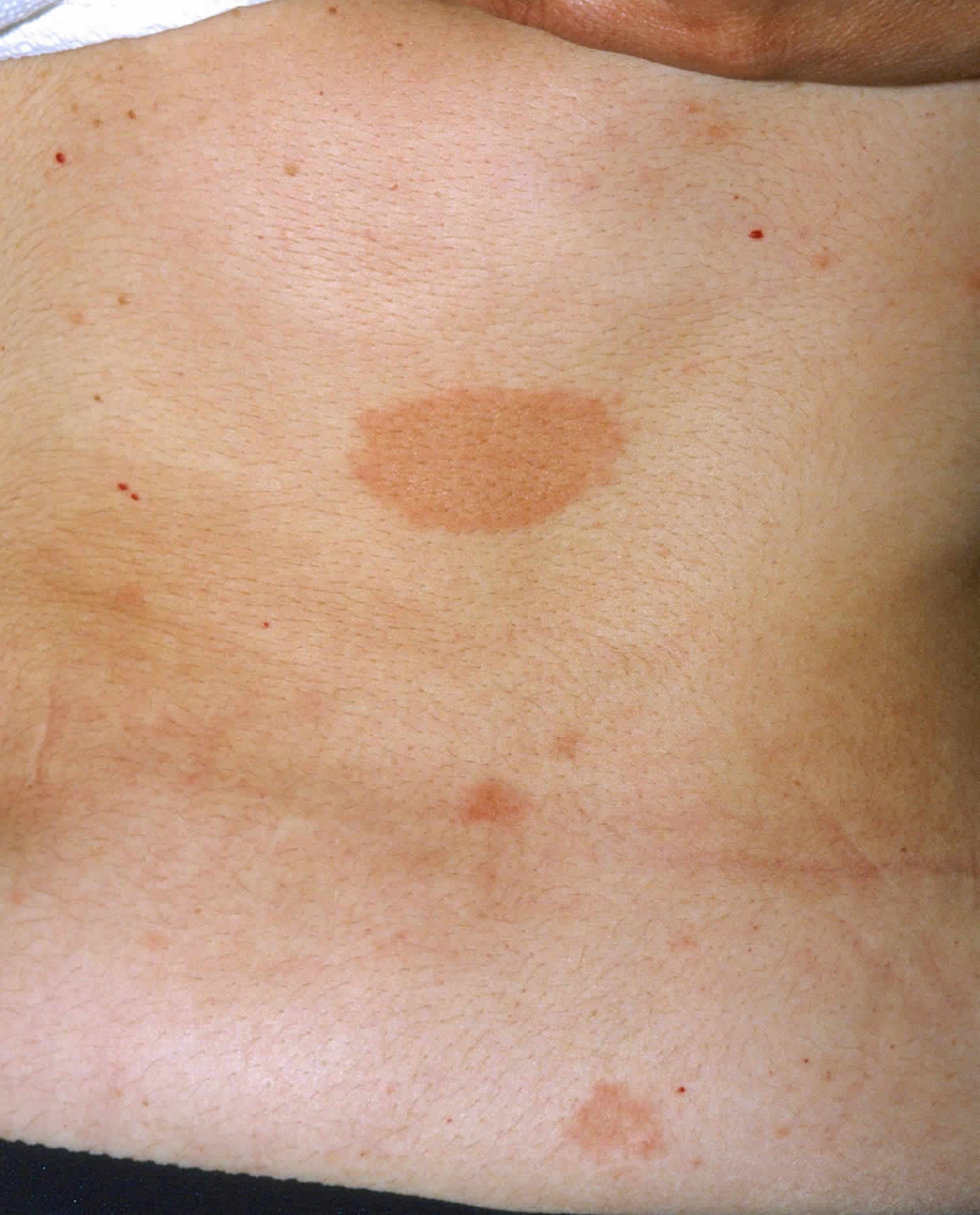
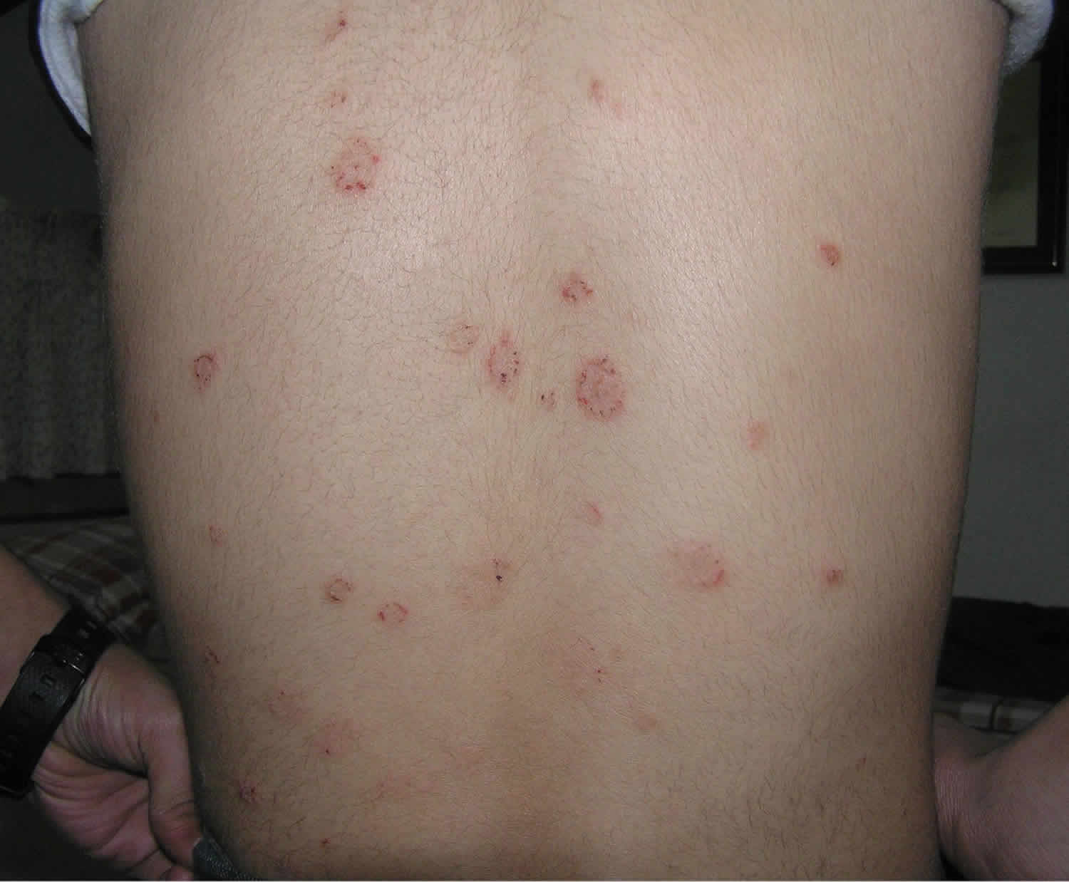
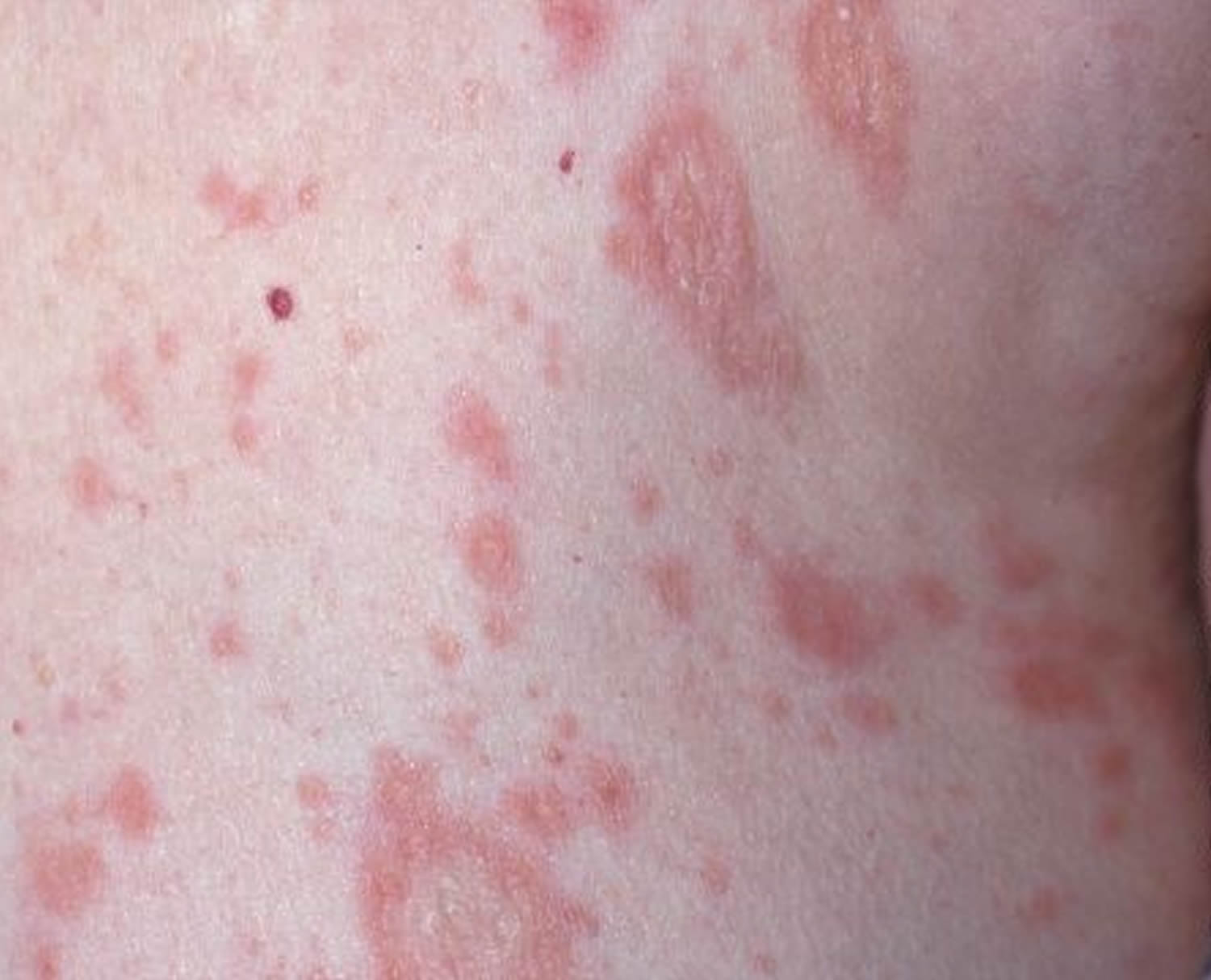
Figure 1. Lupus rash in children

pediatric lupus rash

[Source 1) ]

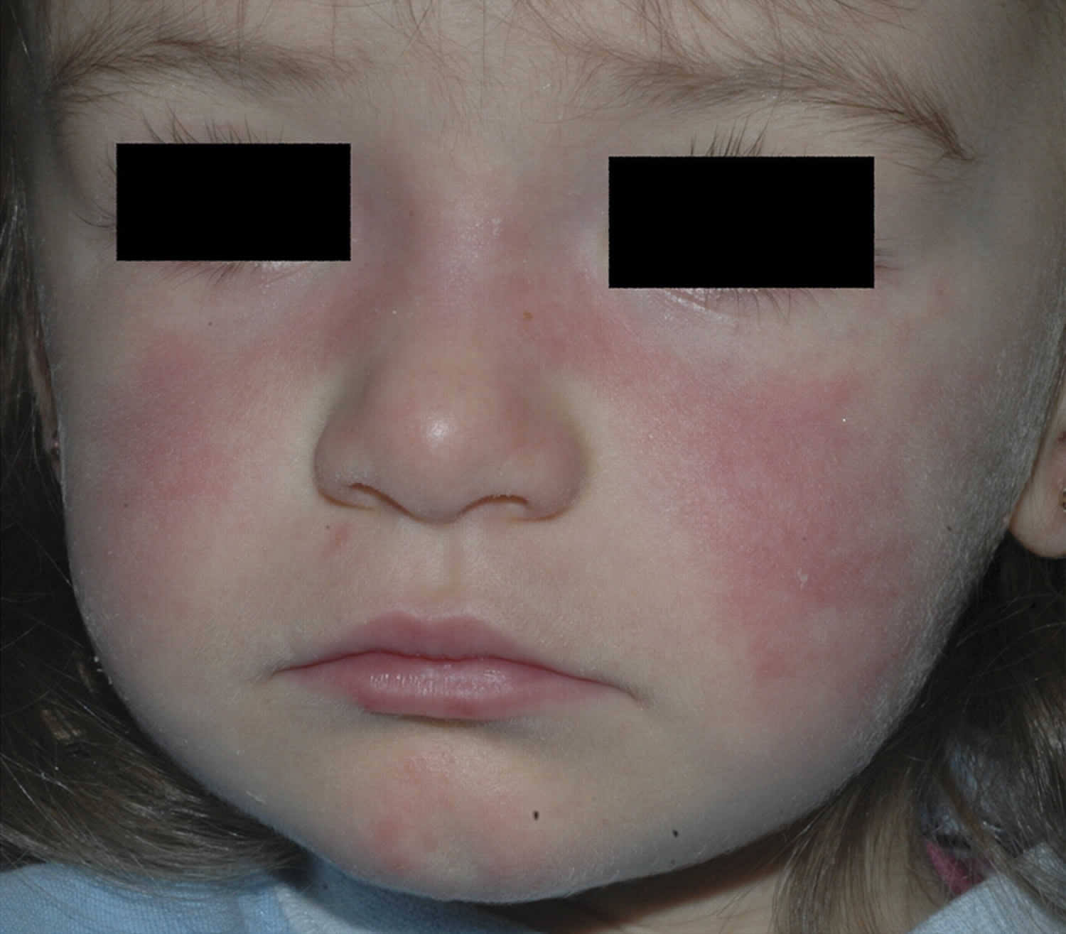
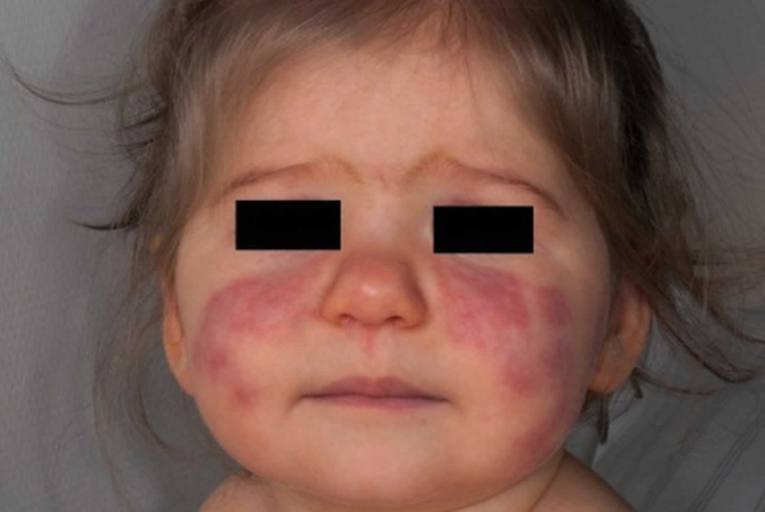
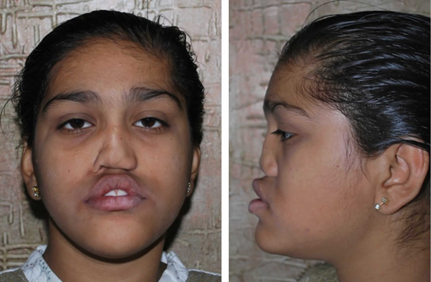
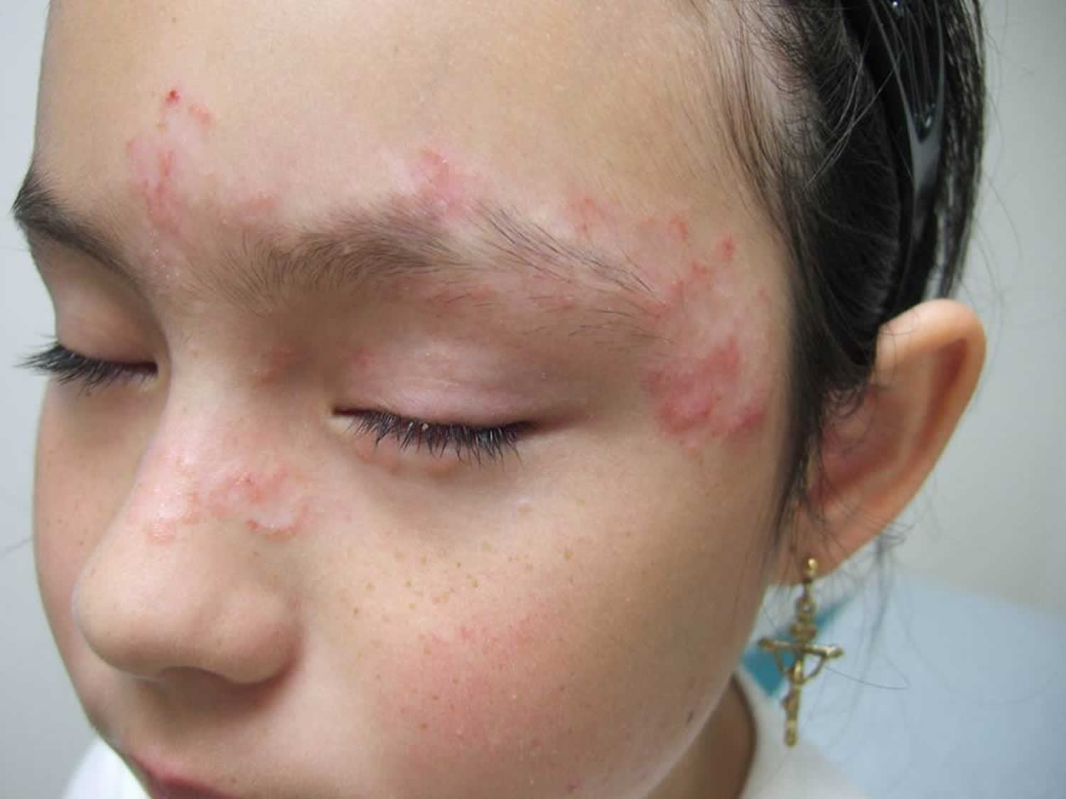
 Figure 2. Malar rash in children

pediatric lupus rash

[Source 2) ]

What is lupus?

Lupus also known as systemic lupus erythematosus (SLE), like all autoimmune diseases, lupus causes the immune system — your natural protection against foreign invaders like viruses and bacteria — to mistakenly attack your body. What makes lupus unusual, and frequently distressing for patients and families, is its unpredictability: It can affect almost any part of the body, and often many parts at the same time. There are different types of lupus, but in general the word “lupus” is shorthand for the most dominant form, systemic lupus erythematosus (SLE). Systemic means it can involve many parts of your body. Erythematosus comes from the Greek word for “red,” and describes the lupus characteristic cheek rash. Lupus is Latin for “wolf,” which some believe refers to the way a check rash resembles a wolf bite.

Lupus or SLE key points:

  • is a chronic (meaning lifelong) autoimmune disorder with no known cause or cure
  • affects about five million people worldwide, most often adolescent girls and younger women (15 to 44)
  • can target various parts of the body, including the skin, joints, blood and vital organs like the kidneys, heart, lungs and brain
  • tends to alternate between being more active (when symptoms surge, or flare) and less active (when symptoms appear to go away) is less common in children

With lupus, doctors can’t predict what part of the body the immune system will choose to strike, or when. But they can use medications to help prevent or blunt these attacks and to extinguish the harmful inflammation. Back in the 1950s, children with lupus faced only about a 30 to 40 percent chance of survival. Today, however, there are powerful medications that can bring this illness under control — often permanently — in the majority of children and allow them to lead full, relatively normal lives.

What are the other forms of lupus?

There are four different types of lupus:

  • Systemic lupus erythematosus
  • Discoid lupus. This type causes a severe red rash that worsens in sunlight.
  • Subacute cutaneous lupus. Skin rashes develop in areas commonly exposed to the sun.
  • Drug-induced lupus (DIL). Certain medications can cause lupus symptoms that go away once the drug is stopped.
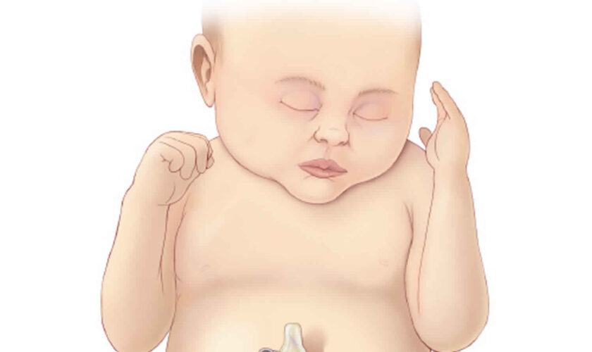
  • Neonatal lupus. This type affects babies of women who have lupus. Skin symptoms usually go away within weeks to a few months after birth. It may cause heart problems.

People of all ages, races and sexes can get lupus, but 9 out of 10 adults with the disease are women between the ages of 15 and 45. African American women are at the highest risk.

Drug-induced lupus

This lupus-like illness can crop up in people who take certain medications for a long time. DIL is fairly rare in children, and when it occurs the most common culprit is the acne drug minocycline. Drug-induced lupus has some of the same symptoms of lupus—fever, fatigue, joint and muscle stiffness—but doesn’t tend to affect vital organs. Symptoms usually disappear within a few weeks after the “triggering” medication is stopped.

Neonatal lupus

This temporary form of lupus affects a small percentage of infants whose mothers have certain lupus autoantibodies. It begins before birth, when these autoantibodies reach the baby via the placenta, and ends within the first several months of life, as the autoantibodies disappear from the baby’s system. The symptoms—skin rash, low blood cell counts—are likewise temporary. However, neonatal lupus does have the potential to cause permanent damage to the baby’s heart (called heart block). If the heart block is significant, a baby may need a pacemaker.

If your child has — or may have — drug-induced lupus or neonatal lupus, his or her doctor will talk with you in detail about what this diagnosis means, and what the next steps will be.
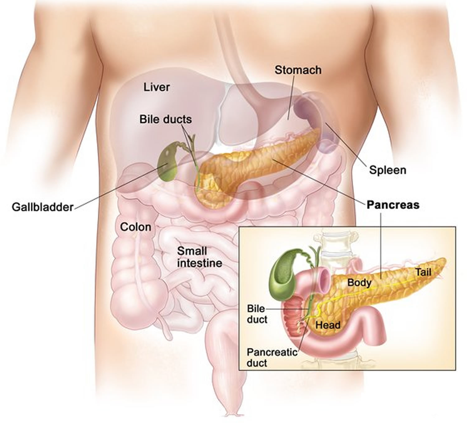
Lupus nephritis

Lupus nephritis is a type of kidney inflammation often found in patients who suffer from systemic lupus erythematosus (SLE) or lupus. SLE, or lupus, is an autoimmune disease that can affect many different parts of the body, including the kidneys.

Lupus nephritis can affect anybody, but it is most common in non-Caucasian females. Depending on age, lupus nephritis can be between 3 to 15 times more common in females than in males, and it is 2 to 3 times more common in women of color.

Lupus nephritis is brought on by lupus or systemic lupus erythematosus, which causes the child’s immune system to attack their own tissues and organs. When the immune system attacks the kidney tissue, it causes inflammation and can destroy parts of kidney.

Types of lupus nephritis

There are six different classifications of lupus nephritis (called Class I through Class VI), sometimes referred to as lupus nephritis stages.

These lupus classifications are based on the form of abnormal cellular structure caused by gene mutation.

What are the signs and symptoms of lupus nephritis?

Lupus nephritis is typically found during the assessment of patients with other signs and symptoms of systemic lupus erythematosus (SLE). Lupus nephritis can also be the first sign that a person has SLE.

Signs and symptoms of systemic lupus erythematosus include:

  • Weakness and fatigue
  • Malaise (a general feeling of discomfort or illness)
  • Fever
  • Weight loss
  • Joint pain
  • Muscle pain
  • Butterfly rash (a red rash across the cheeks and bridge of the nose)
  • Calcinosis (calcium deposits under the skin)
  • Vasculitis (damaged blood vessels)
  • Alopecia (hair loss)
  • Ulcerations (open sores) in the mouth, nose and sometimes the genitals

Lupus nephritis symptoms are the result of kidney dysfunction. Signs and symptoms of lupus nephritis include:

  • Blood in the urine
  • Abdominal swelling due to built-up fluid
  • Low urine output
  • High blood pressure

If your child displays these signs and has not yet been diagnosed with SLE, the next step is to order a laboratory evaluation to tell whether systemic lupus erythematosus is the cause of the kidney dysfunction.

What tests are used to diagnose lupus nephritis?

Your child’s initial laboratory evaluation will include blood and urine tests to assess kidney function.

The tests to diagnose systemic lupus erythematosus (SLE) measure specific immune activity, such as the number and type of antibodies (immune proteins) that are associated with SLE. Your child’s doctor will also look for abnormalities in other immune substances called complement C3 and C4.

If your child is diagnosed with SLE and symptoms suggest that it is affecting the kidney, your child’s doctor will most likely require a kidney biopsy to confirm the lupus nephritis diagnosis.

How is lupus nephritis treated?

Lupus nephritis, as well as systemic lupus erythematosus (SLE) in general, are treated with medications that suppress the immune system. These medications usually include steroids and additional drugs we prescribe very precise dosages and manage closely. Specific treatment regimens depend on the class of lupus nephritis found through the biopsy and the overall extent of how the SLE is affecting the body, outside of the kidneys.

Pediatric lupus causes

Scientists don’t yet know why some children develop lupus and others don’t. Researchers think that people with certain genes are triggered by an external factor, such as stress, a viral infection, medication or regular exposure to chemicals such as silica or pesticides. Since lupus often strikes women during their childbearing years, hormones are believed to play a role.

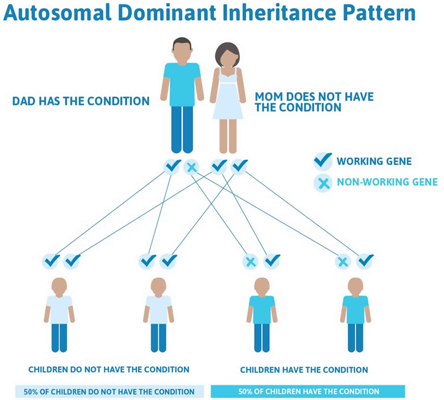
Lupus not contagious, you can’t “catch” it from another person. Systemic lupus erythematosus is not a disease that parents pass directly down to their children; in fact, there’s only about a 5 percent chance that a son or daughter of someone with lupus will also develop it.

While researchers do believe that genes play a big role in causing lupus, there’s more to it than that. Otherwise, you’d expect that if one identical twin has lupus, the other would, too — but that’s often not the case. Instead, there’s likely a two-part process involved in causing lupus:

  • Family history: A child is born with certain genes that make him or her susceptible to lupus. Think of a forest in dry, hot weather: The ingredients for a wildfire are there, but it takes something else to spark the blaze.
  • Environmental factors: The child encounters something—or a combination of things—that causes the disease to “ignite.” The environmental factors that may trigger lupus include infections, ultraviolet light and perhaps extreme stress. And given that so many lupus patients are female, it’s also likely that hormones play an important role in the development of and risk for this disease. However, there’s still a lot we don’t know about these triggers, especially why some affect certain children and not others.

Scientists are now working to discover which genes are involved in lupus and how its potential disease triggers work — in order to bring us closer to curing or even preventing this chronic illness.

Pediatric lupus symptoms

Lupus is known as “the great imitator” because many of its earliest warning signs are common in other illnesses, too. Fever, low energy, no appetite? It could be the beginning of lupus — or it might just be the flu.

Lupus is also a very shifty disease. Symptoms often come and go, new ones may crop up, while others seem to disappear. Symptoms also vary greatly from person to person, depending on what part of the body the disease is affecting at the time.

For all these reasons, diagnosing childhood lupus often requires the expertise of pediatric rheumatologists. These specialists are the best qualified to sort out the signs and symptoms of lupus from other diseases, so your child’s treatment can begin as quickly as possible.

Common symptoms of lupus include:

  • fatigue
  • loss of appetite
  • weight loss
  • swollen or achy joints
  • muscle aches
  • fever of over 100 °F (37.8 °C)
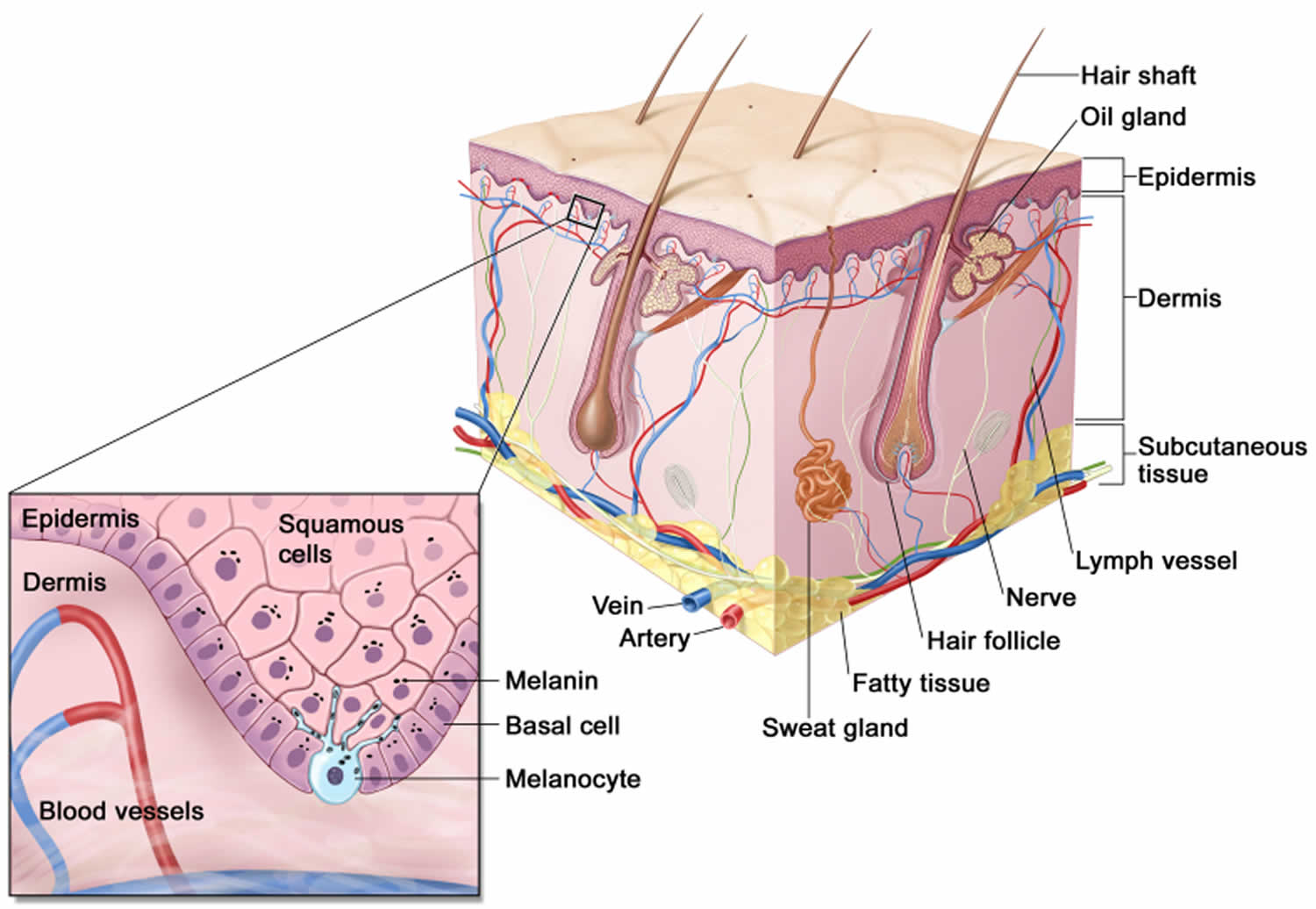
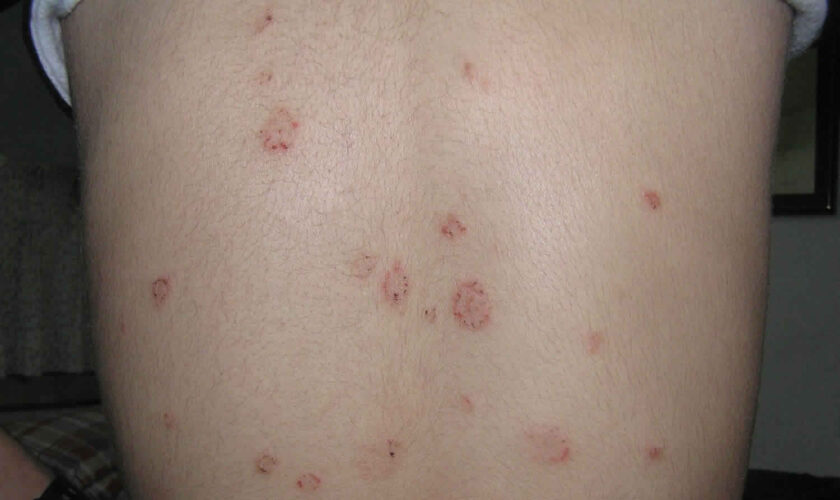
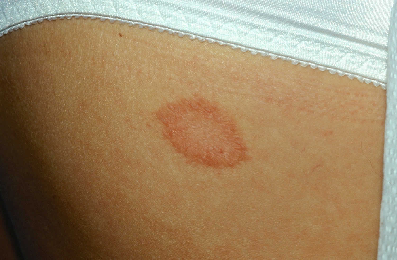
  • skin rashes, especially a butterfly-shaped rash across the cheeks (this so-called malar rash is a hallmark of lupus) and rashes that develop on sun-exposed skin
  • brittle hair, or unusual hair loss
  • ulcers in the mouth or nose
  • fingers that turn white and/or blue from cold or stress (Raynaud’s phenomenon)

Compared with adults, children with lupus are more likely to have problems with vital organs, especially the kidneys and the brain. These symptoms may include:

  • dark urine; swelling around the feet, legs and eyelids and high blood pressure (kidney inflammation, or lupus nephritis)
  • shortness of breath, chest pain (lung inflammation, or pleuritis)
  • headaches, memory problems, seizures (brain inflammation, or cerebritis)
When to seek medical advice

If your child has symptoms such as fever, fatigue, joint stiffness and skin rashes — especially a butterfly-shaped rash across her cheeks and nose — it might mean she has lupus. You should make an appointment with your child’s pediatrician, who will then make a referral to a rheumatologist if lupus is suspected.

If your child has already been diagnosed with lupus, you should call her specialist about any sudden changes in her symptoms or the appearance of new ones. And remember that infections can potentially be more serious in children with lupus: If your child develops a fever or feels increasingly unwell, let her doctor know right away.

Pediatric lupus diagnosis

You may have read that lupus is extremely difficult to diagnose, and that some patients go a long time, even years, before they know what’s wrong with them. And it’s true the symptoms of lupus can mimic many other illnesses, such as infection and cancer. But if you bring your child to a pediatric rheumatologist — the kind of doctor who knows best what this disease looks like in children — odds are that he or she can determine whether it’s lupus relatively quickly, and if treatment is needed, it can begin right away.

Since there is no single symptom or test result that points to lupus, your child’s doctor will collect a lot of information to make a diagnosis. He or she will conduct a thorough physical exam, make a list of your child’s current symptoms and talk with you about your child’s medical history and the medical history of close family members.

Your child’s doctor will also use certain lab tests to help make a diagnosis and, later, to keep tabs on how the lupus is progressing. These tests include:

  • complete blood count (CBC), which is a collection of tests measuring the size, number and maturity of different blood cells in a specific amount of blood. Two important tests include:
    • white blood cell count (WBC): This tests indicates the number of white blood cells present. Low levels may point to an active problem with the immune system, like lupus. High levels, on the other hand, may indicate an infection.
    • hematocrit (Hct): Indicates the number of red blood cells present. Anemia, or low levels of red blood cells, is often a symptom of lupus.
  • antinuclear antibody(ANA), which detects certain abnormal proteins, called antinuclear antibodies, that the immune system often makes when attacking the body’s own tissues. The presence of these antibodies is common in lupus and other autoimmune diseases. However, testing positive for ANA does not equal lupus. Positive tests are also seen in children with other conditions, and even in a handful of perfectly healthy children.
  • anti-DNA, which detects a specific antinuclear autoantibody commonly seen with lupus nephritis.
  • complement (C3 and C4), which measure blood complement levels. The complement system includes a group of proteins that are part of the immune system. Low levels of complement may indicate lupus activity, and increase the risk for infection.
  • C-reactive protein (CRP), which measures the amount of a protein made in the liver. CRP levels tend to increase when there’s an inflammatory process. CRP levels rise very quickly, and may indicate lupus activity or may reflect a new infectious process somewhere in the body.
  • erythrocyte sedimentation rate (ESR or sed rate), which measures how quickly red blood cells fall to the bottom of a test tube. If the cells to clump together and fall more rapidly than normal, it can signal there is inflammation in the body.

Your child’s doctor may order other lab tests or imaging tests to check for signs of lupus in specific organs. A urinalysis, for example, can help show whether lupus is affecting the kidneys, while a chest x-ray may show telltale inflammation around the heart or lungs.

Sometimes a biopsy can be helpful in making a diagnosis or evaluating the health of a specific organ or tissue. Almost any part of the body can be biopsied—in which a small sliver of tissue is removed and examined under a microscope— although in lupus the sample is taken from a rash or the kidneys when symptoms are active.

The 11 criteria for lupus

Since lupus symptoms vary so widely and test results don’t always tell the full story, you may wonder how doctors are able to put the puzzle pieces together to come up with a diagnosis. Much of it depends on their past experience with patients, but they also bear in mind 11 lupus criteria laid out by the American College of Rheumatology.

Typically, at least four of the following things must be present for a doctor to diagnose lupus:

  • malar rash: a butterfly-shaped rash across cheeks and nose
  • discoid rash: raised, scaly patches on the skin
  • photosensitivity: skin rash caused by sun exposure
  • oral ulcers: small, usually painless sores in the mouth
  • arthritis: swelling and achiness in at least two joints
  • cardiopulmonary problems: inflammation around the heart and/or lungs
  • neurological problems: such as seizures and/or psychosis
  • kidney problems: such as blood in the urine
  • hematologic (blood) problems: low levels of red blood cells (anemia), white blood cells or platelets
  • positive antinuclear antibody (ANA) test
  • other positive blood tests that may indicate an autoimmune disease. Antiphospholipid antibodies, including cardiolipin, are common in lupus.

It’s not unusual, though, for experienced physicians to make a diagnosis even when fewer than four criteria are present.

Pediatric lupus treatment

There is no cure for lupus. Treatment involves managing systemic inflammation with medications and lifestyle changes. Treating an unpredictable disease like lupus is like fighting a fire. Doctors can’t know where it might spread, so they focus on what’s actually “on fire”—the places in your child’s body where lupus is active right now. If lupus is affecting your child’s kidneys and central nervous system, for example, the treatment will be very different from what it might be if the disease is affecting your child’s skin. This is why it’s essential to let your child’s doctor know when new symptoms appear, since they could mean another part of the body is under attack.

The medications used to treat lupus fall into two main categories:

  1. Non-immunosuppressants tend to be milder drugs that fight inflammation or help ease discomfort, and have few side effects.
  2. Immunosuppressants are much more powerful drugs aimed at bringing the malfunctioning immune system under control. Some have significant side effects and—because they suppress the immune system — all increase the risk of infection.

Other medications may be used to control conditions that are common in people with lupus:

  • Water pills that help to prevent fluid buildup.
  • Anti-hypertensive drugs to treat high blood pressure.
  • Anticonvulsants to prevent or treat seizures.
  • Antibiotics for infections.
  • Bone-strengthening drugs for osteoporosis.

Nonimmunosuppressants

  • Antimalarials or disease-modifying antirheumatic drugs (DMARDs): DMARDs can modify the course of the disease, prevent progression and slow joint damage. DMARDs are often used with nonsteroidal anti-inflammatory drugs (NSAIDs). Most commonly prescribed DMARD is hydroxychloroquine (Plaquenil) which helps reduce the frequency of lupus flares. They are among the safest, most gentle drugs for lupus, yet go a long way toward preventing its life-threatening complications. Most children can expect to be on antimalarials for an indefinite period of time. A potential side effect of hydroxychloroquine is eye problems, but this can be monitored through twice-yearly visits to an ophthalmologist.
  • Nonsteroidal anti-inflammatory drugs (NSAIDs) ease symptoms like pain, swelling and stiffness, and are used mainly for kids with lupus arthritis. Among the most common NSAIDs are ibuprofen and naproxen, given in therapeutic doses (that is, higher than the over-the-counter versions). Potential side effects, like stomach problems, are monitored for, but tend to be mild.

Immunosuppressants

  • Corticosteroids. Corticosteroids are powerful anti-inflammatory drugs. These drugs are given at the lowest possible dose for the shortest length of time because of side effects. Corticosteroids most often prednisone come in pill form or may be given as an injection into the joint or muscle. Corticosteroids aren’t the same as the anabolic steroids that athletes sometimes take. These are powerful, fast-acting drugs that suppress the entire immune system. Many kids with lupus will need corticosteroids at some point. However, doctors work to phase them out as soon as possible because of their potential side effects, which can include weight gain, facial puffiness (a “cushingoid” appearance), acne, high blood pressure and reduced bone density.
  • Steroid-sparing therapies offer many of the benefits of corticosteroids, usually with fewer side effects, but may take much longer to work. You may also hear these drugs called DMARDs, for disease-modifying anti-rheumatic drugs. This group includes methotrexate;azathioprine (brand name Imuran); and mycophenolate mofetil, or MMF (CellCept). Side effects vary by medication, but may include liver problems and anemia.
  • Biologics are a relatively new class of steroid-sparing therapies that are based on compounds made by living cells. Instead of suppressing the entire immune system, biologics are more like smart bombs—they only target certain parts of it. Biologics used in lupus include rituximab (Rituxan), tocilizumab (Actemra) and belimumab (Benlysta). Side effects vary by medication.
  • Cytotoxins are drugs that destroy rapidly dividing cells in the overactive immune system. Cytotoxins — such as cyclophosphamide (Cytoxan)—are so potent they’re usually reserved for lupus patients with serious kidney disease or central nervous system problems. Side effects may include nausea and vomiting, hair loss and bladder problems.

Aside from medications, your child’s doctor may also prescribe IVIg (intravenous immunoglobulin), which is a blood product made up of healthy antibodies that is delivered by IV, and can help get the immune system back on track. Much more rarely, your child may need to undergo plasmapheresis, a process that removes autoantibodies from the blood. But because it also takes away normal antibodies, it significantly weakens the immune system. That’s why doctors treat only very severe lupus with plasmapheresis.

Complications of lupus require their own medications and treatment procedures, too—dialysis for serious kidney disease, for instance. Your child’s doctor will discuss these with you in detail if and when any complications arise.

Medicine is essential, but it’s not the sum total of your child’s treatment for lupus. Many kids with lupus also require physical and occupational therapy, to increase their mobility and muscle strength and to learn ways to make day-to-day activities easier on their bodies. And because chronic illnesses like lupus can be mentally and emotionally tough to deal with, psychotherapy or counseling can be valuable in helping kids keep the positive outlook they need to beat their disease.

Alternative therapies

When your child is facing a chronic illness like lupus, it’s understandable that you would want to explore all the treatment options. But despite what you may read on the Internet, or hear from friends of friends, there is no “hidden” cure for lupus. No magic herb or special diet will restore your child to perfect health.

That said, there are some things outside conventional medicine — like acupuncture or meditation — that are fairly well studied and do seem to help some people with lupus.

A few words of caution before embarking on any alternative therapies, however:

  • Talk it over with your child’s doctor. It’s important that whatever you have in mind won’t interfere with the overall treatment plan. Also, the doctor may know other patients who have tried the therapy— and whether it helped them.
  • Don’t skip prescribed treatments. Alternative therapies are meant to supplement traditional medicine, not replace it. It’s vital that your child stick to her medications, or else risk letting her illness get out of control.
  • Avoid fly-by-night practitioners. For things like reiki, acupuncture and massage, always ask your doctor for a referral, if appropriate, or recommendations on how to find a qualified care provider.
  • And be prepared to pay: By and large, health insurance plans don’t cover alternative therapies.

Home remedies

Lupus is chronic, incurable and unpredictable. But it’s not unbeatable. And you can fight it by helping your child make some simple lifestyle changes at home.

  • Eat a healthful diet. Make sure your child is getting plenty of fruits and vegetables, whole grains, low-fat dairy products and lean sources of protein. And load up on Vitamin D and calcium, especially if your child is taking corticosteroids, which can weaken bones.
  • Stay active. Encourage your child to walk, swim, bicycle and simply get out and play. While she’ll have to be careful not to get overtired, exercise helps keep muscles strong and joints flexible. And since all lupus patients are at greater risk for cardiovascular disease later in life, making a habit of exercising now will mean better heart health in adulthood.
  • Balance rest and physical activity: When disease is active and joints are painful, swollen or stiff, it is important to rest to reduce inflammation and fatigue. When disease activity is low, regular exercise can ease pain and reduce stress. A balanced program includes low-impact aerobic activity, muscle strengthening and flexibility exercises.
  • Beware of sun exposure. Because the ultraviolet (UV) rays in sunlight or or fluorescent lights can trigger a flare, your child should wear plenty of broad-spectrum sunscreen (SPF 30 or higher) when she goes outside. Protective clothing like hats and long-sleeved shirts are also strongly recommended. Avoid being outside at peak UV hours between 10 a.m. and 4 p.m.
  • Get plenty of rest. People with lupus are prone to fatigue, so remind your child to take a break if she’s feeling run-down — a brief nap can be tremendously helpful in recharging a kid’s batteries. And a full night’s sleep is essential.
  • Pace yourself: Fatigue is a common lupus symptom. It’s important to pace yourself to prevent extreme tiredness from taking over your day. Getting restful sleep will help to keep tiredness at bay.
  • Reduce stress: Develop strong positive relationships so you have emotional support for managing the ups and downs of a chronic disease. Use relaxation techniques, such as meditation, deep breathing or yoga to reduce stress. Stay connected to activities you enjoy to promote a positive mood.
  • Avoid smoking: Smoking including secondhand smoke harms the body and can worsen lupus symptoms.

By doing these things and becoming a full partner in your child’s health care—keeping clinic appointments, alerting doctors to any new symptoms—you may end up feeling more in control of what can be a very daunting illness. The outlook for children with lupus can vary a great deal, however, depending on when the disease begins (called the onset).

  • age 15-18 (more common): In this group, pediatric systemic lupus erythematosus tends to look and behave more like adult lupus; its severity can range from mild symptoms to life-threatening disease.
  • younger than 15 ( less common): Symptoms can be more severe in this group, and there’s a greater chance that vital organs will be affected.
  • younger than than 5 (rare):The smallest children are typically among the sickest lupus patients, largely because most also have complement deficiency, meaning they don’t have enough of certain blood proteins that play a key role in the immune system.

It’s important to remember that everyone with lupus responds to the disease—and the medications used to treat it—in his own unique way. You can’t predict exactly how lupus will progress in your child. But you can greatly improve your child’s odds for a healthy future by helping her understand and stick with the recommended treatments for her condition.

Pediatric lupus prognosis

The outlook for children with lupus can vary a great deal, however, depending on when the disease begins called the onset:

  • age 15-18 (more common): In this group, pediatric lupus tends to look and behave more like adult lupus; its severity can range from mild symptoms to life-threatening disease.
  • younger than 15 ( less common): Symptoms can be more severe in this group, and there’s a greater chance that vital organs will be affected.
  • younger than than 5 (rare):The smallest children are typically among the sickest lupus patients, largely because most also have complement deficiency, meaning they don’t have enough of certain blood proteins that play a key role in the immune system.

It’s important to remember that everyone with lupus responds to the disease—and the medications used to treat it—in his own unique way. You can’t predict exactly how lupus will progress in your child. But you can greatly improve your child’s odds for a healthy future by helping her understand and stick with the recommended treatments for her condition.

References   [ + ]

read more

Cloacal exstrophy

cloacal exstrophy

Cloacal exstrophy

Cloacal exstrophy, also known as OEIS Syndrome, occurs when a portion of the large intestine lies outside of the body and on either side of it and connected to it are the two halves of the bladder. The intestine may be short and the anus may not open. The bony pelvis is also split open like a book. In males, the penis is usually flat and short, with the exposed inner surface of the urethra on top. The penis is sometimes split into a right and left half. In girls, the clitoris is split into a right half and left half and there may be one or two vaginal openings. Cloacal exstrophy (OEIS syndrome) is a very rare birth defect, affecting 1 in every 250,000 births. Although cloacal exstrophy is a serious condition and requires a series of operations, the long-term outcome is good for many children. Patients and families need to be counseled about the complexity of the anomaly, the need for multiple procedures, and long-term expectations for continence, sexual function, and fertility.

Cloacal exstrophy is known as OEIS Syndrome because of the four features that are typically found together 1):

  • Omphalocele: Some of the abdominal organs protrude through an opening in the abdominal muscles in the area of the umbilical cord. The omphalocele may be small, with only a portion of the intestine protruding outside the abdominal cavity, or large, with many of the abdominal organs (including intestine, liver and spleen) protruding outside the abdominal cavity.
  • Exstrophy of the bladder and rectum: The bladder is open and separated into two halves. The rectum and colon are similarly open and the segment of the rectum is placed between the bladder halves on the surface of the abdomen.
  • Imperforate anus: The anus has not been formed or perforated and the colon connects to the bladder.
  • Spinal defects: These defects may either be major or minor. Often children born with cloacal exstrophy are also born with some degree of spina bifida.

With cloacal exstrophy there are often other birth defects, like spina bifida. This occurs in up to 75 percent of cases. Kidney abnormalities and omphalocele are also common. An omphalocele is when an infant’s intestine or other abdominal organs are open to the outside the body. This is from a hole in the belly button (navel) area. The intestines are covered only by a thin layer of tissue and can be easily seen.

Cloacal exstrophy (OEIS Syndrome) is a complex anomaly that often requires several surgical procedures and requires lifelong medical follow-up care.

As soon as possible, surgical reconstruction is done. Surgery is major, and often done in parts. The schedule of surgery depends on the child’s condition and overall health. Surgery can return the bladder and bowel organs back into the body, to a healthy position. It can provide ways for bowel and urinary control, better kidney function, and improve the way the sex organs or genitals look.

Reconstruction surgery often starts within the first few days of life. It is sometimes delayed to allow the baby to grow and develop. Surgical repair is generally divided into steps and include:

  • Repair of spinal abnormalities, and if needed, the repair of a large omphalocele.
  • Once the child has recovered from spinal surgery, the gastrointestinal tract is treated. Many babies require a stoma because the colon is not normal, and the anus is not formed. The stoma will allow for waste to be released from the intestines to a pouch on the outside of the body.
  • Closure of the exposed bladder and bowel and reconstruction of the genitals are next. This may be done in steps if the pelvic bones are widely separated. For a successful closure, a pelvic osteotomy (cutting the bones to allow the pelvis to close more easily) is critical. In some cases, the abdominal wall, the bladder and genitals (genitourinary system) and the bowel may be repaired at the same time. Bladder reconstruction often includes the use of a catheter for some time.

Figure 1. Cloacal exstrophy

cloacal exstrophy

Cloacal exstrophy

[Source 2) ]

Figure 2. Cloacal exstrophy ultrasound

Cloacal exstrophy ultrasound

Footnote: Fetal ultrasound at 36 weeks’ gestation demonstrating bowel loops herniating between 2 bladder plates (“elephant trunk”).

[Source 3) ]

Figure 3. Cloacal bladder exstrophy female infant

cloacal bladder exstrophy female infant

Figure 4. Cloacal bladder exstrophy male infant

Cloacal bladder exstrophy male infant

Will my child be able to have children when they reach adulthood?

In many cases, the answer to this question is: yes. But almost always, assisted fertility is necessary for adults.

With regard to sexuality, males are generally potent, but some report inadequate phallus or residual curvature. Females report normal sexual function 4).

With respect to fertility and childbearing, retrograde ejaculation or iatrogenic obstruction of the ejaculatory ducts or vas deferens after surgical reconstruction may result in abnormal semen analysis. Antegrade ejaculation is preserved after single-stage repair, but abnormal semen parameters are common. However, fertilization, with viable pregnancy, has been achieved by male patients with cloacal exstrophy 5).

Females have had successful pregnancies 6). Cesarean delivery is recommended to avoid injury to continence mechanism. Postpartum uterine prolapse is common because of aggravation of preexisting abnormal pelvic support.

Cloacal exstrophy causes

The cause of cloacal exstrophy is currently unknown, so there is also no known way to prevent it. On the basis of the known embryologic principles of cloacal development, any inciting event would have to occur early in pregnancy.

There is a higher incidence of cloacal exstrophy in families in which one member is affected as compared with the general population. Offspring of patients with exstrophy-epispadias complex have a 1 in 70 risk (500 times that of the general population) of being affected. Nevertheless, familial occurrence is uncommon in large series 7). The heritability of cloacal exstrophy has not been established, because no offspring have been reported. Moreover, there’s no evidence to suggest that anything done by expectant parents leads to the condition.

At present, 22q11.2 duplication is the genetic variant most commonly associated with bladder exstrophy-epispadias complex 8).

Cloacal exstrophy has been reported in twins. Concordance rates show strong evidence of genetic effects 9), but less than 100% concordance among identical twins suggests some role for environmental effect on development of exstrophy-epispadias.

A higher incidence of bladder exstrophy is observed in infants of younger mothers and in those with relatively high parity.

Maternal tobacco exposure is associated with more severe defects (cloacal vs classic exstrophy).

Growing evidence suggests an increased incidence of cloacal exstrophy and bladder exstrophy-epispadias with in-vitro fertilization (IVF) pregnancies 10).

Cloacal exstrophy symptoms

In some cases, cloacal exstrophy is detected from a routine prenatal ultrasound. In other cases, it isn’t diagnosed until birth, when physicians can clearly see the exposed organs.

Antenatal ultrasound findings suggestive of exstrophy-epispadias complex include the following:

  • Repeated failure to visualize the bladder on ultrasound
  • Lower-abdominal-wall mass
  • Low-set umbilical cord
  • Abnormal genitalia
  • Increased pelvic diameter

Additional antenatal ultrasound findings suggestive of cloacal exstrophy include the following:

  • Omphalocele
  • Limb abnormalities
  • Myelomeningocele
  • Trunk sign from prolapsed intestine

Increased use of fetal magnetic resonance imaging (MRI) may further improve the accuracy of antenatal diagnosis, but this test is not necessary if suspicion is high on the basis of ultrasound findings.

Classic bladder exstrophy and cloacal exstrophy are obvious to all in the delivery room. Variants of the exstrophy-epispadias complex exist, including skin-covered bladder exstrophy, duplicate bladders, superior vesical fistula, and epispadias with major bladder prolapse 11). Most exstrophy variants and epispadias are also identifiable at birth. Unrecognized female epispadias may present as persistent childhood incontinence. Unrecognized split-symphysis variants of exstrophy may be identified in childhood only because of persistent incontinence or a waddling gait.

Physical examination

Patients with classic bladder exstrophy or epispadias typically appear as term infants. Patients with cloacal exstrophy, however, are often preterm. They may have respiratory embarrassment requiring mechanical ventilation.

Abdominal findings

In classic cloacal bladder exstrophy (see the images above), the bladder is open on the lower abdomen, with mucosa fully exposed through a triangular fascial defect. The abdominal wall appears long because of a low-set umbilicus on the upper edge of the bladder plate. The distance between the umbilicus and anus is foreshortened. The recti diverge distally, attaching to the widely separated pubic bones. Indirect inguinal hernias are frequent (>80% of males, >10% of females) because of wide inguinal rings and the lack of an oblique inguinal canal.

Nearly all patients with cloacal exstrophy have an associated omphalocele. The bladder is open and separated into two halves, flanking the exposed interior of the cecum. Openings to the remainder of the hindgut and to one or two appendices are evident within the cecal plate. Terminal ileum may prolapse as a “trunk” of bowel onto the cecal plate.

In cloacal exstrophy variants, the pubic symphysis is widely separated, and the recti diverge distally. The umbilicus is low or elongated. A small superior bladder opening or a patch of isolated bladder mucosa may be present. The intact bladder may be externally covered by only a thin membrane. Isolated ectopic bowel segments have been reported.

Genital findings

In describing the anatomy of the penis, the terms dorsal and ventral refer to a normal phallus in the erect state. The dorsal surface is in continuity with the abdominal wall, and the ventral surface is in continuity with the scrotum.

In cloacal exstrophy, the penis is generally quite small and bifid, with a hemiglans located just caudal to each hemibladder. Infrequently, the phallus may be intact in the midline. In females, the clitoris is bifid, and two vaginas are present. The anus is absent.

In exstrophy variants, the genitalia generally are intact (see the image below), though epispadias can occur.

In classic bladder exstrophy in males, the phallus is short and broad with upward curvature (dorsal chordee). The glans lies open and flat like a spade, and the dorsal component of the foreskin is absent. The urethral plate extends the length of the phallus without a roof. The bladder plate and urethral plate are in continuity, with the verumontanum and ejaculatory ducts visible within the prostatic urethral plate. The anus is anteriorly displaced with a normal sphincter mechanism.

In classic bladder exstrophy in females, the clitoris is uniformly bifid with divergent labia superiorly. The open urethral plate is in continuity with the bladder plate. The vagina is anteriorly displaced. The anus is anteriorly displaced with a normal sphincter mechanism.

In male epispadias, the phallus is short and broad with upward curvature (dorsal chordee). The glans lies open and flat like a spade, and the dorsal component of the foreskin is absent. The urethral meatus is located on the dorsal penile shaft, anywhere between the penopubic angle and the proximal margin of the glans.

In female epispadias, the clitoris is most often bifid with divergent labia superiorly. The dorsal aspect of the urethra is open distally. The urethra and bladder neck are patulous and may allow visualization of bladder. Bladder mucosa may prolapse through the bladder neck.

Musculoskeletal findings

In classic bladder exstrophy, the pubic symphysis is widely separated. Divergent rectus muscles remain attached to the pubis. External rotation of the innominate bones results in a waddling gait in ambulatory patients but does not appear to result in orthopedic problems later in life.

In cloacal exstrophy, the examination is the same as for bladder exstrophy. As many as 65% of patients have a clubfoot or major deformity of a lower extremity. As many as 80% of patients have vertebral anomalies.

In split-symphysis variants of exstrophy, the pubic symphysis is widely separated (see the image below), and the rectus muscles are divergent.

Neurologic findings

In cloacal exstrophy, as many as 95% of patients have myelodysplasia, which may include myelomeningocele, lipomeningocele, meningocele, or other forms of occult dysraphism. These patients are at risk for neurologic deterioration, and they should be observed closely. Early neurosurgical consultation is appropriate if a radiographic abnormality of the spinal cord or canal is observed.

Cloacal exstrophy diagnosis

Cloacal exstrophy can usually be diagnosed by fetal ultrasound before an infant is born. Upon birth, a physical exam will confirm the diagnosis.

Laboratory studies

Before complex reconstruction of the urinary tract, it is important to obtain information about the patient’s baseline renal function. In patients with cloacal exstrophy, losses from the terminal ileum short-gut physiology can result in significant electrolyte abnormalities.

Imaging studies

Baseline examination of the kidneys with ultrasonography is recommended for all patients with exstrophy because increased bladder pressure after bladder closure can lead to hydronephrosis and upper urinary tract deterioration. Congenital upper urinary tract anomalies are uncommon with classic exstrophy and epispadias but are present in approximately one third of patients with cloacal exstrophy (eg, ectopic pelvic kidney, renal agenesis, or hydronephrosis).

Spinal ultrasound or radiography may be helpful. Myelodysplasia should be excluded in newborns with cloacal exstrophy. This can be accomplished by means of ultrasound early in life. In cloacal exstrophy, magnetic resonance imaging (MRI) is recommended to help identify occult abnormalities that may predispose to symptomatic spinal cord tethering.

Bilateral vesicoureteral reflux (VUR) is present in nearly all patients with classic bladder exstrophy. Voiding cystourethrography (VCUG) is performed in early childhood to assess bladder capacity in preparation for reconstructive continence surgery. Evaluation of the bladder neck and proximal urethra is recommended in patients with epispadias in order to plan surgical management.

Managing pregnancy after a cloacal exstrophy diagnosis

Because cloacal exstrophy is a high-risk condition, you will need to be monitored throughout your pregnancy. In some cases, pregnancy may be complicated by polyhydramnios (excess amniotic fluid) during the third trimester, which may trigger preterm labor and delivery. Your delivery should also be planned at our state-of-the-art facility. This way, our delivery team can address any complications should they arise and the baby will have immediate access to treatment and the best surgical professionals.

Cloacal exstrophy treatment

Cloacal exstrophy is treated through surgical repair after birth, usually in stages to address each defect. This requires an in-depth treatment plan to be created for your child’s specific needs. The extent of cloacal exstrophy surgery required for your baby depends on the type and severity of his or her abnormalities.

Your child will undergo a series of surgeries over a number of years — referred to as staged reconstruction. The exact timing, nature and outcome of each cloacal exstrophy surgery will depend on your child’s particular situation. Your child’s surgeons will create a treatment plan based on the type and the extent of your child’s condition and discuss the plan with you. Usually surgery begins in the first days of life with the highest-priority procedure. Surgeons usually repair the bladder, create a colostomy (an opening in the colon with an attached “bag” that allows stool to pass) and repair the abdominal wall defect.

Babies with spinal defects usually have them repaired sometime in the first few days of life. Later surgeries include urinary and genital reconstruction, as well as an operation to create a rectum and close the colostomy opening. There are no fetal interventions (surgical procedures while inside the uterus) for cloacal exstrophy.

Treatment may include:

  • Abdominal repair: Typically, soon after your child is born, the surgeons will repair the omphalocele by closing the bladder and creating a colostomy so your child can eliminate stool. With a colostomy, the large intestine is separated from the bladder halves and reclosed. The two halves of the bladder are brought together and placed into the abdomen. The end of the large intestine is brought to the surface of the skin through an opening in the abdomen. A plastic bag, called a colostomy pouch, is placed over the opening to collect the stool.
  • Other surgery, such as surgery to repair the spine, may be planned around the initial stage of the abdominal repair.

After the initial surgery, your child will remain in the hospital where we will monitor the intestine as it begins to function. Our team will work with you and your family to ensure that the plan for your child is clear and that you have access to the supports you need.

  • Osteotomies: Once your child has healed from his first procedure and had some time to grow, we will schedule the second stage of the repair. This primarily involves working on the bladder. The orthopedic surgeon on our team will perform osteotomies to help ensure that your child’s pelvis can best support the bladder over time. During the osteotomy the hip bones are cut and adjusted. Your child will need to be in traction or in a spica cast for several weeks following this surgery.
  • Pull-through procedure: If your child was born with a significant amount of colon and is capable of forming solid stool, a surgical procedure, known as a “pull through” may eventually be performed. The purpose of this procedure is to connect the colon to the rectum.

Subsequent surgeries may also involve major urinary reconstructive surgery and further genital reconstruction. These issues will be discussed with you and your family as your child grows up.

Cloacal exstrophy repair

Reconstruction of exstrophy-epispadias complex remains one of the greatest challenges facing the pediatric urologist. Many modifications in surgical procedures have improved outcomes, but the optimal approach remains uncertain. Longitudinal prospective assessment of the two main current surgical approaches (staged procedure and total reconstruction) is critical for optimizing functional and cosmetic outcomes.

Complete primary reconstruction is now more than 20 years old; however, each approach is in a constant state of minor modification. Data on this approach continue to mature and are updated almost yearly 12). Analysis of each experience focuses on daytime continence with volitional voiding, need for further surgical procedures, and complication rates. In experienced hands, the safety and efficacy of the different approaches are comparable.

Goals of therapy include provision of urinary continence with preservation of renal function and reconstruction of functional and cosmetically acceptable genitalia. Creation of a neoumbilicus is also important to many of these patients.

Surgical techniques used in the treatment of exstrophy-epispadias complex include the following:

  • Staged functional closure for classic bladder exstrophy (ie, modern staged repair of exstrophy) 13)
  • Complete primary repair for classic bladder exstrophy
  • Urinary diversion for classic bladder exstrophy
  • Closure for cloacal exstrophy
  • Gender reassignment

Staged functional closure for classic bladder exstrophy

Modern staged repair of exstrophy, which represents the traditional surgical approach, comprises a series of operations. Initial bladder closure is completed within 72 hours of birth. If this is delayed, pelvic osteotomies are required to facilitate successful closure of the abdominal wall and to allow the bladder to lie within a closed and supportive pelvic ring.

Epispadias repair with urethroplasty is performed at age 12-18 months. This allows enough increase in bladder outlet resistance to improve the bladder capacity.

Bladder neck reconstruction (typically a modified Young-Dees-Leadbetter repair) is performed at age 4 years. This allows continence and correction of vesicoureteral reflux (VUR). Multiple modifications have been proposed. The procedure is delayed until bladder capacity is adequate; better results are reported with a capacity greater than 85 mL.

Chua et al retrospectively studied a modification of staged exstrophy repair aimed at incorporating the advantages of complete primary repair for classic bladder exstrophy by avoiding concurrent epispadias repair and adding bilateral ureteral reimplantation and bladder neck tailoring (staged repair of bladder exstrophy with bilateral ureteral reimplantation) at the initial repair 14). They found staged repair of bladder exstrophy with bilateral ureteral reimplantation to be a safe alternative for exstrophy-epispadias repair, preventing penile tissue loss and yielding long-term outcomes comparable to those of complete primary repair for classic bladder exstrophy.

The radical soft-tissue mobilization (radical soft-tissue mobilization) procedure, also referred to as the Kelly repair, has been suggested as an alternative approach to staged reconstruction of bladder exstrophy 15). Radical soft-tissue mobilization has been performed not only as the second part of a two-step strategy (after bladder closure) but also as part of a combined procedure that includes delayed bladder closure and radical soft-tissue mobilization in a single stage without pelvic osteotomy 16).

Complete primary repair for classic bladder exstrophy

Compared with modern staged repair of exstrophy, complete primary repair for classic bladder exstrophy is a newer approach to exstrophy closure. Primary bladder closure, urethroplasty, and genital reconstruction are performed in a single stage in newborns. This procedure involves complete penile disassembly in males and mobilization of the urogenital complex in females. Hypospadias is a common outcome in males and requires subsequent reconstruction.

The goal is early bladder cycling. A subset of patients have achieved continence without bladder neck reconstruction.

In a study of 34 boys treated with a modified penile disassembly technique (15 with bladder exstrophy who underwent complete primary repair for classic bladder exstrophy, 11 with penopupic epispadias after previous closure of bladder exstrophy, and eight with isolated complete epispadias), Anwar et al found the modified technique to yield excellent cosmetic results 17). Preservation of the distal urethral plate along with both hemiglans avoided shortening and prevented occurrence of hypospadias.

Urinary diversion for classic bladder exstrophy

Urinary diversion was the original surgical treatment of choice. Diversion may be performed in a patient with an extremely small bladder plate not suitable for functional closure 18). In Europe, early diversion has been widely used, with success for most exstrophy patients.

Closure for cloacal exstrophy

Treatment of myelodysplasia and gastrointestinal anomalies has priority over management of urinary and genital anomalies.

Closure can be either staged or performed in a single stage, depending on the overall condition of the child and the severity of the abdominal wall defect. If a large omphalocele is present, successful closure of the abdomen and the bladder in one stage may be difficult to accomplish.

The first stage involves separation of the gastrointestinal and genitourinary (genitourinary) tracts, closure of the colon, creation of a colostomy, and closure of the omphalocele. The bladder plates are brought together in the midline.

Because virtually all of these patients have some element of short-gut syndrome, the hindgut should be incorporated into the gastrointestinal tract to maximize absorptive surface area. Ileostomy should be avoided because of the high incidence of recurrent hospitalizations for dehydration and severe electrolyte abnormalities. The decision between rectal pull-through and permanent colostomy is based on the surgeon’s preference and the projected potential for social fecal continence 19).

Subsequent bladder closure is carried out as in surgical management of classic bladder exstrophy. The principles of complete primary repair have been applied at this point as well. Consideration may be given to continent diversion as the second stage, on the basis of poor potential for volitional voiding and continence.

Because of more severe pubic diastasis, pelvic osteotomies are required. Staged pelvic osteotomy (staged pelvic osteotomy) with gradual closure of the pelvis may be needed in severe cases 20). In a study comparing staged pelvic osteotomy before bladder closure with combined pelvic osteotomy (combined pelvic osteotomy) at the time of closure in cloacal exstrophy patients, Inouye et al found that staged pelvic osteotomy reduced preoperative diastasis more than combined pelvic osteotomy did, without appearing to incur increased rates of complication, closure failure, or incontinence 21).

Gender reassignment

Historically, all males with cloacal exstrophy underwent early gender conversion because of inadequate male genitalia. Testicular histology is normal despite frequent cryptorchidism.

Evidence suggesting that testosterone in utero has a significant impact on the developing brain has led to a change in surgical philosophy, as has anecdotal evidence suggesting that raising a 46,XY cloacal exstrophy patient as female can result in significant gender dysphoria. Cloacal exstrophy is now included as a subset of disorders of sex development 22). Multidisciplinary evaluation and both early and long-term counseling should be offered.

Intraoperative concerns

Multiple or lengthy surgical procedures with exposure to latex antigens increase the risk of latex sensitization or allergy 23). Approximately 30% of patients with bladder exstrophy have demonstrated symptoms of latex allergy, and 70% reveal sensitization (elevation of specific immunoglobulin E [IgE] antibody) to latex antigens. For practical purposes, all patients with exstrophy-epispadias complex should be considered to be latex-sensitive.

Full latex precautions are recommended in the operating room, beginning with preparation for the first operative procedure. Potential latex-containing materials in the operating room include gloves, catheters, drains, masks, anesthesia materials, bandages, and thromboembolic stockings. Polyvinyl chloride and silicone are acceptable alternatives. Latex allergy should be considered seriously in the event of intraoperative anaphylaxis. The offending agent should be removed and the surgical procedure aborted if necessary.

Treatment includes cardiopulmonary resuscitation with fluids, epinephrine, steroids, and histamine blockade. In those with a known latex allergy, premedication with steroids and histamine H1 and H2 blockers should be considered.

After cloacal exstrophy repair

The goal of surgeons and doctors is to help improve the child’s quality of life. Better tools for anesthesia and infant nutrition have helped to increase the survival rate for newborns with this condition.

Postoperatively, patients with exstrophy remain in the hospital in modified Bryant traction (legs adducted and pelvis slightly elevated) for 3 weeks after bladder closure. Alternative techniques of immobilization may be used, based on osteotomies or institutional protocol.

Bladder and kidneys are drained fully with multiple catheters during the first few weeks after closure.

Nutritional support is mandatory for patients with cloacal exstrophy. Patients with classic bladder exstrophy may also have early difficulties feeding because of the body position in traction.

It’s important to work closely with your health care team to prevent infection after surgery, and learn about long-term care. After surgery, a child born with cloacal exstrophy can usually grow to manage urine and stool in a socially acceptable way. Further operations may be needed over time to improve the child’s ability to control their bladder and bowel function. More surgery may also be needed to rebuild and/or make better the outer sex organs.

Time and patience will be important for the parents and child. Neurologic issues from spina bifida, if present, can be managed, but requires ongoing medical care.

Complications

In the treatment of complex congenital anomalies, the distinction between technical complications and problems inherent to the anomaly is not always obvious.

Failure of closure may occur. If the bladder plate is adequate, reclosure with pelvic osteotomies is recommended. In this instance, bladder closure and epispadias repair are performed in one stage. Urinary diversion is the alternative therapy.

A vesicocutaneous fistula or urethrocutaneous fistula may form after primary closure or urethral reconstruction. If spontaneous closure does not occur, surgical repair is required.

Loss of the hemiglans or corporal body has been reported as a result of complete primary repair 24).

Minor orthopedic complications may occur after osteotomy or immobilization.

Upper urinary tract deterioration is a potential complication. Causes include excessive outlet resistance and high pressure in a small-capacity reservoir and persistent VUR.

Abnormal bladder function may result in poor emptying. Clinical problems related to poor emptying include recurrent febrile infections, epididymitis, bladder stones, acute urinary retention, and rupture of the native bladder.

Bladder prolapse is a potential complication. Posterior bladder wall may prolapse through the patulous bladder neck after primary closure (see the image below). Recurrent prolapse, congestion, ischemia of bladder mucosa, or failure of ureteral drainage warrants early surgical correction.

Malignancy is a rare late complication of bladder exstrophy and is more common in untreated patients whose bladders are left exstrophic for many years. Adenocarcinoma is the most common of these malignancies, from the precursor cystitis glandularis, which is caused by chronic irritation and inflammation of exposed mucosa of the exstrophic bladder. Squamous cell carcinoma and rhabdomyosarcoma have also been reported.

Adenocarcinoma may develop adjacent to the ureterointestinal anastomosis in patients with urinary diversions that mix the urinary and fecal streams. This malignancy was reported in more than 10% of patients in one series 25). Patients younger than 25 years with ureterosigmoidostomy have a 7000-fold greater risk of adenocarcinoma of the colon than the general population (mean latency, 10 years).

Complications of short-gut syndrome are as follows:

  • Paucity of hindgut and, in many cases, limited small intestine can result in electrolyte abnormalities in patients with cloacal exstrophy
  • Dehydration is particularly a concern during an acute GI illness with diarrhea
  • Nutritional supplementation may be required

Cloacal exstrophy prognosis

Surgical techniques to treat cloacal exstrophy have improved dramatically in recent years, which means 90% to 100% of babies survive after surgery. Their quality of life and degree of need for ongoing care vary from case to case.

Mortality with classic bladder exstrophy or epispadias is rare. Historically, cloacal exstrophy was associated with significant mortality. Reconstruction was not attempted until the 1970s. Advances in the care of critically ill neonates and recognition of the importance of early parenteral nutritional support have allowed successful reconstruction and survival of children with cloacal exstrophy.

Survival rates after surgical treatment are excellent. With respect to bladder function or continence, reports vary according to the type of reconstruction performed 26). Objective and subjective evidence indicates that many exstrophic bladders do not function normally after reconstruction and may deteriorate over time.

Continence rates of 75-90% have been reported after staged reconstruction in classic exstrophy, but more than one continence procedure may be required (eg, bladder neck reconstruction, bladder augmentation, bladder neck sling, or artificial urinary sphincter). Many of these patients require clean intermittent catheterization (CIC) through the urethra or a continent stoma because they are unable to void spontaneously to completion. Less encouraging results also are reported.

Continence results after staged reconstruction are poor (< 25%) in cloacal exstrophy because of abnormal bladder innervation in many patients. Experience with rectal reservoirs (ureterosigmoidostomy and variants) for exstrophy continence demonstrates rates higher than 95%, but they present long-term malignancy risks 27). Continent reconstruction with intestinal bladder augmentation and clean intermittent catheterization has a success rate greater than 90%.

With regard to psychosocial concerns, education, employment, and social relationships generally are not affected substantially in adults with a history of bladder exstrophy and epispadias 28). Age-appropriate adaptive behaviors may be delayed in children with chronic medical conditions 29). One study revealed below-average daily living skills and socialization but above-average self-esteem. Children may need support in disclosing their condition to new peers.

Multiple anomalies associated with cloacal exstrophy can have a significant impact on daily life. Patients are affected by permanent colostomy, the need for clean intermittent catheterization, and impaired ambulation.

Diet

Some young patients with cloacal exstrophy are seriously affected by short-gut syndrome and may depend on long-term supplemental parenteral nutrition for growth and development.

References   [ + ]

read more

Foreign body aspiration

foreign body aspiration

Foreign body aspiration

Foreign body aspiration is when an object is inhaled and becomes lodged in a child’s airway or lungs. Foreign body aspiration remains a significant cause of death in children for anatomic as well as developmental reasons 1). Foreign body aspiration is the number one cause of accidental infantile deaths 2) and the fourth leading cause of death in preschool and younger age children less than five years of age 3). Foreign body aspiration accounts for a significant number of emergency department visits in the United States. The position of a child’s larynx or voice box, also makes small children susceptible to aspiration of foreign bodies into the airway. It’s natural for children to explore their environment by seeing, touching and tasting objects around them. Unfortunately, their tendency to put non-food objects in their mouth can be dangerous or even life threatening.

Children can also choke on foods given to them too early in their development, before they have the molars and coordinated chewing motions to safely break those foods down. Because infants and young children aged 6 months to 3 years lack molar teeth, have uncoordinated swallowing mechanisms, and, most importantly, engage with their surroundings by placing objects in their mouths, they are prone to foreign body aspiration 4).

Choking is typically defined as an aerodigestive foreign body causing varying amounts of obstruction to the airway. The obstruction can lead to difficulties with ventilation and oxygenation thus resulting in significant morbidity or mortality. The most commonly aspirated foreign bodies in children include vegetable matter, nuts and round foods such as hot dogs and grapes, coins, toys, and balloons. Less common, but more difficult to manage foreign bodies include beads, pins and small plastic toys, among an infinite number of other small objects. Aspirate objects, most commonly peanuts, lodge with the highest frequency within the right main stem bronchus due to its larger size and more vertical alignment as compared with the left. The main cause of death has been attributed to hypoxic-ischemic brain injury and less commonly, pulmonary hemorrhage 5).

According to the National Safety Council, in 2016 the rate of fatal choking in American children <5 years of age in the general population was 0.43 per 100,000. However, a previous study analyzing non-fatal choking data of children under the age of 14 has revealed a comparatively higher rate of 20.4 per 100,000 population. 55.2% of these non-fatal choking cases in children <4 years of age involved candy, with hot dogs and nuts being more likely to require hospitalization. Males accounted for 55.4% of cases but there was no statistically significant difference between the sexes. Analysis of the national trend of inorganic foreign body aspiration has revealed that aspirated coins have decreased in relative frequency with aspirated jewelry conversely becoming more common in the United States. Overall the rate of aspiration from foreign bodies resulting in emergency department visits has remained stable between the years of 2001 and 2014.

A recent meta-analysis of the worldwide literature on foreign body aspiration revealed a sex discrepancy with 60% of patients being males. Nuts were seen to be causative in 40% of cases in high-income and low-middle income nations. Among inorganic foreign bodies, a pooled proportion among the literature stemming from high-income countries revealed that magnets were causative in 34% of the cases. Diagnosis was delayed by more than 24 hours in 60% of those particular cases 6).

Often, diagnosing and treating foreign body aspiration requires special expertise and equipment.

Chest radiography is the first-line imaging study in cases of suspected foreign body aspiration because it is readily available, low in cost, and associated with minimal radiation exposure 7). As only 10% of aspirated foreign bodies are radiopaque 8), however, indirect signs of aspiration including air trapping, focal airspace disease, pleural effusion, mediastinal shift, pneumothorax, or subcutaneous emphysema are important imaging surrogates. Unilateral hyperinflation 9) is the most commonly identified indirect sign of aspiration (Figure 3a) 10). This finding may be further evaluated with bilateral decubitus radiographs to assess for air trapping with the side of foreign body aspiration failing to deflate in the dependent position (Figure 3b and c). Lateral decubitus radiography, however, have only 68–74% sensitivity and 45–67% specificity 11). Furthermore, it has been shown to increase false-positive rates without increasing the rate of true-positive identification. As such, a negative chest radiography in the setting of high clinical suspicion should prompt further evaluation with CT.

CT scan has an accuracy close to 100% for the detection of aspirated foreign body 12) able to identify both an endoluminal mass as well as secondary signs of aspiration. Because of the increased radiation exposure of CT as compared with radiography, study benefits must be weighed. 3D virtual bronchoscopy reconstructions can be utilized to assess foreign body aspiration and may be used for preprocedural planning. Additionally, following bronchoscopic removal, CT can be used to assess for residual aspirated foreign body 13).

The definitive diagnosis and management of a foreign body typically involves bronchoscopy to remove the offending object. However, no universal standard of care has been thoroughly defined. Rigid bronchoscopy has been described to have several key benefits compared to flexible bronchoscopy for definitive management. Some cited reasons include: 1) the ability to ventilate via the rigid bronchoscope, 2) improved visualization with a rigid telescope, and 3) greater versatility to accommodate various sizes of suctioning and optical forceps. Additionally, the rigid scope offers a wider space to manipulate the offending object and to facilitate removal while avoiding obstruction at the level of the glottis. In cases of diagnostic uncertainty or complexity such as in recurrent pneumonia with no clear history of an aspiration, a flexible bronchoscopy may be the preferred initial test.

According to one study, when a combination of three or more highly suggestive clinical and radiological diagnostic data were noted, the risk of identifying a foreign body in the airway was 91%. Diagnostic clinical criteria significantly correlated with the presence of a foreign body included: a history of sudden choking, cyanosis, apnea, and decreased breath sounds. Diagnostic radiological criteria included: atelectasis, mediastinal shift, or signs of air trapping. The absence of the aforementioned criteria was very predictive of a negative flexible bronchoscopy. Thus, it was deemed safe to refer these patients for outpatient follow-up as opposed to initial bronchoscopy 14).

Lungs anatomy and function of the airway

An infant is developmentally able to suck and swallow and is equipped with involuntary reflexes (gag, cough, and glottic closure) that help to protect against aspiration during swallowing. Dentition initially develops at approximately 6 months with eruption of the incisors. Molars are required for chewing and grinding food and do not erupt until approximately 1.5 years of age. However, mature mastication abilities take longer to develop and remain relatively incomplete throughout early childhood 15). Young children and children with developmental and neurologic impairment also do not have the overall cognitive skills, behavioral control, or experience to chew well and eat slowly.

Despite a strong gag reflex, a young child’s airway is more vulnerable to obstruction than that of an adult in several ways. The smaller diameter is more likely to experience significant blockage by small foreign bodies. Resistance to air flow is inversely related to the radius of the airway to the fourth power, so even small changes in the cross-section of the airway of a young child can lead to dramatic changes in airway resistance and air flow. Mucus and secretions around a foreign body in the airway will reduce the radius of the airway even further and may also form a seal around the foreign body, making it more difficult to dislodge by forced air, such as with a cough or Heimlich maneuver. The force of air generated by a cough in an infant or young child is less than that in an adult; therefore, a cough may be less effective in dislodging a complete or partial airway obstruction during early childhood 16).

Airway anatomy in children differs from anatomy in adults. The narrowest portion of the pediatric airway is the cricoid, while in adults the narrowest portion is the glottis. Thus particles may be large enough to be aspirated past the vocal cords (glottis) in children only to become lodged in the subglottis at the area of the cricoid, to potentially devastating effect. When considering aspiration of foreign objects, children have a slight predominance for aspiration into the right mainstem bronchus, but this proclivity increases with age owing to a more vertical orientation of the right mainstem in adults that parallels the orientation of the trachea – it becomes the most dependent and direct portion of the adult airway.

Figure 1. Lung anatomy

Lung anatomy

Figure 2. Bronchial tree of the lungs

Bronchial tree of the lungs

Figure 3. Foreign body aspiration

foreign body aspiration

Footnote: A 30-month-old boy with an endobronchial non-radiopaque foreign body aspiration who presented with acute onset of wheezing and respiratory distress. (a) Frontal chest radiograph shows asymmetric hyperinflation of the right lung (asterisk) as compared with the normally aerated left lung. (b) Left lateral decubitus radiograph demonstrates expected deflation of the left lung (asterisk) in the dependent portion. (c) Right lateral decubitus radiograph shows persistent relative hyperinflation representing air trapping of the right lung (asterisk) despite dependent positioning. Radiographic findings suspicious for aspirated non-radiopaque foreign body in the right main stem bronchus. Subsequently obtained bronchoscopy demonstrated a nearly obstructive foreign body (likely almond) located within the right main stem bronchus

[Source 17) ]

Foreign body aspiration causes

Any object that can be placed into the mouth can potentially be aspirated. This is of particular concern in infants and young children who explore and interact with their environment by placing objects into their mouth; parental vigilance regarding which objects are available to an unsupervised child is paramount. Similarly, infants’ and young children’s swallowing coordination has not fully developed, and there is a proclivity to inhale or aspirate foods when eating. The lack of molars to chew food is also a contributing factor in children 18). Peanuts being the most commonly aspirated food, followed by hotdogs and hard candy, with hotdogs causing the most mortality. Male children are more likely to aspirate than female children 19). Food or other objects with a smooth, round shape are the highest risk for aspiration (nuts, beans, grapes, hotdogs/sausages, etc), and a primary prevention strategy is to prepare such foods in a way so as to change this shape to something more angular and easier to chew and swallow (by quartering grapes, for example).

Foreign body aspiration pathophysiology

Young children are particularly at risk for foreign body aspiration. One study has shown the mean age to be 24 months with 98% of cases involving children < 5 of age. As airway resistance is inversely related to the cross sectional radius by a power of four, the relatively smaller diameter of pediatric airways means that they are more prone to significant airflow obstruction with even small foreign bodies. Dental development also contributes to the risk of foreign body aspiration, as molars typically are not present before the age of 2; thus children in this age group are able to bite pieces of food with their incisors but not effectively able to grind food into smaller pieces. Additionally, young children tend to explore the world with their mouths while playing and exhibit high levels of activity and distractibility while eating, further putting them at risk. Due to the relative anatomical narrowing of the tracheobronchial tree in children, the proximal airway is typically the site of obstruction. In fact, in one retrospective review, 96% of foreign bodies aspirated were found in this location. In children <15 years of age, foreign bodies lodge within the left lung almost as often as in the right lung. This is due to the symmetric tracheal take-off angle found between the two bronchi in many children prior to the development of a prominent aortic indent affecting the trachea and left main bronchus. Regardless of age, if there is noticeable aortic indentation on the trachea when examining radiographs, then the right bronchial angle will be less distinct compared to the left side and aspiration will be more common in the right lung 20).

Foreign body aspiration symptoms

Patients may be completely asymptomatic and the only evidence of an aspiration event may be found during history taking. Upon aspiration of a foreign body into the larynx or proximal trachea, there is always the potential for respiratory compromise or for further inhalation into the distal airways causing subacute symptoms including shortness of breath, wheezing, or coughing. However, sudden onset of cough, choking, and/or shortness of breath (dyspnea) have been found to be the most common symptoms. One prospective study has cited a sensitivity of 91.1% and specificity of 45.2% for choking and acute cough. Wheeze on auscultation has been found to be a major physical finding and in one study was documented in 60% of cases. In the same study, 32% of patients had asymmetric breath sounds. An abnormal physical exam has been seen to have a sensitivity of 80.4% and specificity of 59.5% for foreign body aspiration 21).

Chronic shortness of breath can be related to aspiration of a foreign body, especially in children and developmentally delayed individuals who are unable to articulate the event reliably. This is more likely to be in the smaller, more distal airways and symptoms can relate either to complete occlusion of a terminal bronchus and development of pneumonia or to partial obstruction leading to wheezing, coughing, stridor, and progressive symptoms as the surrounding respiratory epithelium becomes more reactive and edematous. The patient may have several weeks of coughing, shortness of breath, or even complain of chest discomfort. Such patients may present weeks to months later, and the initial aspiration event may have been unknown or forgotten by patients and families.

Foreign body aspiration complications

A myriad of complications including recurrent pneumonia, bronchiectasis, lung abscess, and atelectasis can occur from a missed foreign body aspiration. Bronchial stenosis is also a well-known complication of chronic foreign bodies in the airway. However, nearly universal, tracheal lacerations are the most commonly reported complication among affluent countries. Pneumonia is the most common complication among countries with a poorer socioeconomic status. In a retrospective study evaluating risk factors associated with a missed diagnosis of foreign body aspiration, the incidence of a major complication was often seen to be increased the longer a foreign body was present in the airways. Obstructive emphysema was found to be the most common complication for foreign bodies discovered >3 days after the initial event. Importantly, it should be noted that a normal radiograph with absent physical findings does not exclude the possibility of an aspirated foreign body. Furthermore, patients on bronchodilators and steroid may suppress reactive respiratory symptoms 22).

Foreign body aspiration diagnosis

The diagnosis of an aspirated foreign body is based on a combination of the history of the child’s illness, the child’s presenting symptoms, and chest X-rays. If a foreign body in the airway is strongly suspected, a child needs to go to the operating room and have an airway examination performed under anesthesia. This examination is called a microlaryngoscopy and bronchoscopy (a look at the voice box and windpipe, or airway).

A child may be diagnosed with foreign body in the airway when a family member has seen the child swallow food or a small object then noticed signs of airway distress, like coughing or difficulty breathing. Children with a persistent segmental pneumonia, especially right lower lobe, should also be considered for foreign body aspiration.

There are three primary ways to see if a child has inhaled something into the airway or lungs:

  • Chest X-ray. Some non-food items can be seen in the airway or lungs using a traditional X-ray. However, most food, vegetable matter and plastic toys won’t appear on chest X-ray films.
  • Inspiratory and expiratory phase X-ray. These are X-rays taken when the child has inhaled and then exhaled the air out of their lungs. If a foreign body cannot be seen with a traditional X-ray, then inspiratory and expiratory phase films may show hyperinflation or air-trapping which suggests an aspirated foreign body.
  • Bronchoscopy. When suspicion of aspiration is high enough but the physical exam and X-rays are not definitive for a diagnosis, an instrument called a bronchoscope is inserted through the mouth and used to look at the inside of the airways under anesthesia. Bronchoscopy can be used both to locate the foreign body and to remove it.

Chest radiographs are often utilized as initial tool of investigation for foreign body aspiration. Radiological findings consistent with a foreign body aspiration include atelectasis, pneumothorax, and air trapping. However, chest radiographs have been seen in multiple studies to be frequently normal, with one study citing a normal chest radiograph in 35% of cases of foreign body aspiration. This same study indicated that the most common abnormality found on radiograph was air trapping, present in 53% of cases. The sensitivity of chest radiograph has been documented as 67.9% and the specificity has been seen to be 71.4%. In one study, children with a concerning history of sudden cough, abnormal pulmonary exam, and abnormal chest x-ray have been shown to have a risk of 88.6% for foreign body aspiration. It is important to note that rarely a single finding or historical piece of information can definitively diagnose a foreign body aspiration. Rather, direct identification via bronchoscope is often needed.

In a review of common imaging modalities for diagnosis of foreign body aspiration, a simple algorithm based on feasibility and utility was generated at one institution. Recommendations included initial diagnostic work-up with frontal and lateral chest films with additional neck films if warranted on exam. Abnormalities at this stage would be sufficient to necessitate bronchoscopy. However, if the radiographs are unrevealing in the symptomatic patient with a suggestive history, a CT of the chest should subsequently be performed. If CT is unavailable and the child is > 5 years of age, then inspiratory-expiratory films can be obtained. If the child is younger than 5 years old, bilateral decubitus films are preferred. Fluoroscopy and MRI can be used as adjunctive measures. It should be noted that this was an algorithm formulated at one particular institution and is not universal practice.

To mitigate the potential need for CT scan, one study has evaluated a quantitative method to increase the sensitivity of radiographs to detect foreign bodies, in particular aspirated objects which are radiolucent. In a case-control study, radiodensity ratios were compared between the affected lung and the contralateral lung in patients with definitively diagnosed foreign body aspiration and healthy controls. The radiodensity ratio was seen to be significantly higher in in patients with aspiration as compared to controls. Radiodensity was calculated for each lung by measuring total Hounsfield units and then the ratio developed involved comparison between the affected lung and the normal lung. The authors of this study suggest a radiodensity ratio cutoff of 1.10 is sufficiently positive to warrant further investigation with bronchoscopy, whereas a ratio below this value would warrant further work-up with a CT scan. As there is a relative paucity evaluating this method in the literature, further studies are needed for validation 23).

Foreign body aspiration treatment

Bronchoscopy is the standard method for removal of an aspirated foreign body. An anesthesiologist puts the child into a deep sleep and then topical lidocaine spray is used to further anesthetize the child’s larynx. An instrument called a laryngoscope is inserted into the airway to view the larynx; then a rigid, ventilating bronchoscope is passed beyond it, into the airway, and used to examine the trachea and right and left bronchi to locate the foreign body.

When the object is found, specially designed forceps are inserted into the airway through the bronchoscope to retrieve the object. This procedure requires training, delicacy and skill.

There are three kinds of forceps that may be used to remove airway foreign bodies:

  • Optical forceps with an attached telescope to view the retrieval of the object
  • Non-optical forceps used to remove beads, nails, screws, tacks and other objects that are in a distant tiny space
  • Biopsy forceps used to remove new or granulating tissue or tissue masses, which can form as the body attempts to enclose a foreign body that has been present for an extended period

Very rarely, the doctor may make a tracheotomy incision (an incision that opens the child’s airway) to extract a foreign body that is difficult to remove due to its size or shape.

The child will typically stay in the hospital overnight after the procedure for observation. There may be airway swelling, increased secretions, infection or difficulty breathing after the foreign body is removed. Occasionally, if a foreign body has been left in a child for a long period of time, the child may require additional bronchoscopies to make sure that all of the foreign body has been removed and that there is no residual scarring or granulation tissue. Sometimes the child requires antibiotics, steroids or inhaled bronchodilators for a brief period of time after a foreign body is removed from the airway.

Foreign body aspiration prognosis

A large retrospective review identified the mortality rate among pediatric patients with foreign body aspiration to be 2.5%. Age was seen to be correlated with the anatomic location of the foreign body with increasing age positively correlated to increasing distal anatomic location of the aspiration. Unsurprisingly, neurologic disability was the most common condition among patients with aspiration. This particular co-morbidity was associated with increased odds of death and mechanical ventilation. A lodged foreign body lower in the respiratory tract carried a comparatively higher odds of mortality compared to those proximally positioned. It is believed by the authors that this is secondary to significant mucous plugging along with later dislodgement resulting in contralateral obstruction and hypoxia. Despite this, foreign bodies present in the trachea had the highest odds of requiring mechanical ventilation 24).

References   [ + ]

read more

Skin cancer in children

skin cancer in children

Skin cancer in children

Skin cancer is very rare in children. Skin cancer is a type of cancer that grows in the cells of the skin. Skin cancer can spread to and damage nearby tissue and spread to other parts of the body.

There are 3 main types of skin cancer:

  1. Basal cell carcinoma (BCC). The majority of skin cancers are basal cell carcinoma. It’s a very treatable cancer. It starts in the basal cell layer of the skin (epidermis) and grows very slowly. The cancer usually appears as a small, shiny bump or nodule on the skin. It occurs mainly on areas exposed to the sun, such as the head, neck, arms, hands, and face. It more often occurs among people with light-colored eyes, hair, and skin.
  2. Squamous cell carcinoma (SCC). This cancer is less common. It grows faster than basal cell carcinoma, but it’s also very treatable. Squamous cell carcinoma may appear as nodules or red, scaly patches of skin, and may be found on the face, ears, lips, and mouth. It can spread to other parts of the body, but this is rare. This type of skin cancer is most often found in people with light skin.
  3. Melanoma. This type of skin cancer is a small portion of all skin cancers, but it causes the most deaths. It starts in the melanocyte cells that make pigment in the skin. It may begin as a mole that turns into cancer. This cancer may spread quickly. Melanoma most often appears on fair-skinned people, but is found in people of all skin types.

The term non-melanoma skin cancer refers to all types of skin cancer apart from melanoma. BCC and SCC are also called keratinocyte cancer.

Each subtype of skin cancer has unique characteristics.

Skin cancer most commonly affects older adults. Your risk goes up as you get older.

Skin cancer in children key points:

  • Skin cancer is rare in children.
  • Skin cancer is more common in people with light skin, light-colored eyes, and blond or red hair.
  • Bring your child to see a doctor if you see any unusual changes in your child’s skin.
  • Follow the ABCDE rule to tell the difference between a normal mole and melanoma.
  • Biopsy is used to diagnose skin cancer.
  • Skin cancer can be treated with surgery, medicine, and radiation.
  • Staying out of the sun is the best way to prevent skin cancer.

Figure 1. Melanoma in children

Melanoma in children

Figure 2. Squamous cell carcinoma in children

Squamous cell carcinoma in children

Figure 3. Basal cell carcinoma in children

Basal cell carcinoma in children

Figure 4. Skin anatomy

Skin anatomy

Basal cell carcinoma in children

Basal cell carcinoma (BCC) is a common, locally invasive, keratinocyte cancer also known as non-melanoma cancer. Basal cell carcinoma is very rarely a threat to life. Basal cell carcinoma (BCC) is the most common form of skin cancer. Basal cell carcinoma is also known as rodent ulcer and basalioma. Patients with basal cell carcinoma often develop multiple primary tumors over time.

Basal cell carcinoma main characteristics are:

  • Slowly growing plaque or nodule
  • Skin coloured, pink or pigmented
  • Varies in size from a few millimetres to several centimetres in diameter
  • Spontaneous bleeding or ulceration

A tiny proportion of basal cell carcinomas grow rapidly, invade deeply, and/or metastasise to local lymph nodes.

The cause of basal cell carcinoma is multifactorial:

  • Most often, there are DNA mutations in the patched (PTCH) tumour suppressor gene, part of hedgehog signalling pathway
  • These may be triggered by exposure to ultraviolet radiation
  • Various spontaneous and inherited gene defects predispose to basal cell carcinoma

Risk factors for basal cell carcinoma include:

  • Age and sex: basal cell carcinomas are particularly prevalent in elderly males. However, they also affect females and younger adults
  • Previous basal cell carcinoma or other form of skin cancer (squamous cell carcinoma, melanoma)
  • Sun damage (photoaging, actinic keratosis)
  • Repeated prior episodes of sunburn
  • Fair skin, blue eyes and blond or red hair—note; basal cell carcinoma can also affect darker skin types
  • Previous cutaneous injury, thermal burn, disease (eg cutaneous lupus, sebaceous nevus)
  • Inherited syndromes: basal cell carcinoma is a particular problem for families with basal cell naevus syndrome (Gorlin syndrome), Bazex-Dupré-Christol syndrome, Rombo syndrome, Oley syndrome and xeroderma pigmentosum
  • Other risk factors include ionizing radiation, exposure to arsenic, and immune suppression due to disease or medicines

Types of basal cell carcinoma

There are several distinct clinical types of basal cell carcinoma, and over 20 histological growth patterns of basal cell carcinoma.

Nodular basal cell carcinoma

  • Also known as nodulocystic carcinoma
  • Most common type of facial basal cell carcinoma
  • Shiny or pearly nodule with a smooth surface
  • May have central depression or ulceration, so its edges appear rolled
  • Blood vessels cross its surface
  • Cystic variant is soft, with jelly-like contents
  • Micronodular, microcystic and infiltrative types are potentially aggressive subtypes

Superficial basal cell carcinoma

  • Most common type in younger adults
  • Most common type on upper trunk and shoulders
  • Slightly scaly, irregular plaque
  • Thin, translucent rolled border
  • Multiple microerosions

Morphoeic basal cell carcinoma

  • Also known as morpheic, morphoeiform or sclerosing basal cell carcinoma
  • Usually found in mid-facial sites
  • Waxy, scar-like plaque with indistinct borders
  • Wide and deep subclinical extension
  • May infiltrate cutaneous nerves (perineural spread)

Basosquamous carcinoma

  • Also known as basisquamous carcinoma and mixed basal-squamous cell carcinoma
  • Mixed basal cell carcinoma (basal cell carcinoma) and squamous cell carcinoma (SCC)
  • Infiltrative growth pattern
  • Potentially more aggressive than other forms of basal cell carcinoma

Primary basal cell carcinoma treatment

The treatment for a basal cell carcinoma depends on its type, size and location, the number to be treated, patient factors, and the preference or expertise of the doctor. Most basal cell carcinomas are treated surgically. Long-term follow-up is recommended to check for new lesions and recurrence; the latter may be unnecessary if histology has reported wide clear margins.

Excision biopsy

Excision means the lesion is cut out and the skin stitched up.

  • Most appropriate treatment for nodular, infiltrative and morphoeic basal cell carcinomas
  • Should include 3 to 5 mm margin of normal skin around the tumor
  • Very large lesions may require flap or skin graft to repair the defect
  • Pathologist will report deep and lateral margins
  • Further surgery is recommended for lesions that are incompletely excised

Mohs micrographically controlled excision

Mohs micrographically controlled surgery involves examining carefully marked excised tissue under the microscope, layer by layer, to ensure complete excision.

  • Very high cure rates achieved by trained Mohs surgeons
  • Used in high-risk areas of the face around eyes, lips and nose
  • Suitable for ill-defined, morphoeic, infiltrative and recurrent subtypes
  • Large defects are repaired by flap or skin graft

Superficial skin surgery

Superficial skin surgery comprises shave, curettage, and electrocautery. It is a rapid technique using local anaesthesia and does not require sutures.

  • Suitable for small, well-defined nodular or superficial basal cell carcinomas
  • Lesions are usually located on trunk or limbs
  • Wound is left open to heal by secondary intention
  • Moist wound dressings lead to healing within a few weeks
  • Eventual scar quality variable

Cryotherapy

Cryotherapy is the treatment of a superficial skin lesion by freezing it, usually with liquid nitrogen.

  • Suitable for small superficial basal cell carcinomas on covered areas of trunk and limbs
  • Best avoided for basal cell carcinomas on head and neck, and distal to knees
  • Double freeze-thaw technique
  • Results in a blister that crusts over and heals within several weeks.
  • Leaves permanent white mark

Photodynamic therapy

Photodynamic therapy (PDT) refers to a technique in which basal cell carcinoma is treated with a photosensitising chemical, and exposed to light several hours later.

  • Topical photosensitisers include aminolevulinic acid lotion and methyl aminolevulinate cream
  • Suitable for low-risk small, superficial basal cell carcinomas
  • Best avoided if tumor in site at high risk of recurrence
  • Results in inflammatory reaction, maximal 3–4 days after procedure
  • Treatment repeated 7 days after initial treatment
  • Excellent cosmetic results

Imiquimod cream

Imiquimod is an immune response modifier.

  • Best used for superficial basal cell carcinomas less than 2 cm diameter
  • Applied three to five times each week, for 6–16 weeks
  • Results in a variable inflammatory reaction, maximal at three weeks
  • Minimal scarring is usual

Fluorouracil cream

5-Fluorouracil cream is a topical cytotoxic agent.

  • Used to treat small superficial basal cell carcinomas
  • Requires prolonged course, eg twice daily for 6–12 weeks
  • Causes inflammatory reaction
  • Has high recurrence rates

Radiotherapy

Radiotherapy or X-ray treatment can be used to treat primary basal cell carcinomas or as adjunctive treatment if margins are incomplete.

  • Mainly used if surgery is not suitable
  • Best avoided in young patients and in genetic conditions predisposing to skin cancer
  • Best cosmetic results achieved using multiple fractions
  • Typically, patient attends once-weekly for several weeks
  • Causes inflammatory reaction followed by scar
  • Risk of radiodermatitis, late recurrence, and new tumors

Advanced or metastatic basal cell carcinoma treatment

Locally advanced primary, recurrent or metastatic basal cell carcinoma requires multidisciplinary consultation. Often a combination of treatments is used.

  • Surgery
  • Radiotherapy
  • Targeted therapy

Targeted therapy refers to the hedgehog signalling pathway inhibitors, vismodegib and sonidegib. These drugs have some important risks and side effects.

Basal cell carcinoma prognosis

Most basal cell carcinomas are cured by treatment. Cure is most likely if treatment is undertaken when the lesion is small.

Death from basal and squamous cell skin cancers is uncommon. It’s thought that about 2,000 people in the US die each year from these cancers, and that this rate has been dropping in recent years. Most people who die from these cancers are elderly and may not have seen a doctor until the cancer had already grown quite large. Other people more likely to die of these cancers are those whose immune system is suppressed, such as people who have had organ transplants.

About 50% of people with basal cell carcinoma develop a second one within 3 years of the first. They are also at increased risk of other skin cancers, especially melanoma. Regular self-skin examinations and long-term annual skin checks by an experienced health professional are recommended.

Squamous cell carcinoma in children

Cutaneous squamous cell carcinoma (SCC) is a common type of keratinocyte cancer, or non-melanoma skin cancer. It is derived from cells within the epidermis that make keratin — the horny protein that makes up skin, hair and nails.

Cutaneous squamous cell carcinoma is an invasive disease, referring to cancer cells that have grown beyond the epidermis. Squamous cell carcinoma can sometimes metastasise (spread) and may prove fatal.

Cutaneous squamous cell carcinomas present as enlarging scaly or crusted lumps. They usually arise within pre-existing actinic keratosis or intraepidermal carcinoma.

  • They grow over weeks to months
  • They may ulcerate
  • They are often tender or painful
  • Located on sun-exposed sites, particularly the face, lips, ears, hands, forearms and lower legs
  • Size varies from a few millimetres to several centimetres in diameter.

Types of cutaneous squamous cell carcinoma

Distinct clinical types of invasive cutaneous squamous cell carcinoma include:

  • Cutaneous horn — the horn is due to excessive production of keratin
  • Keratoacanthoma — a rapidly growing keratinising nodule that may resolve without treatment
  • Carcinoma cuniculatum (‘verrucous carcinoma’), a slow-growing, warty tumour on the sole of the foot.
  • Multiple eruptive squamous cell carcinoma/keratoacanthoma-like lesions arising in syndromes, such as multiple self-healing squamous epitheliomas of Ferguson-Smith and Grzybowski syndrome

The histopathologist may classify a tumor as well differentiated, moderately well differentiated, poorly differentiated or anaplastic cutaneous squamous cell carcinoma. There are other variants.

Causes cutaneous squamous cell carcinoma

More than 90% of cases of squamous cell carcinoma are associated with numerous DNA mutations in multiple somatic genes. Mutations in the p53 tumour suppressor gene are caused by exposure to ultraviolet radiation (UV), especially UVB (known as signature 7). Other signature mutations relate to cigarette smoking, ageing and immune suppression (eg, to drugs such as azathioprine). Mutations in signalling pathways affect the epidermal growth factor receptor, RAS, Fyn, and p16INK4a signalling.

Beta-genus human papillomaviruses (wart virus) are thought to play a role in squamous cell carcinoma arising in immune-suppressed populations. β-HPV and HPV subtypes 5, 8, 17, 20, 24, and 38 have also been associated with an increased risk of cutaneous squamous cell carcinoma in immunocompetent individuals.

Risk factors for cutaneous squamous cell carcinoma include:

  • Age and sex: squamous cell carcinomas are particularly prevalent in elderly males. However, they also affect females and younger adults.
  • Previous squamous cell carcinoma or another form of skin cancer (basal cell carcinoma, melanoma) are a strong predictor for further skin cancers.
  • Actinic keratosis
  • Outdoor occupation or recreation
  • Smoking
  • Fair skin, blue eyes and blond or red hair
  • Previous cutaneous injury, thermal burn, disease (eg cutaneous lupus, epidermolysis bullosa, leg ulcer)
  • Inherited syndromes: squamous cell carcinoma is a particular problem for families with xeroderma pigmentosum and albinism
  • Other risk factors include ionising radiation, exposure to arsenic, and immune suppression due to disease (eg chronic lymphocytic leukaemia) or medicines. Organ transplant recipients have a massively increased risk of developing squamous cell carcinoma.

Cutaneous squamous cell carcinoma treatment

Cutaneous squamous cell carcinoma is nearly always treated surgically. Most cases are excised with a 3–10 mm margin of normal tissue around a visible tumor. A flap or skin graft may be needed to repair the defect.

Other methods of removal include:

  • Shave, curettage, and electrocautery for low-risk tumours on trunk and limbs
  • Aggressive cryotherapy for very small, thin, low-risk tumors
  • Mohs micrographic surgery for large facial lesions with indistinct margins or recurrent tumours
  • Radiotherapy for an inoperable tumour, patients unsuitable for surgery, or as adjuvant

Advanced or metastatic squamous cell carcinoma treatment

Locally advanced primary, recurrent or metastatic squamous cell carcinoma requires multidisciplinary consultation. Often a combination of treatments is used.

  • Surgery
  • Radiotherapy
  • Cemiplimab
  • Experimental targeted therapy using epidermal growth factor receptor inhibitors

The exact number of people who develop or die from basal and squamous cell skin cancers each year isn’t known for sure. Death from basal and squamous cell skin cancers is uncommon. It’s thought that about 2,000 people in the US die each year from these cancers, and that this rate has been dropping in recent years. Most people who die from these cancers are elderly and may not have seen a doctor until the cancer had already grown quite large. Other people more likely to die of these cancers are those whose immune system is suppressed, such as people who have had organ transplants.

Cutaneous squamous cell carcinoma prognosis

Most squamous cell carcinomas are cured by treatment. A cure is most likely if treatment is undertaken when the lesion is small. The risk of recurrence or disease-associated death is greater for tumours that are > 20 mm in diameter and/or > 2 mm in thickness at the time of surgical excision.

About 50% of people at high risk of squamous cell carcinoma develop a second one within 5 years of the first. They are also at increased risk of other skin cancers, especially melanoma. Regular self-skin examinations and long-term annual skin checks by an experienced health professional are recommended.

Melanoma in children

Melanoma is a skin cancer that arises from melanocytes (pigment-producing cells). Childhood melanoma usually refers to melanoma diagnosed in individuals under the age of 18 years.

Cutaneous melanoma in children is rare, and extremely rare before puberty 1). Melanoma comprises 3% of all pediatric cancers 2).

Melanoma arising in children has been classified into the following types 3):

  • Melanoma present at birth (congenital melanoma)
  • Melanoma developing in congenital melanocytic nevus (brown birthmark)
  • Melanoma arising in patients with dysplastic or atypical nevi (most often superficial spreading melanoma arising de novo)
  • Malignant blue nevus
  • Nodular melanoma (40-50% of malignant melanoma in children)
  • Spitzoid melanoma

Risk factors for childhood melanoma include 4):

  • Giant congenital nevus
  • Fitzpatrick skin phototypes I-II (i.e. fair skin that burns easily and tans poorly, freckles)
  • Immunodeficiency or immunosuppression
  • History of retinoblastoma
  • Familial atypical naevi (dysplastic naevus syndrome)
  • Many moles
  • Xeroderma pigmentosum (a very rare disorder with extreme sensitivity to sunlight)

Like the adult population, melanoma mainly affects Caucasian children and is associated with sun exposure. There is a slight female preponderance 5).

Melanoma in a congenital melanocytic nevus

Small congenital nevi arise in 1 in 100 births. Melanoma is a rare complication of small to medium congenital nevi. It tends to appear on the edge of the birthmark and is recognized by change within the mole and the ABCDE criteria.

The risk of melanoma is higher in larger congenital nevi 6). Melanoma arises in about 4% of children 10 years or younger that have a giant congenital melanocytic nevus >40 cm in diameter. Giant congenital melanocytic nevi are very rare, arising in 1 in 20,000 births. In giant congenital melanocytic nevi:

  • Melanoma may arise within the centre of the melanoma
  • It tends to arise within deeper dermal naevus cells rather than within superficial naevus cells
  • The melanoma may also arise within the central nervous system due to neurocutaneous melanocytosis
  • The risk of melanoma is greater in giant naevi that cross the midline of the spine and in children with satellite naevi
  • Prophylactic removal of the naevi does not appear to reduce the risk of melanoma

These melanomas can be difficult to detect early. Excision may also be difficult or impossible.

Melanoma in children aged 11 and older

Melanoma in older children appears similar to melanoma in adults; it presents as a growing lesion that looks different from the child’s other lesions. Most are pigmented. About 60% have the ABCDE criteria.

  • A: Asymmetry
  • B: Border irregularity
  • C: Color variation
  • D: Diameter >6 mm
  • E: Evolving

Melanoma in children aged 10 or younger

Superficial spreading melanoma is less common in younger children and melanoma has the ABCDE criteria in 40% of cases.

Melanoma in young children is more commonly amelanotic (red coloured), nodular, and tends to be thicker at diagnosis than in older children and adults.

Cordoro et al 7) have suggested adding additional ABCD detection criteria for skin lesions in children:

  • A: Amelanotic (the lesion is skin colored or red)
  • B: Bleeding, Bump
  • C: Color uniformity
  • D: De novo, any Diameter

Childhood melanoma treatment

Treatment of childhood melanoma is the same as in adults.

  • Lesions that are suspicious for melanoma are completely removed by initial diagnostic excision biopsy, usually with a 2-mm clinical margin.
  • If melanoma is confirmed, a second surgical procedure is undertaken to remove a wider margin of normal skin. This is called wide local excision. The size of the margin depends on the Breslow thickness of the melanoma.
  • If the melanoma has thickness >1 mm or other features of concern, sentinel node biopsy may be offered. However, its role in the pediatric population is not well established.
  • Follow-up is arranged to look for recurrence and new lesions of concern 8).

Metastatic melanoma or advanced melanoma is melanoma that has spread to lymph nodes or elsewhere in the body. Treatment is individualized but may include surgery, radiotherapy, chemotherapy or targeted therapy.

Childhood melanoma prognosis

Prognosis of melanoma depends on the stage of melanoma, ie whether it has spread beyond its original site in the skin. Spread of melanoma to lymph nodes and elsewhere is more likely in thicker tumors (measured by Breslow thickness at the time of removal of a primary tumor).

Survival rates are similar in older children and adults 9). However, melanomas in children under 11 years of age appear to have a less aggressive behavior than those detected in adults 10).

What causes skin cancer in kids?

The common forms of skin cancer listed above are related to exposure to ultraviolet (UV) radiation from sunlight or tanning beds or lamps and the effects of ageing. Skin cancer is more common in people with light skin, light-colored eyes, and blond or red hair.

Other risk factors include:

  • Smoking (especially for squamous cell carcinoma)
  • Human papillomavirus infection (genital warts), particularly for mucosal sites such as oral mucosa, lips and genitals
  • Immune suppression, for example in patients who have received an organ transplant and are on azathioprine and ciclosporin
  • Human immunodeficiency virus infection (HIV)
  • Exposure to ionizing radiation or radiation therapy in the past
  • Exposure to certain chemicals, such as arsenic and coal tar
  • Longstanding skin diseases such as lichen sclerosus, lupus erythematosus, linear porokeratosis or cutaneous tuberculosis
  • A longstanding wound or scar, for example, from a thermal burn (a Marjolin ulcer).
  • Age.
  • Time spent in the sun
  • History of sunburns
  • Actinic keratoses or Bowen disease. These are rough or scaly red or brown patches on the skin.
  • Family history of skin cancer
  • Having many freckles
  • Having many moles
  • Having skin cancer in the past
  • Having atypical moles (dysplastic nevi). These large, oddly shaped moles run in families.
  • Taking a medicine that suppresses the immune system.

Some skin cancers are due to genetic conditions, such as:

  • Albinism
  • Basal cell nevus syndrome (Gorlin syndrome)
  • Bazex–Dupré–Christol syndrome
  • Bloom syndrome
  • Brooke-Spiegler syndrome
  • Cowden syndrome
  • Dyskeratosis congenita
  • Epidermolysis bullosa
  • Epidermodysplasia verruciformis
  • Familial atypical mole-melanoma syndrome (FAMM)
  • Premature ageing syndromes (progeria)
  • Rothmund-Thomson syndrome
  • Torré-Muir syndrome
  • Xeroderma pigmentosum (XP).

Skin cancer in children prevention

The American Academy of Dermatology and the Skin Cancer Foundation advise you to:

  • Limit how much sun your child gets between the hours of 10 a.m. and 4 p.m.
  • Use broad-spectrum sunscreen with an SPF 30 or higher that protects against both UVA and UVB rays. Put it on the skin of children older than 6 months of age who are exposed to the sun.
  • Reapply sunscreen every 2 hours, even on cloudy days. Reapply after swimming.
  • Use extra caution near water, snow, and sand. They reflect the damaging rays of the sun. This can increase the chance of sunburn.
  • Make sure your child wears clothing that covers the body and shades the face. Hats should provide shade for both the face, ears, and back of the neck. Wearing sunglasses will reduce the amount of rays reaching the eye and protect the lids of the eyes, as well as the lens.
  • Don’t let your child use or be around sunlamps or tanning beds.

The American Academy of Pediatrics approves of the use of sunscreen on babies younger than 6 months old if adequate clothing and shade are not available. You should still try to keep your baby out of the sun. Dress the baby in lightweight clothing that covers most surface areas of skin. But you also may use a small amount of sunscreen on the baby’s face and back of the hands.

Skin cancer in children symptoms

Skin cancers generally appear as a lump or nodule, an ulcer, or a changing lesion.

Symptoms of basal cell carcinoma (BCC) appear on areas exposed to the sun, such as the head, face, neck, arms, and hands. The symptoms can include:

  • A small, raised bump that is shiny or pearly, and may have small blood vessels
  • A small, flat spot that is scaly, irregularly shaped, and pale, pink, or red
  • A spot that bleeds easily, then heals and appears to go away, then bleeds again in a few weeks
  • A growth with raised edges, a lower area in the center, and brown, blue, or black areas

Symptoms of squamous cell carcinoma (SCC) appear on areas exposed to the sun, such as the head, face, neck, arms, and hands. They can also appear on other parts of the body, such as skin in the genital area. The symptoms can include:

  • A rough or scaly bump that grows quickly
  • A wart-like growth that may bleed or crust over.
  • Flat, red patches on the skin that are irregularly shaped, and may or may not bleed

Symptoms of melanoma include a change in a mole, or a new mole that has ABCDE traits such as:

  • Asymmetry. One half of the mole does not match the other half.
  • Border irregularity. The edges of the mole are ragged or irregular.
  • Color. The mole has different colors in it. It may be tan, brown, black, red, or other colors. Or it may have areas that appear to have lost color.
  • Diameter. The mole is bigger than 6 millimeters across, about the size of a pencil eraser. But some melanomas can be smaller.
  • Evolving. A mole changes in size, shape, or color.

Other symptoms of melanoma can include a mole that:

  • Itches or hurts
  • Oozes, bleeds, or becomes crusty
  • Turns red or swells
  • Looks different from your child’s other moles.

Complications of skin cancer in children

Skin cancer can usually be treated and cured before complications occur. Signs of advanced, aggressive or neglected skin cancer may include:

  • Ulceration
  • Bleeding
  • Spread of a tumor to lymph glands and other organs such as liver and brain (metastasis).

Possible complications depend on the type and stage of skin cancer. Melanoma is more likely to cause complications. And the more advanced the cancer, the more likely there will be complications.

Complications may result from treatment, such as:

  • Loss of large areas of skin and underlying tissue
  • Scarring
  • Problems with the area healing
  • Infection in the area
  • Damage to nerves
  • Return of the skin cancer after treatment

Melanoma may spread to organs throughout the body and cause death.

Skin cancer in children diagnosis

Skin cancers are generally diagnosed clinically by a dermatologist or family doctor, when learning of an enlarging, crusting or bleeding lesion. The lesion will be inspected carefully, and ideally, a full skin examination will also be conducted. Dermatoscopy (a special magnifying light) may be used to confirm the diagnosis, to detect early skin cancers, and to exclude benign lesions.

Your doctor will examine your child’s skin. Tell your doctor:

  • When you first noticed the skin problem
  • If it oozes fluid or bleeds, or gets crusty
  • If it’s changed in size, color, or shape
  • If your child has pain or itching

Tell your doctor if your child has had skin cancer in the past, and if other your family members have had skin cancer.

Your child’s doctor will likely take a small piece of tissue (biopsy) from a mole or other skin mark that may look like cancer to confirm the diagnosis. The tissue is sent to a lab. A doctor called a histopathologist looks at the tissue under a microscope. He or she may do other tests to see if cancer cells are in the sample. It can take a few days for the report to be issued, or longer if special tests are required. The biopsy results will likely be ready in a few days or a week. Your child’s doctor will tell you the results. He or she will talk with you about other tests that may be needed if cancer is found.

Complete excision is usually undertaken to make a diagnosis if melanoma is suspected, as a partial biopsy can be misleading in melanocytic tumors. Genetic testing for melanoma and blood-based melanoma detection may be available in some centers.

Skin cancer in children treatment

Early treatment of skin cancer usually cures it. The majority of skin cancers are treated surgically, using a local anesthetic to numb the skin. Surgical techniques include:

  • Excision biopsy
    • Simple excision: This is done to cut the cancer from the skin, along with some of the healthy tissue around it. Your child is given a local anesthetic. Then, the doctor uses a scalpel to remove the tumor from the skin. The doctor may also remove some of the normal skin around the tumor. This is called a margin. Stitches or a bandage strip may be used to close the wound. The tissue that was removed is sent to a lab for testing. If the report shows that not all the cancer was removed, your child will likely need another procedure to remove the rest of the cancer.
    • Shave excision: This method is used for cancer that is only in the top layers of the skin. Your child is given a local anesthetic. Then, the doctor uses a small blade to shave off the tumor. The goal is to remove the tumor at its base.
  • Mohs surgery: This procedure removes the cancer and a small amount of normal tissue. It’s done on sensitive areas, such as the face. During Mohs surgery, your child is given a local anesthetic to numb the area being treated. The cancer is removed from the skin one layer at a time. Each layer is checked under a microscope for cancer. If cancer cells are seen, another layer of skin is removed. Layers are removed until the doctor doesn’t see any more cancer. The procedure may take several hours, depending on how many layers need to be removed. After this surgery, the cancer is fully removed and the wound can be repaired.

Treatment options for superficial skin cancers include:

  • Minor surgery including curettage and diathermy/cautery and electrosurgery: This procedure removes tissue and burns (cauterizes) the area. Your child is given a local anesthetic to numb the area. The doctor then uses a sharp spoon-shaped tool called a curette to remove the cancer. This is called curettage. After curettage, the doctor passes an electric needle over the surface of the scraped area to stop bleeding, and destroy any other cancer cells. After it heals, a flat white scar may remain.
  • Cryotherapy: This method uses cold to destroy the cancer cells. This method is best for very small cancers near the skin’s surface. The doctor uses a device that sprays liquid nitrogen onto the tumor. This freezes the cells and destroys them. The dead skin then falls off. Your child may have some swelling and blistering in the area after treatment. A white scar is usually left behind. The procedure may need to be repeated.
  • Topical chemotherapy such as fluorouracil cream, imiquimod cream or ingenol mebutate gel. This kind of medicine is only used if the cancer is just in the top layers of the skin. The medicine is applied several times a week for a few weeks.
  • Photodynamic therapy (photosensitising cream plus light)
  • Radiotherapy (x-ray treatment): This is treatment with high-energy X-rays. Electron beam radiation is often used for skin cancer. This type of radiation doesn’t go deeper than the skin. This helps limit side effects. The radiation damages the cancer cells and stops them from growing. Radiation therapy is a local therapy. This means that it affects the cancer cells only in the treated area.
  • Lasers

Treatment for advanced or metastatic basal cell carcinoma may include targeted therapies vismodegib and sonidegib.

Treatment for advanced and metastatic melanoma may include:

  • Systemic immunotherapy using ipilimumab or checkpoint inhibitors pembrolizumab or nivolumab
  • Topical and intralesional immunotherapy for melanoma metastases
  • Targeted therapy against BRAF mutations using vemurafenib or dabrafenib or MEK inhibition with trametinib. The goal of targeted therapy is to shrink advanced melanoma tumors. This type of therapy is done with medicines that target specific parts of melanoma cells. For example, medicines called BRAF inhibitors target cells with a change in the BRAF gene. This gene is found in about half of all melanomas.
  • Combination medications, such as cometinib.

Patients with skin cancer may be at increased risk of developing other skin cancers. They may be advised to:

  • Practice careful sun protection, including the regular application of sunscreens
  • Learn and practice self-skin examination
  • Have regular skin checks
  • Undergo digital dermatoscopic surveillance (mole mapping), especially if they have many moles or atypical moles
  • Seek medical attention if they notice any changing or enlarging skin lesions
  • Take nicotinamide (vitamin B3) to reduce the numbers of squamous cell carcinomas.

Living with skin cancer in kids

If your child has skin cancer, you can help him or her during treatment in these ways:

  • Your child may have trouble eating. A dietitian or nutritionist may be able to help.
  • Your child may be very tired. He or she will need to learn to balance rest and activity.
  • Get emotional support for your child. Counselors and support groups can help.
  • Keep all follow-up appointments.
  • Keep your child out of the sun.

After treatment, check your child’s skin every month or as often as advised.

References   [ + ]

read more

Familial pancreatic cancer

familial pancreatic cancer

Familial pancreatic cancer

Familial pancreatic cancer is a term to describe families with a high rate of pancreatic cancer or families with a clustering of pancreatic cancer diagnoses. Families are considered to have familial pancreatic cancer if there are at least 2 members of the family with pancreatic cancer who are first-degree relatives, such as a parent, child, or siblings of one another, or if there are at least 3 members of the family who have pancreatic cancer. Healthy individuals who come from a family with familial pancreatic cancer are likely to have an increased risk of developing pancreatic cancer in their lifetime.

At this time, there is no specific test for familial pancreatic cancer. Families are considered to have familial pancreatic cancer if there are:

  • 2 or more members of a family who are first-degree relatives, such as parents, children, or siblings, who have been diagnosed with pancreatic cancer, or
  • 3 or more close relatives from the same side of the family who have been diagnosed with pancreatic cancer.

If you have symptoms of pancreatic cancer, talk with your doctor. They will perform a physical exam and ask you about your medical history. Your doctor will also recommend specific tests to help find pancreatic cancer.

An estimated 57,600 adults (30,400 men and 27,200 women) in the United States will be diagnosed with pancreatic cancer. About 10% of those cases are thought to be caused by familial pancreatic cancer.

Figure 1. Pancreas anatomy

Pancreas anatomy

Pancreas

Figure 2. Pancreas location

Pancreas location

Familial pancreatic cancer causes

Individuals from familial pancreatic cancer families should consider genetic testing to see if there is a specific germline genetic mutation that may have caused the pancreatic cancers in their family. A germline mutation is a genetic mutation found in every cell of a person’s body from birth. Some genes linked to familial pancreatic cancer families include BRCA1, BRCA2, PALB2, CDKN2A, and ATM, and the genes linked to Lynch syndrome (MLH1, MSH2, MSH6, PMS2, and EPCAM). Germline mutations in genes that cause other rare inherited cancer syndromes (TP53 mutations in Li-Fraumeni syndrome and STK11 mutations for Peutz-Jeghers syndrome) can also increase the risk of pancreatic cancer. Individuals who carry germline genetic mutations in these genes are at an increased risk of pancreatic cancer as well as other types of cancers. Genetic testing for these genes is available, but your decision to have genetic testing should be discussed carefully with a medical professional with expertise in this area.

It is important to note that genetic testing is still evolving, and only 10% to 20% of families with familial pancreatic cancer will have a mutation identified by genetic testing. Currently, most families with familial pancreatic cancer will have normal genetic testing results, suggesting that the genes responsible for most familial pancreatic cancer families have not yet been discovered. Researchers continue to search for other specific genes that may be linked to familial pancreatic cancer. Since most familial pancreatic cancer families will have normal genetic testing results, it is important to realize that individuals from familial pancreatic cancer families are still at an increased risk of pancreatic cancer, even when genetic testing results are normal. Talk with a genetic counselor before you have any genetic testing.

Familial pancreatic cancer inheritance pattern

Normally, every cell has 2 copies of each gene: 1 inherited from the mother and 1 inherited from the father. Researchers think that familial pancreatic cancer typically follows an autosomal dominant inheritance pattern, even though the specific genes that cause familial pancreatic cancer are mostly unknown. In autosomal dominant inheritance, a mutation happens in only 1 copy of the gene. This means that a parent with a gene mutation may pass along a copy of their normal gene. Or, that parent may pass along a copy of the gene with the mutation. Therefore, a child who has a parent with a mutation has a 50% chance of inheriting that mutation. A brother, sister, or parent of a person who has a mutation also has a 50% chance of having the same mutation. However, if the parents test negative for the mutation (meaning each person’s test results found no mutation), the risk to the siblings significantly decreases but their risk may still be higher than an average risk.

Options exist for people interested in having a child when a prospective parent carries a gene mutation that increases the risk for this hereditary cancer syndrome. Preimplantation genetic diagnosis (PGD) is a medical procedure done in conjunction with in-vitro fertilization (IVF). It allows people who carry a specific known genetic mutation to reduce the likelihood that their children will inherit the condition. A woman’s eggs are removed and fertilized in a laboratory. When the embryos reach a certain size, 1 cell is removed and is tested for the hereditary condition in question. The parents can then choose to transfer embryos which do not have the mutation. Preimplantation genetic diagnosis has been in use for over 2 decades and has been used for several hereditary cancer predisposition syndromes. However, this is a complex procedure with financial, physical, and emotional factors to consider before starting. For more information, talk with an assisted reproduction specialist at a fertility clinic.

Figure 3. Familial pancreatic cancer autosomal dominant inheritance pattern

Familial pancreatic cancer autosomal dominant inheritance pattern

Genetic counseling and testing

If you’ve been diagnosed with pancreatic cancer, your doctor might suggest speaking with a genetic counselor to determine if you could benefit from genetic testing.

Some people with pancreatic cancer have gene mutations (such as BRCA mutations) in all the cells of their body, which put them at increased risk for pancreatic cancer (and possibly other cancers). Testing for these gene mutations can sometimes affect which treatments might be helpful. It might also affect whether other family members should consider genetic counseling and testing as well.

People with specific questions about genetic risks or genetic testing for themselves or family members should speak with a genetics professional.

Resources for locating a genetics professional in your community are available online:

What are the estimated cancer risks associated with familial pancreatic cancer?

The lifetime risk of pancreatic cancer for the average individual without a family history of pancreatic cancer is approximately 1%. Individuals with a family history of pancreatic cancer are at an increased lifetime risk for developing pancreatic cancer. This risk is likely higher for individuals from a family with familial pancreatic cancer. The following cancer risk estimates are generalized and should be interpreted with caution since the actual risk for each individual may be different:

  • Individuals from familial pancreatic cancer families who have 1 first-degree relative, meaning a parent, sibling, or child, with pancreatic cancer are estimated to have an increased lifetime risk of pancreatic cancer that is 3 to 5 times higher than the general population.
  • Individuals from familial pancreatic cancer families who have 2 first-degree relatives with pancreatic cancer are estimated to have an increased lifetime risk of pancreatic cancer that is 5 to 7 times higher than the general population.
  • Individuals from familial pancreatic cancer families who have 3 or more first-degree relatives with pancreatic cancer are estimated to have an increased lifetime risk of pancreatic cancer that may be up to 30 times higher than the general population.

Individuals who carry germline mutations in known genes linked to pancreatic cancer risk (BRCA1, BRCA2, PALB2, CDKN2A, ATM, TP53, STK11, MLH1, MSH2, MSH6, PMS2, and EPCAM) are also at an increased risk of various cancers, including pancreatic cancer. For individuals with a mutation in 1 of these genes, the risk of pancreatic cancer may be particularly higher if there is also a history of pancreatic cancer in the family. Recent studies have found that 4% to 10% of individuals with pancreatic cancer will have a mutation in 1 of these genes. Individuals with pancreatic cancer who are of Ashkenazi Jewish ancestry are even more likely to carry 1 of these genetic mutations. Some national guidelines now recommend genetic testing for any person diagnosed with pancreatic cancer, regardless of their family history of cancer or age at diagnosis.

Tobacco use increases an individual’s lifetime risk of pancreatic cancer, regardless of their family history. Tobacco use may significantly increase the risk of pancreatic cancer for individuals from familial pancreatic cancer families.

Familial pancreatic cancer signs and symptoms

Early pancreatic cancers often do not cause any signs or symptoms. By the time they do cause symptoms, they have often grown very large or already spread outside the pancreas.

Having one or more of the symptoms below does not mean you have pancreatic cancer. In fact, many of these symptoms are more likely to be caused by other conditions. Still, if you have any of these symptoms, it’s important to have them checked by a doctor so that the cause can be found and treated, if needed.

Jaundice and related symptoms

Jaundice is yellowing of the eyes and skin. Most people with pancreatic cancer and nearly all people with ampullary cancer will have jaundice as one of their first symptoms.

Jaundice is caused by the buildup of bilirubin, a dark yellow-brown substance made in the liver. Normally, the liver releases a liquid called bile that contains bilirubin. Bile goes through the common bile duct into the intestines, where it helps break down fats. It eventually leaves the body in the stool. When the common bile duct becomes blocked, bile can’t reach the intestines, and the amount of bilirubin in the body builds up.

Cancers that start in the head of the pancreas are near the common bile duct. These cancers can press on the duct and cause jaundice while they are still fairly small, which can sometimes lead to these tumors being found at an early stage. But cancers that start in the body or tail of the pancreas don’t press on the duct until they have spread through the pancreas. By this time, the cancer has often spread beyond the pancreas.

When pancreatic cancer spreads, it often goes to the liver. This can also cause jaundice.

There are other signs of jaundice as well as the yellowing of the eyes and skin:

  • Dark urine: Sometimes, the first sign of jaundice is darker urine. As bilirubin levels in the blood increase, the urine becomes brown in color.
  • Light-colored or greasy stools: Bilirubin normally helps give stools their brown color. If the bile duct is blocked, stools might be light-colored or gray. Also, if bile and pancreatic enzymes can’t get through to the intestines to help break down fats, the stools can become greasy and might float in the toilet.
  • Itchy skin: When bilirubin builds up in the skin, it can start to itch as well as turn yellow.

Pancreatic cancer is not the most common cause of jaundice. Other causes, such as gallstones, hepatitis, and other liver and bile duct diseases, are much more common.

Belly or back pain

Pain in the abdomen (belly) or back is common in pancreatic cancer. Cancers that start in the body or tail of the pancreas can grow fairly large and start to press on other nearby organs, causing pain. The cancer may also spread to the nerves surrounding the pancreas, which often causes back pain. Pain in the abdomen or back is fairly common and is most often caused by something other than pancreatic cancer.

Weight loss and poor appetite

Unintended weight loss is very common in people with pancreatic cancer. These people often have little or no appetite.

Nausea and vomiting

If the cancer presses on the far end of the stomach it can partly block it, making it hard for food to get through. This can cause nausea, vomiting, and pain that tend to be worse after eating.

Gallbladder or liver enlargement

If the cancer blocks the bile duct, bile can build up in the gallbladder, making it larger. Sometimes a doctor can feel this (as a large lump under the right side of the ribcage) during a physical exam. It can also be seen on imaging tests.

Pancreatic cancer can also sometimes enlarge the liver, especially if the cancer has spread there. The doctor might be able to feel the edge of the liver below the right ribcage on an exam, or the large liver might be seen on imaging tests.

Blood clots

Sometimes, the first clue that someone has pancreatic cancer is a blood clot in a large vein, often in the leg. This is called a deep vein thrombosis or DVT. Symptoms can include pain, swelling, redness, and warmth in the affected leg. Sometimes a piece of the clot can break off and travel to the lungs, which might make it hard to breathe or cause chest pain. A blood clot in the lungs is called a pulmonary embolism or PE.

Still, having a blood clot does not usually mean that you have cancer. Most blood clots are caused by other things.

Diabetes

Rarely, pancreatic cancers cause diabetes (high blood sugar) because they destroy the insulin-making cells. Symptoms can include feeling thirsty and hungry, and having to urinate often. More often, cancer can lead to small changes in blood sugar levels that don’t cause symptoms of diabetes but can still be detected with blood tests.

Familial pancreatic cancer diagnosis

If a person has signs and symptoms that might be caused by pancreatic cancer, certain exams and tests will be done to find the cause. If cancer is found, more tests will be done to help determine the extent (stage) of the cancer.

Medical history and physical exam

Your doctor will ask about your medical history to learn more about your symptoms. The doctor might also ask about possible risk factors, including smoking and your family history.

Your doctor will also examine you to look for signs of pancreatic cancer or other health problems. Pancreatic cancers can sometimes cause the liver or gallbladder to swell, which the doctor might be able to feel during the exam. Your skin and the whites of your eyes will also be checked for jaundice (yellowing).

If the results of the exam are abnormal, your doctor will probably order tests to help find the problem. You might also be referred to a gastroenterologist (a doctor who treats digestive system diseases) for further tests and treatment.

Imaging tests

Imaging tests use x-rays, magnetic fields, sound waves, or radioactive substances to create pictures of the inside of your body. Imaging tests might be done for a number of reasons both before and after a diagnosis of pancreatic cancer, including:

  • To look for suspicious areas that might be cancer
  • To learn how far cancer may have spread
  • To help determine if treatment is working
  • To look for signs of cancer coming back after treatment

Computed tomography (CT) scan

The CT scan makes detailed cross-sectional images of your body. CT scans are often used to diagnose pancreatic cancer because they can show the pancreas fairly clearly. They can also help show if cancer has spread to organs near the pancreas, as well as to lymph nodes and distant organs. A CT scan can help determine if surgery might be a good treatment option.

If your doctor thinks you might have pancreatic cancer, you might get a special type of CT known as a multiphase CT scan or a pancreatic protocol CT scan. During this test, different sets of CT scans are taken over several minutes after you get an injection of an intravenous (IV) contrast.

CT-guided needle biopsy: CT scans can also be used to guide a biopsy needle into a suspected pancreatic tumor. But if a needle biopsy is needed, most doctors prefer to use endoscopic ultrasound (described below) to guide the needle into place.

Magnetic resonance imaging (MRI)

MRI scans use radio waves and strong magnets instead of x-rays to make detailed images of parts of your body. Most doctors prefer to look at the pancreas with CT scans, but an MRI might also be done.

Special types of MRI scans can also be used in people who might have pancreatic cancer or are at high risk:

  • MR cholangiopancreatography (MRCP), which can be used to look at the pancreatic and bile ducts, is described below in the section on cholangiopancreatography.
  • MR angiography (MRA), which looks at blood vessels, is mentioned below in the section on angiography.

Ultrasound

Ultrasound (US) tests use sound waves to create images of organs such as the pancreas. The two most commonly used types for pancreatic cancer are:

  • Abdominal ultrasound: If it’s not clear what might be causing a person’s abdominal symptoms, this might be the first test done because it is easy to do and it doesn’t expose a person to radiation. But if signs and symptoms are more likely to be caused by pancreatic cancer, a CT scan is often more useful.
  • Endoscopic ultrasound (EUS): This test is more accurate than abdominal US and can be very helpful in diagnosing pancreatic cancer. This test is done with a small US probe on the tip of an endoscope, which is a thin, flexible tube that doctors use to look inside the digestive tract and to get biopsy samples of a tumor.

Cholangiopancreatography

This is an imaging test that looks at the pancreatic ducts and bile ducts to see if they are blocked, narrowed, or dilated. These tests can help show if someone might have a pancreatic tumor that is blocking a duct. They can also be used to help plan surgery. The test can be done in different ways, each of which has pros and cons.

Endoscopic retrograde cholangiopancreatography (ERCP): For this test, an endoscope (a thin, flexible tube with a tiny video camera on the end) is passed down the throat, through the esophagus and stomach, and into the first part of the small intestine. The doctor can see through the endoscope to find the ampulla of Vater (where the common bile duct empties into the small intestine).

X-rays taken at this time can show narrowing or blockage in these ducts that might be due to pancreatic cancer. The doctor doing this test can put a small brush through the tube to remove cells for a biopsy or place a stent (small tube) into a bile or pancreatic duct to keep it open if a nearby tumor is pressing on it.

Magnetic resonance cholangiopancreatography (MRCP): This is a non-invasive way to look at the pancreatic and bile ducts using the same type of machine used for standard MRI scans. Unlike ERCP, it does not require an infusion of a contrast dye. Because this test is non-invasive, doctors often use MRCP if the purpose is just to look at the pancreatic and bile ducts. But this test can’t be used to get biopsy samples of tumors or to place stents in ducts.

Percutaneous transhepatic cholangiography (PTC): In this procedure, the doctor puts a thin, hollow needle through the skin of the belly and into a bile duct within the liver. A contrast dye is then injected through the needle, and x-rays are taken as it passes through the bile and pancreatic ducts. As with ERCP, this approach can also be used to take fluid or tissue samples or to place a stent into a duct to help keep it open. Because it is more invasive (and might cause more pain), PTC is not usually used unless ERCP has already been tried or can’t be done for some reason.

Positron emission tomography (PET) scan

For a PET scan, you are injected with a slightly radioactive form of sugar, which collects mainly in cancer cells. A special camera is then used to create a picture of areas of radioactivity in the body.

This test is sometimes used to look for spread from exocrine pancreatic cancers.

PET/CT scan: Special machines can do both a PET and CT scan at the same time. This lets the doctor compare areas of higher radioactivity on the PET scan with the more detailed appearance of that area on the CT scan. This test can help determine the stage (extent) of the cancer. It might be especially useful for spotting cancer that has spread beyond the pancreas and wouldn’t be treatable by surgery.

Angiography

This is an x-ray test that looks at blood vessels. A small amount of contrast dye is injected into an artery to outline the blood vessels, and then x-rays are taken.

An angiogram can show if blood flow in a particular area is blocked by a tumor. It can also show abnormal blood vessels (feeding the cancer) in the area. This test can be useful in finding out if a pancreatic cancer has grown through the walls of certain blood vessels. Mainly, it helps surgeons decide if the cancer can be removed completely without damaging vital blood vessels, and it can also help them plan the operation.

X-ray angiography can be uncomfortable because the doctor has to put a small catheter into the artery leading to the pancreas. Usually the catheter is put into an artery in your inner thigh and threaded up to the pancreas. A local anesthetic is often used to numb the area before inserting the catheter. Once the catheter is in place, the dye is injected to outline all the vessels while the x-rays are being taken.

Angiography can also be done with a CT scanner (CT angiography) or an MRI scanner (MR angiography). These techniques are now used more often because they can give the same information without the need for a catheter in the artery. You might still need an IV line so that a contrast dye can be injected into the bloodstream during the imaging.

Blood tests

Several types of blood tests can be used to help diagnose pancreatic cancer or to help determine treatment options if it is found.

Liver function tests: Jaundice (yellowing of the skin and eyes) is often one of the first signs of pancreatic cancer. Doctors often get blood tests to assess liver function in people with jaundice to help determine its cause. Certain blood tests can look at levels of different kinds of bilirubin (a chemical made by the liver) and can help tell whether a patient’s jaundice is caused by disease in the liver itself or by a blockage of bile flow (from a gallstone, a tumor, or other disease).

Tumor markers: Tumor markers are substances that can sometimes be found in the blood when a person has cancer. Tumor markers that may be helpful in pancreatic cancer are:

  • CA 19-9. CA 19-9 is a tumor marker that may be helpful in pancreatic cancer. A drop in the CA 19-9 level after surgery (compared to the level before surgery) and low levels of CA 19-9 after pancreas surgery tend to predict a better prognosis (outlook).
  • Carcinoembryonic antigen (CEA), which is not used as often as CA 19-9

Neither of these tumor marker tests is accurate enough to tell for sure if someone has pancreatic cancer. Levels of these tumor markers are not high in all people with pancreatic cancer, and some people who don’t have pancreatic cancer might have high levels of these markers for other reasons. Still, these tests can sometimes be helpful, along with other tests, in figuring out if someone has cancer.

In people already known to have pancreatic cancer and who have high CA19-9 or CEA levels, these levels can be measured over time to help tell how well treatment is working. If all of the cancer has been removed, these tests can also be done to look for signs the cancer may be coming back.

Other blood tests: Other tests, like a complete blood count (CBC) or chemistry panel, can help evaluate a person’s general health (such as kidney and bone marrow function). These tests can help determine if they’ll be able to withstand the stress of a major operation.

Biopsy

A person’s medical history, physical exam, and imaging test results may strongly suggest pancreatic cancer, but usually the only way to be sure is to remove a small sample of tumor and look at it under the microscope. This procedure is called a biopsy. Biopsies can be done in different ways.

Percutaneous (through the skin) biopsy: For this test, a doctor inserts a thin, hollow needle through the skin over the abdomen and into the pancreas to remove a small piece of a tumor. This is known as a fine needle aspiration (FNA). The doctor guides the needle into place using images from ultrasound or CT scans.

Endoscopic biopsy: Doctors can also biopsy a tumor during an endoscopy. The doctor passes an endoscope (a thin, flexible, tube with a small video camera on the end) down the throat and into the small intestine near the pancreas. At this point, the doctor can either use endoscopic ultrasound (EUS) to pass a needle into the tumor or endoscopic retrograde cholangiopancreatography (ERCP) to place a brush to remove cells from the bile or pancreatic ducts.

Surgical biopsy: Surgical biopsies are now done less often than in the past. They can be useful if the surgeon is concerned the cancer has spread beyond the pancreas and wants to look at (and possibly biopsy) other organs in the abdomen. The most common way to do a surgical biopsy is to use laparoscopy (sometimes called keyhole surgery). The surgeon can look at the pancreas and other organs for tumors and take biopsy samples of abnormal areas.

Some people might not need a biopsy

Rarely, the doctor might not do a biopsy on someone who has a tumor in the pancreas if imaging tests show the tumor is very likely to be cancer and if it looks like surgery can remove all of it. Instead, the doctor will proceed with surgery, at which time the tumor cells can be looked at in the lab to confirm the diagnosis. During surgery, if the doctor finds that the cancer has spread too far to be removed completely, only a sample of the cancer may be removed to confirm the diagnosis, and the rest of the planned operation will be stopped.

If treatment (such as chemotherapy or radiation) is planned before surgery, a biopsy is needed first to be sure of the diagnosis.

Lab tests of biopsy samples

The samples obtained during a biopsy (or during surgery) are sent to a lab, where they are looked at under a microscope to see if they contain cancer cells.

If cancer is found, other tests might be done as well. For example, tests might be done to see if the cancer cells have mutations (changes) in certain genes, such as the BRCA genes (BRCA1 or BRCA2) or NTRK genes. This might affect whether certain targeted therapy drugs might be helpful as part of treatment.

Genetic testing for pancreatic cancer risk

It is unknown if screening for pancreatic cancer is effective, and there is no routine screening for pancreatic cancer that is currently recommended for the general population. The medical community continues to research who to screen, which tests to use, and how often to use them.

Given that individuals from familial pancreatic cancer families, or individuals with germline genetic mutations in BRCA1, BRCA2, PALB2, CDKN2A, ATM, MLH1, MSH2, MSH6, PMS2, STK11, and EPCAM, are at increased risk for pancreatic cancer, there is special interest in researching pancreatic cancer screening for these high-risk individuals. It’s important to talk with your doctor about the screening options below, as each person is different.

Current guidelines recommend that healthy individuals from familial pancreatic cancer families should consider pancreatic cancer screening beginning at age 50, or 10 years younger than the earliest pancreatic cancer diagnosis in the family, if at least 1 of the pancreatic cancers in their family was in a first-degree relative. Guidelines also recommend that individuals with germline mutations in the genes listed above should consider screening beginning at age 50, or 10 years younger than the earliest pancreatic cancer diagnosis in the family, if they have a family history of pancreatic cancer. Some experts have recommended that all individuals with germline mutations in STK11 (which causes Peutz-Jeghers syndrome) or CDKN2A (which causes familial atypical multiple mole melanoma [FAMMM] syndrome), have screening regardless of their family history, with Peutz-Jeghers syndrome patients being recommended to begin screening at age 30 to 35 and FAMMM syndrome patients being recommended to begin by age 40. The screening tests that are most commonly used include:

  • Magnetic resonance imaging (MRI) – An MRI uses magnetic fields to produce detailed images of the pancreas.
  • Endoscopic ultrasound (EUS) – A thin, lighted tube is passed through the patient’s mouth and stomach. The tube goes down into the small intestine to take a picture of the pancreas.

Screening options are likely to change over time as new technologies are developed and more is learned about familial pancreatic cancer. It’s important to talk with your doctor about screening tests that are right for you.

Pancreatic cancer staging

After someone is diagnosed with pancreatic cancer, doctors will try to figure out if it has spread, and if so, how far. This process is called staging. The stage of a cancer describes how much cancer is in the body. It helps determine how serious the cancer is and how best to treat it. Doctors also use a cancer’s stage when talking about survival statistics.

The earliest stage pancreas cancers are stage 0 (carcinoma in situ), and then range from stages I (1) through IV (4). As a rule, the lower the number, the less the cancer has spread. A higher number, such as stage IV, means a more advanced cancer. Cancers with similar stages tend to have a similar outlook and are often treated in much the same way.

The staging system used most often for pancreatic cancer is the AJCC (American Joint Committee on Cancer) TNM system, which is based on 3 key pieces of information:

  • The extent of the tumor (T): How large is the tumor and has it grown outside the pancreas into nearby blood vessels?
  • The spread to nearby lymph nodes (N): Has the cancer spread to nearby lymph nodes? If so, how many of the lymph nodes have cancer?
  • The spread (metastasized) to distant sites (M): Has the cancer spread to distant lymph nodes or distant organs such as the liver, peritoneum (the lining of the abdominal cavity), lungs or bones?

The system described below is the most recent American Joint Committee on Cancer (AJCC) system, effective January 2018. It is used to stage most pancreatic cancers except for well-differentiated pancreatic neuroendocrine tumors (NETs), which have their own staging system.

The staging system in the table uses the pathologic stage. It is determined by examining tissue removed during an operation. This is also known as the surgical stage. Sometimes, if the doctor’s physical exam, imaging, or other tests show the tumor is too large or has spread to nearby organs and cannot be removed by surgery right away or at all, radiation or chemotherapy might be given first. In this case, the cancer will have a clinical stage. It is based on the results of physical exam, biopsy, and imaging tests. The clinical stage can be used to help plan treatment. Sometimes, though, the cancer has spread further than the clinical stage estimates, and may not predict the patient’s outlook as accurately as a pathologic stage.

Numbers or letters after T, N, and M provide more details about each of these factors. Higher numbers mean the cancer is more advanced. Once a person’s T, N, and M categories have been determined, this information is combined in a process called stage grouping to assign an overall stage.

Cancer staging can be complex. If you have any questions about your stage, please ask your doctor to explain it to you in a way you understand.

Table 1. Stages of pancreatic cancer

AJCC Stage Stage grouping Stage description*
0 Tis

N0

M0

The cancer is confined to the top layers of pancreatic duct cells and has not invaded deeper tissues. It has not spread outside of the pancreas. These tumors are sometimes referred to as carcinoma in situ (Tis).

It has not spread to nearby lymph nodes (N0) or to distant sites (M0).

IA T1

N0

M0

The cancer is confined to the pancreas and is no bigger than 2 cm (0.8 inch) across (T1).

It has not spread to nearby lymph nodes (N0) or to distant sites (M0).

IB T2

N0

M0

The cancer is confined to the pancreas and is larger than 2 cm (0.8 inch) but no more than 4cm (1.6 inches) across (T2).

It has not spread to nearby lymph nodes (N0) or to distant sites (M0).

IIA T3

N0

M0

The cancer is confined to the pancreas and is bigger than 4 cm (1.6 inches) across (T3).

It has not spread to nearby lymph nodes (N0) or to distant sites (M0).


IIB


T1

N1

M0

The cancer is confined to the pancreas and is no bigger than 2 cm (0.8 inch) across (T1) AND it has spread to no more than 3 nearby lymph nodes (N1).

It has not spread to distant sites (M0).

T2

N1

M0

The cancer is confined to the pancreas and is larger than 2 cm (0.8 inch) but no more than 4cm (1.6 inches) across (T2) AND it has spread to no more than 3 nearby lymph nodes (N1).

It has not spread to distant sites (M0).

T3

N1

M0

The cancer is confined to the pancreas and is bigger than 4 cm (1.6 inches) across (T3) AND it has spread to no more than 3 nearby lymph nodes (N1).

It has not spread to distant sites (M0).

III T1

N2

M0

The cancer is confined to the pancreas and is no bigger than 2 cm (0.8 inch) across (T1) AND it has spread to 4 or more nearby lymph nodes (N2).

It has not spread to distant sites (M0).

OR
T2

N2

M0

The cancer is confined to the pancreas and is larger than 2 cm (0.8 inch) but no more than 4cm (1.6 inches) across (T2) AND it has spread to 4 or more nearby lymph nodes (N2).

It has not spread to distant sites (M0).

OR
T3

N2

M0

The cancer is confined to the pancreas and is bigger than 4 cm (1.6 inches) across (T3) AND it has spread to 4 or more nearby lymph nodes (N2).

It has not spread to distant sites (M0).

OR
T4

Any N

M0

The cancer is growing outside the pancreas and into nearby major blood vessels (T4). The cancer may or may not have spread to nearby lymph nodes (Any N).

It has not spread to distant sites (M0).

IV Any T

Any N

M1

The cancer has spread to distant sites such as the liver, peritoneum (the lining of the abdominal cavity), lungs or bones (M1). It can be any size (Any T) and might or might not have spread to nearby lymph nodes (Any N).

Footnotes: * The following additional categories are not listed on the table above:

  • TX: Main tumor cannot be assessed due to lack of information.
  • T0: No evidence of a primary tumor.
  • NX: Regional lymph nodes cannot be assessed due to lack of information.

Other prognostic factors

Although not formally part of the TNM system, other factors are also important in determining a person’s prognosis (outlook).

Tumor grade

The grade describes how closely the cancer looks like normal tissue under a microscope.

  • Grade 1 (G1) means the cancer looks much like normal pancreas tissue.
  • Grade 3 (G3) means the cancer looks very abnormal.
  • Grade 2 (G2) falls somewhere in between.

Low-grade cancers (G1) tend to grow and spread more slowly than high-grade (G3) cancers. Most of the time, Grade 3 pancreas cancers tend to have a poor prognosis (outlook) compared to Grade 1 or 2 cancers.

Extent of resection

For patients who have surgery, another important factor is the extent of the resection — whether or not all of the tumor is removed:

  • R0: All of the cancer is thought to have been removed. (There are no visible or microscopic signs suggesting that cancer was left behind.)
  • R1: All visible tumor was removed, but lab tests of the removed tissue show that some small areas of cancer were probably left behind.
  • R2: Some visible tumor could not be removed.

Resectable versus unresectable pancreatic cancer

The AJCC staging system gives a detailed summary of how far the cancer has spread. But for treatment purposes, doctors use a simpler staging system, which divides cancers into groups based on whether or not they can be removed (resected) with surgery:

  • Resectable
  • Borderline resectable
  • Unresectable (either locally advanced or metastatic)

Resectable

If the cancer is only in the pancreas (or has spread just beyond it) and the surgeon believes the entire tumor can be removed, it is called resectable. In general, this would include most stage IA, IB, and IIA cancers in the TNM system.

It’s important to note that some cancers might appear to be resectable based on imaging tests, but once surgery is started it might become clear that not all of the cancer can be removed. If this happens, only some of the cancer may be removed to confirm the diagnosis (if a biopsy hasn’t been done already), and the rest of the planned operation will be stopped to help avoid the risk of major side effects.

Borderline resectable

This term is used to describe some cancers that might have just reached nearby blood vessels, but which the doctors feel might still be removed completely with surgery.

Unresectable

These cancers can’t be removed entirely by surgery.

Locally advanced: If the cancer has not yet spread to distant organs but it still can’t be removed completely with surgery, it is called locally advanced. Often the reason the cancer can’t be removed is because it has grown into or surrounded nearby major blood vessels. (This would include some stage III cancers in the TNM system.)

Surgery to try to remove these tumors would be very unlikely to be helpful and could still have major side effects. Some type of surgery might still be done, but it would be a less extensive operation with the goal of preventing or relieving symptoms or problems like a blocked bile duct or intestinal tract, instead of trying to cure the cancer.

Metastatic: If the cancer has spread to distant organs, it is called metastatic (Stage IV). These cancers can’t be removed completely. Surgery might still be done, but the goal would be to prevent or relieve symptoms, not to try to cure the cancer.

Familial pancreatic cancer treatment

If you’ve been diagnosed with pancreatic cancer, your cancer care team will discuss your treatment options with you. Treatment options and recommendations depend on several factors, including the type and stage of cancer, possible side effects, and the patient’s preferences and overall health.

Depending on the type and stage of the cancer and other factors, treatment options for people with pancreatic cancer can include:

  • Surgery
  • Ablation or Embolization
  • Radiation Therapy
  • Chemotherapy
  • Targeted Therapy
  • Immunotherapy
  • Pain Control
  • Clinical trials

The doctors on your cancer treatment team might include:

  • A surgical oncologist: a doctor who specializes in treating cancer with surgery
  • A radiation oncologist: a doctor who specializes in treating cancer with radiation therapy
  • A medical oncologist: a doctor who specializes in treating cancer with chemotherapy, immunotherapy, and targeted therapy
  • A gastroenterologist: a doctor who specializes in diagnosing and treating diseases of the digestive system.

Many other specialists may be involved in your care as well, including nurse practitioners, nurses, psychologists, social workers, rehabilitation specialists, and other health professionals.

Making treatment decisions

It’s important to discuss all of your treatment options, including their goals and possible side effects, with your doctors to help make the decision that best fits your needs. Some important things to consider include:

  • Your age and expected life span
  • Any other serious health conditions you have
  • The stage (extent) of your cancer
  • Whether or not surgery can remove (resect) the cancer
  • The likelihood that treatment will cure the cancer (or help in some other way)
  • Your feelings about the possible side effects from treatment

You may feel that you must make a decision quickly, but it’s important to give yourself time to absorb the information you have just learned. Ask questions if there is anything you’re not sure about.

If time permits, it is often a good idea to seek a second opinion. A second opinion can give you more information and help you feel more confident about the treatment plan you choose.

Surgery for pancreatic cancer

Two general types of surgery can be used for pancreatic cancer:

  • Potentially curative surgery is used when the results of exams and tests suggest that it’s possible to remove (resect) all the cancer.
  • Palliative surgery may be done if tests show that the cancer is too widespread to be removed completely. This surgery is done to relieve symptoms or to prevent certain complications like a blocked bile duct or intestine, but the goal is not to cure the cancer.

Staging laparoscopy

To determine which type of surgery might be best, it’s important to know the stage (extent) of the cancer. But it can be hard to stage pancreatic cancer accurately just using imaging tests. Sometimes laparoscopy is done first to help determine the extent of the cancer and if it can be resected.

For this procedure, the surgeon makes a few small incisions (cuts) in the abdomen (belly) and inserts long, thin instruments. One of these has a small video camera on the end so the surgeon can see inside the abdomen and look at the pancreas and other organs. Biopsy samples of tumors and other abnormal areas can show how far the cancer has spread.

Potentially curative surgery

Studies have shown that removing only part of a pancreatic cancer doesn’t help patients live longer, so potentially curative surgery is only done if the surgeon thinks all of the cancer can be removed.

This is a very complex surgery and it can be very hard for patients. It can cause complications and might take weeks or months to recover from fully. If you’re thinking about having this type of surgery, it’s important to weigh the potential benefits and risks carefully.

Fewer than 1 in 5 pancreatic cancers appear to be confined to the pancreas at the time they are found. Even then, not all of these cancers turn out to be truly resectable (able to be completely removed). Sometimes after the surgeon starts the operation it becomes clear that the cancer has grown too far to be completely taken out. If this happens, the operation may be stopped, or the surgeon might continue with a smaller operation with a goal of relieving or preventing symptoms. This is because the planned operation would be very unlikely to cure the cancer and could still lead to major side effects. It would also lengthen the recovery time, which could delay other treatments.

Surgery offers the only realistic chance to cure pancreatic cancer, but it doesn’t always lead to a cure. Even if all visible cancer is removed, often some cancer cells have already spread to other parts of the body. These cells can grow into new tumors over time, which can be hard to treat.

Curative surgery is done mainly to treat cancers in the head of the pancreas. Because these cancers are near the bile duct, they often cause jaundice, which sometimes allows them to be found early enough to be removed completely. Surgeries for other parts of the pancreas are described below, and are done if it’s possible to remove all of the cancer.

Whipple procedure (pancreaticoduodenectomy)

This is the most common operation to remove a cancer in the head of the pancreas.

During this operation, the surgeon removes the head of the pancreas and sometimes the body of the pancreas as well. Nearby structures such as part of the small intestine, part of the bile duct, the gallbladder, lymph nodes near the pancreas, and sometimes part of the stomach are also removed. The remaining bile duct and pancreas are then attached to the small intestine so that bile and digestive enzymes can still go into the small intestine. The end pieces of the small intestine (or the stomach and small intestine) are then reattached so that food can pass through the digestive tract (gut).

Most often, this operation is done through a large incision (cut) down the middle of the belly. Some doctors at major cancer centers also do the operation laparoscopically, which is sometimes known as keyhole surgery.

A Whipple procedure is a very complex operation that requires a surgeon with a lot of skill and experience. It carries a relatively high risk of complications that can be life threatening. When the operation is done in small hospitals or by doctors with less experience, as many as 15% of patients may die as a result of surgical complications. In contrast, when the operation is done in cancer centers by surgeons experienced in the procedure, fewer than 5% of patients die as a direct result of surgery.

To have the best outcome, it’s important to be treated by a surgeon who does many of these operations and to have the surgery at a hospital where many of them are done. In general, people having this type of surgery do better when it’s done at a hospital that does at least 15 to 20 Whipple procedures per year.

Still, even under the best circumstances, many patients have complications from the surgery. These can include:

  • Leaking from the various connections between organs that the surgeon has to join
  • Infections
  • Bleeding
  • Trouble with the stomach emptying after eating
  • Trouble digesting some foods (which might require taking some pills to help with digestion)
  • Weight loss
  • Changes in bowel habits
  • Diabetes

Distal pancreatectomy

In this operation, the surgeon removes only the tail of the pancreas or the tail and a portion of the body of the pancreas. The spleen is usually removed as well. The spleen helps the body fight infections, so if it’s removed you’ll be at increased risk of infection with certain bacteria. To help with this, doctors recommend that patients get certain vaccines before this surgery.

This surgery is used to treat cancers found in the tail and body of the pancreas. Unfortunately, many of these tumors have usually already spread by the time they are found and surgery is not always an option.

Total pancreatectomy

This operation removes the entire pancreas, as well as the gallbladder, part of the stomach and small intestine, and the spleen. This surgery might be an option if the cancer has spread throughout the pancreas but can still be removed. But this type of surgery is used less often than the other operations because there doesn’t seem to be a major advantage in removing the whole pancreas, and it can have major side effects.

It’s possible to live without a pancreas. But when the entire pancreas is removed, people are left without the cells that make insulin and other hormones that help maintain safe blood sugar levels. These people develop diabetes, which can be hard to manage because they are totally dependent on insulin shots. People who have had this surgery also need to take pancreatic enzyme pills to help them digest certain foods.

Before you have this operation, your doctor will recommend that you get certain vaccines because the spleen will be removed.

Palliative surgery

If the cancer has spread too far to be removed completely, any surgery being considered would be palliative (intended to relieve symptoms). Because pancreatic cancer can spread quickly, most doctors don’t advise major surgery for palliation, especially for people who are in poor health.

Sometimes surgery might be started with the hope it will cure the patient, but once it begins the surgeon discovers this is not possible. In this case, the surgeon might do a less extensive, palliative operation known as bypass surgery to help relieve symptoms.

Cancers growing in the head of the pancreas can block the common bile duct as it passes through this part of the pancreas. This can cause pain and digestive problems because bile can’t get into the intestine. The bile chemicals will also build up in the body, which can cause jaundice, nausea, vomiting, and other problems.

There are two main options to relieve bile duct blockage in this situation:

Stent placement

The most common approach to relieving a blocked bile duct does not involve actual surgery. Instead, a stent (small tube, usually made of metal) is put inside the duct to keep it open. This is usually done through an endoscope (a long, flexible tube) while you are sedated. Often this is part of an endoscopic retrograde cholangiopancreatography (ERCP). The doctor passes the endoscope down the throat and all the way into the small intestine. Through the endoscope, the doctor can then put the stent into the bile duct. The stent can also be put in place through the skin during a percutaneous transhepatic cholangiography (PTC).

The stent helps keep the bile duct open even if the surrounding cancer presses on it. But after several months, the stent may become clogged and may need to be cleared or replaced. Larger stents can also be used to keep parts of the small intestine open if they are in danger of being blocked by the cancer.

A bile duct stent can also be put in to help relieve jaundice before curative surgery is done (which would typically be a couple of weeks later). This can help lower the risk of complications from surgery.

Bypass surgery

In people who are healthy enough, another option for relieving a blocked bile duct is surgery to reroute the flow of bile from the common bile duct directly into the small intestine, bypassing the pancreas. This typically requires a large incision (cut) in the abdomen, and it can take weeks to recover from this. Sometimes surgery can be done through several small cuts in the abdomen using special long surgical tools. This is known as laparoscopic or keyhole surgery.

Having a stent placed is often easier and the recovery is much shorter, which is why this is done more often than bypass surgery. But surgery can have some advantages, such as:

  • It can often give longer-lasting relief than a stent, which might need to be cleaned out or replaced.
  • It might be an option if a stent can’t be placed for some reason.
  • During surgery, the surgeon may be able to cut some of the nerves around the pancreas or inject them with alcohol. Because pancreatic cancer often causes pain if it reaches these nerves, this procedure may reduce or get rid of any pain caused by the cancer.

Sometimes, the end of the stomach is disconnected from the duodenum (the first part of the small intestine) and attached farther down the small intestine during this surgery as well. This is known as a gastric bypass. This is done because over time the cancer might grow large enough to block the duodenum, which can cause pain and vomiting and often requires urgent surgery. Bypassing the duodenum before this happens can sometimes help avoid this.

Bypass surgery can still be a major operation, so it’s important that you are healthy enough to tolerate it and that you talk with your doctor about the possible benefits and risks before you have the surgery.

Ablation or embolization treatments for pancreatic cancer

Ablation and embolization treatments are different ways of destroying tumors, rather than removing them with surgery. They are used much less often for pancreatic cancers but can sometimes be used to help treat pancreatic cancer that has spread to other organs, especially the liver.

These treatments are very unlikely to cure cancers on their own. They are more likely to be used to help prevent or relieve symptoms, when there are only a few areas of spread, and are often used along with other types of treatment.

Ablative treatments

Ablation refers to treatments that destroy tumors, usually with extreme heat or cold. They are generally best for tumors no more than about 2 cm (a little less than an inch) across. Typically, with this type of treatment you will not need to stay in the hospital. There are different kinds of ablative treatments:

Radiofrequency ablation (RFA) uses high-energy radio waves for treatment. A thin, needle-like probe is put through the skin and into the tumor. Placement of the probe is guided by ultrasound or CT scans. The tip of the probe releases a high-frequency electric current which heats the tumor and destroys the cancer cells.

Microwave thermotherapy is similar to radiofrequency ablation, except it uses microwaves to heat and destroy the cancer cells.

Ethanol (alcohol) ablation also known as percutaneous ethanol injection kills the cancer cells by injecting concentrated alcohol directly into the tumor. This is usually done through the skin using a needle guided by ultrasound or CT scans.

Cryosurgery also known as cryotherapy or cryoablation destroys a tumor by freezing it with a thin metal probe. The probe is guided through the skin and into the tumor, using ultrasound. Then very cold gasses are passed through the probe to freeze the tumor, killing the cancer cells. This method may be used to treat larger tumors than the other ablation techniques, but it sometimes requires general anesthesia (where you are put into a deep sleep).

Side effects of ablation treatments

Possible side effects after ablation therapy include abdominal pain, infection, and bleeding inside the body. Serious complications are uncommon, but they are possible.

Embolization

During embolization, substances are injected into an artery to try to block the blood flow to cancer cells, causing them to die. This may be used for larger tumors (up to about 5 cm or 2 inches across) in the liver.

There are 3 main types of embolization:

  1. Arterial embolization also known as trans-arterial embolization (TAE) involves putting a catheter (a thin, flexible tube) into an artery through a small cut in the inner thigh and threaded up into the hepatic artery feeding the tumor. Blood flow is blocked (or reduced) by injecting materials that plug up that artery. Most of the healthy liver cells will not be affected because they get their blood supply from a different blood vessel, the portal vein.
  2. Chemoembolization also known as trans-arterial chemoembolization (TACE) combines embolization with chemotherapy. Most often, this is done by using tiny beads that give off a chemotherapy drug during the embolization. TACE can also be done by giving chemotherapy through the catheter directly into the artery, then plugging up the artery.
  3. Radioembolization combines embolization with radiation therapy. In the United States, this is done by injecting small radioactive beads (called microspheres) into the hepatic artery. The beads lodge in the blood vessels near the tumor, where they give off small amounts of radiation to the tumor site. Since the radiation travels a very short distance, its effects are limited mainly to the tumor.

Side effects of embolization

Possible side effects after embolization include abdominal pain, fever, nausea, infection, and blood clots in nearby blood vessels. Serious complications are not common, but they can happen.

Radiation therapy for pancreatic cancer

Radiation therapy uses high-energy x-rays (or particles) to kill cancer cells. It can be helpful in treating some pancreatic cancers.

Radiation therapy might be used when:

  • Radiation might be given after surgery (known as adjuvant treatment) to try to lower the chance of the cancer coming back. The radiation is typically given along with chemotherapy, which is together known as chemoradiation or chemoradiotherapy.
  • For borderline resectable tumors, radiation might be given along with chemotherapy before surgery (neoadjuvant treatment) to try to shrink the tumor and make it easier to remove completely.
  • Radiation therapy combined with chemotherapy may be used as part of the main treatment in people whose cancers have grown beyond the pancreas and can’t be removed by surgery (locally advanced/unresectable cancers).
  • Radiation is sometimes used to help relieve symptoms (such as pain) in people with advanced cancers or in people who aren’t healthy enough for other treatments like surgery.

The type of radiation most often used to treat pancreatic cancer known as external beam radiation therapy focuses radiation from a source outside of the body on the cancer.

Getting radiation therapy is much like getting an x-ray, but the radiation is stronger. The procedure itself is painless. Each treatment lasts only a few minutes, although the setup time – getting you into place for treatment – usually takes longer. Most often, radiation treatments are given 5 days a week for several weeks.

Possible side effects of radiation therapy

Some of the more common side effects of radiation therapy include:

  • Skin changes in areas getting radiation, ranging from redness to blistering and peeling
  • Nausea and vomiting
  • Diarrhea
  • Fatigue
  • Loss of appetite
  • Weight loss

Radiation can also lower blood counts, which can increase the risk of serious infection.

Usually these effects go away within a few weeks after the treatment is complete. Ask your doctor what side effects to expect and how to prevent or relieve them.

Chemotherapy for pancreatic cancer

Chemotherapy (chemo) is an anti-cancer drug injected into a vein or taken by mouth. These drugs enter the bloodstream and reach almost all areas of the body, making this treatment potentially useful for cancers whether or not they have spread.

Chemo is often part of the treatment for pancreatic cancer and may be used at any stage:

  • Before surgery (neoadjuvant chemotherapy): Chemo can be given before surgery (sometimes along with radiation) to try to shrink the tumor so it can be removed with less extensive surgery. Neoadjuvant chemo is often used to treat cancers that are too big to be removed by surgery at the time of diagnosis (called locally advanced cancers).
  • After surgery (adjuvant chemotherapy): Chemo can be used after surgery (sometimes along with radiation) to try to kill any cancer cells that have been left behind or have spread but can’t be seen, even on imaging tests. If these cells were allowed to grow, they could form new tumors in other places in the body. This type of treatment might lower the chance that the cancer will come back later.
  • For advanced pancreatic cancer: Chemo can be used when the cancer is advanced and can’t be removed completely with surgery, or if surgery isn’t an option, or if the cancer has spread to other organs.

When chemo is given along with radiation, it is known as chemoradiation. It helps the radiation work better, but can also have more side effects.

Chemo drugs are used for pancreatic cancer

In most cases (especially as adjuvant or neoadjuvant treatment), chemo is most effective when combinations of drugs are used. For people who are healthy enough, 2 or more drugs are usually given together. For people who are not healthy enough for combined treatments, a single drug (usually gemcitabine, 5-FU, or capecitabine) can be used.
The most common drugs used for adjuvant and neoadjuvant chemo include:

  • Gemcitabine (Gemzar)
  • 5-fluorouracil (5-FU)
  • Oxaliplatin (Eloxatin)
  • Albumin-bound paclitaxel (Abraxane)
  • Capecitabine (Xeloda)
  • Cisplatin
  • Irinotecan (Camptosar)

Chemotherapy for advanced pancreatic cancer

  • Gemcitabine (Gemzar)
  • 5-fluorouracil (5-FU) or Capecitabine (Xeloda) (an oral 5FU drug)
  • Irinotecan (Camptosar) or Liposomal Irinotecan (Onivyde)
  • Platinum agents : Cisplatin and Oxaliplatin (Eloxatin)
  • Taxanes: Paclitaxel (Taxol), Docetaxel (Taxotere), and Albumin-bound paclitaxel (Abraxane)

How chemotherapy is given for pancreatic cancer

Chemo drugs for pancreatic cancer can be given into a vein (IV) or by mouth as a pill. The infusion can be done in a doctor’s office, chemotherapy clinic, or in a hospital setting.

Often, a slightly larger and sturdier IV is required in the vein system to give chemo. They are known as central venous catheters (CVCs), central venous access devices (CVADs), or central lines. They are used to put medicines, blood products, nutrients, or fluids right into your blood. They can also be used to take out blood for testing.

Doctors give chemo in cycles, with each period of treatment followed by a rest period to give you time to recover from the effects of the drugs. Cycles are most often 2 or 3 weeks long. The schedule varies depending on the drugs used. For example, with some drugs, the chemo is given only on the first day of the cycle. With others, it is given for a few days in a row, or once a week. Then, at the end of the cycle, the chemo schedule repeats to start the next cycle.

Adjuvant and neoadjuvant chemo is often given for a total of 3 to 6 months, depending on the drugs used. The length of treatment for advanced pancreatic cancer is based on how well it is working and what side effects you may have.

Side effects of chemotherapy for pancreatic cancer

Chemo drugs can cause side effects. These depend on the type and dose of drugs given and how long treatment lasts. Common possible side effects include:

  • Nausea and vomiting
  • Loss of appetite
  • Hair loss
  • Mouth sores
  • Diarrhea or constipation

Chemo can also affect the blood-forming cells of the bone marrow, which can lead to:

  • Increased chance of infection (from low white blood cells)
  • Bleeding or bruising (from low platelet counts)
  • Fatigue or shortness of breath (from low red blood cells)

These side effects usually go away after treatment is finished. There are often ways to lessen these side effects. For example, drugs can be given to help prevent or reduce nausea and vomiting.

Some chemo drugs can cause other side effects. For example:

  • Drugs such as cisplatin, oxaliplatin, and paclitaxel can damage nerves, which can lead to symptoms of numbness, tingling, or even pain in the hands and feet (called peripheral neuropathy). For a day or so after treatment, oxaliplatin can cause nerve pain that gets worse with exposure to cold, including when swallowing cold foods or liquids.
  • Cisplatin can damage the kidneys. Doctors try to prevent this by giving the patient lots of intravenous (IV) fluid before and after the drug is given.
  • Cisplatin can affect hearing. Your doctor may ask if you have any ringing in the ears or hearing loss during treatment.

Targeted therapy for pancreatic cancer

As researchers have learned more about the changes in pancreatic cancer cells that help them grow, they have developed newer drugs to specifically target these changes. These targeted drugs work differently from standard chemo drugs. Sometimes they work when standard chemo drugs don’t, and they often have different side effects.

EGFR inhibitor

Erlotinib (Tarceva) is a drug that targets a protein on cancer cells called EGFR, which normally helps the cells grow. In people with advanced pancreatic cancer, this drug can be given along with the chemo drug gemcitabine. Some people may benefit more from this combination than others.

This drug is taken as a pill, once a day.

Common side effects of erlotinib include an acne-like rash on the face and neck, diarrhea, loss of appetite, and feeling tired. Less common but more serious side effects can include serious lung, liver, or kidney damage; holes (perforations) forming in the stomach or intestines; serious skin conditions; and bleeding or blood clotting problems.

PARP inhibitor

In a small number of pancreatic cancers, the cells have changes in one of the BRCA genes (BRCA1 or BRCA2). Changes in one of these genes can sometimes lead to cancer.

Olaparib (Lynparza) is a type of drug known as a PARP inhibitor. PARP enzymes are normally involved in a pathway that helps repair damaged DNA inside cells. The BRCA genes are normally involved in a different pathway of DNA repair, and mutations in one of these genes can block that pathway. By blocking the PARP pathway as well, this drug makes it very hard for tumor cells with a mutated BRCA gene to repair damaged DNA, which often leads to their death.

Olaparib can be used to treat advanced pancreatic cancer in people with a known or suspected BRCA gene mutation, whose cancer has not gotten worse after at least 4 months of chemo that included a platinum drug (such as oxaliplatin or cisplatin).

This drug has been shown to help shrink or slow the growth of some advanced pancreatic cancers, although so far it’s not clear if it can help people live longer.

This drug is taken by mouth as pills, typically twice a day.

Side effects of this drug can include nausea, vomiting, diarrhea or constipation, fatigue, feeling dizzy, loss of appetite, taste changes, low red blood cell counts (anemia), low white blood cell counts (with an increased risk of infection), belly pain, and muscle and joint pain. Less common but more serious side effects can include inflammation in the lungs and the development of certain blood cancers, such as myelodysplastic syndrome (MDS) or acute myeloid leukemia (AML).

NTRK inhibitors

A small number of pancreatic cancers have changes in one of the NTRK genes. These gene changes can sometimes lead to abnormal cell growth and cancer.

Larotrectinib (Vitrakvi) and entrectinib (Rozlytrek) target the proteins made by the NTRK genes. These drugs can be used in people with advanced pancreatic cancer that has been found to have an NTRK gene change, typically when the cancer is still growing despite other treatments.

These drugs are taken as pills, once or twice daily.

Common side effects of these drugs can include dizziness, fatigue, nausea, vomiting, constipation, weight gain, and diarrhea. Less common but more serious side effects can include abnormal liver tests, heart problems, and confusion.

Immunotherapy for pancreatic cancer

Immunotherapy is the use of medicines to stimulate a person’s own immune system to recognize and destroy cancer cells more effectively. Certain types of immunotherapy can be used to treat pancreatic cancer.

Immune checkpoint inhibitors

An important part of the immune system is its ability to keep itself from attacking the body’s normal cells. To do this, it uses “checkpoint” proteins on immune cells, which act like switches that need to be turned on (or off) to start an immune response. Cancer cells sometimes use these checkpoints to keep the immune system from attacking them. But drugs that target these checkpoints hold a lot of promise as cancer treatments.

Drugs called checkpoint inhibitors can be used for people whose pancreatic cancer cells have tested positive for specific gene changes, such as a high level of microsatellite instability (MSI-H), or changes in one of the mismatch repair (MMR) genes. Changes in MSI or in MMR genes (or both) are often seen in people with Lynch syndrome.

The drugs are used for people whose cancer starts growing again after chemotherapy. They might also be used to treat people whose cancer can’t be removed with surgery, has come back (recurred) after treatment, or has spread to other parts of the body (metastasized).

PD-1 inhibitor

Pembrolizumab (Keytruda) is a drug that targets PD-1, a checkpoint protein on immune system cells called T cells, that normally helps keep these cells from attacking normal cells in the body. By blocking PD-1, this drug boosts the immune response against pancreatic cancer cells and can often shrink tumors.

This drug is given as an intravenous (IV) infusion every 2 or 3 weeks.

Side effects can include fatigue, cough, nausea, itching, skin rash, decreased appetite, constipation, joint pain, and diarrhea.

Other, more serious side effects occur less often. This drug works by basically removing the brakes from the body’s immune system. Sometimes the immune system starts attacking other parts of the body, which can cause serious or even life-threatening problems in the lungs, intestines, liver, hormone-making glands, kidneys, or other organs.

It’s very important to report any new side effects to your health care team promptly. If serious side effects do occur, treatment may need to be stopped and you may get high doses of corticosteroids to suppress your immune system.

Pain control for pancreatic cancer

Pain can be a major problem for people with pancreatic cancer. These cancers can invade and press on nerves near the pancreas, which can cause pain in the abdomen (belly) or back.

Treatment is available to help relieve this pain. If you are having any pain, please be sure to tell your doctor or nurse. Pain is easier to control if the treatment is started when you first have it. You and your doctor or nurse can talk about the best ways to treat your pain. A pain specialist can also help develop a treatment plan.

Some proven ways to relieve pain from pancreatic cancer include:

Pain medicines

For most patients, morphine or similar drugs (opioids) can help control the pain. Many people are worried about these drugs because they fear becoming addicted, but studies have shown that the risk of this is low if the patient takes the drug for pain as directed by the doctor.

Pain medicines work best when they are taken on a regular schedule. They do not work as well if they are only used when the pain becomes severe. Several long-acting forms of morphine and other opioids are in pill form and only need be taken once or twice a day. There is even a long-acting form of the drug fentanyl that is applied as a patch every 3 days.

Common side effects of these drugs are nausea and feeling sleepy, which often get better over time. Constipation is a common side effect that does not get better on its own, so it needs to be treated. Most people need to take stool softeners and/or laxatives daily.

Other treatments

Sometimes certain procedures might be needed to treat pain. For example, cutting or injecting alcohol into some of the nerves (that carry pain sensations) near the pancreas can often improve pain and may allow you to use lower doses of pain medicines. If you are having surgery for some reason (such as to remove the cancer or relieve bile duct blockage), this can usually be done as part of the same operation.

This can also be done as a separate procedure. The doctor might do a nerve block by injecting the nerves near the pancreas with either an anesthetic or a medicine that destroys the nerves.

This can be done with the help of an ultrasound or CT scan either by:

  • passing a needle through the skin or
  • by using an endoscope (a long, flexible tube that is passed down the throat and past the stomach) that guides a needle to the nerves.

Treating the cancer with chemotherapy and/or radiation therapy can also sometimes relieve pain by shrinking the size of the cancer.

Pancreatic cancer survival rates

Survival rates can give you an idea of what percentage of people with the same type and stage of cancer are still alive a certain amount of time (usually 5 years) after they were diagnosed. They can’t tell you how long you will live, but they may help give you a better understanding of how likely it is that your treatment will be successful.

Keep in mind that survival rates are estimates and are often based on previous outcomes of large numbers of people who had a specific cancer, but they can’t predict what will happen in any particular person’s case. These statistics can be confusing and may lead you to have more questions. Talk with your doctor about how these numbers may apply to you, as he or she is familiar with your situation.

A relative survival rate compares people with the same type and stage of pancreatic cancer to people in the overall population. For example, if the 5-year relative survival rate for a specific stage of pancreatic cancer is 50%, it means that people who have that cancer are, on average, about 50% as likely as people who don’t have that cancer to live for at least 5 years after being diagnosed.

Table 2. 5-year relative survival rates for pancreatic cancer (based on people diagnosed with pancreatic cancer between 2009 and 2015)

Surveillance, Epidemiology, and End Results Stage 5-year Relative Survival Rate
Localized 37%
Regional 12%
Distant 3%
All SEER stages combined 9%

Footnotes:

The Surveillance, Epidemiology, and End Results (SEER) database does not group cancers by AJCC TNM stages (stage 1, stage 2, stage 3, etc.). Instead, it groups cancers into localized, regional, and distant stages:

  • Localized: There is no sign that the cancer has spread outside of the pancreas.
  • Regional: The cancer has spread from the pancreas to nearby structures or lymph nodes.
  • Distant: The cancer has spread to distant parts of the body such as the lungs, liver or bones.
  • These numbers apply only to the stage of the cancer when it is first diagnosed. They do not apply later on if the cancer grows, spreads, or comes back after treatment.
  • These numbers don’t take everything into account. Survival rates are grouped based on how far the cancer has spread, but your age, overall health, how well the cancer responds to treatment, tumor grade, extent of resection, level of tumor marker (CA 19-9) and other factors will also affect your outlook.
  • People now being diagnosed with pancreatic cancer may have a better outlook than these numbers show. Treatments improve over time, and these numbers are based on people who were diagnosed and treated at least five years earlier.
read more

Cervical insufficiency

cervical incompetence

Cervical insufficiency

Cervical insufficiency also known as incompetent cervix or weakened cervix, means your cervix opens (dilates) too early during pregnancy, usually without pain or contractions (painless cervical dilatation). Cervical insufficiency or cervical incompetence is the inability of the uterine cervix to retain a pregnancy in the second trimester. Cervical insufficiency can cause premature birth and miscarriage. Premature birth is when your baby is born too early, before 37 weeks of pregnancy. Miscarriage is when a baby dies in the womb before 20 weeks of pregnancy.

In a normal pregnancy, the cervix stays firm, long, and closed until late in the 3rd trimester. In the 3rd trimester, the cervix starts to soften, get shorter, and open up (dilate) as a woman’s body prepares for labor.

A cervical insufficiency may begin to dilate too early in pregnancy. If there is an cervical incompetence, the following problems are more likely to occur:

  • Miscarriage in the 2nd trimester
  • Labor begins too early, before 37 weeks
  • Bag of waters breaks before 37 weeks
  • A premature (early) delivery

When you are told that you have an incompetent cervix, it simply means that your cervix begins to open up (dilates) too early during pregnancy, when you are between four and six weeks of your pregnancy. In case you do not know, the cervix mostly remains closed during the 9 months of pregnancy. An incompetent cervix can be thin and widen without any contractions or pain. This causes the amniotic fluid sac to bulge downwards into the cervix opening until it breaks. This leads to premature delivery or miscarriage. Contractions are when the muscles of your uterus get tight and then relax. They help push your baby out of your uterus during labor and birth.

An incompetent or weakened cervix happens in about 1-2% of pregnancies. Almost 25% of babies miscarried in the second trimester are due to incompetent cervix.

Doctors don’t always know why incompetent cervix happens. You’re more likely than other women to have it if:

  • You have defects in your uterus, like if it’s split into two sections.
  • You’ve had surgery on your cervix.
  • You have a short cervix. The shorter the cervix, the more likely you are to have cervical insufficiency.
  • You’ve had injuries to your uterus that happened during a previous birth.

A cervical incompetence or cervical insufficiency, can be treated with an operation to put a small stitch of strong thread around your cervix to keep it closed.

This is usually carried out after the first 12 weeks of your pregnancy.

What is a cervix

The cervix is the opening in the lower part of the uterus (womb) that opens to the top of the vagina (birth canal).  The lumen (internal cavity) of the uterus communicates with the vagina by way of a narrow passage through the cervix called the cervical canal.

During pregnancy, the cervix stays firm and closed until late in the third trimester. It opens, shortens and gets thinner and softer so your baby can pass through the birth canal during labor and birth. In some women, the cervix opens too early during pregnancy or is shorter than normal. These conditions can cause problems during pregnancy.

Cervix function

The cervical canal contains cervical glands that secrete mucus, thought to prevent the spread of microorganisms from the vagina into the uterus. Near the time of ovulation, the mucus becomes thinner than usual and allows easier passage for sperm.

The cervix has two different parts and is covered with two different types of cells.

  1. The part of the cervix closest to the body of the uterus is called the endocervix and is covered with glandular cells.
  2. The part next to the vagina is the exocervix (or ectocervix) and is covered in squamous cells.

These two cell types meet at a place called the transformation zone. The exact location of the transformation zone changes as you get older and if you give birth.

The cervix and superior part of the vagina are supported by cardinal (lateral cervical) ligaments extending to the pelvic wall.

Figure 1. Cervix position

Cervix position

Figure 2. Cervix location

Cervix location

Causes of cervical incompetence

Cervical incompetence may be congenital or acquired 1). The most common congenital cause is a defect in the embryological development of Mullerian ducts. In Ehlers-Danlos syndrome or Marfan syndrome, due to the deficiency in collagen, the cervix is not able to perform adequately, leading to insufficiency.

The most common acquired cause is cervical trauma such as cervical lacerations during childbirth, cervical conization, LEEP (loop electrosurgical excision procedure), or forced cervical dilatation during the uterine evacuation in the first or second trimester of pregnancy.

However, in most patients, cervical changes are the result of infection/inflammation, which causes early activation of the final pathway of parturition 2).

Doctors don’t always know why incompetent cervix happens. You’re more likely than other women to have it if:

  • You have defects in your uterus, like if it’s split into two sections.
  • You’re pregnant with more than 1 baby like twins or triplets
  • You’ve had surgery on your cervix.
  • You have a short cervix. The shorter the cervix, the more likely you are to have cervical insufficiency.
  • You’ve had injuries to your uterus that happened during a previous birth.

The competent human cervix is a complex organ that undergoes extensive changes throughout gestation and parturition. A complex remodeling process of the cervix occurs during gestation, involving timed biochemical cascades, interactions between the extracellular and cellular compartments, and cervical stromal infiltration by inflammatory cells. Any disarray in this timed interaction could result in early cervical ripening, cervical insufficiency, and preterm birth or miscarriage. Current evidence suggests that cervical incompetence functions along with a continuum that is influenced by both endogenous and exogenous factors, such as uterine contraction and decidual/membrane activation 3).

Cervical insufficiency risk factors

No one knows for sure what causes ancervical insufficiency, but these things may increase a woman’s risk:

  • Being pregnant with more than 1 baby (twins, triplets)
  • Having a cervical insufficiency in an earlier pregnancy
  • Having a torn cervix from an earlier birth
  • Having past miscarriages by the 4th month
  • Having past first or second semester abortions
  • Having a cervix that did not develop normally
  • Having a cone biopsy or loop electrosurgical excision procedure (LEEP) on the cervix in the past due to an abnormal Pap smear

Cervical insufficiency symptoms

If your have an incompetent cervix, you may not develop any symptoms or signs early on in pregnancy. Your cervix simply begins to open before 9 months are over without pain or contractions. Some women may feel very mild discomfort or spotting for a few days, but this is only possible if you are between 14 to 20 weeks of pregnancy. However, look out for the following signs and symptoms, because they may indicate you’ve got an incompetent cervix 4):

  • A feeling of pelvic pressure
  • A new backache
  • Mild cramps in your belly (abdomen)
  • A change in vaginal discharge which changes from clear to pink
  • Light vaginal bleeding or spotting

If your health care provider thinks you may have cervical insufficiency, she may check you regularly during pregnancy with transvaginal ultrasound starting at 16 to 20 weeks of pregnancy. Transvaginal ultrasound is an ultrasound in the vagina, not on the outside of your belly. An ultrasound is a prenatal test that uses sound waves and a computer screen to show a picture of your baby in the womb.

Cervical incompetence diagnosis

Often, you will not have any signs or symptoms of cervical insufficiency unless you have a problem it might cause. That is how many women first find out about it. Cervical incompetence is primarily a clinical diagnosis characterized by recurrent painless dilatation and spontaneous midtrimester birth, usually of a living fetus. The presence of risk factors for structural cervical weakness supports the diagnosis. The challenges in making the diagnosis are that relevant findings in prior pregnancy are often not well-documented and only a subjective assessment.

The diagnosis of cervical insufficiency is challenging because of the lack of objective findings and clear diagnostic criteria. Cervical ultrasound has emerged as a proven, clinically useful screening and diagnostic tool in the selected population of high-risk women based on an obstetrical history of a prior (early) spontaneous preterm birth. The transvaginal ultrasound typically shows a short cervical length, less than or equal to 25 mm, or funneling, ballooning of the membranes into a dilated internal os but with the closed external os.

The diagnosis of incompetent cervix is usually made in three different settings:

  1. Women who present with a sudden onset of symptoms and signs of cervical insufficiency
  2. Women who present with a history of second-trimester losses consistent with the diagnosis of cervical incompetence (history-based)
  3. Women with endovaginal ultrasound findings consistent with cervical incompetence (ultrasound diagnosis)

The digital or speculum examination reveals a cervix that is dilated 2 cm or more, effacement greater than or equal to 80%, and the bag of waters visible through the external orifice or protruding into the vagina. The diagnosis is frequently made on the basis of history retrospectively after multiple poor obstetrical outcomes have occurred 5).

If you have any of the risk factors for cervical incompetence:

  • Your health care provider may do a transvaginal ultrasound to look at your cervix when you are planning a pregnancy, or early in your pregnancy.
  • You may have physical exam and ultrasounds more often during your pregnancy.

A cervical insufficiency may cause these symptoms in the 2nd trimester:

  • Abnormal vaginal spotting or bleeding
  • Increasing pressure or cramps in the lower abdomen and pelvis

Cervical insufficiency treatment

Many nonsurgical and surgical modali­ties have been proposed to treat cervical insufficiency. Certain nonsurgical approaches, including activity restriction, bed rest, and pelvic rest have not proven effective in the treatment of cervical incompetence and their use is discouraged. Another nonsurgical treatment to be considered in patients at risk of cervical insufficiency is the vaginal pessary. The evidence is limited for a potential benefit of pessary placement in select high-risk patients 6).

Serial Ultrasounds

If you suffer from premature births, your doctor may recommend ultrasounds after every two weeks to monitor the cervix. This is done from the 15th week to the 26th week. If the cervix is seen to become weaker or open, your doctor may recommend cervical cerclage.

Medication

Some doctors may also recommend taking a medication like progesterone, a hormone that may help prevent premature birth. Progesterone supplementation can be recommended if you have had a history of premature births. Your doctor will recommend weekly shots of progesterone hormone on your 2nd trimester. However, further research is required to prove that progesterone can help women with the risk of cervix incompetence. Talk to your doctor if you have questions about progesterone.

Cerclage is also recommended as a form of treatment. This is especially if you suffered preterm labor when you were between sixteen and thirty four weeks of pregnancy. This procedure can be done on an outpatient basis. You’re required to relax after the treatment.

Steroids are also prescribed together with other drugs to prevent preterm labor. However, this can only be done after the 24 weeks mark where the child has a chance of survival. Steroids also help the baby’s lungs to develop quicker, which helps if the baby is to be born prematurely.

Cervical incompetence cerclage

Your doctor may recommend a cerclage 7). This is a stitch your doctor puts in your cervix to help keep it closed. If your pregnancy has not reached the 26th week mark and you have a history of early births, cerclage can prevent a premature birth. You can get a cerclage as early as 13 to 14 weeks of pregnancy, and your doctor removes the stitch at about 37 weeks of pregnancy. Cerclage may be right for you if you’re pregnant now with just one baby and:

  • You had a cerclage in a past pregnancy.
  • You’ve had one or more pregnancy losses in the second trimester.
  • You had a spontaneous premature birth before 34 weeks in a past pregnancy with a cervix shorter than 25 millimeters (about 1 inch) before 24 weeks of pregnancy. Spontaneous means that labor began on its own.
  • In this pregnancy, your cervix is opening in the second trimester.

However, this procedure is not ideal for every woman at risk of premature labor. It is important to talk to your doctor concerning the benefits and risks of cerclage.

A cerclage is NOT recommended if you’re pregnant with twins, even if your cervix is shorter than 25 millimeters.

A woman would not be eligible for a cerclage if:

  • There is increased irritation of the cervix
  • The cervix has dilated 4cm
  • Membranes have ruptured

Possible complications of cervical cerclage include uterine rupture, maternal hemorrhage, bladder rupture, cervical laceration, preterm labor and premature rupture of the membranes. The likelihood of these risks is very minimal, and most health care providers feel that a cerclage is a life saving procedure that is worth the possible risks involved.

Transvaginal and transabdominal cervical cerclage

Surgical approaches include transvaginal and transabdominal cervical cerclage. The two types of this commonly used vaginal procedure include McDonald and modified Shirodkar. McDonald involves taking four or five bites of number 2 monofilament suture as high as possible in the cervix, trying to avoid injury to the bladder or the rectum, with a placement of a knot anteriorly to facilitate the removal. The Shirodkar procedure involves the dissection of the vesical-cervical mucosa in an attempt to place the suture as close to the cervical internal os as close, otherwise, as possible. The bladder and rectum are dis­sected from the cervix in a cephalad manner, the suture is placed and tied, and mucosa is replaced over the knot. Nonresorbable sutures should be used for cer­clage placement using the Shirodkar procedure.

During an emergency, the cerclage patient is placed in Trendelenburg position and a bag of membranes is deflected cephalad back into the uterus by placing a Foley catheter with a 30 mL balloon through the cervix and inflating it. The balloon is deflated gradually as the cerclage suture is tightened 8).

Transabdominal cerclage with the suture placed at the uterine isthmus is used in some cases of severe anatomical defects of the cervix or cases of prior transvaginal cerclage failure. It can be performed laparoscopically, but it generally requires laparotomy for initial suture placement and subsequent laparotomy for removal of the suture, delivery of the fetus, or both 9).

Bed rest

Instead of the cerclage, some doctors will recommend bed rest. Also, bed rest can be recommended together with the different medical options. Even so, there is no substantial evidence to prove that bed rest works to prevent preterm labor, it works with the theory that relieving the cervix of the pressure can help.

References   [ + ]

read more

Plastic bronchitis

plastic bronchitis

Plastic bronchitis

Plastic bronchitis is a rare lymphatic flow disorder and potentially fatal disease that causes severe respiratory issues. In children with plastic bronchitis, lymph fluid builds in the airways and forms rubbery or caulk-like plugs known as bronchial casts. These casts block the airways and can cause obstruction of an entire lung, making it difficult to breathe 1). Bronchial casts can be seen in diseases associated with diffuse bronchial hypersecretion like asthma, bronchopulmonary aspergillosis, mucoviscoidosis 2). They can also occur in patients with congenital heart disease particularly after surgical correction using the Fontan procedure where it can occur in up to 4% of patients 3). Plastic bronchitis can occur at any age, but most publications interest children population 4).

The formation of bronchial casts in plastic bronchitis is extremely variable; it goes from small fragmented bronchial casts to enormous casts filling the airways of an entire lung.

Plastic bronchitis has been classified based on histology into cellular and acellular 5). Alternatively, plastic bronchitis has been classified according to the associated disease state 6). The cast morphology of plastic bronchitis in patients with congenital heart disease post corrective surgery is typically acellular and presents as a recurrent disease 7).

Lymphatic flow disorders refer to a group of diseases characterized by abnormal circulation of lymph fluid. Lymph vessels carry lymph fluid to veins, where it returns to the bloodstream, playing a crucial role in immune function and fat and protein transport.

Injury to the thoracic duct (the main lymph vessel), congenital abnormalities or excessively high venous pressures can result in lymphatic flow problems and leakage of lymphatic fluid into the chest, abdomen or other body cavities. In plastic bronchitis, the abnormal circulation causes lymph to leak into the airways.

The management of plastic bronchitis consists largely of mechanical evacuation of the casts acutely with bronchoscopy and preventing recurrence 8). A large case series of plastic bronchitis patients found that extraction via bronchoscopy is the only effective treatment 9). Prior to extraction, the treatment includes bronchodilators, corticosteroids, mucolytics, and macrolide antibiotics 10). One case report found that steroid treatment dissolved the secretions 11). In cases where clots cannot be mobilized, chest physiotherapy might be an option. Dissolving the clots can be achieved by either inhaled mucolytic or fibrinolytic agents such as tissue plasminogen activator (tPA) 12). Another study utilized thoracic duct stents to prevent abnormal or retrograde flow back to the pulmonary circulation 13). One group found that utilization of complete parenteral low-fat nutrition decreased the amount of cast formation 14).

Figure 1. Plastic bronchitis

plastic-bronchitis

Figure 2. Plastic bronchitis casts

Plastic bronchitis casts

Footnote: Chest high-resolution computed tomography (HRCT) showing (A) left lower lobe bronchus obstruction and (B) partial atelectasis of the left lower lobe with ventilation defects; (C,D) left lower lobe bronchial obstruction by mucous plug; endoscopic view; (E) removed bronchial cast.

[Source 15) ]

Figure 3. Macroscopic appearance of the bronchial cast

Macroscopic appearance of the bronchial cast

[Source 16) ]

Plastic bronchitis causes

Plastic bronchitis is most prevalent in patients with certain forms of congenital heart disease who have had the Fontan surgery and those with lymphatic abnormalities. It can also be associated with certain lung diseases, infections, and (very rarely) allergies.

The exact pathophysiology of plastic bronchitis in congenital heart disease is unknown. Lymphatic dysfunction, endobronchial lymph leakage, and mucus hypersecretion due to elevated venous pressure have all been proposed as possible mechanisms 17).

Plastic bronchitis symptoms

The accumulation of material in the airways leads to airway obstruction and respiratory symptoms such as cough and oxygen deprivation (hypoxia). In many cases, the early symptoms are nonspecific and can resemble asthma.

Patients with plastic bronchitis have difficulty breathing and are prone to uncomfortable coughing fits, dyspnea, pleuritic chest pain, fever, and wheezing. During these fits, they may cough up the casts. The casts often return within days, as more lymph fluid leaks into the airways. In severe cases, plastic bronchitis can lead to asphyxia (suffocation).

Plastic bronchitis diagnosis

Historically, plastic bronchitis was diagnosed when patients coughed up cast material or cast material was found during bronchoscopy, a procedure used to look inside the airways and lungs. Other tests, such as chest X-ray (CXR), can show that the affected lung is not expanding normally, but this is not a diagnostic finding by itself.

There is no specific cytological, pathologic, or laboratory test that is diagnostic for casts due to lymphatic plastic bronchitis.

Plastic bronchitis is diagnosed using a special type of MRI lymphatic imaging called dynamic contrast magnetic resonance lymphangiography, which can visualize lymphatic abnormalities.

During a dynamic contrast magnetic resonance lymphangiography study, very small needles are placed through your groin into your lymph nodes using ultrasound guidance. Contrast material is injected in to the lymph nodes, allowing the interventional radiologist to see where the “leakage” of the lymph is occurring in your lungs.

Plastic bronchitis treatment

Plastic bronchitis is treated by a team of experts that specialize in lymphatic imaging and interventions. Treatment depends on the exact cause and the patient’s anatomy.

Because plastic bronchitis is an uncommon condition, most reports of effective therapy are based on subjective criteria detailed in case reports or small case series.

Cardiopulmonary stabilization with intubation and mechanical ventilation is the main stay of treatment in acute life threatening emergencies. Airways clearance with immediate rigid or flexile bronchoscopy is often required in plastic bronchitis 18).

High-frequency chest wall oscillation can also be used to vibrate the chest wall at a high frequency to try to loosen and thin the casts 19).

It has been reported that inhalation of tissue plasminogen activator (tPA) and heparin can improve patients with plastic bronchitis, but there are many adverse effects limiting their use 20).

In all patients, the first step involves careful mapping of the anatomy and flow of the lymphatic system called intranodal lymphangiography. This is done using a specialized MRI technique called dynamic contrast magnetic resonance lymphangiography. During this procedure, an MRI contrast agent is injected directly into the lymphatic system. After the MRI procedure a small catheter is placed into the main lymphatic channel, called the thoracic duct, to further outline the abnormal ducts that are surrounding the airway. To further confirm the leakage, a technique called “blue bronchoscopy” is performed. During “blue bronchoscopy”, special blue dye is injected into the thoracic duct while performing a bronchoscopy, which is a standard procedure where a bronchoscope is inserted through your nose or mouth into your airway in order to view your lungs.

After the abnormal ducts are identified, they are sealed using a procedure called selective lymphatic duct embolization. Your or your child’s doctor will use oil, coils, particles, glue or other bonding agents, inserted through a tiny tube (catheter), to seal the ducts. With lymphatic embolization procedure, the interventional radiologists are careful not to block the main thoracic duct if possible, an approach that experts believe is important for the success of this procedure. Because lymphangiography allows clinicians to pinpoint the exact spot of the leak, they can target their intervention to the affected area, preserving the thoracic duct.

The treatment of plastic bronchitis in patients with heart disease may also include cardiac interventional procedures such as balloon dilation, stent dilation of a narrow vessel, or embolization of an abnormal blood vessel with coils.

You or your child may also be prescribed medications that can reduce inflammation, lower venous pressures or dissolve the bronchial casts.

Plastic bronchitis prognosis

The prognosis of plastic bronchitis is generally favorable if the disease is properly treated at the beginning and if the search of associated diseases is negative. By contrast, plastic bronchitis secondary to cyanotic congenital heart disease is often associated with a rather grim prognosis with respiratory failure secondary to central airway obstruction as a common mechanism of death 21).

References   [ + ]

read more

Mitochondrial depletion syndrome

mtdna depletion syndrome

Mitochondrial DNA depletion syndrome

Mitochondrial DNA depletion syndrome are genetically and clinically a heterogeneous group of autosomal recessive mitochondrial disorders that are characterized by a severe reduction in mitochondrial DNA (mtDNA) content in affected tissues without mutations or rearrangements in the mitochondrial DNA (mtDNA) leading to impaired energy production in affected tissues and organs 1). An adequate amount of mtDNA (mitochondrial DNA) is required for the production of key subunits of mitochondrial respiratory chain complexes and therefore for energy production. Therefore, mitochondrial DNA (mtDNA) depletion results in organ dysfunction that is likely due to insufficient synthesis of respiratory chain components needed for adequate energy production 2).

Mitochondrial depletion syndrome are due to defects in mitochondrial DNA (mtDNA) maintenance caused by mutations in nuclear genes that function in either mitochondrial nucleotide synthesis (TK2, SUCLA2, SUCLG1, RRM2B, DGUOK, and TYMP) or mtDNA replication (POLG and C10orf2). Mitochondrial depletion syndrome are phenotypically heterogeneous and can affect a specific organ or a combination of organs and usually classified as myopathic (i.e. hypotonia, muscle weakness, bulbar weakness), encephalomyopathic (i.e. hypotonia, muscle weakness, psychomotor delay), hepatocerebral (i.e. hepatic dysfunction, psychomotor delay) or neurogastrointestinal (i.e gastrointestinal dysmotility, peripheral neuropathy). Additional phenotypes include fatal infantile lactic acidosis with methylmalonic aciduria, spastic ataxia (early-onset spastic ataxia-neuropathy syndrome), and Alpers syndrome.

Myopathic mitochondrial depletion syndrome, caused by mutations in TK2 gene encoding thymidine kinase 2 are among the most common causes of mitochondrial depletion syndrome, usually present before the age of 2 years with hypotonia and muscle weakness, with about 200 patients that have been reported by two groups, Hirano’s 3) and Wang’s 4). Most of the patients with TK2 mutations have an infantile onset form, presenting at or soon after birth with generalized weakness, respiratory insufficiency, and death at 1 to 3 years of age 5). Severe skeletal muscle mitochondrial DNA (mtDNA) depletion is commonly observed. Lately, Michio Hirano and Caterina Garone have developed a life-saving therapeutic approach for these infants. The therapy provided oral deoxynucleosides (the substrates of the TK2 enzyme) and showed efficacy in ameliorating mtDNA depletion and increased lifespan in mice 6). Up to today, this treatment has been used in 16 patients worldwide under compassionate use, the exiting results obtained in this cohort will lead to a forthcoming clinical trial.

Encephalomyopathic mitochondrial depletion syndrome, caused by mutations in SUCLA2, SUCLG1, or RRM2B, typically present during infancy with hypotonia and pronounced neurological features.

Hepatocerebral mitochondrial depletion syndrome, caused by mutations in DGUOK, MPV17, POLG, or C10orf2, commonly have an early-onset liver dysfunction and neurological involvement.

Finally, TYMP mutations have been associated with mitochondrial neurogastrointestinal encephalopathy disease that typically presents before the age of 20 years with progressive gastrointestinal dysmotility and peripheral neuropathy.

Overall, mitochondrial depletion syndrome are severe disorders with poor prognosis in the majority of affected individuals. No efficacious therapy is available for any of these disorders. Affected individuals should have a comprehensive evaluation to assess the degree of involvement of different systems. Treatment is directed mainly toward providing symptomatic management. Nutritional modulation and cofactor supplementation may be beneficial. Liver transplantation remains controversial. Finally, stem cell transplantation in neurogastrointestinal encephalopathy disease shows promising results 7).

What is mitochondria

You have mitochondria present in every cell of your body except red blood cells. Mitochondria are membrane-bound cell organelles (mitochondrion, singular) that generate most of the chemical energy needed to power the cell’s biochemical reactions. The mitochondria in the cells throughout your body are responsible for creating more than 90% of the energy needed by your body to sustain life and support organ function. When mitochondria fail, less and less energy is generated within the cell. Cell injury and even cell death follow. If this process is repeated throughout the body, whole organ systems begin to fail – people get sick, and even die. Chemical energy produced by the mitochondria is stored in a small molecule called adenosine triphosphate (ATP). Mitochondria contain their own small chromosomes. Generally, mitochondria, and therefore mitochondrial DNA, are inherited only from the mother. Problems with mitochondria, the structures that produce energy for all cells, have been linked to the development of Parkinson’s disease.

Mitochondria play a fundamental role in cell physiology; mitochondria organelles are involved in a variety of processes, including bioenergetics, various metabolic pathways, including crucial anabolic and catabolic reactions, such as ATP (adenosine triphosphate) synthesis, the tricarboxylic acid cycle (citric acid cycle or Kreb cycle), and biosynthetic processes, and govern fundamental cellular actions, including proliferation, immunity, and autophagy. Mitochondrial damage and malfunction have been related to the pathogenesis of a large number of human pathologies, such as mitochondrial diseases, neurodegenerative diseases, cancer, cardiovascular diseases, metabolic disorders, and aging. The participation of mitochondria in the redox equilibrium and redox signaling of the cell is also pivotal. Modification of the redox state and increased reactive oxygen species (ROS) production within mitochondria have major consequences for both mitochondrial and extramitochondrial processes and, ultimately, modulate fundamental cellular phenomena such as autophagy and apoptosis.

In people with mitochondrial disease, the parts of the body, such as the heart, brain, muscles and lungs, requiring the greatest amounts of energy are the most affected 8). Based upon recent epidemiological studies, mitochondrial disorders affect at least 1 in 8000 of the general population 9). Mitochondrial disease is difficult to diagnose, because it affects each individual differently. Symptoms can include seizures, strokes, severe developmental delays, inability to walk, talk, see, and digest food combined with a host of other complications. If three or more organ systems are involved, mitochondrial disease should be suspected.

Figure 1. Mitochondria cell

Mitochondria

Mitochondria cell

Mitochondrial DNA depletion syndrome causes

Mitochondrial DNA depletion syndrome are due to defects in mtDNA maintenance caused by mutations in nuclear genes, which function in either maintaining the mitochondrial deoxyribonucleoside triphosphate (dNTP) pool – TK2 (thymidine kinase 2), DGUOK (deoxyguanosine kinase), SUCLA2 [adenosine diphosphate (ADP)-forming succinyl CoA ligase beta subunit], SUCLG1 [guanosine diphosphate (GDP)-forming succinyl CoA ligase alpha subunit], RRM2B (ribonucleotide reductase M2 B subunit), and TYMP (thymidine phosphorylase) or by mutations in genes associated with mtDNA replication [POLG (DNA polymerase gamma) and C10orf2 (Twinkle)]; therefore, mutations in these genes result in insufficient mtDNA synthesis to keep up with mtDNA turnover and segregation to daughter cells during cell divisions resulting in reduction of mtDNA content 10). The function of the MPV17 gene remains unclear (Figure 2).

Figure 2. Mitochondrial DNA depletion syndrome

mtdna depletion syndrome

Footnote: Schematic presentation of protein involved in mitochondrial nucleotide pools maintenance and mitochondrial DNA replication. TK2 = mitochondrial thymidine kinase 2 (encoded by TK2 gene); dGK = mitochondrial deoxyguanosine kinase (encoded by the DGUOK gene); SUCL = succinyl CoA ligase (SUCL is composed of an alpha subunit, encoded by SUCLG1 and a beta subunit, encoded by either SUCLA2 or SUCLG2); NDPK = nucleoside diphosphate kinase; POLG = DNA polymerase gamma (POLG is a heterotrimer enzyme composed of one catalytic subunit encoded by POLG and two accessory subunits encoded by POLG2); TP = thymidine phosphorylase (encoded by TYMP gene); RNR = ribonucleotide reductase (RRM2B encodes the p53-inducible small subunit (p53R2) of the RNR); dNMP = deoxynucleoside monophosphate; dNDP = deoxynucleoside diphosphate; dNTP = deoxynucleoside triphosphate; NDP = nucleoside diphosphate; dTMP = deoxythymidine monophosphate; TK1 = cytosolic thymidine kinase 1; TYMS = thymidylate synthase. The twinkle protein is encoded by C10orf2 and the MPV17 by the MPV17 gene

[Source 11) ]

Defects in maintaining mitochondrial nucleotide pool

Unlike nuclear DNA, which replicates with each cell division, mitochondrial DNA (mtDNA) replicates continuously and independently of cell division. Deoxyribonucleoside triphosphates (dNTPs) can be synthesized via either the de novo pathway, which is cell cycle-regulated, thereby operative only in S-phase cells or the salvage pathway in which deoxyribonucleoside triphosphates are produced by utilizing pre-existing deoxynucleosides to synthesize DNA precursors. As mtDNA synthesis is continuous throughout the cell cycle, the salvage pathway becomes essential for mtDNA maintenance. TK2, DGUOK, SUCLA2, SUCLG1, RRM2B, and TYMP encode proteins that maintain the mitochondrial deoxyribonucleoside triphosphate pool mainly through salvage pathways; therefore, mutations in any of these genes result in depleting the mitochondria from DNA building blocks with subsequent mtDNA depletion.

Mitochondrial thymidine kinase 2 (TK2) is encoded by the nuclear gene TK2 and plays an essential role in the pyrimidine nucleoside salvage pathway 12). It mediates the first, and rate-limiting, step in the phosphorylation of pyrimidine nucleosides in the mitochondrial matrix. Mitochondrial deoxyguanosine kinase is encoded by the nuclear gene DGUOK and is essential for the purine nucleoside salvage pathway as it mediates the first step in the phosphorylation of purine nucleosides in the mitochondrial matrix 13). Mutations in TK2 or DGUOK result in impaired synthesis of mitochondrial deoxyribonucleoside triphosphates (dNTPs), the building blocks for mtDNA, leading to decreased mtDNA amount and mtDNA depletion.

SUCLA2 and SUCLG1 encode subunits of succinyl CoA ligase (SUCL). SUCL is a mitochondrial tricarboxylic acid cycle enzyme that catalyzes the reversible conversion of succinyl-CoA and ADP or GDP to succinate and adenosine triphosphate or guanosine triphosphate. SUCL is composed of an alpha subunit, encoded by SUCLG1 and a beta subunit, encoded by either SUCLA2 or SUCLG2. The alpha subunit forms a heterodimer with either of its beta subunits, resulting in an ADP-forming SUCL and a GDP-forming SUCL, respectively. SUCL also forms a complex with the mitochondrial nucleoside diphosphate kinase, and the lack of this complex formation in SUCL deficiency has been suggested to disturb the kinase function, resulting in decreased mtDNA synthesis leading to mtDNA depletion 14).

RRM2B encodes the p53-inducible small subunit (p53R2) of ribonucleotide reductase, a cytosolic enzyme that catalyzes the terminal step of de novo synthesis of deoxyribonucleoside by direct reduction of ribonucleoside diphosphates to their corresponding deoxyribonucleoside diphosphates. The p53R2 is expressed in post-mitotic cells and therefore has a key function in the maintenance of deoxyribonucleoside triphosphate pools for mtDNA synthesis 15).

TYMP encodes thymidine phosphorylase (TP), which is a cytosolic enzyme that catalyzes the conversion of thymidine to thymine and deoxyuridine to uracil, and is therefore essential for the nucleotide salvage pathway. Low thymidine phosphorylase activity results in the accumulation of thymidine and deoxyuridine, leading to an imbalance of cytosolic deoxyribonucleoside triphosphate pools. Because the mitochondrial deoxyribonucleoside triphosphate pool relies, in part, on deoxyribonucleoside triphosphate imported from the cytosol, an imbalanced cytosolic deoxyribonucleoside triphosphate pool can lead to an imbalanced mitochondrial deoxyribonucleoside triphosphate pool that can impair mtDNA synthesis 16).

Defects in mtDNA replication

POLG encodes the catalytic subunit of DNA polymerase gamma, which is a heterotrimer enzyme composed of one catalytic subunit encoded by POLG and two accessory subunits encoded by POLG2 that assist in binding and processing the synthesized DNA. DNA polymerase gamma is required for mtDNA synthesis as it is the only DNA polymerase in humans that allows for replication and repair of mtDNA 17). The twinkle protein, encoded by C10orf2, serves the important function of a DNA helicase that is required for DNA replication 18). Therefore, POLG and C10orf2 are essential for mtDNA replication and mutations in these genes result in insufficient mtDNA synthesis to keep up with mtDNA turnover and segregation to daughter cells during cell divisions, resulting in a reduction of mtDNA content and mtDNA depletion.

mtDNA depletion caused by defects in a protein of unknown function

MPV17 encodes the MPV17 protein, an inner mitochondrial membrane protein whose function and role in the pathogenesis of mtDNA depletion are as yet unknown. It has been suggested that MPV17 plays a role in controlling mtDNA maintenance and oxidative phosphorylation activity in mammals and yeast 19). A dysfunctional MPV17 protein caused by MPV17 mutations impairs mtDNA maintenance and can cause mtDNA depletion.

Mitochondrial DNA depletion syndrome signs and symptoms

Mitochondrial DNA depletion syndrome are phenotypically heterogeneous and may affect either a specific organ or a combination of organs, including muscle, liver, brain, and kidney. Clinically, mitochondrial DNA depletion syndrome are usually classified as 1 of 4 forms 20):

  1. a myopathic form associated with mutations in TK2;
  2. an encephalomyopathic form associated with mutations in SUCLA2, SUCLG1, or RRM2B;
  3. a hepatocerebral form associated with mutations in DGUOK, MPV17, POLG, or C10orf2; and
  4. a neurogastrointestinal form associated with mutations in TYMP.

Table 1. Clinical phenotypes of different mitochondrial DNA depletion syndromes

Mitochondrial DNA depletion syndromes Age of onset Common clinical features
Myopathic
TK2-related Infancy—early childhood Hypotonia and muscle weakness, facial weakness, bulbar weakness (dysarthria and dysphagia), elevated serum creatine phosphokinase
Encephalomyopathic
SUCLA2– and SUCLG1-related Infancy Hypotonia and muscle weakness, psychomotor delay, scoliosis/kyphosis, abnormal movement disorders (dystonia, athetoid, or choreiform), sensorineural hearing impairment, epilepsy, growth retardation, lactic acidosis, elevated methylmalonic acid in urine and plasma, cortical atrophy and basal ganglia involvement in neuroimaging
RRM2B-related Neonatal—infancy Hypotonia and muscle weakness, psychomotor delay, microcephaly, sensorineural hearing loss, failure to thrive, lactic acidosis
Hepatocerebral
DGUOK-related Neonatal Hepatic dysfunction, psychomotor delay, hypotonia, rotary nystagmus developing into opsoclonus, lactic acidosis, hypoglycemia
MPV17-related Infantile—childhood Hepatic dysfunction, psychomotor delay, hypotonia, peripheral neuropathy, lactic acidosis, hypoglycemia, leukoencephalopathy in neuroimaging
POLG-related Early childhood Hepatic dysfunction, epilepsy, psychomotor delay, ataxia, neuropathy, hyporeflexia and hypotonia evolving into spastic paraparesis, stroke or stroke-like episodes, myoclonus, choreoathetosis, parkinsonism, nystagmus, somnolence, irritability, cortical visual loss, and sensorineural hearing impairment, generalized brain atrophy in neuroimaging
C10orf2-related Neonatal—infancy Hepatic dysfunction, psychomotor delay, epilepsy, peripheral neuropathy, hypotonia, ophthalmoplegia, nystagmus, athetosis, ataxia, sensorineural hearing impairment, lactic acidosis, cerebellar cortical atrophy in neuroimaging
Neurogastrointestinal
TYMP-related Late childhood—adolescence Gastrointestinal dysmotility, weight loss, peripheral neuropathy, ptosis, ophthalmoplegia, elevated thymidine and deoxyuridine in plasma, leukoencephalopathy in neuroimaging

TK2-related myopathic mitochondrial DNA depletion syndrome

To date, approximately 50 affected individuals have been reported with TK2-related mitochondrial DNA depletion syndrome. The clinical presentation of TK2-related mitochondrial DNA depletion syndrome is variable, with a broad phenotype. Initial development is typically normal and the majority of affected children present before the age of 2 years with gradual onset of hypotonia, generalized fatigue, decreased physical stamina, proximal muscle weakness, and feeding difficulty. Some patients develop facial weakness and bulbar weakness, including dysarthria and dysphagia. Hypotonia and weakness is observed in all patients and previously acquired motor skills are lost. However, cognitive function is typically spared 21).

Although TK2-related mitochondrial DNA depletion syndrome has been thought to be associated with a purely myopathic form, other organ system involvements have been reported, including an encephalomyopathic presentation with hypotonia, weakness, epilepsy, and microcephaly 22), and hepatic involvement with hepatomegaly and elevated transaminases accompanied by mtDNA depletion in muscle and liver [19]. Other, less common, presentations include spinal muscular atrophy-like presentation 23) and chronic progressive external ophthalmoplegia with proximal muscle weakness 24). Milder presentations have been reported and include late onset proximal muscle weakness 25), adult-onset progressive myopathy 26) and sensorineural hearing loss 27).

Serum creatine phosphokinase concentration is usually elevated and electromyography (EMG) usually shows non-specific myopathic changes. Histopathological findings on skeletal muscle include prominent variance in fiber size, sarcoplasmic vacuoles, and increased connective tissue. Ragged red fibers are present. Succinate dehydrogenase activity is increased, whereas cytochrome c oxidase activity is low, or absent. Electron microscopy shows abnormal mitochondria with circular cristae. mtDNA content is typically severely reduced in muscle tissue. Electron transport chain (ETC) activity assays in skeletal muscle typically show decreased activity of multiple complexes with complex I, I + III, and IV being the most affected 28).

Typically, muscle weakness rapidly progresses leading to respiratory failure and death within a few years of onset. The most common cause of death is pulmonary infection. Only a few patients have survived to late childhood and adolescence.

SUCLA2 and SUCLG1-related encephalomyopathic mitochondrial DNA depletion syndrome

Nearly 20 individuals have been reported with SUCLA2-related mitochondrial DNA depletion syndrome. Mutations in SUCLG1 have been reported less frequently with a similar phenotype to that observed in SUCLA2-related mitochondrial DNA depletion syndrome 29). Affected infants present with hypotonia typically before the age of 6 months. All affected children develop hypotonia, muscle atrophy, and psychomotor delay. Other frequent manifestations include progressive scoliosis or kyphosis, abnormal movements, including dystonia and athetoid or choreiform movements, feeding difficulty, gastroesophageal reflux, sensorineural hearing impairment, postnatal growth retardation, and respiratory insufficiency that can result in frequent pulmonary infections. Other, less common, manifestations include hyperhidrosis, strabismus, ptosis, and epilepsy presenting with either infantile spasms or generalized convulsions. Urine organic acids analysis consistently shows elevated methylmalonic acid. Similarly, plasma methylmalonic acid concentration is elevated. Lactate is elevated in both plasma and cerebrospinal fluid (CSF) in most affected individuals. EMG may reveal findings suggestive of motor neuron involvement, whereas neuroimaging may show cortical atrophy, bilateral basal ganglia involvement, and delayed myelination. Histopathological findings on skeletal muscle include increased fiber variability, increased number of mitochondria, and extensive intracellular fat accumulation. ETC activity assays in muscle typically show a combined deficiency of respiratory complex I, III, and IV, with normal complex II activity. Quantitation of mtDNA shows a decreased mtDNA content in muscle. Prognosis is poor, with most affected children dying in childhood, most commonly from an intercurrent infection 30).

RRM2B-related encephalomyopathic mitochondrial DNA depletion syndrome

To date, RRM2B mutations have been reported in about 15 infants with severe encephalomyopathic mitochondrial DNA depletion syndrome that is associated with early-onset (neonatal or infantile), multi-organ presentation, and mortality during infancy. Affected individuals typically present during the first months of life with hypotonia, lactic acidosis, failure to thrive, tubulopathy, microcephaly, psychomotor delay, sensorineural hearing loss, and profound mtDNA depletion in muscle. The disease progresses rapidly, leading to death in few months 31).

RRM2B mutations have also been reported to cause a mitochondrial neurogastrointestinal encephalopathy (MNGIE)-like phenotype with mtDNA depletion 32) and autosomal-dominant progressive external ophthalmoplegia (PEO) with multiple mtDNA deletions 33).

DGUOK-related hepatocerebral mitochondrial DNA depletion syndrome

Approximately 100 individuals have been reported with DGUOK-related mitochondrial DNA depletion syndrome, which can present in two forms: multi-organ disease in neonates and isolated hepatic disease later in infancy or childhood 34). The majority of affected individuals have a neonatal-onset multi-organ illness that presents with lactic acidosis and hypoglycemia in the first week of life. Within weeks of birth, all infants develop hepatic disease and neurologic dysfunction. Severe myopathy, developmental regression, and typical rotary nystagmus developing into opsoclonus are also seen. Cholestasis is prominent early in the clinical course. Liver involvement may cause neonatal- or infantile-onset liver failure that is generally progressive with ascites, edema, and coagulopathy. A minority of affected individuals present initially in infancy or childhood with isolated hepatic disease, occasionally following a viral illness. Affected individuals with this form may develop mild hypotonia and renal involvement manifesting as proteinuria and aminoaciduria 35). More recently, DGUOK mutations have been reported in a neonate with clinical and autopsy findings consistent with neonatal hemochromatosis and mtDNA depletion 36), and in individuals with adult-onset mitochondrial myopathy and mtDNA multiple deletions in skeletal muscle 37).

The majority of affected newborns with the multi-organ form of the disease show elevated serum concentration of tyrosine or phenylalanine on newborn screening 38). Findings of intrahepatic cholestasis typically include elevations in serum concentrations of liver transaminases, gamma-glutamyltransferase (GGT), and conjugated hyperbilirubinemia. Increased serum concentration of ferritin is observed in a large number of affected infants. mtDNA content is reduced in liver and muscle 39). Electron transport chain (ETC) activity in liver typically shows a combined deficiency of complexes I, III, and IV 40). Liver histopathology typically reveals microvesicular cholestasis, but may show bridging fibrosis, giant cell hepatitis, or cirrhosis. Liver electron microscopy may reveal an increase in the number of mitochondria and is commonly associated with abnormal cristae 41).

Hepatic dysfunction is progressive in the majority of individuals with both forms of DGUOK-related mitochondrial DNA depletion syndrome and is the most common cause of death. Hepatocellular carcinoma has also been reported in 1 patient. For children with the multi-organ form, liver transplantation provides no survival benefit 42).

MPV17-related hepatocerebral mitochondrial DNA depletion syndrome

MPV17-related hepatocerebral mitochondrial DNA depletion syndrome, an infantile-onset disorder, can present with a spectrum of combined hepatic, neurologic, and metabolic manifestations. Approximately 30 affected individuals have been reported with MPV17-related hepatocerebral mitochondrial DNA depletion syndrome 43). Of note, among those confirmed cases are individuals with Navajo neurohepatopathy who were found to have homozygous p.Arg50Gln mutations in MPV17. Navajo neurohepatopathy, a disorder prevalent in the Native American Navajo population, has the manifestations of MPV17-related hepatocerebral mitochondrial DNA depletion syndrome, as well as painless fractures, acral mutilation, and corneal anesthesia, ulceration, and scarring 44).

Affected individuals typically present with manifestations of liver dysfunction, including jaundice, cholestasis, and coagulopathy. Infancy is the typical age of onset; however, individuals homozygous for the p.Arg50Gln mutation may present later in childhood 45). In the vast majority of affected individuals, liver disease progresses to liver failure typically during infancy or early childhood. Hepatomegaly and liver cirrhosis occur in some affected individuals. Hepatocellular carcinoma has also been reported in 2 affected individuals. The vast majority of affected individuals exhibited neurologic manifestations, including developmental delay, hypotonia, muscle weakness, and motor and sensory peripheral neuropathy. Some affected individuals presented with psychomotor delays during early infancy, while others had normal development early in life followed by loss of motor and cognitive abilities later in infancy or early childhood. Less frequent neurologic manifestations include epilepsy, ataxia, dystonia, microcephaly, cerebrovascular infarction, and subdural hematoma. Failure to thrive is one of the common manifestations, although some children have normal growth, especially early in the course of the disease. The vast majority of affected individuals have metabolic derangements, including lactic acidosis and hypoglycemia, which typically presents during the first 6 months of life. Less frequent manifestations include renal tubulopathy, hypoparathyroidism, and gastrointestinal dysmotility that manifests as gastroesophageal reflux, cyclic vomiting, and diarrhea. Corneal anesthesia and ulcers were reported in individuals homozygous for the mutation p.Arg50Gln 46).

More recently, MPV17 mutations have been reported in adult presentation of neuropathy and leukoencephalopathy with multiple mtDNA deletions in muscle indicating that MPV17 mutations are associated with an evolving broader phenotype 47).

Affected infants demonstrate elevated transaminases and GGT, and hyperbilirubinemia. Liver histopathology may show cholestasis and cirrhosis. Neuroimaging may show white matter abnormalities (leukoencephalopathy). mtDNA content is severely and consistently reduced in liver tissue, and can also be reduced in muscle tissue. Electron transport chain (ETC) activity assays in liver and muscle tissue typically show decreased activity of multiple complexes with complex I or I + III being the most affected 48).

Liver disease typically progresses to liver failure in affected children and liver transplantation remains the only treatment option for liver failure. Approximately half of affected children reported did not undergo liver transplantation and died because of progressive liver failure—the majority during infancy or early childhood. A few children were reported to survive without liver transplantation 49).

POLG-related Hepatocerebral mitochondrial DNA depletion syndrome

POLG-related disorders present a continuum of broad and overlapping phenotypes presenting from early childhood to late adulthood. The clinical phenotypes of POLG-related disorders include autosomal recessive and dominant adult-onset progressive external ophthalmoplegia 50), myoclonic epilepsy, myopathy, sensory ataxia (MEMSA) syndrome 51), ataxia-neuropathy spectrum including mitochondrial recessive ataxia syndrome (MIRAS), and sensory ataxia, neuropathy, dysarthria, ophthalmoplegia (SANDO) syndrome 52), and hepatocerebral mitochondrial DNA depletion syndrome (Alpers-Huttenlocher syndrome) 53). More recently, POLG mutations were identified in individuals with clinical features of mitochondrial neurogastrointestinal encephalomyopathy (MNGIE), but no leukoencephalopathy 54).

The incidence of Alpers-Huttenlocher syndrome has been estimated to be ~1:50,000 55). It is the most severe phenotype associated with POLG mutations and characterized by a progressive encephalopathy with intractable epilepsy and psychomotor delay, neuropathy, and hepatic failure. Affected individuals usually present between the age of 2 and 4 years with seizures (focal, generalized, myoclonic, epilepsia partialis continua, or status epilepticus), headaches that are typically associated with visual sensations or visual auras, hypotonia, and psychomotor regression. Early in the disease course areflexia and hypotonia are present and later followed by spastic paraparesis that evolves over months to years, leading to psychomotor regression. All affected individuals develop neuropathy, ataxia, and loss of cognitive function, including concentration, language skills, and memory. Affected individuals may also develop stroke and stroke-like episodes, myoclonus, choreoathetosis, parkinsonism, nystagmus, somnolence, irritability, loss of normal emotional responses, depression, cortical visual loss, and sensorineural hearing loss. Neurologic signs and symptoms may worsen during infections or other stressful situations. Affected individuals develop liver dysfunction with elevated transaminases, hypoalbuminemia, coagulopathy, hypoglycemia, and hyperammonemia. Liver involvement can progress rapidly to end-stage liver failure within a few months. CSF protein is generally elevated. Neuroimaging may show gliosis and generalized brain atrophy. Liver histology may demonstrate macro- and microvesicular steatosis, centrilobular necrosis, fibrosis, cirrhosis, bile duct proliferation, and mitochondrial proliferation. mtDNA content is reduced in liver. Disease progression is variable, with life expectancy from onset of symptoms ranging from 3 months to 12 years 56).

C10orf2-related hepatocerebral mitochondrial DNA depletion syndrome

Mutations in C10orf2 have been associated with variable phenotypes, including infantile-onset spinocerebellar ataxia 57), autosomal dominant progressive external ophthalmoplegia 58) and hepatocerebral mitochondrial DNA depletion syndrome 59). Mutations in C10orf2 are a rare cause of early-onset hepatocerebral mitochondrial DNA depletion syndrome that has been reported in 5 children from 2 unrelated families. Affected individuals typically present in the neonatal or infantile period with lactic acidosis, hepatomegaly, hypotonia, and psychomotor delay. The neurologic involvement progresses to include hyporeflexia, muscular atrophy, ophthalmoplegia, nystagmus, athetosis, ataxia, epilepsy, sensory neuropathy, sensorineural hearing impairment, psychomotor regression. Liver involvement includes cholestasis, increased transaminases, and coagulopathy. Affected infants may also have feeding difficulties and growth retardation. Lactate is increased in plasma and CSF. Neuroimaging can show cerebellar cortical atrophy. Electron transport chain (ETC) activity assays show reduced activities of complexes I, III, and IV. mtDNA content is severely reduced in liver tissue. Prognosis is poor with 3 of the reported affected children dying between 2 and 3 years of age 60).

Mitochondrial neurogastrointestinal encephalomyopathy (MNGIE) disease

Mutations in TYMP have been reported in about 70 individuals with mitochondrial neurogastrointestinal encephalomyopathy (MNGIE) disease 61). Affected individuals usually present clinical manifestations between the first and fifth decades with the majority starting with symptoms before age 20 years. All affected individuals develop weight loss and progressive gastrointestinal dysmotility manifesting as early satiety, nausea, dysphagia, gastroesophageal reflux, postprandial emesis, episodic abdominal pain and distention, and diarrhea. In addition, all affected individuals have peripheral demyelinating motor and sensory neuropathy that may be accompanied by axonal neuropathy in some cases. The neuropathy typically presents with distal weakness and paresthesias occurring in a symmetric stocking-glove distribution. Ptosis and ophthalmoplegia are common findings. Intellectual disability occurs in some individuals. Other variable manifestations include hepatic cirrhosis with increased liver enzymes and macrovesicular steatosis, anemia, sensorineural hearing loss, short stature, autonomic nervous system dysfunction (usually orthostatic hypotension), bladder dysfunction, ventricular hypertrophy, and diverticulosis 62).

Affected individuals can have elevated CSF protein and plasma lactate. Thymidine and deoxyuridine are increased in plasma. In affected individuals, thymidine phosphorylase enzyme activity in leukocytes is usually less than 10 % of the control mean 63). EMG and nerve conduction velocity show decreased motor and sensory nerve conduction velocities, and myopathic changes. Neuroimaging typically demonstrates diffuse white matter abnormalities (leukoencephalopathy) 64). mtDNA depletion, mitochondrial proliferation, and smooth cell atrophy are observed in the external layer of the muscularis propria in the stomach and in the small intestine. Skeletal muscle generally shows histologic abnormalities of a mitochondrial myopathy including ragged-red fibers and defects in single or multiple ETC complexes with the most common defect in complex IV. However, mitochondrial neurogastrointestinal encephalomyopathy has been reported without skeletal muscle involvement at the morphological, enzymatic, or mtDNA content level 65).

Mitochondrial neurogastrointestinal encephalomyopathy is a progressive disease with mean age of death is approximately 40 years (ranging from 25–60 years) 66).

Mitochondrial DNA depletion syndrome diagnosis

Mitochondrial DNA depletion syndrome are multi-organ disorders; therefore, affected individuals should have a comprehensive evaluation to assess the degree of involvement of different systems, including the neuromuscular, hepatic, gastrointestinal, cardiac, and renal systems.

Almost all affected individuals with mitochondrial DNA depletion syndrome show neuromuscular manifestations; therefore, a neurology consultation with comprehensive neurologic examination and developmental/cognitive assessment are mandatory. The following diagnostic modalities can be used to assess the degree of neurological involvement: neuroimaging (mainly brain magnetic resonance imaging) to establish the degree of central nervous system, nerve conduction velocity to establish the degree of the peripheral nervous system involvement, EMG to assess myopathy, and electroencecephalography if seizures are suspected. A thorough ophthalmologic and hearing evaluation is also required.

The degree of liver involvement in the hepatocerebral forms of mitochondrial DNA depletion syndrome can be assessed by liver function tests, including liver transaminases, GGT, albumin, fasting blood glucose, ammonia, and coagulation profile; ultrasound examination to assess liver size and texture, and for the presence of masses; alpha fetoprotein (AFP) to screen for hepatocellular carcinoma; and hepatology/liver transplantation consultation.

Gastrointestinal evaluation in mitochondrial neurogastrointestinal encephalomyopathy disease may depend on the symptoms and can include the following: gastrointestinal consultation, abdominal imaging (X-ray and computed tomography), upper gastrointestinal contrast radiography, esophagogastroduodenoscopy, sigmoidoscopy, liquid phase scintigraphy, and antroduodenal manometry. These studies may show hypoperistalsis, gastroparesis, dilated duodenum, and diverticulosis. Small bowel manometry shows reduced amplitude of contractions 67).

Echocardiogram and electrocardiogram are needed to determine cardiac involvement. Pulmonary function tests and assessment of blood gases are needed for patients with myopathy to assess for respiratory insufficiency. Nutritional evaluation and swallowing assessment are needed in those with feeding difficulty and growth retardation. Urine analysis, urine electrolytes, and urine amino acids can be performed to assess renal tubulopathy.

Mitochondrial DNA depletion syndrome treatment

Although mitochondrial DNA depletion syndrome are severe disorders with poor prognosis in the majority of affected individuals, no curative therapy is available for any of these disorders.

Management of mitochondrial DNA depletion syndrome should involve a multidisciplinary team, including different specialists and aims to provide supportive care and symptomatic treatment for complications associated with these disorders. Other treatment options for some mitochondrial DNA depletion syndrome include dietary modulation, cofactor supplementation, liver transplantation, and stem cell transplantation.

Symptomatic management for mitochondrial DNA depletion syndrome

Seizures are common features in mitochondrial DNA depletion syndrome with neurological involvement. Seizure control with antiepileptic medications is the goal of treatment; however, refractory epilepsy may be very difficult to control. The use of high-dose anticonvulsants and/or treatment with more than 1 medication often becomes necessary to control refractory seizures. It is very important to avoid valproic acid (Depakene®) and sodium divalproate (divalproex) (Depakote®) in treating seizures in mitochondrial DNA depletion syndrome, particularly POLG-related disorders, because of the risk of precipitating and/or accelerating liver disease 68).

Physical therapy can help maintain muscle function and prevent joint contractures. Feeding difficulties and failure to thrive may require nutritional support by experienced dietitian, occupational therapy to improve oromotor functions, and the use of a nasogastric tube or gastrostomy tube feedings.

Respiratory insufficiency can benefit from chest physiotherapy, aggressive antibiotic treatment of chest infections, and artificial ventilation that could include assisted nasal ventilation or intubation, and the use of a tracheostomy and ventilator.

Other treatment options include bracing to treat scoliosis or kyphosis, surgery for ptosis, and cochlear implantation for sensorineural hearing loss.

Nutritional modulation in mitochondrial DNA depletion syndrome

Formulas with an enriched medium-chain triglyceride content may provide better nutritional support for infants with cholestasis than formulas with predominantly long-chain triglycerides 69).

Prevention of hypoglycemia requires avoidance of fasting by frequent or continuous feeding. In addition, uncooked cornstarch may reduce symptomatic hypoglycemia in individuals with DGUOK and MPV17-related hepatocerebral mitochondrial DNA depletion syndrome 70). Furthermore, cornstarch use may slow the progression of the liver disease in MPV17-related hepatocerebral mitochondrial DNA depletion syndrome 71).

Cofactor use in mitochondrial DNA depletion syndrome

Succinate and ubiquinone were reported to slow the progression of liver impairment in MPV17-related mitochondrial DNA depletion syndrome 72).

Elevated CSF inflammatory cytokines and blocking folate receptor autoantibodies associated with reduced CSF folate were reported in a child with Alpers-Huttenlocher syndrome. Treatment with oral folinic acid (leucovorine) resulted in improvement of CSF folate level and seizure frequency, and communicative abilities improved 73). Therefore, CSF folate may be deficient in disorders that lead to mtDNA depletion. It has been suggested that testing for CSF folate deficiency with treatment offered to those with deficiency can be one option; the other option can be empiric therapy with folinic acid 74).

Levocarnitine, creatine monohydrate, coenzyme Q10, B vitamins, and antioxidants, such as alpha lipoic acid, vitamin E, and vitamin C, have been used as mitochondrial supplements. These cofactors have been used in mitochondrial DNA depletion syndrome; however, there is very limited evidence for their effectiveness 75).

More recently, enteral administration of sodium pyruvate to a child with myopathic mitochondrial DNA depletion syndrome has been reported to improve muscle strength and quality of life score using used the Newcastle Pediatric Mitochondrial Disease Scale. No significant change of the blood lactate level or lactate-to-pyruvate ratio were noticed 76). Further evaluation is need before reaching conclusions about the effectiveness of such therapy.

Studying myotube cells of individuals with mitochondrial DNA depletion syndrome have demonstrated that the application of variable combinations of deoxynucleoside monophosphates in different types of mitochondrial DNA depletion syndrome result in near normalization of mtDNA content in many cases. Therefore, the use of deoxynucleoside monophosphate combinations may be a possible therapeutic approach for individuals with mitochondrial DNA depletion syndrome 77). Further clinical investigation is needed to investigate this approach.

Liver transplantation in mitochondrial DNA depletion syndrome

Although liver transplantation remains the only treatment option for liver failure in hepatocerebral mitochondrial DNA depletion syndrome, liver transplantation in mitochondrial hepatopathy is controversial, largely because of the multi-organ involvement.

Liver transplantation has been performed in about a third of affected individuals with MPV17-related mitochondrial DNA depletion syndrome; the outcome has not been satisfactory, with half of the transplanted children dying in the post-transplantation period because of multi-organ failure and/or sepsis 78).

For children with multi-organ DGUOK-related mitochondrial DNA depletion syndrome, liver transplantation provides no survival benefit. However, several children with isolated hepatic disease have had excellent 10-year survival with liver transplantation and, thus, it is a potential therapeutic option. However, this option warrants careful discussion with parents because at least 1 child with isolated liver disease developed neurologic features after liver transplantation 79).

Liver transplantation is not advised in children with Alpers-Huttenlocher syndrome because transplanting the liver does not alter the rapid progression of the neurological complications 80). However, liver transplantation in adults who have an acceptable quality of life may be of benefit. Two affected individuals with POLG-related disorders were reported to survive after liver transplantation 81).

Thymidine reduction in mitochondrial neurogastrointestinal encephalomyopathy disease

In mitochondrial neurogastrointestinal encephalomyopathy, a correlation between plasma thymidine levels and the severity of the phenotype has been observed 82). Therefore, it has been proposed that the reduction in circulating thymidine levels can result in disease improvement. Peritoneal dialysis has been used to reduce the thymidine levels leading to an improvement of the symptoms in affected individuals with mitochondrial neurogastrointestinal encephalomyopathy disease 83). Enzyme replacement therapy via infusion of platelets from healthy donors to individuals with mitochondrial neurogastrointestinal encephalomyopathy resulted in reduction of circulating thymidine and partially restored thymidine phosphorylase activity 84).

Allogeneic hematopoietic stem cell transplantation (HSCT) offers the possibility of sustained correction of enzyme deficiency and has become an established treatment for many different storage diseases. More than 10 individuals with mitochondrial neurogastrointestinal encephalomyopathy disease have so far been treated with allogeneic hematopoietic stem cell transplantation 85). Allogeneic hematopoietic stem cell transplantation has been shown to restore thymidine phosphorylase activity, lowering thymidine levels and improving the gastrointestinal dysmotility [132–135]. However, neurological assessments remained unchanged 86). Although hematopoietic stem cell transplantation corrects biochemical abnormalities and improves gastrointestinal symptoms, the procedure can be risky in patients already in poor medical condition, as are many mitochondrial neurogastrointestinal encephalomyopathy patients and several affected patients were reported to die in the post-transplantation period 87). As transplant-related morbidity and mortality increase with the progression of the disease and the number of associated comorbidities, it has been suggested that individuals with mitochondrial neurogastrointestinal encephalomyopathy should be referred to hematopoietic stem cell transplantation when they are still relatively healthy in order to minimize the complications of the procedure 88).

References   [ + ]

read more

Binder syndrome

Binder syndrome

Binder syndrome

Binder syndrome also called nasomaxillary hypoplasia or maxilla-facial dysplasia or Binder’s syndrome, is a rare present at birth (congenital) disease affecting the face 1). Binder syndrome results in undergrowth of the central face and may include elements of the nose and upper jaw.

The primary physical characteristic of Binder syndrome is a flat, underdevelopment of the central portion of the face (midfacial hypoplasia) particularly the area including the nose and upper jaw (maxillonasal region) and flattened nose associated with the absence of the anterior nasal spine that supports the nose in normal development. Your child may appear to have an underdeveloped upper jaw and facial imbalance. The specific symptoms and the severity of the disorder can vary from one person to another. Characteristic symptoms include an abnormally short, flattened nose and underdevelopment of the upper jaw bone (maxillary bone).

Although researchers have been able to establish characteristic or “core” symptoms, much about Binder type nasomaxillary dysplasia is not fully understood. Several factors including the small number of identified affected individuals, the lack of large clinical studies, and the possibility of other genes influencing the disorder prevent physicians from developing an accurate picture of associated symptoms and prognosis.

The characteristic finding of the disorder is the abnormal development (dysplasia) of the central or mid portion of the face. The midface appears abnormally flattened. In some patients the frontal sinuses may be underdeveloped or absent. Affected individuals have a short nose and flattened bridge of the nose. The nasal bones may be underdeveloped or abnormally positioned. The bottom of the sheet of cartilage and bone (nasal septum) that separates the right and left nostrils is known as the columella. The columella is abnormally short and the nostrils have a half-moon or comma-shaped appearance. In cases where the columella is severely short, the nostrils may appear triangular. The upper lips may be slanted backward. Despite the various nasal abnormalities, the sense of smell is unaffected.

Underdevelopment (hypoplasia) upper jaw (maxillary bone) is another key feature of Binder type nasomaxillary dysplasia. The maxillae are the large bones of that form the upper jaw and assist in the formation of the nasal cavities, the bony cavities of the eyes (orbits), and the roof of the mouth (palate). The maxillae also contain the sockets of the upper teeth. Hypoplasia of the upper jaw may cause the lower jaw (mandible) to appear to protrude or stick out (relative prognathism). However, in some individuals, the mandible may actually be longer than normal (true prognathism). Affected individuals also develop malocclusion, a condition in which the upper teeth are improperly positioned in relation to the lower teeth. More specifically, affected individuals may be predisposed to a reverse overbite (class III malocclusion), in which the lower jaw is too far forward, the cusps of the lower back teeth are abnormally positioned in front of the corresponding upper teeth, and the lower front teeth (incisors) meet or lie in front of the corresponding upper incisors.

In some cases, additional symptoms and physical findings have been reported in association with this condition. Individuals with Binder type nasomaxillary dysplasia seem to be at an increased risk of various malformations of the spine (vertebrae). Less often, affected individuals exhibit hearing impairment, incomplete closure of the roof of the mouth (cleft palate), misalignment of the eyes (strabismus), structural malformations of the heart (congenital heart defects), mild intellectual disability, and other features. However, the exact relationship between these findings and Binder type nasomaxillary dysplasia is unknown and they may not represent symptoms of the disorder.

Binder type nasomaxillary dysplasia is a rare congenital condition that affects males and females in equal numbers. The exact incidence or prevalence is unknown. One estimate suggests that Binder syndrome occurs in less than 1 per 10,000 live births. However, individuals may go undiagnosed or misdiagnosed making it difficult to determine the true frequency in the general population.

The facial features of Binder syndrome were first described by Noyes in 1939 2), although it was von Binder who in 1962 identified and defined all the features of the syndrome. Von Binder, who called this syndrome “maxillonasal dysostosis”, reported the six most characteristic features of the syndrome: arhinoid face, intermaxillary hypoplasia (associated with malocclusion), abnormal position of the nasal bones, nasal mucosa atrophy, anterior nasal spine agenesis and (in most cases) a lack of frontal sinuses 3).

The exact cause of Binder syndrome is not fully understood. Most cases appear to occur sporadically, but familial cases have been reported as well. Surgical and orthodontic treatment is recommended.

Figure 1. Binder syndrome

Binder syndrome

Binder syndrome causes

The exact, underlying cause of Binder syndrome nasomaxillary dysplasia is not fully understood. In many cases, the disorder is believed to occur spontaneously, for no apparent reason (sporadically). However, there have been reports in the medical literature of families in which more than one family member was affected. This suggests that genetic factors play a role in some affected individuals. Some researchers have suggested that Binder type nasomaxillary dysplasia is a genetic disorder inherited in either an autosomal dominant or recessive manner. Other researchers have suggested that the disorder is caused by complex genetic factors, specifically the interaction of many different genes, possibility in combination with environmental factors (multifactorial inheritance).

Researchers have identified several environmental factors that may be associated with Binder type nasomaxillary dysplasia including birth trauma, vitamin K deficiency 4) or exposure of a developing infant to an anti-seizure drug known as Phenytoin or to an anti-blood clotting (anticoagulant) drug known as warfarin. No suspected environmental agent has been conclusively linked to Binder type nasomaxillary dysplasia.

Some researchers believe that specific cases of Binder type nasomaxillary dysplasia may actually be mild forms or variants of chondrodysplasia punctata 5), a general term for a group of disorders characterized by abnormalities affecting the development of cartilage and bone (skeletal dysplasias). A variety of additional symptoms and physical features can develop. A characteristic finding of chondrodysplasia punctata is the formation of small, hardened spots of calcium on the “growing portion” or heads of the long bones (stippled epiphyses) or inside other areas of cartilage in the body. However, over time there is loss of epiphyseal stippling. Individuals who receive a diagnosis of Binder type maxillofacial dysplasia until their teen-age years or older may actually have chondrodysplasia punctata, but the distinctive epiphyseal stippling is gone so that a diagnosis of chondrodysplasia punctata is not considered.

Binder syndrome genetics

The exact, underlying cause of Binder syndrome nasomaxillary dysplasia is not fully understood. Most reported Binder syndrome nasomaxillary dysplasia cases were sporadic. A few cases of recurrence in pedigrees could be explained by either autosomal recessive or dominant inheritance with reduced penetrance or by multifactorial cause.

Binder syndrome symptoms

Patients with the most severe form of Binder syndrome have a tiny nose and recessed upper jaw, creating an underbite (malocclusion). In milder forms, the position of the upper teeth may be normal and the only difference visible may be the bony deficiency on either side of the nose.

The nasal deformity is characterized by a shortened columella and underdeveloped nasal bridge. The nostrils in children with Binder syndrome are characteristically comma-shaped and the bony tissue at the base of the columella (the anterior nasal spine) is absent.

In some cases other congenital diseases and abnormalities such as Down syndrome, autonomic neuropathy and strabismus are observed 6). According to Nedev 7), 5% of patients are found to present hearing loss and the same number of patients presents congenital heart diseases.

Binder syndrome diagnosis

Binder syndrome is diagnosed based on your child’s appearance with identification of characteristic symptoms, a detailed patient history, and a thorough clinical evaluation. Supplemental tests including X-rays and CT scans can be used to confirm the diagnosis.

Clinical testing and workup

Specialized imaging techniques may be used to help obtain a diagnosis of Binder syndrome. Such tests include computerized tomography (CT) scanning and magnetic resonance imaging (MRI). During CT scanning, a computer and x-rays are used to create a film showing cross-sectional images of certain tissue structures. An MRI uses a magnetic field and radio waves to produce cross-sectional images of particular organs and bodily tissues.

Such exams may yield specific findings including underdevelopment or absence of the bony protrusion that projects from the base of the nasal septum to join with the middle part of the upper jaw (anterior nasal spine); thinness of a portion of the upper jaw known as the alveolar bone, which forms the dental arch over the upper incisors; underdevelopment or absence of the frontal sinuses; and/or certain abnormalities detected with cephalometric studies, which are scientific measurements of particular craniofacial dimensions.

Binder syndrome treatment

The treatment of Binder syndrome is directed toward the specific symptoms that are apparent in each individual. Treatment may require the coordinated efforts of a team of specialists. Pediatricians or general internists, oral and plastic surgeons, craniofacial surgeons, specialists in the diagnosis, prevention, and treatment of crooked teeth (orthodontists), specialists in the diagnosis and treatment of disorders of the bones, joints, ligaments and muscles (orthopedists), and other healthcare professionals may need to systematically and comprehensively plan an affect child’s treatment. Psychosocial support for the entire family is essential as well.

There are no standardized treatment protocols or guidelines for affected individuals. Due to the rarity of the disease, there are no treatment trials that have been tested on a large group of patients. Various treatments have been reported in the medical literature as part of single case reports or small series of patients. Treatment trials would be very helpful to determine the long-term safety and effectiveness of specific medications and treatments for individuals with Binder syndrome.

Recommended treatment may include various orthodontic and surgical measures to help correct abnormalities of the jaw and nose. The specific therapeutic procedures performed will vary depending upon the nature and severity of the disorder in each individual including the specific anatomical abnormalities present, a patient’s general health, a patient’s age, patient preference, and other factors. Often more than one surgical procedure is necessary. The specific type and timing of an individual surgical procedure is determined based upon disease severity and patient age. Some affected children have been treated during childhood, while others are not treated until the late teen-age years, which is when the bone stops growing.

Some individuals may only require treatment with orthodontic devices such as braces that can straighten teeth or reposition the jaw. Nose (nasal) reconstruction can be accomplished with bone or cartilage grafts, or the implantation of alloplastic materials. In some cases, the grafting of cartilage from the ribs has been used successfully to reconstruct the nose (costochondral graft).

If the upper jaw is set back and the teeth retropositioned, the typical approach to treatment is to wait until your child’s facial bones have stopped growing, usually around age 15-19, before surgery is performed.

More severe cases require surgical procedures known as Le Fort I or II osteotomy. During Le Fort I osteotomy, the upper jaw is sectioned and repositioned to treat malocclusion and, if present, cleft palate. Le Fort II osteotomy involves repositioning the upper jaw and nose and correcting the backward displacement (retrusion) of the middle portion of the face.

Surgery for a recessed upper jaw usually involves cutting and repositioning the jaw forward, a procedure known as a Le Fort I osteotomy or advancement. This will be performed by your child’s plastic and reconstructive surgeon. Surgical intervention is usually preceded by a period of orthodontic therapy. In mild cases, surgery to advance the jaw may not be required and your child will be treated by orthodontic therapy alone.

In both cases, bony deficiency along the side of the nose may require the placement of bone grafts or synthetic implants. Treatment of the nasal deformity usually involves adding cartilage grafts to the bridge and to support the tip to give more projection and shape. These may be from the ear, but in most cases one needs more cartilage and the rib may be used. The narrow nasal passages may also require treatment.

If your child’s nose is more mildly affected, he may not require any additional treatment. For others, nose augmentation using cartilage grafts taken from the ribs can add to both the bridge and columella to reshape the nose. This procedure is usually done after your child has reached skeletal maturity to reduce the risk that he will outgrow these grafts.

For patients who have functional appearance-related concerns at a younger age, temporary artificial implants, usually silicone, may be placed. The implants are replaced with larger implants as your child grows. It is usually best to use cartilage as the definitive correction. Cartilage grafts are better tolerated than artificial implants and have fewer long-term complications.

If your child has functional nasal obstruction due to the small size of the nose, surgery of the septum and turbinate membranes may be required. The turbinate membranes are fleshy membranes on the inside of the nose that warm and humidify air but are not functional if severe obstruction occurs.

Even minor degrees of septal deviation and turbinate membrane enlargement can compromise the nose. In this surgical procedure, your plastic surgeon will go through the inside of the nose to remove or straighten the septum and remove a portion of the turbinate membranes. This type of procedure is generally performed on an outpatient basis, and your child can go home the same day.

Binder syndrome prognosis

Affected individuals typically have an unusually flat, underdeveloped midface (midfacial hypoplasia), with an abnormally short nose and flat nasal bridge, underdeveloped upper jaw, relatively protruding lower jaw and/or a ‘reverse overbite’ (or class 3 malocclusion). Other deformities, as well as mental retardation, are also possible. Due to the clinical appearance, patients require surgical and orthodontic treatment. The main surgery performed in these patients is nose reconstruction with bone or cartilage grafts. Usually patients require more than one surgical procedure due to graft resorbtion and an unsatisfactory appearance. Orthodontic treatment is based on class 3 treatment (pseudo-mesio-occlusion) and relieving dental crowding. The treatment of malocclusion may require combined orthodontic and surgical treatment. In younger patients, maxillary protraction with rapid palatal expansion could be an adequate approach.

Once jaw and nasal reconstruction are performed in adolescence, few if any additional procedures will be required. Most patients will experience long-term improved nasal breathing and appearance, and a functionally normal upper jaw and bite.

References   [ + ]

read more

Pityriasis rosea in children

Pityriasis rosea in children

Pityriasis rosea in children

Pityriasis rosea is a common skin rash caused by a virus. Pityriasis rosea tends to be common in autumn and spring, and young adults – particularly women – are most susceptible. Some patients may have a cold before the rash. It is usually seen in children, adolescents, and young adults. Most people with the rash are 10 to 35 years of age. Pityriasis rosea starts with a large scaled spot called a ‘herald patch’, which is then followed within a week by clusters of smaller oval red patches. Often, the patches are confined to the upper body and may follow the ribs in lines. The rash lasts around 6–12 weeks then clears up completely. However, pityriasis rosea often leaves behind patches of lighter (hypopigmented) or darker (hyperpigmented) skin, which are more obvious in darker-skinned people and may take months to return to normal color. Second attacks of pityriasis rosea are uncommon (1–3%), but another viral infection may trigger recurrence years later.

There is no treatment available to speed recovery, but the symptoms can be managed. Generally, pityriasis rosea is a one-off event – once it has gone, the rash doesn’t reappear. No scars are left, although people with darker skin may have spots of skin discoloration for a little while. Pityriasis rosea isn’t thought to be highly contagious.

Figure 1. Pityriasis rosea herald patch

Pityriasis rosea herald patch

Pityriasis rosea herald patch

Figure 2. Pityriasis rosea rash (secondary rash)

Pityriasis rosea rash

Pityriasis rosea rash

Figure 3. Pityriasis rosea kids

Pityriasis rosea kids

Is pityriasis rosea contagious?

Pityriasis rosea isn’t contagious and can’t be spread to other people through physical contact.

How long does pityriasis rosea last?

Pityriasis rosea clears up in about six to twelve weeks. Pale marks or brown discoloration may persist for a few months in darker-skinned people but eventually, the skin returns to its normal appearance.

Second attacks of pityriasis rosea are uncommon (1–3%), but another viral infection may trigger recurrence years later.

Atypical pityriasis rosea

Pityriasis rosea is said to be atypical when diagnosis has been difficult. Atypical pityriasis rosea may be diagnosed when the rash has features such as:

  • Atypical morphology, eg papules (small bumps), vesicles (blisters), urticated plaques (weal-like), purpura (bruising), target lesions (erythema multiforme-like)
  • Large size or confluent plaques
  • Unusual distribution of skin lesions, e.g., inverse pattern, with prominent involvement of the skin folds (armpits and groin), or greater involvement of limbs than the trunk
  • Involvement of mucosal sites, e.g., mouth ulceration
  • Solitary herald patch without generalized rash
  • Multiple herald patches
  • Absence of herald patch
  • Large number of plaques
  • Severe itch
  • Prolonged course of disease
  • Multiple recurrences
When to see your doctor

See your doctor if you have an unexplained rash. They’ll usually be able to confirm whether it’s pityriasis rosea, or another skin condition such as eczema, psoriasis or ringworm.

If your doctor is uncertain, they may refer you to a skin specialist (dermatologist).

Pityriasis rosea stages

Pityriasis rosea usually begins with a single patch of pink-to-red, scaly skin, from 2–5 cm in size. This “herald patch” is usually located on the trunk, neck, or upper arms. The herald patch is followed 1–3 weeks later by the development of a widespread rash, with smaller (0.5–2 cm) oval patches of pink-to-red, scaly skin on the trunk and upper arms. The second rash may form a “Christmas tree” pattern on the back (Figure 2).

Children sometimes have an unusual form of pityriasis rosea with lesions on the face, wrists, and legs rather than on the trunk.

Some children report feeling mildly ill (headache, stuffy nose, muscle aches) for 1–2 weeks before the herald patch forms. Additionally, some children have itching with pityriasis rosea. Becoming overheated by exercising or taking a hot shower may increase itching or make the rash more apparent.

The most common locations for pityriasis rosea rash include:

  • Chest
  • Upper back
  • Neck
  • Abdomen
  • Upper arms
  • Thighs

In an uncommon type of pityriasis rosea, the rash may be concentrated in the armpits and groin or on the face, forearms, and shins.

Pityriasis rosea secondary rash

A few days after the appearance of the herald patch, more scaly patches (flat lesions) or plaques (thickened lesions) appear on the chest and back. A few plaques may also appear on the thighs, upper arms and neck but are uncommon on the face or scalp. These secondary lesions of pityriasis rosea tend to be smaller than the herald patch. They are also oval in shape with a dry surface. Like the herald patch, they may have an inner collaret of scaling. Some plaques may be annular (ring-shaped).

Pityriasis rosea plaques usually follow the relaxed skin tension or cleavage lines (Langers lines) on both sides of the upper trunk. The rash has been described as looking like a fir tree. It does not involve the face, scalp, palms or soles.

Pityriasis rosea may be very itchy, but in most cases it doesn’t itch at all.

Recurrent pityriasis rosea

Recurrent pityriasis rosea and its association with oral ulcers and herald patches for each episode in different locations has been reported 1). However, another study 2), found that the herald patch was always absent, the size and number of the lesions were reduced, and duration was shorter than that of the primary episodes. Constitutional symptoms were present, though less severe than in the primary eruption. Most recurrences occurred within 1 year (16/21, 76.2%). Reactivation of human herpesvirus 6/7, as with other human herpesviruses (varicella zoster virus and Epstein-Barr virus), is proposed.

Persistant pityriasis rosea

In one study of pityriasis rosea that lasted longer than 12 weeks, a persistent reactivation of human herpesvirus-6 and/or human herpesvirus-7 with higher viral loads than in typical pityriasis rosea was found 3). Cases of persistent pityriasis rosea tended to have more frequent and more severe systemic symptoms as well as oral lesions 4).

Pityriasis rosea complications

Pityriasis rosea during early pregnancy has been reported to cause miscarriage in 8 of 61 women studied. Premature delivery and other perinatal problems also occurred in some women. When pityriasis rosea developed on or before the 15th gestational week, there has been reported an abortion rate on the order of 60% 5).

Atypical pityriasis rosea due to reactivation of herpes 6/7 in association with a drug can also lead to the severe cutaneous adverse reaction, drug hypersensitivity syndrome.

Pityriasis rosea causes

The exact cause of pityriasis rosea is unclear. Some evidence indicates the rash may be triggered by a viral infection, particularly by certain strains of the herpes virus. But it’s not related to the herpes virus that causes cold sores.

Pityriasis rosea is associated with reactivation of herpes viruses 6 and 7 6) , which cause the primary rash roseola in infants has being most strongly implicated. Influenza viruses and vaccines have triggered pityriasis rosea in some cases.

Pityriasis rosea and pityriasis rosea-like eruptions have occurred after vaccination. Some eruptions are similar to classic pityriasis rosea with a herald patch and prodromal symptoms. Others are pityriasis rosea-like and do not have prodromal symptoms nor a herald patch. These pityriasis rosea-like eruptions may also occur after drugs including captropril, barbiturates, and isotretinoin.

Pityriasis rosea or atypical pityriasis rosea-like rashes can rarely arise as an adverse reaction to a medicine. Reactivation of herpes 6/7 is reported in some but not all cases of drug-induced pityriasis rosea. Pityriasis-rosea like drug eruptions have been caused by angiotensin-converting enzyme inhibitors, nonsteroidal anti-inflammatory drugs, hydrochlorothiazide, imatinib, clozapine, metronidazole, terbinafine, gold and atypical antipsychotics.

Pityriasis rosea symptoms

Pityriasis rosea usually starts with a large, slightly raised, scaly. pink or tan oval patch called a herald patch or mother patch on your back, chest or abdomen. The main patch is usually followed (after a couple of weeks) by smaller pink or tan scaly marks (generalized rash of pityriasis rosea) elsewhere on the body—usually the back, neck, arms, and legs. Before the herald patch appears, some people experience headache, fatigue, fever, joint pain or sore throat.

The following are other common symptoms of pityriasis rosea. However, each individual may experience symptoms differently. Symptoms may include:

  • Fatigue
  • Aches
  • Itching, sometimes severe. Pityriasis rosea may be very itchy, but in most cases, it doesn’t itch at all.

The symptoms of pityriasis rosea may resemble other skin conditions. Always talk with your healthcare provider for a diagnosis.

Pityriasis rosea diagnosis

The diagnosis of pityriasis rosea is usually made clinically, but may be supported by the finding of a subacute dermatitis on histopathology of a skin biopsy. Eosinophils are typical of drug-induced pityriasis rosea. Blood testing for HHV6 (IgG or PCR) is not indicated because nearly 100% of individuals have been infected with the virus in childhood and existing commercial tests do not measure HHV6 activity.

Fungal scrapings are sometimes sent for mycology to exclude fungal infection (tinea corporis).

Although most people have the classic form of pityriasis rosea, some individuals develop a form of pityriasis rosea with unusual (atypical) features. These atypical types of pityriasis rosea may be more difficult to diagnose and may require a skin biopsy.

The procedure involves:

  • Numbing the skin with an injectable anesthetic.
  • Sampling a small piece of skin by using a flexible razor blade, a scalpel, or a tiny cookie cutter (called a “punch biopsy”). If a punch biopsy is taken, a stitch (suture) or two may be placed and will need to be removed 6–14 days later.
  • Having the skin sample examined under the microscope by a specially trained physician (dermatopathologist).

In addition, the doctor may want to do blood tests for other medical conditions.

Proposed diagnostic criteria for pityriasis rosea 7)

Essential clinical features

  • Discrete circular or oval lesions
  • Scaling on most lesions
  • Peripheral collarette scaling with central clearance on >2 lesions

Optional clinical features

At least one of the following features should be present:

  • Truncal and proximal limb distribution (<10% of lesions distal to mid-upper-arm and mid-thigh)
  • Most lesions along skin cleavage lines
  • Herald patch ≥2 days before other lesions

Pityriasis rosea treatment

Untreated, pityriasis rosea usually resolves spontaneously within 2-3 months although some cases may last 6 months. In asymptomatic patients, no active treatment is necessary. If itching is a problem, a topical steroid may be helpful.

If the condition persists beyond 4-6 months, return to your doctor for further evaluation. Such cases of chronic pityriasis rosea should be biopsied to exclude psoriasis, pityriasis lichenoides or other conditions.

Pityriasis rosea home remedies

The herald patch of pityriasis rosea may be mistaken for ringworm (tinea corporis), but over-the-counter antifungal creams do not improve it. Similarly, the herald patch may look like eczema, but over-the-counter hydrocortisone creams do not affect it. The second, widespread rash of pityriasis rosea will always develop even if the herald patch is treated.

Itching with pityriasis rosea can sometimes be reduced by:

  • Expose skin to sunlight cautiously (without burning). Sunlight chases pityriasis rosea lesions away. The patient may expose his/her skin to the sun several times a week (don’t burn) or if necessary, receive light therapy (UVB) in the dermatologist’s office.
  • Oatmeal baths. You can find oatmeal bath products at your pharmacy.
  • Lukewarm (rather than hot) baths and showers
  • Bathe or shower with plain lukewarm water and bath oil, aqueous cream, or other soap substitute.
  • Take over-the-counter allergy medicine (antihistamines). These include diphenhydramine (Benadryl, others).
  • Apply moisturizing creams to dry skin. Creams that moisturize and soothe the skin; some emollients can be used as soap and are often recommended because normal soap can irritate the rash; you can buy these over the counter from most pharmacists
  • Topical menthol-phenol lotions
  • Medium potency topical steroid (e.g. triamcinolone 0.1% cream) for the itch. Topical steroids only show modest benefit.

Other than relieving the itch, there are no self-care measures for pityriasis rosea. Although the rash should go away on its own within 6–12 weeks, see your child’s doctor for evaluation of any widespread rash.

Prescription treatments

Because pityriasis rosea is benign and self-limited, no treatment is required. However, some people with pityriasis rosea have mild-to-severe itching, and your physician may suggest the following medicines (used off-license) that have been reported to speed up clearance of pityriasis rosea:

  • A 7-day course of high-dose aciclovir
  • A 2-week course of oral erythromycin has also been reported to help, probably because of a nonspecific anti-inflammatory effect. Other studies have found that erythromycin and azithromycin are not effective in pityriasis rosea 8).
  • Oral antihistamine pills

Topical steroid corticosteroid (cortisone) creams, lotions or ointment may reduce the itch while waiting for the rash to resolve.

Acyclovir

Drago et al in 2006 9) conducted a study of oral acyclovir (800 mg 5/day x 7 days) vs placebo in 87 patients, aged. He found on 2 week follow up, 79% of patients had fully regressed compared with 4% of the placebo group. Unfortunately, his study was neither randomized nor double blinded. Rassai et al in 2001 10) conducted a study of low dose acyclovir (400 mg 5day x 7 days) vs no treatment in a randomized, investigator-blinded study of 64 patients. They found resolution of lesions in 78% of acyclovir-treated vs 27% of placebo-treated patients at 2 weeks. Ganguly et al 11) in 2014 conducted a randomized, double-blinded, placebo controlled study of 73 patients with acyclovir 800 mg 5/day for 7 days vs placebo and found resolution of lesions on the 14th day in 87% of treated patients vs 33% of placebo-treated patients. Recently in 2016, Singh et al 12) published a smaller study of 27 patients in an randomized, blinded, placebo-controlled trial using acyclovir 800 mg 5/day x 7 days and complete clearing as the endpoint. They found no benefit to the use of acylovir. The number of days to cure was 27 in the placebo group and 33 days in the acyclovir group.

Of all the published studies, that by Ganguly et al 13) is the most robust, being randomized and double blinded. It does support the use of acyclovir in pityriasis rosea. Although the study by Singh et al gives contrary results, it is much smaller in size. It seems reasonable for more severe cases of pityriasis rosea to give acyclovir 800 mg 5/day x 7 days.

Phototherapy

Extensive or persistent cases can be treated by phototherapy (ultraviolet light, UVB).

References   [ + ]

read more