Uncategorized

Childhood glaucoma

childhood glaucoma

Childhood glaucoma

Childhood glaucoma also called congenital glaucoma, pediatric glaucoma or infantile glaucoma, is glaucoma that occurs in babies and young children. Childhood glaucoma is usually diagnosed within the first year of life. Glaucoma is a condition in which the normal fluid pressure inside the eyes (intraocular pressure or IOP) slowly rises as a result of the fluid aqueous humor – which normally flows in and out of the eye – not being able to drain properly from the anterior chamber of the eye resulting in increased pressure within the eye. Instead, the fluid collects and causes pressure damage to the optic nerve (a bundle of more than 1 million nerve fibers that connects the retina with the brain) and loss of vision.

Glaucoma is classified according to the age of onset. Glaucoma that begins before the child is 3 years old is called infantile or congenital (present at birth) glaucoma. Glaucoma that occurs in a child is called childhood glaucoma. Infantile glaucoma develops between the ages of 1-24 months. Glaucoma with onset after age 3 years is called juvenile glaucoma. Another way to classify glaucoma is to describe the structural abnormality or systemic condition which underlies the glaucoma.

Childhood glaucoma is a rare condition that may be inherited, caused by incorrect development of the eye’s drainage system before birth. This leads to increased intraocular pressure (IOP), which in turn damages the optic nerve. It is estimated that 1 in 5,000 to 10,000 children under 2 years of age will develop primary congenital/ primary infantile glaucoma. However, if a child has cataract surgery or one of the other conditions listed above, the incidence of glaucoma will be much higher. For example, 50% of patients with aniridia will develop glaucoma during their lifetime.

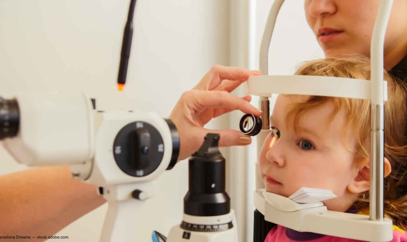
Symptoms of childhood glaucoma include enlarged eyes, cloudiness of the cornea, and photosensitivity (sensitivity to light). Your child needs to be assessed by an ophthalmologist. Ideally the child should be referred to an ophthalmologist with experience in managing childhood glaucoma. The nature of assessment undertaken varies with the age of the child. In children over 7 years the tests used are very similar to those used for adults, i.e. pressure measurement, visual field assessment and examination of the optic disc after dilatation of the pupil.

Both medication and surgery are required in some cases. In an uncomplicated case, surgery can often correct such structural defects.

The treatment for glaucoma in older children is generally medical (eye drops) initially and if these fail, surgery is considered. This is similar to the situation with older adults with glaucoma.

Medical treatments may involve the use of topical eye drops and oral medications. These treatments help to either increase the exit of fluid from the eye or decrease the production of fluid inside the eye. Each results in lower eye pressure.

There are two main types of surgical treatments: filtering surgery and laser surgery. Filtering surgery (also known as micro surgery) involves the use of small surgical tools to create a drainage canal in the eye. In contrast, laser surgery uses a small but powerful beam of light to make a small opening in the eye tissue. Most glaucoma surgery in children can be done safely as a day case without the need for overnight admission. If surgery is required within the first four to five weeks of life the infant may remain in overnight for monitoring after the anesthetic.

Operations such as trabeculectomy or drainage tubes are used. These procedures aim to create a controlled leak or “fistula” by which the aqueous can bypass the trabecular meshwork and escape to drain via the external blood system of the eye.As with adults anti-inflammatory and antibiotic drops are used post operatively. When trabeculectomy is performed in children an anti-metabolite such as 5-fluorouacil (5-FU for short) is very often used as children heal much more rapidly than adults.Laser trabeculoplasty is rarely used in the treatment of glaucoma in children of any age. A cyclo-destructive procedure, such as diode laser treatment of the ciliary body, is sometimes used in the treatment of aphakic glaucoma in children.

Despite timely and aggressive treatment, childhood glaucoma can still cause significant and permanent vision loss. Early diagnosis and treatment, as well as close monitoring are crucial for obtaining a long-term successful outcome.

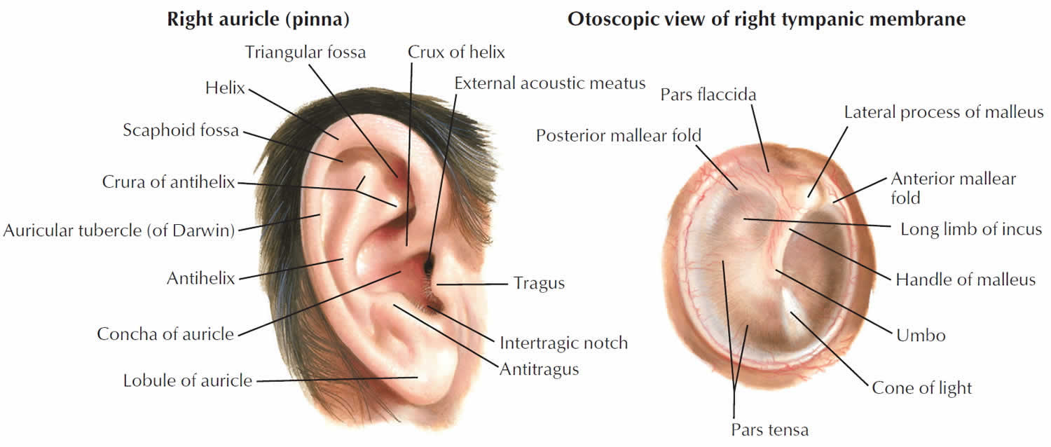
Figure 1. Eye anatomy

Eye anatomy

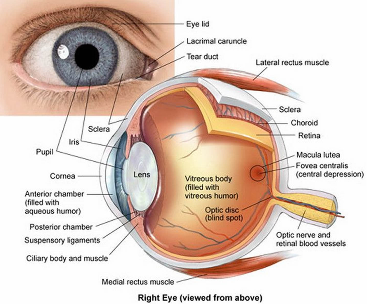
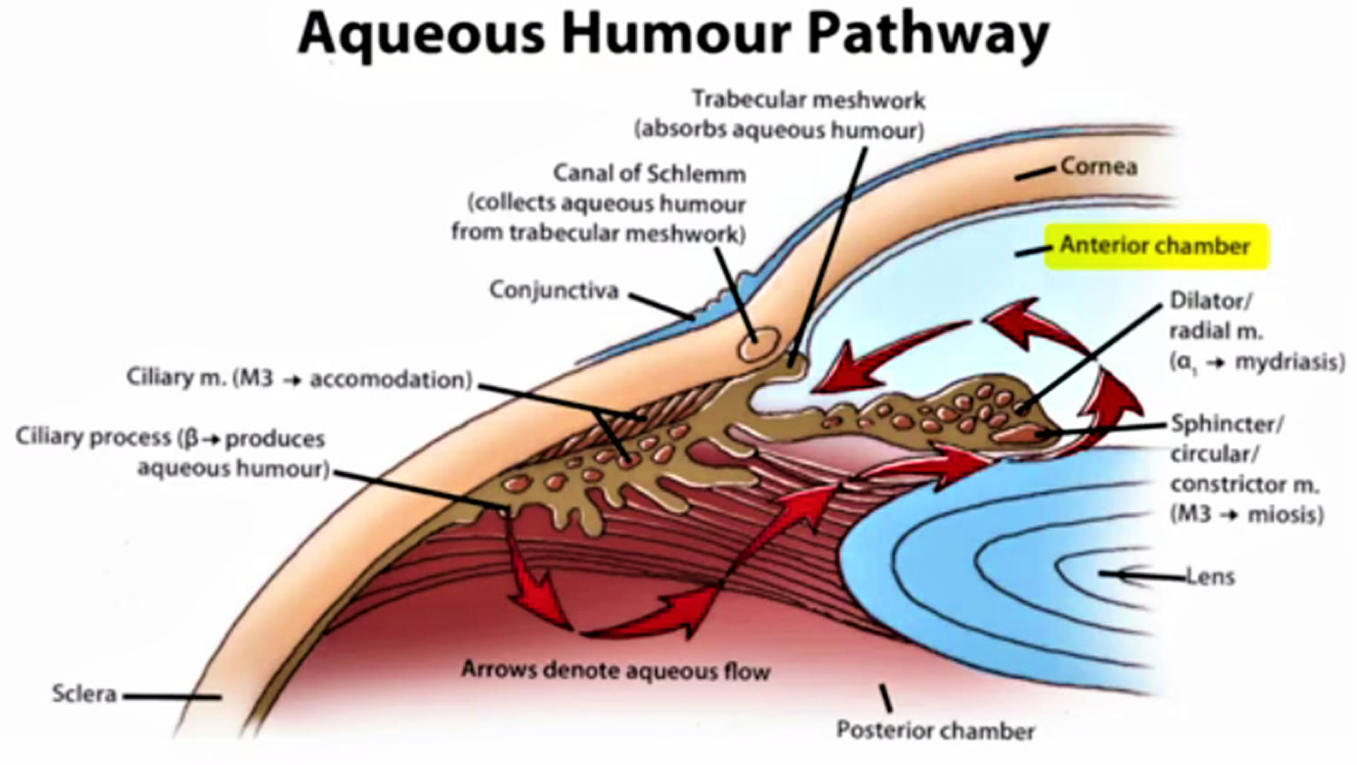
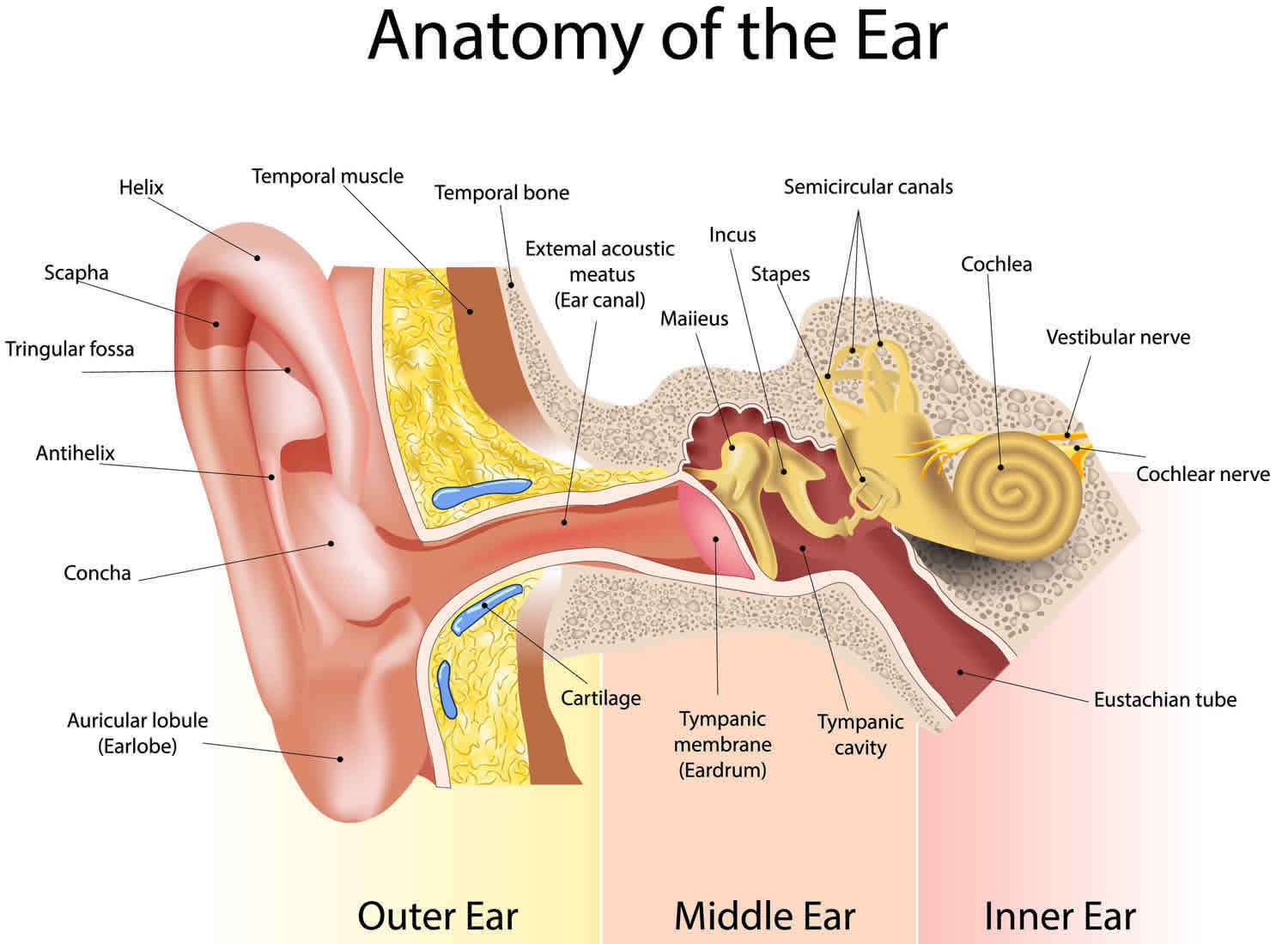
Figure 2. Normal aqueous outflow

Normal aqueous outflow

How would a parent know if a child is suffering from glaucoma?

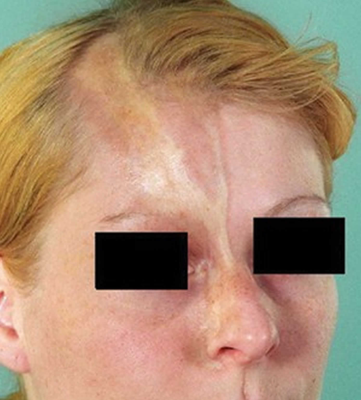
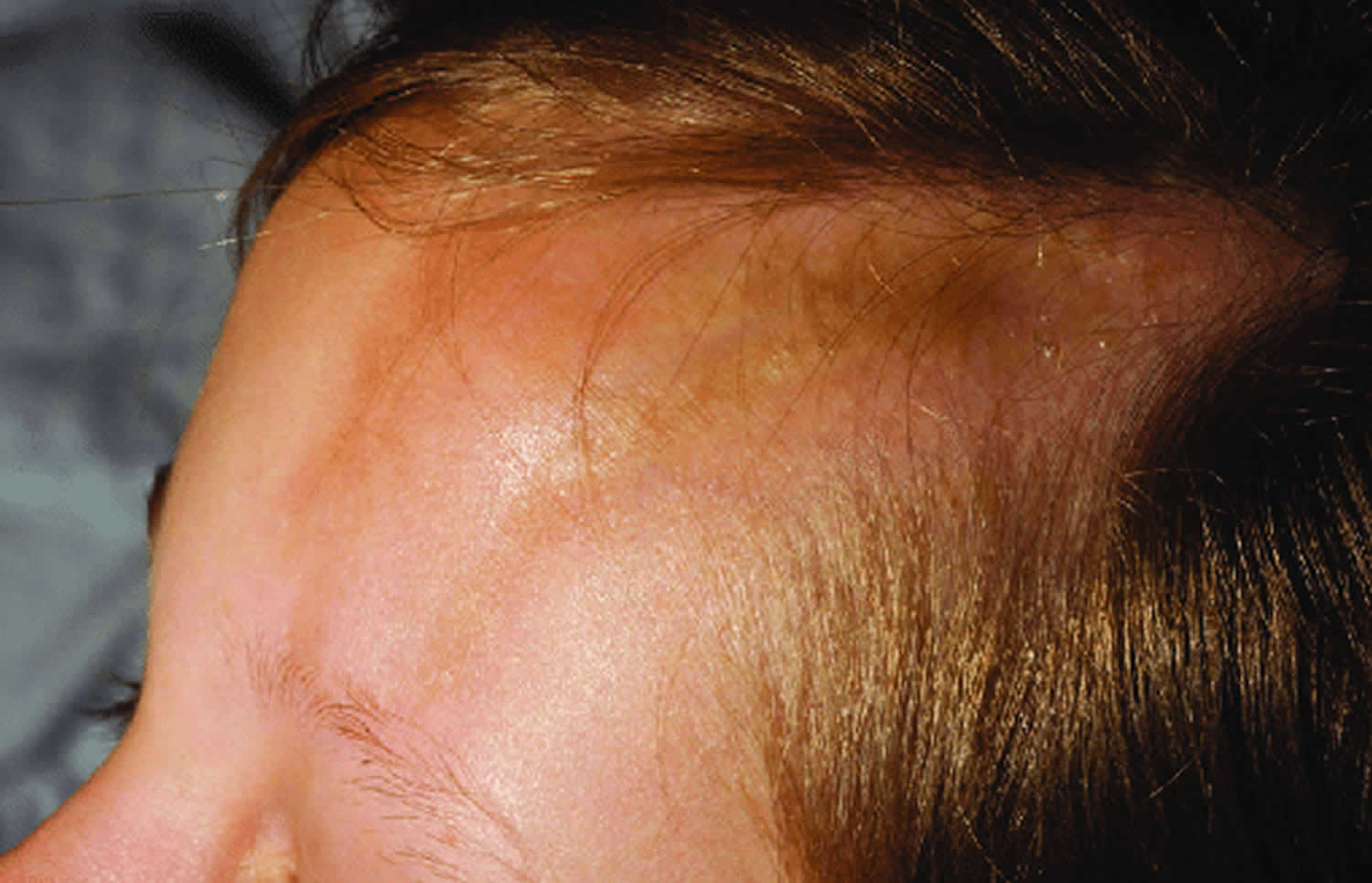
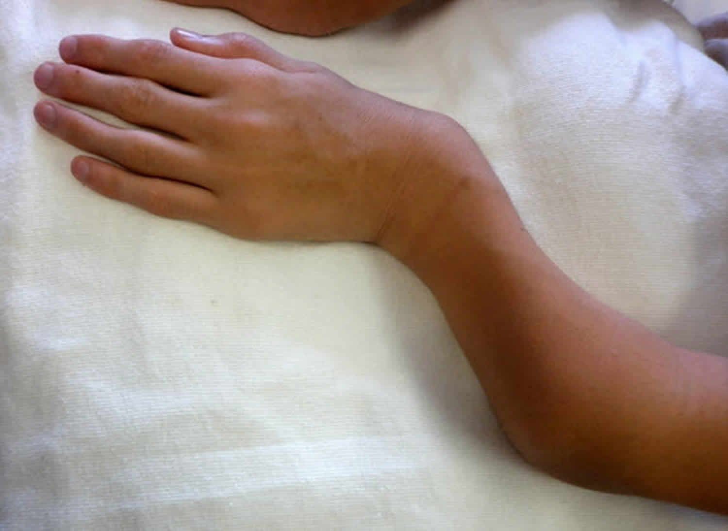
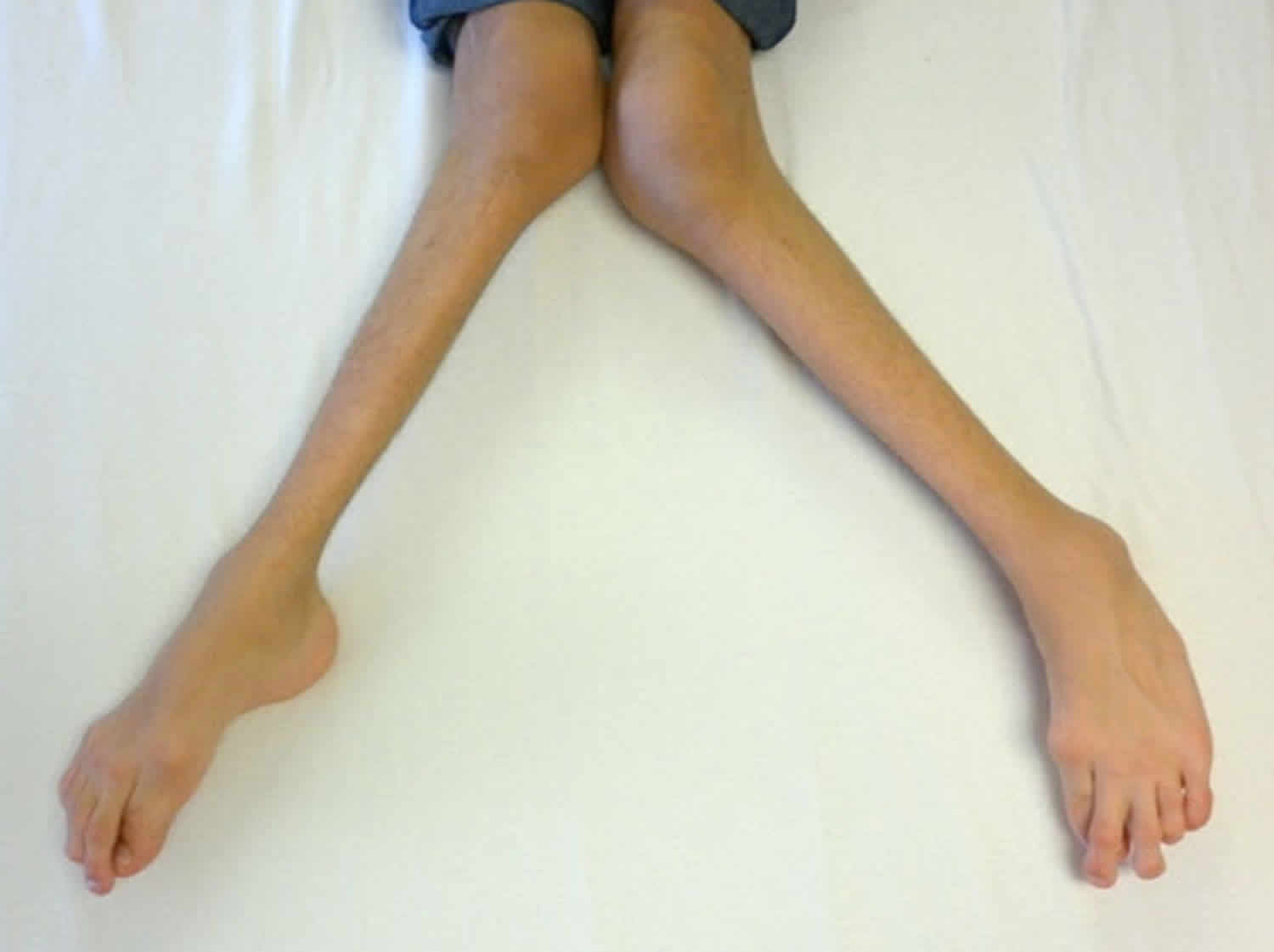
In keeping with the rest of an infant the immature eye is floppy and somewhat elastic. Thus early in life (that is before the second birthday) raised intraocular pressure will stretch the eye and actually cause it to increase in size (it expands in all directions/ rather like a balloon being inflated). Early medical writers termed this buphthalmos (ox eye) as this increase in size of the eye was thought to make the infant’s eye look like an ox’s eye.

The stretching of the eye has a number of harmful effects on the eye. As the eye enlarges the cornea increases in size. One of the many layers of the cornea, Descemet’s membrane, does not have much give and rather than stretch it will split as the eye enlarges. This splitting results in the cornea losing some of its clarity and becoming cloudy. This cloudiness of the cornea is the result of fluid entering the cornea from the anterior chamber via the splits in Descemet’s membrane and is known as corneal edema. Corneal edema causes discomfort and sensitivity to light and increased tear production.

Thus the cardinal features of infantile glaucoma that may be identified by a parent are photophobia (sensitivity to light), increased tearing with an enlarged and cloudy cornea.

What happens when glaucoma is suspected in a young child?

The child needs to be assessed by an ophthalmologist (eye specialist). Ideally the child should be referred to an ophthalmologist with experience in managing childhood glaucoma. Often an examination under anesthetic is required to adequately examine the child and confirm the diagnosis of glaucoma. The diagnosis is confirmed by the presence of typical corneal changes (enlargement, clouding and splits in Descemet’s membrane), raised intraocular pressure and optic disc cupping.

Will my child’s vision be impaired?

Severe loss of vision due to infantile glaucoma is fortunately rare. However, if glaucoma is not appropriately treated there is a risk of progressive visual impairment. Rarely does childhood glaucoma result in severe visual impairment but life-long follow-up is needed for all children after a diagnosis of glaucoma is made. Vision impairment is particularly seen if the onset of the glaucoma is at or before birth. Glasses are commonly required for myopia (short sightedness). This is due to the overall length of the eyeball being increased by the raised intraocular pressure. Photophobia may be a persistent problem if the splits in Descemet’s membrane are severe.

Therefore prompt recognition and timely treatment will improve the chance of a good vision outcome.

Will my child’s lifestyle need to alter in any way?

Most children with infantile glaucoma lead normal lives. Glasses may be required for focusing errors or photophobia. Adolescents often have difficulty accepting the need for long-term medication and regular medical review. Ensuring compliance with regular use of eye drops may be especially difficult. The small number of children with more severe visual impairment will require some degree of help at school.

Is childhood glaucoma hereditary?

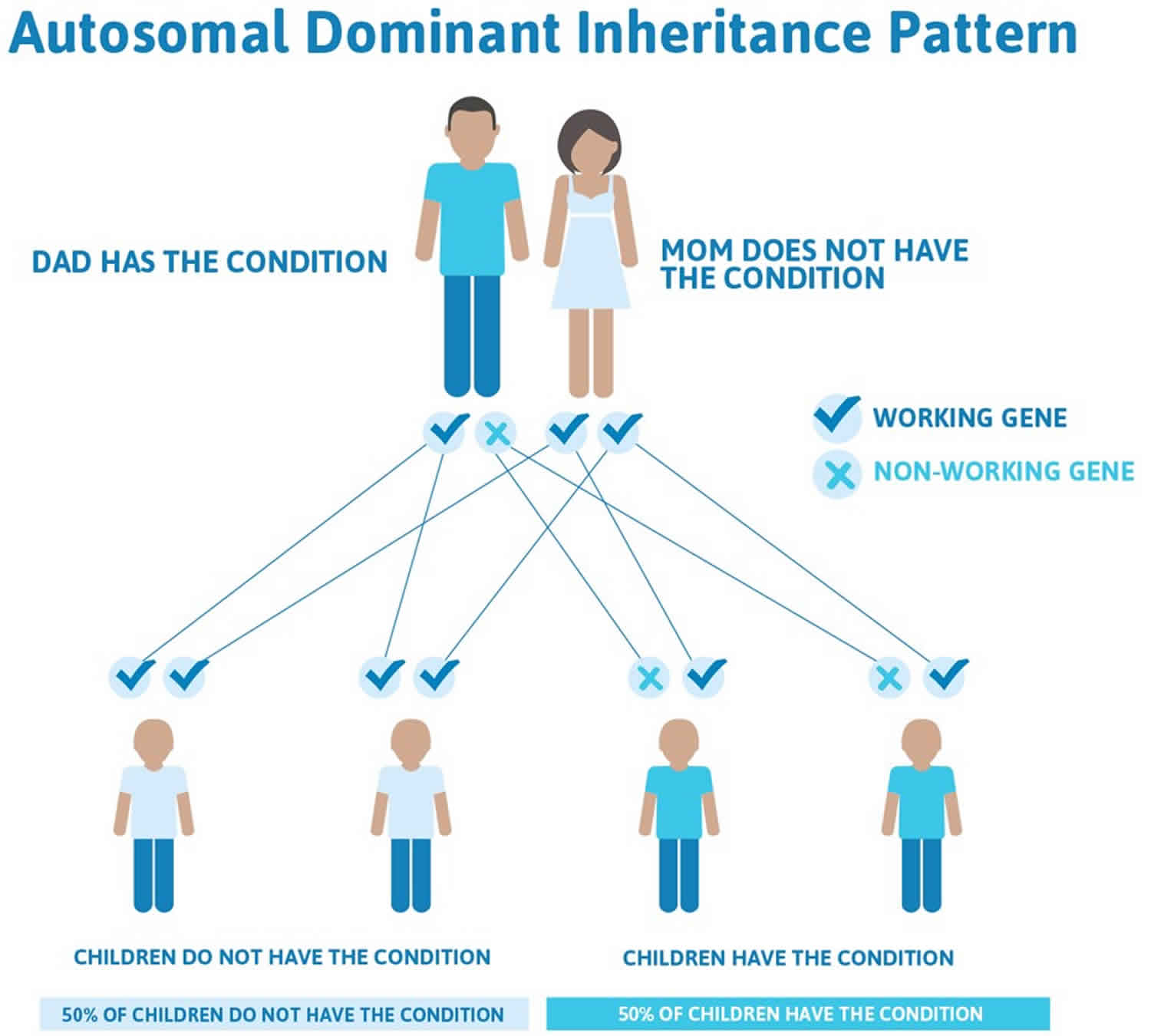
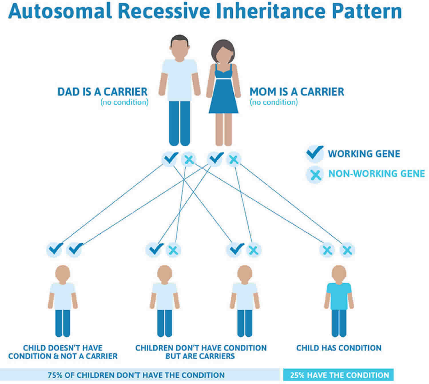
Some types of childhood glaucoma are hereditary. About 10% of primary congenital/infantile glaucoma cases are inherited. Recent research has identified some specific gene mutations linked to this disease; for which genetic testing and counseling for affected families is may be available.

Other conditions that cause secondary glaucoma can be inherited. For example, neurofibromatosis and aniridia are dominantly inherited and are passed on to the children of affected individuals approximately 50% of the time. The incidence of glaucoma that occurs in association with these conditions, however, is less predictable.

What are the chances of another baby of mine developing glaucoma?

The risk is not zero but it is quite low. Primary open angle glaucoma in adolescents may show a familial tendency just as in adult open angle glaucoma. Inherited juvenile open angle glaucoma is well recognized but very rare. This form of glaucoma is generally not detectable till the twenties rather than during later childhood.

Childhood glaucoma causes

Glaucoma occurs when the fluid drainage from the eye is blocked by abnormal development or injury to the drainage tissues, thus, resulting in an increase in the intraocular pressure, damage to the optic nerve, and loss of vision.

There are many causes of childhood glaucoma. It can be hereditary or it can be associated with other eye disorders. If glaucoma cannot be attributed to any other cause, it is classified as primary glaucoma. If glaucoma is a result of another eye disorder, eye injury, or other disease, it is classified as secondary glaucoma.

Approximately 20% of children with primary congenital glaucoma have a mutation in the CYP1B1 gene. If one child in a family has primary congenital glaucoma then the risk for a subsequent child to be affected is about5% and if there are two affected children in one family the risk for subsequent children is 25%. Data suggests that the risk for a parent with a diagnosis of primary congenital glaucoma of having an affected child is 2%.

Most primary congenital glaucoma cases are sporadic (with no family history), however, about 10-40% are familial, with an autosomal recessive inheritance pattern and penetrance varying from 40-100% 1). Autosomal dominant inheritance has also been reported 2). Five loci have been identified by linkage analyses: GLC3A (located on choromosome 2p22-p21), GLC3B (1p36.2-p36.1), GLC3C (14q24.3), GLC3D (14q24.2-q24.3, not overlapping with GLC3C), and GLC3E (9p21) 3).

Thus far, a gene associated with primary congenital glaucoma has been identified in three of these five loci. Further details are below.

The GLC3A loci contains the cyctochrome P4501B1 (CYP1B1) gene, which was the first reported primary congenital glaucoma-causing gene. It codes for an enzyme that metabolizes compounds vital for the developing eye, and is expressed in fetal and adult neuroepithelium and ciliary body 4). Severe trabecular meshwork atrophy is seen in mouse models deficient of CYP1B1 5). In zebrafish, CYP1B1 has been found to indirectly affect neural crest migration to the anterior segment and angle by playing a role in ocular fissure closure 6). While the exact mechanism by which CYP1B1 mutations causes primary congenital glaucoma is unknown, scientists know that CYP1B1 mutations are associated with 15-20% of primary congenital glaucoma cases in Japan and the United States, and all cases in Slovakia Roma 7).

The GLC3D locus contains latent transforming growth factor beta binding protein 2 (LTBP2). LTBP2 is expressed in trabecular meshwork and ciliary processes however its role in the eye is unknown 8). In nonocular tissues, it is involved in tissue repair and cell adhesion 9). LTBP2 null mutations have been reported in consanguineous Iranian and Pakistani families, and Slovakian Roma with primary congenital glaucoma. Homozygosity for a variant of an LTBP2 mutation was associated with worse outcomes even while undergoing more surgical interventions 10).

GLC3E contains the tunica interna endothelial cell kinase (TEK, also known as TIE2) gene. The angiopoietin/TEK (ANGPT/TEK) signaling pathway is required in Schlemm canal development, and includes 3 ligands (ANGPT1, ANGPT2, and ANGPT4) and 2 receptors (TEK and TIE). TEK-knockout mice can have no Schlemm canal and TEK-hemizygous mice have a severely underdeveloped Schlemm canal 11). Additionally, ANGPT1 mutations have been identified in a few primary congenital glaucoma patients who do not have other known primary congenital glaucoma-causing genes, revealing another member of the ANGPT/TEK signaling pathway that may be a cause of primary congenital glaucoma 12).

Lastly, though not associated with the above loci, but originally associated with juvenile open angle glaucoma and primary open angle glaucoma, the myocilin/trabecular meshwork-induced glucocorticoid response protein (MYOC) gene on chromosome 1q24 may also explain a small proportion of primary congenital glaucoma cases, up to 5.5% 13).

Primary congenital glaucoma patients may have one or more genes affected, and the previously discussed genes may regulate or interact with each other 14). CYP1B1 may play a role as a modifier gene for MYOC expression 15) and a digenic mode of inheritance has been considered for CYP1B1 and MYOC 16) and CYP1B1 and TEK 17).

Currently, the chance of identifying a genetic cause is 40% when genetic testing is done 18).

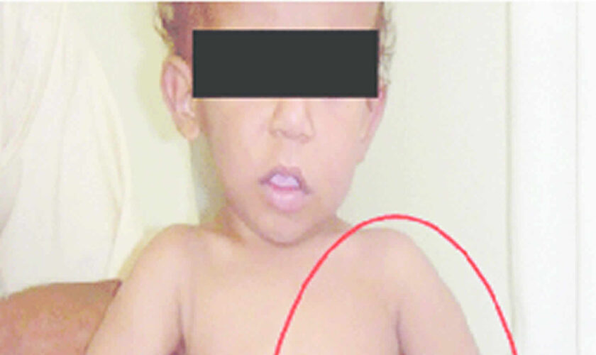
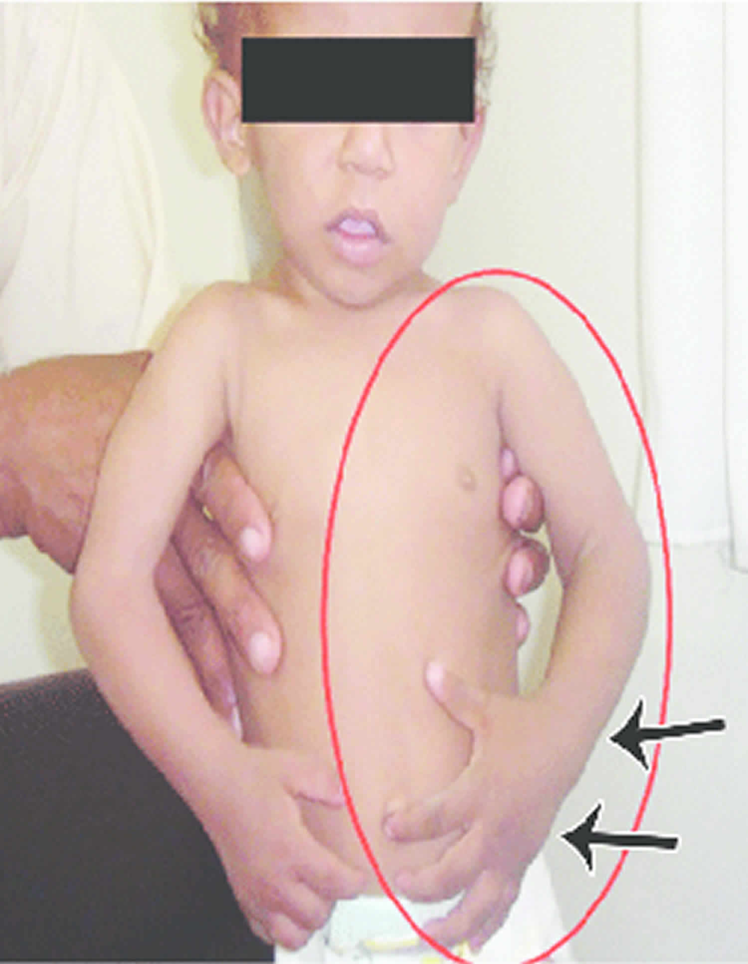
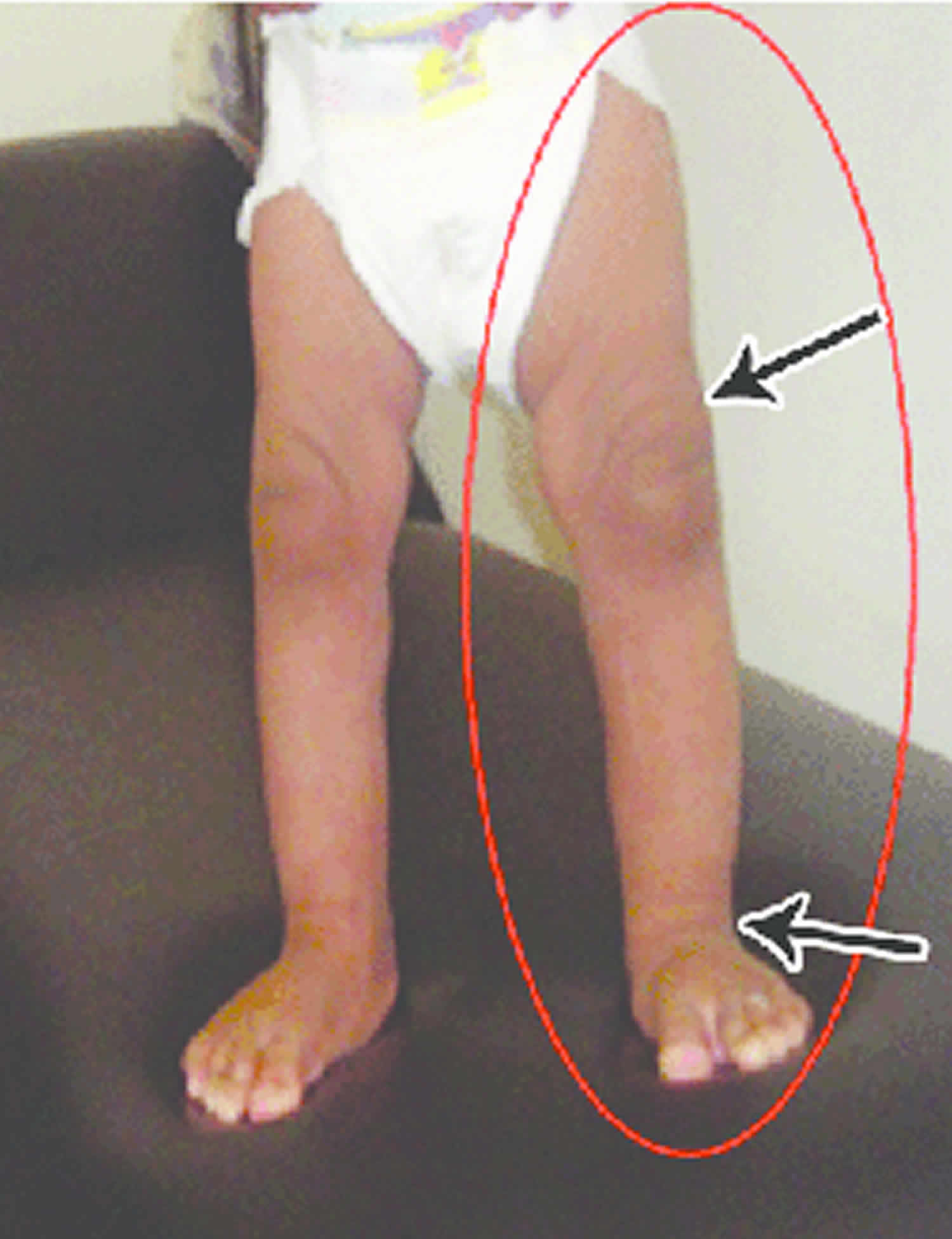
Infantile and childhood glaucoma may be associated with other abnormalities in the eye. The most common of these is a history of having had cataract surgery as an infant, this is called “aphakic glaucoma”. Other eye abnormalities that can be associated with glaucoma are the anterior segment dysgenesis group of disorders which includes Axenfeld-Reiger syndrome. Glaucoma can occur in association with other systemic abnormalities such as the Sturge-Weber syndrome and rubella (German measles) embryopathy. Juvenile arthritis may cause inflammation in the eye (uveitis) that may be complicated by the development of glaucoma. Primary open angle glaucoma does occur rarely in older children and adolescents.

Childhood glaucoma symptoms

Glaucoma is rare in children, as compared to the adult. However, when it does occur, the symptoms may not be as obvious in children. Many children are diagnosed before they are 6 months old. Glaucoma can affect one eye or both.

The most common symptoms of congenital/infantile glaucoma are excessive tearing, light sensitivity and a large, cloudy cornea (the normally clear front surface of the eye) which can cause the iris (colored part of the eye) to appear dull. Excessive tearing accompanied by mattering/discharge in a child is usually not caused by glaucoma but instead is the result of congenital nasolacrimal duct obstruction (blocked tear duct).

Juvenile glaucoma tends to develop without any obvious symptoms, similar to adult glaucoma. Patients with juvenile glaucoma often have a positive family history. On exam the eye pressure will typically be elevated and there may be signs of optic nerve cupping (enlargement of the center “cup” portion of the optic nerve).

The following are the most common symptoms of childhood glaucoma. However, each child may experience symptoms differently. Symptoms may include:

  • excessive tearing
  • light sensitivity (photophobia)
  • closure of one or both eyes in the light
  • cloudy, enlarged cornea (large eye)
  • one eye may be larger than the other
  • vision loss

If the eye pressure increases rapidly, there may be pain and discomfort. Parents may notice that the child becomes irritable, fussy, and develops a poor appetite. Early detection and diagnosis is very important to prevent loss of vision. The symptoms of glaucoma may resemble other eye problems or medical conditions. Always consult your child’s physician for a diagnosis.

Childhood glaucoma diagnosis

In addition to a complete medical history and eye examination of your child, diagnostic procedures for childhood glaucoma may include:

  • visual acuity test – the common eye chart test (with letters and images), which measures vision ability at various distances.
  • pupil dilation – the pupil is widened with eyedrops to allow a close-up examination of the eye’s retina and optic nerve.
  • visual field – a test to measure a child’s side (peripheral) vision. Lost peripheral vision may be an indication of glaucoma.
  • tonometry – a standard test to determine the fluid pressure inside the eye.

Younger children may be examined with hand-held instruments, whereas older children are often examined with standard equipment that is used with adults. An eye examination can be difficult for a child. It is important that parents encourage cooperation. At times, the child may have to be examined under anesthesia, especially young children, in order to examine the eye and the fluid drainage system, and to determine the appropriate treatment.

Childhood glaucoma treatment

Specific treatment for glaucoma will be determined by your child’s opthalmologist (eye specialist) based on:

  • your child’s age, overall health, and medical history
  • extent of the disease
  • your child’s tolerance for specific medications, procedures, or therapies
  • expectations for the course of the disease
  • your opinion or preference

It is important for treatment of childhood glaucoma to start as early as possible. Childhood glaucoma is treated by lowering the intraocular pressure (IOP) via medical and/or surgical means. Treatment may include:

  • Medications: Some medications cause the eye to produce less fluid, while others lower pressure by helping fluid drain from the eye.
  • Conventional surgery: The purpose of conventional surgery is to create a new opening for fluid to leave the eye. Surgical procedures are performed by using microsurgery or lasers. The purpose of surgery is to create an opening for fluid to leave the eye. Surgical procedures used to treat glaucoma in children include the following:
    • Trabeculotomy and goniotomy: A surgical opening is made into the drainage area of the eye (known as the trabecular meshwork drainage system), therefore establishing a more normal anterior chamber angle that allows the fluid to drain more freely, lowering the intraocular pressure (IOP). A goniotomy is an internal trabeculotomy procedure that is used in congenital glaucoma.
    • Trabeculectomy: A surgical procedure that involves the removal of part of the trabecular meshwork drainage system, allowing the fluid to drain from the eye.
  • Iridotomy: In this procedure, a small hole is made through the iris – the colored part of the eye – to allow fluid to flow more freely in the eye. The surgeon may use a laser to create this hole (laser iridotomy).
  • Cyclophotocoagulation: A procedure that uses a laser beam to freeze selected areas of the ciliary body – the part of the eye that produces aqueous humor – to reduce the production of fluid. This type of surgery may be performed with severe cases of childhood glaucoma.

Both medications and surgery have been successfully used to treat childhood glaucoma. Most cases of primary childhood glaucoma are treated with surgery.

Initial treatment may be eye drops or medication by mouth to lower the pressure in the eye. Over the long-term medications have a significant risk of complication in young children and compliance with medical therapy is an even greater problem with young patients than it is with older ones. Surgery is usually required and has a very high success rate.

Childhood glaucoma is the result of blockage of aqueous (fluid) drainage at the trabecular meshwork of the anterior chamber angle. Operations aim to restore the more normal drainage of aqueous. The two most common operations are goniotomy and trabeculotomy. Both involve opening up the tissue in the angle to enable the aqueous to escape more easily from the eye and thus lower the pressure. All surgery for childhood glaucoma is done under a general anesthetic. It is not uncommon for more than one operation to be needed to completely control the raised pressure of infantile glaucoma.

In some instances operations more often used with adult glaucoma such as trabeculectomy or Molteno tubes are used. These procedures aim to create a controlled leak or “fistula” by which the aqueous can bypass the trabecular meshwork and escape from the eye. As with adults anti-inflammatory and antibiotic drops are used post operatively. When trabeculectomy is performed in children an antimetabolite such as 5-fluorouacil (5-FU for short) is very often used as children heal much more rapidly than adults.

Many children with pediatric glaucoma develop myopia (nearsightedness) and require glasses. Also, amblyopia (“lazy eye”) and strabismus (misalignment of the eyes) occur more frequently and may require treatment with patching or surgery.

Follow up

Regular and life long follow up will be required. When the child is quite young it may be necessary for periodic examinations under anaesthetic. As well as monitoring for raised intraocular pressure the follow up will involve monitoring of the development of vision, determine the need for glasses and when older assess any damage to peripheral visual field.

References   [ + ]

read more

Protruding ears

protruding ears

Protruding ears

Protruding ears also called prominent ears, are ears that stick out more than 2 cm from the side of the head. Protruding ears don’t cause any functional problems such as hearing loss.

In most people, protruding or prominent ears are caused by an underdeveloped antihelical fold. When the antihelical fold does not form correctly, it causes the helix (the outer rim of the ear) to stick out.

Most people with protruding ears also have a deep concha, the bowl-shaped space just outside the opening of the ear canal, which pushes the entire ear away from the side of the head.

Figure 1. Ear anatomy

Human ear anatomy

Normal external ear anatomy

The ear is shaped like the letter C, formed by the helix and the earlobe. Inside the C is the letter Y, formed by the antihelix and the superior and inferior crura. The central part of the ear is shaped like a conch sea shell, and is called the concha. There is a small bump in front of the ear canal called the tragus. On the other side of the concha is another bump called the antitragus.

The ear is made primarily of cartilage covered by skin. The earlobe has no cartilage and is made of skin and fat. Although there are some muscles attached to the ear, most people cannot control them, which is why only a small percentage of people can wiggle their ears. The external ear is supplied by four different sensory nerves.

Protruding ears treatment

There are both non-surgical and surgical options for treating protruding ears.

Non-surgical ear molding

If a protruding ear is discovered in the first few weeks after birth, ear molding may correct this deformity and avoid the need for surgery. Ear molding works best in the first few weeks of life when infant ears are soft and pliable. Infant ears have very high levels of maternal estrogen (estrogen which crossed from mother to baby while in the womb and during the birthing process). Because of the increased estrogen levels, infant ears are very moldable, soft and responsive to external molding during the first few weeks and months after birth.

By 6 weeks of age, these levels of maternal estrogen fall to normal, and the ears become more rigid and less pliable. This is why early intervention is so important. If neonatal ear deformities are recognized early enough, they can often be successfully treated by non-surgical molding, preventing the need for surgery later in life.

Because some ear deformities will self-correct over time, your child should be monitored closely for the first 7 to 10 days of life. If the shape or deformity of the ear doesn’t improve in the first week or two, non-surgical infant ear molding may be recommended as the most appropriate treatment approach.

Ear molding uses a combination of commercially available ear molding devices and orthodontic molding materials to reshape the ear.

First, your child will be fit with a non-surgical molding appliance. For the best results, the device should be applied within the first one to two weeks of life. The device is worn continuously for two weeks.

After two weeks, your child’s doctor will examine your child’s ear. If the deformity has not been corrected yet, a new device will be reapplied. This process is repeated every two weeks until acceptable improvement or correction is seen.

Most ears, if treated early, respond to ear molding to improve the shape of the ear. In general, the younger your child is when treatment for prominent ears is started, the shorter the duration of therapy. However, children a few months of age have been treated successfully with non-surgical ear molding.

Protruding ears surgery

Surgery to correct protruding ears is called a setback otoplasty. It can be performed as early as 5 to 6 years of age when ears are almost fully grown.

The procedure to correct protruding ears is usually performed through an incision behind the ears. The cartilage is reshaped to create an antihelical fold. This will support the ear in its new position closer to the head. Sometimes, additional sutures are placed on the back of the conchal to bring the entire ear closer to the side of the head. A postoperative dressing is used to help keep the ears in their new positions. This dressing will typically stay in place for 1 to 2 weeks. Although a general anesthetic is needed, the operation is done on an outpatient basis and your child will be able to return home the same day.

Insurance companies often consider otoplasty to be a cosmetic operation, and therefore they may not cover the cost of this procedure. Before cosmetic ear surgery, discuss the procedure with your insurance carrier to determine what coverage, if any, you can expect.

Ear plastic surgery

To correct prominent ears that lack folds, an ENT (ear, nose, and throat) specialist, or otolaryngologist, places permanent stitches in the upper ear cartilage and ties them in a way that creates a fold to prop up the ear. Scar tissue will form later, holding the fold in place. Corrective surgery, called otoplasty, may be considered on ears that stick out more than 4/5ths of an inch (2 cm) from the back of the head. It can be performed at any age after the ears have reached full size, usually at five- or six-years-old. Having the surgery at a young age has two benefits: (1) the cartilage is more pliable, making it easier to reshape, and (2) the child will experience the psychological benefits of the cosmetic improvement.

An ENT specialist begins the surgery with an incision behind the ear where the ear joins the head. In addition, ears may also be reshaped, reduced in size, or made more symmetrical. The reshaped ear is then secured in position while healing occurs. Typically, otoplasty surgery takes about two hours. The soft dressings over the ears will be used for a few weeks as protection, and the patient usually experiences only mild discomfort. Headbands are sometimes recommended beyond that for a month following surgery.

read more

Myringoplasty

myringoplasty

Myringoplasty

Myringoplasty also called tympanoplasty is microsurgical technique to reconstruct a ruptured or perforated eardrum (tympanic membrane) with the placement of a graft, either medial or lateral to the tympanic membrane annulus, often using the patient’s own tissues. The goal of this surgical procedure is not only to close the perforation but also to improve hearing. The success of the operation depends on the ability to eradicate disease from the middle ear (eg, inflamed granulation tissue and cholesteatoma). Various techniques have been developed and refined, and a number of grafting materials are available. Both the lateral and medial grafting techniques are detailed below.

Myringoplasty can be used for small perforations, such as nonhealing tympanic membranes after pressure-equalizing tube extrusion or traumatic perforations. The technique involves freshening the edges of the perforation to promote healing and placing a carefully trimmed graft lateral to the defect 1). Grafting materials for myringoplasty include fat, Gelfilm, Gelfoam, AlloDerm, and cigarette paper. Gelfoam can also be placed as packing in the middle ear to support the graft.

Myringoplasty is a safe and effective outpatient procedure used to both eradicate disease from the middle ear and restore hearing and middle ear function 2). Your child will need to stay in the hospital overnight. A number of surgical approaches and grafting techniques are available for use by the surgeon. Paramount to success are the preoperative assessment, good hemostasis intraoperatively, and thoughtful surgical planning with careful placement of the graft.

When planning myringoplasty, the surgeon must consider the location of the perforation (marginal versus central), and size (total versus subtotal). Areas of myringosclerosis and tympanosclerosis should be noted. Important comorbidities worth noting include craniofacial disorders and underlying environmental allergies or chronic allergic rhinitis. Critical factors that make myringoplasty less successful include adhesive otitis media, severe eustachian tube dysfunction with either perforation of the contralateral ear or ongoing intermittent otorrhea, cholesteatoma, and previous surgical repair 3).

Myringoplasty key points

  • A myringoplasty is an operation to fix a hole in the eardrum.
  • The operation usually takes about two to three hours.
  • Your child will sleep and feel no pain during the operation.
  • After the operation, your child will have to stay overnight in the hospital.
  • While your child gets better at home, there are some things your child should not do.

Tympanic membrane anatomy

The eardrum also called tympanic membrane is a thin layer of tissue that vibrates in response to sound. An understanding of the tympanic membrane anatomy is critical to successful repair. Myringoplasty (tympanoplasty) procedure mandates an understanding of the layers. The tympanic membrane typically consists of the following 3 layers:

  1. Lateral epithelial layer
  2. Middle fibrous layer
  3. Medial mucosal layer

The outer epithelial layer is composed of stratified squamous epithelium, which is continuous with the skin of the external auditory canal. This is significant because in-growth of this outer epithelial portion through the perforation can result in an epithelial cyst called an acquired cholesteatoma. Untreated, this cyst then releases destructive enzymes that can enlarge the size of the perforation and ultimately cause ossicular erosion. The lateral grafting technique that is discussed later in this text requires that this entire epithelial layer be stripped from the drum remnant prior to placement of the graft so as to avoid iatrogenic cholesteatoma formation.

The middle fibrous layer is composed of connective tissue consisting of outer radial fibers and inner circular fibers. It provides strength to the drum. A healed perforation is also commonly deficient of this middle fibrous layer. The epithelial and endothelial layers regenerate creating a “dimeric” membrane. This miscalculation can be corrected when carefully examined under binocular microscopy. Because this middle layer is absent in the pars flaccida superiorly, the posterior-superior aspect of the drum can be drawn inward toward the middle ear as a retraction pocket.

The inner layer of the tympanic membrane consists of simple cuboidal and columnar epithelium cells. This layer is identical to the mucosal lining of the rest of the middle ear mucosal tissue and is considered to be critical to ensure healing of tympanic membrane perforations, and the surgeon often abrades or rasps the undersurface of the tympanic membrane remnant to stimulate regrowth.

Annulus

The peripheral edge of the tympanic membrane is rimmed by a dense fibrous layer called the annulus, which is essentially a thickening of the pars tensa. Successful elevation of the annulus is critical for medial grafting technique. The annulus is deficient superiorly at the “12 o’clock” location. This area is the notch of Rivinus and can guide the surgeon to a natural plane to elevate the annulus.

Ear canal

The ear canal has bone in the medial component (inner one-third). The lateral portion, which extends into the pinna, is composed of cartilage. The boney/cartilaginous interface is located at the medial two-thirds junction. Most incisions that are made to raise a tympanomeatal flap or perform either an endaural or transcanal approaches are made at this location as well. The superiorly placed vascular strip is another critical area within the ear canal. This region is demarcated by the tympanosquamous suture line superiorly and the tympanomastoid junction line inferiorly. Canal incisions are often made along these junctions.

Middle ear

The middle ear is an air-filled space bordered by the bony labyrinth of the inner ear medially, the tympanic membrane laterally, and the cranium superiorly. This space contains the ossicles, nerves (facial nerve, chorda tympani, Jacobsen nerve), small muscles (stapedius and tensor tympani), ligaments, and blood vessels. The petrous portion of the internal carotid artery and the internal jugular vein, which are both in proximity to the middle ear space, can be dehiscent and should be noted on any preoperative imaging. Rarely, middle ear pathology can involve these structures.

In order for successful grafting of the tympanic membrane to improve hearing, an intact ossicular chain must be present. The malleus transmits energy from the tympanic membrane to the incus, which itself is connected to the stapes superstructure resting on the oval window. Diarthrodial joints connect the 3 ossicles and allow the transmission of acoustic energy from the tympanic membrane to the inner ear. The incudostapedial joint is the most fragile and, hence, has the highest likelihood to require repair.

Mastoid

The middle ear communicates with the mastoid air cells via the mastoid antrum. The temporal bone air cells are usually pneumatized by 3 years of age. However, the air cells can remain underdeveloped and sclerotic in patients with persistent eustachian tube dysfunction. A poorly pneumatized or fluid-filled mastoid bone predisposes a patient to require a more extensive tympanomastoidectomy to improve the chances of successful graft placement.

Eustachian tube

The eustachian tube connects the middle ear with the nasopharynx and allows pressure equilibration in the middle ear. Enlarged adenoids or biofilms within this lymphoid tissue are hypothesized to predispose a patient to persistent middle ear disease. This bony-cartilaginous tube is approximately 45° from the horizontal in adults but only 10° from horizontal in infants. In addition, the infant eustachian tube is about 50% of the adult length.

Inner ear

The inner ear is composed of the cochlea, which is the end-organ for hearing, and the vestibular organs. The vestibular organs include the utricle, saccule, and the 3 semicircular canals and are involved in balance.

Figure 1. Ear anatomy

Ear anatomy

Ear drum anatomy

Figure 2. Tympanic membrane anatomy (right ear)

Tympanic membrane anatomy

Myringoplasty surgery

Myringoplasty (tympanoplasty) is an outpatient procedure for adults and for most children. The operation takes about two to three hours. Your child will need to stay in the hospital overnight.

Your child will sleep and feel no pain during the operation. Just before your child has the operation, they will be given a sleep medicine. This is called a general anesthetic. This means that your child will sleep and feel no pain during the operation.

The ear nose and throat (ENT) doctor will take a tiny piece of tissue from an area around the ear. This is done by making a cut behind your child’s ear. The piece of ear tissue is then used to fix the hole in your child’s eardrum. Your child will have dissolvable stitches behind the ear and gauze packing in the ear to absorb any fluid.

Various techniques and grafting materials can be used. Which approach is used depends on the size and location of the perforation, the presence or absence of cholesteatoma or granulation tissue, the status of the ossicles and mastoid, other anatomical considerations (eg, narrow external auditory canals), as well as the surgeon’s preference and expertise 4).

Examining the middle ear and ossicles and removing any elements of adhesions or cholesteatoma is critical. The chosen approach should provide optimal visualization of the perforation and tympanic membrane. One should be careful not to disrupt an intact and mobile ossicular chain if the hearing loss is only low-frequency conductive, as is often the case with hearing loss secondary to a perforation 5).

Before the operation

Evaluation of the patient for myringoplasty involves a detailed history and physical examination. Important aspects of the history include the duration of the perforation, severity of otalgia, otorrhea, hearing loss, vertigo, previous attempts at repair, otitis media, and otitis externa. The number and frequency of infections (including time of most recent infection) provide insight into the severity of disease. Past otologic surgical history is critical and should include any history of tympanostomy tubes (also called myringotomy tubes, ventilation tubes, or pressure equalization (PE) tubes) and details of any prior tympanoplasties (including approaches, grafts used, outcomes).

Prior to considering surgery in any patient, acute and chronic infections should be controlled using ototopical, oral, and/or intravenous antibiotics or antifungals, if indicated. Ototopical drops with steroids may also be needed if granulation tissue or aural polyps are visualized to improve inflammation. Ideally, an ear should be “dry” for 3-4 months before surgery is performed to enhance the chance of success. Individuals who undergo surgery must keep the operated ear dry for a period of several weeks or until the graft has healed. Operating on an actively infected ear is contraindicated.

Physical examination

The physical examination should focus on pneumatic otoscopy and otomicroscopy. In the office, tuning forks can give an important assessment of hearing; in older patients, tuning forks can be used to confirm audiometric findings. Additional assessments include documentation of facial nerve functionality and inspection for previous incisions. Although a small amount of cerumen is tolerated in the routine otoscopic examination, obstructive cerumen should be removed when evaluating a patient for myringoplasty in order to provide an unimpeded view of the entire tympanic membrane.

Monomeric (or more accurately, dimeric) areas may appear as perforations until inspected more closely under microscopy. Retraction pockets should be closely inspected for accumulation of squamous debris. Considering the status of the contralateral ear when considering repair of a tympanic membrane perforation is essential. The ear with more significant hearing loss is usually operated on first if bilateral perforations exist.

Audiometric testing

Audiometry should be performed preoperatively in all surgical candidates. Tympanometry can add useful information in younger children who are difficult to properly examine. The primary reason for audiometric analysis is to establish the degree of conductive hearing loss. Perforations usually cause low-frequency conductive loss 6). If underlying ossicular discontinuity exists and is not addressed during surgery, then postoperative hearing can be worse despite an intact neo-tympanum. One should consider ossicular involvement if the conductive hearing loss is flat across all frequencies or greater than 35 db. Finally, the presence and degree of any sensorineural hearing loss should be documented preoperatively.

Radiographic testing

Computed tomography (CT) and magnetic resonance imaging (MRI) is not essential but may be indicated in patients in whom a concern for cochlear, labyrinthine, or intracranial pathology exists. Other patients who might be considered for preoperative imaging include patients with a history of facial palsy, children with craniofacial abnormalities, and revision cases in which the anatomy may be distorted.

Pre-operative imaging assists the surgeon in preoperatively identifying pathology and planning surgery. CT scan should be ordered when concern exists for cholesteatoma and in patients with previous mastoid surgery, otalgia, or vertigo. MRI is beneficial for delineating the integrity of the dura as well as detecting small retrocochlear lesions such as acoustic neuromas.

Grafting materials

Various materials exist for use for tympanic membrane grafting. True temporalis fascia is the most common graft because of its ease of harvest and its abundant availability, even in revision cases. Some surgeons prefers loose areolar fascia (also known as “fool’s fascia”) and prefer to save the true fascia for revision cases. Also, the “fool’s fascia” is considered by some to be more pliable, have less donor site morbidity, and to be more transparent after healing. It is available via the same postauricular incision that can be used for tympanoplasty, or a separate incision can be made in or beyond the postauricular hairline if a transcanal or endaural technique is used. A mild amount of donor site morbidity occurs, with postoperative pain over the temporalis muscle being the most common symptom.

  • The postauricular incision is marked and injected with lidocaine with epinephrine.
  • Dissection is carried down onto the fascia (loose areolar /true temporalis).
  • The graft is harvested.
  • Muscle is removed from the fascia graft, and the graft is then set on the back table for later use.

Cartilage is available to be harvested easily from either the tragus or the conchal bowl, if a post-auricular approach is being used. Tragal cartilage is harvested with perichondrium attached via a small incision on the internal surface of the tragus 7). This graft is an appropriate size and carries very little donor site morbidity. In addition, the perichondrium can be reflected to stabilize the graft. Conchal cartilage also carries no additional significant morbidity. Other grafting materials include lobular fat, periosteum, perichondrium, vein, and AlloDerm.

Myringoplasty procedure

Transcanal approach

The transcanal approach is especially good for small posterior perforations, but can be used for medium-sized perforations if the anterior tympanic membrane is easily visualized. This technique can be challenging for significant anterior perforations, narrow/stenotic ear canals, or individuals with a significant anterior canal bulge. Inspecting the perforation prior to preparing the patient and determining that at least a 5-mm speculum can be placed is important. Canalplasty can be used to improve visualization if slightly limited.

Medial graft

When performing a transcanal medial tympanoplasty procedure, the following steps are followed:

  1. The ear canal is suctioned and surgical Betadine used during the surgical prep is removed.
  2. The external auditory canal is cleaned and injected with 1% lidocaine with 1:100,000 epinephrine or 0.5% lidocaine with 1:200,000 epinephrine, primarily for vasoconstriction to optimize visualization during the procedure.
  3. The edge of the perforation is dissected and removed using a sharp pick and cup forceps; this “postage-stamping” and “freshening” of the perforation is critical to ensure that the graft incorporates into the native tympanic membrane remnant.
  4. Next, a tympanomeatal flap is created. It is customized based on the location of the perforation and surgeon’s preference. The flap design should be such that it can be easily and atraumatically raised and the undersurface of tympanic membrane perforation can be readily accessed. A medially-based tympanomeatal flap is usually created with radial incisions at 12 o’clock and 6 o’clock (ie, superiorly and inferiorly) that either connect directly or via a semilunar incision in the posterior canal just medial to the bony-cartilaginous junction.
  5. The tympanomeatal flap is raised medially with a round knife. To avoid traumatic tearing, take great care not to suction on the flap.
  6. When the annulus is reached, the tympanotomy is made such that the instrument of choice (eg, round knife, gimmick, sickle knife, pick) lifts the annulus while hugging the bony groove from which the fibrous annulus can be dissected. The fibrous annulus is then dissected circumferentially with care not to injure the ossicles, the chorda tympani nerve, or residual drum. The flap is then positioned, usually anteriorly, such that the perforation is exposed.
  7. A canalplasty of the posterior or anterior external auditory canal can be performed to optimize visualization. Take care not to injure the facial nerve or temporomandibular joint.
  8. If indicated, the middle ear and ossicles are inspected and palpated to confirm ossicular continuity. Middle ear disease (granulation tissue, tympanosclerosis, adhesions, cholesteatoma) is completely removed. Removing hypertrophic middle ear mucosa with either a McCabe dissector or Duckbill elevator, particularly mucosa abutting the fibrous annulus in anterior tympanic membrane perforations, is important.
  9. Ossicular reconstruction can be performed if necessary, followed by grafting of the perforation. Elevating the tympanic membrane remnant off the long process of the malleus with a sickle knife may be necessary. This allows both closer inspection of the ossicles and better placement of the graft.
  10. The middle ear must be carefully packed with the surgeon’s preferred material – either Gelfilm, Gelfoam, or Surgicel. This is often soaked in either oxymetazoline, antibiotic ear drops, or diluted epinephrine (1:10,000). Packing the mesotympanum and hypotympanum is important, although excess packing should be avoided near the ossicles so as to prevent adhesions.
  11. The graft is trimmed on the back table. The graft should adequately cover the entire defect. Hemostasis is critical to intraoperative visualization and successful placement of the graft. The graft should be well supported so as to avoid shifting or displacement.
  12. Some surgeons advocate that nitrous oxide anesthetic be switched off at this point because this particular agent has a tendency to accumulate in spaces such as the middle ear and can potentially dislodge the graft.
  13. The tympanomeatal flap is laid back down over the graft, and the posterior canal skin edges are laid flat. Pieces of Gelfoam, Surgicel, or antibiotic ointment are placed along the tympanic membrane and graft and layered laterally to cover the canal incisions.
  14. Antibiotic ointment is placed in the lateral canal, and either Vaseline-coated or antibiotic-coated sculpted cotton ball is placed in the external auditory meatus.
  15. An optional Glasscock or mastoid pressure dressing is placed at the end of the case, particularly if a mastoidectomy has been performed.

Endaural approach

The endaural technique is useful with many perforations, especially when a small atticotomy is anticipated (when improved access to and visualization of the epitympanum is needed). Many of the steps involved in the transcanal technique are similarly performed in the endaural tympanoplasty as well. When performing an endaural medial tympanoplasty procedure, the following steps are followed:

  1. The canal is prepared as detailed above, but the injection may continue laterally to the lateral external auditory canal and tragus.
  2. An incision is made at 12 o’clock and extended superolaterally between the tragus and helical root.
  3. A medially-based tympanomeatal flap is raised, and the middle ear is entered with the same care as described previously. The tympanic membrane is freed superiorly and inferiorly.
  4. Middle ear work is carried out as indicated. Atticotomy can be performed using a small curette or drill if access into the epitympanum is needed.
  5. Grafting is performed and the tympanomeatal flap is laid back down. Gelfoam is placed along the tympanic membrane and fills the canal. Antibiotic ointment and a cotton ball are used laterally.

Postauricular approach

The postauricular technique is the most commonly performed approach for either revision tympanoplasties or those in which a mastoidectomy is anticipated. This technique offers the best visualization of the anterior tympanic membrane and is preferred for large anterior perforations. In addition, it can be combined with mastoidectomy if disease is found in the mastoid that requires the surgeon’s attention. A basic outline of the procedure follows:

  1. The canal is prepared in a similar fashion to the transcanal technique.
  2. Radial and horizontal canal incisions are made as described previously, and the canal is packed with cotton soaked in oxymetazoline or epinephrine.
  3. A postauricular incision is marked 5 mm posterior to the auricular crease in a curvilinear fashion, extending form the mastoid tip to the temporal line. The incision is injected with 1% lidocaine with either 1:100,000 epinephrine or 0.5% lidocaine with 1:200,000 epinephrine. The incision is carried down through the skin and subcutaneous tissue with care not to enter the ear canal. When the temporalis fascia is reached, a graft can be harvested using a Freer elevator and scissors. If multiple previous grafts have been harvested, either tissue from the contralateral ear or AlloDerm can be used as well.
  4. A periosteal incision is made in a “T” or “7” fashion, and the periosteum is raised into the lateral ear canal until the previously-made canal incisions are reached. The cotton in the ear canal is removed.
  5. A rubber Penrose drain can be inserted to retract the lateral canal and auricle anteriorly. Self-retaining retractors such as Weitlaner or Perkins retractors are used to provide further exposure. The perforation is visualized and prepared.
  6. The tympanomeatal flap is raised medially and the middle ear is entered as described previously. Do not suction on the flap.
  7. Canalplasty can be performed and middle ear work is carried out as indicated, including ossicular reconstruction. The perforation is grafted, and the tympanomeatal flap is laid back down with Gelfoam layered lateral to the tympanic membrane.
  8. The auricle and lateral ear canal are relaxed and the postauricular incision is closed in a layered fashion. The remainder of the ear canal is packed with Gelfoam and antibiotic ointment. A pressure dressing is applied to prevent a postauricular hematoma.

Grafting technique

Although variations exist, 2 primary grafting techniques exist: medial grafting (or underlay) and lateral grafting (or overlay). These terms refer to the position of the graft in relation to the fibrous annulus, not to the malleus or tympanic remnant.

The medial grafting technique is performed as described previously. The primary advantage of the medial graft technique is that it is quicker and easier to perform than lateral grafting. It also carries a high success rate (approximately 90% in experienced hands). The biggest disadvantage is its limited exposure and poor utility for larger perforations and its difficulty with repair of near-total perforations.

Advantages of the lateral graft technique include wide exposure and versatility for larger perforations and for any needed ossicular reconstruction. Disadvantages include the requirement of a higher technical skill level, a longer operative time, slower healing rate, and the risk of blunting and lateralization of the graft. The lateral graft technique is championed by the some doctors as a technique more suited for total drum replacement. The basic steps involved in lateral grafting are described as follows:

  1. Lateral (overlay) tympanoplasty is performed through the previously-described postauricular incision. Important differences exist in the canal incisions. In this procedure, a vascular strip is created by making radial incisions at about 2 o’clock and 5 o’clock. These incisions are connected medially just lateral to the annulus on the posterior canal wall and laterally just medial to the bony-cartilaginous junction along the anterior canal wall.
  2. The skin of the anterior external auditory canal is raised medially. When the annulus is reached, squamous epithelium is raised off of the tympanic remnant, and the canal skin is removed in continuity with the remnant skin and stored in saline solution. This maneuver is done with a cupped forceps.
  3. Bony canalplasty can be performed anteriorly to ensure visualization of the entire annulus. Protecting the flap with a portion of trimmed Silastic as a shield is helpful. Care must be taken not to enter the glenoid fossa, which risks injuring the temporomandibular joint (TMJ) and causing prolapse into the ear canal.
  4. Antibiotic-soaked Gelfoam is packed into the middle ear to support the tympanic remnant. The fascia graft is placed medial to the malleus and draped onto the posterior canal wall for stabilization. If possible, the graft should not extend onto the anterior canal wall in an effort to prevent blunting of the graft.
  5. The canal skin/tympanic remnant is returned and placed lateral to the graft and carefully positioned. Gelfoam is then packed tightly into the anterior aspect of the medial canal to prevent blunting, and the vascular strip is laid back down, covering the lateral extension of the fascia graft to improve its blood supply. Antibiotic-soaked Gelfoam is then packed into the rest of the external auditory canal.
  6. The postauricular incision is closed in layers, and antibiotic ointment is placed on the incision and in the lateral canal. A cotton ball is placed in the external auditory meatus, and a mastoid pressure dressing is applied.

Myringoplasty recovery

After the operation, your child’s surgical team will take your child to the recovery room, also called the Post Anesthetic Care Unit (PACU). This is where your child will wake up. Your child will stay in PACU for about one hour. Your child’s surgical team will then move your child to a room on the nursing unit.

Your child’s surgical team will give your child fluids through a tube in their arm, called an IV, until they are able to drink easily. Your child will have a gauze bandage around their head, which will be taken off the day after the operation.

Postoperative hearing should be immediately assessed in the recovery room with a tuning fork. If a pressure dressing is applied, it should be removed on the first or second postoperative day depending on surgeon preference.

Although the ear must be kept otherwise dry, patients are allowed to wash their hair, while keeping a cotton ball with Vaseline in the canal for dry ear measures. Pain is usually managed with acetaminophen and/or ibuprofen. Narcotics such as hydrocodone (Vicodin or Norco) are usually prescribed when a post-auricular approach is used. Oral antibiotics are the surgeon’s preference and can be given for 5-7 days. A cotton ball is replaced in the external auditory meatus as needed for bleeding or drainage. Ototopical drops are typically administered postoperatively for 7-21 days after surgery and continued until the first postoperative visit.

At the first postoperative visit (3-4 weeks after surgery), the ear is examined under the microscope, and any canal packing or residual antibiotic is removed. At this time, a good assessment can be made as to the healing and neovascularization of the graft. Granulation tissue at the tympanomeatal flap is addressed. Ototopical drops are continued as the graft continues to heal. Postoperative audiometric testing is delayed until healing is complete (typically 6-12 weeks). Follow-up visits are scheduled to ensure complete proper healing and restoration of hearing.

Medications that may be prescribed after surgery

  • Pain control: Acetaminophen (Tylenol) liquid solution may be given. Some children will requireprescription pain medication. Pain may be worse during evening; some children should be given medication at night.
  • NO ibuprofen (Motrin or Advil) or aspirin for twoweeks after surgeryunless otherwiseinstructed by physician.
  • Antibiotic eardrops may be prescribed twoweeks after surgery. Give drops at room temperature.
  • Antibiotics may be prescribed for 7 to 10 days.
When to see a doctor

Call your child’s ENT doctor, your family doctor or your local medical clinic right away if your child has any of these signs after going home:

  • Fever greater than 101º F (38.5 °C)
  • Severe ear pain, pain that gets worse or pain noted more than 7 days after surgery
  • Excessive drainage or blood leaking from the ear
  • Swelling, redness or drainage from incision site
  • Dizziness that lasts for more than one week
  • Vomiting (throwing up) that does not stop
  • The packing falls out of the ear

If it is an emergency or if you are concerned about your child’s condition, do not wait. Take your child to the closest emergency department.

Special precautions after ear surgery

  1. No nose blowing for two weeks. Sneeze with an open mouth.
  2. Water precautions: Keep ear canal dry for the first twoweeks;place cotton ball coated with Vaseline in the ear(s) when bathing. Hair may be washed twodays after surgery. The sutures may get wet but the ear canal should stay dry. No swimming for usually 4 to 6 weeks. The physician will advise you when the ear can get wet.
  3. Wound and suture line care: A large dressing is usually applied after surgery and should be left in place for one-twodays. After the dressing is removed (at your appointment or at home as instructed by your doctor), clean the incisionwith hydrogen peroxide and apply bacitracin ointment. Use Q-tips or cotton balls to clean the incision. Wash your hands before and after cleaning the incision. Apply a cotton ball to the outside of the ear canal if drainage is present.
  4. Keep the incision protected from the sun for 6 to 12 months, keep covered or apply sunscreen.

Taking care of your child at home

Please follow these steps at home to help your child get better:

  • Your child may have a small gauze bandage over their ear. Please keep this bandage on for one or two days after going home.
  • Do not let the cut behind your child’s ear get wet. Do not get any water in the ear. Your child can have a bath, but take care not to pull on the ear or get it wet if you need to wash their hair.
  • Do not let your child play contact sports like hockey or soccer until the ENT doctor says it is OK.
  • Do not let your child go swimming until the ENT doctor says it is OK.
  • Do not let your child play a musical instrument that you blow in until the ENT doctor says it is OK.
  • Do not let your child blow their nose. Have them cough or sneeze with their mouth open.
  • Your child may return to school or day care when your ENT doctor says it is OK. Usually, this will be one week after the operation.

Pain management at home

Follow these instructions when your child goes home after the procedure.

You may give your child medicine for pain.

You may receive a prescription for pain medication before you leave the hospital. Follow the dosage instructions given to you by the pharmacist. Although these prescription pain medications can be beneficial, they are also potentially very dangerous if not used properly.

When using these medications, if you notice any changes in either breathing or level of drowsiness that concern you, stop the medication and seek medical attention. If your child is unresponsive, call your local emergency services number immediately.

Do not give your child over-the-counter medicine that may have a sedative effect (makes people sleepy) while giving the prescription for pain medicine. Examples of these medicines are decongestants and antihistamines. Discuss these medications with your pharmacist.

You may give your child acetaminophen if they have pain. Give the dose printed on the bottle for your child’s age. Do not give your child ibuprofen or acetylsalicylic acid for two weeks after the surgery. These medications could increase your child’s risk of bleeding after the operation. Check with the nurse or doctor first before giving these medicines to your child.

Myringoplasty recovery time

Routine activities may be resumed in 2-5 days. Most children return to school in 3 to 5 days if eating and sleeping well and pain-free. Vigorous exercise, heavy lifting and physical activities should be avoided for 2 weeks. No swimming until advised by your doctor, typically in 4-6 weeks.

Follow-up care

A follow-up appointment with the ENT doctor

The ENT unit will make a follow-up appointment with the doctor for your child. If everything is normal during the appointment, the doctor will:

  • Check your child’s ear to see how it is healing.
  • Take out the packing from your child’s ear.
  • Tell you when your child can start to play sports again.

Myringoplasty complications

Common complaints after surgery:

  • Nausea and vomiting may occur for the first 24 to 48 hours.
  • Pain: Mild to moderate ear pain and/or pain at theincision site for 3 to 5 daysis expected.
  • Fever: A low-grade fever may be observed several days.
  • Ear drainage after surgery. Packing material is placed in the ear canal; sometimes there is clear, pink, or bloody drainage from the ear for 3-5 days. This may also occurwhen ear drops are started.
  • Dizziness or unsteadiness: Dizziness is common for several days.
  • Decreased hearing in the operated ear for several weeks.

Complications of the surgery include recurrence of the perforation, tympanic membrane retraction, otorrhea, cholesteatoma development, persistence or worsening of any conductive hearing loss, sensorineural hearing loss (rare), and taste disturbances. Post-auricular incisions are at risk for hematoma, and a mastoid pressure dressing is recommended for the first postoperative night. Outcomes can be optimized by a proper and detailed preoperative assessment and the careful construction of an effective surgical plan.

The graft can fail because of infection, failure to pack the graft securely in place, technical error, failure to clear mastoid and middle ear disease, and because of a concurrent undetected cholesteatoma. Excising all tympanosclerosis at the edge of the perforation so as to allow vascularized perimeters to incorporate the graft is critical.

Myringoplasty outcomes

The indications and outcomes vary depending on the specific clinical problem. Success rates of tympanic membrane closure vary greatly in the literature (35-98%) but are usually greater than 80% and depend largely on the size and location of the perforation, surgical technique, and overall health of the middle ear 8).

References   [ + ]

read more

Linear scleroderma

linear scleroderma

Linear scleroderma

Linear scleroderma represents a unique form of localized scleroderma that primarily affects children, with 67% of patients diagnosed before 18 years of age 1). Linear scleroderma manifests as thickened and hardened skin bands, which are most often on the face or extremities. Linear scleroderma is the most common form of scleroderma in children. Women are affected about four times more than men.

When linear scleroderma occurs on the scalp or face, it is referred to as linear scleroderma “en coup de sabre” (a French expression meaning “cut from a sword”), given the resemblance of the skin lesions to the stroke of a sabre. The sclerotic band is generally located on the forehead but can extend to the scalp (causing scarring alopecia), and to the nose as well as the upper lip. The skin is hypo or hyperpigmented, atrophic and adheres to the underlying bone. This form of scleroderma can sometimes be associated with ipsilateral hemiatrophy of the face and is therefore hardly individualizable from Parry-Romberg syndrome.

Linear scleroderma of the limbs is called “monomelic” and often begins in childhood. Sclero-atrophic bands appear gradually and then extend to the muscles and tendons; this may lead to an extreme aspect of pansclerotic morphea with joint and bone deformities sometimes associated with a stop or growth retardation of the limb.

Localized scleroderma typically only affects the skin, although in some cases the underlying muscle and tissue may be involved (subcutaneous morphea). Localized scleroderma is not a fatal disease, but quality of life is often adversely affected because of changes in the appearance of the skin, the occurrence of joint contractures that affect movement, and, rarely, serious deformities of the face and extremities.

Linear scleroderma treatments may include medications, which may include nonsteroidal anti-inflammatories (NSAIDs) or corticosteroids, penicillamine and/or immunosuppressive drugs and therapies to protect the skin, physiotherapy and exercise, and/or surgery as indicated.

Figure 1. Linear morphea scleroderma

linear morphea scleroderma

linear scleroderma

Figure 2. Localized scleroderma face (en coup de sabre)

linear scleroderma face

Figure 3. Linear scleroderma forehead (en coup de sabre)

linear scleroderma forehead

What is scleroderma?

Scleroderma means “hard skin”, which is a rare autoimmune connective tissue disorder of unknown cause in which an overproduction of abnormal collagen causes normal tissues to be replaced with thick, dense scar tissue that can affect underlying bones and muscles if left untreated 2).

  • Although any age can be affected, the peak incidence is 20-40 years of age, and 15% of cases occur before the age of 10 years
  • The female to male ratio is 3:1
  • Scleroderma is less common in black people
  • The onset of scleroderma is generally insidious, and although asymptomatic, affected skin often ceases to sweat
  • The severity and outcome of scleroderma are variable 3).

There are two forms of scleroderma:

  1. Localized scleroderma primarily affects the skin and may have an impact on the muscles and bones. Localized scleroderma is the most common form found in children.
  2. Systemic sclerosis a chronic, degenerative disease rarely seen in children. In systemic scleroderma, there is an involvement of the internal organs, such as the digestive tract, heart, lungs, and kidneys, among others.

It is important to understand that localized scleroderma is different from the form of scleroderma which affects internal organs, called systemic sclerosis, often incorrectly stated, as systemic scleroderma.

Scleroderma has been associated with:

  • Drugs eg bleomycin, carbidopa, penicillamine
  • Chemicals eg polyvinyl chloride, solvents used in dry cleaning, pesticides
  • Graft-versus-host disease following bone marrow transplantation

What is localized scleroderma?

Localized scleroderma is a type of scleroderma that typically only affects the skin, although in some cases the underlying muscle and tissue may be involved (subcutaneous morphea).

Localized scleroderma is characterized by inflammation and thickening of the skin from excessive collagen deposition. Collagen is a protein normally present in our skin. It provides structural support. However, when too much collagen is made, the skin becomes stiff and hard. Some patients with localized scleroderma, an estimated 10 to 20 percent, develop joint pain.

Localized scleroderma is not infectious and cannot be spread by touch or contact with the patient. Localized scleroderma is not hereditary; however, in rare instances similar problems may be present in relatives. Localized scleroderma is thought to be an autoimmune disease but, other than the presence of blood autoantibodies (confusingly similar to those with some internal diseases), patients have no other known or profound defect in the immune system.

Localized scleroderma types

Names and terminology are widely varied and cause a great deal of confusion in localized scleroderma. Patients are often told they have “scleroderma,” which may frighten them. A newly diagnosed patient may think they have systemic sclerosis and will develop internal organ involvement. This is not true.

There are four main types of localized scleroderma. Each type is characterized by the shape and amount of affected skin.

The four types are:

  1. Morphea – This is the most common type of localized scleroderma. It presents as one or a few (3-4) patches of skin thickening with different degrees of pigment changes. Some areas are dark while others are lighter than the surrounding normal skin. Often, the skin lesion is not quite hard to the touch. It is generally painless, but pruritus (itching) may be present. A violet-colored border may be seen when the lesions are still very active and extending. Sometimes, doctors will classify morphea further into other sub-types, according to the shape or depth of the lesions. For example, “guttate” morphea refers to “drop-like” shaped areas of skin involvement, whereas “subcutaneous” morphea indicates a substantial involvement of deeper tissues with relative sparing of the overlying skin. The subcutaneous type may extend deep into muscle tissues in very rare instances, but this does not indicate internal organ involvement.
  2. Generalized morphea – Generalized morphea involves larger skin patches than morphea, often including more of the body surface. Rarely, most of the body may be involved. Some patients with generalized morphea also have a band of thickening on an arm or leg as seen in linear scleroderma, another type of localized scleroderma. Moreover, individual patches of morphea are common in linear scleroderma. Therefore, although one type of localized scleroderma usually predominates, patients may have a combination of different types of skin involvement. Patients with generalized morphea, because of the extensive surface area involved, may encounter considerable cosmetic disability resulting from the appearance of the problem (many dark and light areas of skin). Also, because of skin thickening over the joints, patients may have limited joint function.
  3. Linear scleroderma as the name implies, shows a band or line of skin thickening. It may extend deep into the skin and even involve the underlying muscle. The band of skin thickening is more common on the legs and arms and, when crossing the joints, may prevent proper joint motion. On rare occasions linear scleroderma can be a serious problem in children, especially when it extends deep into the skin. Sometimes, for reasons we do not yet understand, linear scleroderma delays growth of the underlying bones in children who are still in an active growth phase.
  4. En coup de sabre – An unusual form of linear scleroderma on the face or scalp may appear as a white line referred to as “en coup de sabre.” This is a French term meaning “cut from a sword,” because of the way it looks. Some people think it may be completely different from linear scleroderma. En coup de sabre can be very destructive, as when it results in atrophy (loss of tissue) of the face in children; this process may involve the tongue and mouth. Rarely, the condition is associated with abnormalities in the growth of facial bones, which can potentially lead to considerable deformities. There may be some overlap between en coup de sabre and a rare atrophy in the face, known as Parry-Romberg syndrome.

Does localized scleroderma go away?

As a general rule, localized scleroderma is a self-limiting problem, at least in terms of activity of the process; the color changes are likely to remain. Sometimes, new lesions may appear for a few years, but eventually, the process of developing new areas of involvement will subside. The one possible exception to this statement is en coup de sabre, which may run an unpredictable course and become active again, even many years or decades after it first appeared.

Linear scleroderma causes

Scleroderma occurs as a result of the overproduction of collagen by the body. Why exactly this occurs, however, is unknown. There is some evidence that genetic and environmental factors play a role in the genesis of scleroderma. Silica and certain organic solvents are recognized as risk factors of occurrence of systemic scleroderma. The result is an activation of the immune system, causing blood vessel damage and injury to tissues that result in scar tissue formation and the accumulation of excess collagen.

Genetic factors play at least a limited role. According to three US cohorts, the prevalence of the disease was 13 times higher in first-degree relatives of scleroderma patients than in the general population. OX40L gene polymorphism correlates with systemic scleroderma. IRF5 gene was found to correlate with systemic scleroderma as well as with the occurrence of interstitial lung disease during scleroderma 4).

Scleroderma has been associated with:

  • Drugs eg bleomycin, carbidopa, penicillamine
  • Chemicals eg polyvinyl chloride, solvents used in dry cleaning, pesticides
  • Graft-versus-host disease following bone marrow transplantation

Linear scleroderma symptoms

Linear scleroderma is a progressive loss of subcutaneous fat with pigment changes in the skin. Linear scleroderma is a type of localized scleroderma in which the area of skin affected appears in a band. It typically first appears in young children on one side of the body. A shiny, thickened streak of tough darker (or lighter) looking skin that may involve a leg or arm and may spread along a line to feet or hand (sometimes on head, face, scalp, and forehead). This often affects deeper layers of skin, spreads over joints and may limit the movement of the joint, or if extensive may not allow the limb to grow normally.

Symptoms of localized scleroderma may include:

  • Shiny, thickened patches of skin
  • Discolored (lighter or darker) skin
  • Joint tightness

Up to 30% of patients with more severe types of linear scleroderma or generalized scleroderma can have extracutaneous non-specific inflammatory symptoms. These include:

  • Fatigue, lethargy
  • Non-specific joint pain and/or inflammation (arthralgia, arthritis)
  • Muscle pain
  • Reflux/heartburn
  • Raynaud phenomenon (cold hands with red/white/blue colour changes)
  • Eye dryness, irritation or blurred vision due to ocular involvement (most commonly episcleritis, anterior uveitis, keratitis) – related or unrelated to the site of morphoea

These extracutaneous manifestations imply that localized scleroderma is a systemic inflammatory condition. In contrast, systemic sclerosis results in direct damage and fibrosis of the lungs, heart, kidneys and/or gastrointestinal tract — which do not occur in localized scleroderma.

Scleroderma can result in cosmetic problems, scarring, growth abnormalities and limited motion if joints are affected. Symptoms may resemble other medical conditions, so always consult your child’s physician to confirm her diagnosis before pursuing treatment.

Linear scleroderma complications

Linear scleroderma of the limbs may cause functional disabilities, particularly if the disease affects the underlying bone. Linear scleroderma “en coup de sabre” may induce esthetic and/or functional concerns.

Linear scleroderma diagnosis

Doctors who are familiar with scleroderma, or who are experts at examining the skin, can arrive at the diagnosis without much difficulty after a careful examination. In some cases, further tests may be needed to confirm the diagnosis. These tests may include taking a small sample of the skin (a biopsy) and some blood samples. It’s important that the entire skin surface be examined, so that a complete record is made of what is present at first (baseline record). Photographic documentation is also valuable.

Diagnosis of linear scleroderma is usually based on the changes in the skin and internal organs. Because linear scleroderma is often associated with a positive antinuclear antibody, an antibody test may help distinguish the type of scleroderma present.

In addition to a complete medical history and physical examination, your child may undergo additional diagnostic testing, including an echocardiogram, electrocardiogram (EKG or ECG) or X-ray. An EKG can detect abnormal heart rhythms which may be caused by changes in the heart muscle tissue due to scleroderma. X-rays may detect changes in bone and soft tissues, the gastrointestinal tract, and the lungs caused by scleroderma

Linear scleroderma is sometimes confused with Parry-Romberg syndrome because both conditions are characterized by the same progressive loss of subcutaneous fat. Like Parry-Romberg disease, the onset of linear scleroderma occurs is in childhood and may involve the facial region. Unlike Parry-Romberg disease, there is no optic nerve dysfunction, burnout phase and generally no muscle or bone atrophy.

Medical professionals who are experienced in diagnosing and treating children with linear scleroderma and Parry-Romberg syndrome will be able to distinguish between the two and provide an accurate diagnosis.

Linear scleroderma treatment

Treatment for scleroderma depends on your child’s overall health and the severity of the condition. Treatment may include:

  • Medication — Your child’s medical team may recommend medications such as nonsteroidal anti-inflammatory medications (NSAIDs) or corticosteroids to relieve pain; penicillamine to slow the thickening process and delay damage to internal organs; or immunosuppressive medications. Linear scleroderma of the face or limbs generally requires the combination of systemic corticosteroids and methotrexate to avoid functional and/or esthetic disabilities 5).
  • Skin protection — Sunblock or protective padding can be used to protect the affected area.
  • Physical therapy — Physical therapy and exercise can be used to maintain muscle strength.
  • Surgery — Surgical treatment may involve fat transferred by injection or excision of isolated patches of abnormal tissue. In the fat transfer technique, fat is aspirated from elsewhere in the body, cleaned, and reinjected into the tissue under the skin to add volume, contour and shape. This is a minimally invasive procedure and usually can be done on an outpatient basis.

If large areas of discolored, irregular skin are involved, the preferred treatment option may be direct excision of the abnormal tissue with closure of the adjacent normal skin. While this procedure may leave a scar, the scar is typically less noticeable than the existing deformity. This procedure is also typically done on an outpatient basis, and your child can return home the same day.

In severe atrophy, large amounts of tissue may be transferred to the affected area using microsurgical techniques. This transferred tissue often comes from the trunk or legs. The tissue may be placed deeply to add volume, closer to the surface of the skin to replace damaged skin, or both.

Linear scleroderma prognosis

In localized scleroderma, the hardening of the skin generally ceases in the two years after the onset of the disease, and the lesions do not extend to other parts from the body 6). However, the disease can sometimes last several years, and some plaques may become more marked even after the end of inflammation.

Linear scleroderma has the potential to cause serious complications. The linear areas of the skin thickening may extend to the underlying tissue and muscle in children, which may impair growth in the affected leg or arm.

Extensive lesions of linear scleroderma, when cross joint lines, can impair motion of that particular joint. Unless continued efforts are made to maintain a full range of motion to the affected joint with physical therapy, this complication may be permanent and result in the affected area (for example, the elbow, arm, finger, etc.) being in a fixed position (contracture). Many patients with linear scleroderma, especially if older at the age of onset of the disease, will have only minor skin changes and minimal skin thickening. Linear scleroderma remains active for two to five years, but can last longer in some cases. Sometimes patients develop recurrences after a period of what was thought to be inactive disease. This “recurrence” is more frequent in patients with “en coup de sabre.”

En coup de sabre is potentially the most disfiguring form of localized scleroderma, because it affects the face and scalp. It can be mild, with only slight atrophy of the skin. However, depending on its locations on the face, it can lead to considerable problems, especially in children. It is possible that it is an entity by itself, and not truly a type of linear scleroderma. If located on the scalp, it can cause varied degrees of hair loss. When involving the face, it can lead to indentations or depressions of the skin surface, especially on the forehead. The process can extend to the underlying bone. Recurrences can occur, even when it seems the disease has gone into remission.

References   [ + ]

read more

Pseudoachondroplasia

pseudoachondroplasia

Pseudoachondroplasia

Pseudoachondroplasia is an inherited disorder of bone growth, a type of short-limbed dwarfism. Pseudoachondroplasia was once thought to be related to another disorder of bone growth called achondroplasia, but without that disorder’s characteristic facial features. More research has demonstrated that pseudoachondroplasia is a separate disorder.

All people with pseudoachondroplasia have short stature. The average height of adult males with this condition is 120 centimeters (3 feet, 11 inches), and the average height of adult females is 116 centimeters (3 feet, 9 inches). Individuals with pseudoachondroplasia are not unusually short at birth; by the age of two, their growth rate falls below the standard growth curve.

Other characteristic features of pseudoachondroplasia include short arms and legs; a waddling walk; joint pain in childhood that progresses to a joint disease known as osteoarthritis; an unusually large range of joint movement (hyperextensibility) in the hands, knees, and ankles; and a limited range of motion at the elbows and hips. Some people with pseudoachondroplasia have legs that turn outward or inward (valgus or varus deformity). Sometimes, one leg turns outward and the other inward, which is called windswept deformity. Some affected individuals have a spine that curves to the side (scoliosis) or an abnormally curved lower back (lordosis). People with pseudoachondroplasia have normal facial features, head size, and intelligence.

Common characteristics of pseudoachondroplasia:

  • Can affect boys and girls equally
  • Diagnosis may not occur until around 2 years of age, when physical characteristics maybe apparent.
  • Shortened Limbs
  • Short stubby fingers
  • Waddling walk
  • Joint pain (with age)
  • Large range of joint movement (hyperextensibility) in hands, knees and ankles
  • Limited range of motion in the elbows, and hips
  • Some people have legs that turn outwards (valgus) or inwards (varus). Occasionally have one leg turning in, the other turning out (windswept)
  • Sometimes have curved spine (scoliosis)
  • ‘Normal’ facial features
  • Life expectancy is not affected
  • Intelligence is not affected

Pseudoachondroplasia is caused by a mutation in the cartilage oligomeric matrix protein (COMP) gene and is transmitted in an autosomal dominant pattern. Thirty percent of cases are familial with an affected parent transmitting the condition, while 70% occur as a random, new (de novo) mutation in COMP with no previous family history.

The exact birth prevalence pf pseudoachondroplasia is unknown, but estimated to be 1 in 30,000-100,000. Males and females are equally affected.

Treatment for pseudoachondroplasia varies because the condition affects several body systems, and each child’s case is different. Some children will only require careful monitoring. Others will need, non-surgical or surgical treatments to address specific aspects of their condition.

Many children with pseudoachondroplasia are also diagnosed with a variety of orthopaedic conditions including: scoliosis, hip pain, joint stiffness and limb shortening. In most cases, these conditions only become evident – or problematic – as your child grows. Depending on your child’s needs, orthopaedic specialists will treat your child.

Every child’s condition is different, so treatment is determined on a case-by-case basis. For example, if your child has scoliosis, spine specialists will consider the severity of the curve, where it occurs in the spine, and your child’s age and stage of growth, before determining the best course of action. Treatment may include non-surgical options such as bracing and physical therapy, or surgical options such as spinal fusion or implanting growing rods to stabilize your child’s spine as he continues to grow.

For other effects of pseudoachondroplasia, in general, treatment may include:

  • Bracing and/or surgery for scoliosis
  • Medication or pain relievers for joint pain
  • Bracing and/or surgery for knee and lower-leg deformities
  • Bracing and/or surgery to treat hip pain
  • Physical therapy to help your child remain limber

Pseudoachondroplasia causes

Mutations in the COMP (cartilage oligomeric matrix protein) gene cause pseudoachondroplasia. The COMP gene provides instructions for making a protein that is essential for the normal development of cartilage and for its conversion to bone. Cartilage is a tough, flexible tissue that makes up much of the skeleton during early development. Most cartilage is later converted to bone, except for the cartilage that continues to cover and protect the ends of bones and is present in the nose and external ears.

The COMP protein is normally found in the spaces between cartilage-forming cells called chondrocytes, where it interacts with other proteins. COMP gene mutations result in the production of an abnormal COMP protein that cannot be transported out of the cell. The abnormal protein builds up inside the chondrocyte and ultimately leads to early cell death. Early death of the chondrocytes prevents normal bone growth and causes the short stature and bone abnormalities seen in pseudoachondroplasia.

Pseudoachondroplasia inheritance pattern

Pseudoachondroplasia is inherited in an autosomal dominant pattern, which means one copy of the altered gene in each cell is sufficient to cause the disorder.

In some cases, an affected person inherits the mutation from one affected parent. Most cases (70% of cases) result from new mutations in the gene called new (sporadic or de novo) mutation and occur in people with no history of the disorder in their family. In sporadic or de novo mutation case, pseudoachondroplasia is usually not inherited from or “carried” by a unaffected parent. However, once the mutation has occurred, it is transmitted by dominant inheritance from either an affected mother or father to their child. Dominant genetic disorders occur when only a single copy of an abnormal gene is necessary for the appearance of the disorder. The abnormal gene can be inherited from either parent, or can be the result of a new mutation (gene change) in the affected individual. The risk of passing the abnormal gene from affected parent to offspring is 50% for each pregnancy regardless of the sex of the child.

A few families showed recurrence in what appeared to be autosomal recessive inheritance. Mutational analysis revealed that these cases were the result of parental germline mosaicism for a COMP mutation. As a result, one or more of the parent’s children may inherit the germ cell gene COMP mutation, leading to pseudoachondroplasia, while the parent does not have this disorder because the mutation is not present in sufficient number of body cells. The likelihood of a parent passing on a mosaic germline mutation to a child depends upon the percentage of the parent’s germ cells that have the mutation versus the percentage that do not. There is no test for germline mutation prior to pregnancy. Testing during a pregnancy for familial cases with a known mutation is available and should be discussed with a genetic specialist.

Figure 1. Pseudoachondroplasia autosomal dominant inheritance pattern

Pseudoachondroplasia autosomal dominant inheritance pattern

People with specific questions about genetic risks or genetic testing for themselves or family members should speak with a genetics professional.

Resources for locating a genetics professional in your community are available online:

Pseudoachondroplasia symptoms

Signs of pseudoachondroplasia can vary from child to child, but may include:

  • Short arms and legs
  • Delay in crawling and walking
  • Waddling walk
  • Joint pain at an early age
  • Limited range of motion at the elbows and hips
  • Abnormally large range of joint movement (hyperextendibility) in the hands, knees and ankles
  • Knee deformities, such as bow-legs (knees that point outward) or knock-knees (knees that point inward)
  • Curvature of the spine, including scoliosis or lordosis, an inward curvature of the cervical (top) and lumbar (middle) portions of the spine
  • ‘Normal’ facial features

Pseudoachondroplasia shows variable expression with the severity varying within and between families. Infants with pseudoachondroplasia have normal birth parameters and cannot be distinguished from unaffected newborns since birth length and weight are normal. Generally, the first sign is diminished linear growth starting between 9 to 12 months with length and eventually height falling approximately two years behind the standard growth curve. Disproportionate short stature becomes more apparent with age. Affected children usually begin to walk between 12- 18 months but the gait is abnormal and described as ‘waddling’ reflecting underlying skeletal abnormalities involving the hips. The face is attractive and has been described as angular. Disproportionate shortening of the arms and legs become apparent between 3-5 years of age. The hands and feet are short with the fingers and toes showing dramatic shortening and laxity. All the joints exhibit extreme laxity except the elbows which may have limited extension. The joint laxity at the knees contributes to the lower extremity deformities that include bowing (genu varum) or knock knee (genu valgum) deformities. Sometimes bowing can occur in one leg and a knock knee deformity in the other. Surgical correction is generally required but should be delayed to get maximum sustainable correction.

Spinal abnormalities are common and include: 1) scoliosis or S-shaped spinal curve, 2) exaggerated lumbar lordosis, which is an abnormal inward curvature of the lower portion of the spine and 3) kyphosis, which is abnormal front-to-back (or outward) curvature of the spine so that the spine is abnormally rounded at the top. Underdevelopment (hypoplasia) of the small, tooth-like projection (odontoid) at the top of the spine occurs infrequently. Odontoid hypoplasia causes instability in the neck region (cervical instability), which increases the risk of spinal injury (cervical myelopathy). This complication often requires surgical fusion of the upper spine.

Pain, a common and universal complaint, starts in early childhood and is exacerbated by exercise. Activities that stress the joints should be avoided. This includes all contacts sports and trampoline. Early joint pain may reflect an inflammatory process related to the underlying chondrocyte pathology. Osteoarthritis in early adulthood is a universal finding usually developing into chronic joint pain (arthralgia). The hips, ankles, shoulder, elbows and wrists are particularly affected. Degenerative joint disease is progressive and ultimately may require surgery starting with hip replacement followed by other joint replacements. Symptomatic treatment with anti-inflammatory medications is used for pain management with varying degrees of success.

Final adult height on average is 3’8” (116 cm) in women and 3’9” for men (120 cm) but this can vary as some individuals may attain a height of 4’10”. Intelligence and life expectancy are unaffected and most individuals raise families and lead productive, active and full lives.

Pseudoachondroplasia diagnosis

The diagnosis of pseudoachondroplasia is based upon identification of characteristic clinical and radiographic findings, detailed patient history, and genetic testing. The diagnosis is rarely made at birth because short stature is not present. The distinctive features develop over time, and this sets it apart from other short stature conditions.

Clinical testing and workup

A complete set of x-rays (radiographs) can help to establish a diagnosis by revealing abnormal growth centers (epiphyses) and other characteristic skeletal findings. The diagnosis is made clinically and by reviewing the radiographs. More advanced imaging techniques such as magnetic resonance imaging (MRI) and computed tomography (CT) scans can be used later to assess skeletal health, particular in advance of surgery to correct skeletal malformations. An MRI uses a magnetic field and radio waves to produce cross-sectional images of particular organs and bodily tissues. During CT scanning, a computer and x-rays are used to create a film showing cross-sectional images of certain tissue structures.

Genetic counseling helps families understand the genetics and natural history of pseudoachondroplasia as well as providing psychosocial support. Sequencing of the COMP gene confirms the diagnosis and is commercially available. Prenatal diagnosis for pregnancies at increased risk for pseudoachondroplasia is accomplished by chorionic villus sampling or amniocentesis if the COMP gene mutation has been identified in an affected family member.

Pseudoachondroplasia treatment

Treatments are directed toward the specific symptoms as they become apparent and usually require the coordinated efforts of a team of specialists. The team includes geneticist, pediatrician, specialists in treating skeletal disorders (orthopedic surgeons), neurologists, physical and occupational therapists and other healthcare professionals who will systematically and comprehensively plan needed treatments.

Specific therapeutic procedures and interventions may vary, depending upon numerous factors, such as disease severity; the presence or absence of painful symptoms; an individual’s age and general health; and/or other elements. Decisions concerning the use of particular drug regimens and/or other treatments should be made by physicians and other members of the health care team in careful consultation with the patient based upon the specifics of his or her case; a thorough discussion of the potential benefits and risks, including possible side effects and long-term effects; patient preference; and other appropriate factors.

Pain medications may be beneficial in treating pain associated with joint disease. Physical therapy, which can improve joint motion and avoid muscle degeneration (atrophy), is beneficial.

In some patients, surgery may be necessary to achieve better positioning and to increase the range of motion in certain joints. Surgery may be necessary to treat malformation of the hips and, in some individuals, total hip replacement surgery (total hip arthroplasty) may be necessary. Surgical procedures may be recommended to treat abnormalities of the knees and lower legs. Osteotomy, a surgical procedure in which bone is cut to change the alignment, is common in pseudoachondroplasia to treat improper alignment of bones of the lower legs.

In some children, spinal abnormalities may require surgical intervention. Abnormal curvature of the spine, e.g. scoliosis, usually does not require surgery, but in severe cases, surgery has been effective. More serious spinal problems such as cervical instability may require spinal fusion.

Follow-up care

Your child with pseudoachondroplasia should be monitored by an orthopaedic physician throughout his development, into adulthood.

Doctors will watch for degenerative joint disease and neurological problems (such as weakness in an arm or leg), and assess lower-limb alignment and joint pain. It is not uncommon for children with pseudoachondroplasia to have legs that are slightly different lengths, which can affect the way the child walks (gait) in the short-term, and can affect hip function in adulthood.

If your child had spine, hip or leg surgery, he or she will need to see the orthopaedic surgeon about one to two weeks after surgery, then again at three and six months post-surgery. After that, annual monitoring by trained clinicians is strongly encouraged to ensure any problems are spotted and treated as soon as possible.

Additionally, physicians may recommend your child see several specialists because other body systems may be affected by pseudoachondroplasia.

For example, your child may see:

  • An orthopaedist for any bone-, muscle- and joint-related issues
  • Physical therapists and occupational therapists to expand your child’s physical dexterity and skill
  • A neurologist or neuromuscular specialist to address any nerve or muscle weakness
  • A psychologist or social worker to address any body-image and related mental health issues

During follow-up visits, X-rays and other diagnostic testing may be done. The goal of continued monitoring is to help spot any irregularities in growth or development and to address health issues as they develop.

Pseudoachondroplasia prognosis

Children with pseudochondroplasia can lead relatively normal lives. They have normal intelligence and an average life span.

Ongoing medical monitoring is important for people with pseudoachondroplasia. About half will eventually require hip replacement, and some may develop arthritis or further spine problems.

If you have questions about how your child’s condition and any related health issues may affect your child’s prognosis or long-term goals, talk to your child’s healthcare provider.

read more

Couvade syndrome

couvade-syndrome

Couvade syndrome

Couvade syndrome also called sympathetic pregnancy, refers to the various physical symptoms of varying intensity and severity experienced in expectant fathers 1), for which there is no explanation during the period of the transition to parenthood 2). Couvade syndrome is the common but poorly understood phenomenon whereby the expectant father experiences somatic symptoms during the pregnancy for which there is no recognized physiological basis 3). Couvade syndrome symptoms commonly include indigestion, increased or decreased appetite, weight gain, diarrhea or constipation, headache, and toothache. Onset is usually during the third gestational month with a secondary rise in the late third trimester. Symptoms generally resolve with childbirth. Couvade syndrome has been seen as an expression of somatized anxiety, pseudo-sibling rivalry, identification with the fetus, ambivalence about fatherhood, a statement of paternity, or parturition envy. It is likely that the dynamics of couvade may vary between individuals and may be multidetermined 4).

Couvade syndrome is not listed as a diagnostic category in the American Diagnostic and Statistical Manual of Mental Disorders or the World Health Organization’s (WHO) International Classification of Diseases. In addition, Couvade syndrome is not described or discussed in many medical textbooks, although a few handbooks in family practice mention it as a condition of unknown cause.

Couvade syndrome is best examined from the anthropological perspective. The term “couvade” (from the French “couver,” meaning to brood, to hatch) was first used by the anthropologist Edward Burnett Tylor in 1865 to describe the child expectancy habits that he had observed among smaller scale communities 5). Couvade syndrome is used to describe a man’s empathic responses to his wife’s pregnancy 6).

Based on the data analysis, the prevalence of Couvade syndrome among the participants in a study was very high compared to elsewhere. For example, the prevalence of Couvade syndrome in the UK has been estimated between 11% and 50%, although it should be noted that all the data are some decades old 7). In Australia, the proportion of expectant fathers presenting symptoms of Couvade syndrome is 31%, in the United States it is between 25 and 52%, and in Sweden it is 20% 8), while in Thailand an estimated 61% of expectant fathers are reported to have Couvade syndrome 9). In Poland, 72% of expectant fathers experience at least one of the signs of Couvade syndrome during their wife’s pregnancy 10). However, in some international studies 11), the presence of only two symptoms is deemed sufficient to denote the presence of Couvade syndrome.

Most diagnoses are made by the exclusion of physical causes and the condition is self-limiting because it tends to resolve after childbirth 12). In a study published in 1983, it was concluded that men’s symptoms are a reflection of their level of attachment to the unborn child and involvement in the pregnancy 13). Men who have prepared for their parental role, for example by making antenatal visits, report a higher susceptibility to being afflicted by Couvade syndrome 14). According to attachment theory, the man’s closeness to the fetus gives rise to the Couvade syndrome 15).

Couvade syndrome causes

Couvade syndrome cause is unknown. Couvade syndrome (sympathetic pregnancy) is an involuntary disorder whereby an expectant father experiences physiological and/or psychological symptoms for which there is no explanation during the period of the transition to parenthood 16). Couvade syndrome is distinguished from other syndromes by its time course, commencing in the first trimester, temporarily disappears in the second trimester, and emerges again with greater severity in the third trimester and the fact that it is not caused by illness or injury 17). In a study published in 1983, it was concluded that men’s symptoms are a reflection of their level of attachment to the unborn child and involvement in the pregnancy 18). Men who have prepared for their parental role, for example by making antenatal visits, report a higher susceptibility to being afflicted by Couvade syndrome 19). According to attachment theory, the man’s closeness to the fetus gives rise to the Couvade syndrome 20).

Several investigators have reported that the Couvade syndrome is related to various psychosocial factors such as anxiety in expectant fathers; empathy to total identification with the expectant mother as a somatic expression of anxiety; ambivalence about fatherhood and symbolic representation of deeper conflicts; the view of the fetus as a rival for the mother’s attention and a regressive manifestation of the narcissistic injury of losing his position as a “favorite;” paternity issues; roots of fatherliness that develops secondarily to motherliness from the biological dependence on the mother to the biological sexuality as sources of fatherhood; sexuality and gender identity issues as an activation of a passive femininity; parturition envy as the male’s envy of the female’s ability to bear and give birth to children; defense against aggressive impulses as a self-inflicted punishment for his feelings of aggression toward the unborn child; all mostly psychodynamic in nature 21).

There is evidence suggesting that these shifts in behavior in males are mediated by physiological changes similar to those seen in pregnant females that induce parental care. Increases of prolactin and decreases of testosterone are associated with paternal behavior. Also, estradiol levels in men peak in the late prenatal period while cortisol levels increase immediately before birth in both men and women with up to a 75% increase from baseline 22).

Couvade syndrome symptoms are most likely the result of men’s desire to participate, to be more a part of the pregnancy, which will, after all, transform their life 23). While their wife is pregnant, they are preparing for their new role as a father. Overall, research findings could be explained by both the emotional contagion within couples and the general eagerness of fathers to have children as soon as they get married. Although expectant fathers are expected to be actively involved in their wife’s pregnancy, they receive little guidance on how to do so. In a previous study, Mrayan et al. 24) reported that the major goal behind getting married is to have children and create a family. Some decades ago, Weaver and Cranley 25) found a positive association between paternal–fetal attachment and the incidence of physical symptoms resembling pregnancy in the expectant father.

Couvade syndrome symptoms

Couvade syndrome fathers complained of leg cramps (55.8%), increased appetite (55.8%), stomach distention (49.2%), nausea and abdominal pain, weight gain (45.2%), loss of concentration (44.2%) and lack of motivation (42.7%). The least experienced signs, but which were still significant, were sleeping less than usual (35.2%), feeling frustrated (28.7%), and for some sleeping more than usual (23.1%). It has been suggested that these symptoms mimic the pregnant woman’s nocturnal restlessness as pregnancy progresses 26). Usually, couples share a bedroom and a bed, which also may explain men’s vulnerability to sharing their wife’s sleep disturbances during pregnancy. All these reported signs are also very common and considered normal physiological changes in pregnancy 27). These findings are consistent with those reported by Ganapathy 28) who investigated the frequency of Couvade syndrome symptoms among first-time expectant fathers in India. His results revealed that the most commonly reported physical symptoms are related to gastrointestinal disturbances such as changes in appetite, constipation, flatulence, indigestion, nausea, diarrhea, and abdominal pain 29). Another notable result from a study was that toothache was one of the symptoms commonly experienced by the men during their wife’s pregnancy (43%). Steel 30) states that if a patient presents with unexplained toothache and has a pregnant partner, particularly if other unexplained symptoms are also present, perhaps the possibility of Couvade syndrome should be considered. Many years ago, Trethowan 31) identified that more toothache is recorded among expectant fathers compared to a matched control.

Table 1. Frequency of physical symptoms of Couvade syndrome among married males

Physiological Couvade Symptom n Percentage Mild Severe Moderate
Severe
Extremely Severe Mild Distressing Moderate Distressing Severe Distressing
1 Heartburn 218 72.4% 15 (3.3%) 162 (36.1%) 43 (9.6%) 23 (5.1%) 157 (3.5%) 39 (8.7%)
2 Tiredness 208 69.3% 20 (4.5%) 175 (39%) 63 (14%) 37 (8.2%) 169 (37.6%) 50 (11.1%)
3 Back pain 204 68% 21 (4.7%) 188 (41.9%) 42 (9.4%) 30 (6.7%) 179 (39.9%) 42 (9.4%)
4 Leg cramps 168 56% 23 (5.1%) 151 (33.6%) 39 (8.7%) 26 (5.8%) 151 (33.6%) 36 (8 %)
5 Increased appetite 168 55.8% 20 (4.5%) 116 (25.8%) 31 (6.9%) 61 (13.6%) 96 (21.4%) 8 (1.8%)
6 Stomach distension 148 49.2% 13 (2.9%) 13.7 (30.5%) 18 (4%) 24 (5.3%) 126 (28.1%) 17 (3.8%)
7 Weight gain 136 45.2% 29 (6.5%) 135 (30.1%) 8 (1.8%) 49 (10.9%) 106 (23.6%) 14 (3.1%)
8 Toothache 192 43% 21 (4.7%) 142 (31.6%) 28 (6.2%) 26 (5.8%) 133 (29.6%) 32 (7.1%)
9 Being unable to keep food down 126 42% 22 (4.9%) 125 (27.8%) 13 (2.9%) 27 (6%) 116 (25.8%) 16 (3.6%)
10 Vomiting 126 41.7% 24 (5.3%) 113 (25.3%) 15 (3.3%) 23 (5.1%) 116 (25.8%) 12 (2.7%)
11 Indigestion 96 32% 12 (2.7%) 107 (23.8%) 7 (1.6%) 13 (2.9%) 105 (23.4%) 8 (108%)
12 Poor appetite 71 23.5% 12 (2.7%) 83 (18.5%) 12 (2.7%) 27 (6%) 69 (15.4%) 9 (2%)
13 Weight loss 39 12.9% 18 (4%) 44 (9.8%) 3 (0.7%) 24 (5.3%) 37 (8.2%) 2 (0.4%)
[Source 32) ]

Table 2. Frequency of psychological symptoms of Couvade syndrome among married males.

Psychological Couvade Symptom n Percentage Mild
Severe
Moderate Severe Extremely Severe Mild Distressing Moderate Distressing Severe
Distressing
1 Feeling anxious 203 67.4% 19 (4.2%) 176 (39.2%) 33 (7.3%) 28 (6.2%) 160 (35.6%) 38 (8.5%)
2 Feeling low in mood 184 60.9% 27 (6%) 175 (39%) 35 (7.8%) 34 (7.6%) 163 (36.3%) 38 (8.5%)
3 Mood swings 177 58.8% 28 (6.2%) 177 (39.4%) 39 (8.7%) 35 (7.8%) 176 (39.2% 31 (6.9%)
4 Feeling stressed 176 58.5% 19 (4.2%) 157 (35%) 56 (12.5%) 23 (5.1 %) 142 (31.6%) 64 (14.3%)
5 Feeling preoccupied 174 58% 25 (5.6%) 167 (37.2%) 53 (4.8%) 37 (8.2%) 158 (35.2% 49 (10.9%)
6 Early morning waking 152 50.5% 47 (10.5%) 139 (31%) 23 (5.1%) 52 (11.6%) 132 (29.4%) 22 (4.9%)
7 Feeling irritable 148 49.5% 19 (4.2%) 157 (35%) 56 (12.5%) 23 (501%) 142 (31.6%) 64 (14.3%)
8 Feeling annoyed 140 46.8% 24 (5.3%) 153 (34.1%) 24 (5.3%) 29 (6.5%) 148 (33%) 22 (4.9%)
9 Loss of concentration 133 44.2% 25 (5.5%) 129 (28.7%) 25 (5.6%) 32 (7.1%) 127 (28.3%) 19 (4.2%)
10 Lack of motivation 128 42.7% 16 (3.6%) 84 (18.7%) 15 (3.3%) 17 (3.8%) 88 (8.9%) 13 (2.9%)
11 Sleeping less than usual 106 35.2% 20 (4.5%) 110 (24.5%) 24 (5.3%) 19 (4.2%) 114 (25.4%) 18 (4%)
12 Feeling frustrated 86 28.7% 25 (5.6%) 154 (34.3%) 39 (8.7%) 31 (6.9%) 144 (32.1%) 42 (9.4%)
13 Sleeping more than usual 69 23.1% 11 (2.4%) 47 (10.5%) 9 (2%) 20 (4.5%) 41 (9.1%) 6 (1.3%)
[Source 33) ]

Couvade syndrome treatment

Couvade syndrome is normal for the expectant fathers to experience some discomforts during their wife’s pregnancy and it is not considered an illness. Expectant fathers need to know that Couvade syndrome is not a rare or unusual occurrence. In addition, health-care providers need to start monitoring the health status of expectant fathers, and armed with a better understanding of the variety of responses normally experienced by expectant fathers during their wife’s pregnancy, health-care providers will be better able to provide them with the necessary support and education to help them through this transitional period.

References   [ + ]

read more

Morquio syndrome

morquio syndrome

What is Morquio syndrome

Morquio syndrome also called mucopolysaccharidosis type IV (MPS IV), is a rare genetic metabolic condition in which the body is unable to break down long chains of sugar molecules called glycosaminoglycans (GAG) 1). Glycosaminoglycans (GAG) are long chains of sugar molecules used in the building of bones, cartilage, skin, tendons and many other tissues in the body. These sugar chains are submicroscopic and cannot be seen with the eye, but can be studied using special scientific instruments and analytical methods. Toxic levels of glycosaminoglycans (GAG) sugars accumulate in cell structures called lysosomes, leading to the various signs and symptoms associated with the condition. These glycosaminoglycans sugars accumulate in cells, blood, tendons and ligaments, causing damage over time. There is also evidence that GAG are bioactive. This means that their accumulation can cause activation of other chemical reactions in the body (i.e. they may trigger inflammation in joints). Babies may show little sign of the disease, but as more and more glycosaminoglycans (GAG) accumulates, symptoms start to appear. Sugar or foods normally eaten will not affect whether there is more or less buildup of glycosaminoglycans (GAG). Affected children generally develop features of Morquio syndrome (MPS IV) between the ages of 1 and 3. These signs and symptoms may include abnormalities of the skeleton, eyes, heart and respiratory system.

Affected children have a characteristic facial appearance that may include an enlarged head, broad mouth, prominent cheekbones, an unusually small nose, widely spaced and thinly enameled teeth, and widely separated eyes with subtle corneal clouding. The liver and spleen may be mildly enlarged. Children with Morquio syndrome (MPS IV) show marked growth retardation with short trunks and normal limbs from early in life. The elbows, wrists, hips, knees and other large joints are abnormally flexible, causing overall instability. Affected individuals exhibit a waddling gait with frequent falls. Early development and intelligence are typically normal, unlike other MPS storage disorders. High frequency hearing impairment is common.

Individuals with Morquio syndrome (MPS IV) are missing one of two specific enzymes which are essential in the breakdown of certain GAG called keratan sulfate (KS) and chondroitin-6-sulfate (CS).

There are two forms of Morquio syndrome (MPS IV):

  1. MPS IVA (MPS IV type A) is caused by changes (mutations) in the GALNS gene. MPS IVA is caused by a defect in the GALNS gene that instructs the body to make the enzyme N-acetyl- galactosamine-6-sulfate sulfatase (GALNS), which is also called galactosamine-6-sulfatase.
  2. MPS IVB (MPS IV type B) is caused by mutations in the GLB1 gene. MPS IVB is caused by a defect in the GLB1 gene that instructs the body to make the enzyme beta-galactosidase (GLB1). Because of this gene defect, cells either produce the enzymes in low amounts or not at all, and incompletely broken down glycosaminoglycans (GAG) remains stored inside cells in the body and begins to build up, causing progressive damage.

Both forms are inherited in an autosomal recessive manner. Treatment is based on the signs and symptoms present in each person 2).

The exact prevalence of Morquio syndrome (MPS IV) is unknown, although it is estimated to occur in 1 in 200,000 to 300,000 individuals. MPS IVA (95% of individuals affected by MPS IV) occurs more often than MPS IVB (5% of affected individuals).

What causes Morquio syndrome

Mutations in the GALNS and GLB1 genes cause Morquio syndrome (MPS IV). These genes provide instructions for producing enzymes involved in the breakdown of large sugar molecules called glycosaminoglycans (GAGs). GAGs were originally called mucopolysaccharides, which is where this condition gets its name. When Morquio syndrome (MPS IV) is caused by mutations in the GALNS gene it is called Morquio A syndrome (MPS IV type A or MPS IVA), and when it is caused by mutations in the GLB1 gene it is called Morquio B syndrome (MPS IV type B or MPS IVB). In general, the two types of Morquio syndrome (MPS IV) cannot be distinguished by their signs and symptoms.

Mutations in the GALNS and GLB1 genes reduce or completely eliminate the activity of the enzymes produced from these genes. Without these enzymes, GAGs accumulate within cells, specifically inside the lysosomes. Lysosomes are compartments in the cell that break down and recycle different types of molecules. Conditions such as Morquio syndrome (MPS IV) that cause molecules to build up inside the lysosomes are called lysosomal storage disorders. In Morquio syndrome (MPS IV), GAGs accumulate to toxic levels in many tissues and organs, particularly in the bones. The accumulation of GAGs causes the bone deformities in this disorder. Researchers believe that the buildup of GAGs may also cause the features of Morquio syndrome (MPS IV) by interfering with the functions of other proteins inside lysosomes and disrupting the movement of molecules inside the cell.

Morquio syndrome inheritance pattern

Morquio syndrome is inherited in an autosomal recessive pattern, which means both copies of the gene in each cell have mutations. The parents of an individual with an autosomal recessive condition each carry one copy of the mutated gene, but they typically do not show signs and symptoms of the condition.

It is rare to see any history of autosomal recessive conditions within a family because if someone is a carrier for one of these conditions, they would have to have a child with someone who is also a carrier for the same condition. Autosomal recessive conditions are individually pretty rare, so the chance that you and your partner are carriers for the same recessive genetic condition are likely low. Even if both partners are a carrier for the same condition, there is only a 25% chance that they will both pass down the non-working copy of the gene to the baby, thus causing a genetic condition. This chance is the same with each pregnancy, no matter how many children they have with or without the condition.

  • If both partners are carriers of the same abnormal gene, they may pass on either their normal gene or their abnormal gene to their child. This occurs randomly.
  • Each child of parents who both carry the same abnormal gene therefore has a 25% (1 in 4) chance of inheriting a abnormal gene from both parents and being affected by the condition.
  • This also means that there is a 75% ( 3 in 4) chance that a child will not be affected by the condition. This chance remains the same in every pregnancy and is the same for boys or girls.
  • There is also a 50% (2 in 4) chance that the child will inherit just one copy of the abnormal gene from a parent. If this happens, then they will be healthy carriers like their parents.
  • Lastly, there is a 25% (1 in 4) chance that the child will inherit both normal copies of the gene. In this case the child will not have the condition, and will not be a carrier.

These possible outcomes occur randomly. The chance remains the same in every pregnancy and is the same for boys and girls.

Figure 1 illustrates autosomal recessive inheritance. The example below shows what happens when both dad and mum is a carrier of the abnormal gene, there is only a 25% chance that they will both pass down the abnormal gene to the baby, thus causing a genetic condition.

Figure 1. Morquio syndrome autosomal recessive inheritance pattern

Morquio syndrome autosomal recessive inheritance pattern

People with specific questions about genetic risks or genetic testing for themselves or family members should speak with a genetics professional.

Resources for locating a genetics professional in your community are available online:

Morquio syndrome symptoms

People affected by Morquio syndrome (mucopolysaccharidosis type IV or MPS IV) often develop signs and symptoms of the condition in early childhood between ages 1 and 3. Morquio syndrome is considered progressive; however, the rate at which symptoms worsen varies significantly among affected people. All people affected by Morquio syndrome develop skeletal problems such as scoliosis, knock-knees, short stature, pectus carinatum and variety of other abnormalities of the ribs, chest, spine, hips, and wrists 3). Another common feature of Morquio syndrome (MPS IV) is an underdeveloped odontoid process (a peg-like bone in the neck that helps stabilize the cervical vertebrae). This can misalign, compress and damage the spinal cord, leading to paralysis or even death 4).

Other signs and symptoms of Morquio syndrome include 5):

  • Scoliosis or kyphosis
  • Knock knees
  • Heart and vision problems
  • An enlarged liver (mild hepatomegaly)
  • Short height
  • Coarse facial features
  • Hypermobile joints
  • Corneal clouding and vision loss
  • Heart valve abnormalities
  • Respiratory complications, including airway obstruction, sleep apnea and restrictive lung disease
  • Widely-spaced, discolored teeth with thin enamel
  • Mild to moderate hearing loss

Skeletal X-rays typically show marked flattening of the vertebra. The long bones of the arms and legs are characteristically shorter and thicker than normal. The skull is large for the rest of the body. The connection between the first and second vertebrae in the neck is poorly developed and this abnormality can be life threatening. A trivial injury may cause the two vertebrae to slip on each other and compress the spinal cord. Surgery to stabilize the upper cervical spine, usually by spinal fusion, can be lifesaving but life expectancy is decreased somewhat despite surgery. The deformity of the chest causes a strain on the heart and lungs, which may eventually cause respiratory failure.

Figure 2. Morquio syndrome ulnar deviation of both wrists and joint enlargement in a male age 15 years with MPS IVA

Morquio syndrome ulnar deviation of both wrists and joint enlargement

Figure 3. Morquio syndrome with shortened forearm and ulnar deviation of the wrist in a male age 15 years with MPS IVA

Morquio syndrome with shortened forearm and ulnar deviation of the wrist

Figure 4. Morquio syndrome with pectus anomaly and short neck in a male age 15 years with MPS IVA

Morquio syndrome with pectus anomaly and short neck in a male

Morquio syndrome with pectus anomaly and short neck in a male

Figure 5. Morquio syndrome with severe genu valgum (knock-knee) in a male age 15 years with MPS IVA

Morquio syndrome with severe knock-knee

Morquio syndrome diagnosis

Diagnosis of Morquio syndrome starts with a thorough medical history and physical exam.

Physical examination. In severe MPS IVA the following findings are usually observed between ages one and three years; in slowly progressive MPS IVA the following findings may not become evident until as late as the second decade of life:

  • Marked disproportionate short stature with short trunk and normal limbs (arm span exceeds height)
  • Ulnar deviation of the wrists (see Figures 2 and Figure 3)
  • Pectus carinatum and flaring of the lower rib cage (Figure 4)
  • Gibbus (short-segment structural thoracolumbar kyphosis resulting in sharp angulation of the back), kyphosis, and scoliosis
  • Genu valgum (knock-knee) (Figure 5)
  • Hypermobile joints
  • Waddling gait with frequent falls

Your child’s doctor might order:

  • Genetic testing
  • X-ray images to produce images of your child’s bones
  • MRI scans to produce images of organs and other structures
  • Echocardiogram to examine the heart and its functioning
  • Laboratory tests e.g., urine glycosaminoglycans (GAG) analysis. Excessive amounts of keratan sulfate will usually be present in the urine.

Diagnosis of MPS IVA is confirmed by low N-acetylgalactosamine-6-sulfate sulfatase (GALNS) enzyme activity in cultured blood or skin cells and/or molecular genetic testing to identify GALNS gene mutations.

MPS IVB diagnosis is confirmed by the finding of a beta-galactosidase deficiency in blood or skin cells and/or molecular genetic testing to identify GLB1 gene mutations.

Morquio syndrome treatment

The goals of managing Morquio syndrome (MPS IV) are to improve quality of life, to slow down the progression of the disease, and to prevent permanent tissue and organ damage. Currently there is no cure for Morquio syndrome (MPS IV); however, early intervention may help prevent irreversible damage. Treatment options for Morquio syndrome (MPS IV) include those aimed at disease management and supportive or palliative care (care that makes a person with a disease that cannot be cured more comfortable).

In 2014, the FDA approved a recombinant human N-acetylgalactosamine-6-sulfate sulfatase (GALNS) intravenous enzyme replacement therapy (elosulfase alfa, or Vimizim) for the treatment of MPS IVA (Morquio A syndrome). Vimizim is manufactured by BioMarin Pharmaceutical Inc. Vimizim (elosulfase alfa) is administered weekly via intravenous infusion.

There is no treatment for MPS IVB (Morquio B syndrome).

Other treatment of MPS IV is symptomatic and supportive. Surgery to decompress and fuse the bones of the upper neck to the base of the skull can prevent destabilization of the cervical vertebrae and potential damage to the spinal cord.

Management of affected individuals with MPS IV is best undertaken by multiple specialists, including: a physical therapist for physical rehabilitation, a psychiatrist for psychological support, educational professionals for learning optimization, and home care professionals for affected individuals with medical equipment dependence.

Surgeons may also play a crucial role in treating affected individuals. The placement of a bioprosthetic or prosthetic valve may be required for affected individual with ventricular hypertrophy (overgrowth). Enlarged tonsils and adenoids may need to be removed in order to relieve upper-airway obstruction and sleep apnea. Additionally, ventilation tubes and hearing aids may be needed for individuals with hearing loss. Penetrating keratoplasty (corneal replacement) may be needed to treat corneal opacification (scarring or clouding of the cornea), which causes impaired vision.

Since children with MPS IVA are of normal intelligence, they usually attend regular classes, but they made need to sit close to the front of the classroom if they have difficulties hearing or seeing. They may also need to use a wheelchair around school grounds.

Genetic counseling is recommended for affected individuals and their families.

Morquio syndrome life expectancy

Disease severity varies significantly for individuals with Morquio syndrome (MPS IV), and it is not possible to predict the expected life span for a given individual. Those on the more slowly progressing end of the disease spectrum may have a reasonably normal lifespan. However, the availability of new and ever-improving treatments as well as other surgical procedures provides hope for better future outcomes for individuals affected by Morquio syndrome (MPS IV).

Morquio syndrome (MPS IV) has a highly variable phenotype. This means that some children may have many of the symptoms described below and may be severely affected while others may not experience all of the symptoms and have a milder presentation. There is currently no reliable way of telling from biochemical diagnostic tests how severe the disease will be. Detailed studies have shown that in individuals with attenuated, or slowly progressing, Morquio syndrome (MPS IV), a very small amount of active enzyme is working. This small amount of enzyme will digest some of the accumulating GAG, resulting in the disease being less severe than in an individual who has almost no enzyme activity.

DNA tests do not always correctly determine the severity of Morquio syndrome (MPS IV). Many different kinds of mutations(permanent changes) in the gene that produces the enzyme deficiency have been identified. The gene has been studied extensively to see if there is any relationship between specific genetic mutations and the symptoms of the disease. There are some common mutations of the gene that result in absolutely no enzyme being produced. If both copies of the defective gene inherited by an individual are of this kind, evidence suggests that the individual’s condition is likely to be at the severe end of the spectrum. Other common mutations of the gene cause very small amounts of defective enzyme to be produced, and still other mutations are not common at all and may only occur in a single known family. In these cases, it is virtually impossible to predict severity of disease using DNA analysis.

There is therefore no perfectly reliable way to determine the exact course of disease for individuals with Morquio syndrome (MPS IV). Even with the same small amount of enzyme activity, and even within the same family, there can be variations in severity that cannot be explained by the enzyme level or DNA mutation. It is important to remember that whatever name is given to your child’s condition, Morquio syndrome (MPS IV) is a spectrum with a variety of symptoms, and is extremely varied in its effects. This booklet addresses a wide range of possible symptoms that individuals with Morquio syndrome (MPS IV) may encounter; however, parents should be aware that their child(ren) may not experience them all or to the degree described.

Early diagnosis of Morquio syndrome (MPS IV) is critical. The earlier Morquio syndrome (MPS IV) is diagnosed, the sooner potential treatment options can be explored and supportive care may be started to help you or your loved one, and potentially prevent some of the permanent damage that may be caused by the disease.

References   [ + ]

read more

Hemihyperplasia

Hemihyperplasia

Hemihyperplasia

Hemihyperplasia also called hemihypertrophy, is a condition in which there is excessive (hyper) growth (trophy) of only one side (hemi) of one or more body parts 1). The overgrowth may be limited to a portion of the body, such as a leg or arm, or it may involve several different areas of the body, including the arms, face (causing asymmetry of the nose, eyes or cheeks), tongue, jaw, teeth and ears. There may be associated asymmetric hypertrophy of internal organs. All tissue types can be affected, including the bones, skin, muscle, fat and nerves that are connected to the area of overgrowth. Hemihyperplasia may not be apparent at birth, but becomes most noticeable as the child grows.

Hemihyperplasia can be an isolated hemihyperplasia (occur by itself) or be part of well-defined genetic syndromes such as in the case of Beckwith-Wiedemann syndrome, Proteus syndrome, Russell-Silver syndrome, Klippel-Trenaunay-Weber syndrome, McCune-Albright syndrome and Sotos syndrome 2). Isolated hemihyperplasia is usually sporadic, but a number of familial occurrences have been described. In most cases, the cause of isolated hemihypertrophy is unknown. In cases where hemihyperplasia is part of a genetic syndrome, the cause depends on the specific syndrome.

The reported incidence of hemihyperplasia is thought to occur in around one in 86,000 live births, but this number may change as there is more agreement on a definition and more people looking for it.

Rowe 3) proposed a classification system for hemihyperplasia, based on anatomic site of involvement. According to this classification, complex hemihyperplasia is defined as involvement of half of the body (at least one arm and one leg), simple hemihyperplasia is the involvement of a single limb and hemifacial hyperplasia is the involvement of one side of the face. The degree of asymmetry is variable, and mild cases are easily overlooked 4). The prevalence of isolated hemihyperplasia is difficult to establish accurately because many cases may be so mild as not to come to medical attention. The prevalence for hemihyperplasia was reported as approximately 1 in 86,000 5).

Hemihyperplasia can be diagnosed at birth or appear later in childhood, and can follow an irregular growth pattern. At times new growth may be excessive, while at other times it may be modest.

Hemihyperplasia treatment may include surgery to correct the differences in the affected body part(s) 6).

The risk of tumor development in isolated hemihyperplasia is approximately one in 20, or around 5%. Because most of the cancers occur in the abdomen, the recommendation has been made (by the participants of the First International Conference on Molecular and Clinical Genetics of Childhood Renal Tumors–among others) that children with hemihypertrophy receive a screening abdominal ultrasound every 3 months until age 7 and, at minimum, a careful physical examination every 6 months until growth is completed. There is currently inadequate indication to screen children above 6 years of age 7).

Figure 1. Isolated hemihyperplasia

isolated hemihyperplasia

isolated hemihypertrophy

Footnote: Enlargement of the left hand and forearm (arrows) and enlargement of the left foot, leg, and thigh (arrows).

[Source 8) ]

Hemihyperplasia causes

Hemihyperplasia can be an isolated hemihyperplasia (occur by itself) or be part of well-defined genetic syndromes such as in the case of Beckwith-Wiedemann syndrome, Proteus syndrome, Russell-Silver syndrome, and Sotos syndrome 9). Isolated hemihyperplasia is usually sporadic, but a number of familial occurrences have been described. In most cases, the cause of isolated hemihypertrophy is unknown. In cases where hemihyperplasia is part of a genetic syndrome, the cause depends on the specific syndrome.

Hemihyperplasia symptoms

Children with hemihyperplasia may not show any symptoms other than a subtle difference between the two sides of the face. As overgrowth progresses, a greater difference may be seen and the overgrowth may lead to difficulty with eating, chewing seeing and breathing. Appearance-related concerns may arise as the disease progresses.

Hemihypertrophy is often linked with mild mental retardation, genito-urinary anomalies, and an oncogenic potential (Wilms’ tumor) 10). Wilms’ tumor accounts for most renal neoplasms in childhood, and occurs with roughly equal incidence in both genders and all races, with a yearly incidence of 7.8 per million children younger than 15 years 11). An imperative feature of Wilms’ tumor is the association with congenital anomalies, the most common being genitourinary anomalies (4.4%), and hemihypertrophy (29%) 12). The risk of tumor development in isolated hemihyperplasia is approximately one in 20, or around 5%. The best follow-up plan is to follow the patients until the age of 7 years; these children should have abdominal ultrasound scans at 3 monthly intervals. With daily caretaker abdominal examination at the discretion of the doctor or parent. There is currently inadequate indication to screen children above 7 years of age 13).

Hemihyperplasia diagnosis

Hemihyperplasia is diagnosed with clinical examination and supplemented with radiologic studies such as a CT scan or MRI.

Hemihyperplasia treatment

Treatment of hemihyperplasia addresses both functional and appearance-related purposes. Procedures performed include suction-assisted lipectomy, excision of excessive skin and subcutaneous tissue, and contouring or reducing facial bones.

The goal of surgery is to preserve as much nerve and muscle function as possible. Surgery can occur on an outpatient basis, or if it is more extensive it will require hospitalization for a one to two day period.

Incisions can vary around the face depending on the area that is hyperplastic but, in general, are hidden in creases, folds, or intraorally. A moderate amount of soft tissue swelling can occur following surgery and eating may be compromised short term following discharge. Swelling will improve over time and any numbness or the like as a consequence of the surgery will generally improve.

References   [ + ]

read more

Fat pad atrophy

fat pad atrophy

Fat pad atrophy

Fat pad atrophy is the gradual loss of the fat pad in the ball or heel of your foot. Fat pad atrophy of the foot are more common in aging population affecting 30% of patients over the age of 60 as you lose the fat layer under your skin and your body produces less collagen and usually presents with severe foot pain during walking 1). Fat pad atrophy is the thinning of the pad that exposes the delicate connective tissue elements to strain and pressure creating inflammation and micro-injury. As a result, your heels may begin to hurt as the day wears on. In poorly managed cases, patients present with severe pain and discomfort.

The heel contains specialized fat pads which protect the foot from harsh repetitive stress generated during the gait cycle. The fat pad is the the thick pad of connective tissue that runs under the ball and heel of the foot and forms the lower aspect of foot. The purpose of the fat pad is:

  • To provide cushioning to minimize the effect of friction, pressure and gravitational forces on the foot musculature; and,
  • To serve as a mechanical anchor that helps in shifting the body weight without overwhelming connective tissue elements.

These fat pads are divided into a thin, superficial microchamber and a thicker, deeper macrochamber 2). Fat pad atrophy of the deeper macrochamber causes pain with ambulation and can cause substantial disability.

The risk profile and prevalence of fat pad atrophy is fairly comparable in males and females. However, some experts believe that females are relatively more vulnerable to develop this condition because of:

  • High heels which do not support the bottom of the foot; and,
  • Ill-fitting or very tight footwear which aggravates the risk of injuries such as callus formation. If left untreated, such injuries can lead to various degenerative foot changes.

Fat pad atrophy is best managed conservatively with the use of heel cups, soft insoles, gel pads and soft-soled footwear for comfort. The heel cup helps to centralize and increase the bulk of the soft tissue under the calcaneus.

Fat pad atrophy causes

The causes are not totally clear. Some people just seem to develop this and others do not. Fat pad atrophy can occur in a number of rheumatological problems and runners due to the years of pounding on the heel may be at a greater risk for this. Those with a higher arch foot (pes cavus) also get a displacement of the fat pad which can give a similar problem to fat pad atrophy.

Certain factors that may aggravate the risk of developing fat pad atrophy such as:

  • Age: The relationship between aging and fat pad atrophy has been studied. The risk of developing degenerative foot conditions increases with progressing age. With increasing age new cartilage and fat tissue formation decreases which makes the bones weaker and more prone to damage. However, it has not been possible to demonstrate whether fat pad atrophy is a normal part of the aging process or whether it indicates pathology 3). Molines-Barroso et al 4) have not found differences between age groups.
  • Collapsed bone: Degeneration or damage to the long bones of the foot can exert significant pressure over the fat pad leading to increased wear and tear damage..
  • Footwear: Use of high-heeled shoes can cause as well as aggravate the risk of foot pad atrophy.
  • High arch: Certain anatomical characteristics, such as high pedal arches can also cause changes in the foot pad by applying direct pressure on the connective tissue architecture.
  • Injury: Injuries caused by significant trauma such as an accident, or other forms of trauma which leads to multiple fractures or surgeries can also increase the risk of developing fat pad atrophy
  • Family history: Family history or genetics plays a very important role in development of atrophic and degenerative conditions.
  • Arthritis: Inflammation of joints especially in the setting of rheumatoid arthritis aggravates the risk of fat pad atrophy as the bones becomes more vulnerable to damage as a result of ongoing inflammation
  • Diabetes: Individuals with persistently high blood sugar levels are vulnerable to develop neuropathy (which leads to numbness and loss of sensation in the foot) 5). The chances of developing pressure-induced atrophic changes increases resulting in fat pad atrophy.
  • Steroid injections: Steroid injections in the foot, especially if done too frequently can cause fat pad atrophy.
  • Medications: Chronic use of steroids is also known to cause fat pad atrophy in adults.

Fat pad atrophy symptoms

Some characteristic symptoms of fat pad atrophy include:

  • Pan in the foot (metatarsalgia) which becomes worse when wearing high heels or walking over a hard flat surface.
  • Pain in the foot when a person is in standing position for extended periods of time. (62% of diagnosed sufferers report excessive foot pain after a long walk or long period of standing.)
  • Feeling of the development of a mass or swelling in the foot/ heel.
  • The ball of foot may become excessively thick due to callus formation.

Fat pad atrophy diagnosis

Currently, fat pad atrophy is a diagnosis of exclusion, and there are no tissue thickness parameters to define the condition.

Fat pad atrophy treatment

Fat pad atrophy is best managed conservatively with the use of heel cups, soft insoles, and soft-soled footwear. The heel cup helps to centralize and increase the bulk of the soft tissue under the calcaneus.

Additional management for fat pad atrophy includes:

  • Avoiding activities which puts too much pressure on your foot such as walking on hard, flat or uneven surfaces.
  • Avoiding wearing high heel and switch to comfortable footwear.
  • Opting instead for low impact weight bearing exercises to optimize healing and regeneration processes.
  • Use paddings or insoles to allow even distribution of weight to minimize the direct impact of pressure.
  • Chose footwear that supports the foot (especially the heel and arches) to provide cushioning and shock absorbing.

When conventional methods fail, healthcare providers may recommend surgical treatment as a last resort. Existing small clinical trial suggests that fat grafting can restore foot function in patients with heel fat pad atrophy by preserving shock absorbing soft tissue and reducing pain 6). However, these findings will need to be corroborated in a larger sample and longer follow up clinical trials. The fat cells were harvested from patient’s abdomen by manual liposuction, processed and injected by Coleman technique, and introduced into the macrochamber fat compartment 7). Although the fat grafting could increase the tissue thickness under the metatarsal for a prolonged period, data demonstrate that by 6 months, and even more at 12 months, the fat had resorbed under the metatarsal or shifted in position 8). Despite decreasing tissue thickness over time, fat grafting for forefoot fat pad atrophy significantly improves pain and disability outcomes, decreases foot pressures and forces, and prevents against worsening foot pressures and forces. Pedal fat grafting is a safe, minimally invasive approach to treat fat pad atrophy.

There are minimal data describing the use of augmentation of the fat pad with internal techniques. In 1994, a subjective study was performed by Chairman 9). Fifty patients were subjectively interviewed over 9 to 28 months postoperatively after fat grafting in combination with bone surgery. All but two patients had subjective improvement in pain, but no objective data were recorded. Fat was harvested from the calf, with no explanation of how the fat was processed.

Rocchio 10) published a case series of 25 patients treated with acellular dermal graft to treat fat pad atrophy. GRAFTJACKET matrix (Wright Medical, Memphis, Tenn.) was surgically inserted using a “parachute technique” and a tie-over bolster. Patients were non–weight-bearing for 2 weeks and half underwent concomitant bony or soft-tissue operations. Most patients were satisfied with the treatments. Ultrasound thickness demonstrated significant increases over the course of the study, but only nine patients made it to the 6-month ultrasound and only two made it to 12 months, reducing the significance of the long-term conclusions of the study. Objective pain assessments and foot pressure/forces were not measured. A disadvantage of this technique is that incision and dissection of the plantar surface of the foot requires disruption of the natural fibrous septa, potentially leading to neurovascular damage or aberrant scar formation. There remains scant evidence-based research to date on acellular dermis for fat pad augmentation in the foot.

More data have been published about injectable materials, such as silicone 11). Injected liquid silicone increases plantar tissue thickness, decreases plantar pressure, and stimulates proliferation of surrounding collagen fibers. However, after 2 years, the cushioning ability of silicone diminished, resulting in increased plantar pressure 12). Another adverse event of silicone is the potential to migrate and not remain in the allocated fat pad position 13). Although migration appears to be asymptomatic, microscopic droplets can be identified in the groin lymph nodes. In diabetic patients, silicone may be at risk for infection as a foreign body. Other fillers commonly used for facial augmentation have been used off-label by podiatrists and foot and ankle specialists as an off-the-shelf solution to this problem, with no evidence in the literature. The use of 1 to 2 mL of filler per metatarsal head may result in a very expensive temporary solution. Some fillers that require reconstitution with saline may have a dispersion of the product with ambulation that can lead to unpredictable results and likely require multiple treatments with no guarantee of success.

References   [ + ]

read more

Encephalitis in children

encephalitis in children

Encephalitis in children

Encephalitis is a term used to describe inflammation of the brain. The inflammation causes the brain to swell, which leads to changes in the child’s neurological condition, including mental confusion and seizures. Encephalitis can be life threatening and requires urgent treatment in hospital.

It’s not always clear what causes encephalitis, but it can be caused by:

  • viral infections – several common viruses can spread to the brain and cause encephalitis in rare cases, including the herpes simplex virus (which causes cold sores and genital herpes) and the chickenpox virus
  • a problem with the immune system, the body’s defence against infection – sometimes something goes wrong with the immune system and it mistakenly attacks the brain, causing it to become inflamed
  • bacterial or fungal infections – these are much rarer causes of encephalitis than viral infections

Some types of encephalitis are spread by mosquitoes (such as Japanese encephalitis), ticks (such as tick-borne encephalitis) and mammals (such as rabies).

You cannot catch encephalitis from someone else.

Encephalitis often causes only mild flu-like signs and symptoms such as a fever or headache or no symptoms at all. Sometimes the flu-like symptoms are more severe. Encephalitis can also cause confused thinking, seizures, or problems with movement or with senses such as sight or hearing.

In some cases, encephalitis can be life-threatening. Timely diagnosis and treatment are important because it’s difficult to predict how encephalitis will affect each individual.

Encephalitis needs to be treated in a hospital. The earlier treatment is started, the more successful it’s likely to be.

Specific treatment for encephalitis will be determined by your child’s doctor based on:

  • Your child’s age, overall health, and medical history
  • The extent of the condition
  • Your child’s tolerance for specific medications, procedures, or therapies
  • Expectations for the course of the condition
  • Your opinion or preference

The key to treating encephalitis is early detection and treatment. A child with encephalitis requires immediate hospitalization and close monitoring. Sometimes, depending on what doctors think the specific cause of the encephalitis is, certain medications can be used to fight infections that may cause it.

The goal of treatment is to reduce the swelling in the head and to prevent other related complications. Medications to control the infection, seizures, fever, or other conditions may be used.

The extent of the problem is dependent on the severity of the encephalitis and the presence of other organ system problems that could affect the child. In severe cases, a breathing machine may be required to help the child breathe easier.

Treatment depends on the underlying cause, but may include:

  • antiviral medicines
  • steroid injections
  • treatments to help control the immune system
  • antibiotics or antifungal medicines
  • painkillers to reduce discomfort or a high temperature
  • medicine to control seizures or fits
  • support with breathing, such as oxygen through a face mask or a breathing machine (ventilator)

As the child recovers, physical, occupational, or speech therapy may be necessary to help the child regain muscle strength and/or speech skills.

How long someone with encephalitis needs to stay in hospital can range from a few days to several weeks or even months.

Your health care team will educate you and your family after hospitalization on how to best care for your child at home and outlines specific clinical problems that require immediate medical attention by his or her doctor. A child with encephalitis requires frequent medical evaluations following hospitalization.

When to see a doctor

Get immediate care if you are experiencing any of the more-severe symptoms associated with encephalitis. A severe headache, fever and altered consciousness require urgent care.

Infants and young children with any signs or symptoms of encephalitis should receive urgent care.

Is encephalitis contagious?

Brain inflammation itself is not contagious. But the viruses that cause encephalitis can be. Of course, getting a virus does not mean that someone will develop encephalitis.

Encephalitis in children causes

There are more than 100 different recognized causes that can lead to encephalitis in children and many of these differ with respect to the season, the area of the country, and the exposure of the child. According to a new review of medical records 1), viral, bacterial and autoimmune causes account for most cases of encephalitis in children, but more than four in 10 have no recognized cause.

Viruses are the leading cause of encephalitis. Although vaccines for many viruses, including measles, mumps, rubella, and chickenpox have greatly lowered the rate of encephalitis from these diseases, other viruses can cause encephalitis. These include herpes simplex virus (HSV), human herpesvirus 6 (HHV-6), West Nile virus (carried by mosquitoes), varicella zoster virus and rabies (carried by a number of different animals).

Encephalitis can also occur following a bacterial infection, such as Bartonella henselae (the cause of cat scratch fever), Lyme disease (carried by ticks), Streptococcus pneumoniae, Rickettsia rickettsii (the cause of Rocky Mountain spotted fever), tuberculosis and syphilis, and by parasites, such as toxoplasmosis (carried by cats).

Autoimmune and immune-mediated causes of encephalitis represented 45% of all patients with an identified etiology. The most frequently identified single cause in this group was anti-N-methyl-D-aspartate receptor (NMDAR) encephalitis 2). Second most common was acute disseminated encephalomyelitis (ADEM) 3).

Male patients were more likely to present with infectious causes, whereas female patients were more likely to have autoimmune causes. The proportion of autoimmune cases relative to infectious cases increased with increasing age.

Compared with autoimmune encephalitis, infectious encephalitis was more likely to occur in immunocompromised patients and to be associated with abnormal brain MRI findings.

Common viral causes

The viruses that can cause encephalitis include:

  • Herpes simplex virus (HSV). Both HSV type 1 — associated with cold sores and fever blisters around your mouth — and HSV type 2 — associated with genital herpes — can cause encephalitis. Encephalitis caused by HSV type 1 is rare but can result in significant brain damage or death.
  • Other herpes viruses. These include the Epstein-Barr virus, which commonly causes infectious mononucleosis, and the varicella-zoster virus, which commonly causes chickenpox and shingles.
  • Enteroviruses. These viruses include the poliovirus and the coxsackievirus, which usually cause an illness with flu-like symptoms, eye inflammation and abdominal pain.
  • Mosquito-borne viruses. These viruses can cause infections such as West Nile, La Crosse, St. Louis, western equine and eastern equine encephalitis. Symptoms of an infection might appear within a few days to a couple of weeks after exposure to a mosquito-borne virus.
  • Tick-borne viruses. The Powassan virus is carried by ticks and causes encephalitis in the Midwestern United States. Symptoms usually appear about a week after a bite from an infected tick.
  • Rabies virus. Infection with the rabies virus, which is usually transmitted by a bite from an infected animal, causes a rapid progression to encephalitis once symptoms begin. Rabies is a rare cause of encephalitis in the United States.
  • Childhood infections. Common childhood infections — such as measles (rubeola), mumps and German measles (rubella) — used to be fairly common causes of secondary encephalitis. These causes are now rare in the United States due to the availability of vaccinations for these diseases.

Encephalitis in children prevention

It’s not always possible to prevent encephalitis, but some of the infections that cause it can be prevented with vaccinations. Keep your own and your children’s vaccinations current. Before traveling, talk to your doctor about recommended vaccinations for different destinations.

Vaccinations include the:

  • measles, mumps and rubella (MMR) vaccine – a routine vaccination offered to all children in England
  • Japanese encephalitis vaccine – recommended for travelers to at-risk areas, such as parts of Asia
  • tick-borne encephalitis vaccine – recommended for travelers to certain parts of Europe (but not the UK) and Asia
  • rabies vaccination – recommended for travelers to at-risk areas where access to medical care is likely to be limited

Speak to a doctor if you’re not sure whether your vaccinations are up to date, or you’re planning to travel abroad and do not know if you need any vaccinations.

The best way to prevent viral encephalitis is to take precautions to avoid exposure to viruses that can cause the disease.

  • Practice good hygiene. Wash hands frequently and thoroughly with soap and water, particularly after using the toilet and before and after meals.
  • Don’t share utensils. Don’t share tableware and beverages.
  • Teach your children good habits. Make sure they practice good hygiene and avoid sharing utensils at home and school.

Protection against mosquitoes and ticks

To minimize your exposure to mosquitoes and ticks:

  • Dress to protect yourself. Wear long-sleeved shirts and long pants if you’re outside between dusk and dawn when mosquitoes are most active, and when you’re in a wooded area with tall grasses and shrubs where ticks are more common.
  • Apply mosquito repellent. Chemicals such as DEET can be applied to both the skin and clothes. To apply repellent to your face, spray it on your hands and then wipe it on your face. If you’re using both sunscreen and a repellent, apply sunscreen first.
  • Use insecticide. The Environmental Protection Agency recommends the use of products containing permethrin, which repels and kills ticks and mosquitoes. These products can be sprayed on clothing, tents and other outdoor gear. Permethrin shouldn’t be applied to the skin.
  • Avoid mosquitoes. Refrain from unnecessary activity in places where mosquitoes are most common. If possible, avoid being outdoors from dusk till dawn, when mosquitoes are most active. Repair broken windows and screens.
  • Get rid of water sources outside your home. Eliminate standing water in your yard, where mosquitoes can lay their eggs. Common problems include flowerpots or other gardening containers, flat roofs, old tires and clogged gutters.
  • Look for outdoor signs of viral disease. If you notice sick or dying birds or animals, report your observations to your local health department.

Protection for young children

Insect repellents aren’t recommended for use on infants younger than 2 months of age. Instead, cover an infant carrier or stroller with mosquito netting.

For older infants and children, repellents with 10% to 30% DEET are considered safe. Products containing both DEET and sunscreen aren’t recommended for children because reapplication — which might be necessary for the sunscreen component — will expose the child to too much DEET.

Tips for using mosquito repellent with children include:

  • Always assist children with the use of mosquito repellent.
  • Spray on clothing and exposed skin.
  • Apply the repellent when outdoors to lessen the risk of inhaling the repellent.
  • Spray repellent on your hands and then apply it to your child’s face. Take care around the eyes and ears.
  • Don’t use repellent on the hands of young children who may put their hands in their mouths.
  • Wash treated skin with soap and water when you come indoors.

Encephalitis in children symptoms

Encephalitis sometimes starts off with flu-like symptoms, such as a high temperature and headache.

Encephalitis often is preceded by a viral illness, such as an upper respiratory infection, or a gastrointestinal problem, such as diarrhea, nausea, or vomiting. The following are the most common symptoms of encephalitis. However, each child may experience symptoms differently. Symptoms may include:

  • Fever
  • Headache (or bulging of the fontanelles, the soft spots on a baby’s head)
  • Sensitivity to light
  • Neck stiffness
  • Sleepiness or lethargy
  • Increased irritability
  • Seizures or fits
  • Skin rashes
  • Difficulty talking and speech changes
  • Changes in alertness, confusion, or hallucinations
  • Confusion or disorientation
  • Changes in personality and behavior
  • Weakness or loss of movement in some parts of the body
  • Loss of energy
  • Loss of appetite
  • Unsteady gait
  • Nausea and vomiting
  • Loss of consciousness

The symptoms of encephalitis may resemble other problems or medical conditions. Call for an ambulance immediately if you or someone else has these symptoms.

Symptoms of encephalitis may be mild to begin with, but can become more serious over hours or days.

Occasionally the symptoms may develop gradually over several weeks or even months.

Early symptoms

The first symptoms of encephalitis can be similar to flu, such as:

  • a high temperature
  • a headache
  • feeling and being sick
  • aching muscles and joints

Some people may also have a spotty or blistery rash on their skin.

But these early symptoms do not always appear and sometimes the first signs of encephalitis may be more serious symptoms.

Serious symptoms

More severe symptoms develop when the brain is affected, such as:

  • confusion or disorientation
  • drowsiness
  • seizures or fits
  • changes in personality and behavior, such as feeling very agitated
  • difficulty speaking
  • weakness or loss of movement in some parts of the body
  • seeing and hearing things that are not there (hallucinations)
  • loss of feeling in certain parts of the body
  • uncontrollable eye movements, such as side-to-side eye movement
  • eyesight problems
  • loss of consciousness

There may also be symptoms of meningitis, such as a severe headache, sensitivity to bright lights, a stiff neck and a spotty rash that does not fade when a glass is pressed against it.

Call your local emergency services number immediately to request an ambulance if you or someone else has serious symptoms of encephalitis.

It’s a medical emergency that needs to be seen in hospital as soon as possible.

Pediatric encephalitis common complications

Encephalitis can damage the brain and cause long-term problems including:

  • memory problems
  • personality and behavioral changes
  • speech and language problems
  • swallowing problems
  • repeated seizures or fits – known as epilepsy
  • emotional and psychological problems, such as anxiety, depression and mood swings
  • problems with attention, concentrating, planning and problem solving
  • problems with balance, co-ordination and movement
  • persistent tiredness

These problems can have a significant impact on the life of the affected person, as well as their family, friends and carers.

Inflammation can injure the brain, possibly resulting in a coma or death.

Encephalitis in children diagnosis

The diagnosis of encephalitis is made after the sudden or gradual onset of specific symptoms and after diagnostic testing. During the examination, your child’s doctor obtains a complete medical history of your child, including his or her immunization history. Your child’s doctor may also ask if your child has recently had a cold or other respiratory illness, or a gastrointestinal illness, and if the child has recently had a tick bite, has been around pets or other animals, or has traveled to certain areas of the country.

Diagnostic tests that may be performed to confirm the diagnosis of encephalitis may include the following:

  • X-ray. A diagnostic test that uses invisible electromagnetic energy beams to produce images of internal tissues, bones, and organs onto film.
  • Magnetic resonance imaging (MRI). A diagnostic procedure that uses a combination of large magnets, radiofrequencies, and a computer to produce detailed images of organs and structures within the body.
  • Computed tomography scan (also called a CT or CAT scan). A diagnostic imaging procedure that uses a combination of X-rays and computer technology to produce horizontal, or axial, images (often called slices) of the body. A CT scan shows detailed images of any part of the body, including the bones, muscles, fat, and organs. CT scans are more detailed than general X-rays.
  • Blood tests
  • Urine and stool tests
  • Sputum culture. A diagnostic test performed on the material that is coughed up from the lungs and into the mouth. A sputum culture is often performed to determine if an infection is present.
  • Electroencephalogram (EEG). A procedure that records the brain’s continuous, electrical activity by means of electrodes attached to the scalp.
  • Lumbar puncture (spinal tap). A special needle is placed into the lower back, into the spinal canal. This is the area around the spinal cord. The pressure in the spinal canal and brain can then be measured. A small amount of cerebral spinal fluid (CSF) can be removed and sent for testing to determine if there is an infection or other problems. CSF is the fluid that bathes your child’s brain and spinal cord.
  • Brain biopsy. In rare cases, a biopsy of affected brain tissue may be removed for diagnosis.

Encephalitis in children treatment

Encephalitis needs to be treated urgently. Treatment involves tackling the underlying cause, relieving symptoms and supporting bodily functions.

It’s treated in hospital – usually in an intensive care unit (ICU), which is for children who are very ill and need extra care.

How long someone with encephalitis needs to stay in hospital can range from a few days to several weeks or even months.

Treating the cause

If a cause of encephalitis is found, treatment will start straight away.

Possible treatments include:

  • antiviral medicine – used if encephalitis is caused by the herpes simplex or chickenpox viruses; it’s usually given into a vein three times a day for 2 to 3 weeks.
    • Antiviral medications commonly used to treat encephalitis include:
      • Acyclovir (Zovirax)
      • Ganciclovir (Cytovene)
      • Foscarnet (Foscavir)
    • Some viruses, such as insect-borne viruses, don’t respond to these treatments. But because the specific virus may not be identified immediately or at all, doctors often recommend immediate treatment with acyclovir. Acyclovir can be effective against HSV, which can result in significant complications when not treated promptly.
    • Antiviral medications are generally well tolerated. Rarely, side effects can include kidney damage.
  • steroid injections – used if encephalitis is caused by a problem with the immune system and sometimes in cases linked to the chickenpox virus; treatment is usually for a few days
  • immunoglobulin therapy – medicine that helps control the immune system
  • plasmapheresis – a procedure which removes the substances that are attacking the brain from the blood
  • surgery to remove abnormal growths (tumors) – if encephalitis was triggered by a tumor somewhere in the body
  • antibiotics or antifungal medicine – used if encephalitis is caused by a bacterial or fungal infection

If there’s no treatment for the underlying cause, treatment is given to support the body, relieve symptoms, and allow the best chance of recovery.

Supportive care

Encephalitis puts a lot of strain on the body and can cause a range of unpleasant symptoms.

Most children need treatment to relieve these symptoms and to support certain bodily functions until they’re feeling better.

This may involve:

  • fluids given into a vein to prevent dehydration
  • painkillers to reduce discomfort or a high temperature
  • medicine to control seizures or fits
  • medicine to help the person relax if they’re very agitated
  • oxygen given through a face mask to support the lungs – sometimes a machine called a ventilator may be used to control breathing
  • medicine to prevent a build-up of pressure inside the skull

Occasionally, surgery to remove a small piece of the skull may be needed if the pressure inside increases and medicine is not helping.

Follow-up therapy

If you experience complications of encephalitis, you might need additional therapy, such as:

  • Physical therapy to improve strength, flexibility, balance, motor coordination and mobility
  • Occupational therapy to develop everyday skills and to use adaptive products that help with everyday activities
  • Speech therapy to relearn muscle control and coordination to produce speech
  • Psychotherapy to learn coping strategies and new behavioral skills to improve mood disorders or address personality changes

Encephalitis in children prognosis

Encephalitis is a serious condition and, although some children will make a good recovery, it can cause persistent problems and can be fatal.

For example, encephalitis due to the herpes simplex virus (the most common type of encephalitis) is fatal in 1 in 5 cases even if treated, and causes persistent problems in around half the children who have it.

The chances of successful treatment are much better if encephalitis is diagnosed and treated quickly.

Some children eventually make a full recovery from encephalitis, although this can be a long and frustrating process.

Some children never make a full recovery and are left with long-term problems caused by damage to their brain.

Common complications include:

  • memory loss
  • frequent seizures or fits
  • personality and behavioral changes
  • problems with attention, concentration, planning and problem solving
  • persistent tiredness

These problems can have a significant impact on the life of the affected person, as well as their family and friends.

But help and support is available.

Support and rehabilitation

Recovering from encephalitis can be a long, slow and difficult process. Many children will never make a full recovery.

Specialized services are available to aid recovery and help the person adapt to any persistent problems – this is known as rehabilitation.

This may involve support from:

  • a neuropsychologist – a specialist in brain injuries and rehabilitation
  • an occupational therapist – who can identify problem areas in the person’s everyday life and work out practical solutions
  • a physiotherapist – who can help with movement problems
  • a speech and language therapist – who can help with communication

Before leaving hospital, the health and care needs of the affected person will be assessed and an individual care plan drawn up to meet those needs.

This should involve a discussion with the affected person and anyone likely to be involved in their care, such as close family members.

References   [ + ]

read more