Uncategorized

Dysmenorrhea

dysmenorrhea

Dysmenorrhea

Dysmenorrhea is a medical term for painful periods or painful menstruation with either menstrual cramps with no visible cause (primary dysmenorrhea) or secondary to specific pelvic pathology (secondary dysmenorrhea) 1). Period pain is very common: most girls and women have pain of varying intensity at some point during their period. In 10 out of 100 women the pain is so bad that they’re unable to carry out their usual daily activities on one to three days every month. The pain is usually worse in women under the age of 20. It usually gets better or even goes away completely within a few years of their first period. In many women period pain becomes milder after the birth of their first child.

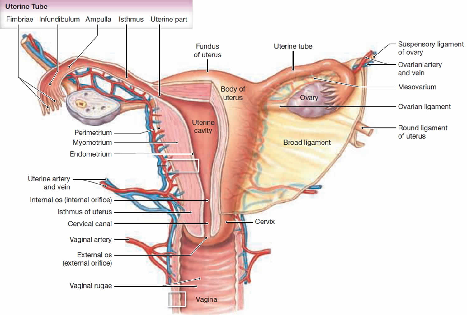
Primary dysmenorrhea occurs in as many as 50% of young women, only in ovulatory cycles, and usually limited to the first 48 or 72 hours of menstruation. Every month the lining of a woman’s womb builds up and is then shed again at the end of the menstrual cycle, when she has her period – unless you are pregnant. To shed the lining during the monthly period, the muscles of the womb tighten (contract) and relax in an irregular rhythm. This helps the tissue lining the womb to detach and flow out of the body, together with blood, through the neck of the womb (cervix) and the vagina. The muscle contractions are sometimes not noticeable or only cause mild discomfort, but they’re also sometimes felt as painful cramps. Period pain might only affect the lower abdomen, or it might be felt in the back or legs too. It can cause nausea, vomiting or diarrhea in some women, as well as headaches or general discomfort. Women who have heavier periods often have more intense pain too.

Secondary dysmenorrhea often first arises after a young woman has already been menstruating for several years. Here, women may also have pain at times of the month other than during menstruation. Secondary dysmenorrhea can be caused by any of a dozen or so disorders such as endometriosis, pelvic inflammatory disease (PID), intrauterine devices (IUDs), irregular cycles or infertility problems, ovarian cysts, adenomyosis, uterine myomas or polyps, intrauterine adhesions or cervical stenosis. Psychological factors are now known not to cause dysmenorrhea, only to add to the reactive component of the pain.

Most of the time, women do not need to see her doctor for menstrual cramps. This may be different if you have severe, lasting pain or pain that is new or different. In these cases, your doctor may want to do a physical exam, pelvic exam, or tests. These can help diagnose or rule out the cause of your pain. An ultrasound test lets your doctor see if you have ovarian cysts. A laparoscopy can check for endometriosis. In this minor surgery, the doctor makes a small cut in your low stomach. Then, they insert a thin tube to look inside your uterus.

Dysmenorrhea key points

  • Dysmenorrhea may begin soon after the menarche (girl’s first period), where it often improves with age, or may originate later in life after the onset of an underlying causative condition.
    • Dysmenorrhea is very common, and in up to 20% of women it may be severe enough to interfere with daily activities.
    • Dysmenorrhea is more likely in women who smoke, and those with an earlier age at menarche or longer duration of menstruation.
  • Nonsteroidal anti-inflammatory drugs (NSAIDs) (e.g., ibuprofen [Advil, Motrin IB, others] or naproxen sodium [Aleve]) reduce moderate to severe pain in women with primary dysmenorrhea compared with placebo, but doctors don’t know whether any one NSAID is superior to the others 2).
    • Simple analgesics such as aspirin and paracetamol may reduce pain in the short term, although few studies have been of good quality.
    • The herbal remedies toki-shakuyaku-san and Iranian herbal remedy (saffron, celery, and anise) may reduce pain compared with placebo. Doctors don’t know whether Chinese herbal remedies are beneficial compared with placebo, but they found limited evidence that they may be effective compared with other treatments for dysmenorrhea.
    • Thiamine and vitamin E may reduce pain compared with placebo in young women with primary dysmenorrhea.
  • Combined oral contraceptives may be more effective at reducing pain in women with primary dysmenorrhea compared with placebo; however, few trials have been of good quality.
  • Topical heat (about 39 °C) may be as effective as ibuprofen and more effective than paracetamol at reducing pain.
  • High-frequency transcutaneous electrical nerve stimulation (TENS) may reduce pain compared with sham TENS, but seems to be less effective than ibuprofen.
  • Acupressure may be more effective than sham acupressure or no treatment at relieving dysmenorrhea.
  • Spinal manipulation may be no more effective than placebo at reducing pain after 1 month in women with primary dysmenorrhea.
  • Relaxation may be better than no treatment at relieving dysmenorrhea.
  • Scientists don’t know whether acupuncture, fish oil, vitamin B12 , magnets, or intrauterine progestogens reduce dysmenorrhea.
  • Surgical interruption of pelvic nerve pathways is not beneficial in treating dysmenorrhea, and may be associated with adverse effects including constipation.
When to see a doctor

See your doctor if:

  • Menstrual cramps disrupt your life every month
  • Your symptoms progressively worsen
  • You just started having severe menstrual cramps after age 25
  • You have pain at times other than the first couple days of your period
  • You have a fever and pelvic pain
  • Sudden or severe pain, especially if your period is more than 1 week late and you have been sexually active.
  • You have abnormal vaginal discharge or bleeding
  • Increased or foul-smelling vaginal discharge.
  • Your pain that doesn’t go away when you take medicine to relieve it.

Also see your doctor if:

  • Treatments do not relieve your pain after 3 months.
  • You have pain and had an intrauterine device (IUD) placed more than 3 months ago.
  • You pass blood clots or have other symptoms with the pain.
  • Your pain occurs at times other than menstruation, begins more than 5 days before your period, or continues after your period is over.

Dysmenorrhea causes

Doctors distinguish between two types of period pain, called primary and secondary dysmenorrhea, depending on the cause.

Painful menstrual periods fall into two groups, depending on the cause:

  • Primary dysmenorrhea: Primary dysmenorrhea is menstrual pain that occurs around the time that menstrual periods first begin in otherwise healthy young women. In most cases, this pain is not related to a specific problem with the uterus or other pelvic organs. Increased activity of the hormone prostaglandin, which is produced in the uterus, is thought to play a role in this condition.
  • Secondary dysmenorrhea: Period pain that is caused by something other than the muscle contractions is called secondary dysmenorrhea. Benign (non-cancerous) growths in the womb, such as fibroids or polyps, are often responsible for secondary dysmenorrhea. Severe period pain may also be caused by endometriosis. In endometriosis, the kind of tissue that lines the womb (endometrium) grows elsewhere in the abdomen too. Sometimes contraceptive coils (IUDs: intrauterine devices) used for birth control can also cause secondary dysmenorrhea.

During your menstrual period, your uterus contracts to help expel its lining. Hormone like substances (prostaglandins) involved in pain and inflammation trigger the uterine muscle contractions. Higher levels of prostaglandins are associated with more-severe menstrual cramps.

Primary dysmenorrhea

Primary dysmenorrhea is the most common kind of period pain. Primary dysmenorrhea is where the period pain is caused by the womb muscle contractions alone. Hormone-like substances called prostaglandins play an important role here. Prostaglandins influence the perception of pain and cause the muscles in your womb to tighten, helping to shed the lining of your womb. Primary dysmenorrhea is more common in women under the age of 30 and women with heavy periods. It can run in families, and stress can play a role too.

The pain can start a day or two before your period. It normally lasts for a few days, though in some women it can last longer.

You usually first start having period pain when you are younger, just after you begin getting periods. Often, as you get older, you have less pain. The pain may also get better after you have given birth.

Secondary dysmenorrhea

Secondary dysmenorrhea is menstrual pain that develops later in women who have had normal periods. Secondary dysmenorrhea is caused by conditions that affect your uterus or other reproductive organs, such as endometriosis and uterine fibroids. This kind of pain often gets worse over time. It may begin before your period starts, and continue after your period ends.

Secondary dysmenorrheais often related to problems in the uterus or other pelvic organs, such as:

  • Endometriosis. The tissue that lines your uterus becomes implanted outside your uterus, most commonly on your fallopian tubes, ovaries or the tissue lining your pelvis.
  • Uterine fibroids. These noncancerous growths in the wall of the uterus can cause pain.
  • Adenomyosis. The tissue that lines your uterus begins to grow into the muscular walls of the uterus.
  • Pelvic inflammatory disease (PID). This infection of the female reproductive organs is usually caused by sexually transmitted bacteria.
  • Cervical stenosis. In some women, the opening of the cervix is small enough to impede menstrual flow, causing a painful increase of pressure within the uterus.
  • Intrauterine device (IUD) made of copper
  • Premenstrual syndrome (PMS). Besides dysmenorrhea, patients with premenstrual dysphoric disorder (formerly premenstrual syndrome) may have bloating, body aches, migraine headaches, breast tenderness, and emotional complaints. The effects of these symptoms are occasionally debilitating. Aside from possible vaginal brownish discharge or bleeding, pelvic examination findings are normal. It is the emergency physician’s responsibility to ensure adequate analgesia and appropriate follow-up with a gynecologist.
  • Sexually transmitted infection (STD or STI)
  • Stress and anxiety.

Risk factors for menstrual cramps

You might be at risk of menstrual cramps if:

  • You’re younger than age 30
  • You started puberty early, at age 11 or younger
  • Early age at menarche (< 12 years)
  • You bleed heavily during periods (menorrhagia)
  • You have irregular menstrual bleeding (metrorrhagia)
  • You have a family history of menstrual cramps (dysmenorrhea)
  • You smoke
  • Nulliparity (woman who has never borne a child)
  • Obesity

Can dysmenorrhea be prevented or avoided?

Menstrual cramps and pain cannot be prevented or avoided.

Various measures have been used to manage dysmenorrhea in the outpatient setting, including the following:

  • Lifestyle modification seems to be helpful
  • Smoking cessation should be encouraged, in that smoking may be a risk factor for dysmenorrhea 3)
  • Exercise has been shown to alleviate symptoms of dysmenorrhea, though the mechanism is not well understood 4)

A Cochrane review of 5 randomized controlled trials showed that certain behavioral interventions may be effective at treating primary and secondary dysmenorrhea 5).

Dysmenorrhea symptoms

Menstrual cramps can feel like a dull ache or a shooting pain. They most often occur in your low stomach. You may also feel them in your low back, hips, or thighs. The pain may start before your period or when your period begins. Menstrual cramps last about 1 to 3 days. The pain may be bad enough to keep you from normal activities.

Primary dysmenorrhea should be distinguished from secondary dysmenorrhea on the basis of clinical features. Clinical features of primary dysmenorrhea include the following 6):

  • Onset shortly after menarche (typically within 6 months)
  • Usual duration of 48-72 hours (often starting several hours before or just after the menstrual flow)
  • Cramping or laborlike pain
  • Background of constant lower abdominal pain, radiating to the back or the anterior or medial thigh
  • Often unremarkable pelvic examination findings (including rectal)

Associated general symptoms, such as malaise, fatigue (85%), nausea and vomiting (89%), diarrhea (60%), lower backache (60%), and headache (45%), may be present with primary dysmenorrhea. Dizziness, nervousness, and even collapse are also associated with dysmenorrhea.

A different pattern of pain is observed with secondary dysmenorrhea that is not limited to the onset of menses; this is usually associated with abdominal bloating, pelvic heaviness, and back pain. Typically, the pain progressively increases during the luteal phase until it peaks around the onset of menstruation.

The following may indicate secondary dysmenorrhea 7):

  • Dysmenorrhea beginning in the 20s or 30s, after relatively painless menstrual cycles in the past
  • Heavy menstrual flow or irregular bleeding
  • Dysmenorrhea occurring during the first or second cycles after menarche, which may indicate congenital outflow obstruction
  • Pelvic abnormality with physical examination (consider endometriosis, pelvic inflammatory disease [PID], pelvic adhesions, and adenomyosis)
  • Little or no response to nonsteroidal anti-inflammatory drugs (NSAIDs) or oral contraceptives
  • Infertility
  • Dyspareunia
  • Vaginal discharge

Dysmenorrhea complications

Menstrual cramps don’t cause other medical complications, but they can interfere with school, work and social activities.

Certain conditions associated with menstrual cramps can have complications, though. For example, endometriosis can cause fertility problems. Pelvic inflammatory disease can scar your fallopian tubes, increasing the risk of a fertilized egg implanting outside of your uterus (ectopic pregnancy).

Dysmenorrhea diagnosis

Your doctor will review your medical history and perform a physical exam, including a pelvic exam. During the pelvic exam, your doctor will check for abnormalities in your reproductive organs and look for signs of infection.

If your doctor suspects that a disorder is causing your menstrual cramps, he or she may recommend other tests, such as:

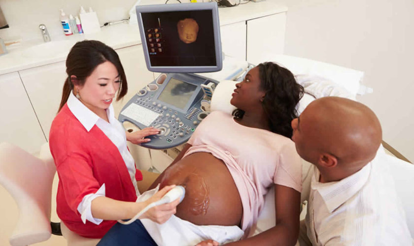
  • Ultrasound. This test uses sound waves to create an image of your uterus, cervix, fallopian tubes and ovaries.
  • Other imaging tests. A CT scan or MRI scan provides more detail than an ultrasound and can help your doctor diagnose underlying conditions. CT combines X-ray images taken from many angles to produce cross-sectional images of bones, organs and other soft tissues inside your body. MRI uses radio waves and a powerful magnetic field to produce detailed images of internal structures. Both tests are noninvasive and painless.
  • Laparoscopy. Although not usually necessary to diagnosis menstrual cramps, laparoscopy can help detect an underlying condition, such as endometriosis, adhesions, fibroids, ovarian cysts and ectopic pregnancy. During this outpatient surgery, your doctor views your abdominal cavity and reproductive organs by making tiny incisions in your abdomen and inserting a fiber-optic tube with a small camera lens.

Dysmenorrhea treatment

The primary dysmenorrhea pain is due to uterine cramps, hypoxia or ischemia, due to overproduction of prostaglandins, leukotrienes or vasopressin.

Treatment of secondary dysmenorrhea involves correction of the underlying organic cause. Specific measures (medical or surgical) may be required to treat pelvic pathologic conditions (eg, endometriosis) and to ameliorate the associated dysmenorrhea. Periodic use of analgesic agents as adjunctive therapy may be beneficial.

To ease your menstrual cramps, your doctor might recommend:

  • Pain relievers. Over-the-counter pain relievers, such as ibuprofen (Advil, Motrin IB, others) or naproxen sodium (Aleve), at regular doses starting the day before you expect your period to begin can help control the pain of cramps. Prescription nonsteroidal anti-inflammatory drugs also are available. Start taking the pain reliever at the beginning of your period, or as soon as you feel symptoms, and continue taking the medicine as directed for two to three days, or until your symptoms are gone.
  • Hormonal birth control. Oral birth control pills contain hormones that prevent ovulation and reduce the severity of menstrual cramps. These hormones can also be delivered in several other forms: an injection, a skin patch, an implant placed under the skin of your arm, a flexible ring that you insert into your vagina, or an intrauterine device (IUD).
  • Surgery. If your menstrual cramps are caused by a disorder such as endometriosis or fibroids, surgery to correct the problem might help your symptoms. Surgical removal of the uterus also might be an option if other approaches fail to ease your symptoms and if you’re not planning to have children.

Medicine for periods cramps

Non-steroidal anti-inflammatory drugs (NSAIDs) are the competitive inhibitors of cyclooxygenase (COX), the enzyme which mediates the bioconversion of arachidonic acid to inflammatory prostaglandins. The NSAIDs specifically approved by the US Food and Drug Administration (FDA) for treatment of dysmenorrhea are as follows:

  • Diclofenac
  • Ibuprofen
  • Ketoprofen
  • Meclofenamate
  • Mefenamic acid
  • Naproxen

Aspirin may not be as effective as these NSAIDs, and acetaminophen may be a useful adjunct for alleviating only mild menstrual cramping pain 8).

NSAIDs that achieve peak serum concentrations within 30-60 minutes and have a faster onset of action (eg, ibuprofen, naproxen, and meclofenamate) may be preferred. However, individual patient response varies, and patients may need to try several agents before finding one that works. Some NSAIDs (eg, indomethacin) should be avoided, because they have a higher incidence of adverse effects.

Cyclooxygenase [COX]-2 specific inhibitors have also proven effective in relieving menstrual pain. Their selectivity reduces the gastrointestinal symptoms caused by inhibition of the COX-1 receptor. Despite some preliminary data suggesting efficacy in patients with primary dysmenorrhea, COX-2 inhibitors have not been demonstrably superior to conventional NSAIDs 9).

However, these agents may be used in patients who cannot tolerate other NSAIDS or in whom these agents are contraindicated. COX-2–derived prostanoids nonetheless appear to be involved in the pathophysiology of primary dysmenorrhea 10).

Other analgesic agents

In an emergency setting, patients who do not respond to NSAIDs may require treatment with narcotics for pain control. Patients whose symptoms are not relieved by NSAIDs are very likely to have an underlying pelvic condition (eg, endometriosis).

In a study comparing montelukast, a leukotriene-receptor antagonist, with placebo in patients with dysmenorrhea, montelukast was effective in reducing pain 11). Clinicians may consider this agent as an alternative to hormonal therapy or in lieu of NSAIDs.

Simple analgesics, such as aspirin and acetaminophen, may also be useful, especially when NSAIDs are contraindicated.

Oral contraceptives

Oral contraceptives, which block monthly ovulation and may decrease menstrual flow, may also relieve symptoms. An update of a Cochrane review showed some evidence of symptomatic benefit in patients with primary dysmenorrhea, though no specific preparation showed superiority over any other 12).In some patients, oral contraceptives can prevent dysmenorrhea altogether, even though these agents are not approved by the FDA for this indication.

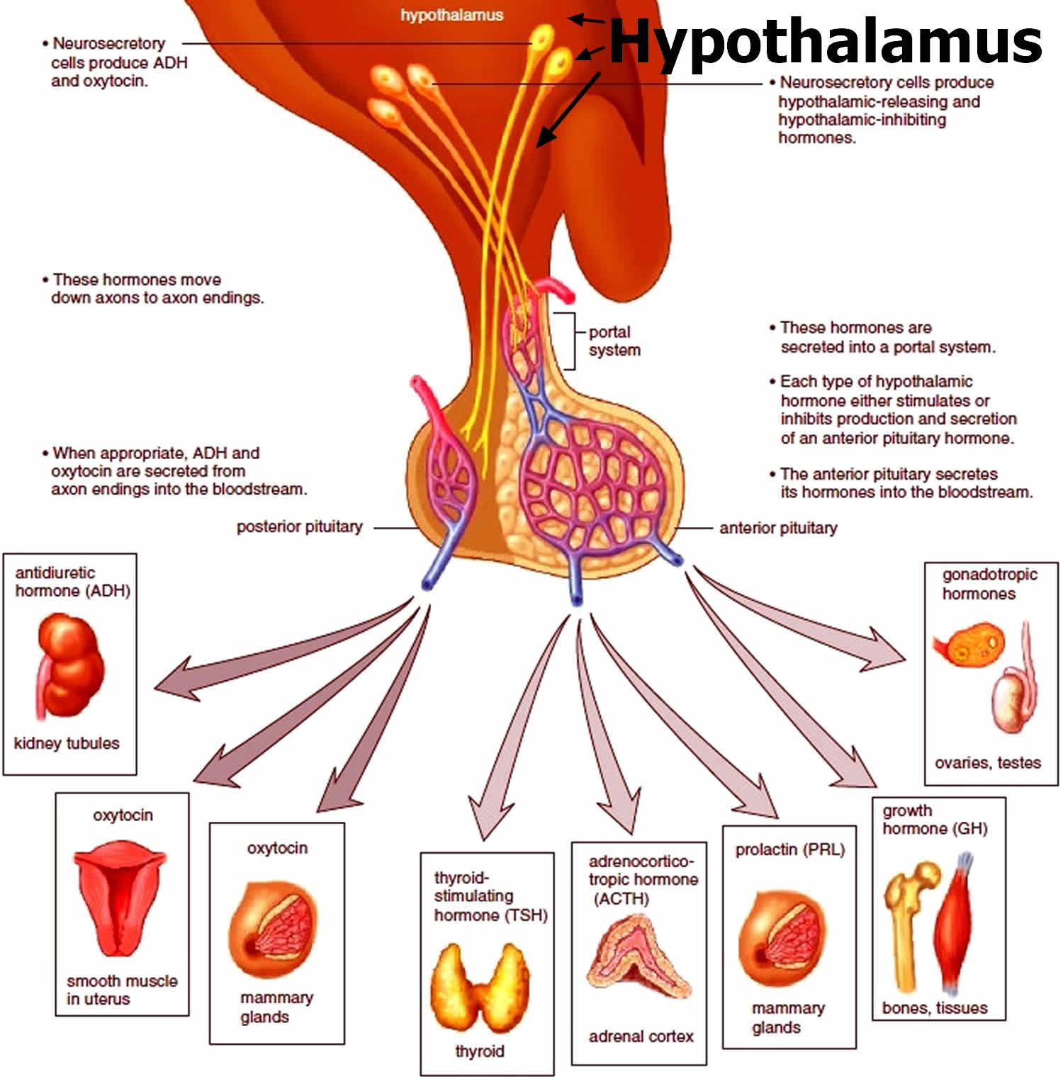
Oral contraceptives may be an appropriate choice for patients who do not wish to conceive. Combination oral contraceptives suppress the hypothalamic-pituitary-ovarian axis, thereby inhibiting ovulation and preventing prostaglandin production in the late luteal phase. This generally reduces the amount of menstrual flow and alleviates primary dysmenorrhea in most patients. Use of oral contraceptives in a manner that reduces the number of menstrual cycles may be beneficial for some patients 13).

Combination oral contraceptives, the levonorgestrel intrauterine device, and depot medroxyprogesterone acetate 14) provide effective pain relief and are associated with reduced menstrual flow. It may be necessary to add an NSAID to the oral contraceptive, especially during the first few cycles after initiation of the oral contraceptive. The ethinyl estradiol dose should generally be less than 50 µg; a monophasic oral contraceptive containing 30 µg is a reasonable choice. To date, studies comparing the efficacy of various oral contraceptive formulations in the management of dysmenorrhea have not been performed.

In a study of women with primary dysmenorrhea, Petraglia et al 15) found that estradiol valerate plus dienogest and ethinyl estradiol plus levonorgestrel were comparably effective in relieving dysmenorrheic pain. Each of the treatments was taken orally by over 200 women daily for three 28-day cycles, with the number of days of pain and the degree of pain being evaluated. Based on the patients’ self-assessments, the investigators determined that pain was reduced by both treatments by approximately the same number of days (by 4.6 days for estradiol valerate plus dienogest, by 4.2 days for ethinyl estradiol plus levonorgestrel).

Lifestyle and home remedies

At-home treatment is available for women who have menstrual cramps. The goal is to relieve symptoms. Over-the-counter (OTC) medicines can reduce pain. These include ibuprofen (brand names: Advil and Motrin) and naproxen (brand name: Aleve). These medicines belong to a group of medications known as non-steroidal anti-inflammatory drugs (NSAIDs). They can relieve period pain by reducing the production of prostaglandins. Start taking it the day before your period is expected to start and continue taking it regularly for the first few days of your period. Although NSAIDs are usually well tolerated, they sometimes have side effects, especially stomach-related problems.

Other medicines are Midol, Pamprin, and Premsyn PMS.

You also can try using heating pads or taking a warm bath. Some evidence suggests that applying warmth, for instance with heat packs, can relieve period pain. A few studies have also suggested that physical activity such as jogging, yoga and exercises can help.

Talk to your doctor if these don’t help. They may suggest a stronger pain reliever. They may want you to try using birth control pills or a birth control shot. These can help make your periods less painful. The birth control pill can relieve period pain because it prevents ovulation. This reduces the production of prostaglandins. It also means that the lining of your womb doesn’t become as thick as usual, and you have a lighter period. The birth control pill can have side effects too, such as headaches and nausea. It also increases the risk of thrombosis.

Other treatments – like acupuncture, dietary supplements or herbal products – haven’t been proven to help. Although these approaches have been tested in a number of studies, the study results were contradictory or the studies weren’t done properly.

The following steps may help you to avoid prescription medicines:

  • Get enough sleep and rest
  • Exercise regularly. Physical activity, including sex, helps ease menstrual cramps for some women.
  • Use heat. Soaking in a hot bath or using a heating pad, hot water bottle or heat patch on your lower abdomen (below your belly button) might ease menstrual cramps. Never fall asleep with the heating pad on.
  • Take warm showers or baths.
  • Try dietary supplements. A number of studies have indicated that vitamin E, omega-3 fatty acids, vitamin B-1 (thiamin), vitamin B-6, calcium, and magnesium supplements might reduce menstrual cramps.
  • Reduce stress. Psychological stress might increase your risk of menstrual cramps and their severity.
  • Practice relaxation techniques such as meditation or yoga.
  • Lose weight if you are overweight. Get regular, aerobic exercise.
  • Do light circular massage with your fingertips around your lower belly area.
  • Drink warm beverages.
  • Eat light but frequent meals.
  • Keep your legs raised while lying down or lie on your side with your knees bent.

Sometimes the pain is so bad that psychological treatment is considered. This may include things like talks with a psychotherapist and learning techniques that can reduce pain (such as relaxation and mindfulness exercises).

If these self-care measures do not work, your health care provider may offer you treatment such as:

  • Birth control pills
  • Mirena IUD
  • Prescription anti-inflammatory medicines
  • Prescription pain relievers (including narcotics, for brief periods)
  • Antidepressants
  • Antibiotics
  • Pelvic ultrasound
  • Suggest surgery (laparoscopy) to rule out endometriosis or other pelvic disease

Alternative medicine

Most alternative therapies for treating menstrual cramps haven’t been studied enough for experts to recommend them. However, some alternative treatments might help, including:

  • Acupuncture. Acupuncture involves inserting extremely thin needles through your skin at strategic points on your body. Some studies have found that acupuncture helps relieve menstrual cramps.
  • Transcutaneous electrical nerve stimulation (TENS). A TENS device connects to the skin using adhesive patches with electrodes in them. The electrodes deliver a varying level of electric current to stimulate nerves. TENS might work by raising the threshold for pain signals and stimulating the release of your body’s natural painkillers (endorphins). In studies, TENS was more effective than a placebo in relieving menstrual cramp pain.
  • Herbal medicine. Some herbal products, such as pycnogenol, fennel or combination products, might provide some relief from menstrual cramps.
  • Acupressure. Like acupuncture, acupressure also involves stimulating certain points on the body, but with gentle pressure on the skin instead of needles. Although research on acupressure and menstrual cramps is limited, it appears that acupressure may be more effective than a placebo in easing menstrual cramps.

Living with dysmenorrhea

Menstrual cramps are painful but can be managed with treatment. Primary dysmenorrhea can be treated with oral contraceptives if the women wishes to take pills for contraception and they are not contraindicated, or with Nonsteroidal anti-inflammatory drugs (NSAIDs) for the full 72 hours after pain begins. Calcium channel-blockers are also used on a research basis; transcutaneous electrical nerve stimulation (TENS) is sometimes effective. If these treatments are not effective, investigation for causes of secondary dysmenorrhea is indicated, preferably using laparoscopy.

Dysmenorrhea prognosis

With the use of NSAIDs, the prognosis for primary dysmenorrhea is excellent. The prognosis for secondary dysmenorrhea varies, depending on the underlying disease process. If a diagnosis of secondary dysmenorrhea is missed, the underlying pathology may lead to increased morbidity, including difficulty conceiving 16).

Although dysmenorrhea itself is not life-threatening, it can have a profound negative impact on a woman’s day-to-day life. Besides missing work or school, she may be unable to participate in sports or other activities and thus experience additional emotional distress. Some 10% of dysmenorrheal women have severe pain that can be incapacitating. Dysmenorrhea is a public health problem associated with substantial economic loss related to work absences (an estimated 600 million work hours and 2 billion dollars in the United States) 17).

A cross sectional study that included 897 adolescent girls reported that those in the dysmenorrhea group had significantly higher depression, aggression, insomnia, daytime sleepiness and sleep apnea scores compared to the control and the premenstrual syndrome groups 18).

References   [ + ]

read more

Amenorrhea

amenorrhea

What is amenorrhea

Amenorrhea is the term used when a woman or adolescent girl is not having menstrual periods or absence of menstruation — one or more missed menstrual periods. Women who have missed at least three menstrual periods in a row have amenorrhea, as do girls who haven’t begun menstruation by age 15. Amenorrhea is not a disease, but it can be a symptom of another condition. A normal menstrual cycle typically occurs every 21 to 35 days 1). Consult your doctor if you’ve missed at least three menstrual periods in a row, or if you’ve never had a menstrual period and you’re age 15 or older.

Amenorrhea is often a sign of another health problem rather than a disease itself, and it can happen for many reasons. It can occur as a natural part of life with the most common cause of amenorrhea is pregnancy. Other causes of amenorrhea include problems with the reproductive organs or with the glands that help regulate hormone levels.

Women naturally stop menstruating during pregnancy, long-term breastfeeding (lactational amenorrhea) and menopause. Birth control pills and injections and hormone-containing IUDs cause amenorrhea in some women. A number of other conditions can cause secondary amenorrhea.

There are two types of amenorrhea:

  • Primary amenorrhea: when a girl has not started having periods by age 15 (or within five years of the first signs of puberty)
  • Secondary amenorrhea: when a girl or woman has been having periods but then stops having them for at least three months

Primary amenorrhea occurs when a girl has not had her first period by age 16. Secondary amenorrhea describes women who experience an absence of more than three menstrual cycles after having regular periods.

Having regular periods is an important sign of overall health. Missing a period, when not caused by pregnancy, breastfeeding, or menopause, is generally a sign of another health problem. If you miss your period, talk to your health care provider about possible causes, including pregnancy.

Missing a period is the main sign of amenorrhea.

Depending on the cause, a woman might have other signs or symptoms as well, such as:

  • Excess facial hair
  • Hair loss
  • Headache
  • Lack of breast development
  • Milky discharge from the breasts
  • Vision changes

Treatment of the underlying condition often resolves amenorrhea.

Who is at risk of amenorrhea?

According to the American Society for Reproductive Medicine, amenorrhea that is not caused by pregnancy, breastfeeding, or menopause occurs in a small percentage (less than 5%) of women during their lifetime.

The risk factors for amenorrhea include 2):

  • Excessive exercise
  • Obesity
  • Eating disorders, such as anorexia nervosa
  • A family history of amenorrhea or early menopause
  • Genetics, such as having a change to the FMR1 gene, which also causes Fragile X syndrome 3)

Primary amenorrhea

Primary amenorrhea, which by definition is failure to reach menarche (failure of menses to occur by age 16), is often the result of chromosomal irregularities leading to primary ovarian insufficiency (e.g., Turner syndrome) or anatomic abnormalities (e.g., Müllerian agenesis). Primary amenorrhea can result from two main causes:

  • Chromosomal or genetic abnormalities can cause the ovaries to stop functioning normally. Turner syndrome, a condition caused by a partially or completely missing X chromosome, and androgen insensitivity syndrome, often characterized by high levels of testosterone, are two examples of genetic abnormalities that can delay or disrupt menstruation 4).
  • Problems with the hypothalamus or pituitary gland in the brain can cause an imbalance of hormones that can prevent periods from starting. Conditions such as eating disorders, excessive exercise, and extreme physical or psychological stress or a combination of these factors can also disrupt the normal functioning of the hypothalamus or pituitary gland, delaying the onset of menstruation.

In rare cases, physical problems—such as missing reproductive organs or blockage of reproductive passageways—can also lead to primary amenorrhea. Missing portions of the reproductive tract can cause endocrine disruptions and may combine with hypothalamic or pituitary problems to prevent menstruation. Blockages may also prevent menstrual bleeding, making it seem like a girl has primary amenorrhea, even if her menstrual cycles are actually normal 5).

Figure 1. Uterus anatomy

Uterus anatomy

Figure 2. Uterus location

Uterus location

Figure 3. Primary amenorrhea diagnosis

Primary amenorrhea diagnosis

Secondary amenorrhea

Secondary amenorrhea (missing three menstrual periods in a row or not having periods for at least 6 months after menstruating normally) can result from various causes.

Secondary amenorrhea causes

  • Natural causes
    • Pregnancy is the most common natural cause of secondary amenorrhea.
    • Other physiologic causes include breastfeeding and menopause.
  • Medications and therapies
    • Certain birth control pills, injectable contraceptives, and hormonal intrauterine devices (IUDs) can cause amenorrhea. It can take a few months after stopping one of these types of birth control for the menstrual cycle to restart and become regular.
    • Some medications, including certain antidepressants and blood pressure medications, can increase the levels of a hormone that prevents ovulation and the menstrual cycle 6).
    • Chemotherapy and radiation treatments for hematologic cancer (including blood, bone marrow, and lymph nodes) and breast or gynecologic cancer can destroy estrogen-producing cells and eggs in the ovaries, leading to amenorrhea. The resulting amenorrhea may be short-term, especially in younger women 7).
    • Sometimes scar tissue can build up in the lining of the uterus, preventing the normal shedding of the uterine lining in the menstrual cycle. This scarring sometimes occurs after a dilation and curettage (D&C), a procedure in which tissue is removed from the uterus to diagnose or treat heavy bleeding or to clear the uterine lining after a miscarriage 8), a cesarean section, or treatment for uterine fibroids.
  • Hypothalamic amenorrhea. This condition occurs when the hypothalamus, a gland in the brain that regulates body processes, slows or stops releasing gonadotropin-releasing hormone (GnRH), the hormone that starts the menstrual cycle.7 Common characteristics of women with hypothalamic amenorrhea include 9):
    • Low body weight
    • Rapid weight loss (any cause)
    • Low percentage of body fat
    • Very low intake of calories or fat
    • Eating disorders such as anorexia
    • Malabsorption
    • Emotional stress
    • Strenuous exercise that burns more calories than are taken in through food
    • Deficiency of leptin, a protein hormone that regulates appetite and metabolism
    • Tumor of the hypothalamus
    • Traumatic brain injury
    • Infection (e.g., meningitis, tuberculosis, syphilis)
    • Some medical conditions or illnesses
    • Gonadotropin deficiency (e.g., Kallmann syndrome)
  • Gynecological conditions, specifically those that lead to or result from hormone imbalances, may also have secondary amenorrhea as a main symptom.
    • Polycystic ovary syndrome (PCOS). PCOS occurs when a woman’s body produces more androgens (a type of hormone) than normal. High levels of androgens can cause fluid-filled sacs or cysts to grow in the ovaries, interfering with the release of eggs (ovulation). Most women with polycystic ovary syndrome either have amenorrhea or experience irregular periods, called oligomenorrhea.
    • Fragile X-associated primary ovarian insufficiency. The term fragile X-associated primary ovarian insufficiency describes a condition in which a woman’s ovaries stop functioning before normal menopause, sometimes around age 40. Fragile X-associated primary ovarian insufficiency results from certain changes to a gene on the X chromosome. Fragile X-associated primary ovarian insufficiency is fairly common among women who seek treatment for amenorrhea 10).
  • Anatomic abnormalities / Outflow tract conditions

    • Congenital
      • Complete androgen resistance
      • Imperforate hymen
      • Müllerian agenesis
      • Transverse vaginal septum
    • Acquired
      • Asherman syndrome (intrauterine synechiae)
      • Cervical stenosis
  • Thyroid problems. The thyroid is a small butterfly-shaped gland at the base of the neck, just below the Adam’s apple. The thyroid produces hormones that control metabolism and play a role in puberty and menstruation 11). A thyroid gland that is overactive (called hyperthyroidism) or underactive (hypothyroidism) can cause menstrual irregularities, including amenorrhea 12).
  • Pituitary tumors. The pituitary gland in the brain regulates the production of hormones that affect many body functions, including metabolism and the reproductive cycle. Tumors on the pituitary gland – increased prolactin by a small benign tumor (prolactinoma) are usually noncancerous (benign) but can interfere with the body’s hormonal regulation of menstruation 13).
  • Pituitary disorders
    • Pituitary damage / radiation to the head
    • Autoimmune disease
    • Cocaine
    • Cushing syndrome
    • Empty sella syndrome
    • Hyperprolactinemia
    • Infiltrative disease (e.g., sarcoidosis)
    • Medications:
      • Antidepressants
      • Antihistamines
      • Antihypertensives
      • Antipsychotics
      • Opiates
    • Other pituitary or central nervous system tumor
    • Sheehan syndrome
  • Primary ovarian insufficiency/premature ovarian failure. Primary ovarian insufficiency, also called premature ovarian failure (menopause before age 40) can be caused by:
    • Congenital
      • Gonadal dysgenesis (other than Turner syndrome)
      • Turner syndrome or variant
    • Acquired
      • Damage to the ovaries from chemotherapy or radiation
      • Autoimmune destruction
  • Other endocrine gland disorders
    • Adrenal disease
    • Adult-onset adrenal hyperplasia
    • Androgen-secreting tumor
    • Chronic disease
    • Constitutional delay of puberty
    • Cushing syndrome
    • Ovarian tumors (androgen producing)
    • Polycystic ovary syndrome (multifactorial)
    • Thyroid disease

Figure 4. The pituitary gland location

pituitary gland location

Figure 5. The hypothalamus and pituitary gland (anterior and posterior) endocrine pathways and target organs

hypothalamus and pituitary gland hormones

Figure 6. Secondary amenorrhea diagnosis

Secondary amenorrhea diagnosis

Amenorrhea causes

What causes amenorrhea

There are a number of reasons why your periods can stop.

The most common reasons for amenorrhea are:

Natural amenorrhea

During the normal course of your life, you may experience amenorrhea for natural reasons, such as:

  • Pregnancy
  • Breast-feeding
  • Menopause

Contraceptives

Some women who take birth control pills may not have periods. Even after stopping oral contraceptives, it may take some time before regular ovulation and menstruation return. Contraceptives that are injected or implanted also may cause amenorrhea, as can some types of intrauterine devices.

Medications

Certain medications can cause menstrual periods to stop, including some types of:

  • Antipsychotics
  • Cancer chemotherapy
  • Antidepressants
  • Blood pressure drugs
  • Allergy medications

Lifestyle factors

Sometimes lifestyle factors contribute to amenorrhea, for instance:

  • Low body weight. Excessively low body weight — about 10 percent under normal weight — interrupts many hormonal functions in your body, potentially halting ovulation. Women who have an eating disorder, such as anorexia or bulimia, often stop having periods because of these abnormal hormonal changes.
  • Excessive exercise. Women who participate in activities that require rigorous training, such as ballet, may find their menstrual cycles interrupted. Several factors combine to contribute to the loss of periods in athletes, including low body fat, stress and high energy expenditure.
  • Stress. Mental stress can temporarily alter the functioning of your hypothalamus — an area of your brain that controls the hormones that regulate your menstrual cycle. Ovulation and menstruation may stop as a result. Regular menstrual periods usually resume after your stress decreases.

Hormonal imbalance

Many types of medical problems can cause hormonal imbalance, including:

  • Polycystic ovary syndrome (PCOS). PCOS causes relatively high and sustained levels of hormones, rather than the fluctuating levels seen in the normal menstrual cycle.
  • Thyroid malfunction. An overactive thyroid gland (hyperthyroidism) or underactive thyroid gland (hypothyroidism) can cause menstrual irregularities, including amenorrhea.
  • Pituitary tumor. A noncancerous (benign) tumor in your pituitary gland can interfere with the hormonal regulation of menstruation.
  • Premature menopause. Menopause usually begins around age 50. But, for some women, the ovarian supply of eggs diminishes before age 40, and menstruation stops.

Structural problems

Problems with the sexual organs themselves also can cause amenorrhea. Examples include:

  • Uterine scarring. Asherman’s syndrome, a condition in which scar tissue builds up in the lining of the uterus, can sometimes occur after a dilation and curettage (D&C), cesarean section or treatment for uterine fibroids. Uterine scarring prevents the normal buildup and shedding of the uterine lining.
  • Lack of reproductive organs. Sometimes problems arise during fetal development that lead to a girl being born without some major part of her reproductive system, such as her uterus, cervix or vagina. Because her reproductive system didn’t develop normally, she can’t have menstrual cycles.
  • Structural abnormality of the vagina. An obstruction of the vagina may prevent visible menstrual bleeding. A membrane or wall may be present in the vagina that blocks the outflow of blood from the uterus and cervix.

Periods can also sometimes stop as a result of a long-term medical condition, such as heart disease, uncontrolled diabetes, an overactive thyroid, or premature ovarian failure.

Anatomic abnormalities

Müllerian agenesis, a condition characterized by a congenital malformation of the genital tract, may present with normal breast development without menarche, and may be associated with urinary tract defects and fused vertebrae 14). Other congenital abnormalities that may cause amenorrhea include imperforate hymen and transverse vaginal septum. In these conditions, products of menstruation accumulate behind the defect and can lead to cyclic or acute pelvic pain. Physical examination, as well as ultrasonography or MRI, is key to diagnosis, and surgical correction is usually warranted 15).

Rare causes of amenorrhea include complete androgen insensitivity syndrome, which is characterized by normal breast development, sparse or absent pubic and axillary hair, and a blind vaginal pouch; and 5-alpha reductase deficiency, which is characterized by partially virilized genitalia.1 In these conditions, serum testosterone levels will be in the same range as those found in males of the same age 16). The karyotype will be 46,XY, and testicular tissue should be removed to avoid malignant transformation 17).

A structural cause of secondary amenorrhea is Asherman syndrome: intrauterine synechiae caused by uterine instrumentation during gynecologic or obstetric procedures, which can be evaluated and treated with hysteroscopy 18).

Hypothalamic and pituitary causes

The ovaries require physiologic stimulation by pituitary gonadotropins for appropriate follicular development and estrogen production. Functional hypothalamic amenorrhea occurs when the hypothalamic-pituitary-ovarian axis is suppressed due to an energy deficit stemming from stress, weight loss (independent of original weight), excessive exercise, or disordered eating 19). It is characterized by a low estrogen state without other organic or structural disease. Laboratory tests usually reveal low or low-normal levels of serum follicle-stimulating hormone, luteinizing hormone, and estradiol; however, these levels can fluctuate, and the clinical context is the discriminating factor 20). Patients with functional amenorrhea may demonstrate the features of the female athlete triad, which consists of insufficient caloric intake with or without an eating disorder, amenorrhea, and low bone density or osteoporosis 21). These patients should be screened for eating disorders, diets, and malabsorption syndromes (e.g., celiac disease) 22).

Treatment of functional hypothalamic amenorrhea involves nutritional rehabilitation as well as reductions in stress and exercise levels 23). Menses typically return after correction of the underlying nutritional deficit 24). Bone loss is best treated by reversal of the underlying process, and the patient should undergo bone density evaluation and take calcium and vitamin D supplements 25). Although the bone loss is partly secondary to estrogen deficiency, estrogen replacement without nutritional rehabilitation does not reverse the bone loss. Combined OCs will restore menses, but will not correct bone density 26). Leptin administration has been reported to restore pulsatility of gonadotropin-releasing hormone and ovulation in these patients, but its effect on bone health is unknown 27). The effect of bisphosphonates on long-term bone health in premenopausal women is unclear, as is their teratogenic potential 28).

Pregnancy

You might be pregnant if you’re sexually active and your period is late. Pregnancy is a common reason why periods unexpectedly stop. It can sometimes happen if the contraception you’re using fails.

It might be that your period is simply late, so you could wait a few days to see if it arrives. If it doesn’t arrive, you can do a pregnancy test to confirm whether or not you’re pregnant.

It’s important to be aware that you can get pregnant in the days after your period is normally due. This can happen if the release of an egg (ovulation) is delayed – for example, as a result of illness or stress.

Stress

If you’re stressed, your menstrual cycle can become longer or shorter, your periods may stop altogether, or they might become more painful.

Try to avoid becoming stressed by making sure you have time to relax. Regular exercise, such as running, swimming and yoga, can help you relax. Breathing exercises can also help.

If you’re finding it hard to cope with stress, cognitive behavioral therapy (CBT) may be recommended. Cognitive behavioral therapy is a talking therapy that can help you manage your problems by changing the way you think and act.

Sudden weight loss

Excessive or sudden weight loss can cause your periods to stop. Severely restricting the amount of calories you eat stops the production of hormones needed for ovulation.

Your doctor may refer you to a dietitian if you’re underweight, where you have a body mass index (BMI) of less than 18.5. The dietitian will be able to advise you about how to regain weight safely.

If your weight loss is caused by an eating disorder, such as anorexia, you’ll be referred to a psychiatrist.

Being overweight or obese

Being overweight or obese can also affect your menstrual cycle. If you’re overweight, your body may produce an excess amount of estrogen, one of the hormones that regulate the reproductive system in women.

The excess estrogen can affect how often you have periods, and can also cause your periods to stop.

Your doctor may refer you to a dietitian if you’re overweight or obese, with a BMI of 30 or more, and it’s affecting your periods. The dietitian will be able to advise you about losing weight safely.

Extreme overexercising

The stress that intense physical activity places on your body can affect the hormones responsible for your periods. Losing too much body fat through intense exercise can also stop you ovulating.

You’ll be advised to reduce your level of activity if excessive exercise has caused your periods to stop.

If you’re a professional athlete, you may benefit from seeing a doctor who specializes in sports medicine. They’ll be able to give you advice about how to maintain your performance without disrupting your periods.

Contraceptive pill

You might miss a period every so often if you’re taking the contraceptive pill. This isn’t usually a cause for concern.

Some types of contraception, such as the progestogen-only pill, contraceptive injection and intrauterine system (IUS), particularly Mirena, can cause periods to stop altogether.

However, your periods should return when you stop using these types of contraception.

Menopause

You may start missing periods as you approach the menopause. This is because estrogen levels will start to decrease, and ovulation will become less regular. After the menopause, your periods will stop completely.

The menopause is a natural part of the ageing process in women, which usually occurs between the ages of 45 and 55. The average age for a woman to reach the menopause is 50 in the US.

However, around 1 in 100 women experience the menopause before the age of 40. This is known as premature menopause or premature ovarian failure.

Polycystic ovary syndrome (PCOS)

Polycystic ovaries contain a large number of harmless follicles, which are underdeveloped sacs in which eggs develop. If you have PCOS, these sacs are often unable to release an egg, which means ovulation doesn’t take place. Polycystic ovary syndrome (PCOS) is a multifactorial endocrine disorder, usually involving peripheral insulin resistance. It is characterized by hyperandrogenism found on clinical or laboratory examination, polycystic ovaries as suggested by ultrasonography, and ovulatory dysfunction.

PCOS is thought to be very common, affecting about 1 in every 10 women in the US. The condition is responsible for as many as one in three cases of stopped periods.

The Rotterdam Consensus Criteria published in 2003 require the presence of two of the three above conditions for diagnosis, whereas the Androgen Excess Society’s 2006 guidelines require hyperandrogenism and either of the remaining two conditions 29). In polycystic ovary syndrome (PCOS), serum androgen levels are typically no greater than twice the upper limit of normal. Thus, higher levels suggest other causes of hyperandrogenism 30).

With insulin resistance contributing to the underlying pathology of PCOS, patients should be screened for dyslipidemia and overall cardiovascular risk. Glucose intolerance should be assessed with a fasting glucose and two-hour glucose tolerance test, because patients may have insulin resistance and beta-cell dysfunction 31). In patients with PCOS who are overweight, weight loss combined with exercise is the first-line treatment 32). Chronic anovulation with resultant unopposed estrogen secretion is a risk factor for endometrial cancer, and low-dose combined oral contraceptives are more frequently prescribed to reduce this risk than higher-dose pills or progestin-only methods 33). Many combined OCs suppress the secretion of ovarian androgen and may be useful in decreasing hirsutism and acne, although data are limited 34). Metformin (Glucophage) can increase insulin sensitivity, thereby improving glucose tolerance. It may also improve ovulation rate, reduce the incidence of menstrual abnormalities, and improve serum androgen concentrations 35).

Primary ovarian insufficiency/premature ovarian failure

Primary ovarian insufficiency, also called premature ovarian failure (menopause before age 40) can be caused by:

  • Abnormal chromosomes
  • Immune disorders
  • Damage to the ovaries from chemotherapy or radiation

Primary ovarian insufficiency, a condition characterized by follicle depletion or dysfunction leading to a continuum of impaired ovarian function, is suggested by a concentration of follicle-stimulating hormone in the menopausal range (per reference laboratory), confirmed on two occasions separated by one month, and diagnosed in patients younger than 40 years with amenorrhea or oligomenorrhea.6 Other terms, including premature ovarian failure, are used synonymously with primary ovarian insufficiency 36). Up to 1% of women may experience primary ovarian insufficiency. This condition differs from menopause, in which the average age is 50 years, because of age and less long-term predictability in ovarian function 37). More than 90% of cases unrelated to a syndrome are idiopathic, but they can be attributed to radiation, chemotherapeutic agents, infection, tumor, empty sella syndrome, or an autoimmune or infiltrative process 38).

Patients with primary ovarian insufficiency should be counseled about possible fertility, because up to 10% of such patients may achieve temporary and unpredictable remission 39). Hormone therapy (e.g., 100 mcg of daily transdermal estradiol or 0.625 mg of daily conjugated equine estrogen [Premarin] on days 1 through 26 of the menstrual cycle, and 10 mg of cyclic medroxyprogesterone acetate for 12 days [e.g., days 14 through 26] of the menstrual cycle 40) until the average age of natural menopause is usually recommended to decrease the likelihood of osteoporosis, ischemic heart disease, and vasomotor symptoms 41). Combined oral contraceptives deliver higher concentrations of estrogen and progesterone than necessary for hormone therapy, may confer thromboembolic risk, and may theoretically be ineffective at suppressing follicle-stimulating hormone for contraceptive purposes in this population; thus, a barrier method or intrauterine device is appropriate in sexually active patients. For optimal bone health, patients with primary ovarian insufficiency should be advised to perform weightbearing exercises and supplement calcium (e.g., 1,200 mg daily) and vitamin D3 (e.g., 800 IU daily) intake 42).

There is evidence of genetic predisposition to primary ovarian insufficiency, and patients without evidence of a syndrome should be tested for FMR1 gene premutation (confers risk of fragile X syndrome in their offspring) and thyroid and adrenal autoantibodies 43).

Turner syndrome, a condition characterized by a chromosomal pattern of 45,X or a variant, can present with a classic phenotype including a webbed neck, a low hairline, cardiac defects, and lymphedema 44). Some patients who have Turner syndrome have only short stature and variable defects in ovarian function (even with possible fertility) 45). Thus, all patients with short stature and amenorrhea should have a karyotype analysis 46). Because patients require screening for a number of systemic problems, including coarctation of the aorta, other cardiac lesions, renal abnormalities, hearing problems, and hypothyroidism, and because they may require human growth hormone treatment and hormone replacement therapy, physicians inexperienced with Turner syndrome should consult an endocrinologist 47).

Elevations in serum prolactin

Prolactin levels can be elevated because of medications, pituitary adenoma, hypothyroidism, or mass lesion compromising normal hypothalamic inhibition 48). Elevated prolactin levels, whatever the cause, inhibit the secretion and effect of gonadotropins, and warrant MRI of the pituitary. Exceptions may occur in cases with a clear pharmacologic trigger and relatively low levels of serum prolactin (i.e., < 100 ng per mL [< 100 mcg per L]) 49). Treatment of prolactinomas may involve dopamine agonists or surgical resection 50).

Risk factors for amenorrhea

Factors that may increase your risk of amenorrhea may include:

  • Family history. If other women in your family have experienced amenorrhea, you may have inherited a predisposition for the problem.
  • Eating disorders. If you have an eating disorder, such as anorexia or bulimia, you are at higher risk of developing amenorrhea.
  • Athletic training. Rigorous athletic training can increase your risk of amenorrhea.

Amenorrhea symptoms

Symptoms vary according to the cause. Women can have hot flashes, discharge of milk from the nipples, vaginal dryness, headaches, and vision changes. Some women develop acne and grow hair on the face and body. Many women have no symptoms other than the lack of periods.

Depending on the cause of amenorrhea, you might experience other signs or symptoms along with the absence of periods, such as:

  • Milky nipple discharge
  • Hair loss
  • Headache
  • Vision changes
  • Excess facial hair
  • Pelvic pain
  • Acne

Amenorrhea complications

Complications of amenorrhea may include:

  • Infertility. If you don’t ovulate and have menstrual periods, you can’t become pregnant.
  • Osteoporosis. If your amenorrhea is caused by low estrogen levels, you may also be at risk of osteoporosis — a weakening of your bones.

Amenorrhea diagnosis

During your appointment, your doctor will perform a pelvic exam to check for any problems with your reproductive organs. If you’ve never had a period, your doctor may examine your breasts and genitals to see if you’re experiencing the normal changes of puberty.

Amenorrhea can be a sign of a complex set of hormonal problems. Finding the underlying cause can take time and may require more than one kind of testing.

Lab tests

A variety of blood tests may be necessary, including:

  • Pregnancy test. This will probably be the first test your doctor suggests, to rule out or confirm a possible pregnancy.
  • Thyroid function test. This test measures the amount of thyroid-stimulating hormone (TSH) in your blood, which can help determine if your thyroid is working properly. A thyroid gland that is overactive (hyperthyroidism) or underactive (hypothyroidism) can cause menstrual irregularities, including amenorrhea.
  • Prolactin test. Low levels of the hormone prolactin may be a sign of a pituitary gland tumor.
  • Ovary function test. This test measures the amount of follicle-stimulating hormone (FSH) or luteinizing hormone (LH)—hormones made by the pituitary gland—in your blood to determine if your ovaries are working properly. Your health care provider may also evaluate the level of anti-Mullerian hormone (AMH), which is produced by the ovarian follicles. Higher levels of anti-Mullerian hormone may be associated with polycystic ovary syndrome (PCOS) 51). Low or undetectable amounts of anti-Mullerian hormone may be associated with menopause or primary ovarian insufficiency.
  • Androgen test / Male hormone test. Androgens are sometimes called “male hormones” because men need higher levels of these hormones than woman do for overall health. However, both men and women need androgens to stay healthy. If you’re experiencing increased facial hair and a lowered voice, your doctor may want to check the level of male hormones in your blood. High levels of androgens may indicate a woman has PCOS.
  • Hormone challenge test. With this test, you will take a hormonal medication for seven to 10 days in an effort to trigger a menstrual cycle. Results from the test can tell your health care provider whether your periods have stopped because of a lack of estrogen.
  • Screening for a premutation of the FMR1 gene. Changes in this gene can cause the ovaries to stop functioning properly, leading to amenorrhea 52).
  • Chromosome evaluation. This test, also known as a karyotype, involves counting and evaluating the chromosomes from cells in the body to identify any missing, extra, or rearranged cells. Results from this evaluation can help determine the cause of the chromosomal abnormality causing primary or secondary amenorrhea.
  • Ultrasound. This painless test uses sound waves to produce images of internal organs. This test can help determine if your reproductive organs are all present and shaped normally.
  • Computerized tomography (CT). CT scans combine many X-ray images taken from different directions to create cross-sectional views of internal structures. A CT scan can indicate whether your uterus, ovaries, and kidneys look normal.
  • Magnetic resonance imaging (MRI). MRI uses radio waves with a strong magnetic field to produce detailed images of soft tissues within the body. Your health care provider may order an MRI to check for a pituitary tumor or to examine your reproductive organs.
  • Hysteroscopy. In this procedure a thin, lighted camera is passed through your vagina and cervix to allow your health care provider to look at the inside of your vagina, cervix and uterus.

Your health care provider might use several of these tests to attempt to diagnose the cause of amenorrhea. In some cases, no specific cause for the amenorrhea can be found. This situation is called idiopathic amenorrhea 53).

Amenorrhea treatment

Treatment depends on the underlying cause of your amenorrhea. In some cases, contraceptive pills or other hormone therapies can restart your menstrual cycles. For example, if the cause is PCOS, you may be advised to take the contraceptive pill or tablets containing a hormone called progesterone. Amenorrhea caused by thyroid or pituitary disorders may be treated with medications. If you have an overactive thyroid gland, you may be given medication to stop your thyroid gland producing too many hormones. If a tumor or structural blockage is causing the problem, surgery may be necessary.

If the cause is early menopause (premature ovarian failure), this means the ovaries no longer function normally. Hormone medication is usually recommended. Treatments to try include the contraceptive pill or hormone replacement therapy (HRT).

If primary or secondary amenorrhea is caused by lifestyle factors, your health care provider may suggest changes in the areas below:

  • Weight. Being overweight or severely underweight can affect your menstrual cycle. Attaining and maintaining a healthy weight often helps balance hormone levels and restore your menstrual cycle.
  • Stress. Assess the areas of stress in your life and reduce the things that are causing stress. If you can’t decrease stress on your own, ask for help from family, friends, your health care provider, or a professional listener such as a counselor.
  • Level of physical activity. You may need to change or adjust your physical activity level to help restart your menstrual cycle. Talk to your health care provider and your coach or trainer about how to train in a way that maintains your health and menstrual cycles.

Be aware of changes in your menstrual cycle and check with your health care provider if you have concerns. Keep a record of when your periods occur. Note the date your period starts, how long it lasts, and any problems you experience. The first day of bleeding is considered the first day of your menstrual cycle.

Treatments for Primary Amenorrhea

For primary amenorrhea, depending on your age and the results of the ovary function test, health care providers may recommend watchful waiting. If an ovary function test shows low follicle-stimulating hormone (FSH) or luteinizing hormone (LH) levels, menstruation may just be delayed. In females with a family history of delayed menstruation, this kind of delay is common 54).

Primary amenorrhea caused by chromosomal or genetic problems may require surgery. Women with a genetic condition called 46, XY gonadal dysgenesis have one X and one Y chromosome, but their ovaries do not develop normally. This condition increases the risk for cancer developing in the ovaries. The gonads (ovaries) are often removed through laparoscopic surgery to prevent or reduce the risk of cancer 55).

Medical Treatments for Secondary Amenorrhea

Common medical treatments for secondary amenorrhea include 56):

  • Birth control pills or other types of hormonal medication. Certain oral contraceptives may help restart the menstrual cycle.
  • Medications to help relieve the symptoms of PCOS. Clomiphene citrate therapy is often prescribed to help trigger ovulation 57).
  • Estrogen replacement therapy. Estrogen replacement therapy may help balance hormonal levels and restart the menstrual cycle in women with primary ovarian insufficiency or Fragile X-associated primary ovarian insufficiency 58). Women with Fragile X-associated primary ovarian insufficiency often experience symptoms of menopause, such as hot flashes and night sweats. Estrogen replacement therapy replaces the estrogen a woman’s body should be making naturally for a normal menstrual cycle. In addition, estrogen replacement therapy may help women with Fragile X-associated primary ovarian insufficiency lower their risk for the bone disease osteoporosis 59). Estrogen replacement therapy can increase the risk for uterine cancer, so your health care provider may also prescribe progestin or progesterone to reduce this risk.

In general, medications are safe, but they can have side effects, some of which may be serious. You should discuss side effects and risks with your health care provider before deciding on any specific medical treatment.

Surgical Treatments for Secondary Amenorrhea

Surgical treatment for amenorrhea is not common, but may be recommended in certain conditions. These include:

  • Uterine scarring. This scarring sometimes occurs after removal of uterine fibroids, a cesarean section, or a dilation and curettage (D&C), a procedure in which tissue is removed from the uterus to diagnose or treat heavy bleeding or to clear the uterine lining after a miscarriage 60). Removal of the scar tissue during a procedure called a hysteroscopic resection can help restore the menstrual cycle 61).
  • Pituitary tumor. Medications may be recommended to shrink the tumor. If this does not work, surgery may be necessary to remove the tumor. Pituitary tumors are not cancerous, but they can cause problems as they grow. Pituitary tumors can put pressure on surrounding blood vessels and nerves such as the optic nerve and may result in loss of vision. Most of the time, pituitary tumors are removed through the nose and sinuses. Radiation therapy may be used to shrink the tumor, either in combination with surgery or, for those who cannot have surgery, by itself.

References   [ + ]

read more

Phimosis

phimosis

What is phimosis

Phimosis is a condition where the foreskin is too tight to be pulled back over the head of the penis (glans) and is common in newborns and young boys. Phimosis is normal in babies and toddlers, but in older children it may be the result of a skin condition that has caused scarring. Phimosis isn’t usually a problem unless it causes symptoms. Phimosis usually eases without treatment. If it interferes with urination, circumcision (removal of the foreskin) might be recommended.

The penis has two main parts, the shaft and the head (called the glans). One continuous layer of skin, called the foreskin, covers the shaft and glans. When a child is not circumcised the foreskin is firmly attached to the glans. Gradually, the foreskin will begin to separate. As this occurs you may notice a white, cheesy material, called smegma, released between the layers of skin. You also may see ‘white pearls’ develop under the fused layers of the foreskin and the glans. These are not signs of an infection or a cyst. Smegma is just skin cells that are shed throughout life; it is normal.

When the foreskin separates from the glans of the penis it can be pulled back (retracted) away from the penis to expose the glans. Never forcibly retract your child’s foreskin. This can cause pain and bleeding and can lead to scarring and adhesions (where skin is stuck to skin).

In adults, phimosis can occasionally be associated with sexually transmitted infections (STIs).

It can also be caused by a number of different skin conditions, including:

  • eczema – a long-term condition that causes the skin to become itchy, red, dry and cracked
  • psoriasis – a skin condition that causes red, flaky, crusty patches of skin covered with silvery scales
  • lichen planus – a non-infectious itchy rash that can affect many areas of the body
  • lichen sclerosus – a scarring condition of the foreskin (and sometimes glans) that’s probably caused by urinary irritation in susceptible men and boys

Treatment for repeated phimosis may involve application of a steroid cream to the foreskin up to three times a day for about a month to loosen the adhesive ring. If the child has ballooning of the foreskin during urination after the age of 10, a circumcision (surgical removal of all or part of the foreskin) may be recommended.

Normal foreskin development

Most uncircumcised baby boys have a foreskin that won’t pull back (retract) because it’s still attached to the glans. This is perfectly normal for about the first 2 to 6 years. By around the age of 2, the foreskin should start to separate naturally from the glans (the head of the penis).

The foreskin of some boys can take longer to separate, but this doesn’t mean there’s a problem – it’ll just detach at a later stage.

Never try to force your child’s foreskin back before it’s ready because it may be painful and damage the foreskin.

Penis hygiene

It’s important to clean your penis regularly to avoid problems developing.

You should:

  • Gently wash your penis with warm water each day while having a bath or shower. Their foreskin might still be attached to the head of the penis and will therefore not retract fully. At this stage of their development, there’s no need to clean inside the foreskin.
  • Gently pull back your foreskin (if you have one) and wash underneath; don’t pull back the foreskin of a baby or young boy because it could be painful and cause harm
  • Use a mild or non-perfumed soap (if you choose to use soap) to reduce the risk of skin irritation
  • Avoid using talc and deodorants on your penis as they may cause irritation

Circumcised men should also regularly clean their penis with warm water and a mild soap (if you choose to use soap).

While regular personal hygiene is important, too much washing with soap and shower gels can cause soreness. Gently washing your penis once a day with warm water is sufficient to maintain good hygiene. If you want to use soap, choose a mild or non-perfumed soap to reduce the risk of skin irritation.

If you don’t wash underneath the foreskin correctly, a cheesy-looking substance called smegma may begin to gather.

Smegma is a natural lubricant that keeps the penis moist. It’s found on the head of the penis and under the foreskin.

If smegma builds up in the foreskin, it can start to smell, stop you easily pulling your foreskin back, and become a breeding ground for bacteria. This can cause redness and swelling of the head of your penis, called balanitis.

What causes phimosis in a child?

Phimosis is caused by a tightening of the opening of the foreskin. This is normal in a newborn baby. Over time the foreskin loosens and can be pulled down more easily. By age 17, most boys will be able to fully retract their foreskin. Phimosis can also occur if the foreskin is forced back before it is ready. This can cause a fibrous scar to form. This can stop the foreskin from retracting in the future.

Phimosis symptoms

Phimosis isn’t usually a problem unless it causes symptoms such as redness, soreness or swelling.

You should seek treatment if your child has the following symptoms of phimosis:

  • Ballooning or bulging of the foreskin during urination
  • Inability to completely retract the foreskin by age 10
  • Frequent infections of the foreskin (balanitis)

If your child’s glans is sore and inflamed, they may have balanitis (inflammation of the head of the penis).

There may also be a thick discharge underneath the foreskin. If both the glans and foreskin are inflamed, it’s known as balanoposthitis.

Take your child to see your doctor if they have these type of symptoms. Your doctor will be able to recommend appropriate treatment.

Most cases of balanitis can be easily managed using a combination of good hygiene, creams or ointments, and avoiding substances that irritate the penis.

Balanoposthitis (inflammation of both the glans and foreskin) can also sometimes be treated by following simple hygiene measures, such as keeping the penis clean by regularly washing it with water and a mild soap or moisturizer.

Urine can irritate the glans if it’s retained for long periods under the foreskin, so if possible you should withdraw the foreskin to wash the glans.

If balanoposthitis is caused by a fungal or bacterial infection, an antifungal cream or a course of antibiotics may be needed.

Phimosis treatments

Treatment of phimosis varies depending on the severity of the condition. Doctors have found that phimosis can be safely and effectively treated with a topical steroid cream. Topical steroids (a cream, gel or ointment that contains corticosteroids) are sometimes prescribed to treat a tight foreskin. They can help soften the skin of the foreskin, making it easier to retract. Your child’s doctor will give you instructions on the proper use of the cream. If the steroid cream fails and the foreskin remains narrowed, a circumcision may be necessary.

Phimosis can cause pain, skin splitting, or a lack of sensation during sex. Using a condom and lubricants while having sex may make your penis more comfortable.

When surgery may be needed

Surgery may be needed if a child or adult has severe or persistent balanitis or balanoposthitis that causes their foreskin to be painfully tight.

Circumcision (surgically removing part or all of the foreskin) may be considered if other treatments have failed, but it carries risks such as bleeding and infection.

This means it’s usually only recommended as a last resort, although it can sometimes be the best and only treatment option.

Alternatively, surgery to release the adhesions (areas where the foreskin is stuck to the glans) may be possible. This will preserve the foreskin but may not always prevent the problem recurring.

read more

Translocation down syndrome

translocation down syndrome

Translocation down syndrome

Translocation Down syndrome refers to the type of Down syndrome (trisomy 21) that occurs when a portion of chromosome 21 becomes attached (translocated) onto another chromosome, before or at conception. In translocation Down syndrome, there are still three chromosome 21 (trisomy 21), just like there are in regular Down syndrome, but one of the chromosome 21 is attached to another chromosome, instead of being separate. The extra copy of the chromosome 21 is what causes the health problems that are associated with Down syndrome. Translocation Down syndrome children have the usual two copies of chromosome 21, but they also have additional genetic material from chromosome 21 attached to another chromosome.

People with translocation Down syndrome still share the same health issues and learning problems with people with other types of Down syndrome. All people with Down syndrome have some learning problems. The specific location of extra genes generally does not help doctors predict how a child will develop.

Without doing a blood test, it is not possible to tell the difference between people with translocation Down syndrome and people with other types of Down Syndrome.

About 3 – 4% of people with Down syndrome (trisomy 21) have translocation Down syndrome. Translocation Down syndrome usually arises when the small arms (p arm) of chromosome 21 and another chromosome break, and the two remaining long arms (q arms) join together at their exposed ends. This process of chromosomes breaking and rejoining to other chromosomes is known as translocation (because the chromosome material has transferred its location).

In translocation Down syndrome, the extra chromosome 21 may be attached to the chromosome 14 or to other chromosome numbers like 13, 15, or 22. In some cases, two chromosome 21 can be attached to each other.

Whenever a translocation is found in a child, the parents’ chromosomes are studied to determine whether the translocation was inherited or not. If one parent has the translocation chromosome, then the doctor knows the baby inherited the translocation from that parent. When a person has a rearrangement of chromosome material, with no extra or missing chromosome material, he or she is said to have a “balanced translocation” or be a “balanced translocation carrier.”

Parents with balanced translocations may have fertility problems (trouble becoming pregnant), miscarriages, or have an increased chance of having a child with health problems. Although the parent can donate the proper amount of genetic material (23 chromosomes) to a pregnancy, he or she also has a risk of donating too much or too little genetic material to a pregnancy. This is not something the parent can control or predict. The chance depends on the type of chromosome rearrangement and which chromosomes are involved.

There is another important factor to remember when a parent is found to have a translocation. The parents’ relatives (brothers, sisters) may also have inherited the translocation and, therefore, may have the same risks for problems with a pregnancy. For these reasons, it is recommended that people with chromosome rearrangements share this information with their relatives so that they can have the option of having their chromosomes studied.

Genetic counselling should always be available to families with a child with Down’s syndrome. Even if parents do not intend to have more children, knowing that one of them is a carrier can be important for all their children or other relatives. Relatives of a person who carries a translocation have an increased chance of being translocation carriers.

Figure 1. Translocation Down syndrome karyotype

translocation down syndrome karyotype

What is chromosomal translocation?

Chromosomal translocation refers to exchange of chromosomal segments between chromosomes. Translocations are the most common type of structural chromosomal abnormalities seen in the general population, having a frequency of about 1/1000 live births 1). Two types of chromosomal translocations are described: Robertsonian translocations and reciprocal translocations 2).

In Robertsonian translocations, the breakpoints commonly occur in the short arms of two acrocentric chromosomes (homologous or nonhomologous) with subsequent fusion and formation of dicentric chromosomes 3). It is reported that either the inactivation of one centromere or the close proximity of the two functional centromeres in these dicentric chromosomes allows them to remain stable 4). Less frequent forms of Robertsonian translocations may be caused by breakage and fusion of centromeres (centric fusion) or from breakage and fusion of one short arm and one long arm of acrocentric chromosomes, resulting in monocentric rearrangements 5). In Robertsonian translocations, the loss of gene-poor short arms of the two acrocentric chromosomes usually does not produce any phenotypic effects. Most of these individuals remain undetected until they attempt to reproduce.

Reciprocal translocations result from breakage of two nonhomologous chromosomes, with at least one of them being a nonacrocentric chromosome, and interchange of chromosomal fragments between them. Consequently, two derivative chromosomes are formed with no loss or gain of genetic material. Unless one or both of the chromosomal breakpoints involve an important functional gene, these balanced chromosomal rearrangements would not produce a significant phenotypic effect either. However, the carriers of both Robertsonian and reciprocal translocations commonly present with reproductive problems, due to unbalanced chromosomal segregation in meiosis, which cause significant chromosomal imbalances (i.e., disomies and nullisomies) in their gametes with subsequent partial aneuploidies in the conceptuses. This can lead to infertility, recurrent miscarriages, or offspring with congenital anomalies due to the unbalanced translocations.

Although all human chromosomes are theoretically susceptible to chromosomal breakage leading to translocations, the frequency of chromosomal translocations shows a nonrandom distribution. Translocations between acrocentric chromosomes 13 and 14 and rob(13q14q) constitute the majority of balanced Robertsonian translocations, with Robertsonian translocations between chromosomes 21 and 14 rob(14q21q) being the second most common type 6). In a Sri Lankan study, Robertsonian translocations between chromosomes 21 and 14 or rob(14q21q) and chromosomes 21 and 21 or rob(21q21q) were commonly observed among children with Down syndrome were more frequent than rob(13q14q) 7). It was suggested that the presence of homologous pericentric regions in chromosomes 13, 14, and 21 contributes to the higher incidence of translocations between these chromosomes 8). Among the balanced reciprocal translocations, translocations involving chromosomes 11 and 22 are commonly described in the scientific literature 9). In a study of 269 balanced translocations among patients with recurrent miscarriages, there was a surplus of chromosomes 6, 7, and 22 in reciprocal translocations 10). Another study with similar design showed an excess of chromosome 7 and 4 in reciprocal translocations 11).

Unbalanced Robertsonian translocations involving chromosome 21 are by far the commonest type of structural abnormalities giving rise to translocation Down syndrome. Other types of homologous or heterologous Robertsonian translocations are comparatively rare 12). A large number of case reports describing children or fetuses with congenital malformations, dysmorphic features, impaired growth, and/or development due to unbalanced reciprocal translocations involving different chromosomes have been reported in the scientific literature. The phenotypic features observed in these cases were attributed to partial monosomy and/or partial trisomy of different chromosomal segments that frequently occurred due to unbalanced chromosomal segregation during meiosis in a parent who is a carrier of a balanced reciprocal translocation.

It is also known that chromosomal translocations are one of the commonest types of inherited chromosomal abnormalities leading to recurrent pregnancy loss 13). Cytogenetic studies on male infertility have also showed a higher frequency of chromosomal translocations among infertile men than in the general population 14). It has been reported that the presence of a chromosomal translocation causes partial or complete spermatogenic arrest, with consequential oligospermia or azoospermia. In contrast, in female carriers of these chromosomal translocations, oogenesis is known to progress without an arrest in meiosis, resulting in the production of abnormal oocytes with unbalanced chromosomal constitution and subsequent partial aneuploidies in the conceptus 15).

What is balanced translocation?

If the extra copy of chromosome 21 is inherited from a parent in the egg or sperm, it means that the parents “carry” this type of Down syndrome. These parents are said to have a “balanced translocation.” Balanced translocation happens when the correct amount of genetic material is present in the wrong location.

Do people with balanced translocation have signs of Down syndrome?

People with balanced translocations do not have any features of Down syndrome. However, it is possible that they may have trouble getting pregnant.

Even though carriers have the right amount of genes, the egg or sperm may have either too much or too few genes. This may cause an unexpected miscarriage.

How does translocation Down syndrome occur?

In two-thirds of people with Down syndrome due to a translocation, the translocation was an isolated event during the formation of the individual egg or sperm involved in their conception. As with regular trisomy 21, there is no known reason why this occurs. It cannot be predicted and it is not a result of anything the parents or other family members have done.

Because it is a new event, this is sometimes called a de novo translocation.

The egg or sperm contains the usual number of chromosomes (i.e. 23) but these include the translocated one. Thus there is one free, whole chromosome 21 and most of a second chromosome 21 attached to another chromosome. If this egg or sperm containing 23 chromosomes (+ translocated part) fuses with an ordinary sperm or egg, the fertilised egg, fetus and baby will have 46 single chromosomes, but one of the chromosomes will have an extra copy of most of the chromosome 21 material attached to it. The translocated chromosome acts like a single chromosome in cell division, and hence all the cells produced from this first cell will contain the extra chromosome 21 portion. This baby will therefore have Down’s syndrome.

In the other one third of people with the translocation type of Down’s syndrome, the translocation is inherited from one of the parents. This parent has two whole number 21 chromosomes in each cell but one of them is attached to another chromosome. As there is no loss or gain of any genetic material this is known as a balanced translocation and the parent is a carrier of the translocation. It is important to realise that because such parents have the usual amount of genetic material, they have no traces of the syndrome themselves and never will have. They cannot be expected to know they are carriers, as the only way of knowing is to study their chromosomes.

When people who carry a translocation produce an egg or sperm, it is possible for them to pass on both the translocated chromosome and the free chromosome 21 in the egg or sperm. This will result in a fertilised egg with two free 21 chromosomes and a translocated chromosome. The baby will therefore have Down’s syndrome.

As 4% of people with Down’s syndrome have the translocation type, and one third of this group have inherited it, only about 1% of people with Down’s syndrome have inherited the condition.

Is translocation Down syndrome inherited?

Yes, translocation Down syndrome can be inherited. An unaffected person can carry a rearrangement of genetic material between chromosome 21 and another chromosome. This rearrangement is called a balanced translocation because there is no extra material from chromosome 21. Although they do not have signs of Down syndrome, people who carry this type of balanced translocation are at an increased risk of having children with the condition.

Could I have another baby with Down syndrome?

Yes. Translocation Down syndrome is the only type of Down Syndrome that can be passed down from a parent who does not have features of Down syndrome.

  • If a parent has balanced translocation, there is an up to 15% chance of having another child with Down syndrome.
  • A genetic counselor or doctor who is a Down Syndrome expert would be happy to discuss this with you before any future pregnancies.
  • These counselors or doctors can also discuss which other family members have a chance of carrying the balanced translocation, such as brothers or sisters.

People with specific questions about genetic risks or genetic testing for themselves or family members should speak with a genetics professional.

Resources for locating a genetics professional in your community are available online:

What is the chance of having another child with translocation Down syndrome?

The recurrence risk depends on the type of translocation. When neither of the parents is a carrier, the translocation was an isolated event with only a small chance of its happening again (geneticists quote less than 1%). In most cases, the recurrence risk for de novo translocations is similar to that of the general population but may be slightly higher in some situations; it is estimated to be 2-3% 16).

Translocation carriers can have children who are carriers, children whose chromosomes show no rearrangement at all, or children with Down syndrome. For translocations involving chromosomes 21 and any other chromosome, the chance of another child with Down syndrome being born is about one in six if the mother is the carrier and about one in twenty if the father is the carrier. A few people are carriers for a translocation between two chromosomes 21; in these people, who are quite ordinary themselves, the only possible outcome is a child with Down’s syndrome.

The theoretic recurrence risk for a Robertsonian carrier parent to have a liveborn offspring with Down syndrome is 1 in 3. However, only 10-15% of the progeny of carrier mothers and only 2-3% of the progeny of carrier fathers have Down syndrome. The reason for this difference is not clear. In a carrier parent with a 21q21q translocation or isochromosome, the recurrence risk is 100%.

In any trisomy 21 patient with a translocation, karyotype testing must be recommended to both parents to look for a translocation. If a translocation is found in one of the parents, the recurrence risk is significantly higher, and further genetic counseling is crucial.

Is age a factor in translocation Down syndrome?

No. unlike regular trisomy 21, translocation Down syndrome occurs equally frequently whatever the age of the parents.

What causes translocation Down syndrome?

Translocation Down syndrome can happen one of two ways:

  • It can be caused by extra genes in the egg or sperm of one of the parents.
  • It can happen by chance.

Translocation down syndrome symptoms

People with translocation Down syndrome still have an extra copy of a large part of chromosome 21. Their clinical features or ability levels of translocation Down syndrome are no different from those in a child with regular trisomy 21. The only way of knowing what type of Down syndrome a person has, is by taking a blood sample and looking at the chromosomes.

A very few children with translocation have partial trisomy 21 – where only a part of chromosome 21 is present in 3 copies. These children may have fewer characteristics of Down syndrome. Like the more usual type of translocation described above, this type may arise de novo, or a parent may carry it.

As one third of people with translocation Down syndrome have inherited the condition, their parents have a high chance of having another affected child and may wish to know whether this is so. To identify these parents, chromosome tests are done on all new babies with Down syndrome. Blood samples can then be taken from parents of babies with translocations, to find out whether one of the parents carries the translocation.

Down syndrome symptoms

Each person with Down syndrome is an individual — intellectual and developmental problems may be mild, moderate or severe. Some people are healthy while others have significant health problems such as serious heart defects.

Children and adults with Down syndrome have distinct facial features. Though not all people with Down syndrome have the same features, some of the more common features include:

  • Flattened face
  • Small head
  • Short neck
  • Protruding tongue
  • Upward slanting eye lids (palpebral fissures)
  • Unusually shaped or small ears
  • Poor muscle tone
  • Broad, short hands with a single crease in the palm
  • Relatively short fingers and small hands and feet
  • Excessive flexibility
  • Tiny white spots on the colored part (iris) of the eye called Brushfield’s spots
  • Short height

Infants with Down syndrome may be average size, but typically they grow slowly and remain shorter than other children the same age.

Intellectual disabilities

Most children with Down syndrome have mild to moderate cognitive impairment. Language is delayed, and both short and long-term memory is affected.

Down syndrome complications

People with Down syndrome can have a variety of complications, some of which become more prominent as they get older. These complications can include:

  • Heart defects. About half the children with Down syndrome are born with some type of congenital heart defect. These heart problems can be life-threatening and may require surgery in early infancy.
  • Gastrointestinal defects. Gastrointestinal abnormalities occur in some children with Down syndrome and may include abnormalities of the intestines, esophagus, trachea and anus. The risk of developing digestive problems, such as gastrointestinal blockage, heartburn (gastroesophageal reflux) or celiac disease, may be increased.
  • Immune disorders. Because of abnormalities in their immune systems, people with Down syndrome are at increased risk of developing autoimmune disorders, some forms of cancer, and infectious diseases, such as pneumonia.
  • Sleep apnea. Because of soft tissue and skeletal changes that lead to the obstruction of their airways, children and adults with Down syndrome are at greater risk of obstructive sleep apnea.
  • Obesity. People with Down syndrome have a greater tendency to be obese compared with the general population.
  • Spinal problems. Some people with Down syndrome may have a misalignment of the top two vertebrae in the neck (atlantoaxial instability). This condition puts them at risk of serious injury to the spinal cord from overextension of the neck.
  • Leukemia. Young children with Down syndrome have an increased risk of leukemia.
  • Dementia. People with Down syndrome have a greatly increased risk of dementia — signs and symptoms may begin around age 50. Having Down syndrome also increases the risk of developing Alzheimer’s disease.
  • Other problems. Down syndrome may also be associated with other health conditions, including endocrine problems, dental problems, seizures, ear infections, and hearing and vision problems.

For people with Down syndrome, getting routine medical care and treating issues when needed can help with maintaining a healthy lifestyle.

Down syndrome life expectancy

Life spans have increased dramatically for people with Down syndrome. Today, someone with Down syndrome can expect to live more than 60 years, depending on the severity of health problems.

How is translocation Down syndrome diagnosed?

Translocation Down syndrome can only be diagnosed with a blood test, because it is not possible to tell the difference between people with translocation Down syndrome and people with other types of Down syndrome.

When Down syndrome is suspected in a person, a genetic test called a chromosome analysis is performed on a blood or skin sample to look for an extra chromosome 21 (trisomy 21). Trisomy 21 means that each cell in the body has three copies of chromosome 21 instead of the usual two copies.

A small number of individuals have Down syndrome because part of chromosome 21 becomes attached (translocated) to another chromosome before or at the time of conception. These individuals have two copies of chromosome 21, and additional material from chromosome 21 that is attached to another chromosome. The chromosomes of parents of a child with Down Syndrome caused by a translocation are studied to see whether the translocation was inherited.

What is the treatment for translocation Down syndrome?

Treatment for Down syndrome is based on the person’s physical problems and intellectual challenges. Many babies who have Down syndrome do not have good muscle tone, which makes it harder for them to roll over and walk. Physical therapy can help with these problems.

About 40 – 60 percent of babies born with Down syndrome have a heart defect. Therefore, all newborns with Down syndrome have their heart checked with an electrocardiogram and an echocardiogram. When there is a heart defect present in an infant with Down syndrome, the infant is referred to a pediatric cardiologist for medical management or to a pediatric cardiac surgeon for early surgical repair.

Some infants with Down syndrome have difficulties with swallowing or they may have blockages in their bowels. Surgery can be performed to correct these problems. Once corrected, they usually cause no further health issues.

Children with Down syndrome may have frequent colds and sinus and ear infections. These are treated early and aggressively to prevent hearing loss and chronic infections.

Low thyroid levels are more common in infants who have Down syndrome. It is recommended that thyroid level testing be performed at least yearly.

Some infants with Down syndrome have eye problems such as cataracts (cloudy lenses) or crossed eyes (strabismus). Surgery can help with these problems.

Sucking problems related to low muscle tone or heart problems may make breast feeding difficult initially. Occupational therapists, speech therapists, breast feeding consultants and support groups usually have specific resources for the mothers of infants with Down syndrome.

Intelligence in individuals with Down syndrome ranges from low normal to very slow to learn. At birth it is not possible to tell the level of intelligence a baby with Down syndrome will have. All areas of development including motor skills, language, intellectual abilities, and social and adaptive skills are followed closely in children with Down syndrome. Early referral, beginning at birth, to an early intervention program will help enhance development. Preschool programs for children with Down syndrome include physical, occupational, speech and educational therapies.

Many adults with Down syndrome have jobs and live independently.

References   [ + ]

read more

PTEN hamartoma tumor syndrome

pten hamartoma tumor syndrome

PTEN hamartoma tumor syndrome

PTEN hamartoma tumor syndrome refers to rare clinical syndromes characterized by germline mutations of the tumor suppressor PTEN that are characterized by multiple hamartomas 1). Hamartomas are benign (non cancer) growth made up of an abnormal mixture of cells and tissues normally found in the area of the body where the growth occurs. PTEN hamartoma tumor syndrome includes 2):

  • Cowden syndrome – Cowden syndrome is a multiple hamartoma syndrome with a high risk for benign and malignant (cancerous) tumors of the thyroid, breast, and endometrium. Affected individuals usually have macrocephaly (large head size), trichilemmomas, and papillomatous papules, and present by the late 20s. The lifetime risk of developing breast cancer is 85%, with an average age of diagnosis between 38 and 46 years. The lifetime risk for thyroid cancer (usually follicular, rarely papillary, but never medullary thyroid cancer) is approximately 35%. The risk for endometrial cancer may approach 28%.
  • Bannayan-Riley-Ruvalcaba syndrome – Bannayan-Riley-Ruvalcaba syndrome is a congenital disorder characterized by macrocephaly (large head size), intestinal hamartomatous polyposis (hamartomas of the intestines), lipomas, and pigmented macules of the glans penis.
  • Proteus syndrome – Proteus syndrome is a complex, highly variable disorder involving congenital malformations and hamartomatous overgrowth of multiple tissues, as well as connective tissue nevi, epidermal nevi, and hyperostoses (overgrowth of the bones).
  • Proteus-like syndrome – people with many of the signs and symptoms associated with Proteus syndrome, but who do not meet the diagnostic criteria.

Cowden syndrome was estimated to affect 1 in 200,000 individuals; this study was conducted just as PTEN hamartoma tumor syndrome was discovered. However, because the disorder is difficult to recognize, researchers believe it is under-diagnosed, making it difficult to determine its true frequency in the general population. Men and women are affected equally with PTEN hamartoma tumor syndrome. PTEN hamartoma tumor syndrome is not more commonly found in persons of a particular racial or ethnic group.

Fortunately most tumors that develop in persons with PTEN hamartoma tumor syndrome are benign,meaning they will not turn into a malignant cancer that can then metastasize, or spread, to other body parts. Persons with PTEN hamartoma tumor syndrome commonly develop benign growths, most of which are small, of the skin, tongue, and gums by adulthood. It has recently been discovered that colon polyps of various types, most of which have a low potential to develop into a malignancy, are seen in most adults who have an endoscopy (colonoscopy or upper). Benign breast lumps, thyroid nodules/goiter, and uterine fibroids are also common. Vascular malformations needing surgical intervention may also occur. A benign tumor of the cerebellum called Lhermitte-Duclos disease develops in a minority of adults with PTEN hamartoma tumor syndrome. Macrocephaly (larger than average head size) is common, and some children with PTEN hamartoma tumor syndrome are identified due to the presence of developmental delays and autism spectrum disorders. However, many persons with PTEN hamartoma tumor syndrome had no developmental challenges early in life and have successful careers as doctors, lawyers, or whatever other path they wished to follow.

For a person with PTEN hamartoma tumor syndrome, three different studies have found increased lifetime risks for specific cancer types 3). The study with the largest number of patients found the following cancer risks (to age 70): breast (85%), thyroid (35%), kidney (34%), uterus (28%), colorectal (9%) and melanoma (6%) 4). This study also recommends the following cancer screening recommendations:

  • Breast (women): start high-risk breast screening (mammogram or MRI) plus clinical breast exam every 6 months starting at age 30
  • Thyroid: annual ultrasound starting at age of diagnosis
  • Kidney: imaging every two years starting at age 40
  • Uterine: see a gynecologic oncologist starting at age 30 to discuss uterine cancer surveillance options
  • Colon: baseline colonoscopy at age 35-40; follow-up dependent on number and type of polyps
  • Skin: yearly dermatologic checks

Some patients wish to consider prophylactic surgeries, like mastectomy (breast) and hysterectomy (uterus) where at-risk tissue is removed prior to cancer development.

While some may develop hundreds of colon polyps, others may only develop a few. Similarly, while some persons have such severe thyroid disease that they require more frequent monitoring or even a thyroidectomy (thyroid removal) surgery, others do not. Given the differing needs of each person, screening and healthcare recommendations often vary from person to person. It can be helpful for persons with a complex condition like PTEN hamartoma tumor syndrome to have a “quarterback” who understands their management needs and helps coordinate visits and screenings with appropriate specialists; for some this quarterback may be their primary care provider, for others it is the genetic counselor or geneticist who made their diagnosis. Persons with PTEN hamartoma tumor syndrome are commonly connected with specialty physicians in the areas of high-risk breast care, endocrinology, gynecology-oncology, dermatology, and gastroenterology to help with their management.

PTEN hamartoma tumor syndrome is caused by changes (mutations) in the PTEN gene and is inherited in an autosomal dominant manner, this means that each child of an affected person has a 50% chance to inherit their PTENgene mutation and thus also have PTEN hamartoma tumor syndrome. It also means that their siblings, parents, and other relatives are at increased risk to share their specific PTEN mutation. Once a PTEN mutation is known to exist in a family, it is much cheaper for relatives to have testing for a known mutation as opposed to testing the entire PTEN gene. If you have PTEN hamartoma tumor syndrome, consider sharing your PTEN testing results with your relatives. If they have a copy of your test result, they can take it to any genetics provider, who will then be able to discuss testing with them and help them figure out if being tested is the right decision for them at this point in their lives.

PTEN hamartoma tumor syndrome treatment is based on the signs and symptoms present in each person 5).

Figure 1. PTEN hamartoma tumor syndrome

PTEN hamartoma tumor syndrome

Footnote: Pathognomonic mucocutaneous features of Cowden syndrome. (a) Trichilemmomas on the nape of the neck of a subject with Cowden syndrome. (b) Palmar keratoses in a subject with Cowden syndrome. (c) Perioral papillomatous papules (arrow head) and nasal polyposis. (d) Gastric hamartomas as seen by endoscopy in a subject with Cowden syndrome.

PTEN hamartoma tumor syndrome causes

PTEN hamartoma tumor syndrome is caused by a germline mutation of PTEN, a tumor suppressor gene. PTEN stands for phosphatase tensin homologue. A tumor suppressor is a gene that slows down cell division, repairs damage to the DNA of cells, and tells cells when to die, a normal process called apoptosis. Mutations in a tumor suppressor gene often lead to cancer. The PTEN gene regulates the production of an enzyme (protein tyrosine phosphatase) which is believed to be important in stopping cell growth and starting apoptosis. Researchers believe that the PTEN gene plays a broad role in the development of human malignancies.

PTEN hamartoma tumor syndrome inheritance pattern

PTEN hamartoma tumor syndrome is inherited in an autosomal dominant pattern. Dominant genetic disorders occur when only a single copy of an abnormal gene is necessary to cause a particular disease. The abnormal gene can be inherited from either parent or can be the result of a mutated (changed) gene in the affected individual. The risk of passing the abnormal gene from an affected parent to an offspring is 50% for each pregnancy. The risk is the same for males and females.

Often autosomal dominant conditions can be seen in multiple generations within the family. If one looks back through their family history they notice their mother, grandfather, aunt/uncle, etc., all had the same condition. In cases where the autosomal dominant condition does run in the family, the chance for an affected person to have a child with the same condition is 50% regardless of whether it is a boy or a girl. These possible outcomes occur randomly. The chance remains the same in every pregnancy and is the same for boys and girls.

  • When one parent has the abnormal gene, they will pass on either their normal gene or their abnormal gene to their child. Each of their children therefore has a 50% (1 in 2) chance of inheriting the changed gene and being affected by the condition.
  • There is also a 50% (1 in 2) chance that a child will inherit the normal copy of the gene. If this happens the child will not be affected by the disorder and cannot pass it on to any of his or her children.

Figure 2 illustrates autosomal dominant inheritance. The example below shows what happens when dad has the condition, but the chances of having a child with the condition would be the same if mom had the condition.

Figure 2. PTEN hamartoma tumor syndrome autosomal dominant inheritance pattern

PTEN hamartoma tumor syndrome autosomal dominant inheritance pattern

People with specific questions about genetic risks or genetic testing for themselves or family members should speak with a genetics professional.

Resources for locating a genetics professional in your community are available online:

PTEN hamartoma tumor syndrome signs and symptoms

The primary findings in PTEN hamartoma tumor syndrome include increased risk for certain types of cancer, benign tumors and tumor-like malformations (hamartomas), and neurodevelopmental disorders. The symptoms of PTEN hamartoma tumor syndrome vary greatly from person to person and can develop at any age.

Cancer in PTEN hamartoma tumor syndrome

Previous data, which focused only on patients with a clinical diagnosis of Cowden syndrome without understanding whether an underlying PTEN mutation was present, estimated lifetime breast cancer risk to be 25-50% and risk for non-medullary thyroid cancer to be 10%. Risks for endometrial (uterine) and renal cell (kidney) cancer were thought to be increased, but an exact risk level was undetermined.

Current data focusing on patients known to have PTEN hamartoma tumor syndrome provide the following lifetime risk estimates, with the majority of diagnoses occurring after age 30:

Table 1. Cancer in PTEN hamartoma tumor syndrome

Cancer Lifetime Risk with PTEN hamartoma tumor syndrome (%) Average Age at presentation
Breast 85 40s
Thyroid 35 30s-40s*
Renal Cell 34 50s
Endometrial 28 40s-50s
Colon 9 40s
Melanoma 6 40s

Footnote: * Earliest age for thyroid cancer in PTEN hamartoma tumor syndrome is as early as 7 years old

Benign tumors in PTEN hamartoma tumor syndrome

Benign skin or oral lesions are very common and tend to appear in early adulthood. The most common types of benign skin lesions seen in PTEN hamartoma tumor syndrome include:

  • Lipomas – benign fatty tumors which can appear just under the skin or elsewhere (breast area, GI tract)
  • Acral keratosis – rough patches of skin most often seen on the extremities (arms, hands, legs, feet)
  • Papillomatous skin papules – wart-like lesions which can appear anywhere, with feet and hands commonly being affected
  • Mucosal papillomas – Benign overgrowth of tissue affecting the tongue, gums, or inside the nose
  • Trichilemmomas – Benign tumor of the hair follicle
  • Fibromas – another kind of overgrowth involving the skin and other connective tissue; may also affect tissue covering organs, such as the ovaries.

Gastrointestinal polyps are very common in adults with PTEN hamartoma tumor syndrome. Among patients who had undergone colonoscopy as part of their clinical care, >90% were found to have polyps with a mix of histological subtypes. The most common types of polyps found are hyperplastic or hamartomatous, which rarely develop into malignancy; however, adenomas, which may develop into a cancer, were also identified. Many polyps were very small and did not cause noticeable symptoms such as pain or rectal bleeding. Supported by this evidence, colorectal surveillance should be offered to any PTEN mutation carrier.

Benign breast, thyroid, and uterine lesions are also common in persons with PTEN hamartoma tumor syndrome. Some women have severe fibrocystic disease or changes which lead to multiple breast biopsies and complications with imaging. Multinodular goiter and Hashimoto’s thyroiditis may develop in children and adults. Uterine fibroids may appear and cause bleeding or discomfort to the extent that a hysterectomy is indicated without an underlying cancer diagnosis.

Vascular tumors, including hemangiomas, arteriovenous malformations, and developmental venous anomalies, have also been observed in patients with PTEN hamartoma tumor syndrome. Treatment of some lesions has been complicated by tendency for regrowth and scarring.

A small percentage of adults develop a rare tumor known as a cerebellar dysplastic gangliocytoma (Lhermitte-Duclos syndrome). Symptoms of Lhermitte-Duclos syndrome include increased intracranial pressure, impaired ability to coordinate voluntary movements (ataxia), and seizures. It is rare when a person with adult-onset Lhermitte-Duclos does not have an underlying PTEN mutation, and observing this tumor type is an automatic indicator for PTEN testing.

Neurodevelopmental concerns in PTEN hamartoma tumor syndrome

Macrocephaly (large head size) is found in 94% of measured patients with PTEN hamartoma tumor syndrome and can be a helpful screening tool to identify patients for PTEN testing. In most patients, large head size is caused by overgrowth of brain tissue. The head shape also tends to be longer than wide (dolicocephaly).

Autism and other developmental disorders, such as intellectual disability and developmental delays, have been observed in patients with PTEN hamartoma tumor syndrome. In previous case series, up to 17% of children presenting with macrocephaly and an autism spectrum disorder alone were found to have an underlying PTEN mutation.

PTEN hamartoma tumor syndrome diagnosis

A diagnosis of PTEN hamartoma tumor syndrome may be suspected based upon a thorough clinical evaluation, a detailed patient history and the presence of characteristic findings. Recently, a mutation risk calculator has been developed which can estimate the risk for adults to have a PTEN mutation based on their personal history characteristics; this tool is available online at (https://www.lerner.ccf.org/gmi/ccscore). The diagnosis can only be confirmed when a mutation of the PTEN gene is identified.

PTEN hamartoma tumor syndrome treatment

Individuals with PTEN mutations should undergo cancer surveillance and screening at the time of diagnosis as follows to enable healthcare providers to detect any tumors at the earliest, most treatable stages. Current suggested screening by age includes:

Pediatric (below age 18)

  • Yearly thyroid ultrasound starting at the time of diagnosis
  • Yearly skin check with physical examination
  • Consider neurodevelopmental evaluation

Adults

  • Monthly breast self-examination
  • Yearly thyroid ultrasound and dermatologic evaluation
  • Women: breast screening (at minimum mammogram) yearly beginning at age 30; MRI may also be incorporated
  • Women: annual transvaginal ultrasound and/or endometrial biopsy beginning at age 30
  • Colonoscopy beginning at age 35-40; frequency dependent on degree of polyposis identified
  • Biannual (every other year) renal imaging (CT or MRI preferred) beginning at age 40

For patients with a family history of a particular cancer type, screening may be considered 5-10 years prior to the youngest diagnosis in the family. For example, a patient whose mother developed breast cancer at 30 may begin breast surveillance at age 25-30.

Additional treatment for PTEN hamartoma tumor syndrome is symptomatic and supportive. Various techniques may be used to treat the mucocutaneous symptoms of Cowden syndrome including topical agents (e.g., 5-fluorouracil), the use of extreme cold to destroy affected tissue (cryosurgery), the removal of tissue or growths by through a process called curettage, in which a surgical tool shaped like a spoon (curette) is used to scrape away affect tissue, or destroying affected tissue by exposing it to laser beams (laser ablation) 6). Cutaneous lesions should be excised only if malignancy is suspected or symptoms (e.g., pain, deformity, increased scarring) are significant. Genetic counseling may be of benefit for affected individuals and their families.

Prevention of primary manifestations

Some women at increased risk for breast cancer consider prophylactic mastectomy, especially if breast tissue is dense or if repeated breast biopsies have been necessary. Prophylactic mastectomy reduces the risk of breast cancer by 90% in women at high risk 7).

Note: The recommendation of prophylactic mastectomy is a generalization for women at increased risk for breast cancer from a variety of causes, not just from PTEN hamartoma tumor syndrome.

No direct evidence supports the routine use of agents such as tamoxifen or raloxifene in individuals with PTEN hamartoma tumor syndrome to reduce the risk of developing breast cancer. Physicians should discuss the limitations of the evidence and the risks and benefits of chemoprophylaxis with each individual. In addition, the clinician must discuss the increased risk of endometrial cancer associated with tamoxifen use in a population already at increased risk for endometrial cancer.

Bannayan-Riley-Ruvalcaba syndrome

Screening recommendations have not been established for Bannayan-Riley-Ruvalcaba syndrome. Given recent molecular epidemiologic studies, however, individuals with Bannayan-Riley-Ruvalcaba syndrome and a germline PTEN pathogenic variant should undergo the same surveillance as individuals with Cowden syndrome.

Individuals with Bannayan-Riley-Ruvalcaba syndrome should also be monitored for complications related to gastrointestinal hamartomatous polyposis, which can be more severe than in Cowden syndrome.

Proteus syndrome and Proteus-like syndrome

Although the observation of germline PTEN pathogenic variants in a minority of individuals who meet the clinical diagnostic criteria for Proteus syndrome and Proteus-like syndrome is relatively new, clinicians should consider instituting the Cowden syndrome surveillance recommendations for individuals with these disorders who have germline PTEN pathogenic variants.

Agents and circumstances to avoid

Because of the propensity for rapid tissue regrowth and the propensity to form keloid tissue, it is recommended that cutaneous lesions be excised only if malignancy is suspected or symptoms (e.g., pain, deformity) are significant.

References   [ + ]

read more

Shwachman Diamond syndrome

Shwachman-Diamond-syndrome

Shwachman Diamond syndrome

Shwachman-Diamond syndrome is an inherited condition that affects many parts of the body, particularly the bone marrow, pancreas, and skeletal system. Most cases of Shwachman-Diamond syndrome are caused by mutations in the SBDS gene 1). In cases where no SBDS mutation is found, the cause of Shwachman-Diamond syndrome is unknown. Shwachman-Diamond syndrome is inherited in an autosomal recessive manner. Researchers are not sure how common Shwachman-Diamond syndrome is. Several hundred cases have been reported in scientific studies.

The major function of bone marrow is to produce new blood cells. These include red blood cells, which carry oxygen to the body’s tissues; white blood cells, which fight infection; and platelets, which are blood cell fragments that are necessary for normal blood clotting. In Shwachman-Diamond syndrome, the bone marrow malfunctions and does not make some or all types of white blood cells. A shortage of neutrophils, the most common type of white blood cell, causes a condition called neutropenia. Most people with Shwachman-Diamond syndrome have at least occasional episodes of neutropenia, which makes them more vulnerable to infections such as pneumonia, recurrent ear infections (otitis media), and skin infections. Less commonly, bone marrow abnormalities lead to a shortage of red blood cells (anemia), which causes fatigue and weakness, or a reduction in the amount of platelets (thrombocytopenia), which can result in easy bruising and abnormal bleeding.

People with Shwachman-Diamond syndrome have an increased risk of several serious complications related to their malfunctioning bone marrow. Specifically, they have a higher-than-average chance of developing myelodysplastic syndrome and aplastic anemia, which are disorders that affect blood cell production, and a cancer of blood-forming tissue known as acute myeloid leukemia (AML).

Shwachman-Diamond syndrome also affects the pancreas, which is an organ that plays an essential role in digestion. One of this organ’s main functions is to produce enzymes that help break down and use the nutrients from food. In most infants with Shwachman-Diamond syndrome, the pancreas does not produce enough of these enzymes. This condition is known as pancreatic insufficiency. Infants with pancreatic insufficiency have trouble digesting food and absorbing nutrients that are needed for growth. As a result, they often have fatty, foul-smelling stools (steatorrhea); are slow to grow and gain weight (failure to thrive); and experience malnutrition. Pancreatic insufficiency often improves with age in people with Shwachman-Diamond syndrome.

Skeletal abnormalities are another common feature of Shwachman-Diamond syndrome. Many affected individuals have problems with bone formation and growth, most often affecting the hips and knees. Low bone density is also frequently associated with this condition. Some infants are born with a narrow rib cage and short ribs, which can cause life-threatening problems with breathing. The combination of skeletal abnormalities and slow growth results in short stature in most people with this disorder.

The complications of this condition can affect several other parts of the body, including the liver, heart, endocrine system (which produces hormones), eyes, teeth, and skin. Additionally, studies suggest that Shwachman-Diamond syndrome may be associated with delayed speech and the delayed development of motor skills such as sitting, standing, and walking.

There is no cure for Shwachman-Diamond syndrome. The treatment is directed to the symptoms or signals that the patient has. It is important to have several specialists taking care of the patients, like hematologists, gastroenterologists, geneticists, orthopedics, endocrinologists, immunologists or others, as needed. Treatment may include enzyme and vitamin supplementation, blood and/or platelet transfusion, administration of granulocyte-colony stimulating factor (G-CSF), and/or hematopoietic stem cell transplantation 2).

Shwachman-Diamond syndrome causes

Mutations in the SBDS gene have been identified in about 90 percent of people with the characteristic features of Shwachman-Diamond syndrome. This gene provides instructions for making a protein whose function is unknown, although it is active in cells throughout the body. Researchers suspect that the SBDS protein may play a role in processing RNA (a molecule that is a chemical cousin of DNA). This protein may also be involved in building ribosomes, which are cellular structures that process the cell’s genetic instructions to create proteins. It is unclear how SBDS mutations lead to the major signs and symptoms of Shwachman-Diamond syndrome.

In cases where no SBDS mutation is found, the cause of this disorder is unknown.

Shwachman Diamond syndrome inheritance pattern

Shwachman Diamond syndrome is inherited in an autosomal recessive pattern, which means both copies of the gene in each cell have mutations. The parents of an individual with an autosomal recessive condition each carry one copy of the mutated gene, but they typically do not show signs and symptoms of the condition.

It is rare to see any history of autosomal recessive conditions within a family because if someone is a carrier for one of these conditions, they would have to have a child with someone who is also a carrier for the same condition. Autosomal recessive conditions are individually pretty rare, so the chance that you and your partner are carriers for the same recessive genetic condition are likely low. Even if both partners are a carrier for the same condition, there is only a 25% chance that they will both pass down the non-working copy of the gene to the baby, thus causing a genetic condition. This chance is the same with each pregnancy, no matter how many children they have with or without the condition.

  • If both partners are carriers of the same abnormal gene, they may pass on either their normal gene or their abnormal gene to their child. This occurs randomly.
  • Each child of parents who both carry the same abnormal gene therefore has a 25% (1 in 4) chance of inheriting a abnormal gene from both parents and being affected by the condition.
  • This also means that there is a 75% ( 3 in 4) chance that a child will not be affected by the condition. This chance remains the same in every pregnancy and is the same for boys or girls.
  • There is also a 50% (2 in 4) chance that the child will inherit just one copy of the abnormal gene from a parent. If this happens, then they will be healthy carriers like their parents.
  • Lastly, there is a 25% (1 in 4) chance that the child will inherit both normal copies of the gene. In this case the child will not have the condition, and will not be a carrier.

These possible outcomes occur randomly. The chance remains the same in every pregnancy and is the same for boys and girls.

Figure 1 illustrates autosomal recessive inheritance. The example below shows what happens when both dad and mum is a carrier of the abnormal gene, there is only a 25% chance that they will both pass down the abnormal gene to the baby, thus causing a genetic condition.

Figure 1. Shwachman Diamond syndrome autosomal recessive inheritance pattern

Shwachman Diamond syndrome autosomal recessive inheritance pattern

People with specific questions about genetic risks or genetic testing for themselves or family members should speak with a genetics professional.

Resources for locating a genetics professional in your community are available online:

Can the brother or sister of a carrier of Shwachman-Diamond syndrome also be a carrier?

Each brother or sister of a carrier of Shwachman-Diamond syndrome has a 50% chance of being a carrier (having the gene mutation) and a 50% chance of not being a carrier.

If a relative is known to carry an SBDS gene mutation, other family members can consider genetic testing to determine whether they are carriers. Meeting with a genetics professional can help determine what, if any, genetic testing is appropriate.

Shwachman-Diamond syndrome symptoms

Shwachman-Diamond syndrome is typically characterized by signs of insufficient absorption (malabsorption) of fats and other nutrients due to abnormal development of the pancreas (pancreatic insufficiency) and impaired functioning of the bone marrow, resulting in low levels of circulating blood cells (hematologic abnormalities). Additional characteristic findings may include short stature; abnormal bone development affecting the rib cage and/or bones in the arms and/or legs (metaphyseal dysostosis); recurrent infections; and/or liver dysfunction.

In addition to short stature, skeletal abnormalities in an individual with Shwachman Diamond syndrome may include the following:

  • Clinodactyly (curvature of a finger or toe)
  • Syndactyly (webbed fingers or toes)
  • Supernumerary metatarsals
  • Coxa vara deformity
  • Genu and cubitus valgus
  • Tooth enamel defects (dental dysplasia) 3)

Along with eczema, dermatologic manifestations in patients with Shwachman Diamond syndrome include ichthyosis and petechiae.

As a result of the bone marrow dysfunction, individuals with Shwachman-Diamond syndrome have a higher-than-average chance of developing myelodysplastic syndrome and aplastic anemia, which are disorders that affect blood cell production, and a cancer of blood-forming tissue known as acute myeloid leukemia (AML).

Less frequently reported conditions include cardiac lesions, developmental and intellectual delays, behavior and eating problems, lung disease, renal tubular malfunction, abnormal pulmonary function tests, testicular fibrosis, dental problems, diabetes mellitus and pubertal delays.

Shwachman-Diamond syndrome diagnosis

The diagnosis of Shwachman-Diamond syndrome relies on clinical findings, including pancreatic dysfunction and characteristic hematologic abnormalities. Variation in severity and clinical findings may complicate the ability to establish a definitive diagnosis. Genetic testing may be used to confirm the diagnosis 4).

Genetic testing is available for SBDS gene, the gene known to cause most cases of Shwachman-Diamond syndrome 5). Carrier testing for at-risk relatives and prenatal testing are possible if both disease-causing changes (mutations) in the family are known. In less than 10% of people with Shwachman Diamond syndrome, no mutation in the SBDS gene is identified 6). The exact underlying cause of the condition in these cases is unknown. Genetic testing is not an option for these families.

In addition to a complete history and physical examination, other diagnostic tests may be used to aid in the diagnosis of Shwachman-Diamond syndrome. Blood work may be performed to evaluate the white blood cells, red blood cells, and platelets under a microscope. Blood testing may also evaluate the function of the kidney, liver, and pancreas. Studies such as pancreatic stimulation testing and stool collection may also be performed. A skeletal survey may be done to evaluate the bones of the body. In addition, samples of bone marrow may be taken to examine blood cell lines (red cells, white cells, and platelets), genetic make up of the bone marrow, and the physical architecture of the bone marrow 7).

Evaluations following initial diagnosis

To establish the extent of disease and needs of an individual following the initial diagnosis of Shwachman-Diamond syndrome, current consensus practice typically recommends the following evaluations to assess the status of the pancreas, liver, bone marrow, and skeleton 8).

  • Assessment of growth: height, weight in relation to age
  • Assessment of nutritional status to determine if supplementation with pancreatic enzymes is necessary and/or effective:
    • Measurement of fat-soluble vitamins (vitamin A, 25-OH-vitamin D, and vitamin E) or their related metabolites
    • Measurement of prothrombin time (to detect vitamin K deficiency)
  • Assessment of serum concentration of the digestive enzyme cationic trypsinogen and, if sufficiency is observed, subsequent confirmation with a 72-hour fecal fat balance study (with discontinuation of enzyme supplementation for at least a 24-hour period)
  • Pancreatic imaging by ultrasound
  • Complete blood count with white cell differential and platelet count
  • Measurement of iron, folate, and B12
  • Bone marrow examination with biopsy and cytogenetic studies at initial assessment
  • Immunoglobulins and lymphocyte subpopulations
  • Skeletal survey with radiographs of at least the hips and lower limbs
  • Bone densitometry as clinically indicated
  • Assessment of serum aminotransferase levels
  • Assessment of developmental milestones (including pubertal development) with neuropsychological evaluation
  • Consultation with a clinical geneticist and/or genetic counselor

Shwachman-Diamond syndrome treatment

Patients with Shwachman-Diamond syndrome usually require care from specialists in hematology, gastroenterology, clinical genetics, orthopedics, endocrinology, immunology, dentistry, child development, psychology, and social work as needed 9).

Treatment of Shwachman Diamond syndrome includes:

  • Oral pancreatic enzyme replacement with meals in those who are pancreatic insufficient – to help break food down into smaller, more absorbable nutrients.
  • Supplementation with fat-soluble vitamins (A, D, E, and K) and a low fat diet is recommended.
  • Blood and/or platelet transfusions may be done if the patient is anemic or have low white cell counts in the blood.
  • Intravenous antibiotics and granulocyte-colony stimulation factor (G-CSF) to stimulate the bone marrow to make more white blood cells, if there are infections and the white cell blood count are low (500/mm³ or less).
  • Hematopoietic stem cell transplantation, which are cells that are isolated from the blood or bone marrow and can renew themselves, in cases of severe pancytopenia (low of all blood cells), myelodysplastic syndrome, or acute myelogenous leukemia (AML).
  • Use of medicines to treat cancer (chemotherapy) before doing the hematopoietic stem cell transplantation when the patient have acute myelogenous leukemia (AML).
  • Growth hormone has been used to treat children with Shwachman-Diamond syndrome who have growth hormone deficiency.
  • Orthopedic surgery – depending on specific skeletal problems
  • Blood transfusion – for children at high risk of anemia and/or bleeding
  • Ongoing regular dental care

Bone marrow abnormalities are not treated unless there is severe aplasia or myelodysplastic changes, or acute myelogenous leukemia (AML). Some children who have bone abnormalities may need surgery if they have respiratory problems (rib problems resulting in asphyxiating thoracic dystrophy), asymmetric growth or joint problems. It is recommended to have consultation with an endocrinologist if the child is having poor growth and delayed puberty, as well as frequent visits to the dentist to reduce the chances of having mouth wounds (ulcers) and gingivitis 10).

Surveillance

The following are recommended given the intermittent nature of some features of Shwachman Diamond syndrome and the evolution of the phenotype over time 11):

  • Complete blood counts with white blood cell differential and platelet counts at least every three to six months, or more frequently if peripheral blood counts are changing or infections are recurrent and debilitating
  • Bone marrow examinations every one to three years following the baseline examination, and more frequently if changes in bone marrow function or cellularity are observed
  • Assessment of nutritional status every six months and measurement of serum concentration of vitamins to evaluate effectiveness of or need for pancreatic enzyme therapy
  • Monitoring for orthopedic complications with x-rays of hips and knees during the most rapid growth stages
  • Bone densitometry before puberty, during puberty, and thereafter based on individual findings. Results must be interpreted in the context of stature and pubertal status.
  • Developmental assessment every six months from birth to age six years and growth every six months
  • Neuropsychological screening in children age 6-8 years, 11-13 years, and 15-17 years

Agents and circumstances to avoid

Prolonged use of cytokine and hematopoietic growth factors (e.g., G-CSF) should be considered with caution in view of their potential contribution to leukemic transformation 12). Some drugs used in standard hematopoietic stem cell transplantation preparative regimens (e.g., cyclophosphamide and busulfan) may not be suitable because of possible cardiac toxicity 13).

Shwachman-Diamond syndrome prognosis

With modern treatment options and ongoing management, most children with Shwachman-Diamond syndrome lead normal lives, although continued medications and regular monitoring through hospital visits are usually required. These are typically annual visits for children without any major problems or more frequently for those with complications.

Children with Shwachman-Diamond syndrome have a small but significant chance of developing blood disorders such as myelodysplastic syndrome or leukemia. Nearly 5 percent of children with the condition will develop leukemia, with the risk rising to 25 percent by adulthood.

In addition, recurring infections, including pneumonia, ear and skin infections, are common. Many children with SDS also have growth problems and vitamin A, D, E and K deficiencies.

References   [ + ]

read more

Fetal circulation

fetal circulation

Fetal circulation

The circulatory system of a fetus called the fetal circulation, exists only in the fetus and contains special structures that allow the developing fetus to exchange materials with its mother (Figure 1). Fetal circulation differs from the neonatal (after birth) circulation because the lungs, kidneys, and gastrointestinal organs do not begin to function until birth. The fetus obtains oxygen (O2) and nutrients from the maternal blood and eliminates carbon dioxide (CO2) and other wastes into it.

The exchange of materials between fetal and maternal circulations occurs through the placenta, which forms inside the mother’s uterus and attaches to the umbilicus (navel) of the fetus by the umbilical cord. The placenta communicates with the mother’s cardiovascular system through many small blood vessels that emerge from the uterine wall. The umbilical cord contains blood vessels that branch into capillaries in the placenta. Wastes from the fetal blood diffuse out of the capillaries, into spaces containing maternal blood (intervillous spaces) in the placenta, and finally into
the mother’s uterine veins. Nutrients travel the opposite route, from the maternal blood vessels to the intervillous spaces to the fetal capillaries. Normally, there is no direct mixing of maternal and fetal blood because all exchanges occur by diffusion through capillary walls.

Blood passes from the fetus to the placenta via two umbilical arteries in the umbilical cord (Figure 1). These branches of the internal iliac (hypogastric) arteries are within the umbilical cord. At the placenta, fetal blood picks up O2 and nutrients and eliminates CO2 and wastes. The oxygenated blood returns from the placenta via a single umbilical vein in the umbilical cord. This vein ascends to the liver of the fetus, where it divides into two branches. Some blood flows through the branch that joins the hepatic portal vein and enters the liver, but most of the blood flows into the second branch, the ductus venosus, which drains into the inferior vena cava (IVC).

The unique aspects of fetal circulation are the umbilical–placental circuit and the presence of three circulatory shortcuts called shunts. The internal iliac arteries give rise to the umbilical arteries, which pass on either side of the bladder into the umbilical cord. The blood in these arteries is low in oxygen (O2) and high in carbon dioxide (CO2) and other fetal wastes; thus, they are depicted in blue in figure 1. The arterial blood discharges its wastes in the placenta, loads oxygen and nutrients, and returns to the fetus by way of a single umbilical vein, which leads toward the liver. The umbilical vein is depicted in red because of its well-oxygenated blood. Some of this venous blood filters through the liver to nourish it. However, the immature liver is not capable of performing many of its postpartum functions, so it doesn’t require a great deal of perfusion before birth. Most of the venous blood therefore bypasses it by way of a shunt called the ductus venosus, which leads directly to the inferior vena cava (IVC).

Deoxygenated blood returning from lower body regions of the fetus mingles with oxygenated blood from the ductus venosus in the inferior vena cava (IVC). This mixed blood then enters the right atrium of the heart. Deoxygenated blood returning from upper body regions of the fetus enters the superior vena cava (SVC) and also passes into the right atrium of the heart.

In the inferior vena cava (IVC), placental blood mixes with venous blood from the fetus’s body and flows to the right atrium of the heart. Most of the fetal blood does not pass from the right ventricle to the lungs, as it does in postnatal circulation, because an opening called the foramen ovale exists in the septum between the right and left atria. Most of the blood that enters the right
atrium passes through the foramen ovale into the left atrium and joins the systemic circulation. The blood that does pass into the right ventricle is pumped into the pulmonary trunk, but little of this blood reaches the nonfunctioning fetal lungs. Instead, most is sent through the ductus arteriosus, a vessel that connects the pulmonary trunk with the aorta. The blood in the aorta is carried to all fetal tissues through the systemic circulation. When the common iliac arteries branch into the external and internal iliacs, part of the blood flows into the internal iliacs, into the umbilical arteries, and back to the placenta for another exchange of materials.

This circulatory pattern changes dramatically at birth, when the neonate is cut off from the placenta and the lungs expand with air (see neonatal circulation). After birth, when pulmonary (lung), renal (kidney), and digestive functions begin, the following vascular changes occur (Figure 1 – Neonatal circulation):

Neonatal circulation

  1. When the umbilical cord is tied off, blood no longer flows through the umbilical arteries, they fill with connective tissue, and the distal portions of the umbilical arteries become fibrous cords called the medial umbilical ligaments. Although the arteries are closed functionally only a few minutes after birth, complete obliteration of the lumens may take 2 to 3 months.
  2. The umbilical vein collapses but remains as the ligamentum teres (round ligament), a structure that attaches the umbilicus to the liver.
  3. The ductus venosus collapses but remains as the ligamentum venosum, a fibrous cord on the inferior surface of the liver.
  4. The placenta is expelled as the after birth.
  5. The foramen ovale normally closes shortly aft er birth to become the fossa ovalis, a depression in the interatrial septum. When an infant takes its first breath, the lungs expand and blood flow to the lungs increases. Blood returning from the lungs to the heart increases pressure in the left atrium. This closes the foramen ovale by pushing the valve that guards it against the interatrial septum. Permanent closure occurs in about a year.
  6. The ductus arteriosus closes by vasoconstriction almost immediately aft er birth and becomes the ligamentum arteriosum. Complete anatomical obliteration of the lumen takes 1 to 3 months.

Figure 1. Fetal blood circulation (fetal and neonatal circulation)

Fetal circulation

Footnotes: Boldface terms in part (a) indicate the three shunts in the fetal circulation, which allow most blood to bypass the liver and lungs. Boldface terms in part (b) indicate the postpartum vestiges of fetal structures.

(a) Fetal circulation

  • 1) Blood bypasses the lungs by flowing directly from the right atrium through the foramen ovale into the left atrium.
  • 2) Blood also bypasses the lungs by flowing from the pulmonary trunk through the ductus arteriosus into the aorta.
  • 3) Oxygen-poor, waste-laden blood flows through two umbilical arteries to the placenta.
  • 4) The placenta disposes of CO2 and other wastes and reoxygenates the blood.
  • 5) Oxygenated blood returns to the fetus through the umbilical vein.
  • 6) Placental blood bypasses the liver by flowing through the ductus venosus into the inferior vena cava (IVC).
  • 7) Placental blood from the umbilical vein mixes with fetal blood from the inferior vena cava (IVC) and returns to the heart.

(b) Neonatal circulation

  • 1) Foramen ovale closes and becomes fossa ovalis.
  • 2) Ductus arteriosus constricts and becomes ligamentum arteriosum.
  • 3) Umbilical arteries degenerate and become median umbilical ligaments.
  • 4) Umbilical vein constricts and becomes round ligament of liver.
  • 5) Ductus venosus degenerates and becomes ligamentum venosum of liver.
  • 6) Blood returning to the heart is now oxygen-poor, systemic blood only.

Figure 2. Scheme of fetal blood circulation

Scheme of fetal blood circulation

read more

Biophysical profile

biophysical profile

Biophysical profile

Biophysical profile also called a fetal biophysical profile, is a prenatal test used to check on your baby’s well-being. A biophysical profile is often done if there is a concern about your baby’s health. The biophysical profile test combines fetal heart rate monitoring (nonstress test) and fetal ultrasound to evaluate a baby’s heart rate, breathing, movements, muscle tone and amniotic fluid level. The nonstress test and ultrasound measurements are then each given a score based on whether certain criteria are met.

The biophysical profile combines 2 tests to check your unborn baby’s overall health: a nonstress test and an ultrasound.

  1. Nonstress test or electronic fetal heart monitoring. This test checks your baby’s heart rate and your contractions. This is done through devices (sensors) that are strapped to belts wrapped around your belly. Nonstress means that nothing is done to cause your baby stress during the test.
  2. Ultrasound evaluation. This is just like the ultrasounds done at other times during pregnancy. A healthcare provider will use an ultrasound machine to see into your uterus and check your unborn baby.

A biophysical profile test is used mainly in the third trimester to assess whether or not your baby is receiving enough oxygen and nourishment from the placenta. Biophysical profile may be considered worthwhile if:

  • You have diabetes, high blood pressure or some other medical condition which could affect your baby
  • You go past your due date
  • Your baby appears to be small or not growing
  • Your baby is less active than normal
  • You have an abnormality found on ultrasound examination
  • You have previously had an unexplained stillbirth or a previous small baby

Typically, a biophysical profile is recommended for women at increased risk of problems that could lead to complications or pregnancy loss. The test is usually done after week 32 of pregnancy, but can be done when your pregnancy is far enough along for delivery to be considered — usually after week 24. A low biophysical profile test score on a biophysical profile might indicate that you and your baby need further testing. In some cases, early or immediate delivery might be recommended.

A biophysical profile is a noninvasive test that doesn’t pose any physical risks to you or your baby. However, it’s not always clear that the test improves pregnancy outcomes. Find out what a biophysical profile involves and whether this prenatal test might benefit your baby.

During the biophysical profile, your provider is looking at 5 main areas to check your baby’s health: body movements, muscle tone, breathing movements, amniotic fluid, and heartbeat.

Each of these 5 areas is given a score of either 0 (abnormal) or 2 (normal). These scores are then added up for a total score ranging from 0 to 10. In general, a score of 8 or 10 is normal, while 6 is borderline. Below 6 is a sign of possible problems. More tests may be needed.

The biophysical profile test results can also help your healthcare provider decide if your baby might need to be born early.

The scientific basis of biophysical profile scoring

When a baby becomes low in oxygen it interferes with brain activity and this affects fetal heart rate patterns, fetal movement, and tone, in both animals and humans. Low amniotic fluid volumes can result from decreased fetal urine production. This is seen with low oxygen levels too, because blood flow in the baby is redistributed away from the fetal kidneys in favor more vital organs such as the brain, heart and adrenal glands. However, there are other reasons which can cause a low biophysical profile test score on the day. Fetuses naturally have sleep-wake cycles which mean that they may not demonstrate active fetal movements for up to 20 minutes, normally. So the fetus has to be examined long enough to account for these normal behaviors.

What’s the biophysical profile test procedure like?

A detailed ultrasound examination during which the sonographer will observe

  • your baby’s body movements
  • your baby’s muscle tone (position of flexion or extension at rest)
  • your baby’s breathing movements (the baby’s ability to move his chest muscles and diaphragm) and
  • the amount of amniotic fluid around the baby

It takes about 30 minutes but it may take less time if the baby demonstrates all of the activities described in quick succession.

If the baby does not show that these four features are normal, you may have to proceed to another test, called a cardiotocograph (CTG).

What is a cardiotocograph (CTG)?

A CTG (cardiotocograph) test assesses whether your baby’s heart rate changes when it is moving.

For this, you sit comfortably on a couch and have two transducers in contact with your tummy for about 20 minutes.
One monitors your baby’s heartbeat (cardio), the other records contractions in your uterus (toco).

You will be asked to press a button when you feel a fetal movement. It is then recorded on a graph while you read a magazine and relax.

How do I get ready for a biophysical profile?

You don’t have to do anything to get ready for a biophysical profile. The test will likely be done in your healthcare provider’s office. No hospital stay is needed. A biophysical profile is typically done after 32 to 34 weeks of pregnancy.

Why is fetal biophysical profile test done?

A biophysical profile is used to evaluate and monitor your baby’s health. The goal of a biophysical profile is to prevent pregnancy loss and detect a low oxygen supply in the baby (fetal hypoxia) early enough so that the baby can be delivered and not sustain permanent damage. For instance, it might be done if there is decreased fetal movement or a fetal growth problem, or your pregnancy goes past 42 weeks. But if your healthcare provider suggests a biophysical profile, it doesn’t mean anything is wrong with your baby. Your provider may have other reasons to recommend a biophysical profile.

The biophysical profile test is most commonly done when there’s an increased risk of problems that could lead to complications or pregnancy loss. Your health care provider will determine the necessity and timing of a biophysical profile based on whether your baby could survive if delivered early, the severity of your condition and the risk of pregnancy loss.

Your health care provider might initially recommend a modified biophysical profile — a simplified version of the test that includes a nonstress test and assesses amniotic fluid through ultrasound. He or she will use the results to determine whether you need a full biophysical profile, which also measures a baby’s breathing, movements and muscle tone, or other tests.

Your health care provider might recommend a biophysical profile if:

  • You have a multiple pregnancy with certain complications
  • You have a medical condition, such as diabetes, high blood pressure, lupus or heart disease
  • Your pregnancy has extended two weeks past your due date (postterm pregnancy)
  • You have a history of pregnancy loss or previous pregnancy complications
  • Your baby has decreased fetal movements or possible fetal growth problems
  • You have too much amniotic fluid (polyhydramnios) or a low amniotic fluid volume (oligohydramnios)
  • You have rhesus (Rh) sensitization — a potentially serious condition that can occur when your blood group is Rh negative and your baby’s blood group is Rh positive
  • You are older than age 35
  • You are obese

Your health care provider might also recommend a biophysical profile if you’re between 40 and 42 weeks pregnant. The benefits of having the test done during this period, however, aren’t clear.

Your health care provider might recommend that you have a biophysical profile once a week or twice a week, depending on your health condition — until you deliver.

Biophysical profile score

Fetal biophysical profile score (BPS or BPP) refers to assessment of four discrete biophysical variables by ultrasound. It is a standard tool in antepartum fetal assessment. It is usually assessed after 28 weeks of gestation.

Each of the five components – body movements, muscle tone, breathing movements, amniotic fluid, and heartbeat – is assigned a score of either 0 (abnormal) or 2 (normal), depending on whether specific criteria were met. A score can be given immediately. For example:

  • Fetal heart rate monitoring or CTG (cardiotocograph) test. This test checks your baby’s heart rate and your contractions. This is done through devices (sensors) that are strapped to belts wrapped around your belly. Nonstress means that nothing is done to cause your baby stress during the test. Results of this portion of the test (nonstress test) are interpreted as reactive or nonreactive. If your baby’s heartbeat accelerates twice or more a certain amount within a 20-minute period, the results are considered reactive and 2 points will be given. If not enough accelerations occur within a 40-minute period, the results are considered nonreactive and 0 points will be given. Keep in mind that nonreactive results might occur because your baby was asleep during the test.
  • Fetal breathing. If your baby displays at least one episode of rhythmic breathing for 30 seconds or more within 30 minutes, 2 points will be given. If your baby’s breathing doesn’t meet the criteria, 0 points will be given.
    • Fetal breathing is considered abnormal if there is:
      • Absent breathing
      • No breathing episode for ≥20 seconds within a 30 minute lapse
  • Fetal movement. If your baby moves his or her body or limbs three times or more within 30 minutes, 2 points will be given. If your baby’s movements don’t meet the criteria, 0 points will be given.
    • Fetal movement (gross body movement) is considered abnormal if there is: <2 episodes of body/limb movements within a 30 minute lapse
  • Fetal muscle tone. If your baby moves a limb from a bent position to an extended position and quickly back to a bent position, 2 points will be given. If your baby’s muscle tone doesn’t meet the criteria, 0 points will be given.
    • Fetal tone is considered abnormal if there is:
      • Slow extension with return to partial flexion
      • Absent fetal movement
  • Amniotic fluid level. The ultrasound technician will look for the largest visible pocket of amniotic fluid. To obtain a score of 2 points, the pocket must be a certain size. If your amniotic fluid level doesn’t meet the criteria, 0 points will be given.
    • Amniotic fluid volume is considered abnormal if the largest pocket is <2 x 2 cm

The individual scores are then added together for a total score ranging from 0 to 10. Typically, a score of 8 to 10 is reassuring. If you receive a score of 6, your health care provider will likely repeat the test within 24 hours (you may be asked to have something to eat and come back for it to be reassessed) or, if your pregnancy is near term, delivery might be recommended. Umbilical arterial Doppler assessment is usually additionally carried out to evaluate fetuses with abnormal BPP scores. A BPP score of 4 or lower means that further testing is needed or that you might need to deliver the baby early or immediately.

In addition, if your health care provider finds that you have a low amount of amniotic fluid, you’ll need further testing and might need to deliver your baby early — regardless of your overall score.

Certain factors can affect the results of a biophysical profile, including the recent use of corticosteroids to speed your baby’s lung maturity. Taking certain medications, such as morphine, also can affect the score.

Be sure to discuss the results of your biophysical profile with your health care provider to fully understand what they might mean for you and your baby.

Table 1. Fetal Biophysical Profile Scoring

Variable Normal (Score = 2) Abnormal (Score = 0)
Fetal breathing movements 1 or more episodes of >20 s within 30 min Absent or no episode of >20 s within 30 min
Body movements 2 or more discrete body/ limb movements within 30 min (episodes of active continuous movement considered as a single movement) <2 episodes of body/limb movements within 30 min
Fetal tone 1 or more episodes of active extension with return to flexion of fetal limb(s) or trunk (opening and closing of hand considered normal tone) Slow extension with return to partial flexion, movement of limb in full extension, absent fetal movement, or partially open fetal hand
Reactive fetal heart rate 2 or more episodes of acceleration of >15 beats per minute* and of >15 seconds associated with fetal movement within 20 min 1 or more episodes of acceleration of fetal heart rate or acceleration of <15 beats per minute within 20 min
Qualitative amniotic fluid volume 1 or more pockets of fluid measuring >2 cm in vertical axis Either no pockets or largest pocket <2 cm in vertical axis

Footnotes:

  • Amniotic Fluid Volume: Measured as the vertical measurement, in centimeters, of the single deepest pocket of amniotic fluid with a transverse measurement of 1 cm or more wide without fetal small parts or umbilical cord 1)
  • * Reactive fetal heart rate: Two or more fetal heart rate accelerations that peak (but do not necessarily remain) at least 15 beats per minute above the baseline and last at least 15 seconds from baseline to baseline during 20 minutes of observation
  • Nonreactive fetal heart rate: Less than two accelerations of fetal heart rate as described above after 40 minutes of observation 2)

Biophysical Profile Test Score Results

A total BPP score of 10 out of 10 or 8 out of 10 with normal fluid is considered normal. A score of 6 is considered equivocal, and a score of 4 or less is abnormal 3). A BPP score of less than 8 indicates the fetus may not be receiving enough oxygen. However, decreased biophysical activities may also be seen for a brief time in the preterm fetus after treatment with ether betamethasone or dexamethasone given to enhance fetal lung maturity 4).

Table 2. Biophysical Profile Test Score Results

Test Score Management
American College of Obstetricians and Gynecologists 5) Society of Obstetrics and Gynaecologists of Canada 6)
10 out of 10, 8 out of 10 (normal fluid), 8 out of 10 nonstress test (NST) not done Deliver for obstetric or maternal factors
8 out of 10 (abnormal fluid) Uncomplicated, isolated persistent oligohydramnios deliver at 36 to 37 weeks. If there is normal urinary tract function  and intact membranes then deliver at term. If < 34 weeks intensive surveillance to maximize maturity
6 out of 10 (normal fluid) At or beyond 37 weeks of gestation, further evaluation and consideration of delivery. Less than 37 weeks repeat BPP in 24 hours Repeat test within 24 hours
6 out of 10 (abnormal fluid) At or beyond 37 weeks of gestation, further evaluation and consideration of delivery. Less than 37 weeks repeat BPP in 24 hours Deliver if at term .If < 34 weeks intensive surveillance to maximize maturity
4 out of 10 Delivery is usually indicated. Pregnancies at less than 32 weeks of gestation, management should be individualized, and extended monitoring may be appropriate. Deliver
2 out of 10 Deliver Deliver
0 out of 10 Deliver Deliver

How reliable is the biophysical profile score?

The biophysical profile (BPP) is a fairly reliable method of predicting fetal survival. Data have been collected on this and other ante-partum testing procedures for more than 20 years.

Testing methods usually are evaluated by comparing the false-negative mortality rate for each method.

The false-negative mortality rate is defined as the number of fetal deaths, corrected for lethal congenital anomalies and unpredictable causes of demise, that occur within 1 week of a normal test result.

The biophysical profile (BPP) has a false-negative mortality rate of 0.77 deaths per 1000 tests.

Furthermore, the score correlates well with the fetal umbilical venous cord pH level and neonatal outcomes.

If you would like more information, please talk to your doctor or midwife or the obstetricians when you have your test.

Biophysical profile test risks

The biophysical profile is an easy, safe, and painless procedure. A biophysical profile is a noninvasive test that poses no physical risks to you or your baby. Some concern has been raised about doing ultrasounds over a long period of time. But having an ultrasound now and then doesn’t seem to be a risk to your baby. A biophysical profile typically requires no special preparation. However, some reports show maternal fasting resulting in reduced fetal breathing movements which can in turn affect the biophysical profile score (BPP score) 7).

For the nonstress test, the provider wraps 2 belts are wrapped around your belly. Devices (sensors) attached to these belts will check your baby’s heart rate and your contractions. For the ultrasound, the provider will put a gel on your belly. Then he or she will move an ultrasound wand (transducer) and press into the gel. This will give a view into your uterus. The images can be seen on a screen.

While a biophysical profile can offer reassurance about your baby’s health, it can also cause anxiety. In addition, a biophysical profile might not detect an existing problem or might suggest that a problem exists when there is none. A test that falsely indicates a problem might cause your health care provider to recommend unnecessary tests or early delivery.

Also, keep in mind that it’s not always clear that the biophysical profile test can improve pregnancy outcomes.

Be sure to talk with your healthcare provider about any concerns you have before the test.

Biophysical profile test procedure

A biophysical profile can be done in your health care provider’s office or in a hospital. The test might take 30 minutes or so to complete. A modified biophysical profile takes less time.

Generally a biophysical profile follows this process:

  1. Your provider will explain the test to you. Ask him or her any questions you have about the test.
  2. You may be asked to undress and put on a hospital gown.
  3. You will lie down on an exam table or bed.
  4. The nonstress test is often done first. The provider puts a belt with a device (sensor) attached around your belly. The sensor checks your baby’s heart rate. This will be displayed on a screen.
  5. The provider puts a second belt and sensor around you right next to the first. This sensor measures your contractions. Even though labor may still be a few weeks off, contractions are normal at this point in your pregnancy. This part of the test often lasts 20 to 30 minutes.
  6. If your baby doesn’t move during the test, don’t panic. The baby might be asleep. If this happens, a nurse may try to wake the baby with a buzzer.
  7. The belts and devices are then taken off. This part of the test is over.
  8. The ultrasound part of the test may take up to 1 hour. Your provider will put warmed ultrasound gel on your bare belly.
  9. The provider will press a small, handheld wand (transducer) into the gel and against your belly. Images of your baby in your uterus will be shown on a screen.
  10. The provider will look at your baby’s breathing movements, body movement, and muscle tone. He or she will also measure the amniotic fluid around the baby.
  11. When this part of the exam is complete, the provider will wipe the ultrasound gel off your belly. You can get dressed.

During the procedure

During the nonstress test, you’ll lie on an exam table and have a belt placed across your abdomen. The belt contains a sensor that measures the fetal heart rate. The heart rate is recorded by a machine. If your baby is asleep, you might need to wait until he or she awakens to ensure accurate results. In some cases, your health care provider might try to awaken the baby by projecting a sound over your abdomen.

During the ultrasound exam, you’ll also lie on an exam table. Your health care provider or an ultrasound technician will apply a small amount of gel to your abdomen. Then he or she will roll a small device called a transducer over your skin. The transducer will emit pulses of sound waves that will be translated into a pattern of light and dark areas — creating an image of your baby on a monitor.

Your health care provider or the ultrasound technician will then evaluate your baby’s breathing movements, body movements, muscle tone and amniotic fluid level. If your baby is asleep, this portion of the test might take a little longer.

After the procedure

When the biophysical profile is complete, your health care provider will likely discuss the results with you right away.

Modified Biophysical Profile

Some testing centers use a modified biophysical profile (MBPP) 8). The modified BPP consists of the nonstress test (NST) and an amniotic fluid volume assessment. The modified BPP is considered normal if the NST is reactive and the deepest vertical pocket of amniotic fluid is greater than 2 centimeters. The modified BPP is considered abnormal if either the nonstress test is nonreactive or the deepest vertical pocket of amniotic fluid is 2 cm or less 9).

When is the modified biophysical profile usually performed?

The modified biophysical profile may be performed for decreased fetal movement. If the nonstress test is nonreactive or the amniotic fluid volume is low a full biophysical profile is usually done.

The American College of Obstetricians and Gynecologists 10) recommends the modified biophysical profile or BPP may also be used for antepartum fetal surveillance in pregnancies at increased risk for bad perinatal outcomes including, but not limited to, pregnancies complicated by hypertension, preeclampsia, pregestational diabetes, poorly controlled or medically treated gestational diabetes, poorly controlled hyperthyroidism, chronic renal disease, systemic lupus erythematosus, antiphospholipid syndrome, hemoglobinopathy (sickle cell disease), maternal cyanotic heart disease, moderate or severe asthma during pregnancy, isoimmunization, oligohydramnios, unexplained or recurrent risk for stillbirth, fetal growth restriction , and late term pregnancy at or beyond 41 weeks 11).

The Society of Obstetricians and Gynaecologists of Canada 12) suggests antenatal fetal surveillance may also be beneficial in pregnancies complicated by preterm premature rupture of membranes, chronic (stable abruption), vaginal bleeding, abnormal maternal serum screening in the absence of confirmed fetal anomaly, motor vehicle accident during pregnancy, morbid obesity, advanced maternal age, assisted reproductive technologies , multiple pregnancy, polyhydramnios, and preterm labor 13).

In addition to many of the above indications an executive summary by a Eunice Kennedy Shriver National Institute of Child Health and Human Development workshop on antenatal testing suggested antepartum testing for cholestasis of pregnancy was appropriate 14). However, the workshop found insufficient data to recommend antenatal testing for other conditions such as obesity, advanced maternal age, abnormal maternal serum markers, thrombophilias, triplets and higher-order multiples 15).

The American College of Obstetricians and Gynecologists has observed that despite a lack of high quality evidence that antepartum surveillance decreases the risk of fetal death …”antepartum fetal surveillance is widely integrated into clinical practice in the developed world” 16). The American College of Obstetricians and Gynecologists advises “… initiating antepartum fetal testing no earlier than 32 0/7 weeks of gestation is appropriate for most at-risk patients. However, in pregnancies with multiple or particularly worrisome high-risk conditions (e.g., chronic hypertension with suspected fetal growth restriction), testing might begin at a gestational age when delivery would be considered for perinatal benefit”. If delivery is not planned (eg, given early gestational age), then antenatal surveillance should not be performed because the results will not inform management 17).

References   [ + ]

read more

Van der Woude syndrome

Van-der-Woude-syndrome

Van der Woude syndrome

Van der Woude syndrome is a rare inherited condition that affects the development of the face 1). Many people with Van der Woude syndrome are born with a cleft lip, a cleft palate (an opening in the roof of the mouth), or both. Affected individuals usually have depressions (pits) near the center of the lower lip, which may appear moist due to the presence of salivary and mucous glands in the pits. Small mounds of tissue on the lower lip may also occur. In some cases, people with van der Woude syndrome have missing teeth.

Van der Woude syndrome represents the mild end of the spectrum of disorders known as IRF6-related disorders. At the more severe end of the spectrum is popliteal pterygium syndrome 2).

People with van der Woude syndrome who have cleft lip and/or palate, like other individuals with these facial conditions, have an increased risk of delayed language development, learning disabilities, or other mild cognitive problems. The average IQ of individuals with van der Woude syndrome is not significantly different from that of the general population.

Van der Woude syndrome is believed to occur in 1 in 35,000 to 1 in 100,000 people and without gender predilection, based on data from Europe and Asia 3). Van der Woude syndrome is the most common cause of cleft lip and palate resulting from variations in a single gene, and Van der Woude syndrome accounts for approximately 1 in 50 such cases.

The indication for surgical treatment of congenital lip sinuses is primarily cosmetic, although recurrent inflammation is also considered 4).

Figure 1. Van der Woude syndrome lip pits

Van der Woude syndrome lip pits

Van der Woude syndrome

Van der Woude syndrome causes

Mutations in the IRF6 gene cause van der Woude syndrome. The IRF6 gene provides instructions for making a protein that plays an important role in early development. This protein is a transcription factor, which means that it attaches (binds) to specific regions of DNA and helps control the activity of particular genes.

The IRF6 protein is active in cells that give rise to tissues in the head and face. It is also involved in the development of other parts of the body, including the skin and genitals.

Mutations in the IRF6 gene that cause van der Woude syndrome prevent one copy of the gene in each cell from making any functional protein. A shortage of the IRF6 protein affects the development and maturation of tissues in the face, resulting in the signs and symptoms of van der Woude syndrome.

Van der Woude syndrome inheritance pattern

Van der Woude syndrome is inherited in an autosomal dominant pattern, which means one copy of the altered gene in each cell is sufficient to cause the disorder. In most cases, an affected person has one parent with the condition. Occasionally, an individual who has a copy of the altered gene does not show any signs or symptoms of the disorder.

Often autosomal dominant conditions can be seen in multiple generations within the family. If one looks back through their family history they notice their mother, grandfather, aunt/uncle, etc., all had the same condition. In cases where the autosomal dominant condition does run in the family, the chance for an affected person to have a child with the same condition is 50% regardless of whether it is a boy or a girl. These possible outcomes occur randomly. The chance remains the same in every pregnancy and is the same for boys and girls.

  • When one parent has the abnormal gene, they will pass on either their normal gene or their abnormal gene to their child. Each of their children therefore has a 50% (1 in 2) chance of inheriting the changed gene and being affected by the condition.
  • There is also a 50% (1 in 2) chance that a child will inherit the normal copy of the gene. If this happens the child will not be affected by the disorder and cannot pass it on to any of his or her children.

Figure 2 illustrates autosomal dominant inheritance. The example below shows what happens when dad has the condition, but the chances of having a child with the condition would be the same if mom had the condition.

Figure 2. Van der Woude syndrome autosomal dominant inheritance pattern

Van der Woude syndrome autosomal dominant inheritance pattern

People with specific questions about genetic risks or genetic testing for themselves or family members should speak with a genetics professional.

Resources for locating a genetics professional in your community are available online:

Van der Woude syndrome symptoms

The most prominent and consistent features of van der Woude syndrome are orofacial anomalies caused by an abnormal fusion of the palate and lips at 30-50 days postconception. Many people with Van der Woude syndrome are born with a cleft lip, a cleft palate (an opening in the roof of the mouth), or both. Affected individuals usually have depressions (pits) near the center of the lower lip, which may appear moist due to the presence of salivary and mucous glands in the pits. Small mounds of tissue on the lower lip may also occur. In some cases, people with van der Woude syndrome have missing teeth.

Intelligence and cognitive testing may demonstrate abnormalities; these have been shown in one study to affect males more than females 5). People with van der Woude syndrome like other individuals with these facial conditions, have an increased risk of delayed language development, learning disabilities, or other mild cognitive problems. However, the average IQ of individuals with van der Woude syndrome is not significantly different from that of the general population.

Orofacial manifestations

  • van der Woude syndrome is characterized by cleft lip and/or cleft palate and distinctive lower lip pits. This combination is present in approximately 70% of overtly affected individuals but is present in less than one half of those who carry the gene.
  • Severity may widely vary, even in members of the same family.

Cleft lip and cleft palate

  • The cleft lip and cleft palate may be isolated.
  • The severity of these anomalies widely varies and may be unilateral or bilateral.
  • Submucous cleft palate is common and may be easily missed during physical examination.
  • Hypernasal voice and cleft or bifid uvula may be present. A bifid uvula is also a possible isolated finding in certain individuals with van der Woude syndrome.

Lip pits

  • Lower lip pits are fairly distinctive. The pits are usually medial, often (but not always) on the vermilion portion of the lower lip. They tend to be centered on small elevations in infancy but become simple depressions by adulthood; however, the presentation varies. They are usually bilateral but are occasionally median or paramedian or unilateral and are most often found on the left side.
  • Visible or expressible saliva may be present in the lip pits because of an association with the accessory salivary glands. Pits may lead to tracts that are surprisingly long, making surgical removal challenging.
  • Lip pits may be the only abnormality.

Teeth

Hypodontia may be observed and most commonly presents as missing maxillary or mandibular second premolars or maxillary lateral incisors. This may be the only symptom. An association of van der Woude syndrome and taurodontism (teeth with greatly enlarged pulp chambers) has been reported 6). Dental fusion has also been reported 7).

Other oral manifestations

Although infrequently reported, other symptoms include syngnathia (congenital adhesion of the jaws); narrow, high, arched palate; and ankyloglossia (short glossal frenulum or tongue-tie). A patient without lip pits, oral clefts, or hypodontia but with a heart-shaped mass of the lower lip has been described 8).

Extraoral manifestations

  • Extraoral manifestations are rare but include limb anomalies, popliteal webs, and brain abnormalities.
  • Accessory nipples, congenital heart defects, and Hirschsprung disease have been reported.
  • Extraoral manifestations may be unassociated additional anomalies or infrequently expressed aspects of van der Woude syndrome.
  • Signs of van der Woude syndrome have been seen in individuals with popliteal pterygium syndrome, which has also been linked to mutations in the same gene. These 2 entities are believed to be allelic variants of the same condition; some have described these entities as being part of a van der Woude syndrome–popliteal pterygium syndrome spectrum 9).

Clinical features of Van der Woude syndrome

The main clinical feature of Van der Woude syndrome is lip pits and/or sinuses of the lower lip associated with cleft lip and /or palate and occasionally hypodontia (developmental absence of one or more teeth) 10). These pits are depressions of the lower lip that represent blind sinuses or fistulas that may extend deep into the orbicularis muscle 11). Sometimes these pits may communicate with the underlying minor salivary gland thereby discharging saliva. These pits are situated usually on the border between vermilion and mucosa. The depth of these pits is between 5mm to 25mm 12). They usually occur on either side of the midline of the lower lip (Figure 1) and are generally bilateral 13). Clinically these pits appear as asymptomatic with only small depression on the vermilion border or fistula that penetrates into the adjacent minor salivary gland discharging saliva.

Most of the times the lip pits are asymptomatic; the only symptom might be the continuous or intermittent drainage of watery or salivary secretion. In the present review, mucous type of secretion was noted in most reports 14). Rizos and Spyropoulos 15) in their review observed that there are rapid accumulation of mucous secretion on mastication and fear of apprehension, before or during mealtime. It was also reported that the secretion worsened during winter seasons in some patients.

Van der Woude syndrome diagnosis

Van der Woude syndrome should be considered in every child born with a cleft lip and/or palate. A clinical evaluation by a medical geneticist is generally performed to document all relevant clinical findings. In addition, the parents should be examined for isolated lip pits, cleft palate, and hypodontia (missing teeth). To make a clinical diagnosis of Van der Woude syndrome, at least one of the following findings must be present 16):

  • Lip pits and cleft lip and/or palate. Lip pits must be paramedian on the lower lip, and can include mounds with a sinus tract leading from a mucous gland of the lip.
  • Lip pits alone and a first-degree relative with cleft lip and/or palate
  • Cleft lip and/or palate and a first-degree relative with lip pits

Genetic testing for mutations in the IRF6 gene can also be used to diagnose this condition 17). Genetic Testing Registry lists the names of laboratories that are performing genetic testing for Van der Woude syndrome. Please note: Most of the laboratories listed through this resource do not accept direct contact from patients and their families; therefore, if you are interested in learning more, you will need to work with a health care provider or a genetics professional.

If someone with cleft lip and lip pits tests negative for Van der Woude syndrome, could this test be incorrect?

Sequence analysis of the IRF6 gene, which is the genetic test most commonly performed, should be performed first. If no mutation is identified, deletion/duplication analysis can be considered. Sequence analysis detects mutations in approximately 72% of individuals with Van der Woude syndrome. Whole gene deletions of the IRF6 gene have been found in fewer than 2% of families with this condition. In many cases, a clinical diagnosis may be more definitive than diagnosing someone through genetic testing 18).

If a person tests negative for Van der Woude syndrome after having sequence analysis, there are two possible explanations 19):

  • The person does not have a mutation in the IRF6 gene and does not have Van der Woude syndrome
  • The person has a mutation that cannot be detected by sequence analysis but may still have Van der Woude syndrome
  • The person has a mutation in a different gene that is causing clinical features similar to Van der Woude syndrome

Van der Woude syndrome treatment

Examination and genetic counseling by a pediatric geneticist (dysmorphologist) is suggested for families that may be affected by van der Woude syndrome. This should include an examination of as many potentially affected family members as possible. Genetic counseling is recommended.

Surgical repair of cleft lip and cleft palate or other anomalies may be required. Indications for surgical intervention of congenital lip sinus are treatment of the associated cosmetic deformity and recurrent inflammation 20).

Reconstruction of the lower lip may involve dermal allograft reconstruction 21).

Even in less severely affected individuals, surgical excision of lip pits is often performed, either to alleviate discomfort or for cosmetic reasons (eg, improving the appearance of lip pits or reducing mucous discharge) 22). Surgical removal of salivary tracts associated with lip pits may be challenging because they may be quite long, extending into other oral structures 23). Recurrent mucocoele formation is common complication noted following excision

Infectious complications of cleft repairs are reportedly more common in patients with van der Woude syndrome than with other children undergoing similar surgery 24).

References   [ + ]

read more

Stickler syndrome

stickler-syndrome

What is Stickler syndrome

Stickler syndrome is a group of hereditary multisystem connective tissue disorder characterized by a distinctive facial appearance, eye abnormalities, hearing loss, skeleton and joint problems 1). These signs and symptoms vary widely among affected individuals. Stickler syndrome signs and symptoms can include ocular findings of myopia, cataract, and retinal detachment; hearing loss that is both conductive and sensorineural; midfacial underdevelopment and cleft palate (either alone or as part of the Robin sequence); and mild spondyloepiphyseal dysplasia and/or precocious arthritis.

Stickler syndrome received its name from Dr. Gunnar B. Stickler, who first studied and documented the syndrome at Mayo Clinic in 1965. A syndrome is a collection of specific symptoms, all with one cause.

A characteristic feature of Stickler syndrome is a somewhat flattened facial appearance. This appearance results from underdeveloped bones in the middle of the face, including the cheekbones and the bridge of the nose. A particular group of physical features called Pierre Robin sequence is also common in people with Stickler syndrome. Pierre Robin sequence includes an opening in the roof of the mouth (a cleft palate), a tongue that is placed further back than normal (glossoptosis), and a small lower jaw (micrognathia). This combination of features can lead to feeding problems and difficulty breathing. 30-40% of patients with Pierre Robin sequence have Stickler syndrome.

Many people with Stickler syndrome have severe nearsightedness (high myopia). In some cases, the clear gel that fills the eyeball (the vitreous) has an abnormal appearance, which is noticeable during an eye examination. Other eye problems are also common, including increased pressure within the eye (glaucoma), clouding of the lens of the eyes (cataracts), and tearing of the lining of the eye (retinal detachment). These eye abnormalities cause impaired vision or blindness in some cases.

In people with Stickler syndrome, hearing loss varies in degree and may become more severe over time. The hearing loss may be sensorineural, meaning that it results from changes in the inner ear, or conductive, meaning that it is caused by abnormalities of the middle ear.

Most people with Stickler syndrome have skeletal abnormalities that affect the joints. The joints of affected children and young adults may be loose and very flexible (hypermobile), though joints become less flexible with age. Arthritis often appears early in life and may cause joint pain or stiffness. Problems with the bones of the spine (vertebrae) can also occur, including abnormal curvature of the spine (scoliosis or kyphosis) and flattened vertebrae (platyspondyly). These spinal abnormalities may cause back pain.

Researchers have described several types of Stickler syndrome, which are distinguished by their genetic causes and their patterns of signs and symptoms. In particular, the eye abnormalities and severity of hearing loss differ among the types. Type I has the highest risk of retinal detachment. Type II also includes eye abnormalities, but type III does not (and is often called non-ocular Stickler syndrome). Types II and III are more likely than type I to have significant hearing loss. Types IV, V, and VI are very rare and have each been diagnosed in only a few individuals. Stickler syndrome affects an estimated 1 in 7,500 to 9,000 newborns.

Stickler syndrome type I (mutations in the dominant genes COL2A1) is the most common form of the condition. Stickler syndrome type 1 – COL2A1 is responsible for Stickler syndrome in about 75% of people diagnosed with the condition. Most will have ‘full’ Stickler syndrome affecting the sight, joints, hearing and any mid-line clefting. Findings show those with this anomaly have an increased incidence of cleft abnormalities.

Stickler syndrome type II – COL112A again causes ‘full’ stickler syndrome, and patients with this anomaly have more incidence of deafness. There is also a recessive variety of type II which has been identified in 3 people with very, severe deafness.

Stickler syndrome type III -COL112A causes a non-ocular Stickler-like syndrome, which affects only the joints and hearing with no eye problems.

Stickler syndrome signs and symptoms

Eyes

  • Short-sight (myopia)
  • Abnormal appearance of the vitreous gel.
  • High risk of retinal detachments (tearing of the lining of the eye), which may affect both eyes.
  • Cataracts
  • Glaucoma

Bones and Joints

  • Hyper-mobile (over flexible) joints and/or stiff joints.
  • Early joint disease leading to osteoarthritis and joint replacements at a younger age

Facial Features

  • A full cleft, submucous or high arched palate and/or bifid uvula
  • Micrognathia – where the lower jaw is shorter than the other resulting in poor contact between the chewing surfaces of the upper and lower teeth. These symptoms are similar to those found in Pierre Robin sequence. It is reported that 30-40% of children diagnosed with Pierre Robin sequence are later re-diagnosed as having Stickler syndrome.
  • Other facial characteristics include a flat face with a small nose and little or no nasal bridge. Appearance tends to improve with age

Hearing

  • Hearing loss ( sensorineural and or conductive. The degree varies in affected individuals and may become more severe over time.
  • Glue ear in childhood caused by cleft palate.

Other symptoms

These may include curvature of the spine (scoliosis), and because of sight and hearing problems, some learning difficulties may be experienced. Many people within the support group, especially children, complain of chronic fatigue.

A condition similar to Stickler syndrome, called Marshall syndrome, is characterized by a distinctive facial appearance, eye abnormalities, hearing loss, and early-onset arthritis. Marshall syndrome can also include short stature. Some researchers have classified Marshall syndrome as a variant of Stickler syndrome, while others consider it to be a separate disorder.

No studies to determine the prevalence of Stickler syndrome have been undertaken 2). However, an approximate incidence of Stickler syndrome among newborns can be estimated from data regarding the incidence of Robin sequence in newborns (1:10,000-1:14,000) and the percent of these newborns who subsequently develop signs or symptoms of Stickler syndrome (35%). These data suggest that the incidence of Stickler syndrome among neonates is approximately 1:7,500-1:9,000 3).

Stickler is believed to be the most common syndrome in the United States and Europe, but one of the rarest to be diagnosed. Most sufferers have such minor symptoms that they do not seek a diagnoses. Those who become patients are generally not correctly diagnosed. One study found a 53% error in original diagnosis of patients found in retrospect to have Stickler. A lot of patients are only diagnosed with one symptom and called, for example, arthritic or near-sighted.

Figure 1. Stickler syndrome

stickler syndrome

Footnote: Physical appearance of the patients. (a, b) Case 1, age 1.5 years. Mild depression of the nasal bridge and micrognathia. (c) Case 2, age 1 month. Buphthalmic eyes, hypertelorism, bilateral epicanthus, flat face, depressed nasal bridge, short stubby nose, and micro-retrognathia. (d, e) Case 4, age 8.5 years. Proptotic eyes, flat face with mild frontal bossing, depressed nasal bridge, and short nose. (f, g) Case 3, age 9 months. Buphthalmic eyes, flat face with frontal bossing, midfacial hypoplasia, depressed nasal bridge, short nose with anteverted nares, long philtrum, and micro-retrognathia. (h–j) Case 3, age 9 years. (h, i) High-frontal area, big proptotic eyes, long palpebral fissures, depressed nasal bridge, short nose, long philtrum, irregular teeth order, micrognathia, and dry rough hairs. (j) Small hands with brachydactyly.

Stickler syndrome types

Stickler syndrome type 1

Stickler syndrome type 1 (STL1) is responsible for approximately 70% of reported cases and presents with a wide variety of symptoms affecting the eye, ear, facial appearance, palate and musculoskeletal system and occurs due to mutations over the entire COL2A1 gene on chromosome 12q13.11. These mutations cause loss of function of the COL2A1 gene. The majority of these mutations are associated with normal stature and early onset osteoarthritis. Only a few non-glycine missense mutations have been reported and among these, the arginine to cysteine substitutions predominate and these mutations cause some unusual disorders which may be described as Stickler-like but have short stature and brachydactyly. The inheritance pattern for Stickler syndrome type 1 is autosomal dominant.

Stickler syndrome type 2

Stickler syndrome type 2 (STL2) occurs due to mutations of the COL11A1 gene on chromosome 1p21. Patients with another condition, called Marshall syndrome, can have mutations of COL11A1 also, but patients with Stickler syndrome type 2 have a milder phenotype with less prominent facial dysmorphism than patients with Marshall syndrome. Patients with Stickler syndrome type 2 have less pronounced midfacial flattening and the nasal bridge better developed than seen in patients with Marshall syndrome. Myopia and retinal degeneration are not always present. Cataracts and more severe early onset hearing loss are more common in Stickler type 2 than in patients with Stickler type 1. The inheritance pattern is autosomal dominant.

Stickler syndrome type 3

Stickler syndrome type 3 (STL3) has been described as the non-ocular form of Stickler syndrome, affecting the joints and hearing without involving the eyes. Stickler syndrome type 3 is caused by mutations of the COL11A2 gene on chromosome 6p21.3. The inheritance pattern is autosomal dominant. This form is now considered the same disorder as heterozygous oto-spondylo-megaepiphyseal dysplasia (OSMED).

Stickler syndrome type 4

A mutation in a fourth gene, COL9A1, located on chromosome 6q13, has been identified in three reported intermarried families in Turkey and Morocco with Stickler syndrome type 4 or STL4.The inheritance pattern is autosomal recessive.

Stickler syndrome type 5

Stickler syndrome type 5 (STL5) is thought to be caused by COL9A2, located on chromosome 1p33. This has been described in one intermarried family in India. The inheritance pattern is autosomal recessive.

Stickler syndrome type 6

Mutations of COL9A3 have recently been reported in three brothers in an intermarried Moroccan family with features of Stickler syndrome and intellectual disability.

Stickler syndrome has also been subdivided based on the vitreous phenotype resulting from mutations in the various loci. However, it has been reported that it is difficult for most ophthalmologists to classify the type of vitreous anomalies in the patients with Stickler syndrome.

Stickler syndrome life expectancy

Because the symptoms of Stickler syndrome are variable, it can be difficult to predict what the long-term outlook is for people who have Stickler syndrome. There is an increased risk for eye problems associated with Stickler syndrome including retinal detachment and cataracts. These symptoms can lead to vision loss. People with Stickler syndrome may also experience arthritis before 40-years-old. In general, people with Stickler syndrome have typical intelligence and can function well in society. Some people do not know they have Stickler syndrome until another family member is diagnosed because the symptoms can be relatively mild 4).

Stickler syndrome causes

Mutations in several genes cause the different types of Stickler syndrome. Between 80 and 90 percent of all cases are classified as type I and are caused by mutations in the COL2A1 gene. Another 10 to 20 percent of cases are classified as type II and result from mutations in the COL11A1 gene. Marshall syndrome, which may be a variant of Stickler syndrome, is also caused by COL11A1 gene mutations. Stickler syndrome types III through VI result from mutations in other, related genes.

All of the genes associated with Stickler syndrome provide instructions for making components of collagens, which are complex molecules that give structure and strength to the connective tissues that support the body’s joints and organs. Mutations in any of these genes impair the production, processing, or assembly of collagen molecules. Defective collagen molecules or reduced amounts of collagen impair the development of connective tissues in many different parts of the body, leading to the varied features of Stickler syndrome.

Not all individuals with Stickler syndrome have mutations in one of the known genes. Researchers believe that mutations in other genes may also cause this condition, but those genes have not been identified.

Stickler syndrome inheritance pattern

Stickler syndrome types I, II, and III are inherited in an autosomal dominant pattern, which means one copy of the altered gene in each cell is sufficient to cause the disorder. In some cases, an affected person inherits a gene mutation from one affected parent. Other cases result from new mutations. These cases occur in people with no history of Stickler syndrome in their family. Marshall syndrome also typically has an autosomal dominant pattern of inheritance.

This means that only one copy of one of the genes causing Stickler syndrome types I, II, and III has a pathogenic variant. You inherit one copy of every gene from your mother and the other from your father. When a person who has Stickler syndrome types I, II, and III has children, for each child there is a:

  • 50% chance that the child will inherit the gene with a pathogenic variant, meaning he or she will have Stickler syndrome
  • 50% chance that the child will inherit the working copy of the gene, meaning he or she will not have Stickler syndrome

In some cases, people who have an autosomal dominant form of Stickler syndrome are the first people to be diagnosed in the family. This may be because they inherited the genetic change from a parent, but the parent has mild symptoms of the syndrome and was never diagnosed. Most people who have an autosomal dominant form of Stickler syndrome inherited the genetic change from a parent 5). In other cases, the genetic change may be new in the person who was diagnosed with Stickler syndrome. Genetic changes that are new in a person are called de novo 6).

Figure 2. Stickler syndrome types I, II, and III are inherited in an autosomal dominant inheritance pattern

Stickler syndrome autosomal dominant inheritance pattern

Stickler syndrome types IV, V, and VI are inherited in an autosomal recessive pattern. Autosomal recessive inheritance means both copies of the gene in each cell have mutations. The parents of an individual with an autosomal recessive condition each carry one copy of the mutated gene, but they typically do not show signs and symptoms of the condition.

This means that both copies of one of these genes must have a pathogenic variant for a person to have signs of Stickler syndrome types IV, V, and VI. People who only have one changed copy of a gene that causes an autosomal recessive form of Stickler syndrome are known as carriers. Carriers do not have signs or symptoms of the syndrome. When two carriers of Stickler syndrome have children together, for each child there is a:

  • 25% chance that the child will inherit both changed copies of the gene, so he or she has Stickler syndrome
  • 50% chance that the child will inherit only one changed copy of the gene, so he or she is a carrier of the syndrome like each of the parents
  • 25% chance that the child will inherit both working copies of the gene, so he or she does not have Stickler syndrome and is not a carrier of the syndrome

Stickler syndrome shows a characteristic known as variable expressivity. This means that people with Stickler syndrome types IV, V, and VI can have different signs and symptoms of the syndrome, even among members of the same family. However, anyone with a pathogenic variant that causes Stickler syndrome is expected to have some symptoms of the syndrome. This is called full penetrance 7).

Figure 3. Stickler syndrome types IV, V, and VI are inherited in an autosomal recessive inheritance pattern

Stickler syndrome autosomal recessive inheritance pattern

People with specific questions about genetic risks or genetic testing for themselves or family members should speak with a genetics professional.

Resources for locating a genetics professional in your community are available online:

Stickler syndrome symptoms

Stickler syndrome is a multisystem connective tissue disorder that can affect the craniofacies, eyes, inner ear, skeleton, and joints.

Craniofacial findings

Craniofacial findings include a flattened facial profile or an appearance that is often referred to as a “scooped out” face. This profile is caused by underdevelopment of the maxilla and nasal bridge, which can cause telecanthus and epicanthal folds. Midface retrusion is most pronounced in infants and young children, but these features usually become less distinctive as affected children grow older; older individuals may have a normal facial profile. Often the nasal tip is small and upturned, making the philtrum appear long. Certain facial features such as cleft palate can cause feeding or breathing difficulties in some children.

Micrognathia is common and may be associated with cleft palate as part of the Pierre Robin sequence (micrognathia, cleft palate, glossoptosis), an assortment of abnormalities that may occur as a distinct syndrome or as part of another underlying disorder. The degree of micrognathia may compromise the upper airway, necessitating tracheostomy. Pierre-Robin sequence is characterized by an unusually small jaw (micrognathia), downward displacement or retraction of the tongue (glossoptosis), and incomplete closure of the roof of the mouth (cleft palate, sub-mucous cleft palate or bifid uvula). Often babies with Pierre-Robin sequence and glossoptosis obstruct their airway when placed on their backs, and it may be recommended that they sleep prone due to this risk. Patients with very small jaws might be recommended to have surgery to extend their jaw forward.

Cleft palate may be seen in the absence of micrognathia.

Dental anomalies such as failure of the upper and lower teeth to meet when biting down (malocclusion) may also occur.

Stickler syndrome eye

Stickler syndrome eye findings include high myopia (>−3 diopters) that is non-progressive and detectable in the newborn period 8) and vitreous abnormalities.

Affected individuals may also develop degeneration of the thick, jelly-like fluid (vitreous) that fills the center of the eyes and the thin layer of nerve cells (retina) that lines the back of the eye (vitreoretinal degeneration). The retina senses light and converts it into nerve signals, which are then relayed to brain through the optic nerve. Vitreoretinal degeneration may cause tiny specks (floaters) that seem to float around obstructing a person’s field of vision. Vitreoretinal degeneration also places individuals with Stickler syndrome at risk for retinal detachment, which can affect one or both eyes.

Retinal detachment occurs when the retina pulls away or is separated (detaches) from the underlying tissue. In some cases, small tears may occur in the retina as well. Symptoms of retinal detachment include an increase in the number of floaters in the eye, increased blurriness of vision, sudden flashes of light and a sudden decrease in vision as if a curtain or veil is pulled over a portion of a person’s field of vision. Retinal detachment can cause significant loss of vision or blindness if left untreated. Retinal detachment can occur at any age.

Additional eye abnormalities associated with Stickler syndrome include clouding (opacity) of the lenses of the eyes (cataracts), crossed eyes (strabismus), and abnormal curvature to the cornea (the clear portion of the eye through which light passes) or lens of the eye (astigmatism), which can contribute to blurred vision. A small percentage of individuals with Stickler syndrome, approximately 5-10 percent, may develop glaucoma, a condition in which increased pressure within the eye causes characteristic damage to the optic nerve, which relays signals from the retina to the brain.

Two types of vitreous abnormalities are observed:

  • Type 1 (“membranous”), which is much more common, is characterized by a persistence of vestigial vitreous gel in the retrolental space that is bordered by a folded membrane.
  • Type 2 (“beaded”), much less common, is characterized by sparse and irregularly thickened bundles throughout the vitreous cavity.

The Stickler syndrome eye findings run true within families 9).

Posterior chorioretinal atrophy was described by Vu et al 10) in a family with vitreoretinal dystrophy, a novel pathogenic variant in COL2A1, and systemic features of Stickler syndrome, suggesting that individuals with Stickler syndrome may have posterior pole chorioretinal changes in addition to the vitreous abnormalities.

Hearing impairment

Hearing impairment is common. The degree of hearing loss is variable and may be progressive. The degree of hearing loss may vary greatly from one individual to another and can range from mild to significant. Hearing loss can occur due to failure of sound waves to be conducted through the middle ear (conductive hearing loss) or the impaired ability of the auditory nerves to transmit sensory input to the brain (sensorineural hearing loss) or from both (mixed hearing loss). Hearing loss is usually less severe and minimally progressive in Stickler syndrome type I as opposed to type II. Chronic (recurrent) infection of the middle ear (otitis media) may occur and can contribute to conductive hearing loss. Some individuals may develop the accumulation of thick, sticky fluid behind the eardrum (glue ear). People with Stickler syndrome can have hypermobility of the middle ear bones.

Some degree of sensorineural hearing impairment (typically high-tone, often subtle) is found in 40% of individuals 11). The exact mechanism is unclear, although it is related to the expression of type II and IX collagen in the inner ear 12). Overall sensorineural hearing loss in type I Stickler syndrome is typically mild and not significantly progressive; it is less severe than that reported for types II and III Stickler syndrome.

Conductive hearing loss can also be seen. This may be secondary to recurrent ear infections that are often associated with cleft palate and/or may be secondary to a defect of the ossicles of the middle ear.

Skeletal manifestations

Skeletal malformations are a common finding in individuals with Stickler syndrome. Affected individuals may have abnormally flexible or lax (hypermobile) joints (double jointedness) that may make them prone to joint dislocation. As affected individuals age, such flexibility becomes reduced. Joint pain and stiffness upon rest are frequent findings, and many individuals develop inflammation of the joints during the third or fourth decade of life (early-onset osteoarthritis).

Skeletal manifestations are early-onset arthritis, short stature relative to unaffected siblings, and radiographic findings consistent with mild spondyloepiphyseal dysplasia. Some individuals have a slender body habitus, but without tall stature.

Joint laxity, sometimes seen in young individuals, becomes less prominent (or resolves completely) with age 13).

Early-onset arthritis is common and may be severe, leading to the need for surgical joint replacement even as early as the third or fourth decade. More commonly, the arthropathy is mild, and affected individuals often do not complain of joint pain unless specifically asked. However, nonspecific complaints of joint stiffness can be elicited even from young children.

Chest deformities such as pectus excavatum (depression of the chest bone) and carinatum (prominent chest bone) can occur. Spinal abnormalities are also common in individuals with Stickler syndrome including abnormal sideways curvature of the spine (scoliosis), front-to-back curvature of the spine (kyphosis), endplate abnormalitiesand forward displacement of one vertebra over another, usually the 4th lumbar vertebra over the 5th or the 5th over the sacrum (spondylolisthesis) 14). Spinal abnormalities associated with Stickler may become progressively worse and may be associated with back pain.

Additional findings may occur in some cases including diminished muscle tone (hypotonia), abnormally long, slender fingers (arachnodactyly), flat feet (pes planus), and osteochondritis deformans of the hips (Legg-Calve-Perthes disease), a degenerative hip disorder with childhood onset.

Other features of Stickler syndrome

Intelligence is unaffected in children with Stickler syndrome, but some children may develop learning disabilities because of hearing and vision abnormalities.

Mitral valve prolapse has been reported in nearly 50% of individuals with Stickler syndrome in one series 15); diagnosis of Stickler syndrome was made on clinical features prior to the identification of the involved genes. A later study 16) reported mitral valve prolapse on echocardiogram in only one of 25 individuals with Stickler syndrome and a COL2A1 pathogenic variant. Ahmad et al 17) screened a group of 75 individuals with molecularly confirmed Stickler syndrome and found no individuals with clinical or echocardiographic evidence of significant mitral valve or other valve abnormality. It was suggested that among those with Stickler syndrome, the prevalence of mitral valve prolapse may be similar to that in the general population. No additional studies reviewing cardiac findings in Stickler syndrome have been reported.

The mitral valve is located between the left upper and left lower chambers (left atrium and left ventricle) of the heart. Mitral valve prolapse occurs when one or both of the flaps (cusps) of the mitral valve bulge or collapse backward (prolapse) into the left upper chamber (atrium) of the heart. In some cases, this may allow leakage or the backward flow of blood from the left lower chamber of the heart (ventricle) back into the left atrium (mitral regurgitation). In some cases, no associated symptoms are apparent (asymptomatic). However, in other cases, mitral valve prolapse can result in chest pain, abnormal heart rhythms (arrhythmias), fatigue, and dizziness.

Stickler syndrome diagnosis

Stickler syndrome can sometimes be diagnosed based on your child’s medical history and a physical exam, additional tests are needed to determine the severity of the symptoms and help direct treatment decisions.

Stickler syndrome should be suspected in individuals with a combination of the following findings:

  • Cleft palate (open cleft, submucous cleft, or bifid uvula)
  • Characteristic facial features including malar hypoplasia, broad or flat nasal bridge, and micro/retrognathia
  • Vitreous changes or retinal abnormalities (lattice degeneration, retinal hole, retinal detachment or retinal tear)
  • High-frequency sensorineural hearing loss
  • Skeletal findings including:
    • Slipped epiphysis or Legg-Perthes-like disease
    • Scoliosis, spondylolisthesis, or Scheuermann-like kyphotic deformity
    • Osteoarthritis before age 40
  • An independently affected first-degree relative

Establishing the Diagnosis

The diagnosis of Stickler syndrome is established in a index case who meets the proposed clinical diagnostic criteria and/or has a heterozygous pathogenic variant in COL2A1, COL11A1, or COL11A2 or biallelic pathogenic variants in COL9A1, COL9A2, or COL9A3.

Clinical Diagnostic Criteria

Clinical diagnostic criteria have been proposed for type 1 Stickler syndrome (in which individuals have the membranous type of vitreous abnormality; see Clinical Description) but not validated [Rose et al 2005]. The proposed criteria are based on assigning points for clinical features, family history data, and molecular data.

Stickler syndrome should be considered in individuals with ≥5 points and absence of features suggestive of an alternative diagnosis. At least one finding should be a major (2-point) manifestation (denoted by *).

Abnormalities (2-pt maximum per category)

  • Orofacial
    • Cleft palate* (open cleft, submucous cleft, or bifid uvula): 2 points
    • Characteristic facial features (malar hypoplasia, broad or flat nasal bridge, and micro/retrognathia): 1 point
  • Ocular. Characteristic vitreous changes or retinal abnormalities* (lattice degeneration, retinal hole, retinal detachment or retinal tear): 2 points
  • Auditory
    • High-frequency sensorineural hearing loss*: 2 points
    • Age <20 years: threshold ≥20 dB at 4-8 Hz
    • Age 20-40 years: threshold ≥30 dB at 4-8 Hz
    • Age >40 years: threshold ≥40 dB at 4-8 Hz
    • Hypermobile tympanic membranes: 1 point
  • Skeletal
    • Femoral head failure (slipped epiphysis or Legg-Perthes-like disease): 1 point
    • Radiographically demonstrated osteoarthritis before age 40: 1 point
    • Scoliosis, spondylolisthesis, or Scheuermann-like kyphotic deformity: 1 point

Family history/molecular data

Independently affected first-degree relative in a pattern consistent with autosomal dominant inheritance or presence of a COL2A1, COL11A1, or COL11A2 pathogenic variant associated with Stickler syndrome**: 1 point

* Denotes major manifestation

** Does not account for families with autosomal recessive Stickler syndrome

Molecular genetic testing approaches can include serial single-gene testing, use of a multigene panel, and more comprehensive genomic testing:

  • Serial single-gene testing can be considered based on the individual’s clinical findings and family history; however, findings should not be used to exclude specific testing:
    • COL2A1 may be tested first in individuals with ocular findings including type 1 “membranous” congenital vitreous anomaly and milder hearing loss.
    • COL11A1 may be tested first in individuals with typical ocular findings including type 2 “beaded” congenital vitreous anomaly and significant hearing loss.
    • COL11A2 may be tested for in individuals with craniofacial and joint manifestations and hearing loss but without ocular findings.
    • COL9A1, COL9A2, and COL9A3 may be tested for in individuals with possible autosomal recessive inheritance.

Sequence analysis of the gene of interest is performed first, followed by gene-targeted deletion/duplication analysis if no pathogenic variant is found.

  • A multigene panel that includes COL2A1, COL11A1, COL11A2, COL9A1, COL9A2, COL9A3 and other genes of interest may be considered. Note: (1) The genes included in the panel and the diagnostic sensitivity of the testing used for each gene varies by laboratory and over time. (2) Some multigene panels may include genes not associated with the condition discussed; thus, clinicians need to determine which multigene panel is most likely to identify the genetic cause of the condition at the most reasonable cost while limiting identification of variants of uncertain significance and pathogenic variants in genes that do not explain the underlying phenotype. (3) In some laboratories, panel options may include a custom laboratory-designed panel and/or custom phenotype-focused exome analysis that includes genes specified by the clinician. (4) Methods used in a panel may include sequence analysis, deletion/duplication analysis, and/or other non-sequencing-based tests.
  • More comprehensive genomic testing (when available) including exome sequencing and genome sequencing may be considered. Such testing may provide or suggest a diagnosis not previously considered (e.g., mutation of a different gene or genes that results in a similar clinical presentation).

Stickler syndrome treatment

There’s no cure for Stickler syndrome. Treatment addresses the signs and symptoms of the disorder.

To establish the extent of disease and needs in an individual diagnosed with Stickler syndrome, the following evaluations are recommended:

  • Evaluation of palate by a craniofacial specialist
  • Baseline ophthalmologic examination
  • Baseline audiogram
  • Directed history to elicit complaints suggestive of mitral valve prolapse, such as episodic tachycardia and chest pain. If symptoms are present, referral to a cardiologist should be made.
  • Consultation with a clinical geneticist and/or genetic counselor

Craniofacial

Infants with Robin sequence need immediate attention from specialists in otolaryngology and pediatric critical care, as they may require tracheostomy to ensure a competent airway. It is recommended that evaluation and management occur in a comprehensive craniofacial clinic that provides all the necessary services, including otolaryngology, plastic surgery, oral and maxillofacial surgery, pediatric dentistry, orthodontics, and medical genetics.

In most individuals, micrognathia tends to become less prominent over time, allowing for removal of the tracheostomy. However, in some individuals, significant micrognathia persists, causing orthodontic problems. In these individuals, a mandibular advancement procedure is often required to correct the malocclusion.

Stickler syndrome eye

Refractive errors should be corrected with spectacles.

Individuals with Stickler syndrome should be advised of the symptoms associated with retinal detachment and the need for immediate evaluation and treatment when such symptoms occur.

Audiologic

Otitis media may be a recurrent problem secondary to palatal abnormalities. Myringotomy tubes are often required.

The ultimate goal in the evaluation and treatment of a child with hereditary hearing loss and deafness is mainstream schooling. Research shows that diagnosis by age three months and habilitation by six months makes this goal possible for children with mild-moderate hearing loss.

Your child may need speech therapy if hearing loss interferes with his or her ability to learn how to pronounce certain sounds.

If your child has problems hearing, you may find that his or her quality of life is improved by wearing a hearing aid. Cochlear implantation in children with severe-to-profound deafness who are part of mainstream education leads to social functioning and educational attainment that is indistinguishable from normal-hearing peers 18).

Special education

Hearing or vision problems may cause learning difficulty in school, so special education services may be helpful.

Joints

Treatment of arthropathy is symptomatic and includes using over-the-counter anti-inflammatory medications before and after physical activity.

In some cases, physical therapy may help with mobility problems associated with joint pain and stiffness. Equipment such as braces, canes and arch supports also may help.

Surgery

  • Tracheostomy. Newborns with very small jaws and displaced tongues may need a tracheostomy to create a hole in the throat so that they can breathe. The operation is reversed once the baby has grown large enough that his or her airway is no longer blocked.
  • Jaw surgery. Surgeons can lengthen the lower jaw by breaking the jawbone and implanting a device that will gradually stretch the bone as it heals.
  • Cleft palate repair. Babies born with a hole in the roof of the mouth (cleft palate) typically undergo surgery in which tissue from the roof of the mouth may be stretched to cover the cleft palate.
  • Ear tubes. The surgical placement of a short plastic tube in the eardrum can help reduce the frequency and severity of ear infections, which are especially common in children who have Stickler syndrome.
  • Eye surgeries. Surgeries to remove cataracts or procedures to reattach the lining of the back of the eye (retina) may be necessary to preserve vision.
  • Joint replacement. Early-onset arthritis, particularly in the hips and knees, may necessitate joint replacement surgeries at a much younger age than is typical for the general population.
  • Spinal bracing or fusion surgeries. Children who develop abnormal curves in their spines, such as those seen in scoliosis and kyphosis, may require corrective surgery. Milder curves often can be treated with a brace.

Home remedies

  • Pain relievers. Over-the-counter drugs such as ibuprofen (Advil, Motrin IB) and naproxen sodium (Aleve) may help relieve joint swelling, stiffness and pain.
  • Avoid contact sports. Strenuous physical activity may stress the joints, and contact sports, such as football, may increase the risk of retinal detachment.
  • Seek educational help. Your child may have difficulty in school due to problems hearing or seeing. Your child’s teachers need to be aware of his or her special needs.

References   [ + ]

read more