Contents
Binder syndrome
Binder syndrome also called nasomaxillary hypoplasia or maxilla-facial dysplasia or Binder’s syndrome, is a rare present at birth (congenital) disease affecting the face 1). Binder syndrome results in undergrowth of the central face and may include elements of the nose and upper jaw.
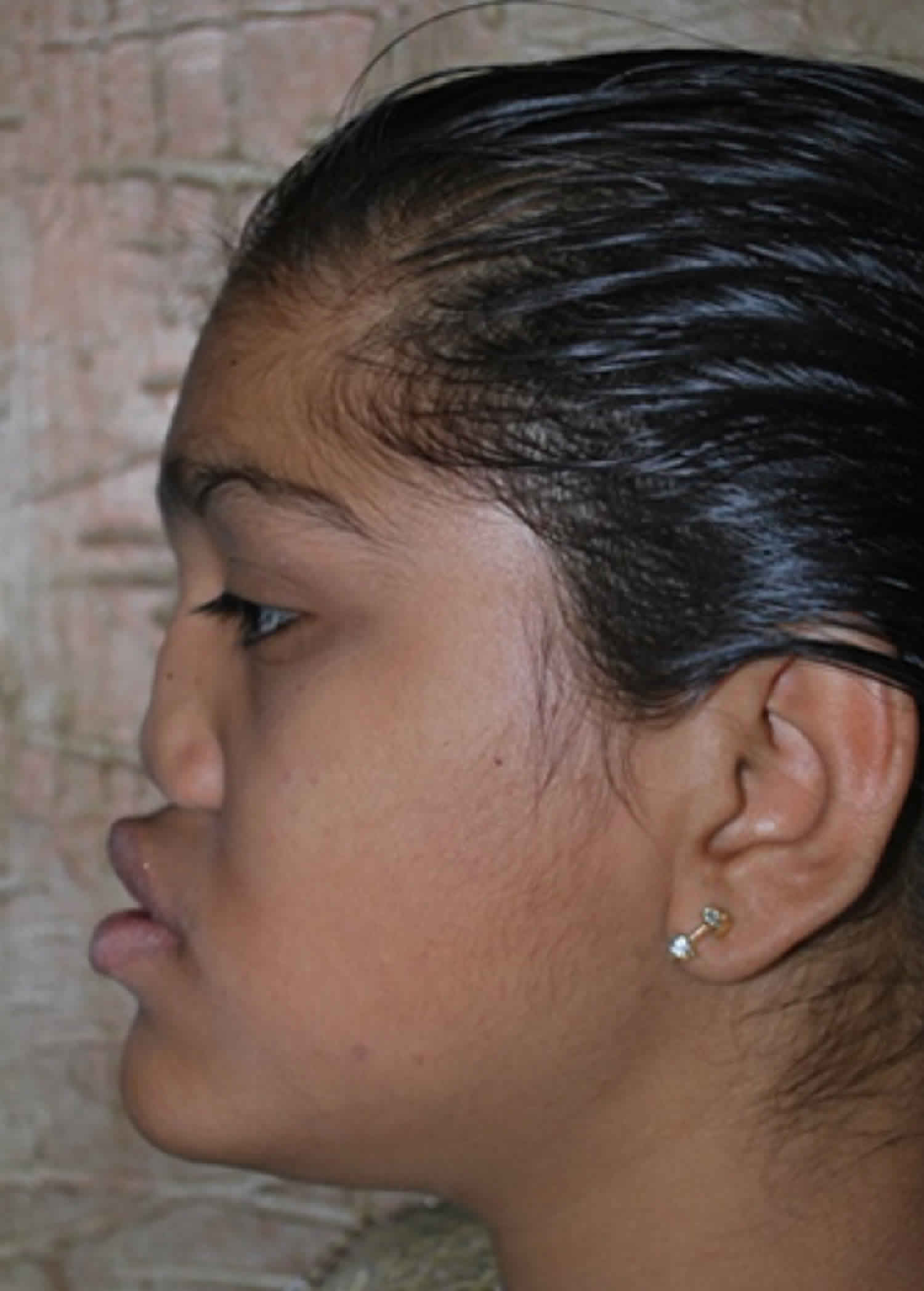
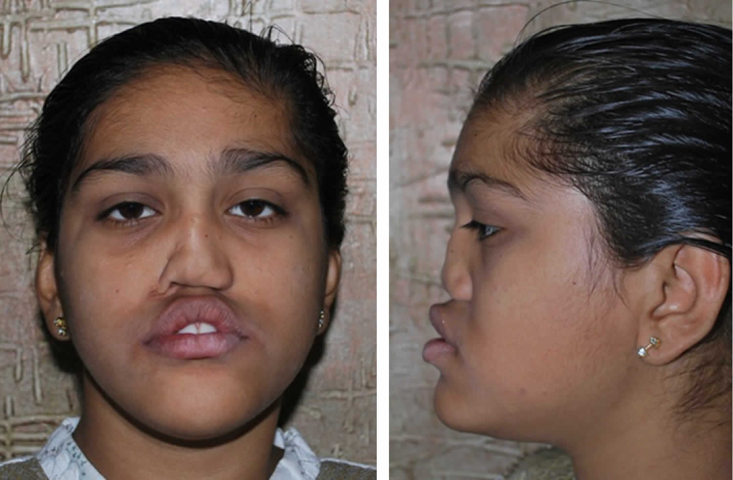
The primary physical characteristic of Binder syndrome is a flat, underdevelopment of the central portion of the face (midfacial hypoplasia) particularly the area including the nose and upper jaw (maxillonasal region) and flattened nose associated with the absence of the anterior nasal spine that supports the nose in normal development. Your child may appear to have an underdeveloped upper jaw and facial imbalance. The specific symptoms and the severity of the disorder can vary from one person to another. Characteristic symptoms include an abnormally short, flattened nose and underdevelopment of the upper jaw bone (maxillary bone).
Although researchers have been able to establish characteristic or “core” symptoms, much about Binder type nasomaxillary dysplasia is not fully understood. Several factors including the small number of identified affected individuals, the lack of large clinical studies, and the possibility of other genes influencing the disorder prevent physicians from developing an accurate picture of associated symptoms and prognosis.
The characteristic finding of the disorder is the abnormal development (dysplasia) of the central or mid portion of the face. The midface appears abnormally flattened. In some patients the frontal sinuses may be underdeveloped or absent. Affected individuals have a short nose and flattened bridge of the nose. The nasal bones may be underdeveloped or abnormally positioned. The bottom of the sheet of cartilage and bone (nasal septum) that separates the right and left nostrils is known as the columella. The columella is abnormally short and the nostrils have a half-moon or comma-shaped appearance. In cases where the columella is severely short, the nostrils may appear triangular. The upper lips may be slanted backward. Despite the various nasal abnormalities, the sense of smell is unaffected.
Underdevelopment (hypoplasia) upper jaw (maxillary bone) is another key feature of Binder type nasomaxillary dysplasia. The maxillae are the large bones of that form the upper jaw and assist in the formation of the nasal cavities, the bony cavities of the eyes (orbits), and the roof of the mouth (palate). The maxillae also contain the sockets of the upper teeth. Hypoplasia of the upper jaw may cause the lower jaw (mandible) to appear to protrude or stick out (relative prognathism). However, in some individuals, the mandible may actually be longer than normal (true prognathism). Affected individuals also develop malocclusion, a condition in which the upper teeth are improperly positioned in relation to the lower teeth. More specifically, affected individuals may be predisposed to a reverse overbite (class III malocclusion), in which the lower jaw is too far forward, the cusps of the lower back teeth are abnormally positioned in front of the corresponding upper teeth, and the lower front teeth (incisors) meet or lie in front of the corresponding upper incisors.
In some cases, additional symptoms and physical findings have been reported in association with this condition. Individuals with Binder type nasomaxillary dysplasia seem to be at an increased risk of various malformations of the spine (vertebrae). Less often, affected individuals exhibit hearing impairment, incomplete closure of the roof of the mouth (cleft palate), misalignment of the eyes (strabismus), structural malformations of the heart (congenital heart defects), mild intellectual disability, and other features. However, the exact relationship between these findings and Binder type nasomaxillary dysplasia is unknown and they may not represent symptoms of the disorder.
Binder type nasomaxillary dysplasia is a rare congenital condition that affects males and females in equal numbers. The exact incidence or prevalence is unknown. One estimate suggests that Binder syndrome occurs in less than 1 per 10,000 live births. However, individuals may go undiagnosed or misdiagnosed making it difficult to determine the true frequency in the general population.
The facial features of Binder syndrome were first described by Noyes in 1939 2), although it was von Binder who in 1962 identified and defined all the features of the syndrome. Von Binder, who called this syndrome “maxillonasal dysostosis”, reported the six most characteristic features of the syndrome: arhinoid face, intermaxillary hypoplasia (associated with malocclusion), abnormal position of the nasal bones, nasal mucosa atrophy, anterior nasal spine agenesis and (in most cases) a lack of frontal sinuses 3).
The exact cause of Binder syndrome is not fully understood. Most cases appear to occur sporadically, but familial cases have been reported as well. Surgical and orthodontic treatment is recommended.
Figure 1. Binder syndrome
Binder syndrome causes
The exact, underlying cause of Binder syndrome nasomaxillary dysplasia is not fully understood. In many cases, the disorder is believed to occur spontaneously, for no apparent reason (sporadically). However, there have been reports in the medical literature of families in which more than one family member was affected. This suggests that genetic factors play a role in some affected individuals. Some researchers have suggested that Binder type nasomaxillary dysplasia is a genetic disorder inherited in either an autosomal dominant or recessive manner. Other researchers have suggested that the disorder is caused by complex genetic factors, specifically the interaction of many different genes, possibility in combination with environmental factors (multifactorial inheritance).
Researchers have identified several environmental factors that may be associated with Binder type nasomaxillary dysplasia including birth trauma, vitamin K deficiency 4) or exposure of a developing infant to an anti-seizure drug known as Phenytoin or to an anti-blood clotting (anticoagulant) drug known as warfarin. No suspected environmental agent has been conclusively linked to Binder type nasomaxillary dysplasia.
Some researchers believe that specific cases of Binder type nasomaxillary dysplasia may actually be mild forms or variants of chondrodysplasia punctata 5), a general term for a group of disorders characterized by abnormalities affecting the development of cartilage and bone (skeletal dysplasias). A variety of additional symptoms and physical features can develop. A characteristic finding of chondrodysplasia punctata is the formation of small, hardened spots of calcium on the “growing portion” or heads of the long bones (stippled epiphyses) or inside other areas of cartilage in the body. However, over time there is loss of epiphyseal stippling. Individuals who receive a diagnosis of Binder type maxillofacial dysplasia until their teen-age years or older may actually have chondrodysplasia punctata, but the distinctive epiphyseal stippling is gone so that a diagnosis of chondrodysplasia punctata is not considered.
Binder syndrome genetics
The exact, underlying cause of Binder syndrome nasomaxillary dysplasia is not fully understood. Most reported Binder syndrome nasomaxillary dysplasia cases were sporadic. A few cases of recurrence in pedigrees could be explained by either autosomal recessive or dominant inheritance with reduced penetrance or by multifactorial cause.
Binder syndrome symptoms
Patients with the most severe form of Binder syndrome have a tiny nose and recessed upper jaw, creating an underbite (malocclusion). In milder forms, the position of the upper teeth may be normal and the only difference visible may be the bony deficiency on either side of the nose.
The nasal deformity is characterized by a shortened columella and underdeveloped nasal bridge. The nostrils in children with Binder syndrome are characteristically comma-shaped and the bony tissue at the base of the columella (the anterior nasal spine) is absent.
In some cases other congenital diseases and abnormalities such as Down syndrome, autonomic neuropathy and strabismus are observed 6). According to Nedev 7), 5% of patients are found to present hearing loss and the same number of patients presents congenital heart diseases.
Binder syndrome diagnosis
Binder syndrome is diagnosed based on your child’s appearance with identification of characteristic symptoms, a detailed patient history, and a thorough clinical evaluation. Supplemental tests including X-rays and CT scans can be used to confirm the diagnosis.
Clinical testing and workup
Specialized imaging techniques may be used to help obtain a diagnosis of Binder syndrome. Such tests include computerized tomography (CT) scanning and magnetic resonance imaging (MRI). During CT scanning, a computer and x-rays are used to create a film showing cross-sectional images of certain tissue structures. An MRI uses a magnetic field and radio waves to produce cross-sectional images of particular organs and bodily tissues.
Such exams may yield specific findings including underdevelopment or absence of the bony protrusion that projects from the base of the nasal septum to join with the middle part of the upper jaw (anterior nasal spine); thinness of a portion of the upper jaw known as the alveolar bone, which forms the dental arch over the upper incisors; underdevelopment or absence of the frontal sinuses; and/or certain abnormalities detected with cephalometric studies, which are scientific measurements of particular craniofacial dimensions.
Binder syndrome treatment
The treatment of Binder syndrome is directed toward the specific symptoms that are apparent in each individual. Treatment may require the coordinated efforts of a team of specialists. Pediatricians or general internists, oral and plastic surgeons, craniofacial surgeons, specialists in the diagnosis, prevention, and treatment of crooked teeth (orthodontists), specialists in the diagnosis and treatment of disorders of the bones, joints, ligaments and muscles (orthopedists), and other healthcare professionals may need to systematically and comprehensively plan an affect child’s treatment. Psychosocial support for the entire family is essential as well.
There are no standardized treatment protocols or guidelines for affected individuals. Due to the rarity of the disease, there are no treatment trials that have been tested on a large group of patients. Various treatments have been reported in the medical literature as part of single case reports or small series of patients. Treatment trials would be very helpful to determine the long-term safety and effectiveness of specific medications and treatments for individuals with Binder syndrome.
Recommended treatment may include various orthodontic and surgical measures to help correct abnormalities of the jaw and nose. The specific therapeutic procedures performed will vary depending upon the nature and severity of the disorder in each individual including the specific anatomical abnormalities present, a patient’s general health, a patient’s age, patient preference, and other factors. Often more than one surgical procedure is necessary. The specific type and timing of an individual surgical procedure is determined based upon disease severity and patient age. Some affected children have been treated during childhood, while others are not treated until the late teen-age years, which is when the bone stops growing.
Some individuals may only require treatment with orthodontic devices such as braces that can straighten teeth or reposition the jaw. Nose (nasal) reconstruction can be accomplished with bone or cartilage grafts, or the implantation of alloplastic materials. In some cases, the grafting of cartilage from the ribs has been used successfully to reconstruct the nose (costochondral graft).
If the upper jaw is set back and the teeth retropositioned, the typical approach to treatment is to wait until your child’s facial bones have stopped growing, usually around age 15-19, before surgery is performed.
More severe cases require surgical procedures known as Le Fort I or II osteotomy. During Le Fort I osteotomy, the upper jaw is sectioned and repositioned to treat malocclusion and, if present, cleft palate. Le Fort II osteotomy involves repositioning the upper jaw and nose and correcting the backward displacement (retrusion) of the middle portion of the face.
Surgery for a recessed upper jaw usually involves cutting and repositioning the jaw forward, a procedure known as a Le Fort I osteotomy or advancement. This will be performed by your child’s plastic and reconstructive surgeon. Surgical intervention is usually preceded by a period of orthodontic therapy. In mild cases, surgery to advance the jaw may not be required and your child will be treated by orthodontic therapy alone.
In both cases, bony deficiency along the side of the nose may require the placement of bone grafts or synthetic implants. Treatment of the nasal deformity usually involves adding cartilage grafts to the bridge and to support the tip to give more projection and shape. These may be from the ear, but in most cases one needs more cartilage and the rib may be used. The narrow nasal passages may also require treatment.
If your child’s nose is more mildly affected, he may not require any additional treatment. For others, nose augmentation using cartilage grafts taken from the ribs can add to both the bridge and columella to reshape the nose. This procedure is usually done after your child has reached skeletal maturity to reduce the risk that he will outgrow these grafts.
For patients who have functional appearance-related concerns at a younger age, temporary artificial implants, usually silicone, may be placed. The implants are replaced with larger implants as your child grows. It is usually best to use cartilage as the definitive correction. Cartilage grafts are better tolerated than artificial implants and have fewer long-term complications.
If your child has functional nasal obstruction due to the small size of the nose, surgery of the septum and turbinate membranes may be required. The turbinate membranes are fleshy membranes on the inside of the nose that warm and humidify air but are not functional if severe obstruction occurs.
Even minor degrees of septal deviation and turbinate membrane enlargement can compromise the nose. In this surgical procedure, your plastic surgeon will go through the inside of the nose to remove or straighten the septum and remove a portion of the turbinate membranes. This type of procedure is generally performed on an outpatient basis, and your child can go home the same day.
Binder syndrome prognosis
Affected individuals typically have an unusually flat, underdeveloped midface (midfacial hypoplasia), with an abnormally short nose and flat nasal bridge, underdeveloped upper jaw, relatively protruding lower jaw and/or a ‘reverse overbite’ (or class 3 malocclusion). Other deformities, as well as mental retardation, are also possible. Due to the clinical appearance, patients require surgical and orthodontic treatment. The main surgery performed in these patients is nose reconstruction with bone or cartilage grafts. Usually patients require more than one surgical procedure due to graft resorbtion and an unsatisfactory appearance. Orthodontic treatment is based on class 3 treatment (pseudo-mesio-occlusion) and relieving dental crowding. The treatment of malocclusion may require combined orthodontic and surgical treatment. In younger patients, maxillary protraction with rapid palatal expansion could be an adequate approach.
Once jaw and nasal reconstruction are performed in adolescence, few if any additional procedures will be required. Most patients will experience long-term improved nasal breathing and appearance, and a functionally normal upper jaw and bite.
References [ + ]


