Uncategorized

Postmaturity

postmature newborn

Postmature baby

Postmaturity also called dysmaturity is a word used to describe babies born after 42 weeks. Very few babies are born at 42 weeks or later. Other terms often used to describe these late births include post-term, post mature infant, prolonged pregnancy, and post-dates pregnancy. Most women go into labor spontaneously by the time they are 42 weeks pregnant.

Typically, tests are started at 41 weeks to evaluate the fetus’s movement and heart rate and the amount of amniotic fluid (the fluid around the fetus), which decreases markedly in postmature pregnancies. Doctors use ultrasonography and may use electronic fetal heart monitoring to monitor the fetus’s status.

Postmature pregnancies increase the risk of problems such as:

  • Difficult labor due to shoulder dystocia (the fetus’s shoulder lodges against the woman’s pubic bone, and the baby is caught in the birth canal)
  • The need for cesarean delivery or operative vaginal delivery (with forceps or a vacuum extractor)
  • Abnormal growth of the fetus (for example, an abnormally large fetus or abnormally small fetus)
  • Too little amniotic fluid around the fetus (oligohydramnios)
  • Problems with blood flow to the fetus, depriving the fetus or newborn of oxygen
  • Passage of meconium (the fetus’s first stool) before delivery
  • A newborn who needs care in a neonatal intensive care unit
  • Death of the fetus or newborn
  • Tears in the area between the opening of the vagina and anus (perineum)
  • Excessive bleeding at delivery (postpartum hemorrhage)

Meconium (baby’s first stool) can sometimes be inhaled before or during delivery, causing the baby to have difficulty breathing shortly after birth. This disorder is called meconium aspiration syndrome.

A postmature fetus may have dry, peeling skin, overgrown nails, a large amount of scalp hair, deep creases on the palms and soles, little body fat, and skin that is stained green or yellow by meconium.

The normal length of pregnancy is 37 to 41 weeks. Early term is from 37 weeks to 38 weeks and 6 days. Full term is 39 weeks to 40 weeks and 6 days. Late term is 41 weeks to 41 weeks and 6 days. However babies rarely keep to an exact timetable, so a full-term pregnancy can be anywhere between 37 and 42 weeks. A baby born before 37 weeks is considered to be premature and anything past 42 weeks is considered overdue or postmature newborn.

If your labor doesn’t start by the time you are 41 weeks pregnant, your doctor may offer you a ‘membrane sweep‘ to see if this will trigger labor.

This involves having a vaginal examination, which stimulates the neck of your womb (known as the ‘cervix’) to produce hormones that may trigger natural labor. You don’t have to have this — you can discuss it with your midwife or doctor.

If your labor still doesn’t start naturally after this, your midwife or doctor will suggest a date to have your labor induced, which is when your doctor or midwife uses drugs or tools and techniques to get your labor to start.

If your pregnancy lasts longer than 42 weeks and you don’t want your labor to be induced, you should be offered increased monitoring to check your baby’s wellbeing every 3 to 4 days.

Your midwife or doctor will check that both you and your baby are healthy by giving you ultrasound scans and checking your baby’s movement and heartbeat. An ultrasound might sometimes show that your placenta isn’t supplying as much oxygen and as many nutrients to your baby as it was. There might also be other concerns about you or your baby. If your baby is not doing well, your doctor will again suggest that labor is induced or a cesarean section (C-section).

Induction is always planned in advance, so you’ll be able to discuss the advantages and disadvantages with your doctor and midwife, and find out why they think your labor should be induced. It’s your choice whether to have your labor induced or not.

If tests show that your baby is fine and your health is good, you might choose to wait and see whether labor starts naturally.

There is a higher risk of stillbirth or fetal compromise (your baby’s health being put at risk) if you go over 42 weeks pregnant, but not every pregnancy over 42 weeks is affected this way. At the moment, there is no way to find out which babies might be affected, so induction is offered to all women who don’t go into labor by 42 weeks.

Key points about postmaturity in the newborn

  • Postmaturity is a word used to describe babies born after 42 weeks.
  • Researchers don’t know why some pregnancies last longer than others.
  • Postmaturity is more likely to happen when a mother has had a post-term pregnancy before.
  • Your doctor may decide to start your labor early.
  • An ultrasound test early in pregnancy can help your doctor figure out your baby’s age by checking the baby’s size.

What is a full-term pregnancy?

Pregnancy usually lasts about 40 weeks (280 days) from the first day of your last menstrual period also called LMP to your due date. Your due date is the date that your doctor thinks you will have your baby.

The American College of Obstetricians and Gynecologists and the Society for Maternal-Fetal Medicine define a full-term pregnancy as a pregnancy that lasts between 39 weeks, 0 days and 40 weeks 6 days. This means your pregnancy lasts between 1 week before your due date and 1 week after your due date. Babies born full term have the best chance of being healthy, compared with babies born earlier or later.

American College of Obstetricians and Gynecologists and Society for Maternal-Fetal Medicine use these definitions to describe term pregnancies:

  • Early term: Your baby is born between 37 weeks, 0 days and 38 weeks, 6 days.
  • Full term: Your baby is born between 39 weeks, 0 days and 40 weeks, 6 days.
  • Late term: Your baby is born between 41 weeks, 0 days and 41 weeks, 6 days.
  • Post term: Your baby is born after 42 weeks, 0 days.

In the past, a pregnancy that lasted anywhere between 37 to 42 weeks was called a term pregnancy. Health care providers once thought this 5-week period was a safe time for most babies to be born. In 2013, American College of Obstetricians and Gynecologists and the Society for Maternal-Fetal Medicine updated the definitions for term pregnancies because research shows that every week of pregnancy counts for the health of your baby. Lots of important things happen to your baby in the last few weeks of pregnancy. For example, your baby’s brain and lungs are still developing. Being pregnant for at least 39 weeks gives your baby’s body the time it needs to grow and develop.

These definitions can help more babies be born healthy by helping to prevent births that are being scheduled a little early for non-medical reasons. If your pregnancy is healthy, wait for labor to begin on its own.

Working out your due date

In order to calculate your baby’s due date, add seven days to the date of your last normal period (LMP) and then add nine months. For example, if your last period started on 1 March, adding seven days will make that 8 March. Then adding nine months will give a due date of 8 December. If your periods are irregular or you are unsure of the date, an ultrasound will help determine the development of the embryo and your due date. Ultrasound scans can be done at any stage of pregnancy after the first six weeks.

Due dates are usually calculated on your last period instead of the date of conception because of a number of reasons.

  • Although the average woman ovulates (releases an egg) approximately 2 weeks after her period, the exact time is not always known.
  • Once an egg has been released, it can remain fertile for up to 24 hours.
  • Sperm can last for up to 7 days after intercourse to fertilize an egg.

Some doctors will refer to your due date as ‘expected date of confinement’ or EDC.

What is gestational age?

Gestational age refers to how far along the fetus is. The gestational age is the number of weeks between the first day of the mother’s last menstrual period and the day of delivery. This time frame is often adjusted according to other information doctors receive, including the results of early ultrasound scans, which give additional information regarding the gestational age. The baby is estimated to be due (the due date) at 40 weeks of gestation.

Newborns are classified by gestational age as:

  • Premature: Delivered before 37 weeks of gestation
  • Full term: Delivered at 37 to before 41 weeks of gestation
  • Late term: Delivered at 41 to before 42 weeks of gestation
  • Post term: Delivered at 42 weeks of gestation

Postterm delivery is much less common than premature (preterm) delivery. Why a pregnancy continues beyond term is usually unknown. Women who have had one postterm delivery are at increased risk of having another one.

Postmaturity in the newborn causes

Researchers don’t know why some pregnancies last longer than others. Postmaturity is more likely when a mother has had one or more previous post-term pregnancies. Sometimes a mother’s pregnancy due date is off because she is not sure of her last menstrual period. Getting the date wrong may mean the baby is born earlier or later than expected. Getting an ultrasound in the first trimester (the first 12 weeks) is the most accurate way to tell the date of pregnancy, unless the date of conception is specifically known, such as with in vitro fertilization (IVF).

Why is postmaturity a concern?

Postmature babies are born after the normal length of pregnancy. The placenta, which supplies babies with the nutrients and oxygen from the mother’s circulation, begins to age toward the end of pregnancy, and may not function as efficiently as before. Other concerns include the following:

  • Amniotic fluid volume may decrease and the fetus may stop gaining weight or may even lose weight.
  • Risks can increase during labor and birth for a fetus with poor oxygen supply.
  • Problems may occur during birth if the baby is large.
  • Postmature babies may be at risk for meconium aspiration, when a baby breathes in fluid containing the first stool.
  • Hypoglycemia (low blood sugar) can also occur because the baby has already used up its glucose-producing stores.

Who is at risk for postmaturity in the newborn?

Postmaturity is more likely to happen when a mother has had a post-term pregnancy before. After one post-term pregnancy, the risk of a second post-term birth increases by 2 to 3 times. Other, minor risk factors include:

  • First pregnancy
  • Male baby
  • Older mother
  • Obese mother
  • Mother or father with personal history of postmaturity
  • White mother.

Postmaturity in the newborn prevention

Knowing your due date is the best way to know if your baby may be post-term. Keep track of the first day of your menstrual period. This can help estimate a baby’s due date. An ultrasound test early in pregnancy can also help your healthcare provider figure out your baby’s age by checking the baby’s size. Ultrasound is also a good way to check the placenta for signs of aging.

Post mature baby

Post mature baby or post mature infant is a baby delivered after gestational age of 42 weeks. Post mature newborns often have dry, peeling, loose skin and may appear abnormally thin (emaciated), especially if the function of the placenta was severely reduced. The fingernails and toenails are long. The umbilical cord and nails may be stained green if meconium was present in the amniotic fluid.

Near the end of a term pregnancy, the function of the placenta decreases, providing fewer nutrients and less oxygen to the fetus. Low blood sugar (glucose) is a particular problem in postterm newborns.

Postmature newborns have dry, peeling, loose skin and may appear abnormally thin because they have not received sufficient nutrition at the end of the pregnancy. The diagnosis is based on the appearance of the newborn and the estimated date of delivery.

Typically treatment focuses on providing good nutrition and general care.

Some post mature newborns are not breathing at birth and need to be revived (resuscitated).

Near the end of a term pregnancy, the level of amniotic fluid decreases and the placenta (the organ that provides nourishment to the fetus) becomes smaller and less effective in providing oxygen and nutrients. To compensate, the fetus begins to use its own fat and carbohydrates (sugars) to provide energy. As a result, its growth rate slows, and its weight may even decrease.

Complications during and after delivery

If the placenta shrinks enough, it may not provide adequate oxygen to the fetus, particularly during labor. A lack of adequate oxygen may result in fetal distress (signs that the fetus is not well) and, in extreme cases, may result in injury to the brain and other organs.

Fetal distress can cause the fetus to pass meconium (the fetus’ stools) into the amniotic fluid. The fetus may reflexively take deep, gasping breaths triggered by the distress and thereby inhale the meconium-containing amniotic fluid into the lungs before birth. As a result, the newborn may have difficulty breathing after delivery (meconium aspiration syndrome).

If the pregnancy continues significantly beyond term, the fetus may die.

After delivery, postterm newborns are prone to developing low blood sugar (glucose) levels (hypoglycemia) because they have exhausted their supply of stored fat and carbohydrates.

Postmaturity in the newborn symptoms

Each baby may show different symptoms of postmaturity. The following are the most common symptoms of postmaturity. However, each baby may show different symptoms of the condition.

Some of those symptoms are:

  • Dry, loose, peeling skin
  • Overgrown nails
  • Large amount of hair on the head
  • Visible creases on palms and soles of feet
  • Small amount of fat on the body
  • Green, brown, or yellow coloring of skin from baby passing stool in the womb
  • More alert and “wide-eyed”

Symptoms of postmaturity sometimes look like other health conditions. Make sure your child sees his or her doctor for a diagnosis.

Postmaturity in the newborn possible complications

Post-term babies are born after the normal length of pregnancy. Because of this they may grow larger than full-term babies. This may be a problem during labor and delivery.

Also, because the placenta ages toward the end of pregnancy, it may not work as well as before. Concerns from placental aging include:

  • Less amniotic fluid. This may stop the baby from gaining weight or may even cause weight loss.
  • Poor oxygen supply. Babies who don’t get enough oxygen may have problems during labor and delivery.
  • Meconium aspiration. Babies who stay in the womb longer are more likely to breathe in fluid containing meconium.
  • Hypoglycemia or low blood sugar. This happens when the baby has already used up his or her stores of glucose.

Postmaturity in the newborn diagnosis

Postmaturity is usually diagnosed by a combination of assessments, including the following:

  • Your baby’s physical appearance
  • The length of your pregnancy
  • How old your baby appears to be or your baby’s assessed gestational age

Postmaturity treatment

Your healthcare provider will check your unborn baby’s health and look for any problems. Testing may be done for a post-term pregnancy to check fetal well-being and identify problems.

Tests may need to be done such as:

  • Ultrasound
  • Nonstress testing. This looks at how the fetal heart rate responds to fetal movement.
  • Checking the amount of amniotic fluid volume

The decision to induce labor for post-term pregnancy depends on many factors. During labor, your baby’s heart rate may be watched with an electronic monitor. This will help spot changes in the heart rate caused by low oxygen levels. Changes in your baby’s condition may require a cesarean delivery.

If a pregnancy goes beyond term, inducing labor in the mother can decrease the risk of newborn death, decrease the need for cesarean delivery, and decrease the possibility that the baby will have meconium aspiration syndrome. Postmature newborns who have low oxygen levels and fetal distress may need to be urgently delivered by cesarean delivery and may need to be revived (resuscitated) at birth.

Special care of the post-term baby may include:

  • Checking for breathing problems caused by baby’s breathing in fluid containing the first stools (meconium)
  • Blood tests for low blood sugar (hypoglycemia).

If the baby has breathed meconium into the lungs or is having trouble breathing because of another problem, doctors may give an injection of surfactant (a material that coats the inside of the air sacs and makes it easy to breathe). A machine that helps air get in and out of the lungs (ventilator) and oxygen may be needed to support breathing.

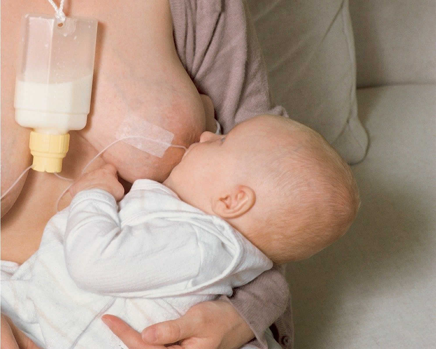
Sugar (glucose) solutions given by vein (intravenously) or frequent breast milk/formula feedings are given to prevent or treat hypoglycemia.

If complications do not occur, the major goal is to provide good nutrition so that postterm newborns can catch up to the weight that is appropriate for them.

read more

Nipple confusion baby

nipple confused baby

Nipple confused baby

Nipple confusion is a broad term for when babies used to sucking from bottles have a hard time getting back on the breast. Healthy babies are born with a vital survival skill—the instinct to breastfeed. Interfering with this instinct in the early days and weeks of a baby’s life can lead to problems. Many a mother has noticed a change in her baby’s sucking patterns after introducing a bottle or dummy. Her baby may struggle and cry, find it difficult to latch on, or simply nurse ineffectively at the breast. These changes in sucking patterns and breast refusal are often referred to as nipple confusion.

How to deal with nipple confused baby

Babies are ‘hard-wired’ to breastfeed so it’s almost always possible to get back to breastfeeding using one or more of the following strategies. Your role is to calm your baby, give him access to the breast and follow his cues. It’s your baby’s job to feed.

Here are some things you can do to get your baby on your breast:

  • Be as patient and calm as you can, even though you may be feeling frustrated or impatient. Forcing your baby to feed is likely to make the situation worse. If he has just been refusing the breast and is upset, distract him by doing something completely different – a walk outdoors, looking at toys, singing a nursery rhyme. When he has settled down he may be eased on to the breast, or he may be happier just being cuddled.
  • Walk around with your baby in an upright position against your body with her head level with your nipple. Walk and feed simultaneously. You could try putting your baby in a baby sling but remember to have your bra undone so that her face is touching the skin of your breast and she can find your nipple. The sling will need to be worn lower than normal for this purpose.
  • Try a completely different feeding position: your baby tucked under your arm (twin style); or lying down on a bed next to your baby with no body contact – this is especially good if it is very hot, or your baby is sensing your tension; or lying down with your baby cuddled in close next to you.
  • Feeding your baby while you are both in the bath may help. You may want to have someone available to help you lift your baby in and out of the bath.
  • Try breastfeeding baby after his bath when he is warm and relaxed (if he likes baths).
  • You could try playing with your baby on the floor while you are bare from the waist up. After some time gradually offer your breast.
  • Anticipate your baby’s waking time and lift her to feed while still sleepy – you may slip in extra night feeds this way.
  • Try to soothe baby with a pacifier (dummy). Walking, singing and rocking while baby sucks the dummy may gradually soothe him so you can gently put your baby to the breast while removing the dummy. It may be necessary to start a very hungry baby sucking on a bottle with a small amount of expressed breastmilk, e.g. 30 ml, then gently replace it with the breast.
  • Some mothers, whose babies have become accustomed to a bottle, have found that putting ice wrapped in a flannel on the nipple or tickling the nipple and areola makes it easier for the baby to grasp. Alternatively, you may use a nipple shield to begin a feed, slipping if off quickly and putting your baby back to the breast once the milk is flowing and she is sucking happily.
  • Feed in a rocking chair.
  • Express some milk into your baby’s open mouth to encourage him.
  • Spend five minutes or so before the feed massaging your baby’s naked body to relax her, if she is receptive to this.
  • Try singing to your baby – he probably won’t mind if it is the same few lines over and over.
  • Try playing some favorite relaxing background music.
  • Once you get your baby on to the breast, it may help to provide an instant milk reward. This can be done with a breastfeeding supplementer. This allows baby to receive additional milk at the breast whilst stimulating your milk supply by his sucking. If your milk supply continues to be low or your let-down slow or your baby is a ‘poor’ sucker, you may like to discuss with a Lactation Consultant counselor the possibility of using a supplementer.

A baby’s behavior can change very quickly, especially in response to gentle repetition, so do persist. With time and patience, most babies will breastfeed well once more. In the meantime, hand express or pump your milk to ensure you still make plenty of milk.

Make breastfeeding pleasant

Handle your baby gently when trying to feed and minimize distractions. Allow him to take the breast at his own pace to help him relax and feel in control. You’ll want your baby to associate being at the breast with pleasure. Pushing him onto the breast can have the opposite effect—he may instinctively fight against it.

Close body contact

Full body contact with you can trigger your baby’s instinct to breastfeed and help him get a deeper latch onto the breast. As you recline, try letting your baby lie upon you so that his whole front is against you and he is supported by your body. In this position babies often start to suck, and may even attach and feed in their sleep. Watch for early feeding cues—bobbing his head or making sucking motions—and gently encourage feeding. Spend as much time like this as you can—it can be done skin-to-skin if you wish. Many mothers find sleeping in close contact helps—a baby may take the breast when sleepy but not when wide awake. Taking a long warm deep bath together may also encourage your baby to relax, latch and feed well. You’ll need help to do this safely.

A faster flow

Swallowing encourages sucking, which will help keep milk flowing. A baby who has become used to a continuous fast flow from a bottle may need help to persist at the breast. Your milk production may also have dipped a little without you realizing. If needed, you could hand express a little to get the flow going before your baby latches on. Use breast compression to encourage him to continue drinking. Then switch sides to stimulate further let downs. Switch sides several times if necessary.

  1. Cup your hand around your breast. Wait until your baby stops swallowing regularly.
  2. Now compress your breast to increase milk flow. Hold it squeezed whilst he continues to swallow.
  3. Then release, rotate your hand and repeat. Do this on both sides or even switch back and forth to keep your baby actively feeding.

Talk to your doctor or Lactation Consultant about using a breastfeeding supplementer also known as Supplemental Nursing System (SNS). This device delivers extra milk through a tube along your breast as your baby nurses, avoiding the need for bottles.

There are two types of breastfeeding supplementer, one the is Medela Supplemental Nursing System that uses a hard sided container for the supplement and the ‘Lactaid’ uses a soft bag to hold the supplement. Each has its own advantages and disadvantages. It is also possible to make a homemade version. Breastfeeding supplementer consists of a container that is worn on a cord around the mother’s neck. Fine tubing carries expressed breastmilk or formula from the container to the nipple. When the baby sucks at the breast, milk is drawn through the tubing into his mouth, along with any milk from the breast.

When extra milk is needed for medical reasons it is better to give it through a breastfeeding supplementer than by bottle:

  • Milk given in this way rewards the baby’s efforts at sucking and he is more likely to be happy to feed from the breast and stay there for a longer time.
  • Since the amount of milk a mother makes depends on how much her baby sucks and takes milk from her breasts, this extra sucking will increase her milk supply.
  • Use of the mother’s own expressed breastmilk in the supplementer helps a weak or easily tired baby to get more milk with the same amount of sucking.
  • The sucking action required during breastfeeding differs from that used with a bottle. Some babies find it hard to do both, or reject the breast in favor of the different bottle.
  • The mother is able to provide milk at the breast and this helps with both the hormonal and physical aspects of breastfeeding.

Figure 1. Breastfeeding supplementer (Supplemental Nursing System)

Supplemental Nursing System

A firmer feel

If improving attachment and increasing milk flow are not enough, a nipple shield may help provide a firmer feel for your baby. A nipple shield is a thin, silicone teat that can be placed over a mother’s nipple during breastfeeding. Holes in the tip allow milk to flow into the baby’s mouth. While a nipple shield can be a useful tool in some situations it should be regarded as only a temporary solution—a transition towards breastfeeding.

Nipple shields come in different sizes, a comfortable fit is very important. A good latch-on is essential with a nipple shield to make sure your baby gets plenty of milk. If your baby latches onto just the tip, he will compress your nipple causing pain.

A deeper latch

If your baby has become used to bottles or dummies he may need encouragement to latch on with a wide-open mouth.

  • Rest comfortably with your breast in its natural position.
  • Position your baby ‘nose to nipple’, so he has to open wide to latch on. Make adjustments as your baby grows in length, so he can comfortably adopt this position. His ear, shoulder and hip should be in line.
  • If you need to support your breast, keep your fingers well away from the areola. Cup your breast by placing your hand with your thumb on one side of the areola, fingers on the other. When your fingers are parallel with your baby’s lips, you can shape your breast to match the oval of your baby’s mouth.
  • Pull your baby’s whole body close against yours. Make sure his head is free to tip back so his jaw can drop freely.
  • Once he opens his mouth wide, quickly roll your breast into his mouth and onto his tongue. Aim his lower jaw as far from the base of your nipple as possible.
  • As the breast lands on his tongue, it pushes his lower jaw open wider, resulting in a bigger and better mouthful of breast.
  • Your nipple is the last part of the breast to enter his mouth. When the nipple reaches the back of his mouth this triggers active sucking.

Sometimes just a small adjustment to the way you hold your baby makes a difference. A Lactation Consultant can provide you with personalized suggestions.

How to avoid nipple confusion

Avoid giving bottles and dummies to your baby. It is perfectly possible to breastfeed without ever using bottles or dummies. A baby who gets used to the firm shape of a bottle teat and the rapid flow it provides before breastfeeding has been established, can find it difficult to nurse effectively when switched back to the breast. If you do decide to use a bottle, use it only once breastfeeding is going well. To avoid using a dummy, encourage your baby to satisfy his need to suck by nursing at your breast.

read more

Breastfeeding while fasting

breastfeeding while fasting

Breastfeeding while fasting

Adherence to the Islamic religion requires fasting from sunrise to sunset throughout the month of Ramadan. Therefore, the duration of a fast can vary from 12–16 hours, depending on whether Ramadan falls in the summer or winter months. It is also an obligation for practising Muslims, and missed fasts must be made up for by fasting at a later date, or fidyah. Therefore, most practising Muslim mums prefer to fast.

A woman who has fasted since she was old enough to do so is capable of making the decision whether to fast. If she feels fit on the day, she can decide to fast. And if she decided to fast and she or baby’s health are affected she can pause fasting until they recover and resume as she sees fit. As a form of religious respect, non-Muslims should refrain from dissuading a Muslim mum from fasting.

Yom Kippur, the Jewish Day of Atonement, requires an almost 25-hour fast. Jewish mothers who are breastfeeding can consult a Rabbi or the Nishmat for more information about fasting for Yom Kippur.

How does fasting affect breast milk?

Milk supply

While severe dehydration can decrease milk supply, breastfeeding research tells us that short-term fasting does not decrease milk supply 1), 2).

Nutrient content of breastmilk

Zimmerman et al 3) studied the breastmilk of 48 healthy mothers who were exclusively breastfeeding a baby aged between 1 and 6 months and who fasted for close to 25 hours during Yom Kippur. This study found that fasting resulted in some short-term changes to breastmilk composition. The author of this study indicated that, ‘The practical significance of these changes should be seen within the context that thousands of babies undergo this exposure yearly without reported clinical effects’. Breastfeeding mothers can help ensure their baby remains well hydrated by continuing to breastfeed as per normal and monitoring their baby’s output (poops and urine). If the mother has any concerns about her ability to fast, she should seek medical advice.

Bener et al 4) also studied the breastmilk of 26 healthy mothers between the second and fourth weeks of Ramadan and 2 weeks after the end of Ramadan. This study found no significant differences in the content of major nutrients of breastmilk taken during and after Ramadan.

Effect on baby

Khoshdel et al 5) studied the effect maternal fasting during Ramadan had on the growth of exclusively breastfed babies aged 15 days – 6 months. The mothers of 36 of these babies fasted throughout Ramadan and 80 mothers did not fast. The babies’ growth was evaluated twice during Ramadan, 3 times in the second month and bimonthly in the next 4 months. This study found no significant differences in the growth of the babies in either group. This study concluded that Ramadan fasting by breastfeeding mothers does not adversely affect the growth of exclusively breastfed babies at least in the short-term.

Likewise, Haratipour et al 6) studied the growth of 55 healthy exclusively breastfed babies aged 1–6 months. Of the 55 babies, 20 of their mothers fasted throughout Ramadan and 35 of their mothers did not. The babies’ growth was evaluated twice in Ramadan and four times in the first, second and third months after Ramadan. This study concluded that Ramadan fasting by breastfeeding mothers did not adversely affect the growth of breastfed babies at least in the short term.

If a mother does not drink fluids for a day, her baby would generally breastfeed as usual the day of the fast, but would often breastfeed more often the next day or two 7).

Effect on mother

Healthy mothers are likely to cope well with short-term fasting. However, it is always a good idea to seek medical advice prior to fasting.

Zimmerman et al 8) recommend for breastfeeding mothers to ‘increase their fluid intake during the 2 days prior to the fast so they begin the fast as well hydrated as possible’. They also recommend for breastfeeding mothers to ‘decrease their activities and heat exposure as much as possible during the fast’.

How do I know my baby is getting enough breast milk?

You know your baby is getting enough breastmilk if she:

  • Is breastfeeding well and frequently
  • Has plenty of pale, wet nappies (at least 5 disposable or 6 cloth nappies in 24 hours)
  • Has 3 or more soft bowel motions a day (babies older than around 6 weeks may have less than this)
  • Is gaining weight and has some periods in which she seems reasonably alert, active and happy.

Crying doesn’t always mean that your baby is hungry. For about the first month, babies usually breastfeed every 2 to 3 hours. As they get older, their bellies are able to hold more and they nurse less often.

Watch your baby for signs that she’s full – like turning her head away or falling asleep. If your baby sucks on her fingers or moves her mouth like she’s eating, it doesn’t always mean she’s hungry – babies do these things to comfort themselves.

In the first few days of life, if your baby is frequently wanting to feed and crying, she may truly be having difficulty with breastfeeding. If you are concerned that your baby isn’t eating enough, talk to your baby’s doctor.

A newborn’s nappies

Wide variation exists in what is seen in a newborn’s nappies. The following gives a general guide as to what might be seen.

The first bowel motions a baby has are black and sticky. This is from the meconium present in the baby’s digestive tract before birth. By day 2, the bowel motions should be softer but still dark in color. Over the next few days, the bowel motions change to a greenish-brown and then to a mustard-yellow. As the color changes they become less sticky and larger in volume.

A baby should have at least 1 wet nappy on day one, at least 2 on day two, at least 3 on day three, at least 4 on day four and at least 5 on day five. From day 5 onwards, the information above is relevant.

Over the first few days, salts of uric acid in your baby’s urine may leave a rusty, orange-red stain on the nappy. This is normal during this time. If you see this after day 4, consult a doctor.

Daily diapers

A newborn’s diaper is a good indicator of whether he or she is getting enough to eat. In the first few days after birth, a baby should have 2 to 3 wet diapers each day. After the first 4 to 5 days, a baby should have at least 5 to 6 wet diapers a day. Stool frequency is more variable and depends whether your baby is breast or formula fed.

Growth charts

During regular health check-ups, your pediatrician will check your baby’s weight and plot it on a growth chart. Your baby’s progress on the growth chart is one way to tell whether or not he or she is getting enough food. Babies who stay in healthy growth percentile ranges are probably getting a healthy amount of food during feedings.

How do I know when my baby is hungry?

For babies born prematurely or with certain medical conditions, scheduled feedings advised by your pediatrician are best. But for most healthy, full-term infants, parents can look to their baby rather than the clock for hunger cues. This is called feeding on demand, or responsive feeding.

Hunger cues

A hungry baby often will cry. But it’s best to watch for hunger cues before the baby starts crying, which is a late sign of hunger and can make it hard for them to settle down and eat.

Other typical hunger cues include:

  • Licking lips
  • Sticking tongue out
  • Rooting (moving jaw and mouth or head in search of breast)
  • Putting his/her hand to mouth repeatedly
  • Opening her mouth
  • Fussiness
  • Sucking on everything around

It is important to realize, however, that every time your baby cries or sucks it is not necessarily because he or she is hungry. Babies suck not only for hunger, but also for comfort; it can be hard at first for parents to tell the difference. Sometimes, your baby just needs to be cuddled or changed.

How often should I breastfeed?

Your breasts make milk in response to your baby’s sucking. The more milk the baby takes, the more milk you make.

You are more likely to establish a good supply of milk if you:

  • Breastfeed frequently, whenever your baby fusses or seems hungry.
  • Let baby finish the first breast. You will know this because the baby will stop sucking and swallowing and will let go of the breast. Then offer the second breast.
  • Breastfeed your baby at night. This also helps prevent your breasts becoming too full and uncomfortable.
  • Many young babies feed between 8-12 times in 24 hours.
  • Avoid giving complementary bottles (‘comps’) unless medically necessary, as these will reduce your baby’s needs to suck at the breast and so reduce your supply.

General Guidelines for Baby Feeding:

It is important to remember all babies are different―some like to snack more often, and others drink more at one time and go longer between feedings. However, most babies will drink more and go longer between feedings as they get bigger and their tummies can hold more milk:

  • Most newborns eat every 2 to 3 hours, or 8 to 12 times every 24 hours. Babies might only take in half ounce per feeding for the first day or two of life, but after that will usually drink 1 to 2 ounces at each feeding. This amount increases to 2 to 3 ounces by 2 weeks of age.
  • At about 2 months of age, babies usually take 4 to 5 ounces per feeding every 3 to 4 hours.
  • At 4 months, babies usually take 4 to 6 ounces per feeding.
  • At 6 months, babies may be taking up to 8 ounces every 4 to 5 hours.

Most babies will increase the amount of formula they drink by an average of 1 ounce each month before leveling off at about 7 to 8 ounces per feeding. Solid foods should be started at about 6 months old.

References   [ + ]

read more

Spicy food while breastfeeding

spicy food and breastfeeding

Does spicy food affect breast milk and breastfeeding?

It’s fine to eat spicy food while you’re breastfeeding. Traces of what you eat enter your breast milk, but it shouldn’t unsettle your baby if you eat spicy food. In fact, it may benefit your baby. The flavor of your breastmilk changes according to what you eat, and this introduces the idea of different tastes to your baby. She may be more adventurous with new flavors once she starts solids at about six months.

Breastfeeding exclusively for the first six months of life has been shown to significantly lessen the risk and severity of food allergies in families with a strong history of them. Exclusive breastfeeding or breastfeeding in combination with partially or extensively hydrolyzed infant formula also reduces the risk for eczema, a condition of excessively dry and easily irritated skin.

A few mothers notice minor reactions to other foods in their diet. Some babies cry, fuss, or even nurse more often after their mother has eaten spicy or “gassy” foods (such as cabbage). These reactions differ from allergies in that they cause less-serious symptoms (no rashes or abnormal breathing) and almost always last less than twenty-four hours.

If your baby reacts negatively every time you eat a certain type of food and you find this troubling, you can just avoid that particular food temporarily. If these symptoms continue on a daily basis and last for long periods, they may indicate colic rather than food sensitivity. Talk with your pediatrician about this possibility, if eliminating various foods has no effect on your child’s symptoms.

You can figure out if your baby is sensitive to spicy food when breastfeeding by observing his/her reaction.

He/she may:

  • turn fussy after feeding
  • sleep little
  • cry for a long time
  • seem uncomfortable
  • wake up abruptly

Other signs which indicate any reaction in your baby are:

  • wheezing
  • skin reaction
  • mucus or green stools
  • congestion

However, these signs may not necessarily signal a response to spicy food, as they could be an indication of allergy to some other foods such as wheat, dairy, citrus or corn in your diet.

If you ate spicy food while nursing and observed any such symptoms, refrain from eating such food for a week and reintroduce later to observe the baby’s behavior. Some babies may turn fussy if the mother eats a significant amount of spicy food so have it in moderate amounts. If needed, schedule an appointment with your doctor for assistance.

How do I know if my child has a food allergy?

A food allergy happens when the body reacts against harmless proteins found in foods. The reaction usually happens shortly after a food is eaten. Food allergy reactions can vary from mild to severe.

Because many symptoms and illnesses could be wrongly blamed on “food allergies,” it is important for parents to know the usual symptoms. The following is information from the American Academy of Pediatrics 1) about food allergies and how to recognize and treat the symptoms.

Symptoms of a food allergy

When the body’s immune system overreacts to certain foods, the following symptoms may occur:

  • Skin problems
    • Hives (red spots that look like mosquito bites)
    • Itchy skin rashes (eczema, also called atopic dermatitis)
    • Swelling
  • Breathing problems
    • Sneezing
    • Wheezing
    • Throat tightness
  • Stomach symptoms
    • Nausea
    • Vomiting
    • Diarrhea
  • Circulation symptoms
    • Pale skin
    • Light-headedness
    • Loss of consciousness

If several areas of the body are affected, the reaction may be severe or even life-threatening. This type of allergic reaction is called anaphylaxis and requires immediate medical attention.

NOT a food allergy

Food can cause many illnesses that are sometimes confused with food allergies. The following are not food allergies:

  • Food poisoning—Can cause diarrhea or vomiting, but is usually caused by bacteria in spoiled food or undercooked food.
  • Drug effects—Certain ingredients, such as caffeine in soda or candy, can make your child shaky or restless.
  • Skin irritation—Can often be caused by acids found in such foods as orange juice or tomato products.
  • Diarrhea—Can occur in small children from too much sugar, such as from fruit juices.

Some food-related illnesses are called intolerance, or a food sensitivity, rather than an allergy because the immune system is not causing the problem. Lactose intolerance is an example of a food intolerance that is often confused with a food allergy. Lactose intolerance is when a person has trouble digesting milk sugar, called lactose, leading to stomachaches, bloating, and loose stools.

Sometimes reactions to the chemicals added to foods, such as dyes or preservatives, are mistaken for a food allergy. However, while some people may be sensitive to certain food additives, it is rare to be allergic to them.

Foods that can cause food allergies

Any food could cause a food allergy, but most food allergies are caused by the following:

  • Cow milk
  • Eggs
  • Peanuts
  • Soy
  • Wheat
  • Nuts from trees (such as walnuts, pistachios, pecans, cashews)
  • Fish (such as tuna, salmon, cod)
  • Shellfish (such as shrimp, lobster)

Peanuts, nuts, and seafood are the most common causes of severe reactions. Allergies also occur to other foods such as meats, fruits, vegetables, grains, and seeds such as sesame.

The good news is that food allergies are often outgrown during early childhood. It is estimated that 80% to 90% of egg, milk, wheat, and soy allergies go away by age 5 years. Some allergies are more persistent. For example, 1 in 5 young children will outgrow a peanut allergy and fewer will outgrow allergies to nuts or seafood. Your pediatrician or allergist can perform tests to track your child’s food allergies and watch to see if they are going away.

Does spicy food affect breast milk?

Unlike formula milk, breast milk alters its taste based on the food you take. For instance, if you eat garlic based foods, the breast milk may have similar taste and your little one would like it too. Your baby may have more feeding spells if the flavor of the milk changes.

Experts even consider it as a wise way to introduce the tastes of solid foods to your baby. You’re helping your baby to develop their sense of taste in preparation for the starting of solids. But basically, the sweetness of the natural sugars in the milk will dominate the flavor.

A study conducted in the early 1990s by researchers Julie Mennella and Gary Beauchamp 2) revealed that nursing babies of mothers who consumed garlic showed longer feeding spells, sucked harder, and drank more garlic-scented milk than babies of mothers who were not exposed to garlic.

Another study published in 2001 indicated that the babies exposed to flavors when they are in the uterus or while breastfeeding may like the same flavors when they grow 3).

However, if you find your baby upset or irritable, or you feel a heartburn then try taking a milder diet for some time. Give the baby a little more time to accept it. To check if your little one loves spicy flavored milk, go by a trial-and-error method.

References   [ + ]

read more

Bladder retraining

bladder retraining

Bladder retraining

The aim of bladder training is to help you gain better control over your bladder. When you do bladder training you are aiming to:

  • decrease the number of times you go to the toilet (frequency)
  • hold on for longer or put off emptying your bladder until your bladder is full, and
  • increase the volume of urine you pass each time when you do go to the toilet.

The Continence Foundation recommends you work with a continence nurse advisor or physiotherapist to design a bladder training program to suit your individual needs. Bladder training programs may take up to three months, with weekly or fortnightly appointments to monitor your progress and measure your improvement.

Your continence professional will teach you how to use your pelvic floor muscles. These muscles support your bladder and urethra (the tube that carries urine from the bladder to outside) and help to hold back the strong urge to pass urine. This will help you defer (put off) going to the toilet, or hold it in until you reach a toilet. The overall aim of bladder training is to restore the normal function of your bladder.

Over time, you should notice not needing to go to the toilet all the time, being able to stop yourself from going and passing more urine when you do go. You will also learn about diet and lifestyle modifications, including how to manage constipation (and straining), which can cause poor bladder control.

Bladder training takes time, so don’t worry if it feels like things are not improving right away. The important thing is to keep trying, make note of the things that make a difference, and stay positive.

Most bladder training programs will start by asking you to keep a bladder diary (see below). Every time you pass urine, you record the date, time and how much urine you passed. You will need to do this for a few days to identify how much your bladder holds and how often you need to empty it. You should also include comments about leaking or other symptoms such as burning or pain.

Step-by-step bladder-training technique

  1. Keep track. For a day or two, keep track of the times you urinate or leak urine during the day.
    • Keeping a bladder diary
      1. Complete the information for two consecutive 24-hour periods. Record both day and night.
      2. Begin with first urination upon arising.
      3. Record intake amount in ounces and type of fluid (for example, coffee, juice, water, etc.).
      4. Record approximate urine output and time of urination.
  2. Calculate. On average, how many hours do you wait between urinations during the day?
  3. Choose an interval. Based on your typical interval between urinations, select a starting interval for training that is 15 minutes longer. If your typical interval is one hour, make your starting interval one hour and 15 minutes.
  4. Hold back. When you start training, empty your bladder first thing in the morning and not again until the interval you’ve set. If the time arrives before you feel the urge, go anyway. If the urge hits first, remind yourself that your bladder isn’t really full, and use whatever techniques you can to delay going. Try the pelvic floor exercises sometimes called Kegels, or simply try to wait another five minutes before walking slowly to the bathroom.
  5. Increase your interval. Once you are comfortable with your set interval, increase it by 15 minutes. Over several weeks or months, you may find you are able to wait much longer and that you experience far fewer feelings of urgency or episodes of urge incontinence.

People with an overactive or weak bladder have to rush off to find a bathroom because of an urge to urinate, or may gradually lose urine drop by drop before arriving at a toilet. Bladder training can then help you to regain at least some control over your bladder.

The bladder muscle can be trained to stretch more so that the bladder is able to hold more urine. Bladder training also includes various behavioral treatment approaches and a particular drinking and voiding schedule (bathroom visit schedule).

Review any lifestyle factors which may be contributing to poor bladder function.

Lifestyle measures

  1. Regulating your fluid intake: Your professional advisor can help you regulate your fluid intake so you drink the correct volume for your body and level of activity at the right times. Too much fluid can overfill the bladder and too little can concentrate the urine, which irritates the bladder. Drinking about 1.5L of fluid per day (mostly water) aim to regularly pass 300ml or more urine.
  2. Limit alcohol and caffeine-based drinks: Alcohol, caffeine-based drinks and fizzy drinks are known to irritate the bladder, which exacerbates symptoms of overactive bladder. Alcohol also has a diuretic effect, which can cause more frequent urination.
  3. Avoid constipation: Constipation can trigger or worsen symptoms of overactive bladder. Straining on the toilet can also weaken the pelvic floor.
  4. Keep your weight in the healthy range: there is evidence that losing weight decreases the severity and frequency of symptoms of overactive bladder. Maintain a healthy weight by walking for at least thirty minutes each day
  5. Only going to the toilet when you need to go (not going “just-in-case”)
  6. Get rid of any cough or sneezing
  7. Exercise your pelvic floor muscles as you have been shown.
  8. It is important to have strong pelvic floor muscles.
  9. Do persevere. You have the ability to make your brain the boss and retrain your bladder!
  10. Do speak to your physiotherapist, doctor or continence consultant about any concerns or problems you encounter.

Figure 1. Bladder diary

bladder diary

Figure 2. Urinary bladder anatomy

Urinary bladder anatomy

Figure 3. Urinary bladder anatomy

Urinary bladder anatomy

What are normal bladder habits?

Your bladder normally stores urine, which is produced by your kidneys. The kidneys produce urine all the time, but the amount of urine you produce depends on how much you drink, eat and sweat.

A healthy bladder can hold one and a half to two cups (300-400 mls) of urine during the day and about four cups (800mls) at night.  It is normal to pass urine five or six times a day and possibly once during the night if you drink between 6-8 glasses of fluid. The amount of fluid you drink will affect how often you need to pass urine. For most people the average fluid intake should be approximately 2 liters (3 and 1/2 pints) a day. It is usual to empty your bladder when you get out of bed in the morning, three times during the day, and before you go to bed at night. As you age this pattern may change, as older people tend to make more urine at night.

Your bladder should act like a balloon, which fills gradually. The outlet for the urine (the urethra) is normally kept closed. This is helped by the pelvic floor muscles beneath the bladder, which sweep around the urethra.

As your bladder fills, the need to pass urine increases. When the bladder functions normally it should be possible for you to ‘hold on’ for a suitable and convenient time to empty your bladder.

Your bladder is made up of a muscle called the ‘detrusor’. When this contracts (squeezes), the muscles in the urethra relax and your bladder will empty. Complex nerve messages are sent between the brain, bladder and the pelvic floor muscles. This influences the sensation of your bladder filling and the use of the right muscles at the right time.

Common bladder problems you may experience

  • Urgency – feeling the sudden need to rush to the toilet.
  • Urge incontinence – feeling the urgent need to go to the toilet and not making it in time.
  • Frequency – going to the toilet often, then only passing small amounts.

Urgency and urge incontinence often occur because the bladder gets over excited and overactive.

What is an overactive bladder?

If your bladder contracts without any warning it can give you an urgent need to pass urine. This gives you little or no time to get to the toilet. This is called urinary urgency. If you have this problem you are likely to need to pass urine more frequently and in small volumes. This is because your bladder often feels fuller than it actually is. It can also cause you to get up in the night more than once to pass urine. If the contractions are large or your pelvic floor muscles are weak, an overactive bladder can cause leakage of urine. This is called urge incontinence.

For most people the cause of an overactive bladder is unknown. An overactive bladder can be a problem for many people of all ages. Other members of your family may suffer from similar symptoms. There could be a neurological cause for the problem (relating to the brain or spinal nerves) but this is uncommon. Stress can make the problem worse and the types of fluid you drink can also influence your symptoms.

How is overactive bladder diagnosed?

Urodynamics is a special test on your bladder which is used to help find out the cause of your bladder symptoms. It may show the cause of symptoms such as incontinence, urgency or difficulty emptying your bladder. If the results of the urodynamic investigation show that you have uncontrolled contractions of the bladder muscle then the diagnosis would be called detrusor over-activity.

Bladder retraining program

Every person will have different needs in regard to bladder training. The first step is to complete a three-day bladder diary to get information about your bladder function. From this your personal goals can be set. Set yourself achievable goals. For example, if you start with a break of one hour between emptying your bladder, your initial goal may be to “hang on” for 5 or 10 minutes. The aim is to gradually increase the time you can “hang-on”. Discuss appropriate goals with your doctor, Continence Nurse or Physiotherapist.

How will I know if bladder training is working?

You may not notice an improvement immediately, but gradually you will become aware of not having to go to the toilet as often, being able to stop yourself going, passing more urine each time you
go, having less urgency, and not getting up to the toilet at night time.

Bladder training takes time. You may have good and bad days. Persevere and you will eventually be the boss of your bladder.

What happens if bladder training doesn’t work?

Despite best efforts, in a few cases, the bladder will not respond to the bladder training program. In these cases, some medications, prescribed by the doctor, may assist. If you decide to take medications it is important to still continue your bladder training strategies.

Bladder diary

A bladder diary will help you record the amount of urine you pass every time you go to the toilet and every time you leak. Take a record over at least 3 days for a bladder diary. Each diary page will cover 24 hours. It will give a clearer picture of your pattern of leaking over the day and night, including any effects of fluids that are “bladder-irritants”. Keeping a diary can be helpful for your doctor or continence nurse.

When collecting urine to measure the volume passed, you will need to measure this fairly accurately. To make recording of drinks easier, note the amount that your favorite tea/coffee-cup or glass holds – use this to record your drinks in “milliliters”. You could use a good-sized measuring jug to “pee” into. Make a note of the volume for your diary before tipping its contents into the toilet. Or you may find it easier to use a large plastic container (such as an ice-cream container) and set this directly down into the toilet. Then tip the contents into the measuring jug.

Your bladder diary is all about recording four important things:

  1. Input (fluids drunk): Record the type or names of all fluids that you drink. Be aware that “fluids” also includes foods that are mainly liquid, such as soups, jellies and custards.
  2. Output: Record the amount of urine you pass over at least 3 days and nights (Make these 3 consecutive days if you can).
  3. Leakage: Record the amount of any leakage of urine, e.g. spot, dribble, flood, a few teaspoons, or similar description.
  4. Circumstances: Where you were, or what you were doing, at the time of leakage is important information, both for your own awareness and for a Continence Assessment. “Circumstances” will include whether you felt an urgent, sudden need to pass urine just before the leakage happened, or you leaked when you coughed or while lifting, or you leaked at aerobics class, for example. Perhaps you arrived home, put the key in the door and lost control before you reached the toilet? Maybe at the time you leaked you weren’t aware you were losing urine? Or perhaps you got out of bed in the morning and, as you stood up, you “lost it”.

Looking back at the entries in your bladder diary could be helpful for you anticipate problems, so you’re ready for a possible leakage episode. Perhaps there’s a problem at a particular time of the day, or overnight, or following certain drinks or foods? It can be difficult to remember every time you empty your bladder (“pee”) so writing this information down will help. Be aware that the part of the week you choose to record may be significant – your weekends may be quite different to a week-day, for example.

How to measure your urine

To measure the amount of urine you pass, put a container (like an ice cream container) in the toilet bowl. Sit on the toilet and pass urine into the container. When you have finished, measure the urine by tipping it into a measuring jug. For men, you may prefer to stand and pass urine directly into the measuring jug. You should note the measurement from the jug in your bladder diary, then tip the urine into the toilet and flush.

You can also use absorbent pads to work out how much urine you leak over one or two days. This is done by using a dry pad that has been weighed in a plastic bag. When you change the wet pad you put it back in the plastic bag and weight it. If you take away the weight of the dry pad from the weight of the wet pad you can work out how much urine you have leaked. One milliliter (ml) of urine weighs one gram.

For example:

  • Wet pad = 350 grams
  • Dry pad = 150 grams

Weight difference

  • 200 grams

Amount leaked

  • 200 mls

Bladder retraining exercises and techniques

Some techniques which may help you control your urge:

  • Stop and relax – do not rush to the toilet.
  • Breathe slowly and evenly.
  • Perineal / Vaginal pressure:
    • Sit on the arm of a chair or the edge of a desk.
    • Sit on a rolled-up towel.
    • Cross your legs.
  • Curl your toes repeatedly or hold firmly.
  • Stretch your calf muscles and hold the stretch.
  • If you are walking, slow your pace and emphasize heel-toe walking.
  • Press or rub the trigger point on the inside of your leg just above your ankle.
  • Press or rub the trigger point at the base of your nose.
  • Squeeze and lift your pelvic floor muscles – hold them tight.
  • Distract yourself. Do not think about the toilet or about leaking. Distract yourself with a physical or mental task e.g. count backwards from 100 by 3’s or 7’s; think of 3 American towns starting with A, then with B, then C etc.
  • When you have performed the technique, walk to the toilet in a slow and controlled manner.

Remember: Your bladder does not have eyes or ears; it cannot see the toilet nor know how far away you are. If you feel you may leak on the way it is just anxiety. Relax, stay calm and walk slowly.

Different techniques work for different people. By practicing these techniques you will find out which ones work for you. Use the above techniques to gradually increase the time between toileting and to stop getting out of bed at night when you feel the urge to go to the toilet.

Retraining your bladder

Review any lifestyle factors which may be contributing to poor bladder function, such as:

  • reducing or ceasing caffeine intake (tea, coffee, cola drinks), alcohol and very sweet drinks
  • not being constipated
  • only going to the toilet when you need to go (not going “just-in-case”)
  • drinking about 1.5L of fluid per day (mostly water) aim to regularly pass 300ml or more urine
  • get rid of any cough or sneezing
  • exercise your pelvic floor muscles as you have been shown
  • maintain a healthy weight by walking for thirty minutes each day
  • do persevere. You have the ability to make your brain the boss and retrain your bladder.
  • do speak to your Physiotherapist or Continence Nurse about any concerns or problems you encounter.

It is important to have strong pelvic floor muscles (see Pelvic Floor Exercises).

Bladder retraining:

  • Instead of rushing to the toilet as soon as you get the urge to pass urine, it important to try to learn to ‘hold on’. If you have an overactive bladder you may tend to go to the toilet more often than you need to, as a precaution to avoid ‘accidents’. Unfortunately, rather than helping the problem this can make it worse, as your bladder will become used to holding less urine. Your bladder then becomes more sensitive, even when there is very little urine in it to stretch it. Try to avoid ‘just in case’ visits to the toilet.
  • Bladder retraining should be carried out in small stages. For example, if you find you are going to the toilet every half an hour, try extending the time (or ‘holding on’) by 10 minutes for a week, then by 15 minutes for a week, and then 30 minutes, etc.
  • Ideally you should be able to hold on for 3-4 hours between toilet visits.

Learn to suppress the urgency

There are different techniques for this. What works for one person may not work for another. Here are some suggestions:

  • Sit straight on a hard seat.
  • Distract yourself – for example, try doing a crossword or word search puzzle; count down from 100; think of girls or boys names beginning with a certain letter; read a book or newspaper. It is important to plan in advance how you will distract yourself, so that you are prepared.
  • Contract your pelvic floor muscles. Contracting these muscles may help to suppress both urgency and incontinence.

The above techniques may help you gradually, but patience is needed as it may take weeks or months before you notice a significant improvement. You will find it helpful to keep a diary of when you go to the toilet at the start of bladder training and then again a few months later. You may see more improvement than you expected.

Change your drinking habits

There are a number of drinks that may irritate the bladder. If you do have these drinks, try to have them less often or stop having them completely, to see if this helps.

Drinks which may irritate the bladder are: 

  • drinks containing caffeine such as tea and coffee• carbonated (fizzy) drinks
  • drinks containing aspartame (an artificial sweetener, which can be found in diet drinks. It can also be labelled as E951)
  • hot chocolate
  • alcohol
  • green tea and mint tea
  • blackcurrant juice
  • citrus fruit juices (e.g. orange and grapefruit).

Drinks which are believed not to irritate the bladder are:

  • water (definitely not a bladder irritant)
  • fruit teas
  • caffeine-free tea and coffee (make sure all the caffeine has been removed). However, some people’s bladders can also be irritated by de-caffeinated drinks. Try de-caffeinated drinks for a month to see if this improves your symptoms.
  • milk
  • diluted fruit juice.

Many people with an overactive bladder reduce the amount they drink. However, this can make the problem worse, as the bladder then never gets full and so loses the ability to stretch. Also, the urine becomes concentrated, which can irritate the bladder even further. Aim to drink 1.5 to 2 liters a day (although this amount will vary depending on the weather, your activity levels and your weight. You need to drink a certain amount of fluid for your weight; your healthcare team will talk about this with you).

Medications

Medicines that may help. There are various medicines that you can try. They work for some people, but not all. It’s worth trying them for one to two months before deciding if they are suitable for you. If they do work, they will work best alongside the bladder techniques described here. You can continue to take them for as long as you need them.

The medicines are called antimuscarinics (anticholinergics) or a newer medication called mirabegron (Betmiga). Antimuscarinics work by blocking certain nerve impulses to the bladder, which stops it contracting and helps it to hold on to more urine. They need to be taken for at least 4 weeks to notice any benefit.

Mirabegron relaxes the bladder muscle. This reduces the activity of an overactive bladder and treats urge and urge incontinence. It starts to work about eight weeks after you begin taking it.

You may need to try different medicines to find one that works best for you. All medicines have possible side effects. The most common side effects from antimuscarinics are a dry mouth and constipation. Ask your doctor or specialist nurse for more information on side effects and make sure they know your medical history before recommending the medicines.

There is another form of therapy using a bacterial toxin (Botox) which can help an overactive bladder. However, it is an invasive procedure involving injections into the bladder and is only performed as a last resort. This would need to be discussed carefully with your doctor or specialist nurse.

Medications that may make the problem worse

If you take diuretics (water tablets) they can make you pass more urine, so it may be worth changing the time you take them. For example, don’t take them late at night as you will pass urine a lot during the night. However, you may not wish to take them in the morning if that is the time of day when you want to go out. Speak to your doctor before you change the pattern of taking your diuretics.

Other medicines can also adversely affect your bladder. Your doctor or specialist nurse will be able to give you more specific information.

Pelvic Floor Exercises

Pelvic floor muscle training exercises also called Kegel exercises, are exercises to make your pelvic floor muscles stronger. The pelvic floor are the muscles that hold up your bladder, rectum and uterus (womb) and help keep them from prolapsing (a condition where organs fall down or slip out of place), resulting in urinary or fecal leakage (incontinence).

Kegel exercises (pelvic floor muscle training exercises) can help both men and women who have problems with urine leakage or bowel control. Kegel exercises (pelvic floor muscle training exercises) can also make sex better.

Building up your pelvic muscles with Kegel exercises (pelvic floor muscle training exercises) can help with your bladder and bowel control. Kegel exercises strengthen the pelvic muscles — the muscles under the uterus, bladder, and bowel (large intestine) and don’t require any equipment. Once you learn how to do “Kegel exercises” you can Kegel anywhere. You can do Kegel exercises while lying down, sitting at a desk, or standing up.

The trick is finding the right muscles to squeeze (see Figures 1 and 2 for reference). Your doctor or nurse can help make sure you are squeezing the right muscles. Your doctor may refer you to a specially trained physical therapist who will teach you to find and strengthen the sphincter muscles. Learning when to squeeze these muscles can also help stop the bladder spasms that cause urge incontinence. After about 6 to 8 weeks, you should notice that you have fewer leaks and more bladder control.

To do Kegel exercises, you just squeeze your pelvic floor muscles. The part of your body including your hip bones is the pelvic area. At the bottom of the pelvis, several layers of muscle stretch between your legs. The muscles attach to the front, back, and sides of the pelvic bone (see Figures 4 and 5). Pelvic floor exercises are designed to make your pelvic floor muscles stronger. These are the muscles that hold up your bladder and help keep it from leaking.

A pelvic floor muscle training exercise is like pretending that you have to urinate, and then holding it. You relax and tighten the muscles that control urine flow. It is important to find the right muscles to tighten.

Find the right muscles. Try one of the following ways to find the right muscles to squeeze.

  1. Imagine that you are trying to stop passing gas. Squeeze the muscles you would use. If you sense a “pulling” feeling, you are squeezing the right muscles for pelvic exercises.
  2. Imagine that you are sitting on a marble and want to pick up the marble with your vagina. Imagine “sucking” the marble into your vagina.
  3. Lie down and put your finger inside your vagina. Squeeze as if you were trying to stop urine from coming out. If you feel tightness on your finger, you are squeezing the right pelvic muscles.

The next time you have to urinate, start to go and then stop. Feel the muscles in your vagina, bladder, or anus get tight and move up. These are the pelvic floor muscles. If you feel them tighten, you have done the exercise right.

If you are still not sure whether you are tightening the right muscles, keep in mind that all of the muscles of the pelvic floor relax and contract at the same time. Because these muscles control the bladder, rectum, and vagina, the following tips may help:

  • Women: Insert a finger into your vagina. Tighten the muscles as if you are holding in your urine, then let go. You should feel the muscles tighten and move up and down.
  • Men: Insert a finger into your rectum. Tighten the muscles as if you are holding in your urine, then let go. You should feel the muscles tighten and move up and down. These are the same muscles you would tighten if you were trying to prevent yourself from passing gas.

It is very important that you keep the following muscles relaxed while doing pelvic floor muscle training exercises:

  • Abdominal
  • Buttocks (the deeper, anal sphincter muscle should contract)
  • Thigh

If you’re having trouble doing Kegel exercises, don’t be embarrassed to ask for help. Your doctor or other health care provider can give you important feedback so that you learn to isolate and exercise the correct muscles.

In some cases, vaginal weighted cones or biofeedback might help. A woman can also strengthen these muscles by using a vaginal cone, which is a weighted device that is inserted into the vagina. To use a vaginal cone, you insert it into your vagina and use pelvic muscle contractions to hold it in place during your daily activities. During a biofeedback session, your doctor or other health care provider inserts a pressure sensor into your vagina or rectum. As you relax and contract your pelvic floor muscles, a monitor will measure and display your pelvic floor activity.

If you are unsure whether you are doing the pelvic floor muscle training correctly, you can use biofeedback and electrical stimulation to help find the correct muscle group to work.

  • Biofeedback is a method of positive reinforcement. Electrodes are placed on the abdomen and along the anal area. Some therapists place a sensor in the vagina in women or anus in men to monitor the contraction of pelvic floor muscles.
  • A monitor will display a graph showing which muscles are contracting and which are at rest. The therapist can help find the right muscles for performing pelvic floor muscle training exercises.

Let your doctor, nurse, or therapist help you. Many people have trouble finding the right muscles. Your doctor, nurse, or therapist can check to make sure you are doing the exercises correctly. You can also exercise by using special weights or biofeedback. Ask your health care team about these exercise aids.

Don’t squeeze other muscles at the same time. Be careful not to tighten your stomach, legs, or other muscles. Squeezing the wrong muscles can put more pressure on your bladder control muscles. Just squeeze the pelvic muscle. Don’t hold your breath.

Repeat, but don’t overdo it. At first, find a quiet spot to practice—your bathroom or bedroom—so you can concentrate. Lie on the floor. Pull in the pelvic muscles and hold for a count of 3. Then relax for a count of 3. Work up to 10 to 15 repeats each time you exercise. Use the Exercise Log below to keep track of your sessions.

Do your pelvic exercises at least three times a day. Every day, use three positions: lying down, sitting, and standing. You can exercise while lying on the floor, sitting at a desk, or standing in the kitchen. Using all three positions makes the muscles strongest.

Be patient. Don’t give up. It’s just 5 minutes, three times a day. You may not feel your bladder control improve until after 3 to 6 weeks. Still, most women do notice an improvement after a few weeks.

Performing Pelvic Floor Exercises

  1. Begin by emptying your bladder.
  2. Tighten the pelvic floor muscles and hold for a count of 10.
  3. Relax the muscles completely for a count of 10.
  4. Do 10 repetitions, 3 to 5 times a day (morning, afternoon, and night).

You can do these exercises at any time and place. Most people prefer to do the exercises while lying down or sitting in a chair. After 4 to 6 weeks, most people notice some improvement. It may take as long as 3 months to see a major change.

After a couple of weeks, you can also try doing a single pelvic floor contraction at times when you are likely to leak (for example, while getting out of a chair).

A word of caution: Some people feel that they can speed up the progress by increasing the number of repetitions and the frequency of exercises. However, over-exercising can instead cause muscle fatigue and increase urine leakage.

If you feel any discomfort in your abdomen or back while doing these exercises, you are probably doing them wrong. Breathe deeply and relax your body when you do these exercises. Make sure you are not tightening your stomach, thigh, buttock, or chest muscles.

When done the right way, pelvic floor muscle exercises have been shown to be very effective at improving urinary continence.

There are physical therapists specially trained in pelvic floor muscle training. Many people benefit from formal physical therapy.

Pelvic Floor Exercises Log

Week: ______________

My Pelvic Muscle Exercise Log

Sunday

  • I exercised my pelvic muscles ____ times.
  • I spent ____ minutes exercising.
  • At each exercise session, I squeezed my pelvic muscles ____ times.

Monday

  • I exercised my pelvic muscles ____ times.
  • I spent ____ minutes exercising.
  • At each exercise session, I squeezed my pelvic muscles ____ times.

Tuesday

  • I exercised my pelvic muscles ____ times.
  • I spent ____ minutes exercising.
  • At each exercise session, I squeezed my pelvic muscles ____ times.

Wednesday

  • I exercised my pelvic muscles ____ times.
  • I spent ____ minutes exercising.
  • At each exercise session, I squeezed my pelvic muscles ____ times.

Thursday

  • I exercised my pelvic muscles ____ times.
  • I spent ____ minutes exercising.
  • At each exercise session, I squeezed my pelvic muscles ____ times.

Friday

  • I exercised my pelvic muscles ____ times.
  • I spent ____ minutes exercising.
  • At each exercise session, I squeezed my pelvic muscles ____ times.

Saturday

  • I exercised my pelvic muscles ____ times.
  • I spent ____ minutes exercising.
  • At each exercise session, I squeezed my pelvic muscles ____ times.

Use this sheet as a master for making copies that you can use to record your exercises week after week.

Muscles of the pelvic floor

The pelvic floor also called the pelvic diaphragm (Figures 4 and 5), is a complex layer of muscles and ligaments which stretches like a hammock from the pubic bone at the front of your pelvis to the coccyx at the bottom your spine.

The pelvic floor is a sheet consisting of two muscles, the levator ani and the small coccygeus (ischiococcygeus). The pelvic floor supports the pelvic organs, seals the inferior opening of the bony pelvis, and lifts superiorly to help release feces during defecation. The pelvic floor is pierced by the rectum and urethra (the tube for urine) and (in females) by the vagina.

The muscles of the pelvic floor are the levator ani and ischiococcygeus. Along with the fascia covering their internal and external surfaces, these muscles are referred to as the pelvic diaphragm, which stretches from the pubis anteriorly to the coccyx posteriorly, and from one lateral wall of the pelvis to the other. This arrangement gives the pelvic diaphragm the appearance of a funnel suspended from its attachments. The pelvic diaphragm separates the pelvic cavity above from the perineum below. The anal canal and urethra pierce the pelvic diaphragm in both sexes, and the vagina also goes through it in females. The three components of the levator ani muscle are the pubococcygeus, puborectalis, and iliococcygeus. Figure 1 shows these muscles in the female and Figure 2 illustrates them in the male. The levator ani is the largest and most important muscle of the pelvic floor. It supports the pelvic viscera and resists the inferior thrust that accompanies increases in intraabdominal pressure during functions such as forced exhalation, coughing, vomiting, urination, and defecation. The muscle also functions as a sphincter at the anorectal junction, urethra, and vagina. In addition to assisting the levator ani, the ischiococcygeus pulls the coccyx anteriorly after it has been pushed posteriorly during defecation or childbirth.

The body region inferior to the pelvic floor is the perineum. In the anterior half of the perineum is a triangular sheet of muscle called the urogenital diaphragm. It contains the external urethral sphincter muscle, which surrounds the urethra. You use this muscle voluntarily to prevent urination. Just inferior to the urogenital diaphragm is the superficial perineal space, which contains muscles (bulbospongiosus, ischiocavernosus) that help maintain erection of the penis and clitoris. In the posterior half of the perineum, circling the anus, lies the external anal sphincter. This muscle is used voluntarily to prevent defecation. Just anterior to this sphincter, at the exact midpoint of the perineum, is the central tendon. Many perineal muscles insert on this strong tendon and, in so doing, are able to support the heavy organs in the pelvis.

The pelvic floor has several functions:

  • it supports your pelvic and abdominal organs, especially when you are standing or straining;
  • it helps your urethra stay closed when you cough, sneeze or strain;
  • it controls leakage of wind or motions from your lower bowel;
  • it helps to heighten sexual awareness by tightening during intercourse.

A weak pelvic floor gives you less control so that you may have urinary stress incontinence (urine leakage) or fecal incontinence (fecal leakage) when you exercise, lift, cough, laugh hard or sneeze.

Often women leak urine when they are pregnant or after they have given birth.

Female athletes of all ages sometimes have urine leakage during strenuous sports activities.

A man’s pelvic floor muscles support the bladder and bowel and affect sexual function. Kegel exercises can help strengthen these muscles.

The pelvic floor muscles can be weakened by:

  • operations on your prostate gland (in men);
  • pregnancy and childbirth (in women);
  • operations on other organs within the pelvis (e.g. the bowel);
  • damage to the nerves of your pelvic floor muscles (by disease, injury, surgery or radiotherapy);
  • repeated straining to empty your bowels, usually due to constipation;
  • a chronic cough such as a smoker’s cough, chronic bronchitis or asthma;
  • being overweight;
  • lack of general fitness;
  • ageing;
  • lack of exercise and the menopause.

Figure 4. Pelvic floor female

Pelvic floor female

Figure 5. Pelvic floor male

Pelvic floor male


Pelvic Floor Exercises for men

Pelvic floor exercises are an important part of preparation before surgery for removal of your prostate for cancer (radical prostatectomy) 1). They ensure that the pelvic floor muscles are in good condition and can help reduce urine leakage after the operation.

How do I contract the pelvic floor muscles?

The first thing you need to do is to identify the muscles to exercise. You can do this by sitting or lying comfortably with the muscles of your thighs, buttock and abdomen relaxed.

Step 1

Tighten the ring of muscle around the back passage as if you are trying to control diarrhea or wind. Relax the muscle again. Practice this movement several times until you are sure you are exercising the correct muscles. Try not to squeeze your buttocks, thighs or abdominal (tummy) muscles

Step 2

Imagine you are trying to pass urine, stop the flow in mid-stream and then re-start it. If your technique is correct, you will feel the base of your penis move upwards slightly towards your tummy. You can check this “for real” while passing urine, but no more than once a week, in case it interferes with normal bladder emptying

How do I do pelvic floor exercises?

Learning how to do these exercises can take a little time but, with practice, you should be able to learn the technique:

  • tighten and draw in the muscles around the anus (back passage) and the urethra (water pipe) all at once. Lift them up inside you and hold this position as you count to five. Release the muscles slowly and relax for a few seconds
  • repeat the contraction and relax again. Once you find it easy to hold the contraction for a count of five, try to hold it for longer (up to ten seconds)
  • repeat this for a maximum of eight to ten squeezes. Try to make each contraction strong, slow and controlled
  • do the same thing again but, this time, using ten short, fast contractions, pulling up rapidly and immediately letting go
  • repeat this whole exercise routine at least four to five times every day. You can do it in a variety of positions – lying, sitting, standing and walking
  • try to avoid holding your breath, pushing down (instead of squeezing) and tightening your abdominal muscles, buttocks or thighs
  • the exercises can be performed standing, sitting or lying down but you may find it easier at first to do them sitting down

Other things that help

Once you have learnt how to do these exercises, they should be done regularly, giving each set your full attention. Find at least five regular times during the day to do them e.g. after going to the toilet, when having a drink, when lying in bed. Tightening the pelvic floor before you cough, lift anything heavy or get up from a chair will also help.

You will probably not notice an improvement for several weeks. It may take a few months before you get maximum benefit. Once you have recovered control over your bowel and bladder, continue doing the exercises twice a day for life, to keep the pelvic floor muscles strong.

When to do your Kegel exercises

Make Kegel exercises part of your daily routine. For example:

  • Fit in a set of Kegel exercises every time you do a routine task, such as brushing your teeth.
  • Do another set after you urinate, to get rid of the last few drops of urine.
  • Contract your pelvic floor muscles just before and during any activity that puts pressure on your abdomen, such as sneezing, coughing, laughing or heavy lifting.

Other things to do include:

  • Share the lifting of heavy loads;
  • Avoid constipation and straining when opening your bowels;
  • Seek medical advice for hay fever, asthma or bronchitis to reduce sneezing and coughing;
  • Keep your weight within the correct range for your height and age; and
  • Other methods which have been shown to help some men include biofeedback and electrical stimulation.

If you would like to explore other methods or you are not sure whether you are performing the exercises correctly, ask your urologist or specialist nurse. They will be able to give you more advice or put you in touch with a continence advisor or physiotherapist.

When you’re having trouble

If you’re having trouble doing Kegel exercises, don’t be embarrassed to ask for help. Your doctor or other health care provider can give you important feedback so that you learn to isolate and strengthen the correct muscles.

In some cases, biofeedback training might help. In a biofeedback session, your doctor or other health care provider inserts a small probe into your rectum. As you relax and contract your pelvic floor muscles, a monitor will measure and display your pelvic floor activity. Research suggests that biofeedback training is more effective in treating fecal incontinence.

When to expect results

If you do your Kegel exercises regularly, you can expect results — such as less frequent urine leakage — within about a few weeks to a few months. For continued benefits, make Kegel exercises a permanent part of your daily routine.

Pelvic Floor Exercises for women and pregnant mothers

If you’re pregnant or planning to get pregnant, you can start doing pelvic floor exercises straight away. Kegel exercises will lower your risk of experiencing incontinence after having your baby. The more active and fit you are during pregnancy, the easier it will be for you to adapt to your changing shape and weight gain. It will also help you to cope with labor and get back into shape after the birth. Exercise is not dangerous for your baby – there is some evidence that active women are less likely to experience problems in later pregnancy and labor.

Kegel exercises help to strengthen the muscles of the pelvic floor, which come under great strain in pregnancy and childbirth. The pelvic floor consists of layers of muscles that stretch like a supportive hammock from the pubic bone (in front) to the end of the backbone (see Figure 1 above).

If your pelvic floor muscles are weak, you may find that you leak urine when you cough, sneeze, laugh or strain. This is quite common and you needn’t feel embarrassed. It’s known as stress incontinence and it can continue after pregnancy.

You can strengthen the muscles by doing pelvic floor exercises. This helps to reduce or avoid stress incontinence after pregnancy. All pregnant women should do Kegel exercises, even if you’re young and not suffering from stress incontinence now.

It is not easy to identify your pelvic floor muscles. Exercising them should not be visible from the outside. Read the section above on “How do you do Kegel exercises.” The steps are essentially the same because doing Kegel exercise (pelvic floor muscle training exercise) is simple. However, if you’re still unsure about what to do, let your doctor, nurse, or therapist help you. Many people have trouble finding the right muscles. Your doctor, nurse, or therapist can check to make sure you are doing the exercises correctly. You can also exercise by using special weights or biofeedback. Ask your health care team about these exercise aids.

Don’t squeeze other muscles at the same time. Be careful not to tighten your stomach, legs, or other muscles. Squeezing the wrong muscles can put more pressure on your bladder control muscles. Just squeeze the pelvic muscle. Don’t hold your breath.

Here is what to do 2):

Step 1

Sit comfortably with your knees slightly apart. Imagine that you are trying to stop yourself passing wind from your bowel. To do this, you must squeeze the muscle around the back passage. Try lifting and squeezing the muscle as if you have wind. If you sense a “pulling” feeling, you are squeezing the right muscles for pelvic exercises. You should be able to feel the muscle move and your buttocks or legs should not move at all. You should notice that the skin around your back passage tightens up and creates the sensation of lifting you from your chair.

Step 2

Imagine that you are sitting on the toilet to pass urine and try to stop yourself from producing a stream of urine. You should be using the same group of muscles that you used before but you will find this a little more difficult. Do not try to stop the urinary stream when you are actually passing water because it can cause problems with bladder emptying.

Or you can lie down and put your finger inside your vagina. Squeeze as if you were trying to stop urine from coming out. If you feel tightness on your finger, you are squeezing the right pelvic muscles.

Step 3

Try to tighten the muscles around your back passage and vagina, by lifting up inside as if you are trying to stop passing wind and urine at the same time. Do not tense your abdomen, squeeze your legs together, tighten your buttocks or hold your breath. If you can master this, most of the muscle contraction should be coming from the pelvic floor.

How do you practice the exercises?

You need to develop two types of muscle activity, slow and fast.

  • to practice slow contractions
    • do the exercises above and try to hold the pelvic floor tight for up to ten seconds. Rest for four seconds and then repeat the contraction as many times as you can, up to a maximum of ten
  • to practice quick contractions
    • draw the pelvic floor rapidly upwards and hold this for one second. Repeat up to a maximum of ten times. This will protect you against sudden leakage during coughing, laughing or exercise
    • Aim to do one set of slow contractions followed by one set of fast contractions up to six times a day. Do not over-do it or the muscles will get tired. The exercises can be performed standing, sitting or lying down but you may find it easier at first to do them sitting down.
  • Don’t make a habit of using Kegel exercises to start and stop your urine stream. Doing Kegel exercises while emptying your bladder can actually lead to incomplete emptying of the bladder — which increases the risk of a urinary tract infection.

Other things that help

  • Get into the habit of doing your exercises regularly and linking them to everyday activities e.g. do them after emptying your bladder, while answering the telephone, standing in a queue or whenever you turn on a tap. Do your pelvic exercises at least three times a day. Every day, use three positions: lying down, sitting, and standing. You can exercise while lying on the floor, sitting at a desk, or standing in the kitchen. Using all three positions makes the muscles strongest.
  • At first, find a quiet spot to practice—your bathroom or bedroom—so you can concentrate. Lie on the floor. Pull in the pelvic muscles and hold for a count of 3. Then relax for a count of 3. Work up to 10 to 15 repeats each time you exercise.
  • If you are not sure you are doing the exercises correctly, insert a thumb or two fingers into the vagina and try the exercises; you should feel a gentle squeeze as the muscles contract
  • Use the pelvic floor exercises to prevent leakage before you do anything which might make you leak; this way, your control will gradually improve
  • Drink normally – six to eight cups (two litres) per day – avoiding caffeine and alcohol if you can
  • Avoid going to the toilet “just in case”; go only when you feel that your bladder is full
  • Watch your weight; extra weight puts more strain on your pelvic floor muscles and your bladder
  • Avoid constipation. Straining can put excessive pressure on your bladder and bowels
  • Pelvic floor exercises take three to six months to produce maximum benefit, but you should continue them for life to prevent problems recurring or worsening; you should seek help from a health professional there is little or no change in your symptoms after exercising for three months
  • Other methods which help some women include weighted vaginal cones, biofeedback and electrical stimulation; consult your doctor, urologist or specialist nurse for more details

Be patient. Don’t give up. It’s just 5 minutes, three times a day. You may not feel your bladder control improve until after 3 to 6 weeks. Still, most women do notice an improvement after a few weeks.

When to do your Kegel exercises

Make Kegel exercises part of your daily routine. You can do Kegel exercises discreetly just about anytime, whether you’re sitting at your desk or relaxing on the couch.

When you’re having trouble

If you’re having trouble doing Kegel exercises, don’t be embarrassed to ask for help. Your doctor or other health care provider can give you important feedback so that you learn to isolate and exercise the correct muscles.

In some cases, vaginal weighted cones or biofeedback might help. To use a vaginal cone, you insert it into your vagina and use pelvic muscle contractions to hold it in place during your daily activities. During a biofeedback session, your doctor or other health care provider inserts a pressure sensor into your vagina or rectum. As you relax and contract your pelvic floor muscles, a monitor will measure and display your pelvic floor activity.

When to expect results

If you do Kegel exercises regularly, you can expect results — such as less frequent urine leakage — within about a few weeks to a few months. For continued benefits, make Kegel exercises a permanent part of your daily routine.

References   [ + ]

read more

Cystoplasty

cystoplasty

Cystoplasty

Cystoplasty also known as bladder augmentation or augmentation cystoplasty, is a surgical procedure to make the bladder larger (increasing the size of the bladder) 1). The bladder can be enlarged or it can be partially or completely replaced, if it is not functioning normally. For many patients, augmentation cystoplasty can provide a safe, functional reservoir that allows for urinary continence and prevention of upper tract deterioration. This is achieved by using part of the bowel to alter the bladder surgically. The bowel is a large organ, so a small part of it can be used safely for this purpose.

There are three different parts of the bowel that can be used to alter the bladder:

  • the small bowel called the ileum, which is used in the ileocystoplasty
  • the large bowel or colon,which is used in the colocystoplasty
  • the large bowel called the cecum, which is used in the cecocystoplasty.

There are two different types of cystoplasty:

  1. the ‘clam’ cystoplasty
  2. substitution cystoplasty.

Your doctor will talk to you about which type of cystoplasty will be best for you and which part of the bowel will be used.

Figure 1. Cystoplasty

cystoplasty

Why is bladder augmentation cystoplasty performed?

Augmentation cystoplasty is performed in people who have trouble urinating because of certain health problems or conditions or when other treatments have failed or do not suit your lifestyle. These include the following:

  • Incontinence, the inability to hold urine (wetting).
  • The muscles of the bladder are not working properly. For instance, the bladder may not stretch normally, which increases pressure in the bladder. This may cause pressure to back up to the kidneys (reflux), which can cause kidney infections and damage the kidneys. Augmentation cystoplasty can decrease pressure in the bladder.
  • Spasticity (stiffness) of the bladder.
  • Shrinking of the bladder caused by treatments (e.g., radiation) or diseases (e.g., spinal cord injury).

In many cases, augmentation cystoplasty is performed after the patient has performed self-catheterization for a period of time. Self-catheterization is a procedure in which the patient inserts a catheter (a thin, hollow tube) through his or her urethra (the tube through which urine comes out of the body) in order to drain urine.

Bladder enlargement surgery types

Clam cystoplasty

The clam cystoplasty involves enlargement of the bladder through a lower abdominal incision (cut) which can be below the bikini line. The bladder is opened up across the top so it forms two halves joined at the bottom end (the bladder neck). It looks like an open clam shell at this point, giving the operation its name.

The piece of bowel to be used is opened along one side so that it is a flat rectangular piece of tissue instead of a tube. It is still attached to its blood supply, even though it is no longer attached to the rest of the bowel. The prepared bowel can then be attached to the opened bladder like a ‘gusset’.

Substitution cystoplasty

The substitution cystoplasty is done through a lower abdominal incision. The damaged or diseased bladder is removed, leaving behind the bladder neck that contains the sphincter muscle which controls continence and the passage of urine. A completely new bladder can then be formed from part of the bowel which has been prepared as shown in Figure 1. This cup-like structure is then attached to the bladder neck to form the new bladder. This image shows the two types of cystoplasty; a clam ileocystoplasty and a substitution cecocystoplasty.

Bladder augmentation cystoplasty indications

Any patient with marked reduction in bladder capacity or compliance may be a candidate for augmentation cystoplasty. Conservative management for these patients usually consists of intermittent self-catheterization 2) and anticholinergic medications. In general, augmentation cystoplasty is considered when bothersome symptoms impair a patient’s lifestyle despite medical treatment or when high-pressure urinary storage places the upper urinary tracts at risk.

Both neuropathic and non-neuropathic causes for severe bladder dysfunction exist in pediatric and adult populations.

Neuropathic causes include the following:

  • Spinal cord injury
  • Multiple sclerosis
  • Myelodysplasia
  • Tethered spinal cord

Neurogenic bladder in the pediatric population is often associated with congenital anomalies, including the following:

  • Spina bifida/myelomeningocele 3).
  • Posterior urethral valves
  • Prune belly syndrome (Eagle-Barrett syndrome)
  • Bladder exstrophy
  • Cloacal exstrophy

Patients with these conditions most commonly undergo augmentation cystoplasty when, despite behavioral and medical management, they experience continued incontinence, debilitating urgency, enuresis, complicated urinary tract infections (UTIs), vesicoureteral reflux (VUR) or impaired renal growth.

Some adult patients who underwent urinary diversion for an embryologic urinary defect as a child seek a healthier cosmetic and functional state. In other cases, adults have an acquired condition that is causing bladder dysfunction. Adults may present with symptoms very similar to those of children with congenital abnormalities. Again, surgery is indicated for adults with refractory symptoms and those with risk or progression of upper tract deterioration.

Nonneuropathic causes include the following:

  • Detrusor instability
  • Chronic cystitis, including tuberculosis and schistosomiasis
  • Interstitial cystitis
  • Radiation cystitis
  • Classic or cloacal exstrophy
  • Defunctionalized bladder in a patient who is on dialysis

Decreased bladder capacity or abnormal compliance may manifest as debilitating urgency, frequency, incontinence, recurrent urinary tract infections (UTIs), pyelonephritis, or progressive renal insufficiency.

Bladder augmentation cystoplasty contraindications

Bladder augmentation cystoplasty surgery is not suitable for:

  • Patients who are unable to self catheterixe.
  • Some patients with severe kidney failure.
  • Patients who produce excessive volumes of urine which would mean they would have to catheterize too frequently to manage.
  • Patients who produce extremely small volumes of urine.
  • Patients with liver failure.
  • Patients with bowel disease or who have had large volumes of bowel removed.
  • Some patients who have had radiotherapy to their bladder and bowel.

Patients who are unable or unwilling to perform life-long intermittent catheterization should not undergo augmentation cystoplasty, because of the high likelihood that catheterization will ultimately be required.

In addition, patients with inflammatory bowel disease (especially Crohn disease), a short or irradiated bowel, bladder tumors, severe radiation cystitis, or severe renal insufficiency should not undergo augmentation cystoplasty.

Poor surgical candidates and patients with a short life expectancy should consider alternatives to augmentation cystoplasty, such as continued medical management or creation of a less complex, temporizing form of urinary drainage.

How is augmentation cystoplasty performed?

Before augmentation cystoplasty surgery

Before the augmentation cystoplasty is performed, you will have a series of tests and examinations, including:

  • Physical exam.
  • Examination of the bladder (usually cystoscopy or radiological imaging).
  • A test to see how well the you holds urine and how full your bladder is before it starts to leak or transmit pressure to the kidneys.
  • Blood tests
  • Electrocardiogram (ECG), which provides a printout of your heart rate and rhythm
  • MRSA screening (a test for infection).

Before your surgery you will need to fast. Fasting means that you cannot eat or drink anything for six hours before surgery. We will give you clear instructions about when to start fasting. It is important to follow the instructions. If there is food or liquid in your stomach during the anesthetic, it could come back up your throat and damage your lungs. Your doctor will therefore defer or re-schedule your operation until it is safe to proceed if you have not followed the fasting 4 of 7 instructions correctly. Your doctor will also ask about you about any medications you take – please bring them with you to your appointments. You will also perform a bowel prep, in which you drinks a solution that will clean out your bowel before surgery.

During augmentation cystoplasty surgery

Augmentation cystoplasty is a type of abdominal surgery, which means that the surgeon makes an incision (cut) in the patient’s abdomen to get to the bladder.

After making the incision in the abdomen, the surgeon cuts open the bladder at the top to prepare it for enlarging.

The surgeon then removes a section of the small or large intestine; detubularizes (cuts open) that link of bowel and attaches it to the top of the bladder. The bladder and bowel are then sewn together to make the bladder larger (Figure 1).

After the augmentation cystoplasty surgery

The cystoplasty surgery usually takes two to five hours depending on the type of operation.

After the augmentation cystoplasty surgery, you will stay in the hospital, usually for five to seven days. You will probably not be able to eat or drink for a couple days after surgery, and will receive fluids through an intravenous (IV) line.

While in the hospital, you will have at least two and possibly more tubes in your abdomen and bladder. Nurses and physicians will instruct you on how to care for these tubes and irrigate mucous out of the bladder, beginning immediately after surgery. The tubes and drains will be removed on your consultant’s instructions. The following timescales are a guideline:

  • Wound drains will usually be removed when they stop draining after about two to three days.
  • The nasogastric tube will be removed when you can drink without feeling nauseated.
  • The drip in your arm or neck will be removed when you are drinking and your bowels are working again.
  • Two very small tubes called stents, which you may have to keep the ureters open, usually stay in for seven to ten days.
  • The clips or sutures (stitches) will be removed from the wound site after seven to ten days. Some sutures may be soluble so will not need to be removed.
  • If you have a catheter in your abdominal wall (a supra pubic catheter), this will remain in place for six weeks.
  • The urethral catheter, if in place, is usually removed after three to seven days.

Because the bowel is now in contact with the bladder system, you should expect that your urine will have mucus in it. You will have to irrigate this mucus on a regular basis (usually every morning) for the remainder of your life. If it is not irrigated out, the mucus can form stones and you will need additional operations to remove these stones. Retained mucus will also lead to a greater risk for infections in the urinary tract. Your nurse will do this for the first few days and then you will be shown how to perform this yourself. The frequency of the bladder washouts will be agreed with you and is different from person to person. Before going home you must feel confident in washing out your reservoir or bladder, so please ask if you are unsure.

Three weeks after the surgery, you will have a radiology test to make sure that the new, larger bladder is not leaking. If that test is deemed okay, then the tubes will be removed and you will be instructed on self-catheterization and irrigation in the clinic. You should be able to resume normal activities, including returning to work, approximately 6 weeks after the surgery.

Because your new bladder is only partially made of bladder muscle, you should expect to not be able to empty your bladder by voiding (urinating) alone after surgery. You will most likely require self-catheterization for the remainder of your life to empty your bladder. In some cases, a new channel is created during bladder augmentation surgery so that you may pass a catheter through you belly button to empty. Patients are usually taught how to do this 3 weeks after surgery.

Bladder augmentation cystoplasty recovery

On leaving hospital

Although you should allow time to rest after you have left hospital, you should also do some gentle exercise, such as a short daily walk, and gradually increase the amount and the intensity of exercise over the following few weeks. You should not do anything too strenuous or lift anything heavy, such as full shopping bags, for six to eight weeks. You should aim to be back to the same level of activity as before your operation within about three months.

Diet and fluids

There is no need to follow any special diet but it is a good idea to eat a healthy nutritious diet including fruit and vegetables. If your surgeon has used a part of your bowels to make a reservoir or to alter your bladder surgically, you may find it takes time for your bowels to return to their usual pattern. If bowel movements are still a problem, please discuss this at your next appointment or talk to your doctor.

Fluid intake is very important – you must drink at least eight to ten cups of fluid a day. This will help prevent the build-up of mucus, which not only blocks the catheters but also acts as a medium on which bacteria can grow, causing infection.

Supplies

When you leave hospital your surgical team will arrange for all the supplies you need to be delivered to your home. This will include drainage bags and equipment to carry out bladder washouts. Your surgical team will give you clear instructions on how to order more supplies. Your surgical team will also refer you to a district nurse. The district nurse will look after your wound sites and offer general advice. The catheters should not be changed or removed by anyone apart from the your surgical team unless instructed by your doctor. If you have any specific questions, please contact the nurse specialist or your surgical team.

Activities 

Your doctor will advise you to refrain from sex for at least six weeks after surgery to allow for healing and to prevent infection and pain. After this time you can resume your sex life when you feel comfortable. If you have any concerns, please discuss them with your surgeon or nurse specialist. Please speak to your surgeon before resuming any sports.

When to contact your doctor

If you experience any problems catheterizing, please do not hesitate to contact your doctor. Contact your nurse, your doctor, or the hospital if:

  • you think you have a urine infection (urine becomes thick or smelly, you have a fever or feel unwell, or there is blood in your urine)
  • your operation scar becomes hard, red, oozes or becomes inflamed
  • you have persistent abdominal (tummy) or back pain
  • you have persistent nausea or vomiting.

Will I have to wash out my bladder?

Yes, especially because it is made out of bowel. Your doctor or nurse will teach you how to do this and tell you how often to do it before you leave the hospital. You will need to perform washouts via the catheter up to 3 times a day to clear any mucus produced by the bowel. Often, when your body is healing after the operation, the bladder needs to be washed more frequently to prevent blockage with mucous or debris. In time you may not need to do it as often. If you leave hospital with a catheter or tube, your doctor or nurse may advise you to perform wash-outs if you feel the tube is blocked and urine is not draining out. If urine stops draining and you cannot get it to flow again, please call your doctor immediately for advice.

You will need to self catheterize every 2-3 hours initially and 1-2 times overnight. Gradually with time you should be able to increase the intervals between passing the catheter to a maximum of every 4-5 hours as the bladder and bowel patch gradually stretches over time and more urine can be held. The bladder capacity usually continues to improve for up to a year afterwards.

What if I don’t catheterize as often as required?

  • The bladder can rupture or burst which can be serious and even fatal.
  • You can get; kidney failure, urine infections, or stones forming in the bladder or kidneys.
  • A build up of mucus in the bladder that can cause blockages.

Follow-up appointment

You will be given a follow-up appointment usually six weeks after leaving hospital. If you need to return to the hospital to have catheters or tubes removed, your doctor will arrange this before you leave. You may wish to wear a medical information bracelet or necklace as a safety precaution if you are ever taken to hospital in an emergency and are unable to communicate. A medical alert bracelet or necklace will alert staff to your condition, enabling them to provide you with the specialist care you need.

Life long follow up is required

  • Blood test for kidney function and urine test for infection every 3 months for the first year
  • An IVP X-Ray is sometimes required at 6 weeks.
  • Review appointment at 6 weeks, 3 months, 6 months and then annually thereafter.
  • Ultrasound of kidneys annually.
  • Vitamin B12 levels if indicated and replacement injections if deficient.
  • Flexible cystoscopy every 1-2 years after 10 years.

How will the new bladder work?

The bladder’s continence mechanism is not affected by the bladder enlargement surgery. This means that, assuming that your bladder worked properly before surgery, it should not leak urine and can often be emptied in the normal way. However, some patients find they are unable to empty out their bladder adequately or at all. This can happen immediately after surgery or later on. The only way to empty the bladder in these cases is to pass a catheter intermittently in to the bladder and drain urine.

Your doctor will give you detailed instructions on how to do this before surgery. If there are problems passing a catheter this way, an alternative will be discussed with you.

Bladder augmentation complications

All treatments and procedures carry some level of risk. The most common complications that can occur after a cystoplasty are:

  • Urine infection.
  • Mucous build up which may cause bladder or kidney stones to form.
  • The blood becoming too acidic (acidosis) due to increased amounts of some salts being lost in the urine. This rarely causes symptoms and can be treated with bicarbonate supplement tablets.
  • Inability to empty the bladder completely, which means you would have to start self-catheterization.
  • Urine incontinence.

Potential risks and complications:

  • Death is rare, < 1/1000.
  • There is a small risk of a major event like a heart attack, stroke, heart failure, leg vein clots and pulmonary embolus, (clots dislodging to the lungs), or pneumonia .
  • Damage to adjacent organs such as: ureters, (tubes from kidney to bladder), bowel, blood vessels or nerves which may require further surgery to correct.
  • Inability to use the bowel segment as planned and an alternative bowel segment is used or rarely being unable to perform the procedure at all.
  • Bleeding; 5% risk of needing a blood transfusion.
  • Wound infection, wound breakdown or infection within the abdomen.
  • Ileus – a delay in the return of bowel activity requiring gastric drainage via a nasogastric tube, intravenous fluids, nil by mouth and even intravenous feeding, sometimes requiring re-exploration of the abdomen by returning to surgery.
  • A small bowel obstruction; this can occur up to many years later and may require an operation/laparotomy and further bowel resection/removal.
  • A leak of urine from the bladder requiring prolonged catheter/tube drainage or even return to theatre.
  • Dying of the bowel patch requiring its removal and possibly replacement with another bowel patch at that time or at a later date.
  • Failure to give adequate bladder capacity or contractions of the bowel segment or bladder resulting in continuing incontinence. Contractions may require ongoing medical therapy with anticholinergic medication, Imodium or even a second augmentation cystoplasty.
  • Development of stress incontinence.
  • Vitamin B12 deficiency due to ileum segment removal requiring lifelong Vitamin B12 replacement injections.
  • Removal of a bowel segment can result in a change in bowel habit and even chronic diarrheoa in some patients.
  • Kidney infections; 11%.
  • Urine infections are common but only need treating if symptomatic.
  • Mucus production usually reduces over time. Some patients continue to require regular washouts via a catheter to prevent mucus causing infections, stone formation or blockage which could result in bladder rupture.
  • Stones requiring surgery to remove occur in up to 50% of patients.
  • Metabolic disturbances may require ongoing medication and can cause osteoporosis. Rarely are they severe enough to require removal of the augmentation.
  • Bleeding or discomfort passing urine
  • Delayed rupture, <5% may be due to mucus blockage, infection, chronic bladder over distension. It requires laparotomy to repair with subsequent increased risk of another rupture.
  • Sometimes complete replacement of the augment is required.
  • Cancer – rare; 1.5% risk. The average time to cancer is > 20 years. It occurs at the join of the bladder to the bowel. After 10 years a screening cystoscopy is required every 1-2 years for the rest of your life.

Bladder rupture

  • This usually results from overfilling of the bladder but could also result from chronic infection or a blow to the lower abdomen.
  • The symptoms are abdominal pain, nausea, vomiting or abdominal distension and eventually sepsis.
  • Immediate presentation to a hospital emergency department is required.
  • Treatment is surgery to repair the rupture. Sometimes replacement of the augment is required.

Your surgeon will discuss any potential complications with you before surgery and you will be able to ask questions. As with any surgery, there are risks with having a general anesthetic. You should have received a copy of our leaflet having an anesthetic which goes into more detail. If you have not, please ask your doctor for one.

Augmentation cystoplasty outcomes

The likelihood of the bladder augmentation surgery working 4):

  • 75% of patients have an excellent result
  • 20% are improved
  • 5% have major ongoing problems
  • 13% have incontinence but only half of these require surgery
  • 16% chance of needing revision surgery over next 3-5 year.

Urine incontinence

  • This occurs in about 13% of patients and is more common at night or if patients don’t catheterize often enough.
  • Some patients have stress incontinence as well as urge incontinence. Patients with stress incontinence may require another procedure for this done at the same time as the augmentation cystoplasty or done at a later date.

In addition to accepting the need to catheterize afterwards it essential patients also realize that following this procedure they will need lifelong follow-up and further surgical procedures are often required.

In properly selected patients, augmentation cystoplasty is an excellent procedure that provides a safe and effective way of improving urinary storage. Bladder emptying is almost universally impaired, and the patient must be prepared to perform lifelong intermittent catheterization. The patient and physician must recognize the need for surveillance to identify potential problems. Stones, metabolic and nutritional abnormalities, renal insufficiency, and malignancy are best treated through early recognition and prompt therapy 5).

Herschorn et al 6), using a survey to address complications and patient satisfaction in an augmentation cystoplasty population, found that at a mean of 6 years after surgery, 41% of the patients had 1 or more complications, with 36% of all patients requiring intervention. Most took medications (eg, anticholinergics, antidiarrheals, or antibiotics). Of 59 patients, 56 required clean intermittent catheterization at a mean interval of 4.6 hours; 18% had postoperative bowel dysfunction, and 7% had preoperative dysfunction.

Schlomer et al 7) analyzed 1622 augmentation cytoplaties performed on children between 2000 and 2009. A complication was identified in 30% of patients. Longer hospital stays and increased risk for having complications were associated with older age and bladder exstrophy-epispadias complex diagnosis.

Alternative approaches

McGuire et al 8) compared patients treated with augmentation cystoplasty to patients treated with autoaugmentation and determined that whereas autoaugmentation causes less morbidity, commonly reduces symptoms, and routinely improves bladder compliance, the gains in bladder capacity were much less than those seen with augmentation cystoplasty.

This study 9) stratified patients by etiology of bladder dysfunction and compared urodynamic and symptomatic improvement. Patients with myelomeningocele did not fare as well with autoaugmentation because of the smaller increases in bladder capacity, and augmentation cystoplasty was more beneficial in these patients. Reportedly, autoaugmentation was unsuccessful in 27% of the patients who required augmentation cystoplasty.

Hedican et al 10) used laparoscopy to mobilize the intestine in complex pediatric procedures, including augmentation cystoplasty. They found laparoscopy useful in mobilizing the right colon and in isolating the appendix for a continent catheterizable stoma. A smaller lower-midline or Pfannenstiel incision can be used for surgery on the bladder or for re-establishment of bowel continuity.

Laparoscopic autoaugmentation has also been shown to be technically feasible. Braren and Bishop 11) performed laparoscopic autoaugmentation in 7 pediatric female patients aged 3 months to 15 years; bladder capacity increased 55-95%, all 7 patients showed symptomatic improvement, and 6 of the 7 were completely dry.

With the advent of robotic instruments with increased range of motion and the facilitation of intracorporeal suturing, many reconstructive procedures including bladder augmentation have become more amenable to fully laparoscopic procedures 12). These procedures may decrease morbidity, reduce intra-abdominal adhesions, and improve body self-image.

Will I still need anticholinergic medication afterwards?

This may be required to inhibit bladder contractions or contractions of the bowel patch.

References   [ + ]

read more

Oligoarticular arthritis

oligoarticular arthritis

Oligoarticular arthritis

Oligoarticular arthritis also called pauciarticular juvenile idiopathic arthritis, is the most common type of juvenile idiopathic arthritis or JIA in young people, affecting four joints or less, typically the large ones (knees, ankles, elbows), in the first six months of symptoms. Juvenile idiopathic arthritis or JIA is the most common type of arthritis in kids and teens. JIA typically causes joint pain and inflammation in the hands, knees, ankles, elbows and/or wrists. But, it may affect other body parts too. JIA used to be called juvenile rheumatoid arthritis (JRA), but the name changed because it is not a kid version of the adult disease. The term “juvenile arthritis” also known as pediatric rheumatic disease, is an umbrella term to describe the inflammatory and rheumatic diseases including JIA that develop in children under the age of 16, often begins in young children between 2 and 4 years of age.

Oligoarticular arthritis occurs in 50% to 60% of children and teenagers who have JIA or juvenile idiopathic arthritis. Oligoarticular arthritis is the most common type of JIA in children and teenagers constituting approximately 50 percent of cases of JIA. Oligoarticular arthritis affects up to four joints in the first six months of symptoms. There are two types of oligoarticular arthritis. These are oligoarticular-persistent arthritis and oligoarticular-extended arthritis 1).

  1. Oligoarticular-persistent arthritis there is no additional joint involvement after the first six months of illness
  2. Oligoarticular-extended arthritis there is involvement of additional joints after the first six months such that more than four joints are ultimately affected.

Approximately 50 percent of children with oligoarticular disease go on to have extended oligoarticular disease 2).

Although no cure exists for oligoarticular arthritis, many therapies do that can be used to ease disease symptoms and improve quality of life. Physicians may prescribe steroids, or non-steroidal anti-inflammatory drugs (NSAIDs) like ibuprofen, to limit pain and inflammation. To treat uveitis, steroids in the form of eye drops may be applied directly to the eye. Disease-modifying anti-rheumatic drugs (DMARDs), available in tablet, liquid, or injectable forms, can be used to slow or stop arthritis progression.

Children with oligoarticular arthritis generally have reduced aerobic and anaerobic capacities. Both high- and low-impact exercise programs have been shown to improve their physical health without exacerbating the symptoms of arthritis.

Oigoarticular-persistent arthritis

About 75% of children with oligoarticular JIA have the oligoarticular-persistent arthritis type.

Oligoarticular-persistent arthritis is generally the mildest form of JIA.

  • Oligoarticular-persistent arthritis affects more affects girls more often than boys.
  • Oligoarticular-persistent arthritis usually begins before four years of age.
  • It most often affects the large joints such as the knee, ankle, wrist, and/or elbow joints.
  • No more than four joints inflamed after six months
  • Usually have joints that work well
  • Good chance of remission before adulthood
  • Oligoarticular-persistent arthritis can be associated with an eye disease called uveitis, which affects up to 20% of young people with this type of JIA.

It is rare to have permanent joint damage with appropriate treatment of this type of JIA. Some young people with this type of JIA go into permanent remission a few years after their symptoms begin. For others, the disease may last into adulthood.

Oligoarticular-extended arthritis

About 25% of children with Oligoarticular JIA have the oligoarticular-extended arthritis type.

This type of JIA also affects four or fewer joints in the first six months after diagnosis. However, after six months or more, patients with oligoarticular-extended arthritis develop arthritis in five or more joints.

Here are a few facts about oligoarticular-extended arthritis:

  • More common in girls than boys, is more likely to start between 1 and 12 years of age and affects five or more joints in the first six months.
  • Approximately 20% to 30% of patients who start out with oligoarticular arthritis will develop extended arthritis at some point.
  • Oligoarticular-extended arthritis can affect both large and small joints.
  • Less chance of going into full remission than in the persistent oligoarticular type
  • With good treatment, usually have joints that work well.

Oligoarticular arthritis causes

The cause and pathogenesis of juvenile idiopathic arthritis (JIA) are not completely understood. Juvenile idiopathic arthritis (JIA) are autoimmune or autoinflammatory diseases. That means the immune system, which is supposed to fight invaders like germs and viruses, gets confused and attacks the body’s cells and tissues . This causes the body to release inflammatory chemicals that attack the synovium (tissue lining around a joint). It produces fluid that cushions joints and helps them move smoothly. An inflamed synovium may make a joint feel painful or tender, look red or swollen or difficult to move.

The word “idiopathic” means unknown, and researchers aren’t sure why kids develop JIA. They believe kids with JIA have certain genes that are activated by a virus, bacteria or other external factors. But there is no evidence that foods, toxins, allergies or lack of vitamins cause the disease. The IL2RA/CD25 gene has been implicated as a JIA susceptibility locus, as has the VTCN1 gene 3). Associations have been found between specific HLA alleles and clinical subtypes of JIA (eg, HLA-A(*)02:06 with susceptibility to JIA accompanied by uveitis, and HLA-DRB1(*)04:05 with polyarticular JIA, in a Japanese cohort) 4).

A study by Ombrello 5) examined the MHC locus in a large collection of systemic juvenile idiopathic arthritis patients and verified the relationship between the class II HLA region and systemic juvenile idiopathic arthritis, implicating adaptive immune molecules in the pathogenesis of the disease.

Humoral and cell-mediated immunity are involved in the pathogenesis of JIA. T lymphocytes have a central role, releasing proinflammatory cytokines (eg, tumor necrosis factor–alpha [TNF-α], interleukin [IL]-6, IL-1) and favoring a type-1 helper T-lymphocyte response. A disordered interaction between type 1 and type 2 T-helper cells has been postulated.

Studies of T-cell receptor expression confirm recruitment of T-lymphocytes specific for synovial nonself antigens. Evidence for abnormalities in the humoral immune system include the increased presence of autoantibodies (especially antinuclear antibodies), increased serum immunoglobulins, the presence of circulating immune complexes, and complement activation.

Chronic inflammation of synovium is characterized by B-lymphocyte infiltration and expansion. Macrophages and T-cell invasion are associated with the release of cytokines, which evoke synoviocyte proliferation. A study by Scola et al 6) found synovium to contain messenger ribonucleic acid (mRNA) for vascular endothelial growth factor and angiopoietin 1, as well as for their receptors, suggesting that induction of angiogenesis by products of lymphocytic infiltration may be involved in persistence of disease.

Some pediatric rheumatologists view systemic-onset JIA as an autoinflammatory disorder, such as familial Mediterranean fever (FMF) or cryopyrin-associated periodic fever syndromes, rather than a subtype of JIA. This theory is supported by work demonstrating similar expression patterns of a phagocytic protein (S100A12) in systemic-onset JIA and familial Mediterranean fever, as well as the same marked responsiveness to IL-1 receptor antagonists 7).

Familial Mediterranean fever is associated with mutations in the MEFV gene; these mutations are associated with activation of the IL-1b pathway, resulting in inflammation. A study by Ayaz et al 8) found an increased frequency of MEFV mutations in Turkish children who were diagnosed with systemic JIA; this study has not been replicated in other populations.

In a nested case–control study of 153 children with juvenile arthritis and 1,530 matched controls, researchers found that exposure to antibiotics during childhood significantly increased the risk for developing JIA in a dose-dependent manner. Compared with those with no exposure, the odds ratio for developing JIA was 3.1 for children exposed to one or two courses of antibiotics, and for those exposed to three to five courses the odds ratio was 3.8 9). The association between antibiotic exposure and JIA was similar for different classes of antibiotics. No association was found between exposure to nonbacterial antimicrobial agents and JIA. Adjustment for the number and type of infections and age at antibiotic exposure did not change the associations significantly 10).

Oligoarticular arthritis symptoms

Oligoarticular JIA most commonly affects larger joints, including the knees, ankles, wrists, elbows, and hips. But, especially in children with extended oligoarticular JIA, it can also affect finger and toe joints. Symptoms include limping and discomfort when standing and walking, pain and swelling in the joints that lasts for more than six weeks, and overall feelings of weakness, fatigue, and fever. Typically, the affected joints do not incur lasting damage, and most children grow out of the disease by adulthood.

Children with oligoarticular JIA, however, are at risk of a serious eye inflammation called uveitis, an inflammation of the middle layer of the eye (called the uvea, and includes the iris). If untreated, uveitis can damage vision and even cause blindness. About 20 percent of oligoarticular JIA patients will develop uveitis.

Uveitis may be difficult to identify because, in about 50 percent of all JIA-related cases, it does not cause distinct symptoms: redness or pain in the eye, blurred vision, or unusual light sensitivity. For this reason, children with oligoarticular JIA are strongly encouraged to undergo regular eye examinations with an ophthalmologist aware of their disease. Patients with positive antinuclear antibody test results, a test used to evaluate the presence of autoimmune disorders, are thought to be at highest risk of developing uveitis.

Oligoarticular arthritis diagnosis

According to American College of Rheumatology a child must have inflammation in one or more joints lasting at least six weeks, be under 16 years old and have all other conditions ruled out before being diagnosed with JIA.

A pediatrician may be the first doctor to start figuring out what’s causing symptoms. It’s likely that parents will be referred to a rheumatologist (a doctor with specialized training in treating arthritis). Some rheumatologists only treat children. Others only treat adults. Some of them treat both. A medical history, physical examination and blood tests helps to make the correct diagnosis.

Medical history

The doctor will ask questions about the child’s health history, when symptoms started and how long they lasted. This helps rule out other causes like trauma or infection. The doctor will also ask about the family’s medical history.

Physical examination

The doctor will look for joint tenderness, swelling, warmth and painful or limited movement and test range of motion. Eyes and skin may also be checked.

Laboratory tests

The doctor may order blood tests that look for certain proteins and chemicals found in some people with arthritis. These tests include:

  • Erythrocyte sedimentation rate (ESR, or “sed rate”) and C-reactive protein (CRP) tests: High ESR rates and CRP levels signal severe inflammation in the body.
  • Antinuclear antibody (ANA) test: A positive ANA test is associated with many types of arthritis, but kids without JIA may also have a positive ANA.
  • Rheumatoid factor (RF) test: May show up in children with polyarthritis. A positive rheumatoid factor may signal more serious disease.
  • HLA-B27 typing (a genetic marker): The HLA-B27 gene is associated with enthesitis-related types of arthritis, such ankylosing spondylitis.
  • Complete blood count (CBC): Raised levels of white blood cells and decreased levels of red blood cells is linked to certain types of arthritis.
  • Imaging. The doctor may order imaging tests, such as X-rays, ultrasound and MRI or CT scans, to look for signs of joint damage.

Oligoarticular arthritis treatment

There is no cure for JIA but remission (little or no disease activity or symptoms) is possible. Early aggressive treatment is key to getting the disease under control as quickly as possible.

The goals of JIA treatment are to:

  • Slow down or stop inflammation.
  • Relieve symptoms, control pain and improve quality of life.
  • Prevent joint and organ damage.
  • Preserve joint function and mobility.
  • Reduce long-term health effects.
  • Achieve remission (little or no disease activity or symptoms).

Treatment for JIA varies depending on disease type and severity. A well-rounded plan includes medication, complementary therapies and healthy lifestyle habits.

Drugs that control disease activity

Disease-modifying antirheumatic drugs (DMARDs). These drugs work to modify the course of the disease. DMARDs relieve symptoms by suppressing the immune system so it doesn’t attack the joints.

  • Traditional DMARDs. These have been used the longest and have a broad immune-suppressing effect. The most commonly-used drug for JIA is methotrexate. These medicines are available in pill or injection form.
  • Biologics. These drugs target certain steps or chemicals in the inflammatory process and may work more quickly than traditional DMARDs. They are self-injected or given by infusion in a doctor’s office.

Drugs that relieve symptoms

Nonsteroidal anti-inflammatory drugs (NSAIDs) and analgesics (pain relievers). These drugs relieve pain but cannot reduce joint damage or change the course of JIA. These medications are available over-the-counter or by prescription.

Every child with JIA is different. The standard of care involves trying methotrexate first, but many doctors start with a biologic/DMARD combination to combat inflammation as quickly as possible. As doctors monitor the disease, drugs may be added or removed.

Exercise

Regular exercise helps ease joint stiffness and pain. Low-impact and joint-friendly activities like walking, swimming, biking and yoga are best, but kids with well-controlled disease can participate in just about any activity they wish, if their doctor or physical therapist approves.

Physical therapies and assistive devices

Physical and occupational therapy can improve a child’s quality of life by teaching them ways to stay active and how to perform daily tasks with ease. Children with JIA may also have trouble with balance and weaker motor skills, or the ability to move and coordinate large muscle groups. Participating in regular physical and occupational therapy can improve coordination and balance, among other things. Here are some other ways physical and occupational therapists can help a child with JIA:

  • Teach and guide them through strengthening and flexibility exercises.
  • Perform body manipulation.
  • Prescribe assistive devices (e.g. braces, splints).

Self care and lifestyle changes

Healthy eating

While there is no special JIA diet, studies show that some foods help to curb inflammation. These include the foods found in the Mediterranean diet, i.e. fatty fish, fruits, vegetables, whole grains and extra virgin olive oil among others. Children with JIA should avoid or cut back on foods that can cause inflammation such as high-fat, sugary and processed foods.

Hot and Cold treatments

Heat treatments, such as heat pads or warm baths, work best for soothing stiff joints and tired muscles. Cold is best for acute pain. It can numb painful areas and reduce inflammation.

Mind-Body therapies

There are different ways to relax and stop focusing on pain. They include meditation, deep breathing and practicing visualization, or thinking about peaceful places or happy memories. Children with JIA may also benefit from certain distraction techniques to lessen pain, especially during shot time. These include listening to music, coloring or drawing, reading and being read to.

Massage and acupuncture can also help reduce pain and ease stress or anxiety. Acupuncture involves inserting fine needles into the body along special points to relieve pain. If there’s a fear of needles, acupressure, which uses firm pressure, may be used instead.

Balancing activity with rest

When JIA is active, and joints feel painful, swollen or stiff, it’s important to balance light activity with rest. Rest helps reduce inflammation and fatigue that can come with a flare. Taking breaks throughout the day protects joints and preserves energy.

Topical products

These creams, gels or stick-on patches can ease the pain in a joint or muscle. Some contain the same medicine that’s in a pill, and others use ingredients that irritate nerves to distract from pain.

Stress and Emotions

Children and teens with JIA are more likely to get depressed because they are living with a chronic disease. Having a strong support system of friends and family can provide emotional support during tough times. Children with JIA can make new friends dealing with similar struggles at various Arthritis Foundation events held throughout the year. Therapist and psychologists can also help kids with JA deal with tough emotions and teach positive coping strategies.

Supplements

The use of supplements is rarely studied in children. But some supplements that adults take may help children too. These include curcumin, a substance found in turmeric, and omega-3 fish oil supplements, which may help with joint pain and stiffness. Taking calcium and vitamin D can help build strong bones. Always discuss supplements and vitamins with a child’s doctor. Some may cause side effects and interact with other medications.

Surgery

Thanks to treatment advances, including biologics, many children will never need surgery. But for children whose disease couldn’t be controlled early enough, surgery can provide much needed relief and restore joint function.

Damaged parts of a joint are replaced with metal, ceramic or plastic prosthesis. Hip and knee replacements are most common, and many surgeries can be performed on an outpatient basis. There are other surgeries that can improve joint function and quality of life but require much less cutting than joint replacement. For example, with arthroscopy, a thin, lighted tube with a camera attached is inserted through a small incision. This helps the surgeon examine the child’s joints and perform procedures, such as removing a loose piece of cartilage. An orthopedic surgeon will evaluate and determine if surgery is the best option.

References   [ + ]

read more

Sensitive gag reflex

overly sensitive gag reflex

Super sensitive gag reflex

The gag reflex also known as the pharyngeal reflex, is a reflex contraction of the muscles of the posterior pharynx after stimulation of the posterior pharyngeal wall, tonsillar area, or base of the tongue 1). The gag reflex is a normal body protective reaction that prevents choking 2). The gag reflex is believed to be an evolutionary reflex that developed as a method to prevent the aspiration of solid food particles from entering the upper airway and your lungs. The gag reflex is started when certain areas of your mouth are touched. These include the roof of your mouth, the back of your tongue, the area around your tonsils and the back of your throat.

Gag reflex is also an essential component of evaluating the medullary brainstem and plays a role in the declaration of brain death 3).

Although the gagging response may be provoked in virtually all humans, the level and type of stimulation necessary to evoke gagging behavior varies across individuals 4). In addition to tactile stimulation in the mouth, gagging also may be induced by visual, auditory, or olfactory stimuli 5). This behavioral response may be induced only by stimulating the front parts of the oral cavity, suggesting that conditioning (i.e., behavioral learning) and other psychological factors, are an important part of the cause of frequent or “overactive” gagging 6).

A hypersensitive gag reflex occurs when this normal reflex has become heightened for some reason. It may be that you have had experiences in the past that have caused particular areas of your mouth to become more likely to trigger the gag reflex. Some people can remember when they were first affected and it is commonly linked to a dental experience, for example, having an impression taken. A hypersensitive gag reflex make dental treatment difficult or impossible. A hypersensitive gag reflex is usually a learned or conditioned response, which occurs following a previous experience. Scientists know this because you are able to tolerate things in your mouth and throat when eating without the gag reflex occurring.

A hypersensitive gag reflex may be either somatogenic or psychogenic in origin 7). In dentistry, the gag reflex may be triggered by the dental injection, x-ray sensor/film, rubber dam, or other procedures 8). Hypersensitive gag reflex may cause self-consciousness, embarrassment, avoidance of dental treatment, fear, anxiety, and a negative impact on overall quality of life 9).

Various behavioral and pharmacological methods have been suggested to reduce hypersensitive gag reflex in dental patients 10). It has been claimed that nitrous oxide and oxygen sedation (N2O) is able to completely blunt the hypersensitive gag reflex 11). This is clinical study 12) showed that nitrous oxide does in fact reduce the hypersensitive gag reflex to varying degrees at various concentrations and allows patients to tolerate a digital x-ray sensor long enough to obtain a dental radiograph.

Gag reflex physiology and neuroanatomy

Control of the gag reflex is by both the glossopharyngeal (cranial nerve 9) and vagus (cranial nerve 10) cranial nerves, which serve as the afferent and the efferent limbs for the reflex arc, respectively. The nerve roots of cranial nerve 9 (glossopharyngeal nerve) and cranial nerve 10 (vagus nerve) exit the medulla through the jugular foramen and descend on either side of the pharynx to finally innervate the posterior pharynx, posterior one-third of the tongue, soft palate, and the stylopharyngeus muscle 13).

The stimulus is provided by sensation to the posterior pharyngeal wall, the tonsillar pillars, or the base of the tongue. These sensations are carried by cranial nerve 9 (glossopharyngeal nerve), which acts as the afferent limb of the reflex to the ipsilateral nucleus solitarius also referred to as the gustatory nucleus after synapsing at the superior ganglion located in the jugular foramen. These nuclei, in turn, send fibers to the nucleus ambiguus, which is a motor nucleus present in the rostral medulla. Efferent nerve fibers to the pharyngeal musculature traverse from the nucleus ambiguus through the cranial nerve 10 (vagus nerve). This ultimately results in the bilateral contraction of the posterior pharyngeal muscles.

Contraction of the pharyngeal musculature ipsilateral to the side of the stimulus is known as the direct gag reflex, and contraction of the musculature on the contralateral side is known as the consensual gag reflex.

Testing the gag reflex can also help to assess damage to cranial nerve 9 (glossopharyngeal nerve) and cranial nerve 10 (vagus nerve). Damage to cranial nerve 9 (glossopharyngeal nerve) leads to the absence of the gag reflex on the ipsilateral (same) side. Damage to the cranial nerve 10 (vagus nerve), however, leads to preserved gag reflex on the unaffected side with a lack of response on the affected side. This presentation is because the sensory afferent is intact, regardless of which side is stimulated. If both the glossopharyngeal (cranial nerve 9) and vagus (cranial nerve 10) nerves suffer damage on one side, there would be no consensual reflex on the affected side.

Stimulation of the soft palate can also elicit the gag reflex; however, the sensory limb, in this case, is the trigeminal nerve (cranial nerve 5). Here, sensory stimulation of the soft palate travels through the nucleus of the spinal tract of the trigeminal nerve.

The gag reflex once served as a method to detect dysphagia in the setting of acute stroke. In one study comparing gag reflex to bedside swallowing assessment in 242 patients, the researchers found that the absence of gag reflex was specific for and consistent with the inability to swallow as assessed at the bedside but not sensitive in stroke patients. The study showed that the specificity of the gag reflex in detecting dysphagia was 96%, and sensitivity was to be 39%. However, an intact gag reflex does indicate the presence of protection against long term swallowing issues and predicts a decreased requirement for enteral feeding in the future 14).

Research has found that the posterior pharyngeal muscles that control the gag reflex are independent of the muscles responsible for swallowing. Therefore, clinicians should not rely upon an absent gag reflex as a predictor for aspiration in stroke patients. Indirect laryngoscopy has demonstrated to be a better alternative to performing the gag reflex to assess airway safety. Researchers have also noted that one out of three people may lack a gag reflex, through habituation, or influenced by emotions through higher centers. Pharyngeal sensation, in contrast, is rarely absent and is thus used as an alternative to gag reflex testing and could prove better at predicting future problems with swallowing 15).

Before intubating a patient, deep sedation is necessary to confirm the absence of the gag reflex, which is in contrast to when patients receive only minimal and moderate sedation, where the presence of the gag reflex should be confirmed so that the airway is protected 16).

Finally, performing the gag reflex is a must when assessing brainstem function as part of determining brain death. Confirmation of brain death is done in part by absent brainstem reflexes, which includes absent gag reflex 17).

Figure 1. Gag Reflex neural pathway

Gag Reflex neural pathway

Gag Reflex neural pathway

Sensitive gag reflex causes

A hypersensitive gag reflex may be either somatogenic or psychogenic in origin 18). In dentistry, the gag reflex may be triggered by the dental injection, x-ray sensor/film, rubber dam, or other procedures 19).

In addition to tactile stimulation in the mouth, gagging also may be induced by visual, auditory, or olfactory stimuli 20). This behavioral response may be induced only by stimulating the front parts of the oral cavity, suggesting that conditioning (i.e., behavioral learning) and other psychological factors, are an important part of the cause of frequent or “overactive” gagging 21).

A hypersensitive gag reflex occurs when this normal reflex has become heightened for some reason. It may be that you have had experiences in the past that have caused particular areas of your mouth to become more likely to trigger the gag reflex. Some people can remember when they were first affected and it is commonly linked to a dental experience, for example, having an impression taken. A hypersensitive gag reflex make dental treatment difficult or impossible. A hypersensitive gag reflex is usually a learned or conditioned response, which occurs following a previous experience. Scientists know this because you are able to tolerate things in your mouth and throat when eating without the gag reflex occurring.

What difficulties can a hypersensitive gag reflex cause?

A hypersensitive gag reflex can give rise to a number of difficulties which include:

  • Brushing your teeth
  • Having dental treatment, impressions and dental x rays
  • Using a denture

How can a hypersensitive gag reflex be treated?

Treatment is simple. It is designed to reduce your hypersensitive reflex so that dental treatment can be carried out in the normal way, and for some people to help them wear dentures.

Your dentist will tell you about the two exercises outlined below. Try to carry these out each day and work at your own pace. Some people find they are able to feel an improvement quickly, for others it takes more practice. There may also be times when you have a relapse that may require additional time and effort. If your dentures are causing a gag reflex, your dentist may use a “training appliance” as the next step in the process. Your dentist will arrange a review appointment to monitor your progress.

Alternatively to behavior therapies, various techniques, such as the salt-on-the-tongue, acupuncture, and hypnosis have been used in the treatment of frequent gagging 22). Whether these strategies truly are efficacious is unknown; however, one of the mechanisms involved is distraction (which itself is a behavioral approach) of the patient, long enough to complete treatment. Such techniques do not eliminate the problem altogether 23). Other distraction methods include having the patient raise his/her legs, counting up or down from 100, and so on. Distraction techniques that include the use of audiovisual equipment have demonstrated success in reducing fear, anxiety, and pain in the dental clinic and may be useful for some, but not all, patients in addressing problems with gagging that are associated with dental care-related fear 24).

Reviews and recent empirical studies have been published on the myriad techniques available to manage gagging problems in dentistry 25). Future work should address which of these techniques are most effective for reducing problems with gagging and whether certain methods are most appropriate for different severities of gagging.

To be certain, more comprehensive behavioral techniques, which seek to eliminate the association between two stimuli (e.g., dental care and gagging), may be more appropriate for patients who desire longer-lasting reduction of dental care-related fear and associated problems with frequent gagging. A psychologist, other behavioral specialist, or dentist with proper training could treat a problem with frequent gagging using these techniques. Behavioral therapies that target a gagging response are theoretically promising, even though not yet well-studied, and should be further explored in randomized clinical trials to assess their efficacy in reducing the gag response and improving dental treatment-seeking behavior.

Sensitive gag reflex exercise 1

This will help you to relearn how to automatically protect your airway:

  1. Relax
  2. Breathing through your nose, lift the back of your tongue up to touch the back of your palate. This means you are forming a seal at the back of your mouth.
  3. Now try doing this again but open your mouth a comfortable amount.
  4. Practice this sequence, increasing the time you are able to hold the position comfortably.

TIP: A good idea is to use this exercise to create a seal at the back when you are brushing your teeth.

Sensitive gag reflex exercise 2

This is a direct desensitization exercise:

  1. Relax
  2. Take a small soft toothbrush and gently massage your palate just behind your upper front teeth for a few minutes. Practice this at least once a day. It is important to just massage to where you can comfortably tolerate the brush. We do not want you to stimulate your gag reflex as this will simply reinforce your hypersensitive response.
  3. When you are comfortable with this, move the brush slightly further backwards over your palate and carry on massaging. Again we do not want you to stimulate your hypersensitive gag reflex, so if it feels too much just move the brush forwards again.
  4. Practice this exercise until you are comfortable massaging the front half of your palate.

TIP: It is a good idea to carry out this exercise when you are relaxing in the evening, for example whilst watching TV.

Remember, without practice this treatment will not be effective. Making a simple diary of your exercises can be a useful way of monitoring your progress.

References   [ + ]

read more

Reflex epilepsy

Reflex epilepsy

Reflex epilepsy

Reflex epilepsies also known as stimulus-sensitive or sensory-evoked seizures, are a group of epilepsy syndromes in which a certain trigger or stimulus brings on seizures. Reflex epilepsies are epileptic seizures that are consistently induced by identifiable and objective-specific triggers, which may be an afferent stimulus or by the patient’s own activity 1). Reflex epilepsies are epileptic events precipitated by external stimuli (light flashes, hot water), internal mental process (emotion, thinking) or both. Some external stimuli are simple such as light flashes, fixation-off, hot water, visual, vestibular, auditory or tactile stimuli, or complex such as reading or listening to music. Intrinsic stimuli can be elementary or higher cerebral functions such as movement, emotion, thinking, calculation, and cognitive functions 2).

The trigger can be something simple in the environment or something more complex:

  • Simple environmental triggers include sensations like touch, light or movement.
  • Complex triggers may include activities like reading, writing, doing arithmetic, or even thinking about specific topics.
  • Other environmental triggers include sounds, such as church bells, a certain type of music or song, or a person’s voice. Simple sound induced seizures in humans are very rare.
  • Seizures also have been triggered in a few people by things like the patterns of a moving escalator step, tooth brushing, taking a hot bath, or being rubbed.
  • For some people, certain rates of blinking or even specific colors may provoke seizures.
  • Sensory triggers can bring seizures on within seconds, while more complex triggers might take minutes to bring on a seizure.

Common types of reflex epilepsy include:

  • Photosensitivity, which is sensitivity to certain light frequencies, sunlight glittering on water, television, etc.
  • Eye closure sensitivity, which is sensitivity to eye closing that is usually repeated eyelid fluttering
  • Orofacial reflex myoclonia, which typically are muscle jerks around the mouth, tongue, or jaw brought on by reading or talking
  • Praxis induction, which are muscle jerks induced by visual motor tasks like playing chess, playing cards, writing, drawing, etc.
  • Musicogenic epilepsy, which is a sensitivity to certain music

Sunflower syndrome is a term often used to describe photosensitive epilepsy, particularly in childhood. Typical symptoms include gazing at the sun and while waving fingers in front of the face. During this time, the child may stare with or without fluttering their eyes.

Reflex seizures are clinically very similar to unprovoked seizures, except for the presence of specific stimuli 3). The prevalence of reflex epilepsy ranges from 4% to 7% for all epilepsy patients and up to 21% in idiopathic generalized epilepsies 4). Factors causing reflex seizures are visual stimuli (75%–80%), thinking, music, eating, praxis, somatic sensory, proprioceptive, reading, hot water, and startling. Seizures induced by other special conditions, such as fever or alcohol withdrawal, are not reflex seizures 5). Table 1 summarizes the main epidemiological, clinical, genetic, therapeutic, and prognostic features of reflex seizures 6).

Table 1. Summary of the main epidemiological, clinical, genetic, therapeutic, and prognostic features of reflex seizures

Type of reflex epilepsy Sex, prevalence Genetics Identified loci or genes Seizure type Epileptic syndromes or associated conditions Prognosis Treatment
Photosensitivity 1/4,000 (2%–10% of patients with epilepsy) female > male (60%) Likely autosomal dominant with reduced penetrance, independent from seizures disorder 6p21, 7q32, 13q31, 16p13 – Absence, myoclonia
– Generalized tonic-clonic seizures
– Focal (mainly occipital)
– Genetic generalized epilepsies (especially juvenile myoclonic epilepsy)
– Idiopathic photosensitive occipital lobe epilepsy
– Progressive myoclonus epilepsies
– Dravet syndrome
Rarely with acquired lesions
Usually favorable response, may remit in 25% after age 30 years Preventive measures (stimulus avoidance, lens, etc.)
Valproic acid first choice, lamotrigine and levetiracetam as the second choice
Musicogenic epilepsy 1:10,000,000 Usually none reported in patients with autosomal dominant temporal lobe epilepsy LGI1/Epitempin SCN1A Usually temporal lobe seizures Epilepsies with epileptogenic lesions also in patients with autosomal dominant temporal lobe epilepsy Variable, usually refractory Stimulus avoidance Medication for focal seizures
Reading epilepsy Rare, male/female: 1.8/1 Autosomal dominant inheritance with incomplete penetrance None Jaw jerks that may progress to generalized tonic-clonic seizures if reading continues
Rare: focal seizures, with alexia and variable degree of dysphasia
Considered a variety of Genetic generalized epilepsies; described in patients with juvenile myoclonic epilepsy Benign, thus well responding to treatment Stimulus avoidance (interruption of reading)
Valproic acid first choice
Levetiracetam and clonazepam as the second choice
Eating epilepsy 1/1,000–2,000 patients with epilepsy, male/female: 3/1 Unknown familial cluster in Sri Lanka MECP2 Focal seizures with or without impairment of awareness Usually epilepsies with epileptogenic lesions Variable Stimulus modification, medication for focal epilepsy clobazam before meal Surgery
Hot water or bathing epilepsy Rare (more common in India and Turkey), male predominance (70%) Likely autosomal dominant 10q21.3–q22.3 and 4q24–q28 Synapsin 1 GPR56 Focal seizures None Relatively benign Stimulus avoidance/modification (shortened bath times, decreasing the bath water temperature) benzodiazepines as needed
Seizures induced by somatosensory stimuli Rare, unknown Unknown Unknown Sensory aura followed by a sensory Jacksonian seizure with tonic motor manifestations. Secondary generalization may occur With malformations of cortical development and post-santral cortical lesions Variable As for other symptomatic or focal epilepsies
Seizures induced by proprioceptive stimuli Rare, unknown Usually none Unknown Myoclonic or somatomotor or somatosensorial seizures Evolvement focal to bilateral may occur Acquired brain lesions non-ketotic hyperglycemia acute diffuse encephalopathies Variable Medication for focal seizures
Seizures induced by orgasm Very uncommon, female predominance None Unknown Focal seizures Usually with acquired lesions Variable Medication or surgery
Seizures induced by thinking or praxis Usually overlapping with juvenile myoclonic epilepsy Usually overlapping with genetic generalized epilepsies Unknown Myoclonia, absence, generalized tonic-clonic seizures Genetic generalized epilepsies Benign (as juvenile myoclonic epilepsy) Same medication as genetic generalized epilepsies
[Source 7) ]

Who gets reflex epilepsy?

Although no major gene has been identified for reflex epilepsies, it has been described in people with other epilepsy types like Dravet syndrome (usually caused by a sodium channel alteration). However, it may also occur with chromosomal (especially chromosomes 4 and 10) and other genetic disorders, including mutations in LGI1 and MECP2.

Photosensitive epilepsy usually begins in childhood and is often (but not always) outgrown before adulthood. It is more common in children with a parent who is also sensitive to flashing lights.

Other reflex epilepsies may occur at any age. They affect only a small number of people with epilepsy.

People who have reflex epilepsies generally are developmentally normal and have normal findings on a neurological examination. However, if due to a genetic mutation, cognitive problems are common.

Reflex epilepsy types

Photosensitive epilepsy

Photosensitivity is an abnormal visual sensitivity response of the brain to light stimuli, intermittent light sources, and more complex stimuli such as television (TV), video games, and visual patterns.

The prevalence in general population is low (1/4000) however it is around 2–5% of all patients with epileptic seizures 8). It is most commonly seen in adolescents and women. The annual incidence of photosensitivity among epilepsy cases is 10% in 7- to 19-year olds 9). Increased artificial light stimulation in recent years has significantly increased the likelihood of clinical manifestation of this phenomenon 10). Seizures often have autosomal dominant inheritance with reduced penetrance but may also have recessive inheritance 11). EEG abnormality with light or pattern stimulation was seen in 0.3%–3% of the whole population. Photosensitivity is associated with many types of seizures. Eyelid myoclonus, generalized myoclonic jerks, absence seizures, generalized tonic-clonic seizures, focal seizures and, more rarely, tonic versive seizures and focal asymmetric myoclonic seizures may be triggered by photic stimulation 12).

In response to intermittent photic stimuli, photosensitivity can simply be examined in three sub-groups:

  1. Only those who have only photically induced seizures without spontaneous seizures (pure photosensitive epilepsy).
  2. Those who have spontaneous and photosensitive seizures.
  3. Asymptomatic people who have light sensitivity in EEG.

Almost all of the photosensitive patients are sensitive to flickering lights.20 Many natural light sources can provoke epileptic seizures like in eyelid myoclonic epilepsy and some of the self-induced seizures.21 Video games and TV are the most common triggers 13).

Photosensitivity can occur in different epilepsy syndromes, such as with juvenile myoclonic epilepsy, childhood epilepsy with occipital paroxysms, absence epilepsy and is also common in progressive myoclonic epilepsies 14). Adult-onset neuronal ceroid lipofuscinoses associated with the CLN6 mutation is a rare form of progressive myoclonus epilepsies severe photosensitivity was reported in two siblings recently 15). The other form of progressive myoclonus epilepsies is Lafora disease where photosensitivity was enhanced during low frequency photic stimulation 16).

EEG findings in photosensitive epilepsy

Resting EEG is usually normal in idiopathic photosensitive epilepsies. Twenty-thirty percent of the cases show paroxysms, which occur within 1–3 seconds when the eyes are closed and last for 1–4 seconds. Similar findings may also occur with intermittent photic stimulation, which is important during EEG recording to induce photosensitivity. It should not be forgotten that long-term intermittent photic stimulation may also provoke generalized convulsive seizures and so patients who have low seizure thresholds for intermittent photic stimulation should be closely monitored. Patients may have one or more types of seizures associated with intermittent photic stimulation, whereas routine intermittent photic stimulation may not be sufficient to elicit photosensitivity in the EEG. For this reason, it may be useful to use more powerful techniques such as sleep deprivation or long-term intermittent photic stimulation additionally.

In some patients with generalized myoclonic epilepsies, there may be photoparoxysmal response in EEG even if seizures are not provoked by intermittent photic stimulation 17). The severity, frequency, and duration of instantaneous frequencies are important in response to intermittent photic stimulation. An abnormal photoparoxysmal response is seen when the intensity of light is high, and when given for a longer time at frequencies of 12–20 Hz. The presence of geometric patterns with Iphotosensitivity is more effective than long-term administration of white light. Binocular stimulation is more effective than monocular. For this reason, in the case of photosensitivity, it is recommended to close one of the eyes if the subject is exposed to intense lights such as in a discotheque. Central stimulation is more effective than peripheral. During intermittent photic stimulation, the range of photosensitivity can vary from lowest to highest in order to obtain a photoparoxysmal response. Alertness, attention, emotion, menstrual cycle, hormones, electrolytes, and some drugs change the photoconvulsive threshold 18). Waltz et al 19) recommended an EEG classification system for photosensitive patients. According to this classification, class I represents occipital spikes; class II represents local parieto-occipital spikes and biphasic slow waves; class III represents parieto-occipital spikes and biphasic slow waves spreading to frontal regions; and class IV represents generalized spikes or polyspikes and waves.

Characteristic of visual stimuli

The likelihood of seizure increases depending on the luminance of the light source. Monocular stimulation may reduce the risk of seizures 20). The majority of patients with photosensitivity are sensitive to certain patterns.25,26 Seizures are most often seen when the frequency of the flash stimulus is 15–25/second 21). The prolonged exposure to light increases the triggering of the seizure, but the intake of certain medicines, especially sodium valproate, decreases the photosensitivity. The red color at the wavelength of 660–720 nm is at a higher risk of seizures compared to the blue and white colors 22). It is especially common in video games. It has been shown that not only photosensitivity but also cognitive function, emotional excitement, and rapid hand movements may also have a role in seizure induction. High-contrast stimuli are more likely to trigger seizures 23).

Mechanism of photic-induced seizures

The stimulation of a critical neuronal mass in the occipital cortex is important in the pathogenesis of seizures in photosensitive patients who have intrinsic hyperexcitability of the visual cortex, which can predispose to a large-scale neuronal activation 24). Diffuse inadequacy of GABAergic inhibition was proposed to be the probable mechanism for photosensitivity 25). Electrophysiology studies suggest that diffuse or multifocal hyperexcitability including cortico-cortical or cortico-subcortical pathways may have a role in addition to occipital cortex hyperexcitability 26). In a study 27), the motor cortex of photosensitive baboons was recruited after stimulation of occipital cortex and produced generalized myoclonic jerks along with hyperactivity of cortico-subcortical loops including the reticular formation and the thalamus. An EEG–fMRI study in patients with juvenile myoclonic epilepsy suggests that the putamen can act as a mediator between the visual and motor-related fields triggering a motor system hyperfunction during generalized photoparoxysmal response 28).

The cortico-cortical propagation from the occipital to premotor cortex along the intraparietal sulcus was determined to cause photoparoxysmal responses in an electrical imaging analysis 29).

Television-induced seizures

The TV-induced seizures, which were first described by Charlton and Hoefer and revised by Dahl, are the most common type of photosensitive epilepsy and the most common external stimuli that provoke photosensitive seizures. Nearly 10% of the patients have a family history of TV epilepsy. Seizures mainly affect children in the age group of 10–12 years and twice as often in girls than boys. Photosensitive patients experience seizures during regular TV watching. Flickering screen, the closeness of the screen distance (less than 1 m), the intensity of the image, and the room light producing contrast or brightness on the screen can induce seizures 30). Ten percent of the patients report that they are “drawn like a magnet” to the screen when they are watching TV too closely followed by a generalized tonic-clonic seizure. This condition is known as “compulsive attraction”.

Flicker frequency is the most important factor in photosensitive epilepsies, with the lower frequency TV sets (50 Hz) more liable to induce a photosensitive response on EEG than a 100 Hz monitor 31). LCD and plasma TV screens have a transistor to keep all the pixels state, which prevents the manifestation of flickering. LCD TV screens are less likely to trigger a seizure than plasma TV screens. There is not any definite symptom that proves 3D movies trigger a seizure more that 2D movies in a patient with photosensitive epilepsy 32).

Video game-induced seizures

Video game-related seizure is one of the most common types of photosensitivity epilepsies. It was reported in 1981 for the first time by Rushton, who presented a 17-year-old boy with seizures during a 15 Hz flashing multicolored part of an arcade game 33). Later on, the scenes with flash lights in another game named “Dark Warrior” also were observed, which provoked seizures 34). It is more common in boys than in girls (probably because boys play more video games than girls). The annual incidence of first seizures triggered by playing electronic screen games was found to be 1.5/100,000 between 7 and 19 years in the UK 35). The role of colors was started to be searched after the referral of hundreds of children to emergency services with seizures when watching a cartoon movie named Pokemon in Japan in 1997 36). Photosensitivity plays a role in 70% of the seizures that are induced by video games. Although video game sensitivity is usually not distinct from photosensitivity, it is note-worthy that 1/3 of the cases do not develop photosensitivity during Iphotosensitivity 37). “Super Mario World” was proved to be more provocative than the standard program (Iphotosensitivity) in the EEG laboratory 38).

The mechanisms that induce seizures in video games are as follows:

  • Photosensitivity
  • Pattern sensitivity
  • Emotional or cognitive excitation (excitement, tension)
  • Proprioceptive stimuli (movement/praxis)

Also, fatigue, insomnia, and playing games for a long time facilitate the seizure activity.

Three criteria were described for the diagnosis of video game-induced seizures. First of all, seizures occur while playing video games, second there is history of photosensitivity with epileptiform activity induced by other types of visual stimuli, and third patients have photoparoxysmal response in EEG records including two during video game playing 39).

Management and treatment of photosensitive epilepsy

More than one factor can trigger epileptic seizures in patients with photic or pattern sensitivity. There are certain recommendations published to prevent their seizures. Antiepileptic drugs are not preferred as the first-line therapy. Patients with pure photosensitive seizures should avoid the factors that provoke seizures. For example, patients with TV-induced seizures should watch TV from a distance of at least 2 m in a well-illuminated room, use a remote control, view on a 100 Hz TV, and try not to watch for a long time especially while they are tired and sleepy. Patients with video game-related seizures should not be allowed to play when they have fatigue and insomnia. Subjects with a history of epilepsy and a family of photosensitivity at the same time need to be careful about playing electronic games. Photosensitive children should not play electronic games when they are alone. Furthermore, patients with photosensitivity may use polarized glasses on sunny days, or can close one eye during the exposure of flash lights. If these precautions are not enough to control seizures, drug treatment may be introduced. If patients have idiopathic generalized epilepsy syndromes and photosensitivity together, antiepileptic drug treatment is required. Valproate is preferred as first-line treatment in video game-related seizures. Low-dose valproate can be used in patients with spontaneous seizures or spontaneous EEG changes at the same time. A single dose of valproate or vigabatrin is demonstrated to inhibit photosensitive responses on EEG, whereas valproate was found to be 78% effective in significantly reducing the photosensitive range and abolished photosensitivity in 50% of patients 40). Levetiracetam can be used in all seizure types, which may reduce both photoparoxysmal response and myoclonic jerks 41). Benzodiazepines, particularly clonazepam, are effective in myoclonic jerks, and ethosuximide is effective in absence seizures 42). Lamotrigine and carbamazepine may be effective in treatment but may increase jerks 43). Psychiatric support is also needed for self-induced seizures. General observations suggest that although photoparoxysmal response is still present in adult life, the prognosis of epilepsy is often better with or without antiepileptic drugs 44). Furthermore phenytoin was reported to aggravate photosensitive epilepsy in a pediatric patient 45).

Musicogenic epilepsy

Musicogenic epilepsy was described by Critchley and classified as a rare form of complex reflex epilepsy with an estimated prevalence of 1/10,000,000 people 46). Although it has been known for more than 75 years, little is known about the underlying mechanism 47). Possibly, it is related to hyperexcitable cortical regions, which could be stimulated to different degrees and extents by various musical stimuli. Not only the acoustic areas but some other cortical areas are also involved and seizures can be generated by external stimulation with emotional content of the melody or rhythm and the associated memory, which is a precipitating factor in individuals with musicogenic epilepsies; conversely, it is well known that epileptiform activity may be prevented or terminated by listening to some other music 48). Seizures can also be provoked by thinking about a melody or different type of sounds, such as machinery, other than music 49). Emotion may have a role in this particular musicogenic epilepsy. The form of musical stimulus shows variability according to type, composer, instrument, or even emotional content of the music. But a very specific stimulus like church bells and the melody of the Marseillaise was reported to trigger the seizure within seconds 50). In musicogenic seizures, ictal or interictal EEG anomalies are recorded, usually due to focal seizures with or without impairment of awareness originating from the right temporal cortex 51). The mean age of onset of musicogenic epilepsy was 28 years, with a female predominance 52). The avoidance of the provocative music is usually the first-line treatment. If preventive measures and avoidance are not possible, antiepileptic medication is suggested 53). Various behavioral therapies, psychotherapy, and deconditioning techniques have been tried in patients with high emotional state 54).

Thinking (noogenic) epilepsy

Thinking epilepsy is a rare form of reflex epilepsy that can be induced by specific cognitive functions, such as calculation, drawing pictures, playing card games or chess, deciding to do something, solving Rubik cube, thinking, making decisions, and abstract reasoning 55). They can also be triggered planning a project, responding to questions at a business talk, or while presenting mathematical calculation orally. Interestingly, such seizures do not occur during reading, writing, or verbal communication 56). Thinking epilepsy is usually presented with generalized seizures where focal seizures were reported in only some exceptional cases 57). They occur in the form of bilateral myoclonus, absence, or generalized tonic-clonic seizures usually preceded by myoclonic jerks. However, myoclonic jerks occur alone in 76% and are absent in 60% of patients where absence seizures alone do not occur by thinking 58). Seizures usually start during adolescence where absence and myoclonic seizures are generally neglected until generalized tonic-clonic seizures arise 59). Local regional abnormalities, when present, are frequently over the right hemisphere and frontal or parietal regions 60). Although there seems to be no Mendelian inheritance, family history as well as clinical pattern is similar to that of patients with idiopathic generalized epilepsies, especially juvenile myoclonic epilepsy or juvenile absence epilepsy 61).

Bilateral synchronous multiple spikes and wave discharges and sometimes temporoparietal or frontal focal abnormalities can be seen in the EEG recordings 62). Preventing the triggering stimuli in this disorder is not always possible, unlike photosensitive patients. Valproate or other drugs commonly used in juvenile myoclonic epilepsy generally can control seizures in majority of the patients 63).

Sellal et al 64) reported a very unusual epilepsy named “Pinocchio syndrome” in which seizures triggered when the patient attempted to lie. He had experienced epigastric sensations, andauditory and visual illusions with intense anxiety followed by generalized convulsions. Almost all of the episodes occurred when the patient was lying for business reasons. MRI revealed a meningioma located on the anterior clinoid process, which compressed the medial part of the right temporal lobe. Seizures resolved after administration of carbamazepine and ablation of the meningioma. This patient suffered from reflex epilepsy triggered by lying, which may be due to the involvement of mesial temporal lobe and amygdala by this particular type of emotional activity.

Seizures induced by somatosensory stimulation

Seizures provoked by somatosensory stimulation are typically triggered by special stimulations such as skin friction, pricking, touching or tapping, tooth brushing, or stimulation of external ear conduct 65). In most cases, the effective stimulus is derived from a localized or regional hypersensitive trigger zone like the head and back. The seizures start with sensory aura, followed by a sensory Jacksonian seizure with tonic motor manifestations implying a supplementary motor area seizure. Consciousness is usually preserved during the seizures. This seizure type is characteristically present in patients with postrolandic cortical lesions. In patients with “rub” epilepsy, seizures are reflex generalized myoclonic jerks provoked by tapping and bilateral spike and wave activities are seen in the EEGs 66). Drugs for focal seizures are effective, and seizures usually reported to respond to valproate 67).

Proprioceptive-induced seizures

These reflex seizures are rare and frequently present as tonic or focal seizures with the passive or active movement of the extremities. It is described as self-induced seizures with compulsive proprioceptive self-stimulation 68). Proprioceptive stimuli can induce focal seizures in patients with acute contralateral rolandic or supplementary motor area lesions, and some acute encephalopathies, especially non-ketotic hyperglycemia where it can be a transient phenomenon that does not recur when the metabolic state improves 69).

Proprioceptive-induced seizures begin gradually and initially have sensory Jacksonian manifestations. Although it can be confused with paroxysmal kinesigenic dystonia, the presence of dystonic and choreoathetotic movements, preserved consciousness, normal EEG findings during the attack, and familial clustering in this rare disorder may help for differential diagnosis 70).

Epileptic nature of the attacks is shown by the ictal EEG recordings 71). The maximum EEG electronegativity is seen at the central vertex electrode in the seizures induced by walking.76 Most of patients respond properly to insulin therapy in non-ketotic hyperglycemia and only a few of them need antiepileptic drugs where carbamazepine and clobazam can be used in the patients requiring treatment 72).

Eating epilepsy

Seizures triggered by eating are very rare, and their estimated prevalence is nearly 1/1,000–2,000 among all patients with epilepsy 73). In Sri Lanka, the incidence of eating epilepsy is very high and shows familial clustering in most cases possibly due to specific habits such as eating bulky meals rich in carbohydrates 74). Physiopathological mechanisms are complex. Seizures are closely related to stereotyped eating behavior, which may differ from a patient to another. Rarely, the smell of food, eating too much or heavy meals, and gastric distension can cause a seizure. Seizures are focal, start short after beginning to eat, and do not repeat during the same meal 75). Because of that, some people induce seizures by themselves to benefit from the following refractory period preventing the occurrence of a seizure later avoiding a more embarrassing situation. EEG has focal epileptiform abnormalities, which may be secondarily generalized and originate from temporolimbic or suprasylvian structures 76). Seizures localized to suprasylvian area can also occur with proprioceptive and somatosensory stimuli as well as with other oral activities. The role of taste and autonomic afferents (eating, gastric distention, and emotional status) is more important in temporolimbic seizures. Eating may also trigger periodic spams in some disorders such as infantile epileptic encephalopathy, focal symptomatic epilepsy, or MECP2 duplication syndrome, possibly by activating the frontal-opercular region that evokes the activity of the brainstem or the regions responsible for seizure initiation 77).

Seizures triggered by eating can be prevented by modifying the trigger. Clobazam intake before meals was shown to be effective to prevent eating-provoked seizures 78). Antiepileptic drugs for focal seizures can be effective; however, they can be generally resistant to drug treatment, which may necessitate further evaluation for surgical treatment 79).

Hot water or bathing epilepsy

Hot water epilepsy (hot water epilepsy) is induced by bathing with hot water usually over 37 degrees and pouring over the head 80). The seizures that occur with exposure to hot water may start with self-induction in some patients as they enjoy this situation. It seems to be the second most common type of reflex epilepsy after photosensitive epilepsy88 and was first described in 1945 by Satishchandra 81). The seizures may occur from infancy to adulthood with a male predominance (male:female ratio, 2–3:1) 82).

Although the physiopathology of hot water epilepsy remains unclear, it can be related to a damage in the thermoregulation center in the hypothalamus. Satishchandra et al 83) mentioned that development of hot water epilepsy in humans can be a result of hyperthermic kindling. Patients with hot water epilepsy were likely to have an abnormal thermoregulation system and were hypersensitive to the rapid rise in temperature during hot water baths, which trigger seizures. This abnormal thermoregulation appears to be genetically determined, and new studies continue to clarify this hypothesis 84).

It usually manifests as focal seizure and can rarely be generalized. Initial symptoms are stunned looking, the feeling of fear, senseless speech, auditory and visual hallucinations, and complex automatisms. They may have spontaneous seizures without hot water exposure. One of the authors (unpublished case) had a patient who experienced a spontaneous generalized tonic-clonic seizure for the first time during sleep, although she had always seizures related to bathing. Further interview revealed that she dreamed of taking shower during sleep, which precipitated her habitual seizure. This unusual condition is another evidence for very complex nature of reflex seizures. Hot water epilepsy is characterized by the trigger of hot water, while bathing epilepsy is characterized by seizure provocation through normothermic water immersion. Appavu et al 85) mentioned that an original form of bathing epilepsy is provoked by the removal of the body from the water.

Water temperature and quantity, over and long-time expo-sure to hot water are important parameters in the provocation of seizures. Startle epilepsy, febrile seizures, and syncope should be considered in the differential diagnosis.

Although the frequency of patients with hot water epilepsy was 0.6% among all epilepsy cases, hot water epilepsy appears to have a high prevalence with 3.6%–6.9% in Southern India 86). The high prevalence may be due to genetic overload, climate conditions, and also some cultural habits like usually bathing with high-temperature water 87). Familial hot water epilepsy patients with more than one affected member have been determined in 18% of Indian and in 10% of Turkish patients 88). There are also isolated cases reported from different countries and small series from Turkey 89). Interictal EEG is usually normal, but 20%–25% of the patient can have epileptiform abnormalities in the temporal region 90). SPECT studies demonstrated ictal hypermetabolic uptake in the medial temporal structures and hypothalamus on the left in three and on the right in two patients, with spread to the opposite hemisphere 91).

It is shown that decreasing the water temperature is effective in the management of hot water epilepsy. However, 1/3 of the cases require treatment of antiepileptic drugs such as carbamazepine, lamotrigine, phenytoin, phenobarbital, sodium valproate, oxcarbazepine, and levetiracetam 92). Satishchandra et al 93) recommended the usage of 5–10 mg of oral clobazam 1.5–2 hours prior to taking a bath.

Reading epilepsy

Reading epilepsy is a rare syndrome that manifests with myoclonic jerks of the masticatory muscles. Involuntary jaw jerks or orofacial myoclonus, usually described as stiffness and clicking sensation may occur while reading and then spread to the limbs if reading continues 94). It is usually seen in adolescence and young adulthood, which is after acquiring reading skills. There is a slight dominance in men (62%). However, interictal EEG is normal in 80% of patients where temporal paroxysmal discharges are present in 5% 95). Visual stimulation and/or psychological activity during reading may precipitate the seizure, although the underlying mechanism is not entirely clear 96). Ictal EEG findings are often imperceptible due to jaw muscle artifacts; usually bilateral sharp waves are detected in the temporoparietal lobe. Koutroumanidis et al pointed out a rare form of reading-induced seizure, which manifests in alexia with various degrees of dysphasia and unilateral focal EEG findings. This syndrome was predicted as a variant form of partial reading epilepsy 97). Valproate or clonazepam controls the attacks in the most of the patients 98).

Seizures induced by orgasm

Epileptic seizures triggered by sexual orgasm are very rare. In 1960, Hoenig and Hamilton 99) reported the first case who was a 23-year-old woman. In 2006, six patients were described with different epileptic syndromes who experienced seizures after sexual intercourse and orgasm. Female preponderance and involvement of right hemisphere were also noted 100). Although it is difficult to exclude the effect of hyperventilation in these seizures, the absence of epileptiform activity in EEG recordings during hyperventilation and a certain delay after sexual intercourse are helpful to exclude such possible effect. The mechanisms of orgasm-related reflex seizures are not exactly disclosed as many other reflex seizures; however, reaching to the sexual climax or orgasm may be responsible for provoking already sensitized neurons within the network localized throughout various responsible centers of the brain that result in seizures.

Praxis-induced reflex seizures

Inoue et al emphasized the role of motor component in seizure provocation for first time introducing the term “praxis-induced epilepsy” where seizures are induced by complex, cognition-guided higher cortical function (arithmetic, decision making) accompanied by execution of movement (praxis) such as drawing, playing cards, chess, other board games or with a Rubik’s cube 101).

Seizures are usually of generalized type and may be part of juvenile myoclonic epilepsy 102). Writing can trigger both praxis-induced and/or primary reading epilepsy 103). It is suggested that these movements stimulate certain functional subsystems especially hyperexcitable and abnormally coupled with other areas, which are necessary for the critical mass needed for seizure initiation 104). This significant network is often identified as a parietofrontotemporal network, which includes the intraparietal sulcus, the superior parietal sulcus, the inferior parietal lobule, the middle frontal gyrus, the premotor cortex, and the inferior frontal cortex 105).

Reflex epilepsy symptoms

The types of seizures that may occur are varied, but 85% are generalized tonic-clonic (grand mal) seizures. Other seizure types include absence seizures (staring) and myoclonic seizures (jerking of the eyes, head, or arms).

Occasionally, focal seizures (arising from a small portion of the brain) may also present as a reflex epilepsy.

Reflex epilepsy diagnosis

Reflex epilepsy diagnosis is made after a careful history and examination.

  • An EEG (electroencephalogram) is ordered most frequently and is likely to show normal background with generalized or focal abnormal activity (polyspikes and spikes, often activated with intermittent light flashes).
  • An MRI (magnetic resonance imaging) may be required in some cases.
  • Advanced imaging like functional MRI (fMRI) might be performed on a research basis.

Reflex epilepsy treatment

The best method where possible is to avoid the stimulus that triggers seizures.

It can be difficult to avoid all flashing lights, since even driving past a line of trees with the sun flickering through can produce the same effect as a strobe light. If flashing lights cannot be avoided, it may help to cover one eye until the flashing is over.

There has been great interest in the safety of video games for children or adults with epilepsy. Certain video games, television shows, and movies can provoke seizures. Sleep deprivation and stress or excitement caused by playing the games for a long time may also provoke seizures. Therefore, getting enough sleep and managing stress are important ways to avoid these triggers.

  • Seizures that are triggered by the flashing lights and changing patterns of video games occur during the game.
  • Seizures that occur later, after the person has finished playing, are not caused by the game.

Most people with reflex epilepsies also require medication because their seizure triggers are unavoidable in everyday life or they also experience seizures without detectable causes.

  • Valproate (divalproex sodium, Depakote) is effective for reflex epilepsies.
  • Other successful medications are clonazepam (Klonopin), clobazam (Onfi), lamotrigine (Lamictal), or phenobarbital.

Reflex epilepsy prognosis

In many cases, seizures in reflex epilepsy are well controlled with low doses of medication.

Some people do outgrow their seizures, but the decrease in the chance for seizures may not happen for many years: 75% of people with photosensitive reflex epilepsy continue to have seizures after age 25 if not treated.

References   [ + ]

read more

Sulfite sensitivity

sulfite sensitivity

Sulfite sensitivity

Sulfite (SO32–) also called sulphites, is the name given to a class of commonly used preservatives (E 220–228) that reduce spoilage, act as antioxidants, and prevent fruit and vegetable browning 1). Sulfites are used as antioxidants for the improvement of food quality and appearance as well as extending shelf life. Sulfites are effective against most bacteria and are widely used antimicrobial agents in the production of meats, in fruit products, fruit juices, potatoes, biscuits and in alcoholic beverages. Thus, sulfites are used in a variety of foods, which have different physical (pH) and ingredients composition, which may influence their fate: stability, solubility, availability and loss during processing. Sulfites are also used extensively in the pharmaceutical industry 2) and have a number of industrial uses. Some common names include sulfur dioxide (SO2) or E 220, sodium sulfite (E 221), sodium bisulfite (E 222), sodium metabisulfite (E 223), potassium metabisulfite (E 224), calcium sulfite (E 226), calcium bisulfite (E 227) and potassium bisulfite (E 228) 3). Sulfites may be present in baked goods, condiments, shrimp, certain wines (white wine usually more than red wine) and beer. Sulfites are present in dried fruits in high quantities. Since 1986, the US Food and Drug Administration (FDA) has banned them in most fresh fruits, vegetables, and salad bars 4).. Sulfite sensitivity occurs mostly in asthmatics and may occur in a small number of non-asthmatic individuals 5). Numerous studies confirm that sensitivity to sulfites is prevalent and after oral intake, may present as asthmatic attacks in people suffering from asthma, but also as urticaria and angiooedema in other individuals. Asthmatic symptoms were shown to be induced by exposure to sulfite in orange drinks in a patient-based study 6). Most studies report a 3-10% prevalence of sulphite sensitivity among asthmatic subjects following ingestion of these additives 7). Also, exposure to sulfite through various routes has been linked to a range of adverse clinical effects in sensitive individuals, ranging from dermatitis to life-threatening anaphylactic and asthmatic reactions 8). Most sulfite sensitivities are not true allergic reactions and to date the mechanisms of sulfite sensitivity remain unclear and likely due to various biological reactions, depending on the individual genetic background. The FDA requires labeling on any food or beverage with greater than 10 parts per million of sulfite preservatives 9). Despite the introduction of these regulations, there continued to be sporadic reports of serious adverse effects following unintended ingestion of sulfites. The potentially severe nature of sulfite sensitivities is highlighted by a number of reports of life-threatening reactions to these additives 10).

There is currently no reliable blood or allergy skin test to test for sulfite reactions.

Endogenous sulfites can be generated as a consequence of the body’s normal processing of sulfur-containing amino acids and that sulfites may occur as a consequence of fermentation and are naturally present in a number of foods and beverages.

Sulfa drugs, antibiotics and other medicines that contain a sulfonamide molecule, are not the same thing as sulfites. Some people will have allergic reactions to sulfonamide containing medications such as sulfonamide antibiotics. This is a very different condition from sulfite sensitivity.

Sulfates are salts of sulfuric acid and are present in many medicines, supplements, and personal care products – they are not the same thing as sulfites or sulfa drugs.

Sulfur or sulphur is an element that is essential for life, and is found in sulfites, sulfates, and sulfonamides, but by itself is not responsible for the reactions people have to these other molecules. Elemental sulfur can rarely cause problems if inhaled.

People who react to sulfites do not need to avoid sulfates or sulfur.

Some medications have a sulfate component (such as morphine sulfate), and most soaps and shampoos contain compounds such as sodium lauryl sulfate. These are not allergenic and do not cause reactions in sulfite-sensitive people. Elemental sulfur which is used in gardening may cause difficulty breathing if inhaled, but is not usually a specific problem for sulfite-sensitive people.

What is sulfite sensitivity?

A sulfite sensitivity is when the body reacts to consuming sulfites. Sulfite (sulphite) sensitive people may have a similar reaction as those with a food allergy. Sulfites can trigger asthma and symptoms of an anaphylactic reaction. Many people who have asthma may also have a sulfite sensitivity. An allergist can confirm sulfite sensitivity. In this case, sulfites need to be avoided.

The mechanism for sulfite adverse reactions

  • Inhaling sulfur dioxide (SO2) may cause reflex contraction of the airways. This mechanism may explain the rapid onset of symptoms when drinking liquids like beer or wine, when SO2 is inhaled during the swallowing process.
  • Some people with asthma who react to sulfites have a partial deficiency of the enzyme sulfite oxidase which helps to break down sulphur dioxide.
  • Some people have positive skin allergy tests to sulfites, indicating IgE-mediated allergy.

Given the wide variations in symptoms, in the severity of reactions, and in the sensitivities of individuals to different forms of sulfite, it is unlikely that any single mechanism can explain all reactions to the sulfite additives.

A number of potential mechanisms that might explain asthmatic reactions to the sulfites have been postulated, although the mode of exposure is a confounding factor 11). Nebulized bisulfite solutions, acidified metabisulfite solutions, encapsulated metabisulfite and sulfite containing food or drinks may or may not provoke reactions in the same individual, and the types of reactions and concentrations of sulfite that provoke reactions may vary widely with different forms of exposure. Inhalation of sulfur dioxide (SO2), generated from ingested sulfites in the warm acidic environments of the mouth and stomach, may cause respiratory symptoms. Although nebulized metabisulfite was also thought to cause bronchoconstriction through generation of sulfur dioxide (SO2) in the airways 12), airway responsiveness to acidic metabisulfite solutions and sulfur dioxide (SO2) were not significantly related 13).

Some studies have suggested that sulfites may stimulate the parasympathetic system, with bronchoconstriction being mediated by a cholinergic pathway 14). The enzyme sulfite oxidase oxidizes sulfite to sulphate, and it was suggested that inadequate activity of this enzyme may result in excessive accumulation of sulfite, resulting in cholinergic mediated bronchoconstriction in some individuals 15). The release of histamine and other mediators as a consequence of mast cell degranulation through IgE or non-IgE mediated mechanisms has also been suggested as a possible mechanism in some individuals 16). There is some evidence supporting a role for prostaglandins in sulfite induced asthma 17), and the inhibition of bronchoconstriction by leukotriene receptor antagonists, in asthmatic subjects exposed to sulfur dioxide, suggests a possible role for leukotrienes 18).

What are other names for sulfites?

Sulfites can have many other names, such as:

  • Potassium bisulphite
  • Potassium metabisulphite
  • Sodium bisulphite
  • Sodium dithionite
  • Sodium metabisulphite
  • Sodium sulphite
  • Sulphur dioxide
  • Sulphurous acid
  • Sulphites
  • Sulfiting or sulphating agents.

Are sulfites safe to eat?

Yes, for most people. However, some people have sulfite sensitivity and may react to sulfites with allergy-like symptoms.

In 1986, the Joint FAO/WHO Expert Committee on Food Additives (JECFA) allocated a group acceptable daily intake (ADI) of 0–0.7 mg sulfur dioxide (SO2) equivalent/kg body weight per day for sulfur dioxide and sulfites. In 1994, the Scientific Committee on Food (SCF) similarly allocated a group acceptable daily intake of 0–0.7 mg sulfur dioxide equivalent/kg body weight per day
based on pigs and rats studies.

Based on the available genotoxicity data, the European Food Safety Authority Panel considered that the use of sulfur dioxide and sulfites (sodium sulfite, sodium bisulfite, sodium metabisulfite, potassium metabisulfite, potassium bisulfite, calcium sulfite and calcium bisulfite) as food additives did not raise a concern with respect to genotoxicity 19).

What foods and drinks have sulfites?

Sulfites preserve many drinks and foods. In many countries it is illegal to add sulfites to foods like fresh salads or fruit salads, or to meats like minced meat or sausage meat.

The addition of sulfites to beer and wine is permitted in most countries.

The following is a list of the most common sources of accidental exposure to sulfites.

Foods and drinks that often contain sulfites include:

  • Canned and frozen fruits and vegetables
  • Fruit and vegetables juices
  • Bottled soft drinks and fruit juice, cordials, cider, beer, wine (including sparkling wine) and instant tea
  • Commercial preparations of lemon and lime juice, vinegar, grape juice
  • Fruit fillings and syrups, jams, jellies and other preserves
  • Dried fruits and vegetables, like dried apricots, fruit bars, coconut, raisins and sweet potato. Dried apricots, and sometimes grapes will be transported with sachets of the sulfite containing preservative. Dried sultanas do not normally contain sulfites.
  • Cereal, cornmeal, cornstarch, crackers and muesli
  • Dehydrated, mashed, peeled and pre-cut potatoes, including French fries
  • Dried potatoes, gravies, sauces and fruit toppings, maraschino cherries, pickled onions, sauerkraut, pickles, maple syrup, jams, jellies, biscuits, bread, pie and pizza dough
  • Salads and fruit salads. Restaurant may add sulfites to preserve their color of salads and fruit salads.
  • Crustaceans. Sulfur powder may be added on top of crustaceans to stop them discoloring.
  • Tomato pastes, pulps and purees
  • Condiments like horseradish, ketchup, mustard, pickles and relishes
  • Vinegar and wine vinegar
  • Bottled lemon and lime juices and concentrates
  • Alcoholic and non-alcoholic wine, beer and cider. Sulfites are generally found at higher levels in cask wine than bottled wine, and are at much higher concentrations in white wine than red wine, which is preserved by natural tannins.

These foods may also contain sulfites:

  • Baked goods, including granola bars (especially with dried fruits)
  • Deli meats, mince meat, hot dogs and sausages. Sulfites are sometimes added illegally to mincemeat or sausage meat.
  • Dressings, gravies, sauces and soups
  • Dehydrated fish, crustaceans and shellfish
  • Noodle and rice mixes
  • Soy products

Other items that may contain sulfites include:

  • Gelatin or pectin
  • Sweeteners like dextrose, glucose solids and syrup and molasses.

Sulfites are not allowed on fresh vegetables and fruits (except sliced potatoes and raw grapes). They are also not allowed on pre-packaged meat, poultry and fish (other than tuna and crustaceans).

In United States, sulfites must be listed on the package label (except for alcohol). Stricter US guidelines require that sulfite-containing products are clearly labelled. The ingredient list will say “contains: sulfites / sulphites” if it contains this ingredient.

Medical and cosmetic uses of sulfites

  • Topical medication: Some eye drops and creams. Topical anti-fungal and corticosteroid creams and ointments (e.g. Trimovate®, Timodine®, Aureocort®, Aureomycin®, Nizoral®, Nystatin®, Lustra®, Psoradrate®), adrenaline, isoprenaline, isoproterenol, isoetharine, phenylephrine, dexamethasone and injectable corticosteroids, dopamine, local anaesthetics, propofol, aminoglycoside antibiotics, metoclopramide, doxycycline and vitamin B complex
  • Oral medication: No adverse reactions to sulfites have been reported from swallowed medication that might have been contaminated with sulfites.
  • Injectable medication: Adrenaline (epinephrine), isoprenaline, phenylephrine, dexamethasone and some other injectable corticosteroids, dopamine, local anaesthetics/dental anaesthetics containing adrenaline and aminoglycoside antibiotics are the most common potential sources of exposure. Even in people with serious sulfite sensitivity, the benefit of adrenaline is considered to outweigh any theoretical risk from sulfites in an emergency.
  • Cosmetic uses of sulfites: hair colors and bleaches, home permanent solutions, skin fading/lighteners, false tan lotions, anti-ageing creams and moisturizers, facial cleansers, around-eye creams, body washes/cleansers, hair sprays, perfumes, blush, bronzers/highlighters

Should I avoid eating sulfites?

Only if you have sulfite sensitivity. You can prevent a sulfite reaction by avoiding all food and products with sulfites. Keep these helpful tips in mind:

  • Look at the ingredients list and avoid foods that say “contains: sulfites / sulphites”
  • Avoid foods that say “may contain” or “may contain traces” of sulfites on the label.
  • If you are unsure if a product contains sulfites, contact the manufacturer. Many food packages have contact information on them.
  • Don’t take chances. Avoid foods that do not have a clear ingredient list. This includes avoiding imported products, as they do not always have an accurate food label.
  • Be informed.
  • Avoid bulk foods where the ingredient lists are not clear and may have come in contact with other bulk items containing sulfites.
  • When eating out, ask if sulphite-containing foods are used. Call ahead if you can.
    Ask at the grocery store if the sliced potatoes, raw grapes and pre-packaged tuna and crustaceans contain sulfites.

Sulfite sensitivity symptoms

Exposure to sulfites has been reported to induce a range of allergy like reactions (intolerances) in sensitive individuals, ranging from dermatitis, urticaria (hives), allergic rhinitis (hay fever), flushing, hypotension, abdominal pain and diarrhea to life-threatening anaphylactic and wheezing in people with asthma 20). Asthma symptoms are the most common adverse reactions. Adverse reactions to sulfites can also occur when there is no preceding history of asthma. Sulfite-induced asthmatic symptoms range from mild in some individuals, to very severe in others, and in some individuals these reactions can be life threatening 21).

Anaphylaxis to sulfites is very rare. Symptoms include flushing, fast heartbeat, wheezing, hives, dizziness, stomach upset and diarrhoea, collapse, tingling or difficulty swallowing.

Exposure to the sulfites arises mainly from the consumption of foods and drinks that contain these additives; however exposure may also occur through the use of pharmaceutical products, as well as in occupational settings.

Although the literature regarding the prevalence of skin reactions to the sulfites is somewhat limited, studies suggest that somewhere between 1 and 5% of those patch tested may demonstrate skin sensitivities to these additives 22).

Reports in the literature describe adverse dermatological responses following exposure to cosmetics, such as facial cosmetic creams 23), hair dyes 24) and false tanning lotion 25).

In addition, topical medications, such as antifungal 26) and haemorrhoid creams 27) and eye drops 28) have been associated with the elicitation of skin symptoms. Similarly, a wide range of occupational exposures have also been linked with adverse skin reactions to the sulfites 29).

Do sulfites cause headaches and migraines?

It’s possible. People who suffer from migraines are often told to avoid sulfites as they may act as a trigger. More research is needed to understand the link between sulfites and migraines. If you think sulfites may be the cause of your migraines, take note of what you ate and drank before your migraine started and talk to your doctor.

Sulfite sensitivity diagnosis

Most people with sulfite sensitivity do not have positive allergy tests and there is currently no reliable blood or skin allergy test for sulfite intolerances.

A food challenge under supervision of a clinical immunology/allergy specialist may confirm or exclude sensitivity

Sulfite sensitivity treatment

Sulfite avoidance is safest. Test strips to test food for the presence of sulfites are available in some countries, but are not 100% reliable.

People with relatively mild reactions such as mild wheezing should carry asthma puffers when eating away from home. People with more serious reactions should have an Action Plan for Anaphylaxis, and carry a prescribed adrenaline autoinjector.

There is no proven way of desensitisation or immunotherapy to reduce the severity of sulfite sensitivity.

References   [ + ]

read more