Uncategorized

Pierre Robin syndrome

Pierre-Robin-syndrome

What is Pierre Robin syndrome

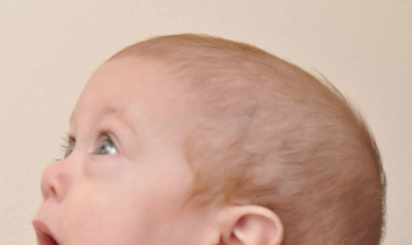
Pierre Robin syndrome is also known as Pierre Robin sequence or Pierre Robin malformation 1). Pierre Robin syndrome is a rare congenital birth defect characterized by a combination of three features: an underdeveloped jaw (micrognathia), backward displacement of the tongue (glossoptosis) and upper airway obstruction. The severity of upper airway obstruction varies, ranging from snoring to life-threatening airway compromise requiring tracheotomy 2). In 80% of the cases of Pierre Robin syndrome, the children also have a U-shaped opening in the roof of their mouths (cleft soft palate). Others may have a high arched palate. While Pierre Robin syndrome is equally common in males and females, there is a higher incidence among twins.

Pierre Robin syndrome was named after Dr. Pierre Robin, a French dental surgeon who first observed its features during the early 20th century.

While the precise cause of Pierre Robin syndrome is not fully clear, the current belief is that multiple contributing factors lead to sequential physical changes within the oral cavity, which ultimately leads to airway obstruction. Pierre Robin syndrome affects approximately 1 per 8,500 to 20,000 neonates 3). The male-to-female ratio is 1:1, except in an X-linked form.

Some people have the features of Pierre Robin sequence as part of a syndrome that affects other organs and tissues in the body, such as Stickler syndrome or campomelic dysplasia. These instances are described as syndromic. When Pierre Robin sequence occurs by itself, it is described as nonsyndromic or isolated. Approximately 20 to 40 percent of cases of Pierre Robin sequence are isolated.

Pierre Robin syndrome is described as a “sequence” because one of its features, underdevelopment of the lower jaw (mandible), sets off a sequence of events before birth that cause the other signs and symptoms. Specifically, having an abnormally small jaw affects placement of the tongue, and the abnormally positioned tongue can block the airways. In addition, underdeveloped jaw (micrognathia) and backward displacement of the tongue (glossoptosis) affect formation of the palate during development before birth, which often leads to cleft palate.

The combination of features characteristic of Pierre Robin syndrome can lead to difficulty breathing and problems eating early in life. As a result, some affected babies have an inability to grow and gain weight at the expected rate (failure to thrive). In some children with Pierre Robin sequence, growth of the mandible catches up, and as adults these individuals have normal-sized chins.

Treatment of Pierre Robin syndrome is multifaceted and individualized, with surgery being performed only to solve the functional problems that a patient may have. Surgical treatments may be indicated for Pierre Robin syndrome patients with more severe clinical conditions, often those associated with airway impairment.

Infants must be kept face down (prone), which allows gravity to pull the tongue forward and keep the airway open. These problems abate over the first few years as the lower jaw grows and assumes a more normal size.

In moderate cases, the patient requires placement of a nasopharyngeal airway (a tube placed through the nose and into the airway) to avoid airway blockage.

In severe cases, surgery is indicated for recurrent upper airway obstruction. A tracheostomy (an operative procedure that creates a surgical airway in the cervical trachea) is sometimes required.

Parents and doctors should continue to monitor child development — particularly jaw and tooth development, growth, and speech.

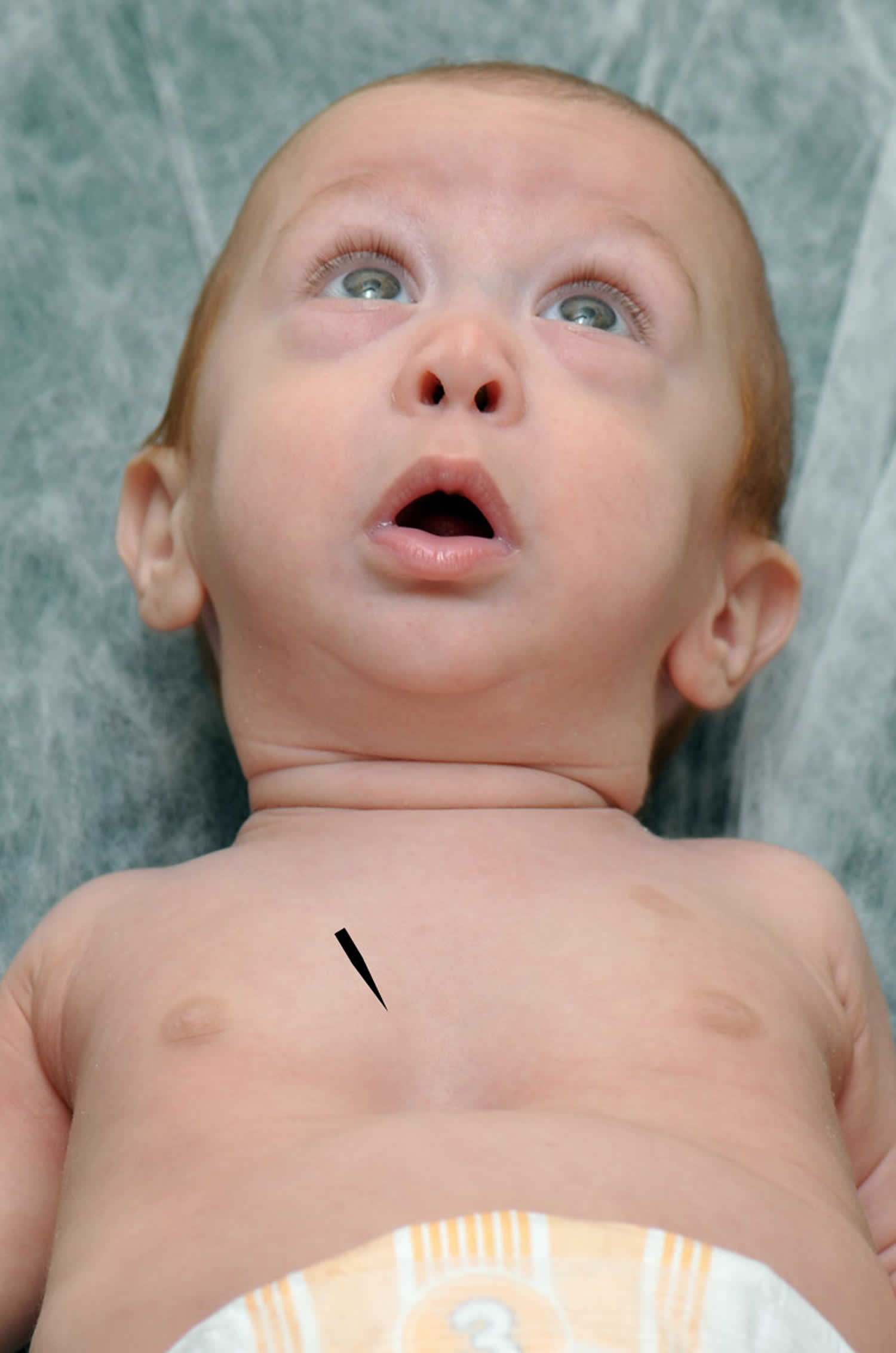
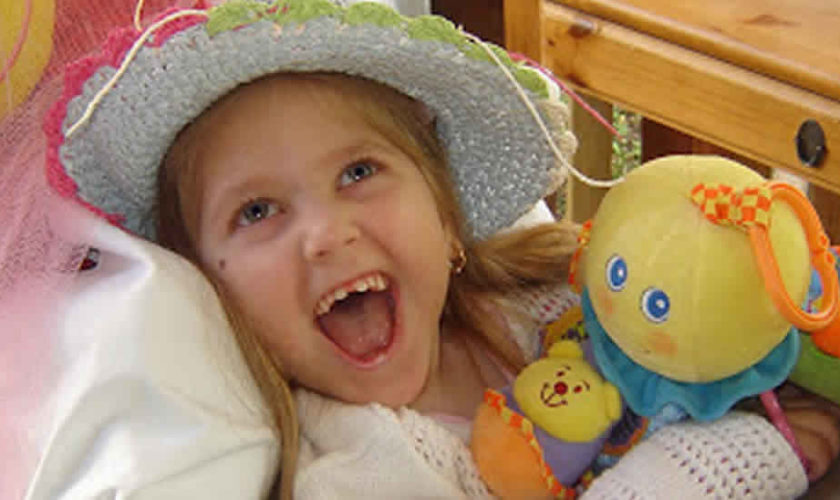
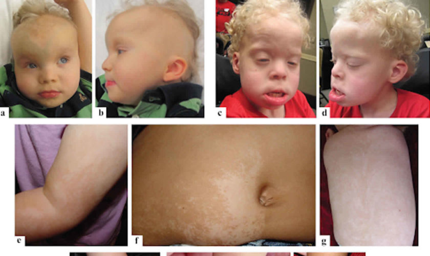
Figure 1. Pierre Robin syndrome (note chest retraction during inhalation due to respiratory distress [arrow])

Pierre Robin syndrome

Why did this happen?

Doctors do no know exactly why Pierre Robin syndrome occurs. They do not believe it is the result of anything the mother did or did not do during pregnancy. If the child only has Pierre Robin syndrome , many experts believe that it is the result of the positioning of the fetus in the early weeks of pregnancy.

Will Pierre Robin syndrome happen to children I have in the future?

Pierre Robin syndrome does not tend to run in families. The chances of you having another child with Pierre Robin syndrome are very small, unless the Pierre Robin Sequence is a part of a syndrome.

What kinds of problems could my child have?

In addition to the physical characteristics common to Pierre Robin syndrome, your child may have the following problems:

  • Feeding problems in infancy
  • Ear infections
  • Reduced hearing
  • About 40% of infants with Pierre Robin have Stickler Syndrome and about 15% have Velocardiofacial Syndrome. Doctors recommends genetic testing be done to determine if your infant has either of these associated syndromes. The Cleft Palate Foundation (https://cleftline.org/) has excellent information concerning genetic testing for babies born with Pierre Robin Sequence.

Will my child need surgery?

Depending on the severity of Pierre Robin syndrome, your child may have some or all of the following surgeries:

  • Surgery to repair the cleft palate
  • Special devices to protect the airway and aid in feeding
  • Surgery to improve breathing
  • The small jaw associated with Pierre Robin syndrome usually grows out on its own during the first two years, and usually no surgery is necessary on the jaw.

Pierre Robin syndrome life expectancy

Since air and food both pass through the mouth and down the throat, breathing and feeding problems are common. Choking and feeding problems may go away spontaneously over the first few years as the lower jaw grows to a more normal size. There is a significant risk of problems if the airway is not protected against obstruction.

Pierre Robin syndrome possible complications

These complications can occur:

  • Breathing difficulties, especially when the child sleeps
  • Choking episodes
  • Congestive heart failure
  • Death
  • Feeding difficulties
  • Low blood oxygen and brain damage (due to difficulty breathing)
  • Type of high blood pressure called pulmonary hypertension

Pierre Robin syndrome causes

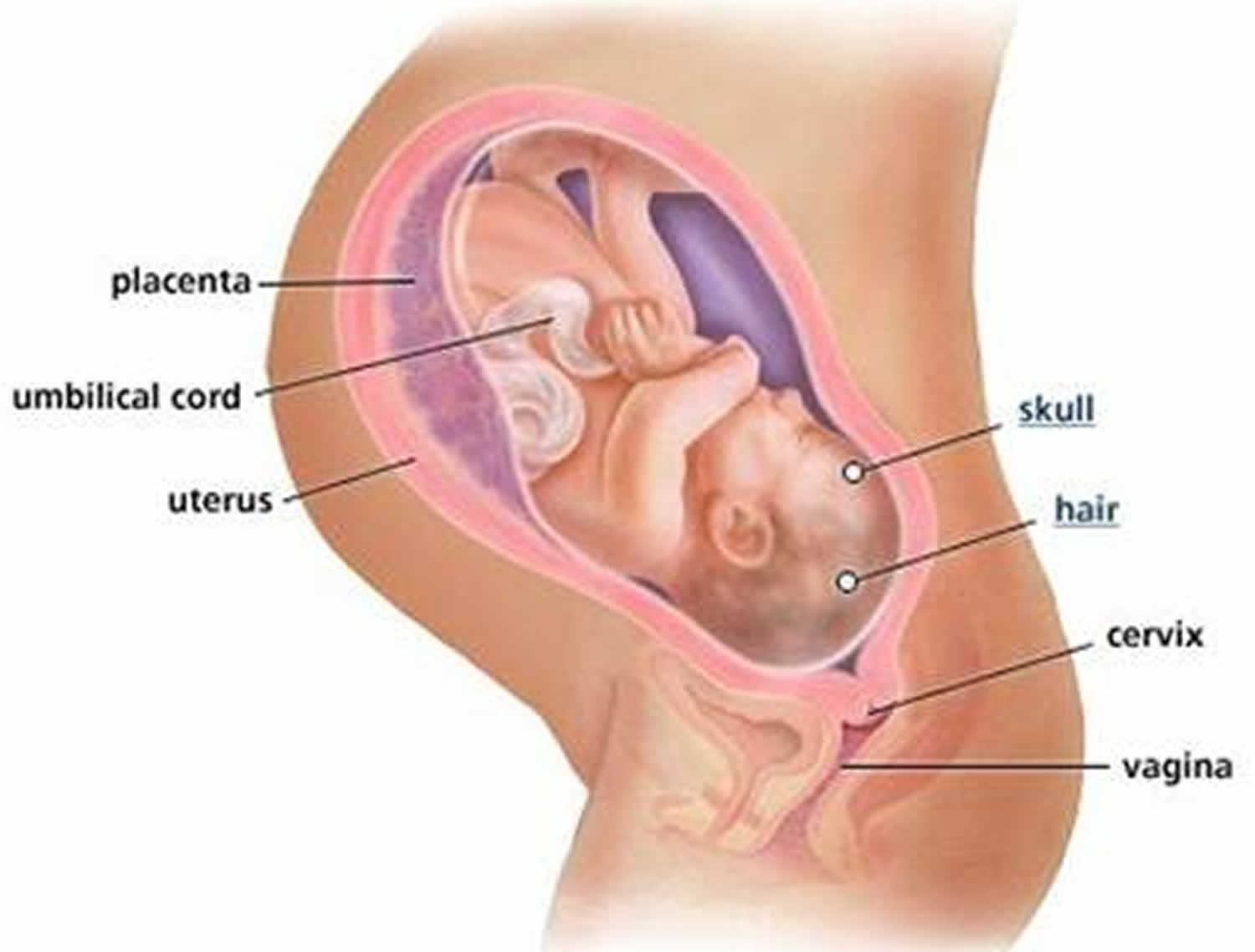
Changes in the DNA near the SOX9 gene located in chromosome 17 (17q24) are the most common genetic cause of isolated Pierre Robin syndrome 4). It is likely that changes in other genes, some of which have not been identified, are also involved in Pierre Robin syndrome. Doctors speculate that nongenetic factors, for example conditions during pregnancy that restrict growth of the jaw, may cause some cases of isolated Pierre Robin syndrome. While some studies point to crowding in the uterus or certain neurological conditions.

The SOX9 gene provides instructions for making a protein that plays a critical role in the formation of many different tissues and organs during embryonic development. The SOX9 protein regulates the activity of other genes, especially those that are important for development of the skeleton, including the mandible.

The genetic changes near the SOX9 gene that are associated with isolated Pierre Robin syndrome are thought to disrupt regions of DNA called enhancers, which normally regulate the activity of the SOX9 gene. These changes reduce SOX9 gene activity. As a result, the SOX9 protein cannot properly control the genes essential for normal development of the lower jaw, causing micrognathia (underdeveloped jaw), and consequently, causes displacement of the tongue (glossoptosis), airway obstruction, and, often, formation of a U-shaped cleft palate.

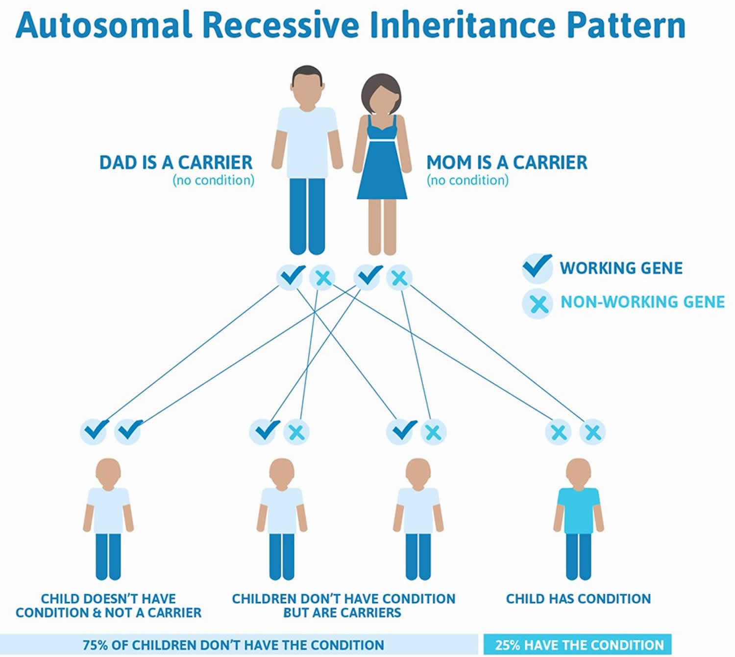
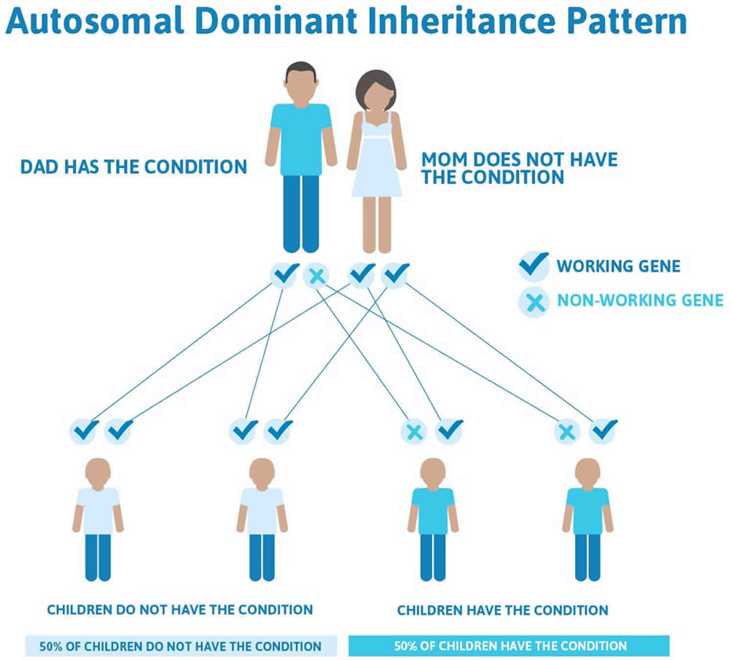
Isolated Pierre Robin sequence is usually not inherited. It typically results from new (de novo) genetic changes and occurs in people with no history of the disorder in their family. When the condition is inherited, it follows an autosomal dominant pattern, which means one copy of the DNA alteration in each cell is sufficient to cause the disorder.

In about 37% of cases, Pierre Robin occurs as part of a syndrome with multiple malformations. Pierre Robin sequence has been reported as occurring in association with Stickler syndrome (20%-25% of these cases), campomelic dysplasia, trisomy 11q syndrome, deletion 4q syndrome, CHARGE association, velocardiofacial syndrome, and Treacher-Collins syndrome 5).

Pierre Robin syndrome symptoms

Babies born with Pierre Robin syndrome commonly experience trouble breathing and feeding early on, resulting from the tongue’s position, smaller jaw size and the cleft palate formation.

Symptoms of Pierre Robin syndrome include:

  • Cleft palate
  • High-arched palate
  • Jaw that is very small with a small chin
  • Jaw that is far back in the throat
  • Repeated ear infections
  • Small opening in the roof of the mouth, which may cause choking or liquids coming back out through the nose
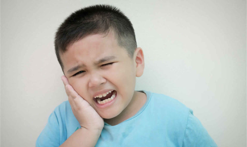
  • Teeth that appear when the baby is born
  • Tongue that is large compared to the jaw
  • Repeated ear infections
  • Natal teeth, or teeth that are present at birth

In Pierre Robin syndrome, the lower jaw (mandible) characteristically has an altered shape and position. Typically, it has a reduced length and is located toward the back (microretrognathia). In turn, these changes in the mandible can influence the tongue’s positioning toward the back of the mouth (a ‘retruded’ tongue). Anatomic anomalies of Pierre Robin syndrome also frequently include a U-shaped cleft palate, which affects the dynamics of breathing and speech development.

Specifically, the displacement of the tongue toward the back (posterior) of the mouth predisposes it to fall toward the throat. This may obstruct the airway and cause difficulty breathing. This can vary in severity, ranging from mild disturbance to life-threatening respiratory distress. Airway obstruction can also occur during the night, in the case of a related condition called ‘obstructive sleep apnea’. This is a sleep disorder characterized by breathing that temporarily stops and restarts because of periodic blockage of the airways.

Since food traveling toward the gastrointestinal tract also passes through the mouth and throat, feeding difficulties can also arise due to abnormal oral cavity anatomy. Depending on the severity, this can lead to issues like choking (aspiration) or gaining less weight gain than expected (which doctors refer to as ‘failure to thrive’). There is also a higher prevalence of acid (gastroesophageal) reflux in children with Pierre Robin syndrome.

Other possible manifestations of Pierre Robin syndrome include cardiovascular and lung conditions, such as heart murmurs, high blood pressure in the arteries of the lungs (pulmonary hypertension), and narrowing of the opening between the lung artery and the right ventricle of the heart (pulmonary stenosis). Anomalies of the musculoskeletal system, including those in the arms, legs, feet, and vertebral column, are also common. Inflammation of the middle ear (otitis media) usually accompanied by repeat ear infections occurs in about 80% of patients, and eye (ocular) defects are noted in about 10% to 30% of patients. Teeth present at birth (natal teeth) are another frequent finding.

Pierre Robin syndrome diagnosis

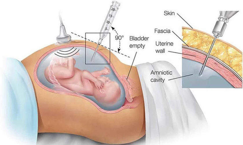
A physical examination is usually sufficient for your health care provider to diagnose Pierre Robin syndrome. A genetics consultation can rule out other associated anomalies and syndromes.

Pierre Robin syndrome treatment

A team of specialists will work together to address affected functions, including breathing, hearing, feeding and sleeping. If your child has Pierre Robin syndrome, you can expect treatment to come in stages. Since the condition affects a variety of functions, including hearing, breathing and feeding, several specialists will be involved in your child’s care.

Breathing

Infants with Pierre Robin syndrome should be observed closely for breathing difficulties.

The first priority will be to keep the upper airway open to allow for proper breathing. Laying your child on his or her stomach (prone position) can help prevent the tongue from falling back toward the throat and blocking off the airway. If placing the infant on his or her stomach does not solve the problem, other treatments aimed at keeping the upper airway open may be recommended. These include a ‘nasopharyngeal airway’ or nasal trumpet (a small tube threaded through the nose into the upper airway).

In cases of severe obstruction, your doctor may recommend surgery to enlarge the lower jaw (so that the tongue can come into the mouth) or a tracheotomy to create an opening in the windpipe to assist the infant in breathing.

Surgery to improve the appearance of the jaw is rarely necessary because the small lower jaw seen at birth most often grows to a more normal size by 18 months of age.

Feeding

Jaw size, tongue placement and cleft palate all contribute to difficulties feeding. Infants with minor degrees of Pierre Robin syndrome can learn to feed using specially adapted nipples and bottles.

However, for babies with more severe Pierre Robin syndrome, the risk of inhaling fluid into the lungs is high. A feeding tube may be recommended as a temporary solution to allow for proper weight gain. While feeding difficulties decrease within the first two years, children that may need long-term assistance could require a gastric tube inserted into the abdominal wall.

Cleft Palate and Hearing Problems

The timing of the cleft palate repair varies depending on the child’s individual growth and development. To close the cleft palate, surgery is typically performed between 12 and 18 months of age. Doctors may postpone the corrective surgery, however, to allow the opening in the palate to close on its own as natural growth occurs.

Cleft palate is repaired with a two- to three-hour surgical procedure and requires a one- to two-night hospital stay. During the procedure, tubes may be inserted into the ear to lessen fluid buildup.

Some children may also require speech therapy following cleft palate repair.

Teeth Problems

Since the lower jaw is smaller in children with Pierre Robin syndrome, teeth crowding is frequently a concern. Orthodontists, pediatric dentists and craniofacial surgeons should work together to monitor dental development.

Symptomatic and supportive treatment

Symptomatic and supportive treatment may be provided using a multidisciplinary team approach, in order to best meet the needs of the affected individual. If speech is impaired, the child should participate in speech therapy or be monitored by a speech pathologist. Ear, nose, and throat doctors (otolaryngologists) and audiologists can provide follow-up on ear- and hearing-related issues. Surgically placed drainage tubes may be recommended if ear infections are recurrent. A combination of orthodontists, maxillofacial surgeons, and dentists may work together to monitor the oral cavity, for example by looking to avoid crowding of the teeth and to ensure proper tooth alignment. Ophthalmology may be consulted to monitor for ocular abnormalities. Genetic counseling may be of benefit for patients and their families.

Pierre Robin syndrome prognosis

All neonates with significant Pierre Robin syndrome are at risk for sudden death. The sudden infant death syndrome (SIDS) data show that the risk of SIDS is increased when infants sleep in the prone position. Neonates with Pierre Robin syndrome already have a compromised airway, and they also typically require prone positioning. Accordingly, monitoring of these neonates should be strongly considered.

Infants with Pierre Robin syndrome deserve to be treated with a multidisciplinary approach that involves a knowledgeable and experienced team capable of providing a comprehensive assessment, a realistic plan of treatment, and appropriate follow-up. Engaging the family in the early stages of the evaluation, the ongoing medical investigations, the issues regarding the child’s care, and future planning generally leads to satisfaction, even in the most difficult of medical scenarios.

In a study of 103 patients followed for a median of 8.6 years (range, 0.1-21.9 years), Logjes et al 6) documented a 10% mortality (n = 10) at a median patient age of 0.8 years (range, 0.1-5.9 years). Of the 10 infants who died, nine had syndromic Pierre Robin syndrome; seven of the nine died of respiratory insufficiency due to various causes, and the other two died of arrhythmia due to hypernatremia and of West syndrome with status epilepticus. The infant with nonsyndromic Pierre Robin syndrome died of brain ischemia after mandibular distraction osteogenesis.

References   [ + ]

read more

Amniocentesis

amniocentesis

What is amniocentesis

Amniocentesis (also called amnio) is a prenatal test that takes amniotic fluid from around your baby in the uterus (also called womb). The amniotic fluid is tested to see if your unborn baby has certain health conditions. A prenatal test is a medical test you get during pregnancy. Amniotic fluid contains cells that are normally shed from the unborn baby (fetus). Samples of these cells are obtained by withdrawing some amniotic fluid. Every cell in the amniotic fluid from the baby contains a complete set of the baby’s DNA. The chromosome analysis of these cells can be performed to determine abnormalities. In addition, the cells may be cultured and analyzed for enzymes, or for other materials that may indicate genetically transmitted diseases. Other studies can be done directly on the amniotic fluid including measurement of alpha-fetoprotein (AFP). Alpha-fetoprotein levels are often higher if your baby has a neural tube defect.

Amniocentesis test may be performed between 15 and 20 weeks of pregnancy to detect certain genetic diseases, chromosomal abnormalities such as Down syndrome, Edwards’ syndrome or Patau’s syndrome and neural tube defects. Amniocentesis test may also be performed at any point after 32 weeks of gestation to evaluate fetal lung maturity when there is an increased risk of or a need for premature delivery. Amniocentesis may also be done when it is suspected that a fetus has an infection or other illness or a blood type incompatibility with the mother and is therefore at risk of developing hemolytic disease.

Amniocentesis gives healthcare professionals direct information about how likely the baby will develop one or more conditions, which may be genetic (inherited) or develop during the pregnancy.

Doctors recommend you rest and avoid physical strain (such as lifting) after amniocentesis. If you experience any complications after the procedure, including abdominal cramping, leakage of fluid, vaginal bleeding, or signs of infection, call your doctor immediately.

The American College of Obstetricians and Gynecologists recommends that all pregnant women should be given the option of having amniocentesis performed. A health practitioner can help a pregnant woman weigh the pros and cons. Some women are at increased risk of birth defects due to their age or family/medical history while others may be advised against having the procedure if they have a history of premature labor, placental problems, or an incompetent cervix, for example. The procedure has some risks associated with it, such as a small chance of miscarriage, and provides information that can have a significant impact of the management of a pregnancy.

There is between a 0.25% and 0.50% risk of miscarriage and a very slight risk of uterine infection (less than .001%) after amniocentesis. In trained hands and under ultrasound guidance, the miscarriage rate may be even lower.

In most cases, your amniocentesis test results will be available within two weeks. Your doctor will explain the results to you and if a problem is diagnosed, give you information about ending the pregnancy or how best to care for your baby after birth.

Talk to your health care provider to see if amniocentesis is right for you.

Key points

  • Amniocentesis is a prenatal test that can diagnose certain birth defects and genetic conditions in your baby.
  • You may want to have an amniocentesis if you’re at increased risk of having a baby with a birth defect or genetic condition.
  • There’s a small risk of having a miscarriage after an amniocentesis. Miscarriage is when a baby dies in the womb before 20 weeks of pregnancy.
  • Having an amniocentesis is your choice. Talk with your partner and your provider to help you decide if an amniocentesis is right for you.
  • Amniocentesis can’t identify all genetic conditions and birth defects.

Amniocentesis test can diagnosis certain birth defects, including:

  • Down’s syndrome
  • Edwards’ syndrome and Patau’s syndrome – conditions that can result in miscarriage, stillbirth or (in babies that survive) severe physical problems and learning disabilities
  • Cystic fibrosis
  • Spina bifida
  • Genetic problems
  • Infection
  • Lung development

It is performed at 14 to 20 weeks.

It may be suggested for couples at higher risk for genetic disorders. It also provides DNA for paternity testing.

While still very much in use, recent advances in testing technology may eventually result in the decline in the use of amniocentesis. For example, there is a newer test called cell-free fetal DNA (cffDNA) that only requires a blood sample from the pregnant woman to screen for certain fetal chromosomal abnormalities, including Down syndrome, Edwards syndrome, and Patau syndrome (trisomy 13), and it can be performed as early as the 10th week of pregnancy. However, at this time, invasive diagnostic tests such as amniocentesis and chorionic villus sampling (CVS) are still needed to confirm the results.

When is amniocentesis done

When you are about 15 to 20 weeks pregnant, your doctor may offer amniocentesis. Amniocentesis is a prenatal test that detects or rules out certain genetic diseases, chromosomal abnormalities and open neural tube defects; after 32 weeks of pregnancy, amniocentesis can be used to evaluate fetal lung maturity; when it is suspected that a fetus has an infection or other illness; serially, about every 14 days, when it is suspected that a pregnant woman has an Rhesus (Rh) or other blood type incompatibility with her fetus. Amniocentesis also assesses lung maturity to see if the fetus can endure an early delivery. You can also find out the baby’s gender.

Doctors generally offer amniocentesis to women with an increased risk of having a baby with particular disorders, including those who:

  • Will be 35 or older when they deliver.
  • Have a close relative with a disorder.
  • Had a previous pregnancy or baby affected by a disorder.
  • Have test results (such as a high or low alpha-fetoprotein count) that may indicate an abnormality.

Doctors also offer amniocentesis to women with pregnancy complications, such as Rh-incompatibility, that necessitate early delivery. There are blood tests and ultrasound tests that can be done earlier in the pregnancy which may avoid the need for amniocentesis at times.

Figure 1. Amniocentesis

amniocentesis procedure

Figure 2. Normal placenta and pregnancy – the placenta attaches to the wall of the uterus (womb) and supplies the baby with food and oxygen through the umbilical cord.

placenta

Why is amniocentesis done?

The American College of Obstetricians and Gynecologists 1) recommends that all pregnant women have the choice of having prenatal tests, like an amniocentesis.

Having an amniocentesis is your choice, even if you’re at risk of having a baby with a birth defect or genetic condition. Talk with your partner, provider and a genetic counselor about your testing options. A genetic counselor is a person who is trained to help you understand about genes, birth defects and other medical conditions that run in families, and how they can affect your health and your baby’s health. You also may want to talk to your religious and spiritual leaders to help you make decisions about testing for birth defects and genetic conditions during pregnancy.

Ask your provider about other prenatal tests and about seeing a provider like a maternal-fetal medicine specialist. This is an obstetrician with education and training to take care of women who have high-risk pregnancies. You can contact the Society for Maternal-Fetal Medicine (https://www.smfm.org/) to find a specialist in your area.

You may want to have an amniocentesis if your baby is at risk for certain conditions like:

  • Birth defects. These are health conditions that are present at birth. Birth defects change the shape or function of one or more parts of the body. They can cause problems in overall health, in how the body develops or in how the body works. Your provider can use amnio to diagnose certain birth defects, like birth defects of the brain and spine called neural tube defects. Examples of neural tube defects are spina bifida and anencephaly. Amnio doesn’t check for every birth defect. For example, it can’t check for certain heart problems or birth defects in a baby’s lip or mouth called cleft lip and palate. Some birth defects are genetic (caused by changes in genes).
  • Genetic and chromosomal conditions. These conditions are caused by changes in genes and chromosomes. A gene is part of your body’s cells that stores instructions for the way your body grows and works. Chromosomes are the structures in cells that hold genes. Genetic conditions include cystic fibrosis (also called CF), sickle cell disease and heart defects. A common chromosomal condition is Down syndrome. Sometimes these conditions are passed from parent to child, and sometimes they happen on their own.

If your baby’s at risk for having these conditions, you may have an amniocentesis at 15 to 20 weeks of pregnancy. It’s not recommended before 15 weeks because it has a higher risk of miscarriage and other complications. Miscarriage is when a baby dies in the womb before 20 weeks of pregnancy.

Later in pregnancy, you may have an amniocentesis to:

  • Check your baby’s lung development (also called fetal lung maturity). Your provider may recommend amniocentesis to check your baby’s amniotic fluid and find out if your baby’s lungs are developed for birth. This kind of amniocentesis is only done if you need to give birth early to help prevent pregnancy complications. It’s usually done between 32 and 39 weeks of pregnancy.
  • Check for infections or other health conditions in your baby. For example, if your baby’s at risk of Rh (Rhesus) incompatibility disease, your provider may use amniocentesis to check your baby for anemia. Rh (Rhesus) disease a dangerous kind of anemia that’s preventable if it’s treated during pregnancy. Anemia is when a person doesn’t have enough healthy red blood cells to carry oxygen to the rest of the body.
  • Treat polyhydramnios (therapeutic amniocentesis). Polyhydramnios is when you have too much amniotic fluid. Amniotic fluid is the fluid that surrounds your baby in the womb. Polyhydramnios may increase your risk of having pregnancy complications, like premature birth (birth before 37 weeks of pregnancy). Your provider can use amnio to drain extra fluid from the womb.

Are you at risk of having a baby with a birth defect or a genetic or chromosomal condition?

You may be at increased risk of having a baby with one of these conditions if:

  • You’re 35 or older. The risk of having a baby with a chromosomal condition, like Down syndrome, increases as you get older.
  • You have a child with a birth defect or you had a previous pregnancy with a birth defect. Having a birth defect in a previous pregnancy makes you more likely to have a birth defect in another pregnancy.
  • You have a family history of a genetic condition. If you, your partner or a member of either of your families has a genetic condition, like cystic fibrosis (CF), fragile X syndrome, sickle cell disease, Tay-Sachs or thalassemia, you may want to have an amniocentesis to find out if your baby also has the condition. You can get carrier screening for genetic conditions before or during early pregnancy. Carrier screening test checks your blood or saliva to see if you’re a carrier of certain genetic conditions that could affect your baby. If you’re a carrier, you don’t have the condition yourself, but you have a gene change for it that you can pass to your baby. If both you and your partner are carriers of the same condition, the risk that your baby has the condition increases.
  • Your prenatal screening tests results are abnormal. There are no risks to you or your baby when you have a screening test, but it doesn’t tell you for sure if your baby has a health condition. A diagnostic test, like amniocentesis, can diagnose a condition. If you have abnormal results from a screening test, like first-trimester screening or cell-free DNA testing, you may want to have a diagnostic test, like amniocentesis.

When is amniocentesis ordered?

While amniocentesis is safe and has been performed for many years, it is an invasive procedure that poses a slight risk of injury to the fetus and of miscarriage. For this reason, it is not performed routinely with each pregnancy.

Genetic amniotic fluid analysis may be offered as part of second trimester prenatal testing and is performed primarily between 15 and 20 weeks gestation if:

  • A woman is 35 years of age or older
  • A woman has an abnormality on a first trimester Down syndrome screen or second trimester maternal serum screen, such as an increased or decreased alpha-feto protein (AFP) level
  • A woman had a previous child or pregnancy with a chromosomal abnormality or birth defect
  • There is a strong family history of a specific genetic disorder
  • A parent has an inherited disorder or both parents have a gene for an inherited disorder
  • An abnormality has been detected on a fetal ultrasound

Fetal lung maturity amniotic fluid testing is ordered when there is a risk of premature delivery, at any time after 32 weeks gestation.

Biochemical testing is sometimes ordered to monitor bilirubin levels when a woman has been sensitized or it is suspected that she has become sensitized (has developed antibodies) to red blood cell antigens and there may be an Rh or other blood type incompatibility with the fetus. In this case, serial testing for bilirubin may be performed, usually about every 14 days.

An amniotic fluid analysis may be performed in late pregnancy to check for fetal distress and to diagnose a fetal infection.

Genetic amniocentesis

Genetic amniocentesis can provide information about your baby’s genetic makeup. Generally, genetic amniocentesis is offered when the test results might have a significant impact on the management of the pregnancy or your desire to continue the pregnancy.

Genetic amniocentesis is usually done between week 15 and 20 of pregnancy. Amniocentesis done before week 15 of pregnancy has been associated with a higher rate of complications.

You might consider genetic amniocentesis if:

  • You had positive results from a prenatal screening test. If the results of a screening test — such as the first trimester screen or prenatal cell-free DNA screening — are positive or worrisome, you might opt for amniocentesis to confirm or rule out a diagnosis.
  • You had a chromosomal condition or a neural tube defect in a previous pregnancy. If a previous pregnancy was affected by conditions such as Down syndrome or a neural tube defect — a serious condition affecting the brain or spinal cord — your health care provider might suggest amniocentesis to confirm or rule out these disorders.
  • You’re 35 or older. Babies born to women 35 and older have a higher risk of chromosomal conditions, such as Down syndrome. Your health care provider might suggest amniocentesis to rule out these conditions.
  • You have a family history of a specific genetic condition, or you or your partner is a known carrier of a genetic condition. In addition to identifying Down syndrome and spina bifida, amniocentesis can be used to diagnose many other conditions — such as cystic fibrosis.
  • You have abnormal ultrasound findings. Your health care provider might recommend amniocentesis to diagnose or rule out genetic conditions associated with abnormal ultrasound findings.

Remember, genetic amniocentesis is typically offered when the test results might have a significant impact on management of the pregnancy. Ultimately, the decision to have genetic amniocentesis is up to you. Your health care provider or genetic counselor can help you weigh all the factors in the decision.

Fetal lung maturity amniocentesis

Fetal lung maturity amniocentesis can determine whether a baby’s lungs are ready for birth. This type of amniocentesis is done only if early delivery — either through induction or C-section — is being considered to prevent pregnancy complications for the mother in a non-emergency situation. It’s usually done between 32 and 39 weeks of pregnancy. Earlier than 32 weeks, a baby’s lungs are unlikely to be fully developed.

Amniocentesis isn’t appropriate for everyone, however. Your health care provider might discourage amniocentesis if you have an infection, such as HIV/AIDS, hepatitis B or hepatitis C. These infections can be transferred to your baby during amniocentesis.

Amniocentesis procedure

Amniocentesis test preparation

You may be instructed to have either a full or empty bladder prior to amniocentesis, depending on when during your pregnancy the testing is being performed; follow any instructions you are given.

If you’re having amniocentesis done before week 20 of pregnancy, it might be helpful to have your bladder full during the procedure to support the uterus. Drink plenty of fluids before your appointment. After 20 weeks of pregnancy, your bladder should be empty during amniocentesis to minimize the chance of puncture.

During the amniocentesis procedure

Here’s what happens when you have an amniocentesis:

  1. You lie on your back on an exam table and expose your abdomen. Your health care provider will apply a special gel to your abdomen and then use a small device known as an ultrasound transducer to show your baby’s position on a monitor.
  2. Your health care provider will use ultrasound to determine the baby’s exact location in your uterus.
  3. Your provider moves an ultrasound wand (also called transducer) across your belly to find your baby and the placenta. The placenta grows in your uterus and supplies your baby with food and oxygen through the umbilical cord. Ultrasound uses sound waves and a computer screen to show a picture of your baby inside the womb.
  4. Your provider cleans your belly with an antibacterial liquid that kills germs on your skin.
  5. Using ultrasound as a guide, your provider puts a thin needle through your belly and uterus into the amniotic sac. The amniotic sac (also called bag of waters) is the sac (bag) inside the uterus that holds your growing baby. It’s filled with amniotic fluid.
  6. Using the ultrasound for guidance, your doctor carefully inserts a long, but thin, hollow needle through your abdomen and into the amniotic sac. Your doctor then extracts about four teaspoons (less than 1 ounce) of amniotic fluid. The amniotic fluid contains fetal cells from your baby. Once the amniotic fluid sample is taken, your provider uses the ultrasound to check that your baby’s heartbeat is healthy.
  7. You should receive Rh (Rhesus) immune globulin (RhIG) at the time of amniocentesis if you are an Rh-negative unsensitized patient.

Your provider sends the amniotic fluid sample to a lab where your baby’s cells are separated from the amniotic fluid. The cells grow for about 10 to 12 days at the lab and then they’re tested for birth defects and genetic conditions. The lab also can test the amniotic fluid for proteins like alpha-fetoprotein (also called AFP). Test results usually are available within 2 to 3 weeks.

If amniocentesis shows that your baby has a health condition, talk to your provider about your options. For example, your baby may be able to be treated with medicines or surgery before or after birth. Knowing about a birth defect before birth may help you get ready to care for your baby. And you can make plans for your baby’s birth with your provider to make sure your baby gets special care or treatment he may need right after he’s born.

After the amniocentesis procedure

After the amniocentesis, your health care provider will continue using the ultrasound to monitor your baby’s heart rate. You might experience cramping or mild pelvic discomfort after an amniocentesis.

You can resume your normal activity level after the procedure. However, you might consider avoiding strenuous exercise and sexual activity for a day or two.

Meanwhile, the sample of amniotic fluid will be analyzed in a lab. Some results might be available within a few days. Other results might take up to four weeks.

Contact your health care provider if you have:

  • Loss of vaginal fluid or vaginal bleeding
  • Severe uterine cramping that lasts more than a few hours
  • Fever
  • Redness and inflammation where the needle was inserted
  • Unusual fetal activity or a lack of fetal movement

Does amniocentesis hurt?

Amniocentesis is done in an examination room, either with or without local anesthesia. It typically takes just a few minutes, during which you must lie very still. Most women have only mild discomfort during an amniocentesis. You may have a stinging feeling when the needle enters your skin, feel cramping when the needle enters the uterus or feel pressure when the fluid is removed. After the test, your provider may tell you to take it easy for the rest of the day and not to exercise or have sex for a day or two.

What does amniocentesis test for?

Amniotic fluid surrounds, protects, and nourishes a growing fetus during pregnancy. Amniotic fluid analysis involves a variety of tests that can be performed to evaluate the health of a fetus.

Amniotic fluid allows a fetus to move relatively freely within the uterus, keeps the umbilical cord from being compressed, and helps maintain a stable temperature. Contained within the amniotic sac, amniotic fluid is normally a clear to pale yellow liquid that contains proteins, nutrients, hormones, and antibodies.

Amniotic fluid begins forming one to two weeks after conception and increases in volume until there is about a quart at 36 weeks of pregnancy. The fluid is absorbed and continually renewed.

The fetus swallows and inhales amniotic fluid and releases urine into it. Cells from various parts of the fetus’s body and chemicals produced by the fetus are present in the amniotic fluid. This allows the fluid to be sampled and tested to evaluate fetal health.

Amniotic fluid contains skin cells shed from the baby that can be used to diagnose chromosomal problems, such as Down syndrome.

Amniotic fluid also contains alpha-fetoprotein (AFP), a substance produced by the baby. Levels of alpha-fetoprotein may also indicate whether a baby has problems affecting the spine or other areas of the body.

Amniocentesis helps in detecting or ruling out Down’s syndrome, which causes intellectual disabilty, congenital heart defects, and physical characteristics such as skin folds near the eyes. Amniocentesis also detects neural tube defects such as spina bifida. Babies born with spina bifida have a backbone that did not close properly. Serious complications of spina bifida can include leg paralysis, bladder and kidney defects, brain swelling (hydrocephalus), and intellectual disability.

If your pregnancy is complicated by a condition such as Rh-incombatibility, your doctor can use amniocentesis to find out if your baby’s lungs are developed enough to endure an early delivery. Many more diagnoses are available through amniocentesis.

For genetic testing and chromosome analysis, fetal cells in the amniotic fluid are cultured and grown for 10-12 days in the laboratory, then are analyzed. Biochemical tests, such as bilirubin and alpha-fetoprotein (AFP), and sometimes genetic tests can be performed directly on the amniotic fluid.

Chromosomal conditions

Chromosomal conditions are conditions that affect the chromosomes (parts of the body’s cells that carry genes). For example:

  • Down syndrome – a condition that affects a person’s physical appearance, mental development and learning ability; it is the result of an extra chromosome, known as trisomy-21
  • Edwards syndrome – a condition that causes severe physical and mental abnormalities; it is the result of an extra chromosome, known as trisomy-18
  • Patow syndrome – a rare but serious condition where babies rarely survive for more than a few days; it is the result of an extra chromosome, known as trisomy-13.

Blood disorders

Amniocentesis can also be used to check for inherited blood disorders, such as:

  • Sickle cell anemia – a condition where red blood cells (which carry oxygen around the body) are an unusual shape and texture
  • Thalassemia – a condition that affects the body’s ability to create red blood cells
  • Hemophilia – a condition that affects the blood’s ability to clot.

Neural tube defects

Amniocentesis can test for neural tube defects. The neural tube is a primitive tissue structure inside which the embryo (fertilized egg) grows during its first month of life. As the embryo develops, the neural tube changes and eventually forms the spine and nervous system.

A neural tube defect can lead to conditions such as spina bifida, which can cause learning difficulties and paralysis (weakness) of the lower limbs.

Musculoskeletal disorders

Amniocentesis can also be used to diagnose conditions that affect the musculoskeletal system (your bones and muscles), such as muscular dystrophy. Muscular dystrophy is an inherited condition that causes muscles to gradually weaken, resulting in an increasing level of disability.
Other genetic conditions

As well as helping diagnose chromosomal conditions, blood disorders, neural tube defects and musculoskeletal disorders, amniocentesis can also help diagnose a number of genetic conditions, such as Marfan syndrome. This condition affects the tissues that provide support.

Amniocentesis accuracy

Amniocentesis is estimated to give a definitive result in 98-99% of cases.

However, amniocentesis can’t test for every birth defect and, in a small number of cases, it’s not possible to get a conclusive result.

For many women who have amniocentesis, the results of the procedure will be “normal”. This means that none of the conditions that were tested for were found in the baby.

However, a normal result doesn’t guarantee that your baby will be completely healthy as the test only checks for conditions caused by faulty genes, and it can’t exclude every condition.

  • An amniocentesis is an accurate way of testing for most chromosome problems. However mosaicism (when some, but not all of the baby’s cells have a chromosomal abnormality) and very small chromosome abnormalities cannot be excluded.
  • Amniocentesis cannot detect all problems with a baby. Having an amniocentesis does not guarantee that your baby will not have a birth defect as most are not caused by abnormalities of the chromosomes.

If your test is “positive”, your baby has one of the conditions they were tested for. In this instance, the implications will be fully discussed with you and you’ll need to decide how to proceed.

What does the amniocentesis test result mean?

Genetic tests, chromosome analysis and testing for birth defects

Women should discuss their test results with their health practitioner and with a genetic counselor.

If a chromosomal abnormality or a genetic disorder is detected, then the baby likely will have the associated condition. However, test results may not predict the condition’s severity or prognosis.

Normal results make it less likely that a fetus has an inherited condition, but all genetic conditions cannot be ruled out. Not every genetic disorder or chromosomal abnormality will be detected with this testing.

If an increased or decreased alpha fetoprotein suggests a structural abnormality, such as an open neural tube defect, then additional testing and imaging may be performed to determine the severity of the condition and the best course of action.

If amniocentesis indicates that your baby has a chromosomal or genetic condition that can’t be treated, you might face wrenching decisions — such as whether to continue the pregnancy. Seek support from your health care team and your loved ones during this difficult time.

Fetal lung maturity

If testing indicates that there are low levels of surfactants, then a fetus’s lungs have not yet matured and measures can be taken to attempt to delay delivery, to promote lung maturity, and – when necessary – to treat the baby as soon as it is born. If the levels of surfactants are deemed high enough, then the baby may be safely delivered without increased risk of complications from lung immaturity.

Rh or other blood type incompatibility

Increasing bilirubin concentrations in a fetus with a fetal-maternal blood type incompatibility indicate increasing destruction of red blood cells (RBCs) and the likelihood that the fetus will be born with hemolytic disease of the newborn, requiring treatment depending on the severity.

Fetal distress or infection

Evaluation of amniotic fluid color:

  • Green-tinged indicates that meconium, the fetus’s first stool, has been released.
  • Yellow to amber may indicate bilirubin in the fluid.
  • Red-tinged indicates blood from the mother or the fetus.

Cultures of the amniotic fluid will indicate whether or not an infection is present.

What happens if a condition is found?

If the test finds that your baby will be born with a condition, you can speak to a number of specialists about what this means.

These could include your midwife, a consultant pediatrician, a geneticist and/or a genetic counselor.

They’ll be able to give you detailed information about the condition – including the possible symptoms your child may have, the treatment and support they might need, and whether their life expectancy will be affected – to help you decide what to do.

A baby born with one of these conditions will always have the condition, so you’ll need to consider your options carefully. Your main options are:

  • continue with your pregnancy while gathering information about the condition, so you’re prepared for caring for your baby
  • have a termination (abortion) – read more about termination for fetal abnormalities

This can be a very difficult decision, but you don’t have to make it on your own.

As well as discussing it with specialist healthcare professionals, talk things over with your partner and speak to close friends and family, if you think it might help.

Is there anything else I should know about amniocentesis test results?

Both blood contamination and stool from the baby (meconium) in the amniotic fluid can affect some chemical test results.

An alternative to amniotic fluid analysis for chromosomal analysis and genetic testing is chorionic villus sampling (CVS), which can be performed earlier, between 10 and 13 weeks of pregnancy. This first trimester procedure collects a placenta tissue sample at the site of implantation and carries about the same risks as amniocentesis. Chorionic villus sampling (CVS) cannot, however, detect neural tube defects.

Performed on a blood sample obtained from the mother, the first trimester screen for Down syndrome and the second trimester screen for Down syndrome and open neural tube defects assess the risk of a fetus having these conditions but are not diagnostic. In most cases, the subsequent amniotic fluid analysis will be normal; only a small percentage of those with an abnormal blood screening test result will actually have an affected baby.

Amniocentesis risks and complications

Serious complications from amniocentesis are rare.

Some women may have other complications from amniocentesis, including:

  • Miscarriage. Less than 1 in 200 women (less than 1 percent) have a miscarriage after an amniocentesis. Research suggests that the risk of pregnancy loss is higher for amniocentesis done before 15 weeks of pregnancy. Second-trimester amniocentesis carries a slight risk of miscarriage — about 0.6 percent.
  • Infection in the uterus. Very rarely, amniocentesis might trigger a uterine infection.
  • Cramping, spotting or leaking amniotic fluid. About 1 to 2 in 100 women (1 to 2 percent) have these problems.
  • Passing infection to your baby. If you have an infection, like HIV/AIDS, hepatitis C, or toxomplasmosis, you may pass it to your baby during amniocentesis. HIV is the virus that causes AIDS. Toxoplasmosis is an infection you can get from eating undercooked meat or touching cat poop.
  • Rhesus (RhD) sensitization. Rarely, amniocentesis might cause a small amount of your baby’s blood to mix with your blood. If you’re Rh-negative and your baby is Rh-positive and you haven’t developed antibodies to Rh positive blood, you may get a shot called Rh immune globulin (RhIG) after amniocentesis to help protect your baby. This will prevent your body from producing Rh antibodies that can cross the placenta and damage the baby’s red blood cells. A blood test can detect if you’ve begun to produce antibodies.
  • Leaking amniotic fluid. Rarely, amniotic fluid leaks through the vagina after amniocentesis. However, in most cases the amount of fluid lost is small and stops within one week, and the pregnancy is likely to proceed normally.
  • Needle injury. During amniocentesis the baby might move an arm or leg into the path of the needle. Serious needle injuries are rare.
  • Club foot. Club foot, also known as talipes, is a congenital (present at birth) deformity of the ankle and foot. Having amniocentesis early (before week 15 of the pregnancy) has been associated with an increased risk of the unborn baby developing club foot. Because of the increased risk of a baby developing club foot, amniocentesis isn’t recommended before 15 weeks of pregnancy.

If you have any of these signs or symptoms after an amniocentesis, call your healthcare provider immediately:

  • Feeling a change in your baby’s movement
  • Bleeding or leaking fluid from your vagina
  • Fever
  • Redness and swelling where your provider inserted the needle
  • Strong belly cramps that last more than a few hours.

Rh (Rhesus) factor

The Rh (Rhesus) blood group was named after the rhesus monkey in which it was first studied. In humans, this group includes several Rh antigens (factors). The most prevalent of these is antigen D, a transmembrane protein.

If the Rh antigens are present on the red blood cell membranes, the blood is said to be Rh-positive. Conversely, if the red blood cells do not have Rh antigens, the blood is called Rh-negative. The presence (or absence) of Rh antigens is an inherited trait. Anti-Rh antibodies (anti-Rh) form only in Rh-negative individuals in response to the presence of red blood cells with Rh antigens. This happens, for example, if an individual with Rh-negative blood receives a transfusion of Rh-positive blood. The Rh antigens stimulate the recipient to begin producing anti-Rh antibodies. Generally, this initial transfusion has no serious consequences, but if an individual with Rh-negative blood—who is now sensitized to Rh-positive blood—receives another transfusion of Rh-positive blood some months later, the donated red cells are likely to agglutinate.

A similar situation of Rh incompatibility arises when an Rh-negative woman is pregnant with an Rh-positive fetus. Her first pregnancy with an Rh-positive fetus would probably be uneventful. However, if at the time of the infant’s birth (or if a miscarriage occurs) the placental membranes that separated the maternal blood from the fetal blood during the pregnancy tear, some of the infant’s Rh-positive blood cells may enter the maternal circulation. These Rh-positive cells may then stimulate the maternal tissues to produce anti-Rh antibodies. If a woman who has already developed anti-Rh antibodies becomes pregnant with a second Rh-positive fetus, these antibodies, called hemolysins, cross the placental membrane and destroy the fetal red blood cells. The fetus then develops a condition called erythroblastosis fetalis, or hemolytic disease of the fetus and newborn.

Erythroblastosis fetalis is extremely rare today because obstetricians carefully track Rh status. An Rh-negative woman who might carry an Rh-positive fetus is given an injection of a drug called RhoGAM at week 28 of her pregnancy and after delivery of an Rh-positive baby. Rhogam is a preparation of anti-Rh antibodies, which bind to and shield any Rh-positive fetal cells that might contact the woman’s cells and sensitize her immune system. RhoGAM must be given within 72 hours of possible contact with Rh-positive cells—including giving birth, terminating a pregnancy, miscarrying, or undergoing amniocentesis (a prenatal test in which a needle is inserted into the uterus).

Amniocentesis vs CVS

CVS (chorionic villus sampling) is a prenatal test carried out during pregnancy to detect specific abnormalities in an unborn baby. During CVS (chorionic villus sampling) a sample of cells is taken from the placenta (the organ that links the mother’s blood supply with her unborn baby’s) and tested for genetic abnormalities. CVS (chorionic villus sampling) is a test you may be offered during pregnancy to check if your baby has a genetic or chromosomal condition, such as Down’s, Edwards’ or Patau’s syndromes.

CVS is different from another prenatal test called amniocentesis. Amniocentesis is performed a little later in pregnancy at around 15 to 20 weeks.

You can get CVS early in pregnancy, between 10 and 13 weeks.

  • Chorionic villus sampling (CVS) cannot, however, detect neural tube defects. These are birth defects affecting the brain and the spinal cord, such as spina bifida, which can usually be detected with an ultrasound scan.
  • Amniocentesis can test for neural tube defects.

CVS isn’t routinely offered to all pregnant women because there’s a small chance of miscarriage after the test. CVS is only offered if there’s a high risk your baby could have a genetic or chromosomal condition.

It’s important to remember that you don’t have to have CVS if it’s offered. It’s up to you to decide whether you want it.

A CVS test done at 10 to 13 weeks to diagnose certain birth defects, including:

  • Chromosomal disorders, including:
    • Down’s syndrome – a condition that typically causes some level of learning disability and a characteristic range of physical features
    • Edwards’ syndrome and Patau’s syndrome – conditions that can result in miscarriage, stillbirth or (in babies that survive) severe physical problems and learning disabilities
  • Genetic disorders, such as:
    • Cystic fibrosis – a condition in which the lungs and digestive system become clogged with thick, sticky mucus
    • Duchenne muscular dystrophy – a condition that causes progressive muscle weakness and disability
    • Thalassaemia – a condition that affects the red blood cells, which can cause anemia, restricted growth and organ damage
    • Sickle-cell disease – where the red blood cells develop abnormally and are unable to carry oxygen around the body properly
    • Phenylketonuria (PKU) – where your body cannot break down a substance called phenylalanine, which can build up to dangerous levels in the brain

CVS may be suggested for couples at higher risk for genetic disorders.

This could be because:

  • an earlier antenatal screening test has suggested there may be a problem, such as Down’s syndrome, Edwards’ syndrome or Patau’s syndrome
  • you’ve had a previous pregnancy with these problems
  • you have a family history of a genetic condition, such as sickle cell disease, thalassaemia, cystic fibrosis or muscular dystrophy, and an abnormality is detected in your baby during a routine ultrasound scan

CVS also provides DNA for paternity testing.

Talk to your doctor about having CVS, amniocentesis or other prenatal tests.

Deciding whether to have CVS

If you’re offered CVS, ask your doctor or midwife what the procedure involves and what the risks and benefits are before deciding whether to have it.

You may also find it helpful to contact a support group, such as Antenatal Results and Choices (https://www.arc-uk.org/). Antenatal Results and Choices is a charity that offers information, advice and support on all issues related to screening during pregnancy.

What are some reasons for having CVS?

The test will usually tell you whether your baby will be born with any of the conditions that were tested for.

Your provider should discuss prenatal testing with you and may offer you CVS. And you can ask to have CVS. You may want to have CVS if you’re at risk for having a baby with a genetic abnormality. These risks include:

  • Being 35 or older: The risk of having a baby with certain birth defects or genetic abnormalities, such as Down syndrome, increases as you get older.
  • Having a previous child or pregnancy with a birth defect: If you had a child or a pregnancy with a birth defect in the past, your provider should offer you testing.
  • Abnormal screening test results: If you had abnormal results from a pregnancy screening test, your provider should discuss CVS with you. CVS can provide specific information to confirm if there is an abnormality in the baby. Most babies with abnormal screening test results don’t have problems and are born healthy.
  • Family history of a genetic health problem: If you or your partner has a certain genetic disease (a health condition that gets passed down to a baby from mom or dad), or a close family member with a disease, such as cystic fibrosis or sickle cell anemia, you may want to have CVS.

If no problem is found, it may be reassuring. A result showing that a condition was detected will give you plenty of time to decide how you want to proceed with your pregnancy.

Reasons not to have CVS

There is a 0.5-1% chance you could have a miscarriage after the procedure. You may feel this risk outweighs the potential benefits of the test.

Some women decide they don’t want to know if there’s a problem with their baby until later on. You may choose to have an alternative test called amniocentesis later in your pregnancy instead, or you might just want to find out when your baby is born.

What are CVS risks or side effects?

CVS does involve a small risk of miscarriage. The American College of Obstetricians and Gynecologists reports that 1 in 100 (1 percent) women has a miscarriage following testing.

However, it’s difficult to determine which miscarriages would have happened anyway, and which are the result of the CVS procedure. Some recent research has suggested that only a very small number of miscarriages that occur after CVS are a direct result of the procedure.

Most miscarriages that happened after CVS occur within three days of the procedure. However, in some cases a miscarriage can occur later than this (up to two weeks afterwards). There’s no evidence to suggest you can do anything during this time to reduce your risk.

The risk of miscarriage after CVS is considered to be similar to that of an alternative test called amniocentesis, which is carried out slightly later in pregnancy.

  • Inadequate sample

In around 1% of procedures, the sample of cells removed may not be suitable for testing. This could be because not enough cells were taken, or because the sample was contaminated with cells from the mother.

If the sample is unsuitable, it may be necessary for the CVS procedure to be carried out again, or to wait a few weeks to have amniocentesis instead.

  • Infection

As with all types of surgical procedures, there’s a risk of infection during or after CVS.

However, severe infection occurs in less than 1 in every 1,000 procedures.

  • Rhesus sensitization

If your blood type is rhesus (RhD) negative, but your baby’s blood type is RhD positive, it’s possible for sensitization to occur during CVS.

This is where some of your baby’s blood enters your bloodstream and your body starts to produce antibodies to attack it. If it’s not treated, this can cause the baby to develop rhesus disease.

If you don’t already know your blood type, a blood test will be carried out before CVS to see if there’s a risk of sensitization. An injection of a medication called anti-D immunoglobulin can be given to stop sensitization occurring, if necessary.

What if you’re not sure about having CVS?

Choosing to have CVS is a personal decision. Talking with genetic counselors and your health care provider may help you make decisions about testing for birth defects during pregnancy.

Ask your doctor about other prenatal test options and how you can find a doctor who is trained and experienced in offering specific tests. Learn as much as you can about any prenatal tests your provider recommends to make the right decisions for you and your baby.

What are the alternatives to CVS?

An alternative to CVS is a test called amniocentesis. This is where a small sample of amniotic fluid (the fluid that surrounds the baby in the womb) is removed for testing.

Amniocentesis usually carried out between the 15th and 20th week of pregnancy, although it can be performed later than this if necessary.

Amniocentesis test has a similar risk of causing a miscarriage, but your pregnancy will be at a more advanced stage before you can get the results, so you’ll have a bit less time to consider your options.

If you’re offered tests to look for a genetic or chromosomal condition in your baby, a specialist involved in carrying out the test will be able to discuss the different options with you, and help you make a decision.

Figure 3. Amniocentesis vs CVS

Amniocentesis-versus-CVS

How is the CVS test done?

The CVS test itself takes about 10 minutes, although the whole consultation may take about 30 to 45 minutes.

A health care provider with expertise in performing CVS takes a tiny piece of tissue from the placenta, which has cells from your unborn baby, to check for problems. The placenta grows with your baby in your uterus (womb). It gives your baby food and oxygen through the umbilical cord.

There are two kinds of CVS:

  1. Testing through the belly (called transabdominal CVS) — Your provider puts a thin needle through your belly into the womb. She then uses the needle to take a small sample of the placenta tissue.
  2. Testing through the cervix (called transcervical CVS) — Your provider places a thin tube through your vagina and cervix (the opening to the uterus that sits at the top of the vagina). The tube gently sucks in a tiny sample of the placenta tissue (see Figure 3 above).

Your healthcare provider sends the tissue sample to a lab where it is examined and tested. Test results are usually ready in about 7 days.

Some women find that CVS is painless. Others feel cramping, similar to period cramps, when the sample is taken. Some women who have testing through the cervix say it feels like having a Pap smear.

After CVS, relax for the rest of the day. You may have spotting or cramping for a few hours after the test. Call your health care provider right away if you have heavy bleeding, fever or contractions.

CVS Test Results

After chorionic villus sampling (CVS) has been carried out, the sample of cells will be sent to a laboratory to be tested.

The number of chromosomes (bundles of genes) in the cells can be counted, and the structure of the chromosomes can be checked for any abnormalities.

If CVS is being carried out to test for a specific genetic disorder, the cells in the sample can also be tested for this.

Getting the results

The first results should be available within three working days, and this will tell you whether a chromosomal condition such as Down’s syndrome, Edwards’ syndrome or Patau’s syndrome has been found.

If rarer conditions are also being tested for, it can take two to three weeks or more for the results to come back.

You can usually choose whether to get the results over the phone or during a face-to-face meeting at the hospital or at home. You’ll also receive written confirmation of the results.

How reliable are the CVS test results?

CVS is estimated to give a definitive result in around 99% of cases. However, it cannot test for every birth defect and it’s not always possible to get a conclusive result.

In a very small number of cases, the results of CVS cannot establish with certainty that the chromosomes in the baby are normal or not. This might be because the sample of cells removed was too small or there’s a possibility the abnormality is just in the placenta and not the baby.

If this happens, it may be necessary to have amniocentesis (an alternative test, in which a sample of amniotic fluid is taken from the mother) a few weeks later to confirm a diagnosis.

What the CVS test results mean

For many women who have CVS, the results of the procedure will be “normal”. This means that none of the conditions that were tested for were found in the baby.

However, a normal result doesn’t guarantee that your baby will be completely healthy, as the test only checks for conditions caused by faulty genes, and it cannot exclude every possible condition.

If your test is “positive”, your baby has one of the conditions they were tested for. In this instance, the implications will be fully discussed with you and you’ll need to decide how to proceed.

What happens if a condition is found

If the test shows that your baby does have a birth defect, you can speak to a number of specialists about what this means. These could include your midwife, a consultant pediatrician, a geneticist and/or a genetic counselor.

They’ll be able to give you detailed information about the condition – including the possible symptoms your child may have, the treatment and support they might need, and whether their life expectancy will be affected – to help you decide what to do.

A baby born with one of these conditions will always have the condition, so you’ll need to consider your options carefully. Your main options are:

  • continue with your pregnancy while gathering information about the condition, so you’re prepared for caring for your baby
  • have a termination (abortion) – read more about termination for fetal abnormalities

This can be a very difficult decision, but you don’t have to make it on your own.

As well as discussing it with specialist healthcare professionals, talk things over with your partner and speak to close friends and family, if you think it might help.

Your baby may be able to be treated with medicines or even surgery before birth. Or there may be treatments or surgery he can have after birth.

Knowing about a birth defect before birth may help you get ready emotionally to care for your baby. You also can plan your baby’s birth with your health care provider. This way, your baby can get any special care she needs right after she is born.

References   [ + ]

read more

Aicardi Goutieres syndrome

aicardi goutieres syndrome

Aicardi Goutieres syndrome

Aicardi-Goutieres syndrome also called familial infantile encephalopathy with calcification of basal ganglia and chronic cerebrospinal fluid lymphocytosis, is an inherited disorder that mainly affects the brain, the immune system, and the skin 1). Most newborns with Aicardi-Goutieres syndrome do not show any signs or symptoms of the disorder. However, about 20 percent are born with a combination of features that include an enlarged liver and spleen (hepatosplenomegaly), elevated blood levels of liver enzymes, a shortage of blood cells called platelets that are needed for normal blood clotting (thrombocytopenia), and neurological abnormalities. While this combination of signs and symptoms is typically associated with the immune system’s response to a viral infection that is present at birth (congenital), no actual infection is found in these infants. For this reason, Aicardi-Goutieres syndrome is sometimes referred to as a “mimic of congenital infection.” Aicardi-Goutieres syndrome is a rare disorder. Its exact prevalence is unknown.

There are several types of Aicardi-Goutieres syndrome, depending on the gene that causes the condition: TREX1, RNASEH2A, RNASEH2B, RNASEH2C, SAMHD1, ADAR and IFIH1, genes 2). Most Aicardi-Goutieres syndrome cases are inherited in an autosomal recessive pattern, although rare autosomal dominant cases have been reported. Treatment of Aicardi-Goutieres syndrome is symptomatic and supportive. This means that you can treat the symptoms, but there is no cure for the disease 3). The prognosis depends mainly on the severity neurologic problems and in the age of onset of these problems 4).

Loss of white matter in the brain (leukodystrophy) and abnormal deposits of calcium (calcification) in the brain leads to an early-onset severe brain dysfunction (encephalopathy) that usually results in severe intellectual and physical disability 5). Additional symptoms may include epilepsy, painful, itchy skin lesion (chilblains), vision problems, and joint and muscle stiffness (spasticity), involuntary muscle twisting and contractions (dystonia), and weak muscle tone (hypotonia) in the torso. Other signs and symptoms may include a very small head (microcephaly), presence of white blood cells and other sign of inflammation in the cerebrospinal fluid, which is the fluid that surrounds the brain and spinal cord (central nervous system). Symptoms usually progress over several months before the disease course stabilizes.

Within the first year of life, most individuals with Aicardi-Goutieres syndrome experience an episode of severe brain dysfunction (encephalopathy), typically lasting for several months. During this encephalopathic phase of the disorder, affected babies are usually extremely irritable and do not feed well. They may develop intermittent fevers in the absence of infection (sterile pyrexias) and may have seizures. They stop developing new skills and begin losing skills they had already acquired (developmental regression). Growth of the brain and skull slows down, resulting in an abnormally small head size (microcephaly). In this phase of the disorder, white blood cells and other immune system molecules associated with inflammation can be detected in the cerebrospinal fluid, which is the fluid that surrounds the brain and spinal cord (central nervous system). These abnormal findings are consistent with inflammation and tissue damage in the central nervous system.

The encephalopathic phase of Aicardi-Goutieres syndrome causes permanent neurological damage that is usually severe. Medical imaging reveals loss of white matter in the brain (leukodystrophy). White matter consists of nerve fibers covered by myelin, which is a substance that protects nerves and insures rapid transmission of nerve impulses. Affected individuals also have abnormal deposits of calcium (calcification) in the brain. As a result of this neurological damage, most people with Aicardi-Goutieres syndrome have profound intellectual disability. They also have muscle stiffness (spasticity); involuntary tensing of various muscles (dystonia), especially those in the arms; and weak muscle tone (hypotonia) in the torso.

Some people with Aicardi-Goutieres syndrome have features characteristic of autoimmune disorders, which occur when the immune system malfunctions and attacks the body’s own systems and organs. Some of these features overlap with those of another disorder called systemic lupus erythematosus (SLE). A feature of SLE that also occurs in about 40 percent of people with Aicardi-Goutieres syndrome is a skin problem called chilblains. Chilblains are painful, itchy skin lesions that are puffy and red, and usually appear on the fingers, toes, and ears. They are caused by inflammation of small blood vessels, and may be brought on or made worse by exposure to cold. Vision problems, joint stiffness, and mouth ulcers are other features that can occur in both disorders.

As a result of the severe neurological problems usually associated with Aicardi-Goutieres syndrome, most people with this disorder do not survive past childhood. However, some affected individuals who develop the condition later or have milder neurological problems live into adulthood.

Figure 1. Aicardi Goutieres syndrome chilblains

Aicardi Goutieres syndrome chilblains

Footnote: (A) Blue toes in a child with AicardieGoutières syndrome caused by RNASEH2B mutations. (B) Less severe—and more typical—chilblain-like lesion on the little toe of a child with RNASEH2C-associated disease.

[Source 6) ]

Aicardi Goutieres syndrome causes

Mutations in several genes can cause Aicardi-Goutieres syndrome. Several of these genes, the TREX1, RNASEH2A, RNASEH2B, and RNASEH2C genes, provide instructions for making nucleases, which are enzymes that help break down molecules of DNA and its chemical cousin RNA when they are no longer needed. These DNA and RNA molecules or fragments may be generated during the first stage of protein production (transcription), copying (replication) of cells’ genetic material in preparation for cell division, DNA repair, cell death (apoptosis), and other processes. Mutations in any of these genes are believed to result the absence or abnormal functioning of the respective nuclease enzyme. Researchers suggest that absent or impaired enzyme function may result in the accumulation of unneeded DNA and RNA in cells. The unneeded DNA and RNA may be mistaken by cells for the genetic material of viral invaders, triggering immune system reactions in multiple body systems that result in encephalopathy, skin lesions, and other signs and symptoms of Aicardi-Goutieres syndrome.

Mutations in other genes, including the SAMHD1, IFIH1, and ADAR genes, can also cause Aicardi-Goutieres syndrome. These genes provide instructions for making proteins that are involved in the immune system. Mutations in these genes cause inappropriate activation of the body’s immune response, resulting in inflammatory damage to the brain, skin, and other body systems that lead to the characteristic features of Aicardi-Goutieres syndrome.

Aicardi Goutieres syndrome inheritance pattern

Aicardi-Goutieres syndrome can have different inheritance patterns. In most cases caused by mutations in the ADAR, TREX1, RNASEH2A, RNASEH2B, RNASEH2C, and SAMHD1 genes, it is inherited in an autosomal recessive pattern, which means both copies of the gene in each cell have mutations. The parents of an individual with an autosomal recessive condition each carry one copy of the mutated gene, but they typically do not show signs and symptoms of the condition.

When caused by mutations in the IFIH1 gene or by certain severe mutations in the TREX1 or ADAR gene, Aicardi-Goutieres syndrome is inherited in an autosomal dominant pattern, which means one copy of the altered gene in each cell is sufficient to cause the disorder. These cases result from new mutations in the gene and occur in people with no history of the disorder in their family.

Figure 2. Aicardi-Goutieres syndrome autosomal recessive inheritance pattern

Aicardi-Goutieres syndrome autosomal recessive inheritance pattern

Figure 3. Aicardi-Goutieres syndrome autosomal dominant inheritance pattern

Aicardi-Goutieres syndrome autosomal dominant inheritance pattern

People with specific questions about genetic risks or genetic testing for themselves or family members should speak with a genetics professional.

Resources for locating a genetics professional in your community are available online:

Aicardi Goutieres syndrome symptoms

In its most characteristic form, Aicardi-Goutieres syndrome can be considered an early-onset encephalopathy associated with significant intellectual and physical disability. Symptoms of Aicardi-Goutieres syndrome usually appear within the first six months of life. Aicardi-Goutieres syndrome is generally either fatal or else it results in a persistent vegetative state in early childhood. Generally, the first symptoms observed are vomiting, feeding difficulties, and lack of progress in motor and social skills. A subset of patients has a later onset of disease, which usually occurs between six-twelve months of age, and is marked by loss of previously acquired motor skills and spasticity. The course of the disease is severe and progressive; death occurs in 25% of patients before 17 years of age. However, in some cases, there can be less impairment, and some retention of contact with surroundings and social interactions. Below is a list of symptoms that may be present for Aicardi-Goutieres syndrome, along with definitions as necessary. Please note that all of these symptoms are not present in all cases.

  • Microcephaly: abnormally small head
  • Early progressive encephalopathy: abnormalities of the brain
  • Lack of progress of motor and social skills; no or very poor contact with surroundings
  • Feeding difficulties
  • Irritability
  • Vomiting
  • Spasticity: presence of spasms
  • Dystonia: Abnormal muscle tone, characterized by prolonged, repetitive muscle contractions that may cause twisting or jerking movements of the body or a body part.
  • Visual Inattention
  • Ocular jerks: abnormal eye movements
  • Sterile CSF lymphocytosis: cerebrospinal fluid that has elevated levels of lymphocytes (a certain cell of the immune system), but in which there are no indications of infection (sterility)
  • Skin lesions of the toes, fingers, ear lobes looking like chilblains (itchy red swelling of the skin), puffy hands and feet, and cold feet
  • Intracerebral calcification: presence of calcium deposits on a particular area of the brain.

Pregnancy, delivery, and the neonatal period are normal in approximately 80% of infants with Aicardi-Goutieres syndrome 7). However, brain calcifications can be identified in utero 8) and 20% of cases, mainly those caused by biallelic pathogenic variants in TREX1, present at birth with abnormal neurologic findings, hepatosplenomegaly, elevated liver enzymes, and thrombocytopenia, a picture reminiscent of congenital infection.

All other affected infants present at variable times after the first few weeks of life, frequently after a period of apparently normal development. The majority of these later-presenting infants exhibit subacute onset of a severe encephalopathy characterized by extreme irritability, intermittent sterile pyrexias, loss of skills, and slowing of head growth. The encephalopathic phase usually lasts several months. The opinion of most pediatricians caring for such children is that the disease does not progress beyond the encephalopathic period; occasionally, however, affected individuals do appear to show progression and/or episodes of regression. Death is usually considered to be secondary to the neurologic damage incurred during the initial disease episode, not to further regression. Several affected individuals older than age 30 years show no obvious signs of disease progression.

Neurologic features. Typically, affected individuals have peripheral spasticity, dystonic posturing (particularly of the upper limbs), truncal hypotonia, and poor head control. Seizures are reported in up to half of affected children, but are usually relatively easily controlled 9). A number of children demonstrate a marked startle reaction to sudden noise, and the differentiation from epilepsy can be difficult. Most affected individuals have severe intellectual and physical impairment. Variability in the severity of the neurologic outcome can be observed among sibs. Most affected children exhibit a severe acquired microcephaly; in children with preserved intellect head circumference can be normal.

Hearing is almost always normal.

Ophthalmology. Visual function varies from normal to cortical blindness. Ocular structures are almost invariably normal on examination. However, there is a risk of congenital glaucoma or later-onset glaucoma 10).

Skin lesions. As many as 40% of affected individuals 11) have skin lesions with chilblains on the fingers and toes and sometimes the ears and other pressure points (e.g., elbows) 12). The cutaneous lesions may be complicated by periungual infection and necrosis.

Other

  • Intracranial large-vessel disease. An additional previously undescribed feature of Aicardi-Goutieres syndrome, which so far appears to be almost exclusively related to biallelic pathogenic variants in SAMHD1, is intracranial large-vessel disease causing both intracranial stenoses (in some cases reminiscent of moyamoya syndrome) and aneurysms 13).
  • Refractory four-limb dystonia. Several individuals with ADAR pathogenic variants have been reported to demonstrate an acute or subacute onset of refractory four-limb dystonia starting between age eight months and five years 14).
    • Individuals can be developmentally normal at initial presentation.
    • This phenotype can occur due to either biallelic pathogenic variants in ADAR or the autosomal dominant pathogenic variant p.Gly1007Arg in ADAR.
    • Like other Aicardi-Goutieres syndrome-related phenotypes, bilateral striatal necrosis due to biallelic pathogenic variants in ADAR or the recurrent dominant pathogenic p.Gly1007Arg variant in ADAR1 are typically associated with an upregulation of ISGs.

Infrequent features seen in a cohort of 123 individuals with molecularly confirmed Aicardi-Goutieres syndrome

  • Scoliosis
  • Cardiomegaly
  • Abnormal antibody profile
  • Preserved language
  • Demyelinating peripheral neuropathy
  • Congenital glaucoma
  • Micropenis
  • Hypothyroidism
  • Insulin-dependent diabetes mellitus
  • Transitory deficiency of antidiuretic hormone

Aicardi Goutieres syndrome diagnosis

Aicardi-Goutieres syndrome is difficult to diagnose, as many of the symptoms overlap with other disorders. In its most characteristic form, Aicardi-Goutières syndrome can be considered an early-onset encephalopathy associated with significant intellectual and physical disability. The clinical symptoms of the disease are taken into consideration, as well as certain brain abnormalities seen by MRI. A sample of the cerebrospinal fluid (CSF) will be taken from a spinal tap. This fluid can then be tested for increased levels of a certain type of cell of the immune system (lymphocytes), a condition known as chronic lymphocytosis. These cells are normally only elevated during infection, so the combination of lymphocytosis combined with a lack of evidence of infection can support a diagnosis of Aicardi-Goutieres syndrome. The CSF may also be tested for elevated levels of a molecule known as interferon-gamma, which can also be suggestive of this disease.

Suggestive findings

Aicardi-Goutieres syndrome should be suspected in individuals with the following clinical, neuroimaging, and supportive laboratory findings 15).

Clinical features

  • Encephalopathy and/or significant intellectual disability
  • Acquired microcephaly during the first year of life
  • Dystonia and spasticity
  • Sterile pyrexias
  • Hepatosplenomegaly
  • Chilblain lesions on the feet, hands, ears, and sometimes more generalized mottling of the skin.

Exclusion criteria include the following:

  • Evidence of prenatal/perinatal infection including, but not limited to, CMV, toxoplasmosis, rubella, herpes simplex, Zika, and HIV
  • Evidence of a known other metabolic disorder or neurodegenerative disorder

Neuroimaging

  • Calcification (best visualized on CT scan) of the basal ganglia, particularly the putamen, globus pallidus and thalamus but also extending into the white matter, sometimes in a para- (rather than true peri-) ventricular distribution 16). See Figure 4.
    • Note: Intracranial calcification is not always recognized on MRI, the initial imaging modality employed in most units.
  • White matter changes, particularly affecting the frontotemporal regions with (in severe cases) temporal lobe cyst-like formation. See Figure 5. On MRI, appears on T2-weighted images as a hyperintense signal most commonly located around the horns of the ventricles.
  • Cerebral atrophy, which may be progressive and involve the periventricular white matter and sulci. Cerebellar atrophy and brain stem atrophy may also be prominen 17).
  • Bilateral striatal necrosis
  • Intracerebral vasculopathy including intracranial stenosis, moyamoya, and aneurysms

Figure 4. Cerebral calcifications on CT scan in individuals with Aicardi-Goutieres syndrome

erebral calcifications on CT scan in individuals with Aicardi-Goutieres syndrome

Footnote: Cerebral calcifications. (A) Axial nonenhanced CT image shows numerous punctuate calcifications within the basal ganglia and the cerebral white matter, a pattern typical in patients with Aicardi-Goutieres syndrome. (B) Contrast-enhanced CT scan shows large calcifications in the white matter. Although the CT examination was performed in the acute phase of the disease, no signs of contrast enhancement are seen. Atrophy, microcephaly, and areas of hypoattenuation in the periventricular white matter are also evident.

[Source 18) ]

Figure 5. MRI characteristics in Aicardi-Goutieres syndrome

MRI characteristics in Aicardi-goutieres syndrome

Footnote: MRI obtained in 2 patients with Aicardi-Goutieres syndrome at ages 4 months (upper row) and 11 months (lower row) in A and the second patient with images at 2 and 17 months in B. The images demonstrate early frontal and temporal lobe swelling (thin white arrow) that gives way to severe frontal and temporal lobe atrophy (thick white arrows), as well as the prominent global atrophy. Note also the T 2 dark signal abnormalities presumed to be calcifications present at 2 months and present at 17 months in the second patient (dashed arrows).

[Source 19) ]

Supportive laboratory findings

Peripheral blood

  • Positive interferon signature identified using quantitative PCR analysis of RNA/cDNA 20)
  • Elevated liver enzymes
  • Thrombocytopenia

Cerebrospinal fluid (CSF)

  • Chronic CSF leukocytosis, defined as more than five lymphocytes/mm³ CSF.
    • Typical values range from five to 100 lymphocytes/mm³ 21).
    • A decrease in the number of lymphocytes occurs with time, although high cell counts may persist for several years.
    • A normal cell count can be observed in the presence of elevated concentrations of IFN-α in the CSF even at an early stage of the disease 22).
  • Increased interferon-alpha (IFN-α) activity in the CSF (normal: <2 IU/mL)
    • Recorded IFN-α activity is usually highest in the early stages of the disease. The IFN-α CSF activity can normalize over the first three to four years of life 23).
    • Recorded IFN-α activity is usually higher in CSF than in blood, where it may be normal.
    • High IFN-α activity has been identified in fetal blood at 26 weeks’ gestation 24).
  • Increased concentration of neopterin in the CSF 25)
    • Levels are highest in the early stages of the disease and can normalize over time.
    • Levels of the neurotransmitter metabolites 5HIAA, HVA, and 5MTHF are normal.

Is prenatal diagnosis possible?

Since Aicardi-Goutieres syndrome is, with rare exceptions, inherited as an autosomal recessive trait, most couples with an affected child have a 25% risk of the disease recurring in each future pregnancy. There exist rare cases in which it has been found to be inherited as an autosomal dominant trait (heterozygous TREX1, ADAR1 and IFIH1 mutations); in the majority of cases, these seem to be de novo mutations (except in rare families with mutations in ADAR1 and IFIH1 in which the mutation was transmitted by asymptomatic family members. These are real exceptions, to date found only for these two genes).

Today, genetic tests allow doctors to obtain a definite diagnosis of Aicardi-Goutieres syndrome in a large proportion of cases: however, it is important to note that diagnostic prenatal testing in at-risk pregnancies is possible only in families that already have an affected child, in whom the disease-causing gene has been identified. If the affected child’s mutation is known, then, in the event of a further pregnancy, the DNA of foetal cells obtained by chorionic villus sampling (at 10-12 weeks of gestation) or by amniocentesis (15-18 weeks of gestation) can be examined for the presence of the same mutation.

Establishing the diagnosis

The diagnosis of Aicardi-Goutieres syndrome is established in a proband with typical clinical findings and characteristic abnormalities on cranial CT (calcification of the basal ganglia and white matter) and MRI (leukodystrophic changes) and/or by the identification of biallelic pathogenic variants in ADAR, RNASEH2A, RNASEH2B, RNASEH2C, SAMHD1, or TREX1; specific heterozygous autosomal dominant pathogenic variants in TREX1 and ADAR; or a variety of heterozygous autosomal dominant pathogenic variants in IFIH1.

Aicardi Goutieres syndrome treatment

Treatment of Aicardi-Goutieres syndrome is currently only symptomatic and supportive. Chest physiotherapy and treatment of respiratory complications; attention to diet and feeding methods to assure adequate caloric intake and avoid aspiration; management of seizures using drugs to control epilepsy.

The use of botulinum toxin and myorelaxant drugs to treat spasticity should be evaluated on a case by case basis in the context of the individual’s overall rehabilitation and care programme.

As regards the chilblain lesions, neither vasodilators nor immunosuppressants have been reported to show any real therapeutic efficacy in Aicardi-Goutieres syndrome and the treatment of these symptoms is limited to protecting the vulnerable parts from the cold and preventing infections that could complicate the situation.

Blau, in 2003, suggested that oral treatment with folinic acid might produce a general improvement of the clinical conditions in Aicardi-Goutieres syndrome patients presenting reduced folates in the CSF, but there are, as yet, no data in the literature confirming this hypothesis. In addition, a neurosurgical treatment to address the vascular complications (occlusive arterial and aneurysmal problems) has been proposed for patients with mutations in SAMHD1.

Surveillance: Patients must be regularly screened for the symptoms of the disease which are treatable, such as glaucoma or endocrine problems (e.g. diabetes or hypothyroidism). Monitoring for signs of diabetes insipidus in the neonatal period; repeat ophthalmologic examinations at least for the first few years of life to evaluate for evidence of glaucoma; monitoring for evidence of scoliosis, insulin-dependent diabetes mellitus, and hypothyroidism.

New therapies

Given the involvement and activation of the immune system in the pathogenesis of Aicardi-Goutieres syndrome, treatment with immunomodulatory therapies (prednisone + azathioprine, intravenous methylprednisolone + intravenous immunoglobulin (IVIG), methylprednisolone or IVIG alone) has been hypothesised and in some cases proposed. To date, however, these treatments have been used in an empirical manner and in a limited number of patients, and have not been clearly shown to modify the evolution of the disease, even in the rare cases treated in the early stages. It is, in fact, difficult to evaluate the efficacy of these treatments, considering the small number of patients involved and the differences between them, both in genotype and in disease stage at the time of treatment.

The progress in the understanding of the molecular mechanisms underlying the pathogenesis of Aicardi-Goutieres syndrome has led various authors to envisage new therapeutic strategies:

1) Biotech drugs

Given the suggested primary role of interferon in the pathogenesis of Aicardi-Goutieres syndrome, a treatment has been proposed based on blocking of interferon (IFN) activity through the use of monoclonal antibodies selective for different subtypes of IFN and for the type I IFN receptor (alpha and beta subunits). To date, this therapy has been used only on an experimental basis in SLE, and no data are available for Aicardi-Goutieres syndrome.

2) Immunomodulatory drugs

On the basis of the role of immune system dysregulation in the pathogenesis of Aicardi-Goutieres syndrome, it has been suggested that treatment could be based on the use of substances capable of destroying B cells (such as the monoclonal antibody rituximab) or of inhibiting autoreactive T cells (such as mycophenolate mofetil). However, these substances, often approved for paediatric use, can cause serious side effects and they have yet to be applied in therapeutic trials in the field of Aicardi-Goutieres syndrome.

3) Reverse transcriptase inhibitors

In view of the role of reverse transcriptase in the accumulation of DNA originating from endogenous RNA fragments (retroelements), normally metabolised by the proteins encoded by the genes that are mutated in Aicardi-Goutieres syndrome, it has been suggested that there might be a role in treatment of Aicardi-Goutieres syndrome for antiretroviral drugs (reverse transcriptase inhibitors, i.e. substances capable of interrupting the replication cycle both of retroviruses and of endogenous retroelements). The pharmacodynamics, safety and toxicity of these therapies are already well known, given that they are commonly prescribed to adults and children infected with HIV-1. At present, the only available data refer to an experimental trial in an animal model: Beck-Engeser showed that neurologically normal mice with the lethal TREX1-null phenotype could be kept alive using a combination of antiretroviral agents. Crow et al. have thus suggested that the next step could be to conduct a trial using drugs of this kind (even though their effectiveness could be limited by their difficulty crossing the blood-brain barrier) in order to test their applicability and safety in patients with Aicardi-Goutieres syndrome, and to assess whether this therapeutic approach is capable of “lowering” the “interferon signature” in those subjects in whom it can be considered, as we have seen, a biomarker of the disease, present even many years after the diagnosis.

It is indeed important to underline that the identification of an appropriate therapeutic strategy demands, in addition to a homogeneous sample of subjects, the use of easily quantifiable parameters of outcome (response to treatment), such as the interferon signature.

These new drugs, if applied in the early stage of the disease, could lead to an attenuation of the inflammatory process and thus a lessening of the tissue damage; instead, if applied in patients who are in an advanced stage of the disease and already show marked neurological impairment, they could result in some improvement of the clinical manifestations associated with the immune system dysregulation (e.g. chilblains). At present, there is no proof of the effectiveness of these possible therapeutic approaches, since the first pilot clinical trial is ongoing and has not yet provided results.

References   [ + ]

read more

Kids toothache

kids toothache

Kids toothache

Toothaches also called pulpitis, are caused by an inflammation of the pulp inside the tooth. The pulp is part of the inside of the tooth that has tissue and nerves (see Figure 1). A toothache is usually from dental caries, or a tooth cavity. This is often a result of poor dental hygiene.

Toothaches key points

  • Pulpitis is inflammation of the dental pulp due to deep cavities, trauma, or extensive dental repair.
  • Sometimes infection develops (eg, periapical abscess, cellulitis, osteomyelitis).
  • Toothaches may be reversible or irreversible.
  • In reversible pulpitis, the pulp is not necrotic, a cold or sweet stimulus causes pain that typically lasts 1 or 2 seconds, and repair requires only drilling and filling.
  • In irreversible pulpitis, the pulp is becoming necrotic, the stimulus (often heat) causes pain that typically lasts minutes, and root canal or extraction is needed.
  • Pulpal necrosis is a later stage of irreversible pulpitis; the pulp does not respond to hot or cold but often responds to percussion, and root canal or extraction is needed.

Figure 1. Tooth anatomy

Tooth anatomy

Figure 2. Tooth pulpitis

Tooth pulpitis

When does tooth decay start?

Decay can start as soon as the tooth appears in the mouth.

Whitish marks on the tooth surface close to the gum line maybe an early sign of tooth decay. At this early ‘white spot’ stage, the decay process can be stopped and/or reversed by the use of fluoride.

If it is left untreated it can quickly progress to become a hole that will need dental treatment. This more advanced stage of decay will have a yellow-brown or black appearance on teeth.

How do young children get dental decay?

Decay is more likely to occur in infants or toddlers who:

  • fall asleep sucking a bottle filled with a sugary liquid
  • fall asleep sucking a dummy dipped in a sweet substance such as honey
  • have prolonged (more than one year) on-demand breastfeeding
  • have poor oral hygiene
  • have a diet high in sugar, with lots of snacks.

Falling asleep while sucking a bottle or dummy dipped in a sweet substance is particularly damaging to the teeth because less saliva is produced during sleep. Saliva has an important role in washing away the harmful plaque acids.

Frequent snacking can also contribute to tooth decay because there is less time between eating to allow teeth to recover from plaque acid attacks.

Will a dummy or thumb sucking harm my child’s teeth?

No, but they will encourage an open bite, which is when teeth move to make space for the dummy or thumb. They may also affect speech development. That’s why you should avoid using dummies after 12 months of age.

Thumb sucking won’t cause permanent problems, as long as the habit stops by the time your child gets their second teeth, but it can be a hard habit to break.

Discourage your children from talking or making sounds with their thumb or a dummy in their mouth, and don’t dip dummies in anything sweet, such as sugar or jam.

Why do children need fluoride?

Fluoride is an important mineral for all children. Bacteria in the mouth combine with sugars and produce acid that can harm tooth enamel and damage teeth. Fluoride protects teeth from acid damage and helps reverse early signs of decay. Make sure your children are drinking plenty of water and brushing with toothpaste that has fluoride in it.

Is fluoridated water safe for my children?

Yes. The American Academy of Pediatrics, along with the American Dental Association and the Centers for Disease Control and Prevention (CDC), agree that water fluoridation is a safe and effective way to prevent tooth decay.

What is dental fluorosis and will fluoridated water mixed with infant formula increase the risk?

Although using fluoridated water to prepare infant formula might increase the risk of dental fluorosis, most cases are mild.

Fluorosis usually appears as very faint white streaks on the teeth. Often it is only noticeable by a dental expert during an exam. Mild fluorosis is not painful and does not affect the function or health of the teeth.

Once your child’s adult teeth come in (usually around age 8), the risk of developing fluorosis is over.

Should I mix infant formula with fluoridated water?

According to the American Dental Association, it is safe to use fluoridated water to mix infant formula. The risk if mixing infant formula with fluoridated water is mild fluorosis (see below for more information on this condition). However, if you have concerns about this, talk with your pediatrician or dentist.

What if I prefer not to use fluoridated water for infant formula?

If you prefer not to use fluoridated water with formula, you can:

  • Breastfeed your baby.
  • Use bottled or purified water that has no fluoride with the formula.
  • Use ready-to-feed formula that does not need water to be added.

What if we live in a community where the water is not fluoridated? What can we do?

Check with your local water utility agency to find out if your water has fluoride in it. If it doesn’t, ask your pediatrician or dentist if your child is at HIGH risk for dental caries (also known as tooth decay or a cavity). He or she may recommend you buy fluoridated water or give you a prescription for fluoride drops or tablets for your child.

How else can my child get fluoride?

There are many sources of fluoride. Fluoridated water and toothpaste are the most common. It is also found in many foods and beverages. So making sure your child eats a balanced diet with plenty of calcium and vitamin D is a great way to keep teeth healthy. Your dentist or pediatrician may also recommend a topical fluoride treatment during well child or dental visits at various stages of your child’s development.

When should my child start using fluoride toothpaste?

The American Academy of Pediatrics and the American Dental Association recommend using a “smear” of toothpaste on children once the first tooth appears and until your child is 3. Once your child has turned 3, a pea-sized amount can be used.

What causes a toothache in kids?

Most toothaches are a result of a cavity. Sugar and starch in foods are the substances that cause damage to teeth. The bacteria in the mouth feed on sugar and starch and produce an acid that can eat through the teeth that damages the enamel (the surface of a tooth) and causes holes (dental caries or cavities) in the tooth leading to tooth decay. Different types of bacteria are involved in this process that can lead to an infection in the inside of the tooth.

Children who clean their teeth properly are still at risk of tooth decay if their diet is unbalanced. More and more children have tooth decay because of increased consumption of sugar-sweetened drinks (soft drinks, cordials, fruit drinks, juice, sports drinks and flavored milk) and foods (lollies, chocolate, fruit bars).

Healthy choices for food and drink will help look after your child’s teeth. Also avoid putting your child to bed with a bottle of anything other than water, as this can increase the chances of tooth decay.

Which children are at risk for tooth decay?

All children have bacteria in their mouth. So all children are at risk for tooth decay. But the following may raise your child’s risk for it:

  • High levels of the bacteria that cause cavities
  • A diet high in sugars and starches
  • Water supply that has limited or no fluoride in it
  • Poor oral hygiene
  • Less saliva flow than normal

Stages of tooth decay

  1. Demineralization: During this stage, white or light brown spots begin to develop on the surface of the tooth. This usually occurs on teeth in the back of the mouth. Because of the location and shape of these teeth, food can get easily trapped within the cusps of teeth. As food remains, it develops into harmful bacteria. The longer this bacteria remains on teeth, the more likely that demineralization will occur. Although you may experience some dental sensitivity at this stage, side effects are generally minimal. The tooth can be remineralized and strengthened with a deeper dental cleaning, fluoride treatment, and dental sealants.
  2. Enamel decay: Although enamel is one of the strongest substances in the body, it is susceptible to damage. As bacteria linger on teeth, they will begin to erode the enamel, causing small holes, known as cavities. During this stage, you may feel even more dental sensitivity or have difficulty chewing on the affected side of your mouth.
  3. Dentin decay: The layer directly under the enamel is known as dentin. If enamel decay is left untreated, the cavity will continue to widen and deepen, causing more surface damage to the tooth. At this stage, patients generally experience stronger symptoms, such as intensified sensitivity and tooth pain, making simple tasks, such as biting and chewing uncomfortable. Although a tooth-colored filling can often repair a tooth at this stage, more aggressive treatments, such as a dental crown may be necessary.
  4. Pulpitis (pulp decay): Once bacteria penetrate the inner chamber of the tooth, the roots have been compromised. The pulp chamber also houses your nerves. Most patients experience severe, persistent pain that can radiate to different areas of the face and jaw. Conservative treatments include root canal therapy and a crown to protect and strengthen the tooth. In some cases a tooth extraction may be necessary.
  5. Abscess growth: Once an abscess develops, the tooth is no longer functional and it can place surrounding teeth and gums in danger of further decay and gum disease. Symptoms at this stage are debilitating and the tooth may need to be extracted.

Kids toothache prevention

Most children want sweets, but you can help to prevent problems by making sure they don’t have a large amount or very often, and particularly not before bed, when saliva flow lessens. Try not to give sweets or sweet drinks as rewards.

The best snacks for your child are fruit and raw vegetables. Try tangerines, bananas, pieces of cucumber or carrot sticks. Other good snacks include toast, rice cakes and plain popcorn.

Dried fruit is high in sugar and can be bad for teeth, so only ever give it to children with meals – for instance, as a dessert – and never as a snack between meals.

Fizzy drinks can contain large amounts of sugar, which will increase the risk of tooth decay. Fizzy drinks (both those containing sugar and sugar-free or “diet” versions) also contain acids that can erode the outer surface of the tooth. The best drinks for children over one year old are plain still water or plain milk.

Even unsweetened juices and smoothies contain sugars and acids. Restrict your child to no more than one small glass (about 150ml) of fruit juice or smoothie each day and only at mealtimes.

Teeth are at most risk at night because there is less saliva in the mouth to protect them. Water is the best drink to give at bedtime, but if you do give milk, don’t add anything to it. Chocolate-flavored drinks and milkshake powder usually contain sugars, which will increase the risk of decay.

A regular teeth-cleaning routine is essential for good dental health. Follow these tips and you can help keep your kids’ teeth decay-free.

From brushing their first tooth to their first trip to the dentist, here’s how to take care of your children’s teeth.

You can help prevent tooth decay in your child with these simple steps:

  • Start brushing your child’s teeth as soon as the first one appears. Brush the teeth, tongue, and gums twice a day with a fluoride toothpaste. Or watch as your child brushes his or her teeth.
  • For children younger than 3 years old, use only a small amount of toothpaste, about the size of a grain of rice. Starting at age 3, your child can use a pea-sized amount of toothpaste.
  • Floss your child’s teeth daily after age 2.
  • Make sure children don’t eat or lick toothpaste from the tube.
  • Make sure your child eats a well-balanced diet. Limit snacks that are sticky and high in sugars. These include chips, candy, cookies, and cake.
  • Prevent the transfer of bacteria from your mouth to your child’s. Don’t share eating utensils. And don’t clean your baby’s pacifier with your saliva.
  • If your child uses a bottle at bedtime, only put water in it. Juice or formula contain sugars that can lead to tooth decay.
  • Do not allow a bottle containing milk or sweetened liquids to remain in your child’s mouth after they have fallen asleep.
  • If you child does need a bottle for comfort or for sleep, only provide cooled, boiled water in the bottle.
  • Do not give cordial or juices in the bottle. Water is the best thirst quencher.
  • Replace the bottle with a cup when your child is 6 to 12 months old.
  • Do not dip your child’s dummy in any sweetened substances.
  • Avoid sweet and sticky snacks.
  • Talk with your child’s dentist about using a fluoride supplement if you live in an area without fluoridated water. Also ask about dental sealants and fluoride varnish. Both are put on the teeth.
  • Schedule routine dental cleanings and exams for your child every 6 months.

Toothbrushing tips

  • Brush your child’s teeth for about two minutes twice a day: once just before bedtime and at least one other time during the day.
  • Encourage them to spit out excess toothpaste, but not to rinse with lots of water. Rinsing with water after tooth brushing will wash away the fluoride and make it less effective.
  • Supervise tooth brushing until your child is seven or eight years old, either by brushing their teeth yourself or, if they brush their own teeth, by watching how they do it. From the age of seven or eight, they should be able to brush their own teeth, but it’s still a good idea to watch them now and again to make sure they brush properly and for about two minutes.

How to help children brush their teeth properly

  • Guide your child’s hand so they can feel the correct movement.
  • Use a mirror to help your child see exactly where the brush is cleaning their teeth.
  • Make tooth brushing as fun as possible by using an egg timer to time it for about two minutes.
  • Don’t let children run around with a toothbrush in their mouth, as they may have an accident and hurt themselves.

Dentist visit

Maintaining good oral hygiene involves going to the dentist every 6-12 months. The dentist is able to:

  • Recommend cleaning techniques and products;
  • Clean plaque and calculus from the teeth;
  • Fill cavities that could lead to further tooth decay;
  • Administer fluoride treatments;
  • Treat mild gingivitis before it turns into periodontitis;
  • Take radiographs; and
  • Reinforce oral hygiene instruction over long term.

Fluoride varnish and fissure sealants

  • Fissure sealants can be done once your child’s permanent back teeth have started to come through (usually at the age of about six or seven) to protect them from decay. This is where the chewing surfaces of the back teeth are covered with a special thin plastic coating to keep germs and food particles out of the grooves. The sealant can last for as long as 5 to 10 years.
  • Fluoride varnish can be applied to both baby teeth and adult teeth. It involves painting a varnish that contains high levels of fluoride on to the surface of the tooth every six months to prevent decay. It works by strengthening tooth enamel, making it more resistant to decay.
  • From the age of three, children should be offered fluoride varnish application at least twice a year. Younger children may also be offered this treatment if your dentist thinks they need it.

Ask your dentist about fluoride varnish or fissure sealing.

How to brush your child’s teeth

Brush your child’s teeth by placing the tip of the toothbrush bristles towards the gum line and gently jiggling the brush, or moving it in tiny circles over the teeth and gums.

  • Repeat the same brushing method on the inside surfaces of all teeth. For the chewing surfaces, use a light backward and forward motion.
  • Remember plaque is soft, use the toothbrush gently as there is no need to scrub.
  • Jiggle the toothbrush or move in tiny circles on the outside surfaces of the teeth and gums.
  • Repeat the same method on the inside surfaces of the teeth.
  • Use a light backward and forward motion on the chewing surfaces of the teeth.

What type of toothpaste should I use for my child?

It’s important to use a toothpaste with the right concentration of fluoride. Check the packaging to find out how much fluoride each brand contains.

  • Children don’t need to use special “children’s toothpaste”. Children of all ages can use family toothpaste, as long as it contains 1,350-1,500 ppm (parts per million) fluoride.
  • Children aged six and under who don’t have tooth decay can use a lower-strength children’s toothpaste, but make sure it contains at least 1,000 ppm fluoride.
  • Below the age of three, children should use just a smear of toothpaste. Children aged three to six years should use a pea-sized blob of toothpaste. Make sure children don’t lick or eat toothpaste from the tube.

Your dentist may advise you or your child to use a toothpaste with a higher concentration of fluoride, if you need it.

What are the signs and symptoms of a toothache?

The following are the most common signs and symptoms of a toothache. However, each child may experience symptoms differently. Signs and symptoms may include:

  • Constant, throbbing pain in a tooth
  • A tooth is painful to touch
  • Pain in the tooth that worsens with hot or cold foods or liquids
  • A sore and tender jaw in the area of the tooth
  • Fever
  • Malaise (generally tired and feeling badly)

How is a toothache diagnosed?

The symptoms of a toothache may resemble other medical conditions or dental problems. Always consult your child’s doctor or dentist for a diagnosis. A toothache is usually diagnosed based on a complete history and physical examination of your child and your child’s mouth. Your child’s doctor will probably refer your child to a dentist for complete evaluation and treatment. At the dentist, X-rays (a diagnostic test that uses invisible electromagnetic radiation to produce images of internal tissues, bones, teeth, and organs onto film or captured digitally on a computer) of the teeth may be taken to help in the diagnosis and treatment of the problem.

What is the treatment for a toothache in kids?

Specific treatment for a toothache will be determined by your child’s dentist based on:

  • Your child’s age, overall health, and medical history
  • Extent of the condition
  • Your child’s tolerance for specific medications, procedures, or therapies
  • Expectations for the course of the condition
  • Your opinion or preference

Treatment may include:

  • Antibiotics by mouth may be prescribed
  • Pain medications may be prescribed
  • Warm salt water rinses to the mouth
  • Tooth extraction
  • Draining of an abscess, if present
  • Root canal (a surgical procedure that involves the removal of the damaged nerve and tissue from the middle of the tooth)

If the infection is severe, the child may need to be treated in the hospital and receive antibiotics through an intravenous (IV) catheter.

read more

Kids sunburn

kids sunburn

Kids sunburn

Sunburn is superficial skin damage that occurs when your child’s unprotected skin is exposed to the sun’s harmful ultraviolet (UV) rays. Two types of UV waves, called ultraviolet A (UVA) and ultraviolet B (UVB), are responsible for most of the damage to the skin. If your baby or young child is sunburned, the sunburn can damage the DNA of skin cells. Damaged skin cells can lead to moles or even skin cancer.

Sunburn can happen within 15 minutes of being in the sun, but the redness and discomfort may not be noticed for a few hours. Repeated sunburns can lead to skin cancer. Unprotected sun exposure is even more dangerous for kids who have moles or freckles, very fair skin and hair, or a family history of skin cancer.

Any part of the body can burn – from the scalp, to ear tips, to arms, chest and face. Only minutes of intense sun exposure can cause sunburn. Sunburns usually appear a few hours after your child has been exposed to the sun.

Sunburn is typically at its worst at 24 to 36 hours after sun exposure and resolves in 3 to 5 days.

After a few days, the skin tries to repair itself by peeling away the top layer of damaged skin.

Most sunburns occur during daily playtime and not necessarily special trips to the beach.

First-aid for sunburn

  • Have your child get out of the sun right away.
  • Have your child take a cool (not cold) shower or bath, or apply cool compresses on the sunburned area as often as needed.
  • Give extra liquids for the next 2 to 3 days.
  • Give acetaminophen or ibuprofen for discomfort. Be sure to follow the directions on the container
  • Apply a topical moisturizer, aloe gel, hydrocortisone cream, or a topical pain reliever to sunburned skin. Avoid commercial products that contain Benadryl or benzocaine, because of the possibility of skin irritation or allergy
  • If blisters are present, do not break them open, as infection can occur
  • Keep the child out of the sun until the burn is healed
  • When going outside, all sunburned areas should be fully covered to protect the skin from the sun until healed.
  • Do not attempt to break any blisters that may form; you can cover these with gauze if necessary. If any break on their own, a topical antibiotic ointment can be applied. A moisturizer can help with skin peeling afterward. Avoid topical products that end in “-caine” as they can sometimes further irritate the skin.

When should a doctor be consulted?

Specific treatment for sunburn will be determined by your physician and may depend on the severity of the sunburn. In general, see a doctor if:

  • The sunburn is severe or forms blisters
  • The child has symptoms of heat stress such as fever, chills, nausea, vomiting, or feeling faint

What causes sunburn?

To better understand the causes of sunburn we need to take a look at some basic principles of the electromagnetic (light) spectrum. This spectrum is divided according to wavelength into the ultraviolet (< 400 nm), visible (400–760 nm), and infrared (> 760 nm). The ultraviolet (UV) spectrum is divided into three broad areas:

  • Ultraviolet A (UV-A) = 320–400 nm. UV-A is less potent than UV-B but is the wavelength that reaches the surface of the earth most (about 90% at midday). Penetrates the middle skin layer (dermis) and subcutaneous fat causing damage to the site where new skin cells are created. Long-term exposure causes injury to the dermis resulting in ageing skin.
  • Ultraviolet B (UV-B) = 290–320 nm. UV-B is much more potent at causing erythema. About 90% is absorbed by the surface skin layer (epidermis). Epidermis responds by releasing chemicals that cause the reddening and swelling characteristic of the early signs of sunburn. Repeated exposure causes injury to the epidermis resulting in ageing skin.
  • Ultraviolet C (UV-C) = < 290 nm.

UV-C radiation is filtered out or absorbed in the outer atmosphere so does not pose a problem to humans. UV-A and UV-B radiation are the primary causes of sunburn. The skin reacts differently to each waveband.

Figure 1. Spectrum of solar radiation

Spectrum of solar radiation

Figure 2. Ultraviolet radiation

Ultraviolet radiation

Sunburn prevention

Prevention is very important. To reduce risk of skin cancer later in life, sunburns should be prevented.

One of the most important tips for preventing sunburns is to remember that infants younger than 6 months old should be kept out of direct sunlight, according to the American Academy of Pediatrics. The best way to prevent sunburn in children over 6 months of age is to follow the ABCs recommended by The American Academy of Dermatology:

  • Avoid direct sun in the middle of the day (10 AM to 3 PM). Remember: snow and water reflect light to the skin, and clouds still let a lot of light through, so you may still be exposed to ultraviolet light even on cloudy days. Keep babies less than 6 months old out of direct sunlight at all times.
  • Block. Block the sun’s rays using a SPF 30 or higher sunscreen. Apply the lotion 30 minutes before going outside and reapply it often during the day. Sunscreen should not be used on infants under 6 months of age.
  • Cover up. Cover up using protective clothing, such as a long sleeve shirt and a hat with a wide brim when in the sun. Use clothing with a tight weave to keep out as much sunlight as possible. Some manufacturers make specialty clothing with a high sun protection factor (SPF) rating, or you can purchase a special ingredient to be added to your washer that can “wash” SPF into your clothing.
  • Use sunscreen on all exposed skin areas, including the lips, before going outdoors. A broad spectrum (blocks UVB and UVA light), with an SPF of at least 30, is best. Apply generously 30 minutes before going outdoors, and reapply every 2 hours or after swimming or sweating a lot.
  • Do not use tanning beds!

Here are more tips from the American Academy of Pediatrics:

  • Dress yourself and your kids in cool, comfortable clothing that covers the body, like lightweight cotton pants, long-sleeved shirts and hats.
  • Select clothes made with a tight weave – they protect better than clothes with a looser weave. If you’re not sure how tight a fabric’s weave is, hold it up to see how much light shines through. The less light, the better.
  • Wear a hat or cap with a brim that faces forward to shield the face.
  • Limit your sun exposure between 10 am and 4 pm, when UV rays are strongest.
  • Wear sunglasses with at least 99 percent UV protection (look for child-sized sunglasses with UV protection for your child).
  • Use sunscreen.
  • Set a good example. You can be the best teacher by practicing sun protection yourself. Teach all members of your family how to protect their skin and eyes.
  • Ask your physician if extra care is needed for sun exposure if your child is taking an antibiotic, anti-seizure medication or acne preparation.

What are sunscreens?

Sunscreens protect the skin against sunburns and play an important role in blocking the penetration of ultraviolet (UV) radiation. However, no sunscreen blocks UV radiation 100 percent.

The terms used on sunscreen labels can be confusing. The protection provided by a sunscreen is indicated by the sun protection factor (SPF) listed on the product label. A product with an SPF higher than 15 is called a sunblock.

Choosing a sunscreen

According to the U.S. Food and Drug Administration (FDA), no sunscreen is fully waterproof, so parents should reapply often.

  • Read the label – choose a sunscreen that is labeled “broad spectrum” to protect you and your family from both UVA and UVB rays.
  • Buy a sunscreen with an SPF of at least 30+.
  • Apply sunscreen 15 minutes before sun exposure.
  • Maintain caution on overcast days because UV rays can penetrate cloud cover.
  • Reapply sunscreen at least every two hours; more often if you are swimming or sweating.

How are sunscreens used?

A sunscreen protects from sunburn and minimizes suntan by absorbing UV rays. Using sunscreens correctly is important in protecting the skin. Consider the following recommendations:

  • Choose a sunscreen for children and test it on the child’s wrist before using. If the child develops skin or eye irritation, choose another brand. Apply the sunscreen very carefully around the eyes.
  • Choose a broad-spectrum sunscreen that filters out both ultraviolet A (UVA) and ultraviolet B (UVB) rays.
  • Apply sunscreens to all exposed areas of skin, including easily overlooked areas, such as the rims of the ears, the lips, the back of the neck, and tops of the feet.
  • Use sunscreens for all children over 6 months of age, regardless of skin or complexion type, because all skin types need protection from UV rays. Even dark-skinned children can have painful sunburns.
  • Apply sunscreens 30 minutes before going out into the sun to give it time to work. Use it liberally and reapply it every two hours after being in the water or after exercising or sweating. Sunscreens are not just for the beach – use them when the child is playing outdoors in the yard or participating in sports.
  • Use a waterproof or water-resistant sunscreen.
  • Using a sunscreen with SPF of 50+ offers substantial protection from sunburn and prevents tanning. High SPF sunscreens protect from burning for longer periods of time than sunscreens with lower a SPF.
  • Talk with older children or teenagers about using sunscreen and why it’s important. Set a good example for them by using sunscreen.
  • Teach teenagers to avoid tanning beds and salons. Most tanning beds and salons use ultraviolet-A bulbs. Research has shown that UVA rays may contribute to premature aging of the skin and skin cancer.
  • The American Academy of Pediatrics states that sunscreen may be used on infants younger than 6 months old if adequate clothing and shade are not available. Using sunscreen on small areas of skin on an infant is safe, according to the American Academy of Pediatrics.
  • Avoid sun exposure for infants and dress the infant in lightweight clothing that covers most surface areas of skin. Apply a minimal amount of sunscreen to the infant’s face and back of the hands. Consult the infant’s physician for more information.

Sunburn signs and symptoms

The following are the most common symptoms of sunburn. However, each child may experience symptoms differently. Symptoms may include:

  • Redness
  • Swelling of the skin
  • Pain
  • Blisters
  • Fever
  • Chills
  • Weakness
  • Dry, itching, and peeling skin days after the burn

Sunburn may occur on any sun-exposed area. Sunburn appears as redness within 4 hours following exposure followed by deep redness and blister formation in severe situations. Long-lasting redness may be present for weeks after the actual burn.

The symptoms of sunburn may resemble other skin conditions. Always consult a physician for the correct diagnosis.

Mild sunburn:

  • skin redness and warmth
  • pain
  • itchiness

Severe sunburn:

  • skin redness and blistering
  • pain and tingling
  • swelling
  • headache
  • nausea
  • fever and chills
  • dizziness.

Severe sunburns may cause a person to become dehydrated and even go into shock. This is characterized by fainting, low blood pressure, and profound weakness. Immediate medical attention is necessary if this happens. By the time redness and pain appear, the damage has been done. Pain is usually at its worst 6 to 48 hours after the burn. While the symptoms of a sunburn may be temporary, the skin damage is permanent. The symptoms of a sunburn may resemble other skin conditions. Always talk with your healthcare provider for a diagnosis.

Although it may seem like a temporary condition, sunburn—a result of skin receiving too much exposure from the sun’s ultraviolet (UV) rays—can cause long-lasting damage to the skin. This damage increases a person’s risk for getting skin cancer, making it critical to protect the skin from the sun.

Long-term consequences of sunburn

It is now clearly apparent that the long-term consequences of overexposure to the sun or other sources of UV radiation are significant. One blistering sunburn at least doubles the likelihood of developing skin cancer later. Skin cancer is the most common type of cancer in the U.S., and exposure to the sun is the leading cause of skin cancer.

  • Premature skin ageing and wrinkling
  • Brown spots and freckles (lentigines)
  • Development of premalignant lesions (actinic keratoses)
  • Development of skin cancer (eg, melanoma, basal cell carcinoma, squamous cell carcinoma)

Sunburn treatment

The treatment of sunburn is to provide relief of the discomfort it can cause with the use of analgesics (pain-killers), cool baths, aloe vera lotions and moisturizers.

However, sunburn is better prevented than treated. Sun protection is your best defense against sunburn and other damaging effects of UV radiation.

  • Avoid sun exposure, especially between 10 am to 3 pm
  • Wear protective clothing, including wide-brimmed hats
  • Regularly apply sunscreen with a Sun Protection Factor (SPF) of 50+

An oral food supplement containing Polypodium leucotomas may provide additional oral photoprotection and reduce sunburn.

If you are inadvertently exposed and expect to be sunburned, you may lessen the severity of the burn with the following measures:

  • To alleviate pain and heat (skin is warm to the touch) caused by the sunburn, take a cool (not cold) bath, or gently apply cool, wet compresses to the skin.
  • Cool milk soaks are an alternative.
  • As soon as you get out of the bathtub or shower, gently pat yourself dry, but leave a little water on your skin. Then, apply a moisturizer to help trap the water in your skin. This can help ease the dryness.
  • To rehydrate (add moisture to) the skin and help reduce swelling, apply topical moisturizing cream, aloe vera or 1% hydrocortisone cream that you can buy without a prescription. Apply a topical steroid to exposed areas twice daily for two or three days.
  • Do not treat sunburn with “-caine” products (such as benzocaine), as these may irritate the skin or cause an allergic reaction.
  • Drink extra water. A sunburn draws fluid to the skin’s surface and away from the rest of the body. Drinking extra water when you are sunburned helps prevent dehydration.
  • Acetaminophen and ibuprofen to help decrease the redness and relieve the discomfort.
  • If your skin blisters, allow the blisters to heal. Blistering skin means you have a second-degree sunburn. You should not pop the blisters, as blisters form to help your skin heal and protect you from infection.
  • If the sunburn is severe and blisters form, talk with your healthcare provider right away.
  • Stay in the shade until the sunburn is healed. Additional sun exposure will only increase the severity and pain of the sunburn.
  • Take extra care to protect sunburned skin while it heals. Wear clothing that covers your skin when outdoors. Tightly-woven fabrics work best. When you hold the fabric up to a bright light, you shouldn’t see any light coming through.
  • For severe reactions, prednisone, an oral steroid, may also help reduce the inflammation.
read more

Acne in children

acne in children

Acne in children

Acne also known as pimples or zits is a totally normal part of growing up. Kids get acne because of changes that occur during puberty, the time when kids’ bodies begin the many changes that turn them into adults. Acne occurs on the face, neck, chest, back, shoulders, and even the upper arms. The most common type of acne is acne vulgaris.

Acne is a disorder of the hair follicles and sebaceous glands. Hair follicles are the areas around the base or root of each hair. Sebaceous glands are the tiny glands that release oil (sebum) into the hair follicles. The sebum moistens the skin and hair. The sebum and hair get to the skin surface through tiny holes called pores.

Acne is very common. Most children and young adults between ages 11 and 30 will have acne at some point. Acne most often begins in puberty. But it can happen at any age. There are different types of acne that affect newborns, infants, younger children, and adults.

Acne may occur when the pores gets clogged with dead skin cells and oil. Bacteria that are normally on the skin may also get into the clogged pore. Acne comes in several types. One type is a comedone. This is a plug of sebum in the hair follicle. They are either closed whiteheads, or open blackheads. These are not inflamed or infected.

Inflamed acne causes red, painful bumps or sores. The sores may be infected with bacteria. This type of acne includes:

  • Pustule: Bacteria cause the hair follicle to be inflamed. Pustules are closer to the skin surface.
  • Papule: The wall of the hair follicle gets irritated. Papules are deeper in the skin.
  • Nodule: These are larger, deeper, and more solid.
  • Cyst: This is a nodule with pus.

Treatment will depend on your child’s symptoms, age, and general health. It will also depend on how severe the condition is. The goal of acne treatment is to improve the skins appearance and to lessen the chance of scarring. Treatment for acne will include gentle, regular skin care. Your child’s healthcare provider may advise:

  • Non-prescription cleansers and creams, lotions, gels, or other products
  • Prescriptions that are put on the skin (topical) or taken by mouth (oral)
  • Other therapies or procedures, such as laser therapy, light therapy, or chemical peels
  • Draining of a cyst, or injecting it with medicine

Topical medicines are often prescribed to treat acne. These can be in the form of a cream, gel, lotion, or liquid. These may include:

  • Benzoyl peroxide: This kills bacteria.
  • Antibiotics: These help stop or slow down the growth of bacteria. They also reduce inflammation.
  • Tretinoin: This stops new comedones from forming. It also encourages new skin cell growth and unplugs pimples.
  • Adapalene: This helps stop new comedones from forming.

Medicines to take by mouth may be prescribed, such as:

  • Antibiotic medicines: These may include tetracycline, doxycycline, or erythromycin. They are used to treat moderate to severe acne.
  • Isotretinoin: This may be prescribed for severe acne that can’t be treated by other methods. It helps to prevent new acne and scarring.

Acne in children key points

  • Acne is a disorder of the hair follicles and sebaceous glands.
  • Acne may happen when the pores gets clogged with dead skin cells and oil. Bacteria that are normally on the skin may also get into the clogged pore.
  • Most teens and young adults between 11 and 30 years old will have acne at some point.
  • Both over-the-counter and prescription medicines are available to treat acne.
  • Acne can have an emotional effect. This can lead to depression, anxiety, and even suicidal thoughts.
  • Scarring can result from severe or long-term acne.

Figure 1. Acne formation

Acne formation

Figure 2. Skin structure

Skin structure

When should I see my child's doctor?

See your child’s healthcare provider if:

  • Your child is upset by his or her acne
  • The acne is getting worse
  • Over-the-counter treatments are not working

Why is acne often most severe during teenage years?

The precise reasons that acne is most severe during the teenage years are being studied. There are several theories.

There are higher levels of sex hormones after puberty than in younger children.

  • Sex hormones are converted in the skin to dihydrotestosterone (DHT), which stimulates sebaceous (oil) glands at the base of hair follicles to enlarge.
  • The sebaceous glands produce sebum. Changes in sebum composition may lead to acne lesions.
  • The activated sebaceous gland cells (sebocytes) also produce pro-inflammatory factors, including lipid peroxides, cytokines, peptidases and neuropeptides.
  • Hair follicles are tiny canals that open into skin pores (tiny holes) on the skin surface. The follicles normally carry sebum and keratin (scale) from dead skin cells to the surface. Inflammation and debris leads to blockage of the skin pores — forming comedones.
  • The wall of the follicle may then rupture, increasing an inflammatory response.
  • Bacteria within the hair follicle may enhance inflammatory lesions.

While acne is most common in adolescents, acne can affect people of all ages and all races. It usually becomes less of a problem after the age of 25 years, although about 15% of women and 5% of men continue to have acne as adults. It may also start in adult life.

Why is acne worse in some people?

Some people have particularly severe acne. This may be because of:

  • Genetic factors (family members have bad acne)
  • Hormonal factors (higher levels of male/androgenic hormones) due to:
  • Polycystic ovaries (common). Hyperinsulinaemia and insulin resistance are characteristically found in women with polycystic ovarian syndrome, who are prone to acne among other problems
  • Psychological stress and depression
  • Excessive corticosteroids eg Cushing disease (rare)
  • Enzyme deficiency eg sterol hydroxylase deficiency (very rare)
  • Environmental factors such as:
  • High humidity causing swelling of the skin
  • Cosmetics especially certain moisturisers, foundation and pomades. Watch out for products that contain lanolin, petrolatum, vegetable oils, butyl stearate, lauryl alcohol and oleic acid.
  • Pressure from headbands and chin straps (eg “fiddler’s neck”, a condition seen in violin or viola players, where continual pressure from the violin against the neck causes skin irritation)
  • Excessive dairy products, meat protein and sugars in the diet. Diets low in zinc or high in iodine can worsen pustular acne.
  • Certain medications may provoke acne.
  • Much of the individual variation in acne severity is due to variation in the innate immune system and the production of inflammatory mediators such as cytokines, defensins, peptidases, sebum lipids, and neuropeptides. Evidence has emerged that inflammation leads to distension and occlusion of the hair follicle, which then ruptures.

Do certain foods cause acne?

Some studies suggest there is a link between the food we eat and acne. It is very difficult to study the role of diet and acne.

Acne is reported to be less common in people that have a diet with lower glycaemic index, eg, natives from Kitava and Papua New Guinea, the Ache people of Paraguay, Inuits and rural residents of Kenya, Zambia and Bantu. These people tend to become sexually mature at a later age than in the cities where higher glycaemic index foods are consumed. Early puberty is associated with earlier onset and more severe acne that tends to peak at the time of full maturity (age 16 to 18).

Several studies, criticised for their quality, have shown benefits in acne from a low-glycaemic, low-protein, low-fat and low-dairy diet. The reasons for these benefits are thought to relate to the effects of these foods on insulin and insulin-like growth factor-1 (IGF-1).

Insulin induces male hormones (androgens), glucocorticoids and growth factors. These provoke keratinisation (scaling) of the hair follicle and sebum production. An increase in sebum production and keratinisation is a factor in the appearance of acne.

On the other hand, a large prevalence study of acne in military recruits showed a lower prevalence in severely obese adolescents than in those of normal weight.

Foods that increase insulin production

Foods that increase insulin levels have a high glycemic index. The glycemic index is a measurement of how carbohydrates have an effect on your blood sugar levels. When you eat foods with a high glycemic index, such as white bread and baked goods, your blood sugar level rises. This increases the amount of insulin produced in your body.

Although cow’s milk has a low glycemic index, it contains androgens, estrogen, progesterone and glucocorticoids, which also provoke keratinization and sebum production. Milk also contains amino acids (eg arginine, leucine, and phenylalanine) that produce insulin when combined with carbohydrates. Other components of milk that might induce comedones include whey proteins and iodine.

Caffeine, theobromine, and serotonin found in chocolate may also increase insulin production.

Food containing fatty acids

Fatty acids are needed to form sebum. Studies show that some monounsaturated fatty acids, such as sapienic acid and some vegetable oils, can increase sebum production. However, the essential fatty acids linoleic, linolenic and gamma-linolenic acid can unblock the follicles and reduce sebum production.

Why does acne eventually clear up?

We do not understand why acne eventually clears up. It does not always coincide with a reduction in sebum production or with a reduction in the number of bacteria. It may relate to changes in the sebaceous glands themselves or to the activity of the immune system.

Classification of acne in children

Prepubertal acne has been classified into the following age groups by a panel convened by the American Acne and Rosacea Society 1):

  • Neonatal acne: Neonatal acne — birth to 6 weeks of age.
  • Infantile acne: Infantile acne — 6 weeks to 1 year of age.
  • Mid-childhood acne: Mid-childhood acne — 1–6 years of age
  • Preadolescent acne: Preadolescent acne — 7–12 years (or up to menarche if female)
  • Adolescent acne: Adolescent acne ≥12 to ≤19 years or after menarche if female.

Neonatal acne

Neonatal acne — birth to 6 weeks of age. Neonatal acne is estimated to affect 20% of newborns. Neonatal acne takes the form of comedones (whiteheads and blackheads) that extend from the scalp, upper chest, and back, and inflammatory lesions (erythematous papules and pustules) on the cheeks, chin, and forehead. Neonatal acne can be mistaken for neonatal cephalic pustulosis (shown above).

Neonatal acne does not usually result in scarring. It is more likely to affect boys more than girls, at a rate of 5:1.

Infantile acne

Infantile acne — 6 weeks to 1 year of age. Infantile acne is rare. It occurs in infants up to 16 months of age and presents as comedones, papules, pustules, and occasional nodules. It predominantly affects the cheeks. Occasionally, it leaves scarring.

Infantile acne can rarely persist until puberty, but it is not associated with underlying endocrine abnormalities. Male infants are affected more often than girls, at a rate of 3:1.

Mid-childhood acne

Mid-childhood acne — 1–6 years of age. Acne in this age group is very rare. An endocrinologist should be consulted to exclude possible hyperandrogenism.

Preadolescent acne

Preadolescent acne — 7–12 years (or up to menarche if female). Acne can be the first sign of puberty, and it is common to find acne in this age group.

It often presents as comedones in the ‘T-zone’, the region of the face covering the central forehead and the central part of the face (eg, the brow, nose, and lips).

Baby acne

Babies can develop blemishes on their face that looks exactly like acne commonly seen in teens. Although the cause of baby acne is unknown, it may be the result of maternal or infant hormones (androgens) stimulating glands in the face to produce oil, or sebum.

Baby acne can essentially be divided into 2 groups:

  1. Neonatal acne, which affects babies in their first month of life. Neonatal acne occurs in about 20% of newborns.
  2. Infantile acne, which typically affects babies 3–16 months of age. Infantile acne appears to be less common.

Neonatal acne that is confined to the face is called benign cephalic pustulosis, while infantile acne is usually more severe than neonatal acne and consists of more lesions. Infantile acne may last a few weeks to a few months, but most cases usually resolve by age 3. Males tend to be more affected than females, although this reason is unknown.

In general, baby acne is harmless and does not require urgent care. If you have any questions or feel that the acne on your baby’s skin is worsening despite using daily cleansing with a gentle soap, it is best to see your pediatrician. Additionally, if your baby is prone to scratching or picking at these lesions, there is a risk the affected areas could develop a bacterial skin infection, and it is best to seek further medical care.

Figure 3. Baby acne

Baby acne

Baby acne signs and symptoms

Baby acne consists of multiple red, raised pimples and pus-filled bumps, commonly found on the baby’s face, neck or trunk. Skin can have blackheads and whiteheads present as well. Pitting and scarring of the affected areas can occur in approximately 10–15% of affected infants.

Baby acne treatment

In mild cases of baby acne, using a daily cleanser is usually the first step in treatment. Gentle, fragrance-free cleansers are best and should be applied to the affected area daily. Newborns and infants have very sensitive skin, so vigorous scrubbing should be avoided.

Treatments your doctor may prescribe

In mild cases, prescription therapy is generally unnecessary, and the lesions may resolve with gentle cleansing of the skin. The first-line treatment most physicians prescribe is 2.5% benzoyl peroxide. This is an gel that is applied to the skin; it is a commonly used acne product. It is generally well tolerated but may cause dryness. The next line of therapy, in severe cases, is to add an oral antibiotic. Most infants are able to stop oral antibiotics within 18 months. Rarely, cases of acne could be made worse by a fungus, which would require a topical antifungal applied to the skin for treatment. Your baby’s pediatrician may request the help of a pediatric dermatologist for severe cases of acne. Furthermore, in severe cases or those resistant to therapy, an investigation for an underlying hormonal (endocrine) disorder may be warranted.

How does acne develop?

Acne is caused by clogged sebaceous glands in the pores of the skin. The sebaceous glands produce oil (sebum), which normally travels via hair follicles to the skin surface. If skin cells plug the follicles, blocking the oil, skin bacteria called Cutibacterium acnes (formerly Propionibacterium acnes) grow inside the follicles, causing inflammation. Acne progresses in the following manner:

  • Incomplete blockage of the hair follicle results in blackheads (a semisolid, black plug)
  • Complete blockage of the hair follicle results in whiteheads (a semisolid, white plug)
  • Infection and irritation cause whiteheads to form
  • The plugged follicle bursts, spilling oil, skin cells, and the bacteria onto the skin surface. In turn, the skin becomes irritated and pimples or lesions develop

Acne can be superficial (pimples without abscesses) or deep (when the inflamed pimples push down into the skin, causing pus-filled cysts that rupture and result in larger abscesses).

What causes acne in children?

Neonatal acne is thought to be a result of hyperactive sebaceous glands responding to neonatal androgens and maternal androgens that have crossed through the placenta. Androgen levels wane after approximately 1 year. At around 7 years of age, androgen production restarts, with the onset of adrenarche.

From birth to around 12 months of age, luteinizing hormone (LH) levels are similar to those during puberty. In males, this results in increased testosterone production and may explain the higher incidence of acne in boys of this age compared to girls.

Sebum production leads to increased colonization of the hair follicles by the acne bacteria, Cutibacterium acnes (formerly Propionibacterium acnes) and, as in adult acne, this results in follicular obstruction by sebum and keratin debris, and to inflammation.

What are the symptoms of acne in children?

Acne can occur anywhere on the body. It is most common in areas where there are more sebaceous glands, such as:

  • Face
  • Chest
  • Upper back
  • Shoulders
  • Neck

Symptoms can occur a bit differently in each child. They can include:

  • Small bumps that are skin-colored or white (whiteheads)
  • Small bumps that are dark in color (blackheads)
  • Red, pus-filled pimples that may hurt
  • Solid, raised bumps (nodules)
  • Darker areas of skin
  • Scarring

The symptoms of acne can be like other health conditions. Make sure your child sees his or her healthcare provider for a diagnosis.

Acne in children diagnosis

In pre-pubertal children with acne, a clinical history and examination may detect accelerated growth, early sexual development, and signs of hyperandrogenism, such as hirsutism. A bone-age X-ray of the left hand and a wrist X-ray should be considered for children with indications of accelerated growth.

The majority of children with acne will not require further investigations.

However, if the findings on a clinical history and examination in children aged 1–6 years old indicate that further investigation is required, or if the acne is severe or unresponsive to treatment, an endocrinology referral may be required. The levels of the following hormones should be measured:

  • Free and total testosterone
  • Dehydroepiandrosterone (DHEA)
  • Luteinizing hormone (LH)
  • Follicle-stimulating hormone (FSH)
  • Prolactin
  • 17-hydroxyprogesterone.

Acne treatment for kids

Treatment for children with acne is generally the same as for adults with acne, with the exception of restrictions by age for tetracyclines. All treatments take at least 1–2 months to result in significant improvement.

Treatment of mild acne

The general management of mild acne involves gently washing the skin twice daily and using oil-free moisturizers.

Avoid greasy emollients, hair pomades, and the use of comedogenic products on the affected area.

Benzoyl peroxide

Benzoyl peroxide is a topical antiseptic and is available as a wash, gel, or lotion that can be bought over the counter. It can be used alone for mild acne or in combination with oral therapy for more severe cases.

Benzoyl peroxide should be applied to all the areas affected by acne. If the skin is particularly sensitive, benzoyl peroxide treatment can be started at a low concentration of 2.5%, as higher concentrations are more likely to cause dryness and irritation.

Topical retinoids — tretinoin and adapalene

Topical retinoids are creams, lotions, and gels enriched with a derivative of vitamin A (eg, tretinoin and adapalene). If the skin is sensitive, an oil-free moisturiser or sunscreen can be added.

A topical retinoid should be applied to the whole of the affected skin. It is often initially used 2–3 times a week, and applications are increased to daily, as tolerated, if there is no improvement in the acne.

Topical retinoids are also available in combination with benzoyl peroxide or a topical antibiotic.

Treatment of moderate acne

The treatment for children with moderate acne is 250–500 mg of the oral antibiotic, erythromycin, in single or split dosing. Erythromycin is best used in combination with a topical regimen, such as benzoyl peroxide and/or a topical retinoid, to reduce Cutibacterium acnes (formerly Propionibacterium acnes) resistance.

Trimethoprim and combined trimethoprim and sulphamethoxazole, have both been used if there is bacterial resistance to erythromycin or if erythromycin is contraindicated due to adverse effects. Doxycycline and minocycline should be used only in children over 12 years of age.

Isotretinoin is sometimes used in moderate acne when antibiotics and topical therapy have been unsuccessful.

Treatment of severe acne

The treatment of severe acne is the same as for moderate acne. Isotretinoin can be prescribed if there is an inadequate response to oral antibiotics.

Doses of isotretinoin ranging from 0.2 to 1 mg/kg/day have been used safely in infants from 5 months of age and in children with severe acne. The isotretinoin capsules can be frozen to make it easier to divide them into halves or quarters, and freezing can help mask the unpleasant taste.

Premature epiphyseal closure is a theoretical concern with isotretinoin, but this has only been reported once when isotretinoin was used to treat acne in a 14-year-old boy at a dose of 0.75 mg/kg/day.

Deep nodules can be treated by injections with low-concentration intralesional triamcinolone acetonide at a dose of 2.5 mg/mL.

Suitable food if you have acne

Some people with acne have reported improvement in their skin when they follow a low-glycemic index diet and increase their consumption of whole grains, fresh fruits and vegetables, fish, olive oil, garlic, while keeping their wine consumption moderate.

It’s a good idea to drink less milk and eat less of high glycemic index foods such as sugar, biscuits, cakes, ice creams and bottled drinks. Reducing your intake of meat and amino acid supplements may also help.

Seek medical help if you are concerned about your skin, as changing diet does not always help.

References   [ + ]

read more

Alexander disease

Alexander-disease

Alexander disease

Alexander disease is an extremely rare, usually progressive and fatal, neurological disorder. It is one of a group of disorders, called leukodystrophies (diseases of the white matter of the brain), that involve the destruction of myelin. Myelin is the fatty covering that insulates nerve fibers and promotes the rapid transmission of nerve impulses. If myelin is not properly maintained, the transmission of nerve impulses could be disrupted. As myelin deteriorates in leukodystrophies such as Alexander disease, nervous system functions are impaired.

There is a marked deficit in myelin formation in most early onset patients with Alexander disease, and sometimes in later onset patients, particularly in the front (frontal lobes) of the brain’s two hemispheres (cerebrum). However, white matter defects are sometimes not observed in later onset individuals. Instead, the unifying feature among all Alexander disease patients is the presence of abnormal protein deposits known as “Rosenthal fibers” throughout certain regions of the brain and spinal cord (central nervous system [CNS]). These aggregates occur inside specialized cells called astroglial cells or astrocytes, a common cell type in the CNS that helps support and nourish other cells in the brain and spinal cord (central nervous system). Accordingly, it is more appropriate to consider Alexander disease a disease of astrocytes (an astrogliopathy) than a white matter disease (leukodystrophy).

The prevalence of Alexander disease is unknown. Alexander disease has been estimated to occur at a frequency of about 1 in 1 million births 1). About 500 cases have been reported since the disorder was first described in 1949 2). No racial, ethnic, geographic, or sex preference has been observed, nor is any expected given the de novo (new) nature of the mutations responsible for most cases. Although initially diagnosed primarily in young children, it is now being observed with similar frequency at all ages.

Most cases of Alexander disease begin before age 2 and are described as the infantile form. Signs and symptoms of the infantile form typically include an enlarged brain and head size (megalencephaly), seizures, stiffness in the arms and/or legs (spasticity), intellectual disability, and developmental delay. Less frequently, onset occurs later in childhood (the juvenile form) or in adulthood. Common problems in juvenile and adult forms of Alexander disease include speech abnormalities, swallowing difficulties, seizures, and poor coordination (ataxia). Rarely, a neonatal form of Alexander disease occurs within the first month of life and is associated with severe intellectual disability and developmental delay, a buildup of fluid in the brain (hydrocephalus), and seizures.

No specific therapy is currently available for Alexander disease. Management is supportive and includes attention to general care, physical and occupational therapy, nutritional requirements, antibiotic treatment for any infection, and antiepileptic drugs for seizure control 3).

Physical and occupational therapy and speech therapy may be recommended depending on the specific signs and symptoms present. Physical and occupational therapy may be indicated in people with developmental and language delays.

Figure 1. Alexander disease MRI brain

alexander disease

Footnote: Late MR imaging study of a patient with presumed juvenile Alexander disease, obtained at the age of 10 years. A and B, There is extensive white matter involvement with frontal preponderance (A). The basal ganglia are dark and atrophic on T2-weighted images (A). A thin periventricular rim of low signal intensity is just visible (arrows, A). After contrast administration, enhancement of the entire cerebellar surface and dentate nucleus is seen (B).

[Source 4) ]

Infantile Alexander disease

Infantile Alexander disease leads to symptoms in the first two years of life; while some children die in the first year of life, a larger number live to be 5-10 years old. The usual course of infantile Alexander disease is progressive, leading to eventual severe mental retardation and spastic quadriparesis (spasms that may involve all four limbs). However, in some children the degree of disability develops slowly over several years, and some children retain responsiveness and emotional contact until near the end of their lives. Feeding often becomes a problem, and assisted feeding (as with a nasogastric tube) may become necessary. Their head circumference is often enlarged. Children with hydrocephalus caused by Alexander disease usually have increased intracranial pressure and a more rapid progression of the disease. Generally, the earlier the age of onset of Alexander disease, the more severe and rapid the course.

Below is a list of the clinical terms of some of the symptoms and pathologies of infantile Alexander disease, along with definitions of each term. Please keep in mind that severity and symptoms will vary, and so all children will not have all symptoms.

  • Megalencephaly: Megalencephaly means that the brain is abnormally large; this can be associated with delayed development, convulsive disorders, corticospinal (brain cortex and spinal cord) dysfunction, and seizures.
  • Hydrocephaly: Literally means “water on the brain.” Characterized by the accumulation of fluid in the brain or between the brain and the skull. Can cause pressure on the brain, resulting in developmental defects. Also can lead to an abnormally large head size (to greater than 90% of normal).
  • Failure to thrive: A general term meaning the the child is not growing and gaining weight at the expected rate.
  • Seizures: The brain controls how the body moves by sending electrical signals. Seizures (also called convulsions) occur when the normal signals from the brain are changed. Severity of a seizure can vary dramatically. Some people may only shake slightly and do not lose consciousness. Other people may become unconscious and have violent shaking of the entire body.
  • Spasticity/spastic quadriparesis: This means that the child tends to suffer spasms, or involuntary contractions of muscles. Muscles are abnormally stiff and movement is restricted. Quadriparesis means that all four limbs are involved.
  • Progressive psychomotor retardation: This can include difficulties with walking, speech difficulties, and mental regression. Eventually this can lead to loss of all meaningful contact with the environment. Progressive means that the condition worsens as time goes on.

Juvenile Alexander disease

Juvenile Alexander disease is characterized by difficulty with talking and swallowing and the inability to cough. There can also be weakness and spasticity of the extremities, particularly the legs. Unlike in the infantile form of the disease, mental ability and head size may be normal. Age of onset is usually between the ages of 4 and 10. Survival can extend several years following onset of symptoms, with occasional longer survival into middle age.

The course of juvenile Alexander disease may involve signs of swallowing or speech difficulty, vomiting, ataxia, and/or spasticity. Kyophoscoliosis can occur. Mental function often slowly declines, although in some cases the intellectual skills remain intact.

Pathologically, whereas the infantile form of Alexander disease generally affects the brain, the juvenile form generally leads to changes in the brain stem rather than in the brain. There are many Rosenthal fibers (as in infantile Alexander Disease), but the lack of myelin is less prominent than in the infantile form.

Adult-onset Alexander disease

Adult-onset Alexander disease is the most rare of the forms, and also is generally the most mild. Onset can be anywhere from the late teens to very late in life. In older patients ataxia (impaired coordination) often occurs and difficulties in speech articulation, swallowing, and sleep disturbances may occur. Symptoms can be similar to those in juvenile disease, although the disease may also be so mild that symptoms are not even noticed until an autopsy reveals the presence of the Rosenthal fibers. Symptoms may resemble multiple sclerosis or a tumor.

Alexander disease cause

About 95% of Alexander disease cases are caused by mutations in a gene called GFAP gene. The cause of the other 5% of cases is not known. The GFAP gene provides instructions for making a protein called glial fibrillary acidic protein that is found exclusively in astrocytes in the central nervous system (brain and spinal cord). Several molecules of glial fibrillary acidic protein bind together to form intermediate filaments, which provide support and strength to cells. Mutations in the GFAP gene lead to the production of a structurally altered glial fibrillary acidic protein. The altered protein is thought to impair the formation of normal intermediate filaments. As a result, the abnormal glial fibrillary acidic protein likely accumulates in astroglial cells, leading to the formation of Rosenthal fibers, which impair cell function. It is not well understood how impaired astroglial cells contribute to the abnormal formation or maintenance of myelin, leading to the signs and symptoms of Alexander disease.

How the GFAP mutations produce Alexander disease is not known. The Rosenthal fibers, which contain GFAP, accumulate throughout the surfaces of the brain (cerebral cortex), in the white matter of the brain, and in the lower regions of the brain (brainstem), and the spinal cord, and primarily appear under the innermost of the protective membranes (meninges) surrounding the brain and spinal cord (pia mater); under the lining of the fluid-filled cavities (ventricles) of the brain (subependymal regions); and around blood vessels (perivascular regions). Studies in mice indicate that the mutations act by producing a new, toxic effect, rather than by interfering with the normal function of GFAP. This toxic effect may be due to the presence of the Rosenthal fibers, or to the very large, abnormal amounts of GFAP that accumulate in Alexander astrocytes, or both. Astrocytes perform many critical functions in the CNS, and several of these are affected by the GFAP mutations, but the importance of these changes to the disease is not yet known.

Alexander disease inheritance pattern

Alexander disease is inherited in an autosomal dominant pattern, which means one copy of the altered gene in each cell is sufficient to cause the disorder. The risk of transmitting Alexander disease from an affected parent to an offspring is 50 percent for each pregnancy. The risk is the same for males and females.

Most Alexander disease patients have a new mutation in the GFAP gene, indicating that neither of their parents has the mutation, but the mutation arose at some point during the development of sperm or ova or an embryo. As Alexander disease becomes better diagnosed, familial cases, in which the disease is passed from one generation to the next, are being increasingly recognized. Rarely, an affected person inherits the mutation from one affected parent.

Figure 2. Alexander disease autosomal dominant inheritance pattern

Alexander disease autosomal dominant inheritance pattern

People with specific questions about genetic risks or genetic testing for themselves or family members should speak with a genetics professional.

Resources for locating a genetics professional in your community are available online:

Alexander disease symptoms

The symptoms of Alexander disease vary depending on the form of the condition (neonatal, infantile, juvenile, and adult). Even within the different forms there may be huge differences in respect to symptoms and severity 5):

  • Neonatal Alexander disease – Leads to severe disability or death within two years. Characteristics include seizures, hydrocephalus, severe motor and intellectual disability.
  • Infantile Alexander disease – The most common type of Alexander disease. It has an onset during the first two years of life. Usually there are both mental and physical developmental delays, followed by the loss of developmental milestones, an abnormal increase in head size, and seizures.
  • Juvenile Alexander disease – Less common and has an onset between the ages of two and thirteen. These children may have excessive vomiting, difficulty swallowing and speaking, poor coordination, and loss of motor control.
  • Adult Alexander disease – Rare and is generally the most mild. Onset can be anywhere from the late teens to very late in life. In some cases the symptoms mimic those of Parkinson disease or multiple sclerosis.

Historically, three forms of Alexander disease have been described based on age of onset, Infantile, Juvenile and Adult; but an analysis of a large number of patients concluded that Alexander disease is better described as having two forms, Type 1, which generally has an onset by age 4, and Type 2, which can have onset at any age, but primarily after age 4 6). Each type accounts for about half of the reported patients. Symptoms associated with the Type 1 form include a failure to grow and gain weight at the expected rate (failure to thrive); delays in the development of certain physical, mental, and behavioral skills that are typically acquired at particular stages (psychomotor impairment); and sudden episodes of uncontrolled electrical activity in the brain (seizures). Additional features typically include progressive enlargement of the head (macrocephaly); abnormally increased muscle stiffness and restriction of movement (spasticity); lack of coordination (ataxia); and vomiting and difficulty swallowing, coughing, breathing or talking (bulbar and pseudobulbar signs). Nearly 90% of infantile patients display developmental problems and seizures, and over 50% the other symptoms mentioned; however, no single symptom or combination of symptoms is always present.

Patients with type 2 Alexander disease rarely show delay or regression of development, macrocephaly or seizures, and mental decline may develop slowly or not at all. Instead, about 50% display bulbar/pseudobulbar signs, about 75% have ataxia and about 33% spasticity. Because these symptoms are not specific, adult Alexander disease is sometimes confused with more common disorders such as multiple sclerosis or the presence of tumors.

The two different forms of Alexander disease are generalizations rather than defined entities. In actuality there is an overlapping continuum of presentations; a one year old could present with symptoms more typical of a 10 years old, and vice-versa. However, in all cases the symptoms almost always worsen with time and eventually lead to death, with the downhill course generally (but not always) being swifter the earlier the onset.

Alexander disease diagnosis

For many years a brain biopsy to determine the presence of Rosenthal fibers was required for the diagnosis of Alexander disease. However, even this procedure can be ambiguous, because Rosenthal fibers are also found in certain other disorders, such as tumors of astrocytes. More recently, MRI criteria have been developed that have a high degree of accuracy for diagnosing typical Type I (early onset) disease. These criteria have been less useful for some of the Type II cases, which have little or no white matter deficits in the brain, although abnormalities in the brainstem, cerebellum, and spinal cord can suggest the diagnosis. Accordingly, when making a diagnosis of Alexander disease, more common diseases that have similar symptoms for which tests are available should first be ruled out. These include adrenoleukodystrophy, Canavan’s disease, glutaricacidurias, Krabbe leukodystrophy, Leigh syndrome, metachromic leukodystrophy, Pelizaeus-Merzbacher and Tay-Sachs disease. A definitive diagnosis of Alexander disease rests on the identification of a GFAP mutation in the patient’s DNA, which can be obtained from a blood sample or a swab of the inside of the cheek. DNA analysis is provided by several commercial and research laboratories. However, since no GFAP mutation has been found in about 5% of known cases, a negative result does not completely rule out the disease. Presently, Alexander patients without a GFAP mutation can be definitively diagnosed only at autopsy by the presence of disseminated, large numbers of Rosenthal fibers.

Alexander disease treatment

There is no cure for Alexander disease, nor is there a standard course of treatment 7). Treatment of Alexander disease is symptomatic and supportive.

Genetic counseling may be of benefit for patients and their families. Fetal diagnosis is an option for a couple who have had a previously affected child.

Current research on Alexander disease is focused on identifying the genetic change in all cases and investigating the mechanism of how the mutations in the GFAP gene lead to the disease. Also being investigated is the exact composition of the Rosenthal fibers and the factors responsible for their formation and growth. Research is also underway to try to find ways to prevent the mutant GFAP from being made or accumulating. Together, these studies may eventually lead to new methods of diagnosis and, in time, to the development of new treatments for Alexander disease.

Alexander disease prognosis

The prognosis for individuals with Alexander disease is generally poor and typically depends of the specific form. People with the neonatal form usually have the worst prognosis. Most children with the infantile form do not survive past the age of 6. The juvenile and adult forms of the disorder have a slower, more lengthy course. The adult form varies greatly and, in some cases, there are no symptoms 8).

Alexander disease life expectancy

Alexander disease life expectancy depends on the specific form.

Infantile Alexander disease leads to symptoms in the first two years of life; while some children die in the first year of life, a larger number live to be 5-10 years old. The usual course of infantile Alexander disease is progressive, leading to eventual severe mental retardation and spastic quadriparesis (spasms that may involve all four limbs). However, in some children the degree of disability develops slowly over several years, and some children retain responsiveness and emotional contact until near the end of their lives. Feeding often becomes a problem, and assisted feeding (as with a nasogastric tube) may become necessary. Their head circumference is often enlarged. Children with hydrocephalus caused by Alexander disease usually have increased intracranial pressure and a more rapid progression of the disease. Generally, the earlier the age of onset of Alexander disease, the more severe and rapid the course.

Juvenile Alexander disease is characterized by difficulty with talking and swallowing and the inability to cough. There can also be weakness and spasticity of the extremities, particularly the legs. Unlike in the infantile form of the disease, mental ability and head size may be normal. Age of onset is usually between the ages of 4 and 10. Survival can extend several years following onset of symptoms, with occasional longer survival into middle age.

Adult-onset Alexander disease is the most rare of the forms, and also is generally the most mild. Onset can be anywhere from the late teens to very late in life. In older patients ataxia (impaired coordination) often occurs and difficulties in speech articulation, swallowing, and sleep disturbances may occur. Symptoms can be similar to those in juvenile disease, although the disease may also be so mild that symptoms are not even noticed until an autopsy reveals the presence of the Rosenthal fibers. Symptoms may resemble multiple sclerosis or a tumor.

References   [ + ]

read more

Li Fraumeni syndrome

li fraumeni syndrome

What is Li-Fraumeni syndrome

Li-Fraumeni syndrome is a rare inherited disorder that greatly increases the risk of developing several types of cancer, particularly in children and young adults. People with Li-Fraumeni syndrome often develop cancer at an earlier age than expected and may be diagnosed with more than one cancer during their lifetime. They also seem to have a higher risk of getting cancer from radiation therapy, so doctors treating these patients might try to avoid giving them radiation when possible.

The cancers most often associated with Li-Fraumeni syndrome include breast cancer, a form of bone cancer called osteosarcoma, and cancers of soft tissues (such as muscle) called soft tissue sarcomas. Other cancers commonly seen in Li-Fraumeni syndrome include brain tumors, cancers of blood-forming tissues (leukemias), and a cancer called adrenocortical carcinoma that affects the outer layer of the adrenal glands (small hormone-producing glands on top of each kidney). Several other types of cancer also occur more frequently in people with Li-Fraumeni syndrome 1).

A very similar condition called Li-Fraumeni-Like syndrome shares many of the features of classic Li-Fraumeni syndrome. Both conditions significantly increase the chances of developing multiple cancers beginning in childhood; however, the pattern of specific cancers seen in affected family members is different.

Li-Fraumeni syndrome is caused by changes (mutations) in the TP53 gene, which is a tumor suppressor gene and is inherited in an autosomal dominant manner 2). A normal TP53 gene makes a protein that helps stop abnormal cells from growing. Li-Fraumeni syndrome can also be caused by mutations in a tumor suppressor gene called CHEK2, which also normally helps stop cells with DNA damage from growing.

The exact prevalence of Li-Fraumeni is unknown. One U.S. registry of Li-Fraumeni syndrome patients suggests that about 400 people from 64 families have this disorder.

If someone has Li-Fraumeni syndrome, their close relatives (especially children) have an increased chance of having a mutation, too. They may wish to be tested, or even without testing they may wish to start screening for certain cancers early or take other precautions to help lower their risk of cancer.

There is no evidence of ethnic or geographic disparity in the occurrence of Li-Fraumeni syndrome, but a uniquely high prevalence of Li-Fraumeni syndrome has been reported in southern and southeastern Brazil. The population with Li-Fraumeni syndrome in this area has been associated with a highly specific mutation of the TP53 referred to as R337H. Having this particular alteration in the region led researchers to suspect one point of origin, and family lineages were traced to a common ancestor who migrated long ago from Portugal. Interestingly, though, as opposed to the 90% lifetime risk of developing cancer in most people with Li-Fraumeni syndrome, the population in Brazil with this “founder mutation” has roughly a 60% lifetime risk of cancers, which have relatively favorable survival rates.

Management may include high-risk cancer screening and/or prophylactic surgeries 3).

People with a strong family history of cancer may want to learn their genetic makeup. This may help the person or other family members plan their health care for the future. Since inherited mutations affect all cells of a person’s body, they can often be found by genetic testing done on blood or saliva (spit) samples. Still, genetic testing is not helpful for everyone, so it’s important to speak with a genetic counselor first to find out if testing might be right for you.

Resources for locating a genetics professional in your community are available online:

Li-Fraumeni syndrome cancers

Li-Fraumeni syndrome is associated with high risks of a diverse spectrum of childhood- and adult-onset malignancies 4).

The most common cancers observed in families with Li-Fraumeni syndrome per age group are:

  • 0-10 yrs. Soft tissue sarcomas, brain tumors, and adrenocortical carcinoma
  • 11-20 yrs. Bone sarcomas
  • >20 yrs. Breast cancer and brain tumors

At-risk individuals who remain cancer free into their 50s and 60s are at lower risk of having the familial TP53 pathogenic variant. However, males who did not develop childhood cancers may present in their 50s with multiple primary tumors 5).

The tumors most closely associated with Li-Fraumeni syndrome are: soft tissue sarcoma, osteosarcoma, pre-menopausal breast cancer, brain tumors, and adrenocortical carcinoma. These core cancers, which are described below, account for about 70% of all Li-Fraumeni syndrome-related tumors 6):

Sarcomas

Individuals with Li-Fraumeni syndrome are at increased risk of developing soft tissue and bone sarcomas. The International Agency for Research on Cancer (IARC) TP53 database found that sarcomas represented 25% of the cancers reported in people with Li-Fraumeni syndrome. The most commonly occurring sarcomas in the IARC TP53 database were rhabdomyosarcomas before age five years and osteosarcomas at any age. Other forms of sarcoma included leiomyosarcomas, liposarcomas, and histiosarcomas; 16 other histology types were also noted 7). Li-Fraumeni syndrome-related sarcomas can occur in childhood or adulthood, with most Li-Fraumeni syndrome-associated sarcomas occurring before age 50 years. Sarcomas that were not reported in the IARC TP53 database, and are less likely to be features of Li-Fraumeni syndrome, include gastrointestinal stromal tumors, desmoid tumors/fibromatosis, Ewing sarcomas, and angiosarcomas 8).

Breast cancer

Women with Li-Fraumeni syndrome are at greatly increased risk of developing pre-menopausal breast cancer. The median age of breast cancer diagnosis in women with Li-Fraumeni syndrome is about 33 years 9). In one series of women with Li-Fraumeni syndrome-related breast cancers, 32% of the cancers occurred before age 30 years and none of the breast cancers occurred after age 50 years 10). Recent data suggest that Li-Fraumeni syndrome-related breast cancers are predominantly positive by immunohistochemistry and FISH (for HER2/neu) for hormone receptors and/or Her2/neu 11). In one series, 84% of the Li-Fraumeni syndrome-related breast tumors were positive for estrogen and/or progesterone hormone markers, and 63% of the invasive breast cancers and 73% of in situ breast cancers were Her2/neu positive 12). In another study, 67% of the Li-Fraumeni syndrome-related breast cancers were Her2/neu positive compared to 25% of the controls 13). Malignant phyllodes tumors of the breast may also be associated with Li-Fraumeni syndrome 14). To date, male breast cancer has rarely been reported in families with Li-Fraumeni syndrome.

Brain tumors

Individuals with Li-Fraumeni syndrome are at increased risk of developing many types of brain tumors (e.g., astrocytomas, glioblastomas, medulloblastomas, choroid plexus carcinomas). Li-Fraumeni syndrome-related brain tumors can occur in childhood or adulthood; the median age of onset is 16 years 15). The likelihood of germline TP53 pathogenic variants in children with choroid plexus carcinoma is high, even in the absence of a family history suggestive of Li-Fraumeni syndrome 16). A rare peripheral nerve sheath tumor termed malignant triton tumor has also been reported in a child with a germline TP53 pathogenic variant 17). Malignant triton tumors contain schwannoma cells and rhabdomyoblasts.

Adrenocortical carcinomas

Individuals with Li-Fraumeni syndrome are at increased risk of developing adrenocortical carcinoma. Children with adrenocortical carcinoma have a 50%-80% chance of having a germline TP53 pathogenic variant, even in the absence of additional family history 18). Individuals with adult-onset adrenocortical carcinoma may also be at increased risk for a germline TP53 pathogenic variant, especially if diagnosed before age 50 years 19). In one series, 5.8% of individuals diagnosed with adrenocortical carcinoma after age 18 years tested positive for a germline TP53 pathogenic variant 20).

Excess of early-onset cancers

Individuals with Li-Fraumeni syndrome are at increased risk of developing cancer at younger than typical ages. It is estimated that 50% of Li-Fraumeni syndrome-associated malignancies occur by age 30 years 21). In one series of individuals who have a germline TP53 pathogenic variant, the median age at diagnosis was 25 years 22).

When assessing the likelihood that a family could have Li-Fraumeni syndrome, the age at diagnosis is important 23). For example, one series found that in six individuals with germline TP53 pathogenic variants who had developed colorectal cancer, four occurred before age 21 years 24). Therefore, in assessing families with possible Li-Fraumeni syndrome, an unusually young age at cancer diagnosis may be as important as the specific type of malignancy observed.

Excess of multiple primary cancers

Individuals with Li-Fraumeni syndrome are also at increased risk of developing multiple primary tumors 25). A retrospective study on 200 affected members of families with Li-Fraumeni syndrome found that 15% had developed a second cancer, 4% a third cancer, and 2% a total of four cancers. In this cohort, survivors of childhood cancers were found to have the highest risks for developing additional malignancies 26). The risk to individuals with Li-Fraumeni syndrome of developing a second cancer has been estimated at 57%, and the risk of a third malignancy 38%. The subsequent malignancies are not all clearly related to the treatment of the previous neoplasms.

Additional cancers

Although consensus holds that sarcomas, breast cancer, brain tumors, and adrenocortical carcinomas constitute the core cancers of Li-Fraumeni syndrome, there is much less agreement about the non-core cancers which account for about 30% of malignancies in Li-Fraumeni syndrome.

The following malignancies have been found to occur excessively in at least some families who have met criteria for Li-Fraumeni syndrome and/or have tested positive for germline TP53 pathogenic variants 27):

  • Gastrointestinal cancers. Colorectal, esophageal, pancreatic, and stomach cancers have all been reported in families with Li-Fraumeni syndrome. One series reported a 2.8% incidence of colorectal cancer in people with TP53 pathogenic variants with a mean age of diagnosis of 33 years 28). Another series reported that 22.6% of families with Li-Fraumeni syndrome had at least one relative with gastric cancer with a mean age of diagnosis of 43 years 29).
  • Genitourinary cancers. Renal cell carcinomas have been reported in families with Li-Fraumeni syndrome. Endometrial, ovarian, prostate and gonadal germ cell tumors have all been reported in families with Li-Fraumeni syndrome.
  • Leukemias and lymphomas. Leukemias, especially the acute form, were initially considered a cardinal feature of Li-Fraumeni syndrome; however, more recent studies have determined that leukemias are not a major feature of Li-Fraumeni syndrome. Hodgkin and non-Hodgkin lymphomas have also been reported in families with Li-Fraumeni syndrome.
  • Lung cancers – Increased risks for lung cancers have been reported in individuals with Li-Fraumeni syndrome, especially in those who use tobacco products 30). A rare form of lung cancer, termed bronchoalveolar cancer, is associated with Li-Fraumeni syndrome 31).
  • Neuroblastomas and other childhood cancers – Children with germlineTP53 pathogenic variants may be at increased risk of developing neuroblastoma as well as other cancers of early childhood.
  • Skin cancers. Increased rates of melanoma and non-melanoma skin cancers have been reported in families with Li-Fraumeni syndrome.
  • Thyroid cancers. Non-medullary thyroid cancers have been reported in families with Li-Fraumeni syndrome.

Cancer risk

Li-Fraumeni syndrome is associated with high lifetime risks of cancer. The risk of cancer is estimated at 50% by age 30 years and 90% by age 60 years 32).

Age-specific cancer rates have also been assessed. One study, based on five families with Li-Fraumeni syndrome, estimated age-specific cancer risks as 42% at ages 0-16 years, 38% at ages 17-45 years, and 63% after age 45 years; overall lifetime cancer risk was calculated at 85%. In another series, 56% of cancers in families with Li-Fraumeni syndrome occurred prior to age 30 years and 100% were diagnosed by age 50 years 33).

The cancer risks in Li-Fraumeni syndrome demonstrate significant gender differences. For women with Li-Fraumeni syndrome, the lifetime risk of cancer is nearly 100% and for men with Li-Fraumeni syndrome, the lifetime risk of cancer is about 73% 34). This gender difference in cancer risk is primarily the result of the high incidence of breast cancer among women with Li-Fraumeni syndrome 35). However, in one series, the excessive cancer risk in females with Li-Fraumeni syndrome was observed at all stages of life, including childhood 36).

Li-Fraumeni syndrome causes

The CHEK2 and TP53 genes are associated with Li-Fraumeni syndrome.

More than half of all families with Li-Fraumeni syndrome have inherited mutations in the TP53 gene. TP53 is a tumor suppressor gene, which means that it normally helps control the growth and division of cells. Mutations in this gene can allow cells to divide in an uncontrolled way and form tumors. Other genetic and environmental factors are also likely to affect the risk of cancer in people with TP53 mutations.

A few families with cancers characteristic of Li-Fraumeni syndrome and Li-Fraumeni-like syndrome do not have TP53 mutations, but have mutations in the CHEK2 gene. Like the TP53 gene, CHEK2 is a tumor suppressor gene. Researchers are uncertain whether CHEK2 mutations actually cause these conditions or are merely associated with an increased risk of certain cancers (including breast cancer).

Li-Fraumeni syndrome inheritance pattern

Li-Fraumeni syndrome is inherited in an autosomal dominant pattern, which means one copy of the altered gene in each cell is sufficient to increase the risk of developing cancer. In most cases, an affected person has a parent and other family members with cancers characteristic of the condition.

In cases where the autosomal dominant condition does run in the family, the chance for an affected person to have a child with the same condition is 50% regardless of whether it is a boy or a girl. These possible outcomes occur randomly. The chance remains the same in every pregnancy and is the same for boys and girls.

  • When one parent has the abnormal gene, they will pass on either their normal gene or their abnormal gene to their child. Each of their children therefore has a 50% (1 in 2) chance of inheriting the changed gene and being affected by the condition.
  • There is also a 50% (1 in 2) chance that a child will inherit the normal copy of the gene. If this happens the child will not be affected by the disorder and cannot pass it on to any of his or her children.

Figure 1 illustrates autosomal dominant inheritance. The example below shows what happens when dad has the condition, but the chances of having a child with the condition would be the same if mom had the condition.

Figure 1. Li-Fraumeni syndrome autosomal dominant inheritance pattern

Li-Fraumeni syndrome autosomal dominant inheritance pattern

People with specific questions about genetic risks or genetic testing for themselves or family members should speak with a genetics professional.

Resources for locating a genetics professional in your community are available online:

Li-Fraumeni syndrome symptoms

Li-Fraumeni syndrome may be suspected if someone has a personal or family history of cancers featured in Li-Fraumeni syndrome. In addition, there are certain rare cancers that are characteristic of the syndrome that should alert clinicians to the potential of a diagnosis of Li-Fraumeni syndrome. Patients and families with multiple childhood cancers, or specific rare cancers such as adrenocortical or choroid plexus carcinoma, should alert practitioners to the potential of a hereditary cancer syndrome such as Li-Fraumeni syndrome. Although increasingly identified as a hereditary cancer syndrome, not all physicians are aware of the diagnosis of Li-Fraumeni syndrome.

Cancers most closely associated (core cancers) with Li-Fraumeni syndrome include:

  • Soft tissue sarcoma
  • Osteosarcoma
  • Breast cancer
  • Brain and CNS tumors (glioma, choroid plexus carcinoma, SHH subtype medulloblastoma, neuroblastoma)
  • Adrenocortical carcinoma
  • Acute leukemia

Other cancers may also appear, but risks are lower than for the core cancers:

  • Lung adenocarcinoma
  • Melanoma
  • Gastrointestinal tumors (such as colon, pancreas)
  • Kidney
  • Thyroid
  • Gonadal germ cells (such as ovarian, testicular, and prostate)

Individuals with Li-Fraumeni syndrome have an approximately 50% of developing cancer by age 40, and up to a 90% percent chance by age 60, while females have nearly a 100% risk of developing cancer in their lifetime due to their markedly increased risk of breast cancer. Many individuals with Li-Fraumeni syndrome develop two or more primary cancers over their lifetimes.

Li-Fraumeni syndrome diagnosis

Li-Fraumeni syndrome is diagnosed based on clinical criteria and/or genetic testing for the mutation in the TP53 gene. Genetic testing is typically considered with the below delineated criteria.

Clinical Testing (Clinical Screening & Genetic Testing)

The potential of genetic testing (and the implications of the results) should always involve discussions with a genetic counselor, medical providers, and family.

As delineated by the American Society of Clinical Oncology, the below criteria can be used in determining if genetic testing should be considered:

Classic Li-Fraumeni syndrome is diagnosed when a person has all of the following criteria:

  • A sarcoma diagnosed before age 45
  • A first-degree relative, meaning a parent, sibling or child, with any cancer before age 45
  • A first-degree relative or second-degree relative, meaning a grandparent, aunt/uncle, niece/nephew, or grandchild, with any cancer before age 45 or a sarcoma at any age

Chompret Criteria for Clinical Diagnosis of Li-Fraumeni syndrome is a recent set of criteria that has been proposed to identify affected families beyond the Classic criteria listed above. A diagnosis of Li-Fraumeni syndrome and performing TP53 gene mutation testing is considered for anyone with a personal and family history that meets 1 of the following 3 criteria:

Criterion 1

  • A tumor belonging to the Li-Fraumeni syndrome tumor spectrum, before the age of 46. This means any of the following diseases: soft-tissue sarcoma, osteosarcoma, pre-menopausal breast cancer, brain tumor, adrenal cortical carcinoma, leukemia, or lung cancer, and
  • At least 1 first-degree or second-degree family member with an Li-Fraumeni syndrome-related tumor, except breast cancer if the individual has breast cancer before the age of 56 or with multiple tumors

Criterion 2

  • A person with multiple tumors, except multiple breast tumors, 2 of which belonging to the Li-Fraumeni syndrome tumor spectrum and the first of which occurred before age 46

Criterion 3

  • A person who is diagnosed with adrenocortical carcinoma or a tumor in the choroid plexus, meaning a membrane around the brain, regardless of family history.

Li-Fraumeni-Like Syndrome is another, similar set of criteria for affected families who do not meet Classic criteria (see above). There are 2 suggested definitions for Li-Fraumeni-Like Syndrome:

LFL Definition 1, called the Birch definition:

  • A person diagnosed with any childhood cancer, sarcoma, brain tumor, or adrenal cortical tumor before age 45 and
  • A first-degree or second-degree relative diagnosed with a typical Li-Fraumeni syndrome cancer, such as sarcoma, breast cancer, brain cancer, adrenal cortical tumor, or leukemia, at any age and
  • A first-degree or second-degree relative diagnosed with any cancer before age 60

Li-Fraumeni-Like Syndrome Definition 2, called the Eeles definition:

  • 2 first-degree or second-degree relatives diagnosed with a typical Li-Fraumeni syndrome cancer, such as sarcoma, breast cancer, brain cancer, adrenal cortical tumor, or leukemia, at any age

Other risk factors to consider, specific to breast cancer:

A woman who has a personal history of breast cancer at a younger age and does not have an identifiable mutation in breast cancer genes 1 or 2, called BRCA1 or BRCA2, may have a TP53 mutation.

A woman who is diagnosed with breast cancer before age 30 and is not found to have a BRCA mutation has an estimated 4% to 8% likelihood of having a TP53 mutation.

Women with breast cancer diagnosed between ages 30 and 39 may also have a small increased risk of having a TP53 mutation.

In younger woman with breast cancer, a TP53 mutation may also occur with any of the following features: a family history of cancer, especially Li-Fraumeni syndrome-related cancers, a personal history of a breast tumor that is positive for estrogen (ER), progesterone (PR), and HER2/neu markers, also known as “triple-positive” breast cancer, and a personal history of an additional Li-Fraumeni syndrome-related cancer.

Li-Fraumeni syndrome treatment

At this time, there is no standard treatment or cure for Li-Fraumeni syndrome or a germline TP53 gene mutation. With some exceptions, cancers in people with Li-Fraumeni syndrome are treated the same as for cancers in other patients, but research continues on how to best manage those cancers involved in Li-Fraumeni syndrome.

Research has indicated that those individuals with Li-Fraumeni syndrome appear to be an elevated risk for radiation-induced cancers, so the use of radiotherapy should be approached with caution. For this reason, computed tomography (CT) scans and other diagnostic techniques involving ionizing radiation should be limited. However, radiation therapy should not be avoided if the benefits outweigh the risks.

Since those living with Li-Fraumeni syndrome are susceptible to the development of a number of different cancers, individuals should ensure that they incorporate simple measures into a healthy lifestyle, such as sun protection and the avoidance of tobacco products.

It has been widely accepted that early cancer detection can greatly increase overall survival, and those diagnosed with Li-Fraumeni syndrome should seek to adhere to preventive screening. An expert panel of Li-Fraumeni syndrome researchers, oncologists, and genetic counselors has published surveillance recommendations that utilize whole body MRI screening for patients that fit the definition of Li-Fraumeni syndrome. This should be offered as soon as the diagnosis of Li-Fraumeni syndrome is established. In brief, the screening recommendations involve 37):

Children (birth to age 18 years)

General assessment

  • Complete physical exam every 3-4 months
  • Prompt assessment with primary care physician for any medical concerns

Adrenocortical carcinoma

  • Ultrasound of abdomen and pelvis every 3-4 months
  • In case of unsatisfactory ultrasound, blood tests every 3-4 months

Brain tumor

  • Annual brain MRI (first MRI with contrast – thereafter without contrast if previous MRI normal with and no new abnormality

Soft tissue and bone sarcoma

  • Annual whole body MRI

Adults

General assessment

  • Complete physical exam every 6 months
  • Prompt assessment with primary care physician for any medical concerns

Breast cancer

  • Breast awareness (age 18 years and forward)
  • Clinical breast exam twice a year (age 20 years and forward)
  • Annual breast MRI screening (ages 20-75) – ideally, alternating with annual whole body MRI (one scan every 6 months)
  • Consider risk-reducing bilateral mastectomy (Note that the use of ultrasound and mammography has been omitted)

Brain tumor (age 18 years and forward)

  • Annual brain MRI (first MRI with contrast – thereafter without contrast if previous MRI normal)

Soft tissue and bone sarcoma (age 18 years and forward)

  • Annual whole body MRI
  • Ultrasound of abdomen and pelvis every 12 months

Gastrointestinal cancer (age 25 years and forward)

  • Upper endoscopy and colonoscopy every 2-5 years)

Melanoma (age 18 years and forward)

  • Annual dermatologic examination

Also noted, for families in which breast cancer has already made an appearance at or around age 20 – awareness and screening can be considered 5 to 10 years before the earliest age of onset known. The same is recommended for gastrointestinal cancers – consider screening 5 years before the earliest known onset of a gastrointestinal cancer in the family.

Li-Fraumeni syndrome life expectancy

Li-Fraumeni syndrome is associated with a high lifelong cancer risk. Individuals with Li-Fraumeni syndrome have an approximately 50% of developing cancer by age 40, and up to a 90% percent chance by age 60 38), while females have nearly a 100% risk of developing cancer in their lifetime due to their markedly increased risk of breast cancer. Many individuals with Li-Fraumeni syndrome develop two or more primary cancers over their lifetimes. It has been shown that TP53 mutation carriers enrolled in a surveillance program have an improved survival.

Children in families with Li-Fraumeni syndrome who survive an initial cancer have a relative risk of developing a second cancer that is 83 times greater than that of the general population. The risk for a second cancer increases with younger age at diagnosis of the first cancer.

A second cancer generally occurs 6-12 years after the first cancer. The cumulative probability of a person affected by Li-Fraumeni syndrome developing a second cancer is 57% at 30 years after developing the first cancer 39).

The risk for a second cancer increases with radiation exposure. Patients with Li-Fraumeni syndrome have a predilection for developing subsequent primary tumors (especially sarcomas) in prior radiation fields.

A study by Teepen et al evaluated individual chemotherapeutic agents and solid cancer risk in childhood cancer survivors and reported that compared to other cancers, the doxorubicin-breast cancer dose response was stronger in survivors of Li-Fraumeni syndrome-associated childhood cancers 40).

Individuals with Li-Fraumeni syndrome may also be prone to the carcinogenic risks associated with certain lifestyle or environmental exposures, such as tobacco smoking or radiation exposure. Li-Fraumeni syndrome patients should take preventive measures to reduce their exposures to behavioral risk factors and carcinogens.

References   [ + ]

read more

Mosaic Down syndrome

mosaic down syndrome

Mosaic Down syndrome

About two to four percent of people with Down syndrome (trisomy 21) have mosaic Down syndrome, where only some of the cells in a person’s body have an extra chromosome 21 1). Mosaicism is caused by an error in cell division very early in the development of the unborn baby. In mosaic Down syndrome, only some cells have the extra chromosome 21. The rest of the cells have the right number of chromosomes. A person with mosaic Down syndrome typically has 46 chromosomes in some cells, and 47 chromosomes (with the extra chromosome 21) in others. The features and severity in people with mosaic trisomy 21 may vary widely. Mosaic Down syndrome sometimes leads to a milder level of intellectual disability and less obvious physical features of Down syndrome.

What is mosaicism?

Every cell in the human body comes from one initial cell: the fertilized egg, which is also called the zygote. After fertilization, the zygote then proceeds to divide.

As new cells form, the chromosomes duplicate themselves so that the resulting cells have the same number of chromosomes as the original cell. However, mistakes sometimes happen and one cell ends up with a different number of chromosomes. From then on, all cells originating from that cell will have the different chromosomal number, unless another mistake happens. (All like cells originating from a single type of cell is called a cell line ; for example, the skin cell line, the blood cell line, the brain cell line, etc.)

When a person has more than one type of chromosomal makeup, that is called mosaicism, like the mosaic style of art in which a picture is made up of different colors of tiles. In Down syndrome, mosaicism means that some cells of the body have trisomy 21, and some have the typical number of chromosomes.

Mosaicism is a condition in which cells within the same person have a different genetic makeup. This condition can affect any type of cell, including:

  • Blood cells
  • Egg and sperm cells
  • Skin cells.

How many people have mosaic Down syndrome?

  • Approximately 1 in 27,000 people are diagnosed with mosaic Down syndrome.
  • Approximately 15% of individuals diagnosed with Trisomy 21 Down syndrome are misdiagnosed and actually have mosaic Down syndrome.
  • There are many individuals who are never diagnosed with mosaic Down syndrome.

What did I do to cause my child to have mosaic Down syndrome?

Mosaic Down syndrome happens during cell division at and/or after conception. You did nothing to cause this to happen.

Will my mosaic Down syndrome child need special help?

The majority of children with mosaic Down syndrome do experience delays in developmental milestones compared to their typically developing peers. The most prominent delay noted is speech and communication; however delays can be present in all areas of development.

The majority of children with mosaic Down syndrome require special therapy. Your child may have developmental delays with speech, fine and gross motor skills. With the help of Speech, Occupational, and Physical Therapists, your child’s delays can be helped and often he/she will overcome these delays

One noted learning problem is with mathematics. However, many excel in reading and writing. Some children require special education once they reach school age. Most children are “mainstreamed” in regular education classrooms leaving for extra support in academic areas. But, some children with mosaic Down syndrome require no special education at all.

There is no way to determine how your child will be affected. With time, you will be able to see his/her strengths and if there is a delay you will be able to help your child with appropriate teaching and therapy.

What health concerns does a child with mosaic Down syndrome have?

Because children with mosaic Down syndrome have a percentage of affected cells in their body, they can have the same health concerns of a person with Down syndrome. It is important to talk with your doctor about having scheduled Down syndrome health check-ups as described by the Down Syndrome Medical Interest Group.

People with Down syndrome on the whole do not have medical problems that differ from those in the general population. However some medical conditions are over represented. Most of these are treatable disorders which, if undiagnosed, impose an additional but preventable burden of secondary handicap.

The Down Syndrome Medical Interest Group surveillance guidelines have been developed on the basis of available evidence by a group of clinicians with a special interest in Down syndrome. They are updated as new research and audit evidence becomes available. The guidelines purpose is to set out a minimum safe standard of basic medical surveillance which experts consider essential for all those with Down syndrome.

Down Syndrome Medical Interest Group surveillance guidelines:

  • cardiac disease
  • thyroid
  • hearing
  • ophthalmic problems
  • and the appropriate monitoring of growth.

For the American Academy of Pediatrics guidelines for children with Down Syndrome go here (https://pediatrics.aappublications.org/content/128/2/393.full).

What does the future hold for my child with mosaic Down syndrome?

No one can tell you what your child will grow up to be. People with mosaic Down syndrome can grow up to be just like everyone else. They have jobs, and families, fun and friends. They have the same feelings as anyone and the same ambitions of their peers. Some individuals do go on to college and have careers. This all depends upon the individual’s wants and capabilities. Some individuals do drive cars, and some do not. This depends on the individual’s comfort level with this and their reflexes that are required for driving. Some individuals do live independently and marry. The majority of individuals do not have speech impairments as adults.

If the affected cells are not located in the reproductive organs, individuals with mosaic Down syndrome have a higher likelihood of having children without extra chromosomes.

Mosaic Down syndrome causes

There are two different ways mosaicism can occur 2):

  • The initial zygote had three 21st chromosomes, which normally would result in simple trisomy 21, but during the course of cell division one or more cell lines lost one of the 21st chromosomes.
  • The initial zygote had two 21st chromosomes, but during the course of cell division one of the 21st chromosomes were duplicated.

It’s possible to determine the origin of mosaicism in individual cases using special DNA markers, but this isn’t done on a regular basis.

Mosaic Down syndrome characteristics

At the present time, there is not much research on the similarities and differences between simple Down syndrome and mosaic Down syndrome 3). One report published in 1991 4) on mental development in Down syndrome mosaicism compared 30 children with mosaic Down syndrome with 30 children with typical Down syndrome. IQ testing showed that the mean IQ of the mosaic group was 12 points higher than the mean of the non-mosaic group. However, some children with typical Down syndrome did score higher on the IQ tests than some of the children with mosaic Down syndrome.

The Department of Human Genetics at the Medical College of Virginia has had an ongoing study project of children with mosaic Down syndrome. In a survey of 45 children with mosaicism, they found that these children did show delayed development compared to their siblings. When 28 of these children with mosaicism were matched up with 28 children with typical Down syndrome for age and gender, the children with mosaicism reached certain motor milestones earlier than children with typical Down syndrome, such as crawling and walking alone. However, the speech development was equally delayed in both groups.

Mosaic Down syndrome diagnosis

The usual way in which mosaic Down syndrome is discovered is through genetic testing of the baby’s blood. Typically, 20 to 25 cells are examined. If some of the cells have trisomy 21 and some don’t, then the diagnosis of mosaicism is made.

However, this blood test can only determine the level of mosaicism in the blood cell line.

While mosaicism can occur in just one cell line (some blood cells have trisomy 21 and the rest don’t), it can also occur across cell lines (skin cells may have trisomy 21 while other cell lines don’t). In the latter case, it may be more difficult to diagnose mosaicism. When mosaicism is suspected but not confirmed through the blood test, other cell types may be tested: skin and bone marrow are most commonly the next cells checked. Because skin cells and brain cells arise from the same type of cell at the beginning of fetal development (ectoderm), many doctors believe that skin cell tests reflect the chromosomal makeup of the brain cells as well.

Mosaic Down syndrome life expectancy

Currently experts have no research that indicates individuals with mosaic Down syndrome will live longer than their peers with Down syndrome. Current research suggests that individuals with Down syndrome live to 55 yrs of age. The oldest living woman with mosaic Down syndrome lived to the age of 83 years old. With the advances of medical development individuals born in this century should have the same life expectancy as those without extra chromosomes. It is important to remember that people do not die from having mosaic Down syndrome, they die because of medical complications surrounding this syndrome.

References   [ + ]

read more

Pallister Killian syndrome

pallister killian mosaic syndrome

Pallister Killian mosaic syndrome

Pallister-Killian mosaic syndrome also called Pallister-Killian syndrome or PKS, is a rare sporadic developmental disorder caused by mosaic tissue-limited tetrasomy of the short arm of chromosome 12 (12p) that affects many parts of the body 1). The clinical features are highly variable, ranging from mild to severe. Pallister-Killian mosaic syndrome is characterized by extremely weak muscle tone (hypotonia) in infancy and early childhood, intellectual disability, distinctive facial features, sparse hair, areas of unusual skin coloring (pigmentation), and other birth defects.

Most babies with Pallister-Killian mosaic syndrome are born with significant hypotonia, which can cause difficulty breathing and problems with feeding. Hypotonia also interferes with the normal development of motor skills such as sitting, standing, and walking. About 30 percent of affected individuals are ultimately able to walk without assistance. Additional developmental delays result from intellectual disability, which is usually severe to profound. Speech is often limited or absent in people with this condition.

Pallister-Killian mosaic syndrome is associated with a distinctive facial appearance that is often described as “coarse.” Characteristic facial features include a high, rounded forehead; a broad nasal bridge; a short nose; widely spaced eyes; low-set ears; rounded cheeks; and a wide mouth with a thin upper lip and a large tongue. Some affected children are born with an opening in the roof of the mouth (cleft palate) or a high arched palate.

Most children with Pallister-Killian mosaic syndrome have sparse hair on their heads, particularly around the temples. These areas may fill in as affected children get older. Many affected individuals also have streaks or patches of skin that are darker or lighter than the surrounding skin. These skin changes can occur anywhere on the body, and they may be apparent at birth or occur later in life.

Additional features of Pallister-Killian mosaic syndrome can include hearing loss, vision impairment, seizures, extra nipples, genital abnormalities, and heart defects. Affected individuals may also have skeletal abnormalities such as extra fingers and/or toes, large big toes (halluces), and unusually short arms and legs. About 40 percent of affected infants are born with a congenital diaphragmatic hernia, which is a hole in the muscle that separates the abdomen from the chest cavity (the diaphragm). This potentially serious birth defect allows the stomach and intestines to move into the chest, where they can crowd the developing heart and lungs.

The signs and symptoms of Pallister-Killian mosaic syndrome vary, although most people with this disorder have severe to profound intellectual disability and other serious health problems. The most severe cases involve birth defects that are life-threatening in early infancy. However, several affected people have had milder features, including mild intellectual disability and less noticeable physical abnormalities.

Pallister-Killian mosaic syndrome is usually caused by the presence of an abnormal extra chromosome 12 called isochromosome 12p. An isochromosome is a chromosome with two identical arms. Normal chromosomes have one long (q) arm and one short (p) arm, but isochromosomes have either two q arms or two p arms. Isochromosome 12p is a version of chromosome 12 made up of two p arms. Cells normally have two copies of each chromosome, one inherited from each parent. In people with Pallister-Killian mosaic syndrome, cells have the two usual copies of chromosome 12, but some cells also have the isochromosome 12p. These cells have a total of four copies of all the genes on the p arm of chromosome 12. The extra genetic material from the isochromosome disrupts the normal course of development, causing the characteristic features of Pallister-Killian mosaic syndrome.

Although Pallister-Killian mosaic syndrome is usually caused by an isochromosome 12p, other, more complex chromosomal changes involving chromosome 12 are responsible for the disorder in rare cases.

Treatment depends upon the specific symptoms present in each individual. Treating medical and developmental problems early can help to optimize outcome 2).

Pallister-Killian mosaic syndrome appears to be a rare condition, although its exact prevalence is unknown. This disorder may be underdiagnosed because it can be difficult to detect in people with mild signs and symptoms. As a result, most diagnoses are made in children with more severe features of the disorder. More than 150 people with Pallister-Killian mosaic syndrome have been reported in the medical literature.

Pallister Killian mosaic syndrome causes

Pallister-Killian mosaic syndrome is usually caused by the presence of an abnormal extra chromosome called an isochromosome 12p or i(12p). An isochromosome is a chromosome with two identical arms. Normal chromosomes have one long (q) arm and one short (p) arm, but isochromosomes have either two q arms or two p arms. Isochromosome 12p is a version of chromosome 12 made up of two p arms.

Cells normally have two copies of each chromosome, one inherited from each parent. In people with Pallister-Killian mosaic syndrome, cells have the two usual copies of chromosome 12, but some cells also have the isochromosome 12p. These cells have a total of four copies of all the genes on the p arm of chromosome 12. The extra genetic material from the isochromosome disrupts the normal course of development, causing the characteristic features of this disorder.

Although Pallister-Killian mosaic syndrome is usually caused by the presence of an isochromosome 12p, other, more complex chromosomal changes involving chromosome 12 are responsible for the disorder in rare cases.

Pallister-Killian mosaic syndrome inheritance pattern

Pallister-Killian mosaic syndrome is not inherited. The chromosomal change responsible for the disorder typically occurs as a random event during the formation of reproductive cells (eggs or sperm) in a parent of the affected individual, usually the mother. Affected individuals have no history of the disorder in their families.

An error in cell division called nondisjunction likely results in a reproductive cell containing an isochromosome 12p. If this atypical reproductive cell contributes to the genetic makeup of a child, the child will have two normal copies of chromosome 12 along with an isochromosome 12p.

As cells divide during early development, some cells lose the isochromosome 12p, while other cells retain the abnormal chromosome. This situation is called mosaicism. Almost all cases of Pallister-Killian mosaic syndrome are caused by mosaicism for an isochromosome 12p. If all of the body’s cells contained the isochromosome, the resulting syndrome would probably not be compatible with life.

Pallister Killian mosaic syndrome symptoms

Pallister-Killian mosaic syndrome has the following characteristics:

  • Low muscle tone or floppiness (hypotonia)
  • Facial features that are common to the syndrome-high forehead, broad nasal bridge, wide space between the eyes
  • Sparse scalp hair at birth
  • High arched palate
  • Hypopigmentation
  • Extra nipples
  • Cognitive and developmental delays (cognitive and motor skills). Althought most Pallister-Killian mosaic syndrome children have these delays, many children are only mildly handicapped.
  • Delayed speech development or no speech
  • Repetitive behaviors (stereotypy)
  • Difficulties with walking
  • Feeding difficulties
  • Impaired vision
  • Hearing loss
  • Seizures
  • Diaphragmatic hernias

Common facial features of Pallister-Killian mosaic syndrome:

  • Macroglossia (an enlarged tongue that may protrude from the mouth)
  • Wide-set eyes (hypertelorism)
  • Wide mouth with thin upper lip
  • Highly arched or cleft palate
  • High, rounded forehead
  • Broad nasal bridge
  • Downslanting eyes
  • Low-set ears
  • Short neck

Individuals with Pallister-Killian mosaic syndrome typically have low muscle tone at birth (hypotonia), sparse scalp hair, a high forehead, a coarse face, an abnormally wide space between the eyes, a broad nasal bridge, a highly arched palate, a fold of the skin over the inner corner of the eyes, and large ears with lobes that are thick and protrude outward.

Infants that are born with significant hypotonia can experience problems with feeding, breathing, walking and standing. About seventy percent of affected individuals are unable to walk without assistance.

Additional features frequently found in affected individuals may include streaks of skin in which there is no color (hypopigmentation); extra nipples; seizures; droopy upper eyelids, crossed eyes (strabismus); joints that will not move (contractures); and delays in perceiving, recognizing, judging, sensing, reasoning or imagining (cognitive delays). Intellectual disability and difficulties with speech development often occur as well. In rare cases, affected children may experience hearing loss.

Congenital heart defects, hernias of the diaphragm, a narrowing of the external auditory canal (stenosis) and an abnormal opening in the anus have also been associated with Pallister-Killian mosaic syndrome. Some affected individuals may have an underdeveloped (hypoplastic) lung, abnormalities of the genitourinary system, and skeletal malformations. Symptoms may vary according to which tissue has the additional chromosomal material, and may also affect each side of the body unevenly.

Heart

Approximately 25% of children with Pallister-Killian mosaic syndrome will have a congenital heart difference. Sometimes these can be detected by simply listening to the heart to see if there is a murmur (note: while a murmur may indicate a congenital heart difference many murmurs can be normal or “benign”), but some heart differences (such as atrial septal defects (ASDs) which are not uncommon in Pallister-Killian mosaic syndrome) do not cause a murmur and can cause significant problems to the child. For this reason experts recommend that all children with Pallister-Killian mosaic syndrome be evaluated by a cardiologist as early as possible and have an echocardiogram performed whether there is a murmur or not. If the heart is normal then the child is cleared, as a congenital heart defect will be present at birth and not develop later on.

Experts recommend periodic cardiac evaluations for all children with Pallister-Killian mosaic syndrome (once every year or two, or as recommended by their cardiologist) even for those who do not have a congenital heart difference, as changes in the heart muscle called hypertrophic cardiomyopathy have been reported. This is rare but should be checked for at regular intervals.

Stomach and intestines

Individuals with Pallister-Killian mosaic syndrome may have both functional and structural differences of their gastrointestinal (GI) system. The most common functional difference is gastroesophageal reflux disease (GERD). GERD usually manifests as “spitting up” but sometimes the refluxing stomach contents are not actually “spit up” but do cause discomfort in the esophagus (the tube between the mouth and the stomach) which may result in arching of the back or irritability. Some reflux may even be “silent” (without any clinical signs in your child). This should be evaluated for in all newborns and young children with Pallister-Killian mosaic syndrome and may involve studies called a milk scan or pH probe. This is an important evaluation because if left untreated this can result in damage to the esophagus due to the acid from the stomach, which if left untreated for many years can result in cancer. Depending on the severity of the reflux some children may need surgical correction of the connection between the stomach and the esophagus, while others will do fine on anti-reflux medicines.

All kids with Pallister-Killian mosaic syndrome should also be evaluated in the newborn period for structural differences in the gastrointestinal tract. The most concerning is intestinal malrotation, where the small intestines are looped in the abdomen in a reverse manner. Children with this finding are at increased risk for the intestine to become twisted (“volvulus”) and if left untreated can rupture, which can be a life-threatening event. This is especially important to identify early as many kids with Pallister-Killian mosaic syndrome are unable to vocalize where their pain and discomfort lies and may make diagnosis of a volvulus difficult in an emergent situation with physicians that are unfamiliar with Pallister-Killian mosaic syndrome. A simple test called an “upper GI with small bowel follow-through” which is basically a series of X-rays following the passage of a liquid that can be visualized on the x-rays that the child drinks or has placed in the feeding tube, can easily rule a malrotation in or out. This is a one time test as a malrotation is either present at birth or not.

Other gastrointestinal issues that should be evaluated for in the newborn period include diaphragmatic, umbilical and inguinal hernias, and differences in the anus like narrowing of the opening (anal stenosis) or complete closure of the opening (anal atresia or imperforate anus).

Your gastroeneterologists and nutritionists are also excellent resources for feeding issues and concerns about growth and weight gain.

Bones and joints

While some orthopedic problems are readily apparent at birth, such as extra fingers or toes, there are a few potential problems that will need the targeted attention of an orthopedist. Because of their low muscle tone and lax joints, all children with Pallister-Killian mosaic syndrome should be evaluated for congenital hip dislocations. This can usually be easily done with an ultrasound of the hips. Early diagnosis and correction is important and will save later complications and difficulties for ambulation. Another fairly common issue, again likely related to the low muscle tone in Pallister-Killian mosaic syndrome, is kyphoscoliosis (curvature of the spine). This usually develops as a later complication but kids with Pallister-Killian mosaic syndrome should be monitored regularly for this both by physical exam and radiologic studies if indicated.

Eyes

While children with Pallister-Killian mosaic syndrome can have the same ophthalmologic differences as children without Pallister-Killian mosaic syndrome, identifying any visual differences early and treating it will help in optimizing learning and should be undertaken early and on a regular basis. In addition to general visual issues, kids with Pallister-Killian mosaic syndrome may be at increased risk for ptosis (droopy eyelids) that may need to be surgically corrected in order to be sure that the child’s visual fields aren’t obstructed.

Ears, nose, and throat

A good ENT evaluation is warranted as there are increased rates of various differences involving the mouth and palate that should be evaluated. Children with Pallister-Killian mosaic syndrome have a higher rate of cleft palate (incomplete closure of the roof of the mouth), which may be easily seen on exam or be very subtle and only involve the muscle that can’t be easily visualized (called velopharyngeal incompetence). Untreated clefts can result in feeding difficulties, infections and delays in development of speech. Individuals with Pallister-Killian mosaic syndrome may also be at increased risk of enlargement of the tonsils and adenoid glands as well as enlargement of the tongue that may result in obstruction of the airway.

There may be structural differences of the ear as well as hearing loss, which may need to be managed by an ENT.

Although the exact prevalence of hearing loss in Pallister-Killian mosaic syndrome is not known (as high as 90% of kids has been reported), it is a significant issue and it is recommended that all newborns with Pallister-Killian mosaic syndrome have a thorough audiologic evaluation. This should be retested at regular intervals depending on the presence and severity of hearing loss in the child, and should be done by an audiologist and not simply in the pediatrician’s office. Early recognition of a hearing loss and appropriate interventions will help to optimize developmental outcomes.

Kidney, bladder and genitalia

Children wit Pallister-Killian mosaic syndrome can have a variety of kidney and bladder differences. All newborns with Pallister-Killian mosaic syndrome should have a renal (another word for kidney) and bladder ultrasound to rule out any structural differences. Any child with Pallister-Killian mosaic syndrome who has had a urinary tract infection should have a functional study called a
vesicoureterogram (VCUG) to rule out reflux between the bladder and the ureters (the tubes that drain the urine from the kidney into the bladder).

Boys with Pallister-Killian mosaic syndrome are at increased risk of having genital differences such as undescended testes, hypospadias (where the opening in the penis (urethra) is not at the tip of the penis) and other differences. These often need surgical correction and should be evaluated by the urologist early in the newborn period.

Teeth

Although tooth eruption may be delayed, most of the dental issues faced by kids with Pallister-Killian mosaic syndrome are the same as other children. Sometimes treatment can be complicated by developmental delays and it may be necessary to do exams and procedures under general anesthesia. For this reason it is best to develop a relationship with a pediatric dentist who has experience with special needs children and to try and be seen by 1 year of age.

Developmental delay

One of the major, and most challenging, issues that parents have to deal with is that of cognitive delays in their child. While there are no specific treatments targeted at kids with Pallister-Killian mosaic syndrome for these issues, having annual visits with a pediatric developmental specialist has many benefits. These evaluations will help identify those areas that may need more, or less, focused therapies and help develop an individualized therapy plan for your child. The developmental pediatrician is also a valuable advocate for implementing appropriate programs in your child’s
school.

Neurological

Most families with a child with Pallister-Killian mosaic syndrome end up seeing a neurologist at some point, often early on due to the low muscle tone (hypotonia). Low muscle tone is almost universally present in children with Pallister-Killian mosaic syndrome but may evolve as they get older into increased muscle tome (hypertonia). This can result in contractures (tightening of joints) that may need surgical treatment if left to advance. The neurologist can monitor this and recommend targeted physical therapy to help keep these joint loose.

Seizures are another major concern for families with a child with Pallister-Killian mosaic syndrome. Up to 80% of children with Pallister-Killian mosaic syndrome will develop seizures at some point. These can be severe and if undiagnosed or untreated can have a devastating impact on the child’s growth and development. Early diagnosis and management is critical, although some children’s seizures are quite refractory to treatment. An early and close relationship with a good pediatric neurologist is essential.

Pallister Killian mosaic syndrome diagnosis

Pallister Killian mosaic syndrome diagnosis is usually made from a chromosome study of skin cells (fibroblasts) that reveals 47 chromosomes including an extra small chromosome that has two short (p) arms and no long (q) arm (isochromosome). Chromosomal microarray, the first tier analyses in malformation syndromes, can also reveal Pallister Killian syndrome, if the right tissue is selected. Blood chromosome analyses usually shows normal number of chromosomes, but some affected persons have some blood cells (lymphocytes) with an isochromosome 12p. Cells with high cell turnover such as blood may lose the additional chromosomal material over time, and thereby give a false negative result on blood. Therefore, a normal blood chromosomal analysis does not completely rule out Pallister-Killian mosaic syndrome.

Two individuals with Pallister-Killian mosaic syndrome have been reported with five copies (hexasomy) for chromosome 12p.

Pallister-Killian mosaic syndrome can be diagnosed before birth (prenatally) by removing a small amount of fluid that is in the womb during pregnancy (amniocentesis) or by removing a small number of cells from outside the sac where the fetus develops (chorionic villous sampling). Cell cultures may yield a false negative however, and a normal chromosomal analysis does not completely rule out the condition.

Pallister Killian mosaic syndrome treatment

There is no specific therapy for individuals with Pallister-Killian mosaic syndrome. Affected children may benefit from early intervention programs and special education. Genetic counseling may be of benefit for affected individuals and their families. Other treatment is symptomatic and supportive.

Pallister Killian mosaic syndrome life span

Life expectancy in Pallister-Killian mosaic syndrome has not been formally looked at but what experts do know is this: there seems to be two or three main time points to consider. For the very severely affected newborn with a diaphragmatic hernia or severe congenital heart defect or other severe structural or functional anomaly this will have the most significant effect on morbidity and mortality and many newborns succumb early form these congenital differences.

The most concerning issues that impact life expectancy are undiagnosed or untreated medical conditions, such as reflux, malrotation of the intestine or seizures. Experts advocate for aggressive clinical evaluation’s for these issues and if present for aggressive treatment as this will not only improve health and life expectancy but also optimize learning and happiness for the children (any parent should understand the impact that untreated gastroesophageal reflux, for example, can have on a child resulting in constant/recurrent pain and discomfort, risk for aspiration and damage to the esophagus).

For the child with Pallister-Killian mosaic syndrome who has overcome these neonatal issues and is being managed effectively for the commonly associated medical issues, lifespan should be long or close to normal. For any individual with chronic medical issues like seizures or cognitive impairment there is an overall impact on life expectancy but not a large one. Experts know of some individuals with Pallister-Killian mosaic syndrome in their 40s and 50s, and it is likely that there are many older individuals out there who have never been diagnosed since the type of testing
available in the past decade or so is primarily used in the pediatric setting.

It would be great for parents to report the ages of their kids as well as for parents who have lost a child with Pallister-Killian mosaic syndrome to report how old their child was were when they passed away and what the cause of death was. This could form the initial database that can eventually be used to better answer this question as well as to help identify causes of death in individuals with Pallister-Killian mosaic syndrome so that experts can better identify those problems early and avoid bad outcomes.

References   [ + ]

read more