Uncategorized

Atopic dermatitis

atopic dermatitis

Atopic dermatitis

Atopic dermatitis also called atopic eczema, is the most common form of eczema (also known as dermatitis) that often begins in infancy or early childhood before their first birthday but can also begin for the first time in young adults or even later in life. The skin is often dry, becomes red, swollen, cracked and very itchy (pruritus). The itchiness may interfere with sleep. Scratching leads to: redness, swelling, cracking, “weeping” clear fluid, crusting and scaling. The inflammation and itchiness wax and wane in severity. Atopic dermatitis is usually a long-term (chronic) condition, although it can improve significantly, or even clear completely, in some children as they get older.

Atopic dermatitis is most common in infants. It may start as early as age 2 to 6 months. In infants, atopic dermatitis often affects the cheeks, scalp, outsides of the arms and legs and the trunk. In children and adults the inflammation involves the creases in the front of the arms and behind the knee, often the wrists, ankles and buttocks.

Atopic dermatitis is due to a skin reaction in the skin. The reaction leads to ongoing itching, swelling and redness. People with atopic dermatitis may be more sensitive because their skin lacks certain proteins that maintain the skin’s barrier to water.

Atopic dermatitis is seen in approximately 10% to 30% of children and 2% to 10% of adults in developed countries 1). This prevalence has increased two to three-fold in recent decades. Atopic dermatitis has a higher incidence at higher latitudes, which may be related to decreased sun exposure and lower humidity levels. Atopic dermatitis is divided into three subsets based on the age of onset:

  1. Early-onset atopic dermatitis (birth to 2 years old): most common type of atopic dermatitis, with approximately 60% of cases starting by age 1. Sixty percent of cases resolve by 12 years old
  2. Late-onset atopic dermatitis: symptoms begin after the onset of puberty
  3. Senile onset atopic dermatitis: an unusual subset with onset in patients older than 60 years old.

The type and location of the rash can depend on the age of the person:

  • In children younger than age 2, the rash may begin on the face, scalp, hands, and feet. The rash is often itchy and forms blisters that ooze and crust over.
  • In older children and adults, the rash is more often seen on the inside of the knees and elbow. It can also appear on the neck, hands, and feet.
  • In adults, the rash may be limited to the hands, eyelids, or genitals.
  • Rashes may occur anywhere on the body during a bad outbreak.

Intense itching is common. Itching may start even before the rash appears. Atopic dermatitis is often called the “itch that rashes” because the itching starts, and then the skin rash follows as a result of scratching.

Many people outgrow it by early adulthood.

Atopic dermatitis can vary in severity between different individuals. Some children have dry skin and dermatitis that can be kept under control with simple treatments, while others may need a variety of more complex treatments. You will get to know what your child’s dermatitis looks like, what treatments will be needed for flares (when the skin becomes hot, inflamed, itchy and sore) and when your child needs to visit a healthcare professional. However, if the dermatitis gets worse or looks different, you should always ask for medical help.

There are a number of different topical treatments for atopic dermatitis – that is, treatments that can be applied to the skin: emollients (medical moisturizers), topical steroids and topical calcineurin inhibitors. For more severe dermatitis, treatments include phototherapy, oral steroids, oral immunosuppressant drugs, and a biologic drug.

Atopic dermatitis is usually treated with medicines placed directly on the skin or scalp. These are called topical medicines:

  • Your doctor will probably prescribe a mild cortisone (steroid) cream or ointment at first. Topical steroids contain a hormone that helps “calm” your child’s skin when it is swollen or inflamed.
  • Your child may need a stronger medicine if this does not work.
  • Medicines called topical immunomodulators may be prescribed for anyone over 2 years old.
  • Creams or ointments that contain coal tar or anthralin may be used for thickened skin areas.
  • Moisturizers and creams containing ceramides that restore the barrier of the skin are also helpful.
  • A newer medicine called crisaborole (Eucrisa) may also help.

Other treatments that may be used include:

  • Antibiotic creams or pills if your child’s skin is infected
  • Drugs that suppress the immune system
  • Phototherapy, a medical treatment in which your child’s skin is carefully exposed to ultraviolet (UV) light
  • Short-term use of systemic steroids (steroids given by mouth or through a vein)
  • A biologic injection called dupilumab (Dupixent) may be used for moderate to severe atopic dermatitis.

Your child’s doctor will tell you how much of these medicines to use and how often. DO NOT use more medicine or use it more often than your doctor says.

Figure 1. Atopic dermatitis baby

Atopic dermatitis baby

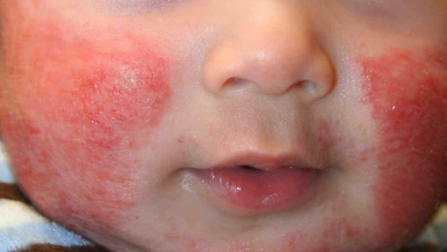
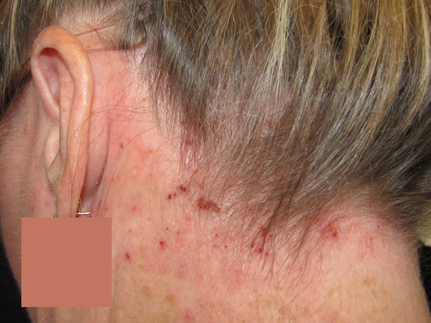
Figure 2. Atopic dermatitis neck

Atopic dermatitis neck

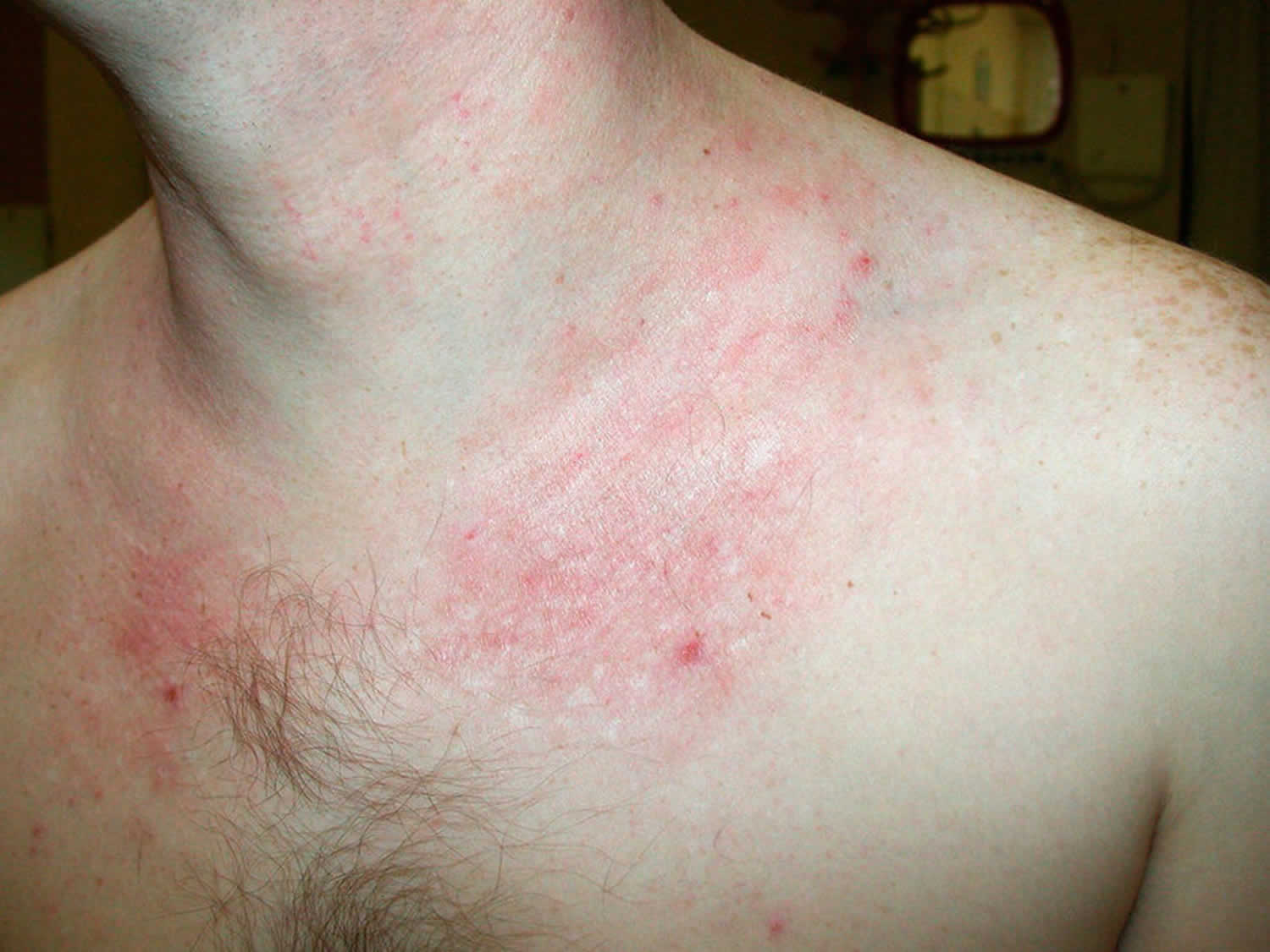
Figure 3. Atopic dermatitis eyelid

Atopic dermatitis eyelid

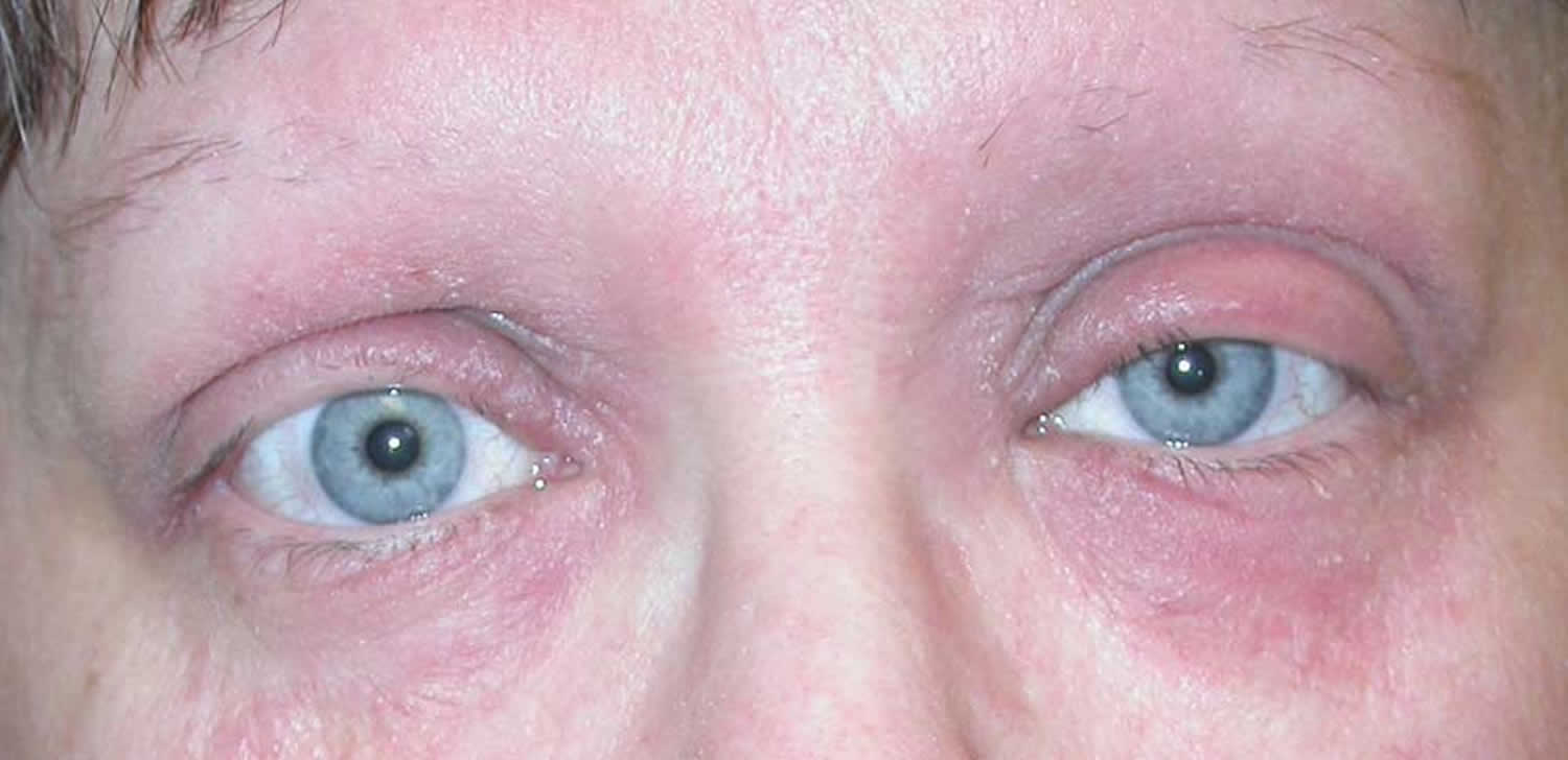
Figure 4. Atopic dermatitis scalp

Atopic dermatitis scalp

When to see a doctor

See your child’s doctor if:

  • Atopic dermatitis does not get better with home care
  • Symptoms get worse or treatment does not work
  • Your child has signs of infection, such as redness, pus or fluid-filled bumps on skin, fever, or pain

Is there a cure for atopic dermatitis?

Although there is currently no known cure for atopic dermatitis, when it is well-managed it is possible to limit its impact on day-to-day life.

Will my child grow out of atopic dermatitis?

Children often ‘grow out of’ the symptoms of atopic dermatitis, but it can return at any time, unfortunately. If you have atopic dermatitis at an early age, your skin is likely to remain sensitive even if there is no recurrence of dermatitis.

Atopic dermatitis causes

Atopic dermatitis has a complex causes including genetic and environmental factors which lead to abnormalities in the epidermis and the immune system 2). In atopic dermatitis, dry skin is due to a genetically defective skin barrier. Skin without eczema (dermatitis) provides an effective barrier that protects the body from infection and irritation. If you think of the skin as a brick wall, the outer cells are the bricks, while fats and oils are the mortar, holding everything together and acting like a seal. The cells attract and keep water inside, and the fats and oils also help to keep moisture in.

If you have dermatitis, your skin may not produce as many fats and oils and will be unable to retain water. Also, some everyday substances (e.g. soap, bubble bath and detergents) will dry out the skin. Gaps open up between the skin cells as they are not sufficiently plumped up by water. This means that the skin barrier is not as effective as it should be, and bacteria or irritants can more easily pass through. These then trigger an inflammatory response, which causes the redness in dermatitis flares. Although the exact cause of atopic dermatitis is not known, an ‘over-reactive’ immune system is understood to be involved.

Atopic dermatitis is part of the atopic triad (atopic dermatitis, allergic rhinoconjunctivitis, and asthma) which may start simultaneously or in succession in what is known as the “atopic march.” Patients with the atopic triad have a defective barrier of the skin, upper respiratory, and lower respiratory tract which leads to their symptomatology 3). If one parent is atopic, there is more than a 50% chance that their offspring will develop atopic symptoms. If both parents are affected, up to 80% of offspring will be affected. Genetic alterations include loss of function mutations of filaggrin (Filament Aggregating Protein), an epidermal protein that is broken down into natural moisturization factor. Filaggrin mutations are present in up to 30% of atopic dermatitis patients and may also predispose patients to ichthyosis vulgaris, allergic rhinitis, and keratosis pilaris. Food hypersensitivity may also cause or exacerbate atopic dermatitis in 10% to 30% of patients. Ninety percent of such reactions or flares are caused by eggs, milk, peanuts, soy, and wheat 4).

Recent studies indicate that there may be an association between smoking and adult-onset atopic dermatitis.

Atopic dermatitis patients have a defective skin barrier that is susceptible to xerosis (dry skin) and environmental irritants and allergens that lead to inflammation, pruritus, and the classic clinical findings of atopic dermatitis. The barrier defect may be caused in part by decreased levels of ceramides, which are sphingolipids in the stratum corneum which play a role in the skin’s barrier function and prevent transepidermal water loss. The defective skin barrier allows irritants and allergens to penetrate the skin and cause inflammation via an overactive Th2 response (with increased IL-4, IL-5 cytokines) in acute lesions and Th1 response (with IFN-gamma and IL-12) in chronic lesions. Scratching of the skin also stimulates keratinocytes to release inflammatory cytokines such as TNF-alpha, IL-1, and IL-6. Decreased anti-microbial peptides (human beta-defensins, cathelicidins) in the epidermis of atopic patients also contribute to Staphylococcus aureus colonization seen in more than 90% of atopic dermatitis patients. Staphylococcus aureus may worsen the inflammation of atopic dermatitis lesions and lead to secondary infection and impetiginization 5).

In atopic dermatitis, there is significant water loss across the epidermis, but why there is dysregulation of the epithelial barrier is not fully understood. It is believed that filaggrin, which is critical for epithelial integrity, may be dysfunctional.

The following can make atopic dermatitis symptoms worse:

  • Allergies to pollen, mold, dust mites, or animals
  • Cold and dry air in the winter
  • Colds or the flu
  • Contact with irritants and chemicals
  • Contact with rough materials, such as wool
  • Dry skin
  • Emotional stress
  • Drying out of the skin from taking frequent baths or showers and swimming very often
  • Getting too hot or too cold, as well as sudden changes of temperature
  • Perfumes or dyes added to skin lotions or soaps

Atopic dermatitis prevention

Moisturization is important on an ongoing basis and may prevent flares. Daily skin care may cut down on the need for medicines.

To help you avoid scratching your rash or skin:

  • Use a moisturizer, topical steroid cream, or other medicine your provider prescribes.
  • Take antihistamine medicines by mouth to reduce severe itching.
  • Keep your fingernails cut short. Wear light gloves during sleep if nighttime scratching is a problem.

Keep your skin moist by using ointments (such as petroleum jelly), creams, or lotions 2 to 3 times a day. Choose skin products that do not contain alcohol, scents, dyes, and other chemicals. A humidifier to keep home air moist will also help.

Avoid Triggers

The following triggers can make atopic dermatitis symptoms worse:

  • Allergies to pollen, mold, dust mites, or animals
  • Cold and dry air in the winter
  • Colds or the flu
  • Contact with irritants and chemicals
  • Contact with rough materials, such as wool
  • Dry skin
  • Emotional stress
  • Taking frequent baths or showers and swimming often, which can dry out skin
  • Getting too hot or too cold, as well as sudden changes of temperature
  • Perfumes or dyes added to skin lotions or soaps

To prevent flare-ups, try to avoid:

  • Foods, such as eggs, that may cause an allergic reaction in a very young child. Always discuss with your provider first.
  • Wool, lanolin, and other scratchy fabrics. Use smooth, textured clothing and bedding, such as cotton.
  • Sweating. Be careful not to over dress your child during warmer weather.
  • Strong soaps or detergents, as well as chemicals and solvents.
  • Sudden changes in body temperature, which may cause sweating and worsen your child’s condition.
  • Stress. Watch for signs that your child feels frustrated or stressed and teach them ways to reduce stress such as taking deep breaths or thinking about things they enjoy.
  • Triggers that cause allergy symptoms. Do what you can to keep your home free of allergy triggers such as mold, dust, and pet dander.
  • Avoid using skin care products that contain alcohol.

Using moisturizers, creams, or ointments every day as directed may help prevent flares.

When washing or bathing:

  • Expose your skin to water for as short a time as possible. Short, cooler baths are better than long, hot baths.
  • Use gentle body washes and cleansers instead of regular soaps.
  • Do not scrub or dry your skin too hard or for too long.
  • Apply lubricating creams, lotions, or ointment to your skin while it is still damp after bathing. This will help trap moisture in your skin.

Medicines

Antihistamines taken by mouth may help with itching or allergies. You can often buy these medicines without a prescription.

Atopic dermatitis is usually treated with medicines placed directly on the skin or scalp. These are called topical medicines:

  • You will probably be prescribed a mild cortisone (steroid) cream or ointment at first. You may need a stronger medicine if this does not work.
  • Medicines called topical immunomodulators may be prescribed for anyone over 2 years old. Ask your provider about concerns over a possible cancer risk with the use of these medicines.
  • Creams or ointments that contain coal tar or anthralin may be used for thickened areas.
  • Barrier repair creams containing ceramides may be used.

Wet-wrap treatment with topical corticosteroids may help control the condition. But, it may lead to an infection.

Other treatments that may be used include:

  • Antibiotic creams or pills if your skin is infected
  • Drugs that suppress the immune system
  • Targeted biologic medicines that are designed to affect parts of the immune system involved in atopic dermatitis
  • Phototherapy, a treatment in which your skin is carefully exposed to ultraviolet (UV) light
  • Short-term use of systemic steroids (steroids given by mouth or through a vein)

Atopic dermatitis symptoms

Atopic dermatitis affects up to 30% of the childhood population and causes considerable distress and ill health. Its prevalence is greatest in childhood, generally starting in the first few months of life, becoming more severe in infancy and often improving in school years.

Affected sites vary with age. Infantile eczema commonly affects the face, sparing around the mouth and later the hands, feet and elsewhere. Erythrodermic eczema refers to involvement of the entire body at any age.

In older children, eczema tends to affect flexures, particularly antecubital and popliteal fossae. Flexural dermatitis often persists into adult life. Occasionally, a ‘dirty neck’ is observed in teenagers. Irritant hand eczema may be a problem for those who do wet work. Bilateral nipple eczema is not uncommon.

Diagnosis depends on clinical findings, which vary with the age and stage of the disease. The main features are:

  • Marked pruritus, frequently resulting in lichenification
  • Intermittent exacerbations (acute flare-ups)
  • Association with personal or family history of atopic dermatitis, allergic rhinitis and/or asthma
  • Dry skin

Skin changes may include:

  • Blisters with oozing and crusting
  • Dry skin all over the body, or areas of bumpy skin on the back of the arms and front of the thighs
  • Ear discharge or bleeding
  • Raw areas of the skin from scratching
  • Skin color changes, such as more or less color than the normal skin tone
  • Skin redness or inflammation around the blisters
  • Thickened or leather-like areas, which can occur after long-term irritation and scratching

A scoring index (SCORAD) combining extent, severity and subjective symptoms, is often used in clinical trials that assess the effectiveness of treatments.

Atopic dermatitis flare-ups may be precipitated by:

  • Staphylococcus aureus exotoxins, which act as superantigens
  • Irritants, especially skin dehydration by over-washing, woollen clothing
  • Inhalant allergens especially house dust mite (which may also be a contact allergen)
  • Ingested allergens in some infants (eggs, milk, soybeans, peanuts and wheat account for most of these)
  • Emotional stress

Atopic dermatitis baby

  • Infants less than one year of age often have widely distributed eczema. The skin is often dry, scaly and red with small scratch marks made by sharp baby nails.
  • The cheeks of infants are often the first place to be affected by eczema.
  • The napkin area is frequently spared due to the moisture retention of nappies. Just like other babies, they can develop irritant napkin dermatitis, if wet or soiled nappies are left on too long.

Atopic dermatitis toddlers and pre-schoolers

  • As children begin to move around, eczema becomes more localised and thickened. Toddlers scratch vigorously and eczema may look very raw and uncomfortable.
  • Eczema in this age group often affects the extensor (outer) aspects of joints, particularly the wrists, elbows, ankles and knees. It may also affect the genitals.
  • As the child becomes older the pattern frequently changes to involve the flexor surfaces of the same joints (the creases) with less extensor involvement. The affected skin often becomes lichenified i.e. dry and thickened from constant scratching and rubbing,
  • In some children, the extensor pattern of eczema persists into later childhood.

Atopic dermatitis school-age children

  • Older children tend to have the flexural pattern of eczema and it most often affects the elbow and knee creases. Other susceptible areas include the eyelids, earlobes, neck and scalp.
  • They can develop recurrent acute itchy blisters on the palms, fingers and sometimes on the feet, known as pompholyx or vesicular hand/foot dermatitis.
  • Many children develop a ‘nummular’ pattern of atopic dermatitis. This refers to small coin-like areas of eczema scattered over the body. These round patches of eczema are dry, red and itchy and may be mistaken for ringworm (a fungal infection).
  • Mostly eczema improves during school years and it may completely clear up by the teens, although the barrier function of the skin is never entirely normal.

Atopic dermatitis adults

  • Adults who have atopic dermatitis may present in various different ways.
    They may continue to have a diffuse pattern of eczema but the skin is often more dry and lichenified than in children.
  • Commonly adults have persistent localised eczema, possibly confined to the hands, eyelids, flexures, nipples or all of these areas.
  • Recurrent staphylococcal infections may be prominent.
  • Atopic dermatitis is a major contributing factor to occupational irritant contact dermatitis. This most often affects hands that are frequently exposed to water, detergents and /or solvents.
  • Having atopic dermatitis does not exclude contact allergic dermatitis (confirmed by patch tests) in children and adults)
  • Hand dermatitis in adult atopics tends to be dry and thickened but may also be blistered.

Atopic dermatitis complications

Atopic dermatitis may be complicated by microbial colonization or infection:

  • Staphylococcus aureus (impetiginised eczema)
  • Streptococcus pyogenes
  • Herpes simplex (eczema herpeticum)
  • Warts
  • Molluscum contagiosum
  • Malassezia spp.

The pathogenesis appears involve release of vasoactive substances from mast cells and basophils as an IgE-mediated hypersensitivity reaction. There is a TH2 pattern of cytokine release from T helper lymphocytes in the epidermis and dermis with low levels of TH1 lymphocytes and gamma interferon. There is also a non-allergic or intrinsic type of atopic dermatitis. These patients have no associated respiratory diseases, show normal total serum IgE levels, no specific IgE, and have negative skin-prick tests to aeroallergens or foods.

It is not known why the incidence of atopic dermatitis appears to be increasing; theories include increased hygiene and decreased exposure to micro-organisms and greater exposure to house dust mite.

A proportion of patients have been found to be deficient in fillagrin, resulting in abnormal barrier function of the stratum corneum and increased susceptibility to the effect of contact irritants. These individuals have dry skin (ichthyosis vulgaris).

Atopic dermatitis diagnosis

Your health care provider will look at your skin and do a physical exam. You may need a skin biopsy to confirm the diagnosis or rule out other causes of dry, itchy skin. The presence of associated findings (e.g., keratosis pilaris) may facilitate diagnosis. A biopsy will show an eczematous pattern. In childhood cases that are recalcitrant to treatment, fluorescent enzyme immunoassays or skin prick testing can be performed to detect immunoglobulin E (IgE) antibodies against specific allergens, which may or may not be a clinically relevant exacerbating factor 6).

Atopic dermatitis diagnosis is based on:

  • How your skin looks
  • Your personal and family history

In most cases, no specific investigations are required. However, on occasion the following may be useful:

  • Skin swabs for bacteriology: to identify methicillin resistant strains of Staphylococcus. Your doctor may order cultures for infection of the skin. If you have atopic dermatitis you may get infections easily.
  • Viral culture: to confirm eczema herpeticum
  • Iron studies: severe eczema can result in iron deficiency; iron deficiency aggravates pruritus
  • Total IgE: elevated IgE confirms atopy but normal levels may occur in non-allergic patients
  • RAST tests (specific IgE): negative tests have a high predictive value, positive test results are not so useful
  • Prick tests: positive test results may simply confirm an atopic diathesis
  • Patch tests: to rule out specific contact allergy for example to an applied medicament

Allergy skin testing may be helpful for people with:

  • Hard-to-treat atopic dermatitis
  • Other allergy symptoms
  • Skin rashes that form only on certain areas of the body after exposure to a specific chemical

Atopic dermatitis treatment

The best way to manage atopic dermatitis is unknown. At present, management of atopic dermatitis in patients of all ages involves:

  • Avoidance of aggravating factors: use soap substitutes; don’t wear woollen garments or perfumed cosmetics; ensure adequate rest; reduce stress
  • Fans and wet dressings to cool hot and inflamed skin
  • Emollients applied as often as is required to keep the skin hydrated and comfortable
  • Topical immunomodulators, also known as calcineurin inhibitors (pimecrolimus cream)
  • Topical corticosteroid creams and/or ointments for flare-ups
  • Oral antibiotics, usually flucloxacillin or dicloxacillin, for bacterial infection
  • Non-sedating antihistamines for patients with an element of urticaria.
  • Sedative antihistamines at night to allow adequate rest.
  • In severe cases, refer for phototherapy, azathioprine, methotrexate or ciclosporin

Treatment of atopic dermatitis may be required for many months and possibly years.

Atopic dermatitis treatment nearly always requires:

  • Reduction of exposure to trigger factors (where possible)
  • Regular emollients (moisturizers)
  • Intermittent topical steroids

Daily skin care includes the application of emollients twice daily, with the application within three minutes of exiting lukewarm shower or bath to prevent skin drying. Ointments are the most occlusive but may be more greasy. Topical steroids, which should be applied before emollients to “lock-in” their effect, are first-line agents for acute flares. The potency should be strong enough to control a flare quickly, and consideration should be given for tapering every other day and for maintenance therapy twice weekly (e.g., weekends) in the usual areas of involvement. Reversible side effects of steroid use include skin atrophy and telangiectasia.

Potent topical steroids should be used with caution in infants because of their greater proportional absorption due to higher surface area to body weight ratio. Blistered or weeping eczema is brought under control most rapidly using wet dressings.

Oral corticosteroids are used for crisis intervention but may be followed by a severe rebound flare at discontinuation. It is important to taper the dose and begin intensified skin care with topical steroids and bathing followed by application of emollients.

Sensitive areas (including the intertriginous areas of the axilla and groin, in addition to the face) may require topical nonsteroidal agents including calcineurin inhibitors such as tacrolimus and pimecrolimus. Newer non-steroidal agents include crisaborole, which exerts its effect by blocking PDE-4. When atopic dermatitis is not controlled with topical agents, systemic agents include phototherapy (ultraviolet (UV) A, UVB, and narrow-band UVB), cyclosporine, azathioprine, mycophenolate mofetil, and methotrexate.

A newly FDA-approved biologic therapy is dupilumab, which is a monoclonal antibody that blocks the IL-4 receptor and thus the effect of IL-4 and IL-13. Other complementary therapies include bleach baths (0.5 cup bleach in full 40 gallon tub) one to two times weekly to decrease S. aureus colonization, low allergen maternal diets during breastfeeding, and probiotic and prebiotic use in pregnant mothers and at-risk infants which has shown 50% decreased frequency of atopic dermatitis at ages 1 to 4 years old compared to placebo.[10][11][12]

Recently Crisaborole topic ointment was approved for mild to moderate atopic dermatitis. The drug is a phosphodiesterase inhibitor and shown to improve skin symptoms.

Some patients may benefit from probiotics; it is believed that the bacterial products may enhance the immune system and prevent the development of allergic IgE antibody response. Further, probiotics are recommended during pregnancy and in breast feeding women.

Numerous studies show that bleach baths may help relieve the symptoms of atopic dermatitis by lowering the risk of

In some cases, management may also include one or more of the following:

  • Topical calcineurin inhibitors, such as pimecrolimus cream or tacrolimus ointment
  • Crisabarole ointment
  • Antibiotics
  • Antihistamines
  • Phototherapy
  • Oral corticosteroids

Longstanding and severe eczema may be treated with an immunosuppressive agent.

  • Methotrexate
  • Ciclosporin
  • Azathioprine

New biologics are under investigation. The first to be approved for the treatment of atopic dermatitis is:

  • Dupilumab

Atopic dermatitis face

Use light emollients in adolescents and adults to avoid provoking acne or perioral dermatitis. It is safe to use 0.5-2% hydrocortisone cream on active dermatitis indefinitely, although in time it is likely to lose its efficacy. Pimecrolimus cream can also be used as maintenance treatment. Severe dermatitis may require moderate potency topical steroids for a course of 5 to 10 days or as pulse therapy. A trial of antifungal agents may be warranted in adults with prominent facial dermatitis.

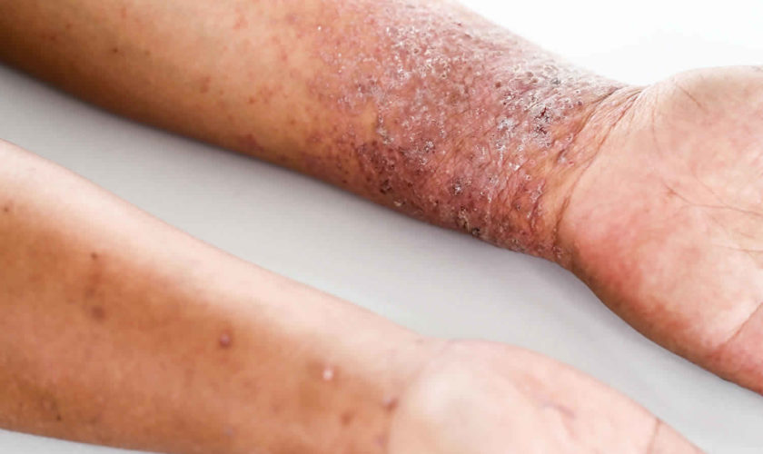
Atopic dermatitis trunk and limbs

These sites may require thicker emollients (sorbolene cream, white soft paraffin, emulsifying ointment, fatty cream) and moderate potency topical steroid fatty cream or ointment, with potent topical steroids for flare-ups. It is often useful to apply the potent preparation prophylactically on two consecutive days each week (pulse therapy). Ultrapotent products may be required for severe lichenification.

Atopic dermatitis hands and feet

The very thick stratum corneum of palmoplantar sites necessitates the use of ultrapotent topical steroids such as clobetasol propionate, generally in an ointment base, for two to four weeks. Advise frequent use of emollient barrier creams that contain dimethicone and/or petrolatum, and careful protection against irritants.

Atopic dermatitis armpits and groins

Flexures do not generally require emollients. Potent topical steroids are rarely required, and should only be used for a few days. Hydrocortisone cream or pimecrolimus cream is generally adequate.

Atopic dermatitis treatment with topical therapies

Non-pharmacologic interventions such as the role of moisturizers and bathing practices to help with treatment, maintenance, and prevention of flares 7).

  • Moisturizers: The application of moisturizers should be an integral part of the treatment of patients with atopic dermatitis. They are also important components of maintenance therapy and prevention of flares.
  • Bathing practices, including additives: Bathing is suggested in patients with atopic dermatitis as part of treatment and maintenance; however, there is no standard for the frequency or duration of bathing appropriate for those with atopic dermatitis. Moisturizers should be applied soon after bathing to improve skin hydration. Limited use of non-soap cleansers (that are neutral to low pH, hypoallergenic, and fragrance-free) is recommended, and there is no data to support the use of bath water additives (oils, emollients, etc.).
  • Wet-wrap therapy with or without topical corticosteroid can be recommended for patients with moderate to severe atopic dermatitis to decrease severity and water loss during flares.

Topical corticosteroids are used on both adults and pediatrics for the management of atopic dermatitis. A variety of factors must be considered to choose the proper topical corticosteroids for treatment, such as patient preference and age. No specific monitoring is required for side effects. However, based on patient risk factors and response some monitoring may be required. Patient education may be key to address misconceptions of topical corticosteroids.

Topical calcineurin inhibitors can be used for the treatment of acute and chronic atopic dermatitis as well as maintenance therapy in both adults and children. They can serve as a steroid-sparing treatment; however, careful considerations must be made before prescribing. Patient education regarding topical calcineurin inhibitor for atopic dermatitis is crucial since there are some adverse effects they may experience. Concomitant use of topical corticosteroid and topical calcineurin inhibitor can be used to treat atopic dermatitis. No specific monitoring is required for topical calcineurin inhibitors; however, based on individual patient risk factors it may be warranted.

Topical antimicrobials and antiseptics: Bleach baths with intranasal mupirocin have been shown to be beneficial for atopic dermatitis patients; however, they are not routinely recommended. This therapy is mostly for patients with clinical signs of secondary bacterial infection to help reduce disease severity. All other forms of antimicrobial and antiseptic therapies have not been shown to be clinically helpful for atopic dermatitis.

Topical antihistamines are not suggested for the treatment of atopic dermatitis due to the risk of absorption and contact dermatitis 8).

Other topical agents are currently being studied for their use in treating atopic dermatitis. However, no conclusive data is available.

Complications

If topical corticosteroids are used inappropriately or if superpotent steroids are used in teenagers during rapid growth, striae may occur. Skin thinning can result if steroids are used inappropriately in older patients.

Whether verrucae vulgaris and mollusca contagiosa are more frequent is difficult to assess, but they are more widespread and difficult to eliminate.

Tachyphylaxis to topical steroids occurs if they are not used on a stop-start basis.

Patients may develop other related allergic disorders such as urticaria, food allergy, asthma and allergic rhinitis.

Superinfection with Staphylococcus aureus may require topical and/or systemic antibiotic treatment with antistaphylococcal agents.

Superinfection with herpes simplex virus, referred to as eczema herpeticum, can require admission to the hospital in children for systemic treatment with acyclovir and evaluation of other complications such as herpes keratitis.

Atopic dermatitis treatment with phototherapy and systemic agents

Phototherapy is typically used as a treatment for both acute and chronic atopic dermatitis in pediatric and adult patients. Narrowband ultraviolet-B (NB-UVB) is the most commonly used phototherapy due to its low-risk profile, efficacy, and availability. Phototherapy can be used as monotherapy or in combination with other topical therapies. However, caution must be taken due to drug interactions and increased risk of adverse effects.

Phototherapy can be used in children; however, additional factors such as their psychological perspective may need to be considered when administering therapy.

Various factors must be considered when prescribing systemic agents, such as previous therapy failure or contraindications, as well as quality of life and disease severity. When using systemic agents, the minimal effective dose should be used, because there is no optimal dosing, duration, or monitoring protocol due to the lack of data. Treatment is highly individualized and based on patient response, comorbidities, and history.

The following systemic therapies can be used off-label to treat atopic dermatitis. Some should only be considered as an alternative when other more commonly used off-label systemic therapies are not an option:

  • Cyclosporine: off-label use for atopic dermatitis
  • Azathioprine: off-label use for atopic dermatitis
  • Methotrexate: off-label use for atopic dermatitis
  • Mycophenolate Mofetil: alternative off-label use for atopic dermatitis
  • Interferon Gama: alternative use for atopic dermatitis

Systemic steroids should be avoided when possible for the treatment of atopic dermatitis. They mainly serve for short-term bridge therapy to other systemic therapies or for acute severe exacerbations.

There is insufficient data to make proper recommendations for the use of the following systemic therapies for the treatment of atopic dermatitis 9):

  • Omalizumab
  • Oral Calcineurin Inhibitors
  • Other systemic therapies (TNF-alpha inhibitors, IV immunoglobulin, theophylline, papaverine, or thymopentin)

The use of systemic antibiotics is not encouraged unless there is clinical evidence of bacterial infection or eczema herpeticum.

There is no data to support the use of oral antihistamines as a treatment of atopic dermatitis, they can be used to help with pruritus and some sedating antihistamines can help (short-term) with sleep loss due to atopic dermatitis.

Prevention of atopic dermatitis flares

Continued use of topical corticosteroids or topical calcineurin inhibitors after disease stabilization can help prevent relapse or flares 10).

Patient education is important to inform the patient about atopic dermatitis and can be done through educational programs, video training, or nurse-led workshops. Education should always be an adjunct to conventional therapy.

Overall, allergy testing without a history of allergies in atopic dermatitis patients is not supported since atopic dermatitis can be affected by other non-allergic factors such as diet and the environment. If a patient does have a history of allergies or signs of contact dermatitis then it may be helpful to test for allergies (food allergies, inhalant/aeroallergens, allergic contact dermatitis).

Dietary interventions based solely on food allergies are not supported due to atopic dermatitis being affected by different non-food allergy/diet-based factors.

  • In children < 5 years of age with persistent atopic dermatitis despite optimized treatment or history of reaction to certain foods can be considered for food allergy tests.
  • If a food avoidance diet is being undertaken, then it should be done with the assistance of a dietician.

The following dietary supplements are NOT supported by data for the treatment of atopic dermatitis:

  • Probiotics/prebiotics
  • Fish oils, primrose oil, borage oil, multivitamin supplements, zinc, vitamin D, E, B12, or B6

Environmental modifications are not supported by the currently available data and more studies are needed.

  • Measures to avoid and reduce contact with house dust mites may be helpful in patients with high sensitivity to house dust mites.
  • Modification of laundering techniques such as double rinsing or use of certain detergents and laundry products is not supported by the current data.
  • There is limited data supporting the use of certain clothing fabrics and fibers to help reduce irritation, further studies are needed.

Other allergen-based interventions such as immunotherapy or sublingual immunotherapy for the treatment of atopic dermatitis are not supported by the currently available data.

There is insufficient evidence to support the use of complementary alternative therapies for the treatment of atopic dermatitis such as the following: traditional Chinese medicine, acupuncture, aromatherapy, homeopathy, naturopathy, acupressure, autologous blood injections.

Allergens

Dermatologists rarely recommend dietary manipulation for atopic dermatitis because it is troublesome, expensive and not often helpful. However, about 30% of children with dermatitis also have food allergies causing urticaria and anaphylactic responses. In some of these, certain foods may consistently aggravate their dermatitis. The most well established food allergies associated with dermatitis are to egg, milk, peanut, wheat, and soy.

If there is a strong suspicion of food allergy, a specific RAST test reaction or positive prick test may be supportive but should be confirmed with controlled food challenges and a limited elimination diet that results in consistent clinical improvement. Extensive elimination diets can be nutritionally deficient and are useless. Most affected children outgrow their food hypersensitivity.

Extended avoidance of house dust mites in sensitised patients with atopic dermatitis is reported to be helpful but is difficult to achieve. Avoidance measures include use of house dust mite-proof encasings on pillows, mattresses, and duvets; washing bedding in hot water weekly; removal of bedroom carpets; and decreasing indoor humidity levels.

Immunotherapy has not been found to be useful.

Atopic dermatitis treatment over the counter

Antihistamines taken by mouth may help if allergies cause your child’s itchy skin. These medicines are often available over the counter and do not require a prescription. Ask your child’s doctor what kind is right for your child.

Help for itching and scratching

Severe itching is common. Itching may start even before the rash appears. Atopic dermatitis is often called the “itch that rashes” because the itching starts, and then the skin rash follows as a result of scratching.

To help your child avoid scratching:

  • Use a moisturizer, topical steroid cream, barrier repair cream, or other medicine the child’s doctor prescribes.
  • Keep your child’s fingernails cut short. Have them wear light gloves while sleeping if scratching at night is a problem.
  • Give antihistamines or other medicines by mouth as prescribed by your child’s doctor.
  • As much as possible, teach older children not to scratch itchy skin.

Day-to-day skin care

Daily skin care with allergen-free products may cut down on the need for medicines.

Use moisturizing ointments (such as petroleum jelly), creams, or lotions. Choose skin products that are made for people with eczema or sensitive skin. These products do not contain alcohol, scents, dyes, and other chemicals. Having a humidifier to keep air moist will also help.

Moisturizers and emollients work best when they are applied to skin that is wet or damp. After washing or bathing, pat the skin dry and then apply the moisturizer right away. Your provider may also recommend placing a dressing over these skin moisturizing ointments.

When washing or bathing your child:

  • Bathe less often and keep water contact as brief as possible. Short, cooler baths are better than long, hot baths.
  • Use gentle skin care cleansers rather than traditional soaps, and use them only on your child’s face, underarms, genital areas, hands, and feet.
  • Do not scrub or dry the skin too hard or for too long.
  • Right after bathing, apply lubricating cream, lotion, or ointment while skin is still damp to trap moisture.

Dress your child in soft, comfortable clothing, such as cotton clothes. Have your child drink plenty of water. This may help add moisture to the skin.

Teach older children these same tips for skin care.

The rash itself, as well as the scratching, often causes breaks in the skin and may lead to infection. Keep an eye out for redness, warmth, swelling, or other signs of infection. Call your child’s doctor at the first sign of infection.

Atopic dermatitis prognosis

Atopic dermatitis affects 10–30% of children but is much less common in adults. One third of patients develop allergic rhinitis. One third of patients develop asthma. It is impossible to predict whether atopic dermatitis will improve by itself or not in an individual. Sensitive skin persists life-long. A meta-analysis including over 110,000 subjects found that 20% of children with atopic dermatitis still had persistent disease 8 years later. Fewer than 5% had persistent disease 20 years later. Children who developed atopic dermatitis before the age of 2 had a much lower risk of persistent disease than those who developed atopic dermatitis later in childhood or during adolescence.

In a longitudinal study of 7157 children and adolescents with atopic dermatitis from the Pediatric Eczema Elective Registry 11), researchers found that symptoms of mild to moderate atopic dermatitis are likely to persist into the teen years or beyond. Approximately two-thirds of the patients were followed for at least 2 years and the rest were followed for at least 5 years. From ages 2 to 26 years, more than 80% of patients reported having continued symptoms and/or use of topical medications to control symptoms. By age 20, approximately half of the patients had experienced at least one 6-month symptom- and medication-free period. Living in southern states, having a relative with an atopic illness, and exposure to pollen, wool, pets, cigarettes, fumes, some foods or drinks, and soaps/detergents were linked to persistent symptoms 12).

It is unusual for an infant to be affected with atopic dermatitis before the age of four months but they may suffer from infantile seborrheic dermatitis or other rashes prior to this. The onset of atopic dermatitis is usually before two years of age although it can manifest itself in older people for the first time.

Atopic dermatitis is often worst between the ages of two and four but it generally improves after this and may clear altogether by the teens.

Certain occupations such as farming, hairdressing, domestic and industrial cleaning, domestic duties and care-giving expose the skin to various irritants and, sometimes, allergens. This aggravates atopic dermatitis. It is wise to bear this in mind when considering career options — it is usually easier to choose a more suitable occupation from the outset than to change it later.

References   [ + ]

read more

Allergy shots

Allergy shots

Allergy shots

Allergy shots also called allergen immunotherapy, is a form of long-term treatment that decreases symptoms for many people with allergic rhinitis, allergic asthma, conjunctivitis (eye allergy) or stinging insect allergy. Allergy shots decrease sensitivity to allergens and often leads to lasting relief of allergy symptoms even after treatment is stopped. This makes it a cost-effective, beneficial treatment approach for many people. If you want want to start allergy shots, find a board certified allergist/immunologist here (https://allergist.aaaai.org/find).

Allergy shots can treat some types of allergies. Allergy shots or allergen immunotherapy is appropriate for patients with immunoglobulin E (IgE)-mediated allergic diseases such as seasonal allergic rhinitis, perennial rhinitis, allergic asthma, and insect venomanaphylaxis whose symptoms are not well controlled with avoidance measures and pharmacotherapy and in whom an IgE-mediated reaction to antigen has been documented 1). Some recent reports have suggested that immunotherapy may also aid in atopic dermatitis 2).

Allergy shots sometimes used for children with allergies to:

  • things in the environment, like pollen, mold, or dust mites
  • pet dander
  • insect stings

Allergy shots aren’t helpful for food allergies. The best option for people with food allergies is to strictly avoid that food.

Allergy injections or allergen immunotherapy has been clinically demonstrated to provide long-term clinical benefits, including symptomatic disease remission and a reduction in allergic disease progression from rhinitis to asthma 3).

Aside from allergy shots, the other available treatment options for allergic diseases include aeroallergen avoidance and pharmacotherapy. In addition, surgery may be offered as an adjunct in appropriate patients (eg, septoplasty for nasal septal deviation, sinus surgery for chronic sinusitis, turbinate reduction for turbinate hypertrophy, nasal polypectomy for nasal polyps).

Effective treatment reduces symptoms and medication utilization while improving quality of life. Allergy shots has demonstrated long-term effectiveness in symptom reduction more than 3 years following treatment cessation and has prevented the development of new sensitizations in children.

Why are allergy shots used?

An allergy is when the body’s immune system overreacts to a usually harmless substance. Things that cause allergic reactions are called allergens. Common allergens include dust mites, molds, pollen, pets with fur or feathers, stinging insects, and foods.

The body reacts to the allergen by releasing chemicals, one of which is histamine. This release can cause symptoms such as wheezing, trouble breathing, coughing, a stuffy nose, and more. Some allergic reactions can be serious.

The best way to prevent or control allergy symptoms is to avoid allergens. Allergists (doctors who identify and treat allergies) look for causes of an allergic reaction with skin tests and blood tests. Based on the test results, they can recommend treatments, including medicines and ways to avoid allergens.

If these treatments don’t help, the allergist might recommend allergy shots.

Who can benefit from allergy shots?

Both children and adults can receive allergy shots, although it is not typically recommended for children under age five. This is because of the difficulties younger children may have in cooperating with the program and in articulating any adverse symptoms they may be experiencing. When considering allergy shots for an older adult, medical conditions such as cardiac disease should be taken into consideration and discussed with your allergist / immunologist first.

You and your allergist / immunologist should base your decision regarding allergy shots on:

  • Length of allergy season and severity of your symptoms
  • How well medications and/or environmental controls are helping your allergy symptoms
  • Your desire to avoid long-term medication use
  • Time available for treatment (allergy shots requires a significant commitment)
  • Cost, which may vary depending on region and insurance coverage

Allergy shots are not used to treat food allergies. The best option for people with food allergies is to strictly avoid that food.

How do allergy shots work?

Allergy shots work like a vaccine. Your body responds to injected amounts of a particular allergen, given in gradually increasing doses, by developing immunity or tolerance to the allergen.

There are two phases:

  1. Build-up phase. This involves receiving injections with increasing amounts of the allergens about one to two times per week. The length of this phase depends upon how often the injections are received, but generally ranges from three to six months.
  2. Maintenance phase. This begins once the effective dose is reached. The effective maintenance dose depends on your level of allergen sensitivity and your response to the build-up phase. During the maintenance phase, there will be longer periods of time between treatments, ranging from two to four weeks. Your allergist / immunologist will decide what range is best for you.

You may notice a decrease in symptoms during the build-up phase, but it may take as long as 12 months on the maintenance dose to notice an improvement. If allergy shots are successful, maintenance treatment is generally continued for three to five years. Any decision to stop allergy shots should be discussed with your allergist / immunologist.

How do allergy shots help?

Allergy shots help the body build immunity to specific allergens, so it’s not as bothered by them. Allergy shots also can help people who have allergies and asthma have fewer asthma flare-ups.

Allergy shots contain a tiny amount of a purified form of the allergen causing problems. Doctors increase the dose slowly over the first 3–6 months. This lets the immune system safely adjust and build immunity to the allergens. This is called the buildup phase.

The highest effective safe dose becomes that person’s monthly maintenance dose. Health care providers give this to patients for about 3 to 5 years. Most people will need fewer shots over time.

Some people’s allergy symptoms ease during the buildup phase. Others don’t feel better until they’re into the maintenance phase. After years of getting allergy shots, some may have lasting relief from symptoms.

Where should allergy shots be given?

This type of treatment should be supervised by a specialized physician in a facility equipped with proper staff and equipment to identify and treat adverse reactions to allergy injections. Ideally, immunotherapy should be given in your allergist / immunologist’s office. If this is not possible, your allergist / immunologist should provide the supervising physician with comprehensive instructions about your allergy shot treatments.

Allergy shots risks and side effects

Allergy shots given by a trained health professional are safe and effective. Kids as young as 5 years old can get them.

A typical reaction is redness and swelling at the injection site. This can happen immediately or several hours after the treatment. Applying an ice pack to the area and taking an antihistamine can help. In some instances, symptoms can include increased allergy symptoms such as sneezing, nasal congestion or hives. More widespread reactions, like hives and itching all over the body, are less common. And more severe reactions (like wheezing, breathing problems, throat swelling, and nausea) are rare.

Serious reactions to allergy shots are rare. When they do occur, they require immediate medical attention. Symptoms of an anaphylactic reaction can include swelling in the throat, wheezing or tightness in the chest, nausea and dizziness. Most serious reactions develop within 30 minutes of the allergy injections. This is why it is recommended you wait in your doctor’s office for at least 30 minutes after you receive allergy shots.

Before you or your child gets allergy shots, be sure to tell your doctor about any other medicines you or your child takes.

Complication prevention

While adverse systemic reactions are uncommon, allergy injections should be administered by only trained personnel, and resuscitative medications and equipment should be immediately available.

Adequate equipment and medications should be immediately available in case of anaphylaxis. The following are suggested equipment and medications for the management of systemic immunotherapy reactions. Modifications of this suggested list might be based on anticipated emergency medical services’ response time and physician’s airway management skills 4):

  • Stethoscope and sphygmomanometer
  • Tourniquet, syringes, hypodermic needles, and intravenous catheters (eg, 14-18 gauge)
  • Aqueous epinephrine HCL 1:1,000 weight/volume
  • Equipment to administer oxygen by mask
  • Intravenous fluid setup
  • Antihistamine for injection (second-line agents for anaphylaxis, but H1 and H2 antihistamines work better together than either one alone)
  • Corticosteroids for intramuscular or intravenous injection (second-line agents for anaphylaxis)
  • Equipment to maintain an airway appropriate for the supervising physician’s expertise and skill
  • Glucagon kit available for patients receiving beta-blockers

How effective are allergy shots?

Allergy shots have shown to decrease symptoms of many allergies. It can prevent the development of new allergies, and in children it can prevent the progression of allergic disease from allergic rhinitis to asthma. The effectiveness of allergy shots appears to be related to the length of the treatment program as well as the dose of the allergen. Some people experience lasting relief from allergy symptoms, while others may relapse after discontinuing allergy shots. If you have not seen improvement after a year of maintenance therapy, your allergist / immunologist will work with you to discuss treatment options.

Failure to respond to allergy shots may be due to several factors:

  • Inadequate dose of allergen in the allergy vaccine
  • Missing allergens not identified during the allergy evaluation
  • High levels of allergen in the environment
  • Significant exposure to non-allergic triggers, such as tobacco smoke.

Alternative to allergy shots

Aside from allergen immunotherapy, the other available treatment options for allergic diseases include aeroallergen avoidance and pharmacotherapy. In addition, surgery may be offered as an adjunct in appropriate patients (eg, septoplasty for nasal septal deviation, sinus surgery for chronic sinusitis, turbinate reduction for turbinate hypertrophy, nasal polypectomy for nasal polyps).

Sublingual immunotherapy

Sublingual immunotherapy experienced renewed interest in England after a review of safety and protocols of subcutaneous immunotherapy identified a 0.5%-5.6% rate of systemic reactions and fatalities, most commonly due to “preventable errors.” These findings reduced the use of subcutaneous immunotherapy in Britain and encouraged research into allergy treatment with sublingual immunotherapy. Most of the data address monotherapy, and research has yet to delineate ideal dosing concentrations, dosing schedules, dosing duration, and duration of patient response.

In sublingual immunotherapy, the theory is that the antigen comes into contact with Langerhans-like dendritic cells residing in the oral mucosa on the floor of the mouth under the tongue. These cells capture the antigen and migrate to local lymph nodes, resulting in the production of blocking antibodies and induction of T-regulator cells, which then suppress Th-1 and Th-2 cellular response via IL-10 and TGFβ mechanisms, both of which are described in detail above 5).

Sublingual immunotherapy has been demonstrated to decrease allergen-specific IgE, bronchial reactivity, and nasal and conjunctival eosinophils and neutrophils and to increase IgG4, IL-10, TGFβ, and interferon (INF)–gamma. Significant long-lasting symptom control of allergic rhinitis, rhinoconjunctivitis, and asthma has been demonstrated in multiple studies. This method is very convenient for patients, decreases blood and latex exposure, and is believed to be quite safe , although several cases of anaphylaxis have been reported in the literature 6).

References   [ + ]

read more

Seafood allergy

seafood allergy

What is shellfish allergy

Shellfish is one of the more common food allergies. Shellfish allergy usually is lifelong. About 60 percent of people with shellfish allergy experience their first allergic reaction as adults. If you have shellfish allergy, your immune system overreacts to a particular protein found in that shellfish. Shellfish allergy can develop at any age. Even people who have eaten shellfish in the past can develop an allergy. Some people outgrow certain food allergies over time, but those with shellfish allergies usually have the allergy for the rest of their lives.

There are two groups of shellfish:

  • Crustacea (such as shrimp, crab and lobster) and
  • Mollusks (such as clams, mussels, oysters, scallops, octopus and squid).

Most people with one shellfish allergy are allergic to other species within the same class. For example, if you are allergic to crab, you may also be allergic to lobster, shrimp and other crustaceans. Likewise, if you are allergic to clams, you may also be allergic to other mollusks, such as mussels or scallops.

Crustacea cause most shellfish reactions, and these tend to be severe. Shellfish allergic reactions can range from mild symptoms — such as hives or a stuffy nose — to severe and even life-threatening.

A shellfish allergy is not exactly the same as a seafood allergy. Seafood includes fish (like tuna or cod) and shellfish (like lobster or clams). Even though they both fall into the category of “seafood,” fish and shellfish are biologically different. So fish will not cause an allergic reaction in someone with a shellfish allergy, unless that person also has a fish allergy.

  • Finned fish and shellfish are not related. Being allergic to one does not always mean that you must avoid both.

Carrageenan, or “Irish moss,” is not shellfish. It is a red marine algae used as an emulsifier, stabilizer and thickener in many foods like dairy foods. It is safe for most people with food allergies.

Shellfish allergy is sometimes confused with iodine allergy because shellfish is known to contain the element iodine. But iodine is not what triggers the reaction in people who are allergic to shellfish. If you have a shellfish allergy, you do not need to worry about cross-reactions with iodine or radiocontrast material (which can contain iodine and is used in some radiographic medical procedures).

Another misconception is that shellfish-allergic patients cannot take glucosamine. Glucosamine is normally safe to consume because it is made from shells, not the protein that causes allergy to shellfish.

Shellfish can cause severe and potentially life-threatening allergic reactions (such as anaphylaxis). Allergic reactions can be unpredictable, and even very small amounts of shellfish can cause one. Most allergic reactions to shellfish happen when someone eats shellfish, but sometimes a person can react to touching shellfish or breathing in vapors from cooking shellfish.

If you have a shellfish allergy, keep an epinephrine auto-injector (such as an EpiPen®, Auvi-Q™ or Adrenaclick®) with you at all times. Epinephrine is the first-line treatment for anaphylaxis.

To prevent a reaction, it is very important to avoid all shellfish and shellfish products. Always read food labels and ask questions about ingredients before eating a food that you have not prepared yourself.

Most people who are allergic to one group of shellfish are allergic to other types. Your allergist will usually recommend you avoid all kinds of shellfish. If you are allergic to a specific type of shellfish but want to eat other shellfish, talk to your doctor about further allergy testing.

Steer clear of seafood restaurants, where there is a high risk of food cross-contact. You should also avoid touching shellfish and going to fish markets. Being in any area where shellfish are being cooked can put you at risk, as shellfish protein could be in the steam.

Shellfish is one of the eight major allergens that must be listed on packaged foods sold in the U.S., as required by federal law. However, mollusks are not considered major allergens under food labeling laws and may not be fully disclosed on a product label.

Many people who think they are allergic to shellfish may actually be intolerant to it. Some of the symptoms of food intolerance and food allergy are similar, but the differences between the two are very important. An intolerance to shellfish can make you feel miserable. An allergy to shellfish can result in symptoms of anaphylaxis, a life-threatening allergic reaction.

An allergist / immunologist has advanced training and experience to determine if you are intolerant or allergic to shellfish and help you manage your condition.

What Happens in a Shellfish Allergy

When someone is allergic to shellfish, the body’s immune system, which normally fights infections, overreacts to proteins in the shellfish. Every time the person eats (or, in some cases, handles or breathes in) shellfish, the body thinks these proteins are harmful invaders.

The immune system responds by working very hard to fight off the invader. This causes an allergic reaction, in which chemicals like histamine are released in the body. The release of these chemicals can cause someone to have these symptoms:

  • wheezing
  • trouble breathing
  • coughing
  • hoarseness
  • throat tightness
  • stomachache
  • vomiting
  • diarrhea
  • itchy, watery, or swollen eyes
  • hives
  • red spots
  • swelling
  • a drop in blood pressure causing lightheadedness or loss of consciousness

Your could have different reactions to different types of shellfish or react differently at different times. Some reactions can be very mild and involve only one system of the body, like hives on the skin. Other reactions can be more severe and involve more than one part of the body.

What is anaphylaxis

Anaphylaxis is a severe and potentially life-threatening reaction to a trigger such as an allergy. It’s also known as anaphylactic shock.

  • Anaphylaxis is a medical emergency that requires immediate medical assistance and treatment.
  • Anaphylaxis can occur within seconds or minutes of exposure to something you’re allergic to, such as peanuts or bee stings.

Anaphylaxis causes your immune system to release a flood of chemicals that can cause you to go into shock — your blood pressure drops suddenly and your airways narrow, blocking breathing. Signs and symptoms include a rapid, weak pulse; a skin rash; and nausea and vomiting. Common triggers include certain foods, some medications, insect venom and latex.

Anaphylaxis requires an injection of epinephrine and a follow-up trip to an emergency room. If you don’t have epinephrine, you need to go to an emergency room immediately. If anaphylaxis isn’t treated right away, it can be fatal.

Signs and symptoms of anaphylaxis
  • Trouble breathing or noisy breathing
  • Difficulty talking more than a few words and/or hoarse voice
  • Wheeze
  • Cough
  • Swelling and tightness of the throat
  • Collapse
  • Light-headedness or dizziness
  • Diarrhea
  • Tingling in the hands, feet, lips or scalp
  • Swelling of tongue
  • Pale and floppy (in young children)

A severe allergic reaction (anaphylaxis) is a medical emergency. Call your local emergency immediately. Lay the person down. If they have an adrenaline injector and you are able to administer it, do so.

What to do if someone has anaphylaxis

Anaphylaxis is a medical emergency. It can be very serious if not treated quickly.

If someone has symptoms of anaphylaxis, you should:

  • Call your local emergency number for an ambulance immediately – mention that you think the person has anaphylaxis
  • Remove any trigger if possible – for example, carefully remove any wasp or bee sting stuck in the skin
  • Lie the person down flat and elevate his or her legs – unless they’re unconscious, pregnant or having breathing difficulties
  • Use an adrenaline auto-injector if the person has one – but make sure you know how to use it correctly first
  • Check the person’s pulse and breathing and, if necessary, administer CPR (cardiopulmonary resuscitation) or other first-aid measures
  • Give another injection after 5-15 minutes if the symptoms don’t improve and a second auto-injector is available

If you’re having an anaphylactic reaction, you can follow these steps yourself if you feel able to.

How to use Adrenaline auto-injectors

People with potentially serious allergies will often be given an adrenaline auto-injector to carry at all times. This can help stop an anaphylactic reaction becoming life threatening.

This should be used as soon as a serious reaction is suspected, either by the person experiencing anaphylaxis or someone helping them.

If you’ve been given an auto-injector, make sure you’re aware how to use it correctly.

There are three main types of adrenaline auto-injector, which are used in slightly different ways.

These are:

  1. EpiPen – see Figure 1 on how to use Epipen
  2. Jext
  3. Emerade

Instructions are also included on the side of each injector if you forget how to use it or someone else needs to give you the injection.

Figure 1. How to use the Epipen

how to use epipen for anaphylaxis

Demonstration of appropriate deployment of the (A) Epipen (released in 2008). For demonstration purposes, a trainer device is being used instead of a live device; however, the coloring of key components is the same as the actual adult device. (B) Open the yellow cap of the carrying case and remove the device from its storage tube (the Epipen Jr. has a green cap). (C) Grasp and form a fist around the unit with the orange tip facing down. (D) With the other hand, remove the blue safety release. (E) Aim the orange tip toward the outer thigh. (F) Swing the arm and jab the device firmly into the outer thigh, at a 90-degree angle, until the device clicks. The needle will deploy at this time into thigh (the autoinjector is designed to work through clothing). Hold the device firmly against the thigh for 10 seconds, so the entire dose will be delivered. (G) Remove the device from the thigh and massage the injection area for 10 seconds. (H) The safety feature of the device, extension of the orange tip that locks into place, will completely cover the needle immediately after use. The used device should be taken to the hospital emergency department with the patient for disposal.

Positioning and resuscitation

Someone experiencing anaphylaxis should be placed in a comfortable position.

  • Most people should lie flat.
  • Pregnant women should lie on their left side to avoid putting too much pressure on the large vein that leads to the heart.
  • People having trouble breathing should sit up to help make breathing easier.
  • People who are unconscious should be placed in the recovery position to ensure the airway remains open and clear – place them on their side, making sure they’re supported by one leg and one arm, and open their airway by lifting their chin.
  • Avoid a sudden change to an upright posture such as standing or sitting up – this can cause a dangerous fall in blood pressure.

If the person’s breathing or heart stops, cardiopulmonary resuscitation (CPR) should be performed immediately.

How to perform a Cardiopulmonary Resuscitation (CPR)

Hands-only CPR

To carry out a chest compression:

  1. Place the heel of your hand on the breastbone at the centre of the person’s chest. Place your other hand on top of your first hand and interlock your fingers.
  2. Position yourself with your shoulders above your hands.
  3. Using your body weight (not just your arms), press straight down by 5-6cm (2-2.5 inches) on their chest.
  4. Keeping your hands on their chest, release the compression and allow the chest to return to its original position.
  5. Repeat these compressions at a rate of 100 to 120 times per minute until an ambulance arrives or you become exhausted.

When you call for an ambulance, telephone systems now exist that can give basic life-saving instructions, including advice about CPR. These are now common and are easily accessible with mobile phones.

Cardiopulmonary Resuscitation (CPR) with rescue breaths

If you’ve been trained in CPR, including rescue breaths, and feel confident using your skills, you should give chest compressions with rescue breaths. If you’re not completely confident, attempt hands-only CPR instead (see above).

Adults

  1. Place the heel of your hand on the centre of the person’s chest, then place the other hand on top and press down by 5-6cm (2-2.5 inches) at a steady rate of 100 to 120 compressions per minute.
  2. After every 30 chest compressions, give two rescue breaths.
  3. Tilt the casualty’s head gently and lift the chin up with two fingers. Pinch the person’s nose. Seal your mouth over their mouth and blow steadily and firmly into their mouth for about one second. Check that their chest rises. Give two rescue breaths.
  4. Continue with cycles of 30 chest compressions and two rescue breaths until they begin to recover or emergency help arrives.

Children over one year old

  1. Open the child’s airway by placing one hand on the child’s forehead and gently tilting their head back and lifting the chin. Remove any visible obstructions from the mouth and nose.
  2. Pinch their nose. Seal your mouth over their mouth and blow steadily and firmly into their mouth, checking that their chest rises. Give five initial rescue breaths.
  3. Place the heel of one hand on the center of their chest and push down by 5cm (about two inches), which is approximately one-third of the chest diameter.
  4. The quality (depth) of chest compressions is very important. Use two hands if you can’t achieve a depth of 5cm using one hand.
  5. After every 30 chest compressions at a rate of 100 to 120 per minute, give two breaths.
  6. Continue with cycles of 30 chest compressions and two rescue breaths until they begin to recover or emergency help arrives.

Infants under one year old

  1. Open the infant’s airway by placing one hand on their forehead and gently tilting the head back and lifting the chin. Remove any visible obstructions from the mouth and nose.
  2. Place your mouth over the mouth and nose of the infant and blow steadily and firmly into their mouth, checking that their chest rises. Give five initial rescue breaths.
  3. Place two fingers in the middle of the chest and push down by 4cm (about 1.5 inches), which is approximately one-third of the chest diameter. The quality (depth) of chest compressions is very important. Use the heel of one hand if you can’t achieve a depth of 4cm using the tips of two fingers.
  4. After 30 chest compressions at a rate of 100 to 120 per minute, give two rescue breaths.
  5. Continue with cycles of 30 chest compressions and two rescue breaths until they begin to recover or emergency help arrives.

Recovery position

If a person is unconscious but is breathing and has no other life-threatening conditions, they should be placed in the recovery position.

Putting someone in the recovery position will keep their airway clear and open. It also ensures that any vomit or fluid won’t cause them to choke.

Figure 2. Recovery position

seizure recovery position

The video shows a step-by-step guide to putting someone in the recovery position.

Or you can follow these steps:

  1. with the person lying on their back, kneel on the floor at their side
  2. place the arm nearest you at a right angle to their body with their hand upwards, towards the head
  3. tuck their other hand under the side of their head, so that the back of their hand is touching their cheek
  4. bend the knee farthest from you to a right angle
  5. carefully roll the person onto their side by pulling on the bent knee
  6. the top arm should be supporting the head and the bottom arm will stop you rolling them too far
  7. open their airway by gently tilting their head back and lifting their chin, and check that nothing is blocking their airway
  8. stay with the person and monitor their condition until help arrives.

Figure 3. Anaphylaxis Emergency Action Plan

Anaphylaxis-Emergency-Action-Plan

For a Shellfish-Free Diet

*Note: The federal government does not require mollusks to be fully disclosed on product labels.

Avoid foods that contain shellfish or any of these ingredients:

  • Barnacle
  • Crab
  • Crawfish (crawdad, crayfish, ecrevisse)
  • Krill
  • Lobster (langouste, langoustine, Moreton bay bugs, scampi, tomalley)
  • Prawns
  • Shrimp (crevette, scampi)

Shellfish are sometimes found in the following:

  • Bouillabaisse
  • Cuttlefish ink
  • Glucosamine
  • Fish stock
  • Seafood flavoring (e.g., crab or clam extract)
  • Surimi

Your may want to avoid mollusks or these ingredients:

  • Abalone
  • Clams (cherrystone, geoduck, littleneck, pismo, quahog)
  • Cockle
  • Cuttlefish
  • Limpet (lapas, opihi)
  • Mussels
  • Octopus
  • Oysters
  • Periwinkle
  • Sea cucumber
  • Sea urchin
  • Scallops
  • Snails (escargot)
  • Squid (calamari)
  • Whelk (Turban shell)

Keep the following in mind:

  • Any food served in a seafood restaurant may contain shellfish protein due to cross-contact.
  • For some individuals, a reaction may occur from inhaling cooking vapors or from handling fish or shellfish.

Carrageenan, or “Irish moss,” is not shellfish. It is a red marine algae used as an emulsifier, stabilizer and thickener in many foods like dairy foods. It is safe for most people with food allergies.

Shellfish allergy is sometimes confused with iodine allergy because shellfish is known to contain the element iodine. But iodine is not what triggers the reaction in people who are allergic to shellfish. If you have a shellfish allergy, you do not need to worry about cross-reactions with iodine or radiocontrast material (which can contain iodine and is used in some radiographic medical procedures).

What causes shellfish allergy

All food allergies are caused by an immune system overreaction. Your immune system identifies a certain shellfish protein as harmful, triggering the production of antibodies to the shellfish protein (allergen). The next time you come in contact with the allergen, your immune system releases histamine and other chemicals that cause allergy symptoms.

Types of shellfish

There are several types of shellfish, each containing different proteins:

  • Crustaceans include crabs, lobster, crayfish, shrimp and prawn.
  • Mollusks include squid, snails, octopus, clams, oysters and scallops.

Some people are allergic to only one type of shellfish but can eat others. Other people with shellfish allergy must avoid all shellfish.

Risk factors for shellfish allergy

You’re at increased risk of developing a shellfish allergy if allergies of any type are common in your family.

Though people of any age can develop a shellfish allergy, it’s more common in adults. Among adults, shellfish allergy is more common in women. Among children, shellfish allergy is more common in boys.

Shellfish allergy prevention

If you have a shellfish allergy, the only way to avoid an allergic reaction is to avoid all shellfish and products that contain shellfish. Even trace amounts of shellfish can cause a severe reaction in some people.

Avoiding shellfish

  • Be cautious when dining out. When dining at restaurants, always check to make sure that the pan, oil or utensils used for shellfish aren’t also used to prepare other foods, creating cross-contamination. It might be necessary to avoid eating at seafood restaurants, where there’s a high risk of cross-contamination.
  • Read labels. Cross-contamination can occur in stores where other food is processed or displayed near shellfish and during manufacturing. Read food labels carefully. Shellfish is rarely a hidden ingredient, but it may be in fish stock or seafood flavoring. Companies are required to label any product that contains shellfish or other foods that often cause allergic reactions, but the regulations don’t apply to mollusks, such as clams, oysters and scallops.
  • Keep your distance. You may need to completely avoid places where shellfish are prepared or processed. Some people react after touching shellfish or inhaling steam from cooking shellfish.

If you have a shellfish allergy, talk with your doctor about carrying emergency epinephrine. Consider wearing a medical alert bracelet or necklace that lets others know you have a food allergy.

It’s untrue that people with shellfish allergy also will be allergic to iodine or radiocontrast dye used in some lab procedures. Reactions to radiocontrast material or iodine aren’t related.

Shellfish Allergy Safety Tips

To prevent allergic reactions, your child must not eat shellfish. He or she also must not eat any foods that might contain shellfish as ingredients. For detailed information, visit food allergy websites, such as the Food Allergy Research and Education 1) or others that your doctor recommends.

Also, read food labels to see if a food is free of shellfish. Makers of foods sold in the United States must state in understandable language whether foods contain any of the top eight most common allergens, including crustacean shellfish. The label should list “shellfish” in the ingredient list or say “Contains shellfish” after the list.

Also look for advisory statements such as “May contain shellfish,” “Processed in a facility that also processes shellfish,” or “Manufactured on equipment also used for shellfish.” These are cross-contamination warnings, but manufacturers are not required to list them.

Because products without precautionary statements also might be cross-contaminated and the company simply chose not to label for it, it is always best to contact the company to see if the product could contain shellfish. You might find this information on the company’s website or you can email a company representative.

Manufacturers also do not have to list mollusk shellfish ingredients because mollusk shellfish (clams, mussels, oysters, or scallops) are not considered a major food allergen. When labels say a food contains shellfish, they refer to crustacean shellfish. Contact the company to see about cross-contamination risk with mollusks.

Even if a food did not cause a reaction in the past, it still could be a problem. Manufacturers may change processes or ingredients at any time.

Shellfish ingredients also might be used in some non-food products, like nutritional supplements, lip gloss, pet foods, and plant fertilizer. Talk to your doctor if you have questions about what is safe.

Cross-Contamination

Cross-contamination is common in restaurants, which is where many people often mistakenly eat shellfish. This happens in kitchens when shellfish gets into a food product because the staff use the same surfaces, utensils (like knives, cutting boards, or pans), or oil to prepare both shellfish and other foods.

This is particularly common in seafood restaurants, so some people find it safer to simply avoid these restaurants altogether. Since shellfish is also used in a lot of Asian cooking, there’s a risk of cross-contamination in Chinese, Vietnamese, Thai, or Japanese restaurants. It’s a good idea to avoid a restaurant’s fried foods, like French fries and fried chicken, because the restaurant may use the same oil to fry shrimp.

Eating Away From Home

When you eat in a restaurant or at a friend’s house, find out how foods are cooked and exactly what’s in them. It can be hard to ask a lot of questions about cooking methods, and to trust the information you get. If you can’t be certain that a food is shellfish-free, it’s best to bring safe food from home.

Also talk to the staff at school about cross-contamination risks for foods in the cafeteria. It may be best to pack lunches at home so you can control what’s in them.

If you are eating at a restaurant, take these precautions:

  • Stay away from steam tables or stove tops when shellfish is cooked (especially places where food is cooked on a communal grill, like hibachi restaurants).
  • Tell the restaurant waitstaff that your child has as serious shellfish allergy.
  • Carry a personalized “chef card” for your child, which can be given to the kitchen staff. The card details your child’s allergies for food preparers. Food allergy websites provide printable chef card forms in many different languages 2).
  • Don’t eat at a restaurant if the manager or owner seems uncomfortable about your requests for a safe meal.

Figure 4. Chef card

How to use your chef card:

In addition to asking a lot of questions about the ingredients and preparation methods, carry a “chef card” that outlines the foods you must avoid. Present the card to the chef or manager for review.

Fold your card in half, then tape it together and store in your wallet. You can even laminate it to make it more durable. Be sure to make several copies in case you forget to retrieve it from the restaurant or to store in multiple locations.

Shellfish allergy signs and symptoms

Shellfish allergy symptoms generally develop within minutes to an hour of eating shellfish. They may include:

  • Hives, itching or eczema (atopic dermatitis)
  • Swelling of the lips, face, tongue and throat, or other parts of the body
  • Wheezing, nasal congestion or trouble breathing
  • Abdominal pain, diarrhea, nausea or vomiting
  • Dizziness, lightheadedness or fainting
  • Tingling in the mouth

Allergies can cause a severe, potentially life-threatening reaction known as anaphylaxis. An anaphylactic reaction to shellfish or anything else is a medical emergency that requires treatment with an epinephrine (adrenaline) injection and a trip to the emergency room.

Signs and symptoms of anaphylaxis include:

  • A swollen throat or a lump in your throat (airway constriction) that makes it difficult for you to breathe
  • Shock, with a severe drop in your blood pressure
  • Rapid pulse
  • Dizziness, lightheadedness or loss of consciousness

Shellfish allergy complications

In severe cases, shellfish allergy can lead to anaphylaxis, a dangerous allergic reaction marked by a swollen throat (airway constriction), rapid pulse, shock, and dizziness or lightheadedness. Anaphylaxis can be life-threatening.

When you have shellfish allergy, you may be at increased risk of anaphylaxis if:

  • You have asthma
  • You have allergic reactions to very small amounts of shellfish (extreme sensitivity)
  • You have a history of food-induced anaphylaxis

Anaphylaxis can be treated with an emergency injection of epinephrine (adrenaline). If you are at risk of having a severe allergic reaction to shellfish, you always should carry injectable epinephrine (EpiPen, Auvi-Q, others).

Shellfish allergy diagnosis

Your doctor will ask about your symptoms and may perform a physical exam to find or rule out other medical problems. He or she may also recommend one or both of the following tests:

  • Skin test. In this test, your skin is pricked and exposed to small amounts of the proteins found in shellfish. If you’re allergic, you’ll develop a raised bump (hive) at the test site on your skin.
  • Blood test. Also called allergen-specific IgE antibody test or radioallergosorbent (RAST) test, this test can measure your immune system’s response to shellfish proteins by measuring the amount of certain antibodies in your bloodstream, known as immunoglobulin E (IgE) antibodies.

A history of allergic reactions shortly after exposure to shellfish can be a sign of a shellfish allergy, but allergy testing is the only sure way to tell what’s causing your symptoms and to rule out other possibilities, such as food poisoning.

Shellfish allergy treatment

The only sure way to prevent an allergic reaction to shellfish is to avoid shellfish. But despite your best efforts, you may come into contact with shellfish.

Your doctor may instruct you to treat a mild allergic reaction to shellfish, with medications such as antihistamines to reduce signs and symptoms, such as rash and itchiness.

If you have a severe allergic reaction to shellfish (anaphylaxis), you’ll likely need an emergency injection of epinephrine (adrenaline). If you’re at risk of having a severe reaction, carry injectable epinephrine (EpiPen, Auvi-Q, others) with you at all times. If you’re at risk for anaphylaxis to shellfish, your doctor may instruct you to administer epinephrine even at the first sign of an allergic reaction. After you use epinephrine, seek emergency medical care.

References   [ + ]

read more

Costochondritis

costochondritis

What is costochondritis

Costochondritis is an inflammation of the cartilage that connects a rib to the breastbone (sternum) or chondrosternal joints of the anterior chest wall. Pain caused by costochondritis might mimic that of a heart attack or other heart conditions. Costochondritis is sometimes known as chest wall pain, costosternal syndrome or costosternal chondrodynia. Sometimes, swelling accompanies the pain (Tietze syndrome). Cartilage is tough but flexible connective tissue found throughout the body, including in the joints between bones. It acts as a shock absorber, cushioning the joints.

Costochondritis is a self-limited condition and most commonly affects the upper ribs (the second to fifth costal cartilages) on the left-hand side of your body, usually at multiple levels and lacking swelling or induration 1). Pain is often worst where the rib cartilage attaches to the breastbone (sternum) [chondrosternal joints], but it can also occur where the cartilage attaches to the rib [costochondral junctions of ribs].

Costochondritis usually has no apparent cause. Treatment focuses on easing your pain while you wait for the condition to improve on its own, which can take several weeks or longer.

Costochondritis can affect children as well as adults. A study of chest pain in an outpatient adolescent clinic found that 31 percent of adolescents had musculoskeletal causes, with costochondritis accounting for 14 percent of adolescent patients with chest pain 2). In this series, no definite cause of chest pain was found in 39 percent of cases 3). In a prospective series of children three to 15 years of age presenting to an emergency department or cardiac clinic with chest pain, chest wall pain was the most common diagnosis, with respiratory and psychogenic conditions the next most common diagnoses 4).

Costochondritis is a common diagnosis in adults with acute chest pain. It is present in 13 to 36 percent of these patients, depending on the study and the patient setting 5). In a prospective study of adult patients presenting to an emergency department with chest pain, 30 percent had costochondritis 6). A prospective study of episodes of care for chest pain in a primary care office network found musculoskeletal causes in 20 percent of episodes of care, with costochondritis responsible for 13 percent 7). These data are similar to a study of patients with noncardiac chest pain that found reproducible chest wall tenderness (although not specifically defined as costochondritis) in 16 percent of patients 8). A European study found a higher prevalence of musculoskeletal diagnoses in patients with chest pain presenting in primary care settings compared with hospital settings (20 versus 6 percent, respectively) 9).

Costochondritis usually goes away on its own, although it might last for several weeks or longer. Treatment focuses on pain relief.

Costochondritis may be confused with a separate condition called Tietze’s syndrome. Both conditions involve inflammation of the costochondral joint and can cause very similar symptoms. However, Tietze’s syndrome is much less common and often causes chest swelling, which may last after any pain and tenderness has gone. Tietze syndrome usually occurs in a single rib 70 percent of the time, usually within costal cartilages of ribs two through three, predominantly in rib two 10).

Costochondritis also tends to affect adults aged 40 or over, whereas Tietze’s syndrome usually affects young adults under 40.

Figure 1. Costochondritis

Costochondritis

Figure 2. Sternum and chest wall anatomy

sternum and chest wall

Figure 3. Important organs behind the sternum

important organs behind the sternum

Table 1. Costochondritis vs Tietze Syndrome

Feature Tietze syndrome Costochondritis

Prevalence

Rare

More common

Age

Younger than 40 years

Older than 40 years

Number of sites affected

One (in 70 percent of patients)

More than one (in 90 percent of patients)

Costochondral junctions most commonly affected

Second and third

Second to fifth

Local swelling

Present

Absent

[Source 11)]

How long does costochondritis last?

Costochondritis may improve on its own after a few weeks, although it can last for several months or more. The condition doesn’t lead to any permanent problems, but may sometimes relapse.

When to seek emergency medical attention or a doctor

If you have chest pain, you should seek emergency medical attention to rule out life-threatening causes such as a heart attack.

Call your local emergency number for an ambulance.

Costochondritis causes

What causes costochondritis

Costochondritis usually has no clear cause. Occasionally, however, costochondritis may be caused by:

  • Injury. A blow to the chest is one example.
  • Physical strain. Heavy lifting, strenuous exercise and severe coughing have been linked to costochondritis.
  • Arthritis. Costochondritis might be linked to specific problems, such as osteoarthritis, rheumatoid arthritis or ankylosing spondylitis.
  • Joint infection. Viruses, bacteria and fungi — such as tuberculosis, syphilis and aspergillosis — can infect the rib joint. Respiratory tract infections can also cause costochondritis.
  • Infections after surgery or from IV drug use
  • Tumors. Noncancerous and cancerous tumors can cause costochondritis. Primary cancers of the rib, lung, pleura, and muscle or cancer might travel to the joint from another part of the body, such as the breast, thyroid or lung 12).

Risk factors for costochondritis

Costochondritis occurs most often in women and in people older than 40.

Tietze syndrome usually occurs in teenagers and young adults, and with equal frequency in men and women.

Costochondritis symptoms

The symptoms may develop gradually or start suddenly.

The pain associated with costochondritis usually:

  • Occurs on the left side of your breastbone
  • Is sharp, aching or pressure-like
  • Affects more than one rib
  • Worsens when you take a deep breath, sneeze or cough
  • Gets worse if you move
  • If you put pressure on your chest by using a tight seatbelt or hugging someone.

Costochondritis is sometimes confused with a rare condition called Tietze syndrome, which has similar symptoms but also causes chest swelling.

  • Costochondritis might feel like you’re having a heart attack. If you are in doubt, see your doctor as soon as possible. If you have chest pain and have trouble breathing, feel sick or are sweaty, dial your local emergency number for an ambulance.

Pain that is reproduced by palpation of the typically affected areas suggests costochondritis, but depends on the exclusion of underlying causes (Table 1) 21,2,7,8,12,16-20). Although pain reproduced by chest wall palpation is considered atypical for a cardiac cause, it does not exclude it. In a study of costochondritis in an emergency department, 6 percent of patients with pain reproduced by chest wall palpation were also diagnosed with myocardial infarction, compared with 27 percent of the control group who had chest pain without pain to palpation.1 In another study of noncardiac chest pain in an emergency department, almost 3 percent of patients had adverse coronary events at 30 days follow-up.21

Table 1. Differential Diagnosis and Treatment of Chest Wall Conditions

Condition Diagnostic considerations Treatment principles

Arthritis of sternoclavicular, sternomanubrial, or shoulder joints

Tenderness to palpation of specific joints of the sternum; evidence of joint sclerosis can be seen on radiography

Analgesics, intra-articular corticosteroid injections, physiotherapy 13)

Costochondritis

Tenderness to palpation of costochondral junctions; reproduces patient’s pain; usually multiple sites on same side of chest 14)

Simple analgesics; heat or ice; rarely, local anesthetic injections or steroid injections 15)

Destruction of costal cartilage by infections or neoplasm

Bacterial or fungal infections or metastatic neoplasms to costal cartilages; infections seen postsurgery or in intravenous drug users; chest computed tomography imaging useful to show alteration or destruction of cartilage and extension of masses to chest wall; gallium scanning may be helpful in patients with infection

Antibiotics or antifungal drugs; surgical resection of affected costal cartilage; treatment of neoplasm based on tissue type 16)

Fibromyalgia

Symmetric tender points at second costochondral junctions, along with characteristic tender points in the neck, back hip, and extremities, and widespread pain 17)

Graded exercise is beneficial; cyclobenzaprine (Flexeril), antidepressants, and pregabalin (Lyrica) may be beneficial

Herpes zoster of thorax

Clusters of vesicles on red bases that follow one or two dermatomes and do not cross the midline; usually preceded by a prodrome of pain; postherpetic neuralgia is common

Oral antiviral agents (e.g., acyclovir [Zovirax], famciclovir [Famvir], valacyclovir [Valtrex]); analgesics as needed for pain; may require narcotics or topical lidocaine patches (Lidoderm) to control pain

Painful xiphoid syndrome

Tenderness at sternoxiphoid joint or over xiphoid process with palpation 18)

Usually self-limited unless associated with congenital deformity of xiphoid; analgesics; rarely, corticosteroid injections 19)

Slipping rib syndrome

Tenderness and hypermobility of anterior ends of lower costal cartilages causing pain at lower anterior chest wall or upper abdomen; diagnosis by “hooking maneuver”: curving fingers under costal margin and gently pulling anteriorly—a “click” and movement is felt that reproduces patient’s pain 20)

Rest, physiotherapy, intercostal nerve blocks; or, if chronic and severe: surgical removal of hypermobile cartilage segment 21)

Tietze syndrome

A single tender and swollen, but nonsupportive costochondral junction; usually in costochondral junction of ribs two or three 22)

Simple analgesics; usually self-limiting; rarely, corticosteroid injections 23)

Traumatic muscle pain and overuse myalgia

History of trauma to chest or recent new onset of strenuous exercise to upper body (e.g., rowing); may be bilateral and affecting multiple costochondral areas; muscle groups may also be tender to palpation 24)

Simple analgesics; refrain from doing or reduce intensity of strenuous activities that provoke pain 25)


[Source 26)]

Costochondritis diagnosis

During the physical exam, your doctor will feel along your breastbone for areas of tenderness or swelling. The doctor may also move your rib cage or your arms in certain ways to try to trigger your symptoms.

The pain of costochondritis can be very similar to the pain associated with heart disease, lung disease, gastrointestinal problems and osteoarthritis. While there is no laboratory or imaging test to confirm a diagnosis of costochondritis, your doctor might order certain tests — such as an electrocardiograph (ECG), X-ray, CT or MRI — to rule out other conditions.

Costochondritis treatment

Costochondritis often gets better after a few weeks, but self-help measures and medication can manage the symptoms.

Treatment for costochondritis focuses on pain relief.

Medications

Your doctor might recommend:

  • Nonsteroidal anti-inflammatory drugs (NSAIDs). Although certain medications, such as ibuprofen (Motrin IB) or naproxen sodium (Aleve) are available over the counter, your doctor might prescribe stronger varieties of these nonsteroidal anti-inflammatory medications. Side effects might include damage to your stomach lining and kidneys.
  • Narcotics. If your pain is severe, your doctor might prescribe medications containing codeine, such as hydrocodone/acetaminophen (Vicodin, Norco) or oxycodone/acetaminophen (Tylox, Roxicet, Percocet). Narcotics can be habit-forming.
  • Antidepressants. Tricyclic antidepressants, such as amitriptyline, are often used to control chronic pain — especially if it’s keeping you awake at night.
  • Anti-seizure drugs. The epilepsy medication gabapentin (Neurontin) also has proved successful in controlling chronic pain.

Physical therapy

Physical therapy treatments might include:

  • Stretching exercises. Gentle stretching exercises for the chest muscles may be helpful.
  • Nerve stimulation. In a procedure called transcutaneous electrical nerve stimulation (TENS), a device sends a weak electrical current via adhesive patches on the skin near the area of pain. The current might interrupt or mask pain signals, preventing them from reaching your brain.

Transcutaneous electrical nerve stimulation (TENS) is a method of pain relief where a mild electric current is delivered to the affected area using a small, battery-operated device. The electrical impulses can reduce the pain signals going to the spinal cord and brain, which may help relieve pain and relax muscles. They may also stimulate the production of endorphins, which are the body’s natural painkillers. Although TENS may be used to help relieve pain in a wide range of conditions, it doesn’t work for everyone. There isn’t enough good-quality scientific evidence to say for sure whether TENS is a reliable method of pain relief. Speak to your doctor if you’re considering TENS.

Surgical and other procedures

If conservative measures don’t work, your doctor might suggest injecting numbing medication and a corticosteroid directly into the painful joint.

Corticosteroids are powerful medicines that can help reduce pain and swelling. They can be injected into and around your costochondral joint to help relieve the symptoms of costochondritis.

Corticosteroid injections may be recommended if your pain is severe, or if NSAIDs are unsuitable or ineffective.

They may be given by your doctor, or you may need to be referred to a specialist called a rheumatologist.

Having too many corticosteroid injections can damage your costochondral joint, so you may only be able to have this type of treatment once every few months if you continue to experience pain.

Home remedies

It can be frustrating to know that there’s little your doctor can do to treat your costochondritis.

Costochondritis can be aggravated by any activity that places stress on your chest area, such as strenuous exercise or even simple movements like reaching up to a high cupboard.

Any activity that makes the pain in your chest area worse should be avoided until the inflammation in your ribs and cartilage has improved.

You may also find it soothing to regularly apply heat to the painful area – for example, using a cloth or flannel that’s been warmed with hot water.

Self-care measures might make you feel more comfortable. They include:

  • Over-the-counter nonsteroidal anti-inflammatory drugs pain relievers. Ask your doctor about using ibuprofen (Advil, Motrin IB, others) or naproxen sodium (Aleve, others).
  • Heat or ice. Try placing hot compresses or a heating pad on the painful area several times a day. Keep the heat on a low setting. Ice also might be helpful.
  • Rest. Avoid activities that make your pain worse.

References   [ + ]

read more