Uncategorized

Juvenile idiopathic arthritis

juvenile idiopathic arthritis

Juvenile idiopathic arthritis

Juvenile idiopathic arthritis (JIA) is a heterogeneous group of conditions, which encompasses all forms of arthritis (joint inflammation) of unknown cause, lasting for at least 6 weeks, with onset before the age of 16 years 1). Juvenile idiopathic arthritis is the most common type of arthritis in kids and teens. Juvenile idiopathic arthritis typically causes joint pain and inflammation in the hands, knees, ankles, elbows and/or wrists. But, it may affect other body parts too. Juvenile idiopathic arthritis used to be called juvenile rheumatoid arthritis (JRA), but the name changed because it is not a kid version of the adult disease. The term “juvenile arthritis” is used to describe all the joint conditions that affects kids and teens, including juvenile idiopathic arthritis.

Juvenile idiopathic arthritis is an autoimmune disorder, which means that the immune system malfunctions and attacks the body’s organs and tissues, in this case the joints.

Researchers have described seven types of juvenile idiopathic arthritis 2). The types are distinguished by their signs and symptoms, the number of joints affected, the results of laboratory tests, and the family history (see Table 1 below).

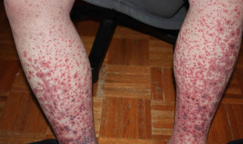
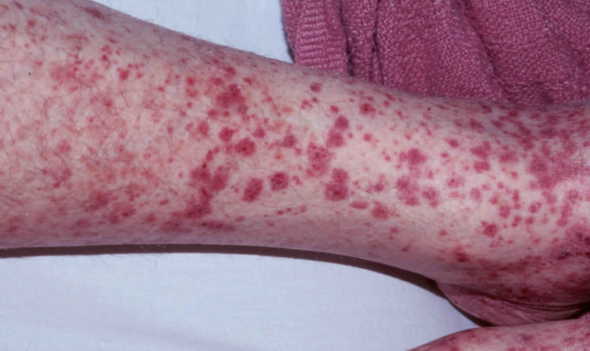
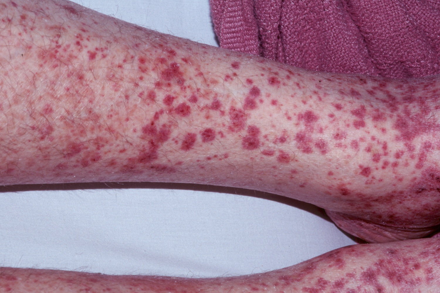
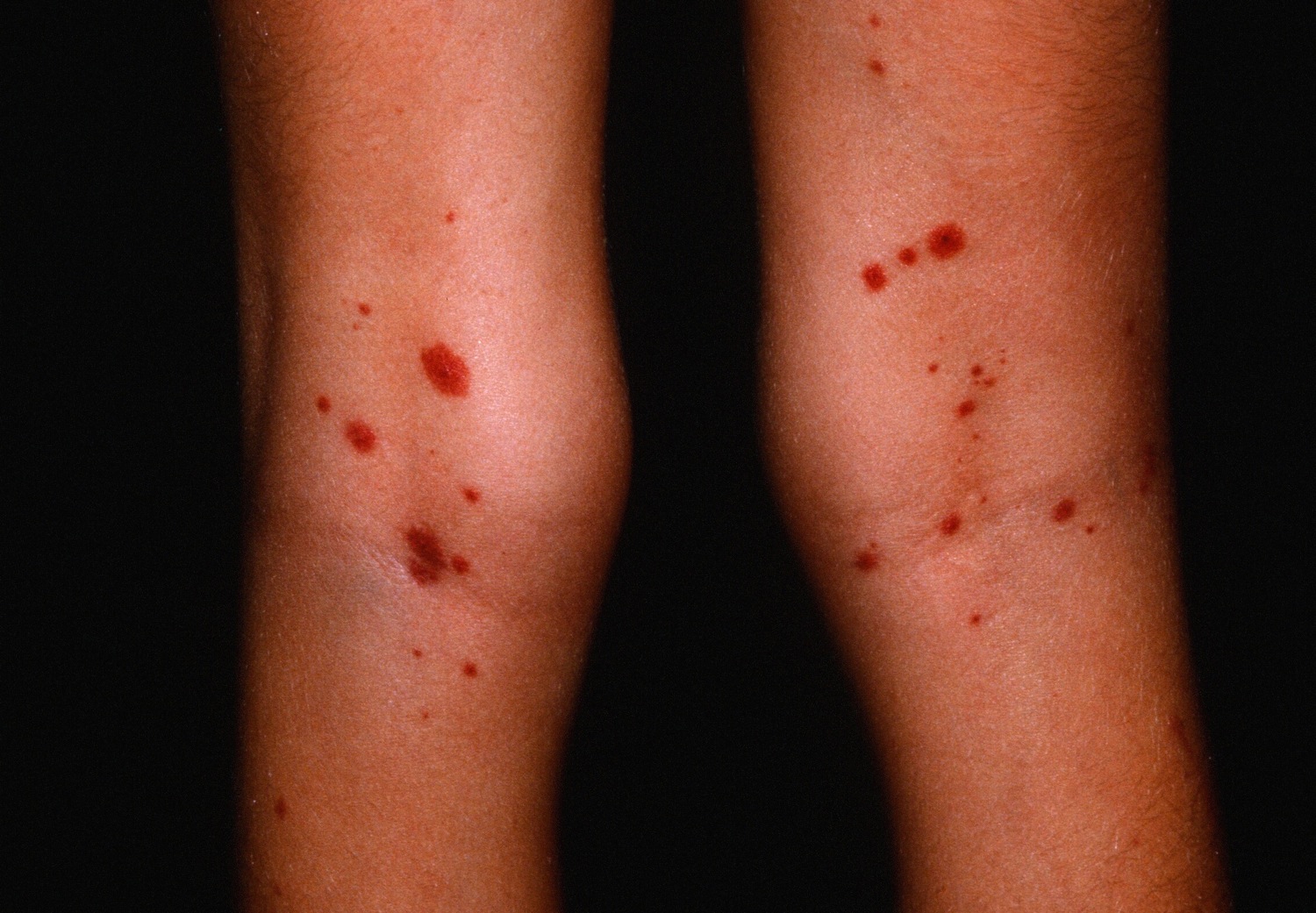
  1. Systemic juvenile idiopathic arthritis causes inflammation in one or more joints. A high daily fever that lasts at least 2 weeks either precedes or accompanies the arthritis. Individuals with systemic arthritis may also have a skin rash or enlargement of the lymph nodes (lymphadenopathy), liver (hepatomegaly), or spleen (splenomegaly).
  2. Oligoarticular juvenile idiopathic arthritis also known as oligoarthritis, is marked by the occurrence of arthritis in four or fewer joints in the first 6 months of the disease. It is divided into two subtypes depending on the course of disease. If the arthritis is confined to four or fewer joints after 6 months, then the condition is classified as persistent oligoarthritis. If more than four joints are affected after 6 months, this condition is classified as extended oligoarthritis. Individuals with oligoarthritis are at increased risk of developing inflammation of the eye (uveitis).
  3. Rheumatoid factor positive polyarticular juvenile idiopathic arthritis also known as polyarthritis or rheumatoid factor positive, causes inflammation in five or more joints within the first 6 months of the disease. Individuals with this condition also have a positive blood test for proteins called rheumatoid factors. This type of arthritis closely resembles rheumatoid arthritis as seen in adults.
  4. Rheumatoid factor negative polyarticular juvenile idiopathic arthritis also known as polyarthritis or rheumatoid factor negative, is also characterized by arthritis in five or more joints within the first 6 months of the disease. Individuals with this type, however, test negative for rheumatoid factor in the blood.
  5. Psoriatic juvenile idiopathic arthritis involves arthritis that usually occurs in combination with a skin disorder called psoriasis. Psoriasis is a condition characterized by patches of red, irritated skin that are often covered by flaky white scales. Some affected individuals develop psoriasis before arthritis while others first develop arthritis. Other features of psoriatic arthritis include abnormalities of the fingers and nails or eye problems.
  6. Enthesitis-related juvenile idiopathic arthritis is characterized by tenderness where the bone meets a tendon, ligament, or other connective tissue. The most commonly affected places are the hips, knees, and feet. This tenderness, known as enthesitis, accompanies the joint inflammation of arthritis. Enthesitis-related arthritis may also involve inflammation in parts of the body other than the joints.
  7. The last type of juvenile idiopathic arthritis is called undifferentiated arthritis. This classification is given to affected individuals who do not fit into any of the above types or who fulfill the criteria for more than one type of juvenile idiopathic arthritis.

The incidence of juvenile idiopathic arthritis in North America and Europe is estimated to be 4 to 16 in 10,000 children. One in 1,000, or approximately 294,000, children in the United States are affected. The most common type of juvenile idiopathic arthritis in the United States is oligoarticular juvenile idiopathic arthritis, which accounts for about half of all cases. For reasons that are unclear, females seem to be affected with juvenile idiopathic arthritis somewhat more frequently than males. However, in enthesitis-related juvenile idiopathic arthritis males are affected more often than females. The incidence of juvenile idiopathic arthritis varies across different populations and ethnic groups.

Table 1. Categories of Juvenile Idiopathic Arthritis

International League of Associations for Rheumatology Category Previous Nomenclature International League of Associations for Rheumatology Definition Exclusion* Adult Equivalent
Systemic-onset juvenile idiopathic arthritis Systemic-onset juvenile rheumatoid arthritis Arthritis and fever (≥2 weeks, documented quotidian × 3+ days) 1, 2, 3, 4 Adult Still disease
Plus 1 or more:
• Evanescent erythematous rash
• Generalized lymphadenopathy
• Hepatosplenomegaly
• Serositis
Polyarticular rheumatoid factor-negative Arthritis ≥5 joints during the first 6 months of disease and rheumatoid factor-negative 1, 2, 3, 4, 5
Polyarticular rheumatoid factor-positive Arthritis ≥5 joints during the first 6 months of disease and rheumatoid factor-positive × 2 at least 3 months apart 1, 2, 3, 5 Rheumatoid arthritis (RF-positive)
Oligoarthritis Pauciarticular juvenile rheumatoid arthritis Arthritis ≤4 joints during the first 6 months of disease; 2 subtypes are identified: 1, 2, 3, 4, 5
• Persistent oligoarticular juvenile idiopathic arthritis affecting no more than 4 joints
• Extended oligoarticular juvenile idiopathic arthritis, affecting a total of >4 joints after the first 6 months of disease
Enthesitis-related arthritis Seronegative enthesitis and arthritis syndrome Spondyloarthropathy Arthritis and enthesitis 1, 4, 5 Ankylosing spondylitis (if bilateral sacroiliitis)
or
Arthritis or enthesitis
Plus 2 of:
• Sacroiliac joint tenderness or inflammatory lumbosacral pain
• HLA-B27+
• Onset of arthritis in a male older than age 6 years
• Acute anterior uveitis
• History of ankylosing spondylitis, enthesitis-related arthritis, sacroiliitis with inflammatory bowel disease, reactive arthritis, or acute anterior uveitis in first-degree relative
Psoriatic arthritis Arthritis and psoriasis or Arthritis plus 2 of: 2, 3, 4, 5 Psoriatic arthritis
• Dactylitis
• Nail pitting or onycholysis
• Psoriasis in a first-degree relative
Undifferentiated arthritis Arthritis that fulfills criteria for:
• No category
or
• Two or more categories

Footnotes: * Exclusion definitions: 1. Psoriasis in patient or first-degree relative; 2. HLA-B27+ male older than age 6 years; 3. Ankylosing spondylitis, enthesitis-related arthritis, sacroiliitis with inflammatory bowel disease, Reiter syndrome, or acute anterior uveitis in a first-degree relative; 4. Rheumatoid factor-positive in 2 assessments 3 months apart; 5. Systemic-onset juvenile idiopathic arthritis in patient.

Abbreviation: HLA=human leukocyte antigen

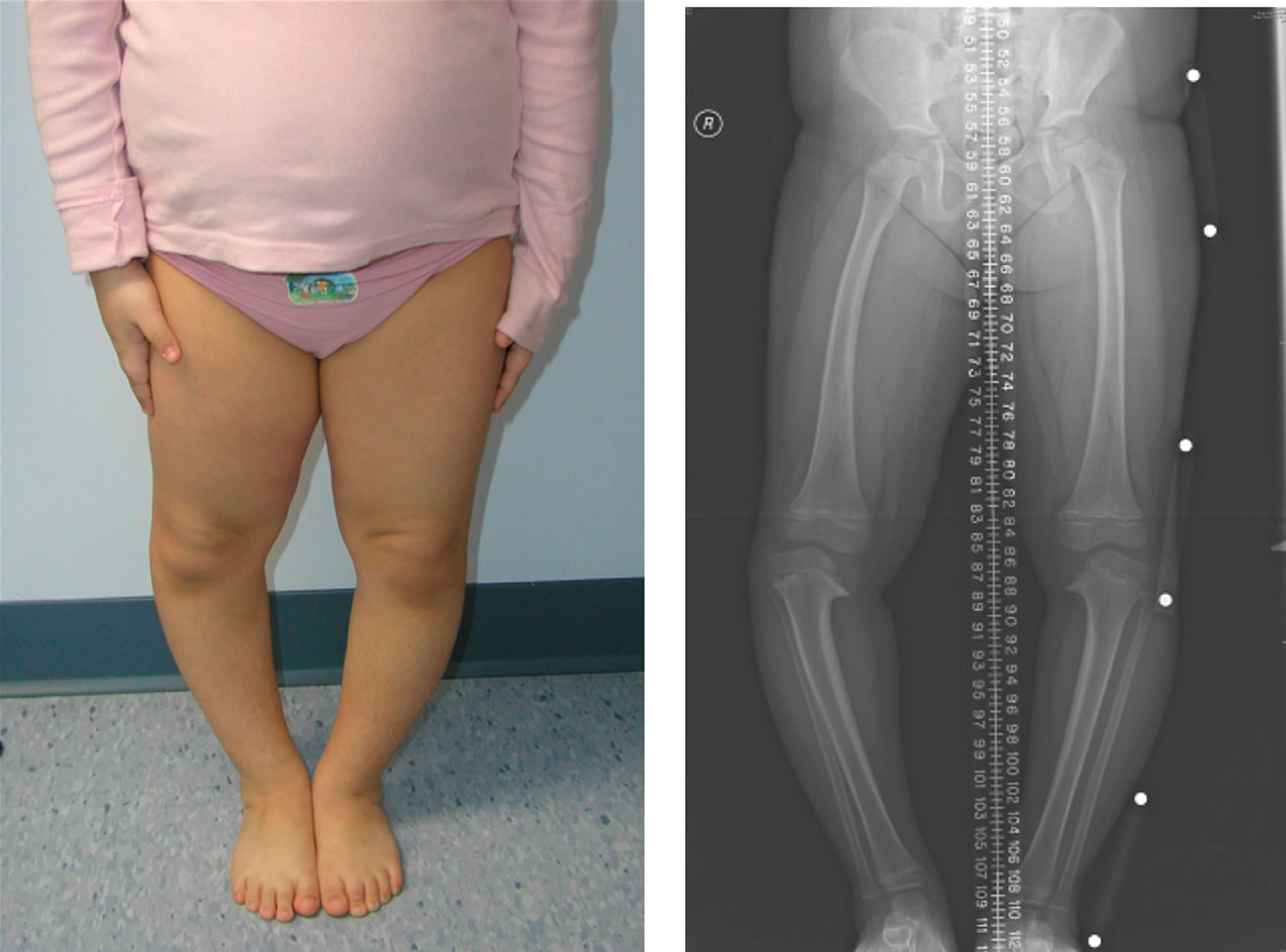
Figure 1. Juvenile idiopathic arthritis

Juvenile idiopathic arthritis

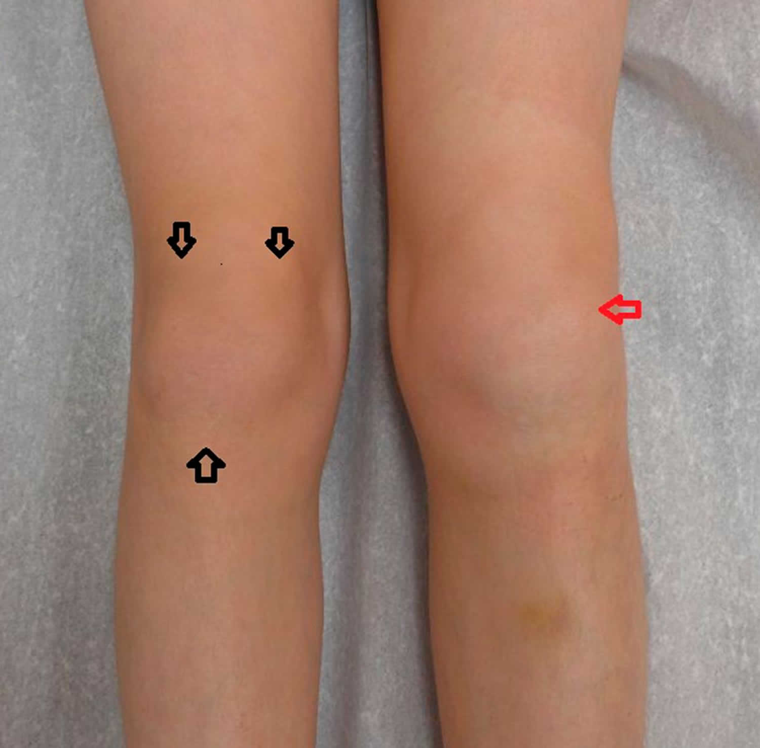
Footnote: Knee in juvenile arthritis patient showing active arthritis of left knee (red arrow) and the positions for checking enthesitis (2, 6, and 10 o’clock, black arrows).
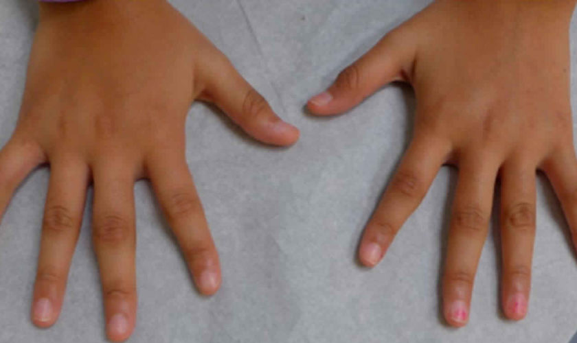
[Source 3) ]

Figure 2. Juvenile idiopathic arthritis

juvenile idiopathic arthritis

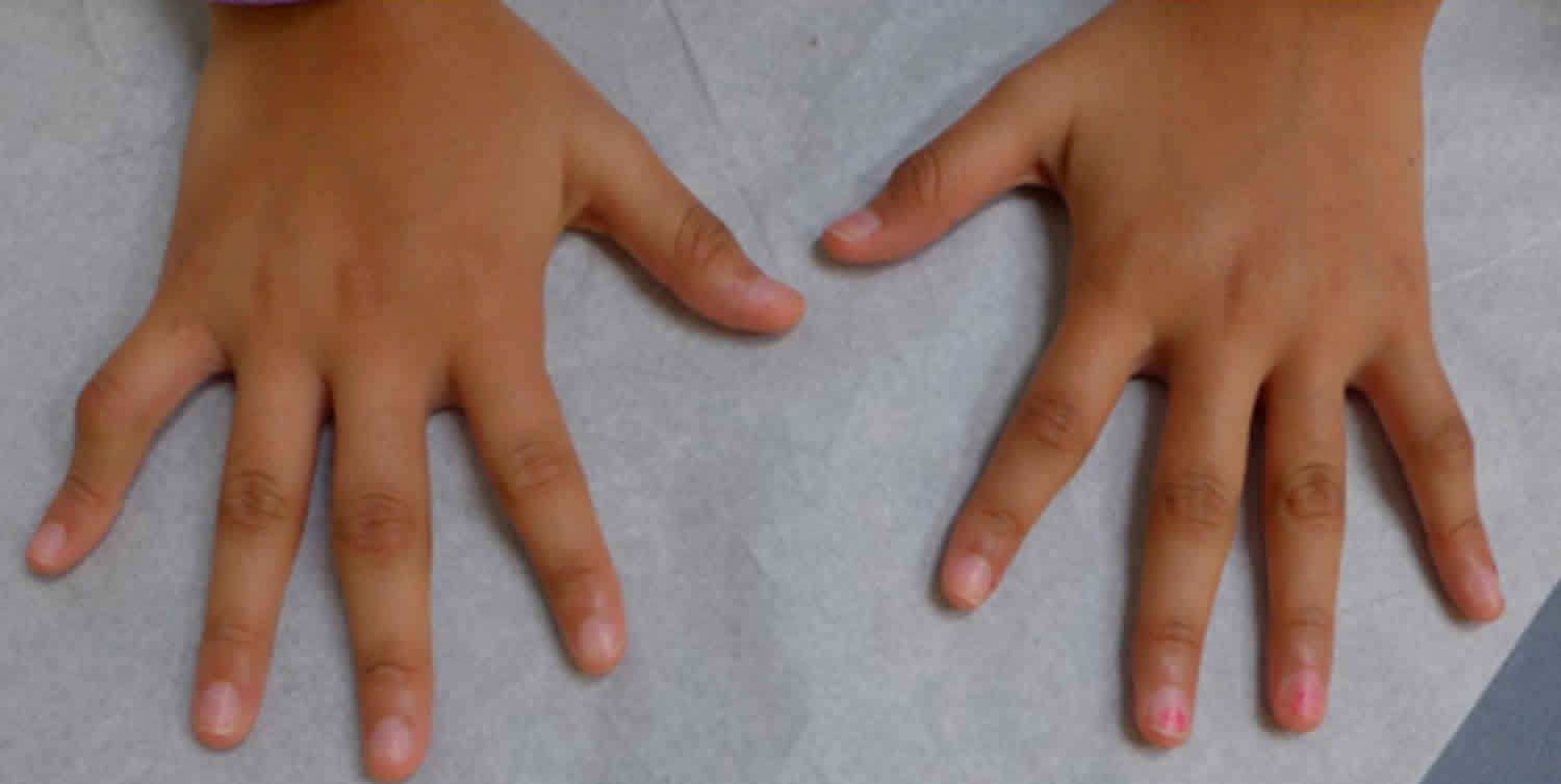
Footnote: Arthritis of several proximal interphalangeal joints bilaterally and flexion contractures with boutonniere deformity of bilateral fifth fingers in a child who has polyarticular juvenile idiopathic arthritis.

[Source 4) ]

Juvenile idiopathic arthritis causes

Juvenile idiopathic arthritis is thought to arise from a combination of genetic and environmental factors. The term “idiopathic” indicates that the specific cause of the disorder is unknown and researchers aren’t sure why kids develop juvenile idiopathic arthritis. Scientists believe kids with juvenile idiopathic arthritis have certain genes that are activated by a virus, bacteria or other external factors. But there is no evidence that foods, toxins, allergies or lack of vitamins cause the disease.

Juvenile idiopathic arthritis is a type of autoimmune or autoinflammatory disease. That means the immune system, which is supposed to fight invaders like germs and viruses, gets confused and attacks the body’s cells and tissues. This causes the body to release inflammatory chemicals that attack the synovium (tissue lining around a joint). It produces fluid that cushions joints and helps them move smoothly. An inflamed synovium may make a joint feel painful or tender, look red or swollen or difficult to move.

Juvenile idiopathic arthritis signs and symptoms result from excessive inflammation in and around the joints. Inflammation occurs when the immune system sends signaling molecules and white blood cells to a site of injury or disease to fight microbial invaders and facilitate tissue repair. Normally, the body stops the inflammatory response after healing is complete to prevent damage to its own cells and tissues. In people with juvenile idiopathic arthritis, the inflammatory response is prolonged, particularly during joint movement. The reasons for this excessive inflammatory response are unclear.

Researchers have identified changes in several genes that may influence the risk of developing juvenile idiopathic arthritis. Many of these genes are thought to play roles in immune system function. Some of these genes belong to a family of genes that provide instructions for making a group of related proteins called the human leukocyte antigen (HLA) complex. The HLA complex helps the immune system distinguish the body’s own proteins from proteins made by foreign invaders (such as viruses and bacteria). Each HLA gene has many different normal variations, allowing each person’s immune system to react to a wide range of foreign proteins. Certain normal variations of several HLA genes seem to affect the risk of developing juvenile idiopathic arthritis, and the specific type of the condition that a person may have.

Additional unknown genetic influences and environmental factors, such as infection and other issues that affect immune health, are also likely to influence a person’s chances of developing this complex disorder.

Juvenile idiopathic arthritis inheritance pattern

Most cases of juvenile idiopathic arthritis are sporadic, which means they occur in people with no history of the disorder in their family. A small percentage of cases of juvenile idiopathic arthritis have been reported to run in families, although the inheritance pattern of the condition is unclear. A sibling of a person with juvenile idiopathic arthritis has an estimated risk of developing the condition that is about 12 times that of the general population.

Juvenile idiopathic arthritis symptoms

It’s also possible that a child may start off with one type of juvenile idiopathic arthritis but develop symptoms of another type later.

The most common symptoms of juvenile idiopathic arthritis include:

  • Joint pain or stiffness; may get worse after waking up or staying in one position too long.
  • Red, swollen, tender or warm joints.
  • Feeling very tired or rundown (fatigue).
  • Blurry vision or dry, gritty eyes.
  • Rash.
  • Appetite loss.
  • High fever.

Certain symptoms are specific to the type of arthritis a child has. There are six types of juvenile idiopathic arthritis:

  1. Oligoarthritis: Affects four or fewer joints, typically the large ones (knees, ankles, elbows). Most common subtype of juvenile idiopathic arthritis.
  2. Polyarthritis: Affects five or more joints, often on both sides of the body (both knees, both wrists, etc.). May affect large and small joints. Affects about 25% of children with juvenile idiopathic arthritis.
  3. Systemic: Affects the entire body (joints, skin and internal organs). Symptoms may include a high spiking fever (103°F or higher) that lasts at least two weeks and rash. Affects about 10% of children with juvenile idiopathic arthritis.
  4. Psoriatic arthritis (PsA): Joint symptoms and a scaly rash behind the ears and/or on the eyelids, elbows, knees, belly button and scalp. Skin symptoms may occur before or after joint symptoms appear. May affect one or more joints, often the wrists, knees, ankles, fingers or toes.
  5. Enthesitis-related: Also known as spondyloarthritis. Affects where the muscles, ligaments or tendons attach to the bone (entheses). Commonly affects the hips, knees and feet, but may also affect the fingers, elbows, pelvis, chest, digestive tract (Crohn’s disease or ulcerative colitis) and lower back (ankylosing spondylitis). More common in boys; typically appears in children between the ages of eight and 15.
  6. Undifferentiated: Symptoms don’t match up perfectly with any of the subtypes, but inflammation is present in one or more joints.

Juvenile idiopathic arthritis symptoms may also come and go. Periods of lots inflammation and worsening symptoms are called flares. A flare can last for days or months.

Juvenile idiopathic arthritis complications

If juvenile idiopathic arthritis inflammation goes unchecked, it can damage the lining that covers the ends of bones in a joint (cartilage), and the bones themselves. Here are some other ways juvenile idiopathic arthritis can affect the body:

  • Eyes. Dryness, pain, redness, sensitivity to light and trouble seeing properly caused by uveitis (chronic eye inflammation). More common with oligoarthritis.
  • Bones. Chronic inflammation and use of corticosteroids may cause growth delay in some children with juvenile idiopathic arthritis. Bones may get thinner and break more easily (osteoporosis).
  • Mouth/Jaw. Difficulty chewing, brushing or flossing. More than half of children with juvenile idiopathic arthritis have jaw involvement.
  • Neck. Inflammation of the cervical spine can cause neck pain or stiffness. Swollen neck glands could also signal an infection for kids with Sjuvenile idiopathic arthritis or who take immunosuppressing drugs.
  • Ankles/feet. Foot pain and difficulty walking. More common in polyarthritis and enthesitis-related arthritis.
  • Skin. Symptoms can range from a faint salmon colored rash (Sjuvenile idiopathic arthritis) to a red, scaly rash (psoriatic arthritis).
  • Lungs. Inflammation and scarring that can lead to shortness of breath and lung disease. May occur in Sjuvenile idiopathic arthritis.
  • Heart. Inflammation may cause damage to the heart muscle. May occur in Sjuvenile idiopathic arthritis.
  • Digestive Tract. Abdominal pain and diarrhea. More common in children with spine arthritis or ankylosing spondylitis.
  • Reproductive organs. Late onset of puberty. Certain drugs such as cyclophosphamide may lead to fertility problems later.
  • Weight loss or gain. Due to changes in appetite, jaw involvement or difficulty exercising. Being overweight puts extra stress on the joints.

Controlling inflammation and managing disease can prevent damage and complications from these health effects.

Juvenile idiopathic arthritis diagnosis

According to American College of Rheumatology a child must have inflammation in one or more joints lasting at least six weeks, be under 16 years old and have all other conditions ruled out before being diagnosed with juvenile idiopathic arthritis.

A pediatrician may be the first doctor to start figuring out what’s causing symptoms. It’s likely that parents will be referred to a rheumatologist (a doctor with specialized training in treating arthritis). Some rheumatologists only treat children. Others only treat adults. Some of them treat both. A medical history, physical examination and blood tests helps to make the correct diagnosis.

Medical history. The doctor will ask questions about the child’s health history, when symptoms started and how long they lasted. This helps rule out other causes like trauma or infection. The doctor will also ask about the family’s medical history.

Physical examination. The doctor will look for joint tenderness, swelling, warmth and painful or limited movement and test range of motion. Eyes and skin may also be checked.

Laboratory tests. The doctor may order blood tests that look for certain proteins and chemicals found in some people with arthritis. These tests include:

  • Erythrocyte sedimentation rate (ESR, or “sed rate”) and C-reactive protein (CRP) tests: High ESR rates and CRP levels signal severe inflammation in the body.
  • Antinuclear antibody (ANA) test: A positive ANA test is associated with many types of arthritis, but kids without juvenile idiopathic arthritis may also have a positive ANA.
  • Rheumatoid factor (RF) test: May show up in children with polyarthritis. A positive rheumatoid factor may signal more serious disease.
  • HLA-B27 typing (a genetic marker): The HLA-B27 gene is associated with enthesitis-related types of arthritis, such ankylosing spondylitis.
  • Complete blood count (CBC): Raised levels of white blood cells and decreased levels of red blood cells is linked to certain types of arthritis.
  • Imaging. The doctor may order imaging tests, such as X-rays, ultrasound and MRI or CT scans, to look for signs of joint damage.

Juvenile idiopathic arthritis treatment

There is no cure for juvenile idiopathic arthritis but remission (little or no disease activity or symptoms) is possible. Early aggressive treatment is key to getting the disease under control as quickly as possible.

The goals of juvenile idiopathic arthritis treatment are to:

  • Slow down or stop inflammation.
  • Relieve symptoms, control pain and improve quality of life.
  • Prevent joint and organ damage.
  • Preserve joint function and mobility.
  • Reduce long-term health effects.
  • Achieve remission (little or no disease activity or symptoms).

Treatment for juvenile idiopathic arthritis varies depending on disease type and severity. A well-rounded plan includes medication, complementary therapies and healthy lifestyle habits.

Intra-articular steroid injections (triamcinolone hexacetonide or methylprednisolone acetate as the most used drugs), systemic steroids and methotrexate (MTX) have played a major role in the treatment of juvenile idiopathic arthritis for many years. A recent trial has confirmed that the effectiveness of intra-articular steroid injections is not enhanced by concomitant administration of methotrexate 5). Nevertheless, juvenile idiopathic arthritis patients’ prognosis has dramatically changed in the last decades, thanks to the introduction of biologic agents at the beginning of 2000s. These medications have shown to be effective in a sizeable proportion of patients refractory or intolerant to methotrexate, although methotrexate remains the most widely used conventional treatment in the management of juvenile idiopathic arthritis for its effectiveness at achieving disease control, its acceptable toxic effects, worldwide availability and low cost 6). Among biologics, there are currently five anti-tumor necrosis factor (TNF) drugs available in juvenile idiopathic arthritis: four with published results [etanercept (ETN), adalimumab (ADA), infliximab (IFX) and golimumab], and one which has recently completed enrollment (certolizumab pegol) 7).

Table 2 shows the tumor necrosis factor (TNF) inhibitors approved by the European Medicines Agency (EMA) and the U.S. Food and Drug Administration (FDA) for use in juvenile idiopathic arthritis. Etanercept was the first TNF inhibitor to gain approval nearly two decades ago. The sustained efficacy and the good safety profile of etanercept have been confirmed in the open-label, multicentre CLIPPER study 8), by the national juvenile idiopathic arthritis German database BiKer 9), as well as in other multicentric studies, which showed a low rate of serious infections, just slightly increased compared with methotrexate alone 10).

Among TNF inhibitors, a recently approved treatment in polyarticular-course juvenile idiopathic arthritis is golimumab, a fully human monoclonal antibody that can be administered by subcutaneous injection (a trial with intravenous infusion is on-going). Although the primary endpoint of the study was not met in the withdrawal phase (juvenile idiopathic arthritis flare rates with golimumab vs. placebo: 41 vs. 47%), golimumab caused a significant improvement in juvenile idiopathic arthritis and, therefore, has been approved by European Medicines Agency for the treatment of children with polyarticular-course juvenile idiopathic arthritis and weight above 40 kg 11). Other alternatives to TNF inhibitors for treatment of polyarticular-course juvenile idiopathic arthritis include tocilizumab and abatacept, both approved for use in polyarticular-course juvenile idiopathic arthritis 12).

The IL-6 inhibitor tocilizumab (TCZ) and the IL-1 inhibitors (anakinra and canakinumab) are the most commonly prescribed biologic agents in systemic juvenile idiopathic arthritis, whereas, rilonacept has not gone on to receive an approval for a juvenile idiopathic arthritis indication 13). The introduction of biologics in the treatment of systemic juvenile idiopathic arthritis has significantly increased the proportion of patients with inactive disease and reduced the occurrence of functional limitations because of the disease 14). Yokota et al.15) described more than 400 patients with sjuvenile idiopathic arthritis, all treated with tocilizumab and reported that, compared with previous studies, there were more serious adverse events, especially infections, but with an overall acceptable safety profile. Among 26 cases of macrophage activation syndrome (MAS), no deaths occurred, underling the importance of early detection and treatment of such a severe complication 16). Grom et al. 17). reported episodes of macrophage activation syndrome occurring in individuals with systemic juvenile idiopathic arthritis treated with canakinumab, but implicated infectious triggers rather than being caused by canakinumab.

Table 2. Interventional studies in juvenile idiopathic arthritis sponsored by pharmaceutical companies or investigator’s initiated studies

Drug Target Dosage Approval
Abatacept CD80/86 10 mg/kg week 0, 2, 4, then q4 weeks EMA/FDA
Adalimumab TNF-α 24mg/m2 every 2 weeks; max 40mg EMA/FDA
Anakinra IL-1 1–4 mg/kg/day EMA
Canakinumab IL-1 ≥2 years: 4 mg/kg/dose every 4 weeks Maximum dose: 300mg EMA/FDA
Etanercept TNF-α,β 0.8 mg/kg/week or 0.4 mg/kg twice weekly; Maximum dose: 50mg EMA/FDA
Infliximab TNF-α 6–10 mg/kg every 2 weeks to 2 months
Tocilizumab IL-6 polyarticular juvenile idiopathic arthritis more than 2 years, less than 30 kg 10 mg/kg every 4 weeks
polyarticular juvenile idiopathic arthritis >2 years, more than 30 kg 8 mg/kg every 4 weeks
systemic juvenile idiopathic arthritis more than 2 years, less than 30 kg 12 mg/kg every 2 weeks more than 2 years, >30 kg 8 mg/kg every 2 weeks
EMA/FDA

Abbreviations: EMA = European Medicines Agency; FDA = U.S. Food and Drug Administration

Medications

Drugs that control disease activity

Disease-modifying antirheumatic drugs (DMARDs). These drugs work to modify the course of the disease. DMARDs relieve symptoms by suppressing the immune system so it doesn’t attack the joints.

  • Traditional disease-modifying antirheumatic drugs (DMARDs). These have been used the longest and have a broad immune-suppressing effect. The most commonly-used drug for juvenile idiopathic arthritis is methotrexate. These medicines are available in pill or injection form.
  • Biologics. These drugs target certain steps or chemicals in the inflammatory process and may work more quickly than traditional DMARDs. They are self-injected or given by infusion in a doctor’s office.

Drugs that relieve symptoms

Nonsteroidal anti-inflammatory drugs (NSAIDs) and analgesics (pain relievers). These drugs relieve pain but cannot reduce joint damage or change the course of juvenile idiopathic arthritis. These medications are available over-the-counter or by prescription.

Every child with juvenile idiopathic arthritis is different. The standard of care involves trying methotrexate first, but many doctors start with a biologic/disease-modifying antirheumatic drug (DMARD) combination to combat inflammation as quickly as possible. As doctors monitor the disease, drugs may be added or removed.

Exercise

Regular exercise helps ease joint stiffness and pain. Low-impact and joint-friendly activities like walking, swimming, biking and yoga are best, but kids with well-controlled disease can participate in just about any activity they wish, if their doctor or physical therapist approves.

Physical therapies and assistive devices

Physical and occupational therapy can improve a child’s quality of life by teaching them ways to stay active and how to perform daily tasks with ease. Children with juvenile idiopathic arthritis may also have trouble with balance and weaker motor skills, or the ability to move and coordinate large muscle groups. Participating in regular physical and occupational therapy can improve coordination and balance, among other things. Here are some other ways physical and occupational therapists can help a child with juvenile idiopathic arthritis:

  • Teach and guide them through strengthening and flexibility exercises.
  • Perform body manipulation.
  • Prescribe assistive devices (e.g. braces, splints).

Surgery

Thanks to treatment advances, including biologics, many children will never need surgery. But for children whose disease couldn’t be controlled early enough, surgery can provide much needed relief and restore joint function.

Damaged parts of a joint are replaced with metal, ceramic or plastic prosthesis. Hip and knee replacements are most common, and many surgeries can be performed on an outpatient basis. There are other surgeries that can improve joint function and quality of life but require much less cutting than joint replacement. For example, with arthroscopy, a thin, lighted tube with a camera attached is inserted through a small incision. This helps the surgeon examine the child’s joints and perform procedures, such as removing a loose piece of cartilage. An orthopedic surgeon will evaluate and determine if surgery is the best option.

Lifestyle and home remedies

Certain habits can help manage disease and relieve symptoms.

Healthy eating

While there is no special juvenile idiopathic arthritis diet, studies show that some foods help to curb inflammation. These include the foods found in the Mediterranean diet, i.e. fatty fish, fruits, vegetables, whole grains and extra virgin olive oil among others. Children with juvenile idiopathic arthritis should avoid or cut back on foods that can cause inflammation such as high-fat, sugary and processed foods.

Balancing activity with rest

When juvenile idiopathic arthritis is active, and joints feel painful, swollen or stiff, it’s important to balance light activity with rest. Rest helps reduce inflammation and fatigue that can come with a flare. Taking breaks throughout the day protects joints and preserves energy.

Hot and cold treatments

Heat treatments, such as heat pads or warm baths, work best for soothing stiff joints and tired muscles. Cold is best for acute pain. It can numb painful areas and reduce inflammation.

Topical products

Creams, gels or stick-on patches can ease the pain in a joint or muscle. Some contain the same medicine that’s in a pill, and others use ingredients that irritate nerves to distract from pain.

Mind-Body Therapies

There are different ways to relax and stop focusing on pain. They include meditation, deep breathing and practicing visualization, or thinking about peaceful places or happy memories. Children with juvenile idiopathic arthritis may also benefit from certain distraction techniques to lessen pain, especially during shot time. These include listening to music, coloring or drawing, reading and being read to.

Massage and acupuncture can also help reduce pain and ease stress or anxiety. Acupuncture involves inserting fine needles into the body along special points to relieve pain. If there’s a fear of needles, acupressure, which uses firm pressure, may be used instead.

Supplements

The use of supplements is rarely studied in children. But some supplements that adults take may help children too. These include curcumin, a substance found in turmeric, and omega-3 fish oil supplements, which may help with joint pain and stiffness. Taking calcium and vitamin D can help build strong bones. Always discuss supplements and vitamins with a child’s doctor. Some may cause side effects and interact with other medications.

Stress and emotions

Children and teens with juvenile idiopathic arthritis are more likely to get depressed because they are living with a chronic disease. Having a strong support system of friends and family can provide emotional support during tough times. Children with juvenile idiopathic arthritis can make new friends dealing with similar struggles at various Arthritis Foundation Juvenile Arthritis events held throughout the year. Therapist and psychologists can also help kids with juvenile arthritis deal with tough emotions and teach positive coping strategies.

Living with juvenile idiopathic arthritis

If you have juvenile idiopathic arthritis, it may be hard to get out of bed some mornings. Periods of inactivity, like sleeping for 8 hours, can be followed by stiffness.

It may be tempting to roll back into bed and sleep the day away, but that can make things worse. Even though you may feel lousy sometimes, gentle movement can help you feel better. Just as runners, bodybuilders, and other athletes do stretching exercises to warm up, gentle massaging and stretching can help soothe the muscles and ligaments around sore joints.

Once a person is up and moving, the discomfort usually lessens. Exercise can help keep full motion in your joints and strengthen your muscles and bones. A physical therapist can help you plan an effective exercise program to do at home.

Proper nutrition can improve anyone’s overall health. A dietitian can help you to understand the basics of a healthy diet. For example, when your symptoms flare up, you might feel sick and unable to eat as much. A dietitian can help you find foods that have a higher nutritional value to make up for having a poor appetite.

A positive mental outlook is just as important as exercise and a healthy diet. If you feel depressed or angry sometimes, talk to someone who can support you. Tell your parents, your doctor, or a friend about how you feel. It also may help to do simple things that we often take for granted. For example, each day try to do something that you enjoy and that makes you happy.

Most teens with juvenile idiopathic arthritis do the same stuff as other teens — go to regular schools, hang out with friends, and stay active physically, academically, and socially. Learning more about juvenile idiopathic arthritis and taking charge of your medical care can help some people feel more in control, too.

Your doctor and other medical professionals are there to support you and can help you manage the condition so that it has the least possible impact on your life.

Juvenile idiopathic arthritis prognosis

In a study of 251 children from southern Sweden with a validated diagnosis of juvenile idiopathic arthritis 18), consisting of all subgroups in the total follow-up period, 40.0% of the years were with inactive disease (defined as no arthritis or uveitis), 54.8% were active due to arthritis with or without uveitis, and 5.2% were active because of uveitis only. The median follow-up time was 8.0 years. In the subgroups, the percentages of inactive disease presents as follows: enthesitis-related arthritis 38.4%, oligoarthritis 42.5% (with extended oligoarthritis 33.3% and persistent oligoarthritis 46.5%), rheumatoid factor-negative 37.3%, rheumatoid factor-positive 25.9%, juvenile psoriatic arthritis 33.3%, systemic juvenile idiopathic arthritis 64.0%, and undifferentiated juvenile idiopathic arthritis 43.5%. 28.8% of the inactive years were without treatment. One patient that was lost to follow-up was later found out to have died.

Uveitis was seen in 27 (10.8%) of the children, 8.0% had chronic uveitis, and 4.0% had acute uveitis (3 individuals have had both manifestations). Fourteen of the children have had uveitis in their debut year (10 chronic). The median debut age of chronic uveitis is 5.5 years (range 0–16 years). There are no cases of uveitis in the rheumatoid factor-positive, juvenile psoriatic arthritis, or systemic juvenile idiopathic arthritis groups. The risk of chronic uveitis is 10.0% at 12 years of follow-up.

In total, 23 (9.2%) individuals have been treated with joint corrective orthopedic surgery, 8 of them with multiple procedures (3 with rheumatoid factor-positive juvenile idiopathic arthritis, 2 with oligoarticular disease, 1 with rheumatoid factor-negative juvenile idiopathic arthritis, 1 with juvenile psoriatic arthritis and 1 with ujuvenile idiopathic arthritis). However, only 11 individuals (4.4%) have undergone large orthopedic surgery (arthroplasty, osteotomies, or arthrodesis). The procedures were 17 synovectomies (5 with diagnostic purpose), 7 arthrodeses, 6 osteotomies, 4 medial knee epiphysiodeses, 1 arthrolysis, 1 arthroplasty (hip prosthesis), 1 volar tenosynovectomy, and 1 finger tendon transposition. The need for orthopedic surgery is the highest (23.5%) in the group with rheumatoid factor-positive juvenile idiopathic arthritis. The risk for joint corrective surgery is 17.9% at 12 years of follow-up using. The risk for a serious orthopedic procedure is 9.4% at 12 years of follow-up.

References   [ + ]

read more

Hay fever

hay fever

What is hay fever

Hay fever, also known as seasonal allergic rhinitis, is an allergic reaction to allergens like molds, pollens, dust mites, insects and animal dander. Hay fever affects millions of people worldwide. Symptoms of hay fever are seasonal. You will feel worse when the pollens that affect you are at their highest levels. Symptoms include sneezing, stuffiness, a runny nose and itchiness in your nose, the roof of your mouth, throat, eyes or ears. These allergic reactions are most commonly caused by pollen and mold spores in the air, which start a chain reaction in your immune system.Sometimes symptoms are caused by molds or flakes of animal skin (dander) from pets. If pollen is the cause, you will feel worse when pollen levels are high.

Pollen comes from flowering trees, grass, and weeds. If you are allergic to pollen, you will notice your symptoms are worse on hot, dry days when wind carries the pollen. On rainy days, pollen often is washed to the ground, which means you are less likely to breathe it.

  • Allergies that occur in the spring (late April and May) are often due to tree pollen.
  • Allergies that occur in the summer (late May to mid-July) are often due to grass and weed pollen.
  • Allergies that occur in the fall (late August to the first frost) are often due to ragweed.

Allergens that can cause perennial (year-round) allergic rhinitis include:

  • Mold. Mold is common where water tends to collect, such as shower curtains and damp basements. It can also be found in rotting logs, hay, and mulch. This allergy is usually worse during humid and rainy weather.
  • Animal dander.Proteins found in the skin, saliva, and urine of furry pets such as cats and dogs are allergens. You can be exposed to dander when handling an animal or from house dust that contains dander.
  • Dust. Many allergens, including dust mites, are in dust. Dust mites are tiny living creatures found in bedding, mattresses, carpeting, and upholstered furniture. They live on dead skin cells and other things found in house dust.

Is hay fever contagious?

Hay fever, also called allergic rhinitis, causes cold-like signs and symptoms, such as a runny nose, itchy eyes, congestion, sneezing and sinus pressure. But unlike a cold, hay fever isn’t caused by a virus. Hay fever is caused by an allergic response to outdoor or indoor allergens, such as pollen, dust mites or tiny flecks of skin and saliva shed by cats, dogs and other animals with fur or feathers (pet dander).

Hay fever lasts longer than a cold or the flu—up to several weeks—and does not cause fever. With hay fever, the fluid from your nose is thin, watery, and clear. With a cold or the flu, the fluid tends to be thicker. Itching (mostly eyes, nose, mouth, throat, and skin) is common with hay fever but not with a cold or the flu. Sneezing happens more with hay fever. Hay fever does not cause fever.

Besides making you miserable, hay fever can affect your performance at work or school and generally interfere with your life. But you don’t have to put up with annoying symptoms. You can learn to avoid triggers and find the right treatment.

Figure 1. Hay fever rash

hay fever rash

What causes hay fever?

Allergens cause your body to respond with an allergic reaction. When you are exposed to something you are allergic to, your body releases chemicals. One of theses chemicals is histamine, which is your body’s defense against the allergen. The release of histamine causes swelling, itching, sneezing, runny nose, and watery eyes.

If you are allergic to pollen, you will notice that your symptoms are worse on hot, dry days when wind carries the pollen. On rainy days, pollen often is washed to the ground, which means you are less likely to breathe it.

Hay fever symptoms are most often triggered by tree pollen in the early spring. Grasses are often the culprit during the late spring and early summer. Weeds are most often the cause of late summer and fall hay fever symptoms.

What is pollen?

The word pollen is derived from the Greek word meaning ‘fine flour’. Pollen is the male fertilizing agent (male gametes or sperm cells) of flowering plants, trees, grasses and weeds and the role of the pollen grain is to fertilize the female flower to reproduce plant species. Pollen is also a major allergen that causes symptoms of seasonal allergic rhinitis (hay fever).

Pollen from plants with colorful flowers, like roses, usually do not cause allergies. These plants large, waxy pollen are carried from plant to plant by bees and other insects for fertilization. On the other hand, many trees, grasses and low-growing weeds have small, light, dry powdery pollen that are easily spread by wind. These are the pollen that trigger allergy symptoms.

Each plant has a period of pollination that does not vary much from year to year. However, weather conditions affect pollen levels. For instance, wind and humidity may affect pollen counts. Because pollen are small, light and dry, they can be easily spread by wind, which keeps pollen airborne and carries it over long distances. When the air is humid, such as during or after it rains, pollen becomes damp and heavy with moisture, keeping it still and on the ground. Seasonal allergic rhinitis is often caused by tree pollen in the early spring. During the late spring and early summer, grass pollen often cause symptoms. Late summer and fall hay fever is caused by weed pollen. In warmer places, pollination can be year-round.

Allergy symptoms are often minimal on days that are rainy, cloudy or windless, because pollen does not move about during these conditions. Hot, dry and windy weather signals greater pollen and mold distribution and therefore, increased allergy symptoms.

Pollen grains can be spread by birds, bees or wind:

  • Some plants (such as flowering plants, including wattle trees) produce small amounts of pollen which are distributed by birds and bees from one plant to another.
  • Other plants (such as pasture grasses and weeds) rely on the wind to spread their pollen. These pollen are produced in vast quantities, blow long distances and cause allergies in people, even if they live a long way from the source.

Most of the pollen that cause seasonal allergies is produced by airborne pollen from grasses, trees and weeds:

  • Improved pasture grasses are more allergenic than native grasses.
  • Pollen from exotic trees, which are planted for their autumn colors, is more allergenic than pollen from native trees.

How long does hay fever last

Pollen seasons can last for several months. For more information you may want to visit the American Academy of Allergy, Asthma & Immunology’s National Allergy Bureau (NAB™) to find information on the current pollen and mold spore levels in your area: http://www.aaaai.org/global/nab-pollen-counts

The National Allergy Bureau currently provides the most accurate and reliable pollen and mold levels from approximately 84 counting stations throughout the United States, one counting station in Canada, and six counting stations in Argentina. The stations use air sampling equipment to collect airborne pollen and spores which are then examined microscopically. This information is also used for research to aid in the diagnosis, treatment and management of allergic diseases.

Pollen counts from the National Allergy Bureau stations are based on actual counts, which reflect the real day to day weather events. Predicted counts reported on many websites may be based on pollen data from previous years and general weather forecasts.

Ragweed Pollen

Ragweed and other weeds such as curly dock, lambs quarters, pigweed, plantain, sheep sorrel and sagebrush are some of the most prolific producers of pollen allergens .

Although the ragweed pollen season runs from August to November, ragweed pollen levels usually peak in mid-September in many areas in the country.

In addition, pollen counts are highest between 5:00 a.m. – 10:00 a.m. and on dry, hot and windy days.

Preventive Strategies

  • Avoid the outdoors between 5:00 a.m. – 10:00 a.m. Save outside activities for late afternoon or after a heavy rain, when pollen levels are lower.
  • Keep windows in your home and car closed to lower exposure to pollen. To keep cool, use air conditioners and avoid using window and attic fans.
  • Be aware that pollen can also be transported indoors on people and pets.
  • Dry your clothes in an automatic dryer rather than hanging them outside. Otherwise pollen can collect on clothing and be carried indoors.

Grass Pollen

As with tree pollen, grass pollen is regional as well as seasonal. In addition, grass pollen levels can be affected by temperature, time of day and rain.

Of the 1,200 species of grass that grow in North America, only a small percentage of these cause allergies. The most common grasses that can cause allergies are:

  • Bermuda grass
  • Johnson grass
  • Kentucky bluegrass
  • Orchard grass
  • Sweet vernal grass
  • Timothy grass

Preventive Strategies:

  • If you have a grass lawn, have someone else do the mowing. If you must mow the lawn yourself, wear a mask.
  • Keep grass cut short.
  • Choose ground covers that don’t produce much pollen, such as Irish moss, bunch, and dichondra.
  • Avoid the outdoors between 5:00 a.m. – 10:00 a.m. Save outside activities for late afternoon or after a heavy rain, when pollen levels are lower.
  • Keep windows in your home and car closed to lower exposure to pollen. To keep cool, use air conditioners and avoid using window and attic fans.
  • Be aware that pollen can also be transported indoors on people and pets.
  • Dry your clothes in an automatic dryer rather than hanging them outside. Otherwise pollen can collect on clothing and be carried indoors.

Tree Pollen

Trees can aggravate your allergy whether or not they are on your property, since trees release large amounts of pollen that can be distributed miles away from the original source.

Trees are the earliest pollen producers, releasing their pollen as early as January in the Southern states and as late as May or June in the Northern states.

Most allergies are specific to one type of tree such as:

  • catalpa
  • elm
  • hickory
  • olive
  • pecan
  • sycamore
  • walnut

or to the male cultivar of certain trees. The female of these species are totally pollen-free:

  • ash
  • box elder
  • cottonwood
  • date palm
  • maple (red)
  • maple (silver)
  • Phoenix palm
  • poplar
  • willow

Some people, though, do show cross-reactivity among trees in the alder, beech, birch and oak family, and the juniper and cedar family.

Preventive Strategies

  • If you buy trees for your yard, look for species that do not aggravate allergies such as crape myrtle, dogwood, fig, fir, palm, pear, plum, redbud and redwood trees or the female cultivars of ash, box elder, cottonwood, maple, palm, poplar or willow trees.
  • Avoid the outdoors between 5:00 a.m. – 10:00 a.m. Save outside activities for late afternoon or after a heavy rain, when pollen levels are lower.
  • Keep windows in your home and car closed to lower exposure to pollen. To keep cool, use air conditioners and avoid using window and attic fans.
  • Be aware that pollen can also be transported indoors on people and pets.
  • Dry your clothes in an automatic dryer rather than hanging them outside. Otherwise pollen can collect on clothing and be carried indoors.

Hay fever causes

Your immune system controls how your body defends itself. For instance, if you have an allergy to pollen, your immune system identifies pollen as an invader or allergen. Your immune system overreacts by producing antibodies called Immunoglobulin E (IgE) 1). These antibodies travel to mast cells that release histamine and other chemicals, causing an allergic reaction that leads to the signs and symptoms of hay fever — such as sneezing, stuffiness, a runny nose, itching and post-nasal drip.

Risk factors for hay fever

The following can increase your risk of developing hay fever:

  • Having other allergies or asthma
  • Having atopic dermatitis (eczema)
  • Having a blood relative (such as a parent or sibling) with allergies or asthma
  • Living or working in an environment that constantly exposes you to allergens — such as animal dander
  • Having a mother who smoked during your first year of life

Complications of hay fever

Problems that may be associated with hay fever include:

  • Reduced quality of life. Hay fever can interfere with your enjoyment of activities and cause you to be less productive. For many people, hay fever symptoms lead to absences from work or school.
  • Poor sleep. Hay fever symptoms can keep you awake or make it hard to stay asleep, which can lead to fatigue and a general feeling of being unwell (malaise).
  • Worsening asthma. Hay fever can worsen signs and symptoms of asthma, such as coughing and wheezing.
  • Sinusitis. Prolonged sinus congestion due to hay fever may increase your susceptibility to sinusitis — an infection or inflammation of the membrane that lines the sinuses.
  • Ear infection. In children, hay fever often is a factor in middle ear infection (otitis media).

Some people with allergic rhinitis also have asthma. Better control of allergic rhinitis has been shown to result in better asthma control in both adults and children. Emerging evidence shows that untreated allergic rhinitis can also increase the risk of developing asthma.

Prevention of hay fever

There’s no way to avoid getting hay fever. If you have hay fever, the best thing to do is to lessen your exposure to the allergens that cause your symptoms. Take allergy medications before you’re exposed to allergens, as directed by your doctor.

You can help your symptoms by avoiding the things that you are allergic, including:

  • Keeping windows closed. This is especially important during high-pollen seasons.
  • Washing your hands after petting animals.
  • Using dust- and mite-proof bedding and mattress covers.
  • Wearing glasses outside to protect your eyes.
  • Showering before bed to wash off allergens from hair and skin.

You can also avoid things that can make your symptoms worse, such as:

  • aerosol sprays
  • air pollution
  • cold temperatures
  • humidity
  • irritating fumes
  • tobacco smoke
  • wind
  • wood smoke.

Hay fever symptoms

They vary depending on how bad your allergies are.

The signs and symptoms of hay fever include:

  • Runny nose
  • Stuffy nose or blocked nose
  • Loss of smell
  • Itching
  • Sneezing and coughing
  • Itchy, red and/or watery eyes
  • Itchy throat, mouth, nose and ears
  • Sore throat
  • Allergic shiners: dark circles under eyes caused by nasal congestion.
  • Allergic salute: a rubbing motion done by children with the palm of the hand due to nasal itching, which can result in a crease along the nose
  • Mouth-breathing from nasal congestion, which can interfere with the normal development of the face in children
  • Lining of the nasal cavity may appear swollen and pale blue
  • The back of the throat may show “cobblestoning” or bumps
  • Pain around your temples and forehead
  • Headache
  • Earache
  • Feeling tired

An examination of the nose is important to rule out other causes of blockage such as polyps (growth of tissue on the mucous membranes), a deviated septum or a foreign body.

If you have asthma, you might also:

  • have a tight feeling in your chest
  • be short of breath
  • wheeze and cough

Hay fever will last for weeks or months, unlike a cold, which usually goes away after 1 to 2 weeks.

Hay fever season

Your hay fever signs and symptoms may start or worsen at a particular time of year. Triggers include:

  • Tree pollen, which is common in early spring.
  • Grass pollen, which is common in late spring and summer.
  • Ragweed pollen, which is common in fall.
  • Dust mites, cockroaches and dander from pets can occur year-round (perennial). Symptoms to indoor allergens might worsen in winter, when houses are closed up.
  • Spoors from indoor and outdoor fungi and molds are considered both seasonal and perennial.

Hay fever diagnosis

If your symptoms interfere with your daily life, see your family doctor. Your doctor will ask you questions about your symptoms and medical history and perform a physical exam. Keeping a record of your symptoms over a period of time can help your doctor determine what triggers your allergies.

Your doctor may want to do an allergy skin test to help determine exactly what you are allergic to. During an allergy skin test, tiny amounts of allergens are applied to your skin. Your doctor will observe and record the way your skin reacts to each allergen.

Your doctor may also decide to do a blood test, such as the radioallergosorbent test (RAST). This test identifies antibodies in your blood that determine what you’re allergic to. Once your allergens are identified, you and your doctor can decide the best treatment.

Your doctor will possibly recommend one or both of the following tests:

  • Skin prick test. You’re watched for an allergic reaction after small amounts of material that can trigger allergies are pricked into the skin of your arm or upper back. If you’re allergic, you develop a raised bump (hive) at the site of that allergen. Allergy specialists usually are best equipped to perform allergy skin tests.
  • Allergy blood test. A blood sample is sent to a lab to measure your immune system’s response to a specific allergen. Also called the radioallergosorbent test (RAST), this test measures the amount of allergy-causing antibodies in your bloodstream, known as immunoglobulin E (IgE) antibodies.

Hay fever treatment

There’s currently no cure for hay fever and you can’t prevent it. But you can do things to ease your symptoms when the pollen count is high.

It’s best to limit your exposure to substances that cause your hay fever as much as possible. If your hay fever isn’t too severe, over-the-counter medications may be enough to relieve symptoms. For worse symptoms, you may need prescription medications.

Many people get the best relief from a combination of allergy medications. You might need to try a few before you find what works best.

If your child has hay fever, talk with your doctor about treatment. Not all medications are approved for use in children. Read labels carefully.

Hay fever medications

  • Nasal corticosteroids. These prescription nasal sprays help prevent and treat the nasal inflammation, nasal itching and runny nose caused by hay fever. For many people they’re the most effective hay fever medications, and they’re often the first type of medication prescribed.

Examples include fluticasone propionate (Flonase), triamcinolone (Nasacort AQ), mometasone (Nasonex) and budesonide (Rhinocort Aqua). An over-the-counter version (Flonase Allergy Relief) recently became available. A newer prescription nasal spray combines an antihistamine with a steroid (Dymista).

Nasal corticosteroids are a safe, long-term treatment for most people. Side effects can include an unpleasant smell or taste and nose irritation. Steroid side effects are rare.

  • Antihistamines. These preparations are usually given as pills. However, there are also antihistamine nasal sprays and eye drops. Antihistamines can help with itching, sneezing and a runny nose but have less effect on congestion. They work by blocking histamine, a symptom-causing chemical released by your immune system during an allergic reaction. Over-the-counter examples include loratadine (Claritin, Alavert), cetirizine (Zyrtec Allergy) and fexofenadine (Allegra Allergy). The prescription antihistamine nasal sprays azelastine (Astelin, Astepro) and olopatadine (Patanase) can relieve nasal symptoms. Antihistamine eye drops help relieve eye itchiness and eye irritation caused by hay fever.
  • Decongestants. These medications are available in over-the-counter and prescription liquids, tablets and nasal sprays. Over-the-counter oral decongestants include pseudoephedrine (Sudafed, Afrinol, others). Nasal sprays include phenylephrine (Neo-Synephrine) and oxymetazoline (Afrin). Oral decongestants can cause a number of side effects, including increased blood pressure, insomnia, irritability and headache. Don’t use a decongestant nasal spray for more than two or three days at a time because it can actually worsen symptoms when used continuously (rebound congestion).
  • Cromolyn sodium. This is available as an over-the-counter nasal spray that must be used several times a day. It’s also available in eye drop form with a prescription (Crolom). It helps relieve hay fever symptoms by preventing the release of histamine. Most effective when you start using before you have symptoms, Cromolyn sodium doesn’t have serious side effects.
  • Leukotriene modifier. Montelukast (Singulair) is a prescription tablet taken to block the action of leukotrienes — immune system chemicals that cause allergy symptoms such as excess mucus production. It’s especially effective in treating allergy-induced asthma. It’s often used when nasal sprays can’t be tolerated or when you have mild asthma. Montelukast can cause headaches. In rare cases, it has been linked to psychological reactions such as agitation, aggression, hallucinations, depression and suicidal thinking. Seek medical advice right away for any unusual psychological reaction.
  • Nasal ipratropium. Available in a prescription nasal spray, ipratropium (Atrovent) helps relieve severe runny nose by preventing the glands in your nose from producing excess fluid. It’s not effective for treating congestion, sneezing or postnasal drip.

Mild side effects include nasal dryness, nosebleeds and sore throat. Rarely, it can cause more-severe side effects, such as blurred vision, dizziness and difficult urination. The drug is not recommended for people with glaucoma or men with an enlarged prostate.
Oral corticosteroids. Corticosteroid pills such as prednisone sometimes are used to relieve severe allergy symptoms. Because the long-term use of corticosteroids can cause serious side effects such as cataracts, osteoporosis and muscle weakness, they’re usually prescribed for only short periods of time.

Other treatments for hay fever include:

  • Allergy shots (immunotherapy). If medications don’t relieve your hay fever symptoms or cause too many side effects, your doctor may recommend allergy shots (immunotherapy or desensitization therapy). Over three to five years, you’ll receive regular injections containing tiny amounts of allergens. The goal is to get your body used to the allergens that cause your symptoms, and decrease your need for medications. Immunotherapy might be especially effective if you’re allergic to cat dander, dust mites, or pollen produced by trees, grass or weeds. In children, immunotherapy may help prevent the development of asthma.
  • Under-the-tongue (sublingual) allergy tablets. Rather than getting shots, you have tiny amounts of allergen in pill form dissolve in your mouth, usually daily.
  • Rinsing your sinuses. Rinsing your nasal passages with distilled, sterile saline (nasal irrigation) is a quick, inexpensive and effective way to relieve nasal congestion. Rinsing flushes out mucus and allergens from your nose. Look for a squeeze bottle or a neti pot — a small container with a spout designed for nose rinsing — at your pharmacy or health food store. Use water that’s distilled, sterile, previously boiled and cooled, or filtered using a filter with an absolute pore size of 1 micron or smaller to make up the saline irrigation solution. Also be sure to rinse the irrigation device after each use with similarly distilled, sterile, previously boiled and cooled, or filtered water and leave open to air-dry.

Hay fever home remedies

It’s not possible to completely avoid allergens, but you can reduce your symptoms by limiting your exposure to them. If you know what you’re allergic to, you can avoid your triggers.

Use over-the-counter hay fever medicines:

  • antihistamines may ease mild symptoms
  • a steroid nasal spray may ease a blocked or runny nose
  • eye drops may help relieve itchy or watery eyes.

Pollen or molds

  • Close doors and windows during pollen season.
  • Don’t hang laundry outside — pollen can stick to sheets and towels.
  • Shower and change your clothes after you’ve been outside to wash pollen off.
  • Wash your hands, then bathe your eyes with cold water if they are itchy or sore
  • Avoid smoking or breathing in other people’s smoke, as it can make symptoms worse – see our other article on quitting smoking,
  • Use air conditioning in your house and car.
  • Use an allergy-grade filter in your home ventilation system and change it regularly.
  • Avoid outdoor activity in the early morning, when pollen counts are highest.
  • Stay indoors on dry, windy days.
  • Use a dehumidifier to reduce indoor humidity.
  • Use a high-efficiency particulate air (HEPA) filter in your bedroom and other rooms where you spend a lot of time.
  • Avoid mowing the lawn or raking leaves.
  • Wear a dust mask when cleaning house or gardening.
  • Wear wraparound sunglasses to stop pollen getting into your eyes
  • Put Vaseline around your nostrils to trap pollen

Dust mites

  • Use allergy-proof covers on mattresses, box springs and pillows.
  • Wash sheets and blankets in water heated to at least 130 F (54 C).
  • Use a dehumidifier or air conditioner to reduce indoor humidity.
  • Vacuum carpets weekly with a vacuum cleaner equipped with a small-particle or HEPA filter.
  • Spray insecticide designed to kill dust mites (acaricides) and approved for indoor use on carpets, furniture and bedding.
  • Consider removing carpeting, especially where you sleep, if you’re highly sensitive to dust mites.

Cockroaches

  • Block cracks and crevices where roaches can enter.
  • Fix leaky faucets and pipes.
  • Wash dishes and empty garbage daily.
  • Sweep food crumbs from counters and floors.
  • Store food, including pet food, in sealed containers.
  • Consider professional pest extermination.

Pet dander

  • Keep pets out of your home, if possible.
  • Bathe dogs twice a week, if possible. The benefit of bathing cats hasn’t been proven.
  • Keep pets out of the bedroom and off furniture.

References   [ + ]

read more

Broken collarbone

broken collarbone

Broken collarbone

A broken collarbone also known as clavicle fracture, is a break in the collarbone (the clavicle), one of the main bones in your shoulder. A broken collarbone or clavicle fracture, is fairly common—accounting for about 5 percent of all adult fractures. Most clavicle fractures occur when a fall onto the shoulder or an outstretched arm puts enough pressure on the bone that it snaps or breaks. A broken collarbone can be very painful and can make it hard to move your arm.

While waiting to see a doctor, stabilize your arm using a towel as a sling – this goes under the forearm and then around the neck. Try to move your arm as little as possible.

Over-the-counter painkillers, such as acetaminophen or ibuprofen, can help reduce the pain. Don’t give aspirin to children under the age of 16.

Holding an ice pack to the injured area can also help reduce the pain and swelling. A bag of frozen peas wrapped in a tea towel works well. Avoid applying ice directly to the skin because it can burn.

Clavicle fractures are fairly common and occur in people of all ages. Most fractures occur in the middle portion, or shaft, of the bone. Occasionally, the bone will break where it attaches at the ribcage or shoulder blade.

Clavicle fractures vary. The bone can crack just slightly or break into many pieces (comminuted fracture). The broken pieces of bone may line up straight or may be far out of place (displaced fracture).

Most clavicle fractures can be treated by wearing a sling to keep the arm and shoulder from moving while the bone heals. With some clavicle fractures, however, the pieces of collarbone move far out of place when the injury occurs. For these more complicated clavicle fractures, surgery may be needed to realign the collarbone.

Figure 1. Clavicle fracture

clavicle fracture

Collarbone Anatomy

The clavicle is located between the ribcage (sternum) and the shoulder blade (scapula). It is the bone that connects the arm to the body.

The clavicle lies above several important nerves and blood vessels. However, these vital structures are rarely injured when a fracture occurs.

Figure 2. Shoulder girdle (pectoral girdle)

shoulder girdle

Broken collarbone causes

Clavicle fractures are most often caused by a direct blow to the shoulder. This can happen during a fall onto the shoulder or a car collision. A fall onto an outstretched arm can also cause a clavicle fracture. In a baby, a clavicle fracture can occur during the passage through the birth canal.

Broken collarbone signs and symptoms

A clavicle fracture can be very painful and may make it hard to move your arm. Other signs and symptoms of a broken collarbone may include:

  • Sagging of the shoulder downward and forward under the weight of the arm, as the broken collarbone is no longer providing support.
  • Inability to lift the arm because of pain
  • A grinding sensation or snapping noise when you try to raise the arm
  • A deformity or “bump” over the break
  • Bruising, swelling, and/or tenderness over the collarbone
  • Bleeding if the bone has damaged the tissue and skin, also called a ‘compound fracture’ (this is rare)
  • Numbness or pins and needles if nerves in the arm are injured

Figure 3. Sign of a broken collarbone (the broken ends of the bone may cause tenting of the skin or “bump” over the fracture site)

Sign of a broken collarbone

Broken collarbone diagnosis

Physical Examination

Your doctor will want to know how the injury occurred and will ask about your symptoms. He or she will then carefully examine your shoulder.

In a clavicle fracture, there is usually an obvious deformity, or “bump,” at the fracture site. Gentle pressure over the break will bring about pain. Although it is rare for a bone fragment to break through the skin, it may push the skin into a “tent” formation.

Your doctor will also perform tests to ensure that no nerves or blood vessels were damaged when the fracture occurred.

Imaging Studies

X-rays. X-rays provide images of dense structures, such as bone. Your doctor will order an x-ray to help pinpoint the location of the fracture and to learn more about the severity of the break.

He or she may also order x-rays of your entire shoulder to check for additional injuries. If other bones are broken, your doctor may order a computerized tomography (CT) scan to see the fractures in better detail.

Figure 4. Distal clavicle fracture (note how far out of place (displaced) the broken ends of the bone are)

distal clavicle fracture

Broken collarbone treatment

Nonsurgical treatment

If the broken ends of the collarbone have not significantly shifted out of place, you may not need surgery. Most broken collarbones can heal without surgery.

Nonsurgical treatment may include:

  • Arm support. A simple arm sling is usually used for comfort immediately after the break and to keep your arm and shoulder in position while the injury heals. The arm sling is usually fitted in hospital after an X-ray has confirmed the collarbone is broken and have not significantly shifted out of place.
  • Medication. Pain medication, including acetaminophen, can help relieve pain as the fracture heals.
  • Physical therapy. Although there will be some pain, it is important to maintain arm motion to prevent stiffness. Often, patients will begin doing exercises for elbow motion immediately after the injury.

After a clavicle fracture, it is common to lose some shoulder and arm strength. Once the bone begins to heal, your pain will decrease and your doctor may start gentle shoulder exercises. These exercises will help prevent stiffness and weakness. More strenuous exercises will be started gradually once the fracture is completely healed.

Figure 5. Broken collarbone arm sling

Broken collarbone arm sling

Follow-up care

You will need to see your doctor regularly until your clavicle fracture heals. During these visits, he or will take x-rays to make sure the collarbone is healing in a good position. After the collarbone has healed, you will be able to gradually return to your normal activities.

Complications

In some cases, a clavicle fracture can move out of place before it heals. It is important to follow up with your doctor as scheduled to make sure the collarbone stays in position.

If the clavicle fracture fragments do move out of place and the bones heal in that position, it is called a “malunion”. Treatment for this is determined by how far out of place the bones are and how much this affects your arm movement.

A large bump over the fracture site may develop as the fracture heals. This usually gets smaller over time, but a small bump may remain permanently.

Broken collarbone surgery

Surgery under a general anaesthetic is only needed if the injury is severe. If the broken ends of the bones have significantly shifted out of place or where the bone has broken through the skin (compound fracture), your doctor will recommend surgery.

Surgery typically involves putting the broken pieces of bone back into position and preventing them from moving out of place until they are healed. This can improve shoulder strength when you have recovered.

Open reduction and internal fixation. This is the procedure most often used to treat clavicle fractures. During the procedure, the bone fragments are first repositioned (reduced) into their normal alignment. The pieces of bone are then held in place with special metal hardware.

Common methods of internal fixation include:

  • Plates and screws. After being repositioned into their normal alignment, the bone fragments are held in place with special screws and metal plates attached to the outer surface of the bone.

After surgery, you may notice a small patch of numb skin below the incision. This numbness will become less noticeable with time. Because the clavicle lies directly under the skin, you may be able to feel the plate through your skin.

Plates and screws are not routinely removed after the bone has healed, unless they are causing discomfort. Problems with the hardware are not common, but some patients find that seatbelts and backpacks can irritate the collarbone area. If this happens, the hardware can be removed after the fracture has healed.

Figure 6. Broken collarbone plates and screws

Broken collarbone plates and screws

  • Pins or screws. Pins or screws can also be used to hold the fracture in good position after the bone ends have been put back in place. The incisions for pin or screw placement are usually smaller than those used for plates.

Pins or screws often irritate the skin where they have been inserted and are usually removed once the fracture has healed.

Figure 7. Broken collarbone screw

Broken collarbone screw

Pain management

After surgery, you will feel some pain.This is a natural part of the healing process. Many patients find that using ice and simple, non-prescription medications for pain relief are all that is needed to relieve pain.

If your pain is severe, your doctor may suggest a prescription-strength medication, such as an opioid, for a few days.

Be aware that although opioids help relieve pain after surgery, they are a narcotic and can be addictive. Opioid dependency and overdose has become a critical public health issue. For this reason, opioids are typically prescribed for a short period of time. It is important to use opioids only as directed by your doctor. As soon as your pain begins to improve, stop taking opioids.

Rehabilitation

Specific exercises will help restore movement and strengthen your shoulder. Your doctor may provide you with a home therapy plan or suggest that you work with a physical therapist.

Therapy programs typically start with gentle motion exercises. Your doctor will gradually add strengthening exercises to your program as your fracture heals.

Although it is a slow process, following your physical therapy plan is an important factor in returning to all the activities you enjoy.

Complications

There are risks associated with any type of surgery. These include:

  • Infection
  • Bleeding
  • Problems with wound healing
  • Pain
  • Blood clots
  • Damage to blood vessels or nerves
  • Reaction to anesthesia

Risks that are specific to surgery for clavicle fractures include:

  • Difficulty with bone healing
  • Lung injury
  • Hardware irritation

Patients who smoke or use tobacco products, have diabetes, or are elderly are at a higher risk for complications both during and after surgery. They are also more likely to have problems with wound and bone healing.

Before your surgery, your doctor will discuss each of the risks with you and will take specific measures to avoid complications.

Broken collarbone recovery time

Whether your treatment involves surgery or not, it can take several months for your collarbone to heal. Healing may take longer in diabetics or in people who smoke or use tobacco products.

  • In adults, it usually takes about 6-8 weeks for a broken collarbone to heal, although it can take longer. In children, it usually takes about 3-6 weeks to heal. However, it will take at least the same period again to restore full strength to your shoulder.
  • While the fracture heals, a lump may develop along your collarbone. This is normal, and often improves over the following months. 

Occasionally, the fracture doesn’t heal (non-union) and you may need surgery. This should be discussed with your surgeon.

Most people return to their regular activities within 3 months of their injury. Your doctor will tell you when your injury is stable enough to do so. Returning to regular activities or lifting with your arm before your doctor advises may cause the fracture fragments to move or the hardware to break. This may require you to start your treatment from the beginning.

Once your fracture has completely healed, you can safely return to sports activities.

Broken collarbone recovery advice

You may find the following advice helpful while recovering from a broken collarbone:

  • use extra pillows at night to keep yourself more upright if you find sleeping uncomfortable
  • use ice packs and painkillers if pain and swelling continues while your arm is in a sling
  • move your elbow, hand and fingers regularly as soon as it’s comfortable to do so
  • when you think the fracture has started to heal, remove the sling for short periods of time if it isn’t too painful
  • don’t play contact sports for at least 10-12 weeks after the injury – your doctor will tell you when you can go back to work and resume normal activities
read more

Blount’s disease

Blount’s disease

What is Blount’s disease

Blount’s disease is a growth disorder of the shin bone (tibia) in which the lower leg turns inward, making it look like a bowleg. Blount’s disease can affect people at any time during the growing process, but it’s more common in kids younger than 4 and in teens.

There are two types of Blount’s disease (early-onset and late-onset), based on whether symptoms begin before or after four years of age 1). Blount’s disease may occur in one or both legs and can lead to shortening of the affected leg and other changes within bones of the legs.

Infantile Blount disease or early-onset (occurring before 4 years of age) usually involves both legs. It is estimated that about 50-60% of individuals with infantile Blount disease have symptoms in both legs 2). Individuals with late-onset Blount disease are more likely to have only one affected leg 3).

The epidemiology of Blount’s disease is not well documented. Large series of patients with Blount disease indicates the estimated prevalence is less than 1% 4) in the United States. Predisposition for Blount’s disease has been attributed to race, genetics, age at walking, and obesity. Blount’s disease has increased prevalence in the overweight African American population and in the Scandinavian population. Increased occurrence has been seen in South Africa 5).

The cause of Blount’s disease is not well understood and remains controversial 6); however, a variety of hereditary and genetic factors are likely involved. The condition is more common among certain populations and is associated with obesity and early walking 7).

In Blount disease, a lot of pressure is put on the growth plate at the top of the tibia. This is called the physis and it’s made out of cartilage, which is weaker than bone. The job of the physis is to allow the bone to lengthen and grow.

But this excess pressure prevents the bone from growing normally. Instead, the lateral (outer) side of the tibia keeps growing but the medial (inner) side of the bone does not. This uneven bone growth causes the tibia to bend outward instead of grow straight.

Blount’s disease is very different from the bowlegs that babies and toddlers have. Their legs are naturally bowed and usually straighten out when they start walking.

But with Blount disease — whether it starts in early childhood or the teen years — the curve gets worse if it’s not treated. So early diagnosis is very important. Treatment may involve bracing and/or surgery 8).

In a child under the age of 2 years, it may be impossible to distinguish infantile Blount’s disease from physiologic genu varum. By the age of 3 years, however, the bowing will worsen and an obvious problem can often be seen in an x-ray.

Figure 1. Blount disease

Blount disease

Footnote: 5 year old girl with bilateral early-onset Blount disease

[Source 9) ]

Is Blount disease hereditary?

The cause of Blount disease remains controversial, but it is most likely secondary to a combination of hereditary and developmental factors 10).

Blount disease has an increased incidence in overweight children who walk at an early age 11). These findings have lead to theories that mechanical overload of the proximal tibia contribute to Blount disease. The mechanical overload of the physis is attributed to obesity and varus deformity 12). However, mechanical factors in isolation cannot cause Blount disease, given that the infantile form is often seen in children with normal weight 13).

Blount disease has a genetic component as well, but a direct pattern of inheritance has not been shown. Clearly, the cause of Blount disease is multifactorial and may differ in the early- and late-onset forms of Blount disease 14).

An association between vitamin D deficiency and Blount disease has been suggested 15), but an independent association between the two, without regard to obesity, has not been proved 16).

Blount’s disease causes

Blount’s disease occurs in young children and adolescents. The cause is unknown. It is thought to be due to the effects of weight on the growth plate 17). The inner part of the shin bone, just below the knee, fails to develop normally.

Most people who get Blount disease are overweight or gained weight very quickly.

Unlike bowlegs, which tend to straighten as the child develops, Blount disease slowly gets worse. It can cause severe bowing of one or both legs.

Blount’s disease is more common among African American children. Blount’s disease is also associated with obesity and walking at an early age 18) and those with a family member who had it.

Risks factors for Blount disease

Most kids who get Blount disease are overweight or gained weight very quickly. It’s also more common in people of African heritage, kids who started walking at an early age, and those with a family member who had it.

In Blount disease, a lot of pressure is put on the growth plate (an area of growing bone tissue) at the top of the tibia. As a result, the bone can’t grow normally. The lateral (outer) side of the tibia keeps growing but the medial (inner) side of the bone does not.

This uneven bone growth causes the tibia to bend outward instead of grow straight. One leg may also become slightly shorter than the other.

Blount’s disease signs and symptoms

The most obvious sign a person might have Blount disease is bowing of the leg below the knee. In young kids this is usually not painful, though it can affect the way they walk. For preteens and teens, Blount disease may cause knee pain that gets worse with activity (it may feel like a growing pain in the knee area). The pain may come and go. Most teens have already been taking over-the-counter pain relievers for it by the time they see a doctor.

The tibia can be rotated as well as bowed, causing a condition called in-toeing (when the feet point inward instead of straight out).

One or both of the lower legs turn inward. This is called “bowing.” It may:

  • Look the same on both legs
  • Occur just below the knee
  • Rapidly get worse

It can cause other problems, too, mainly due to the way the lower leg bears the weight of the body. The tibia can be rotated as well as bowed, causing a condition called in-toeing (when the feet point inward instead of straight out).

Over time (usually decades), Blount disease can lead to arthritis of the knee joint and in severe cases, trouble walking. One leg may also become slightly shorter than the other.

Blount’s disease prognosis

In long-term follow-up of infantile Blount disease, Doyle et al 19) found that the outcome depends on the patient’s age and the severity of deformity at the time of intervention. An understanding of the natural history of Blount disease is important for treatment. The prognosis in the infantile form of Blount disease must be considered separately from that in the adolescent Blount disease. Infantile Blount disease has a good prognosis, and recurrence rates of deformity are low when treated at a young age and early stage.

Untreated infantile Blount disease is believed to be progressive. While partial or complete regression may occur in the early stages of Blount disease, later stages continue to progress and eventually lead to joint degeneration. In the late-onset form of Blount disease, regression does not occur and the varus deformity may worsen over time. These patients may eventually develop complications as a result of joint malalignment.

Data on long-term follow-up of Blount disease are limited, and further studies are needed to characterize the relationship between the deformity and the development of arthroses 20). Severity of deformity has been shown to correlate with severity proximal tibial deformity, and poor outcomes appear to be related to the degree of physeal damage 21). With advances in treatment, retrospective studies on different treatment groups may show whether progression to arthrosis is a significant concern.

If being overweight caused the Blount disease, it’s important for parents to help their child reach and maintain a healthy weight. This can reduce stress on the bones and joints and prevent other long-term problems from weight gain (like type 2 diabetes and heart disease).

If you need help getting your child to adopt a healthier lifestyle that includes a balanced diet and exercise, talk to your doctor. Most kids who are treated for Blount disease get better and have active lives.

Blount’s disease possible complications

Failure to treat Blount disease may lead to progressive deformity. The condition may lead to differences in leg lengths, which can result in disability if not treated.

Blount disease may come back after surgery, especially in younger children.

Blount disease diagnosis

If your child’s legs start bowing — especially if he/she also has knee pain that seems to be getting worse and can’t be traced to an injury — your doctor may consider Blount disease as a possibility. If so, your doctor will refer you to an orthopedic specialist (a doctor who treats bone problems).

Mild bone changes can be hard to spot in kids younger than 2 because their bowed legs might be normal and straighten out on their own. It’s easier for doctors to diagnose Blount disease in kids after age 2.

The orthopedic doctor will do a complete physical exam and also take X-rays of your child’s legs. These let the doc look for the abnormal bone growth patterns at the top of the tibia that are the telltale sign of Blount disease. They also help the doctor measure how severe the bowing is.

Blount disease treatment

Treatment of Blount disease depends on a child’s age and how curved the bone is.

Usually, doctors will just keep a close eye on the condition in children younger than 2. Kids 2 to 4 years old and those with severe bowing might need leg braces, called KAFOs (knee-ankle-foot orthotics). Knee-ankle-foot orthotics, which go from the thigh to the toes, are created for kids using a mold of their leg. The hope is that the braces gradually shift leg bones to a straighter position over time. However, doctors have differing opinions on KAFOs. If you have questions about them, talk to your doctor.

Older kids and teens, or kids who don’t get better wearing knee-ankle-foot orthotics, might need surgery:

  • The surgeon can cut the bone, straighten it, and fasten it with plates and screws. This is called an osteotomy.
  • Another procedure can slow or stop the growth of half of the growth plate to allow the other side to catch up and straighten the leg.
  • A device called an external fixator can be put on the outside of the leg and attached to the bones after they are cut. It holds the bones in place while gradually straightening the leg.

If surgery is necessary, it will be done under general anesthesia. This means your child will be sedated and asleep and won’t feel anything. Afterward, your child might wear a cast or use crutches and a wheelchair for a while. Physical therapy also might be needed.

Blount disease surgery

Many different types of surgeries can correct Blount disease — some involve cutting the tibia, realigning it, and holding it in place with a plate and screws (this is called an osteotomy); some involve removing the damaged growth plate; and some use a device called an external fixator to hold the bones in place from the outside. If a person’s toes turn in, surgeons may correct the twist that’s causing that, too.

If surgery is necessary, it will be done under general anesthesia (you will be completely asleep and not feel anything). Afterward, you might wear a cast and use crutches for a while. You’ll also probably need physical therapy. The good news is that most teens make a complete recovery.

Recent studies have investigated individual aspects of the surgical management of Blount’s disease including closing wedge tibial osteotomies 22), fibular osteotomy 23), hemi-epiphyseal stapling 24) and different types of external fixators 25).

The medial tibial plateau elevation is performed acutely and there is ongoing debate on the relative merits of iliac crest graft or gradual correction as an alternative. A prospective randomised clinical trial by Zorzi et al 26) compared high tibial opening-wedge osteotomies with and without autologous iliac crest bone grafts. Blinded investigators concluded that all osteotomies had achieved bone union and that the difference in time to union was not statistically significant. The potential complications of bone grafting include infection, pain and failure 27). As patients requiring high degrees of correction often yield less than adequate bone 28), this method is not advocated 29).

Gradual correction has been reported to be a reliable and safe method of treating multi-planar deformities 30). Following a percutaneous osteotomy with progressive opening wedge correction using a modified Wagner monolateral fixator in ten patients (20 tibias) with late-onset Blount’s disease, de Pablos et al 31) noted, apart from one case of under-correction, no major complications. Similar results were noted when Coogan et al 32) reviewed their use of a circular external fixator in eight obese adolescents (12 tibias) undergoing gradual correction. A single case of premature consolidation was noted and this required a repeat osteotomy. The low incidence of neurovascular injury is likely to be due to the avoidance of acute intra-operative traction on the neurovascular structures.

Acute correction in Blount’s disease can be accomplished with a proximal tibial metaphyseal osteotomy 33). A variety of techniques have been suggested, including closing wedge 34), serrated 35), inclined 36), dome 37) and as was done in this study, an opening wedge osteotomy 38). Edwards et al 39) have used an acute opening wedge osteotomy without grafting and have observed uncomplicated union in all cases. Occasionally, this produced a medial metaphyseal prominence. This was not resected and it remodelled with time and did not produce a clinical or cosmetic abnormality 40).

Alternative methods of stabilization have been reported and include smooth pins and wires 41), cast immobilisation, plates and screws 42), inter-fragmentary screws32 and external fixators 43). Chotigavanichaya et al 44) conducted a retrospective review of osteotomies performed for late-onset Blount’s disease. Of these, 34 were cross-pinned while 14 were held with an external fixator. At six years follow-up, the recurrence of varus deformity was 94% in those who were pinned and 72% in those with an external fixator. Following external fixator use in this study, three of eight (37.5%) limbs had recurrence after initial complete correction. This is due to the effect of residual growth in the proximal tibial physis and the procedure has been modified to include formal drill epiphysiodesis at the time of fixator removal. An alternative approach would involve physeal ablation at the time of initial surgery, but this would potentially compromise the stability and viability of the epimetaphyseal fragment and is considered unwise, from a theoretical perspective alone.

Blount disease surgery risks

Regardless of the type of osteotomy and fixation device, there is a potential for serious adverse effects including compartment syndrome and focal neurological injury 45). In this report 46), one patient had mild paraesthesia lasting weeks, while one patient had a long episode of sensory and motor deficit of the peroneal nerve. A retrospective review of 116 children with 129 tibial osteotomies performed by Payman et al 47) noted 35 cases of delayed union, two cases with transient peroneal nerve palsy, one case of nonunion and one case of malunion. More recently, Wilson et al 48) reported a complication rate of 153% following 38 high tibial osteotomies with external fixation in patients with Blount’s disease; both studies highlighted Blount’s disease and obesity as factors increasing the complication rate.

The peroneal nerve is at risk at the time of fibular osteotomy and the recommendation is to perform the osteotomy through an incision that permits sub-periosteal exposure and osteotomy under direct vision. Paradoxically, this may increase the risk of neurapraxia due to a traction injury as seen in transient sensory loss in one patient 49). The site of the proximal fibular osteotomy is determined by the geometry of the tibial deformity and whilst a more distal division would be less likely to injure the peroneal nerve, it would produce a secondary fibular deformity that is frequently associated with local soft-tissue irritation. The fact that the complete peroneal nerve injury recovered without intervention indicates that this was also a traction injury, most likely at the time of fibular osteotomy.

References   [ + ]

read more

Asthma

asthma

What is asthma

Asthma is a disease that causes the airways of the lungs to swell (inflammation) and narrow. Normally, when someone breathes in, air goes in through the nose or mouth, down the windpipe (trachea), and into the airways (bronchioles) of the lungs. When people breathe out, air exits the body in the opposite direction. With asthma, air has a harder time passing through. Airways swell and fill with mucus. The muscles around the airways tighten, making airways narrower. It leads to wheezing, shortness of breath, chest tightness, and coughing.

According to data from the National Health Interview Survey, the prevalence of asthma in the United States in 2014 was 7.7% in all ages, with 44.7% of persons with asthma reporting having had one or more asthma attacks. Nearly 9 million of them are children. Children have smaller airways than adults, which makes asthma especially serious for them. Children with asthma may experience wheezing, coughing, chest tightness, and trouble breathing, especially early in the morning or at night. In 2011, there were 1.8 million emergency department visits for asthma and in 2012 there were 10.5 million physician office visits with asthma as the primary diagnosis (Centers for Disease Control) 1). Given the prevalence of this disease, the impact on patients and caregivers, and the health care resources it demands, clinical guidelines are critical to standardizing and improving care throughout health care systems.

What is Asthma Attack ?

Asthma can flare-ups are when asthma symptoms get worse. They happen when airways get more irritated and inflamed (swollen) than usual.

During an asthma attack (flare-up), you might have:

  • trouble breathing
  • a tight chest
  • a whistling sound while breathing (wheezing)
  • a cough
  • a fast heartbeat

Some flare-ups are serious, but others are mild. Flare-ups can happen suddenly or build up over time, especially if people don’t take their asthma medicines as directed.

Things that bring on a flare-up are called triggers. Triggers vary from person to person, but common ones include:

  • allergies to things like pollen, mold, and pet dander
  • irritants and pollutants in the air
  • respiratory infections, like colds or flu
  • weather conditions
  • exercise (some kids only have asthma symptoms during or after exercise)
  • gastroesophageal reflux

An important part of managing asthma is avoiding triggers. Your child’s doctor will work with you to create a care plan that helps prevent flare-ups as much as possible.

Signs of an asthma attack

If you do not know if you have asthma, these 4 symptoms could be signs that you do:

  1. Coughing that sometimes wakes you up at night.
  2. Wheezing, or a whistling sound when you breathe. You may hear it more when you breathe out. It can start as a low-sounding whistle and get higher.
  3. Breathing problems that include having shortness of breath, feeling like you are out of breath, gasping for air, having trouble breathing out, or breathing faster than normal. When breathing gets very difficult, the skin of your chest and neck may suck inward.
  4. Chest tightness

Other Warning Signs

Other early warning signs of an asthma attack are:

  • An itchy neck
  • Dark bags under your eyes
  • Fatigue
  • Being short-tempered or irritable
  • Feeling nervous or edgy

Danger Signs

Call your local emergency number right away if you have any of the following symptoms. These are signs of a serious medical emergency.

  • You are having trouble walking or talking because it is so hard to breathe.
  • You are hunching over.
  • Your lips or fingernails are blue or gray.
  • You are confused or less responsive than usual.

If your child has asthma, the child’s caregivers must know to call 911 if your child has any of these symptoms. This includes teachers, babysitters, and others who take care of your child.

Exercise induced asthma

Sometimes exercise triggers asthma symptoms. This is called exercise-induced asthma 2).

The symptoms of exercise induced asthma are coughing, wheezing, a feeling of tightness in your chest, or shortness of breath. Most times, these symptoms start soon after you stop exercising. Some people may have symptoms after they start exercising.

Be Careful Where and When you Exercise

Having asthma symptoms when you exercise does not mean you cannot or should not exercise. But be aware of your exercise induced asthma triggers.

Cold or dry air may trigger your asthma symptoms. If you do exercise in cold or dry air 3):

  • Breathe through your nose.
  • Wear a scarf or mask over your mouth.
  • DO NOT exercise when the air is polluted.
  • DO NOT exercise near fields or lawns that have just been mowed.

Warm up before you exercise, and cool down afterward:

  • To warm up, walk or do your exercise activity slowly before you speed up.
  • The longer you warm up, the better.
  • To cool down, walk or do your exercise activity slowly for several minutes.

Some kinds of exercise may be less likely to trigger asthma symptoms than others.

  • Swimming is a good sport for people with exercise induced asthma. The warm, moist air helps keep asthma symptoms away.
  • Football, baseball, and other sports with periods when you do not move fast are less likely to trigger your asthma symptoms.

Activities that keep you moving fast all the time are more likely to trigger asthma symptoms, such as running, basketball, or soccer.

Use Your Asthma Medicine Before you Exercise

Take your short-acting, or quick-relief, inhaled medicines before you exercise.

  • Take them 10 to 15 minutes before exercise.
  • They can help for up to 4 hours.

Long-acting, inhaled medicines may also help.

  • Use them at least 30 minutes before exercise.
  • They can help for up to 12 hours. Children can take this medicine before school, and it will help for the whole day.
  • Be aware that using this kind of medicine every day before exercise will make it less effective over time.

Follow your doctor’s advice on which medicines to use and when.

When to see a Medical Professional

Call for an appointment with your provider if asthma symptoms develop.

See your provider or go to the emergency room if:

  • An asthma attack requires more medicine than recommended
  • Symptoms get worse or do not improve with treatment
  • You have shortness of breath while talking
  • Your peak flow measurement is 50% to 80% of your personal best

Go to the emergency room if these symptoms occur:

  • Drowsiness or confusion
  • Severe shortness of breath at rest
  • A peak flow measurement of less than 50% of your personal best
  • Severe chest pain
  • Bluish color to the lips and face
  • Extreme difficulty breathing
  • Rapid pulse
  • Severe anxiety due to shortness of breath.

Causes of asthma

Asthma is caused by swelling (inflammation) in the airways. When an asthma attack occurs, the lining of the air passages swells and the muscles surrounding the airways become tight. This reduces the amount of air that can pass through the airway.

No one knows exactly why some people develop asthma. Experts think it might be a combination of environmental factors and genes.

People with asthma may have a parent or other close relative with asthma. Those who are overweight may be more likely to have it.

In people who have sensitive airways, asthma symptoms can be triggered by breathing in substances called allergens or triggers.

Common asthma triggers include:

  • Animals (pet hair or dander)
  • Dust mites
  • Certain medicines e.g. aspirin and other non-steroidal anti-inflammatory drugs (NSAIDS)
  • Changes in weather (most often cold weather)
  • Chemicals in the air or in food
  • Exercise
  • Mold
  • Pollen
  • Respiratory infections, such as the common cold
  • Strong emotions (stress)
  • Tobacco smoke

Substances in some workplaces can also trigger asthma symptoms, leading to occupational asthma. The most common triggers are wood dust, grain dust, animal dander, fungi, or chemicals.

Many people with asthma have a personal or family history of allergies, such as hay fever (allergic rhinitis) or eczema. Others have no history of allergies.

How Is Asthma Diagnosed ?

To diagnose asthma, doctors will ask questions about your health, problems with breathing, and family medical history. They’ll also ask about any allergies, illnesses, and exposure to things that may make your breathing worse.

You will have a physical exam and may have a lung function test. This usually involves testing breathing with a spirometer, a machine that analyzes airflow through the airways.

Asthma Prevention

You can reduce asthma symptoms by avoiding triggers and substances that irritate the airways.

  • Cover bedding with allergy-proof casings to reduce exposure to dust mites.
  • Remove carpets from bedrooms and vacuum regularly.
  • Use only unscented detergents and cleaning materials in the home.
  • Keep humidity levels low and fix leaks to reduce the growth of organisms such as mold.
  • Keep the house clean and keep food in containers and out of bedrooms. This helps reduce the possibility of cockroaches. Body parts and droppings from cockroaches can trigger asthma attacks in some people.
  • If someone is allergic to an animal that cannot be removed from the home, the animal should be kept out of the bedroom. Place filtering material over the heating outlets to trap animal dander. Change the filter in furnaces and air conditioners often.
  • Eliminate tobacco smoke from the home. This is the single most important thing a family can do to help someone with asthma. Smoking outside the house is not enough. Family members and visitors who smoke outside carry smoke residue inside on their clothes and hair. This can trigger asthma symptoms. If you smoke, now is a good time to quit.
  • Avoid air pollution, industrial dust, and irritating fumes as much as possible.

Stay away from asthma triggers

It is important to know what things make your asthma worse. These are called asthma “triggers.” Avoiding them is your first step toward feeling better.

Your homes can be filled with asthma triggers, found within:

  • The air we breathe
  • Furniture and carpets
  • Your pets

Stay Away From Smoking

If you smoke, ask your health care provider for help quitting. No one should smoke in your house. This includes you and your visitors.

Smokers should smoke outside and wear a coat. The coat will keep smoke particles from sticking to their clothes. They should leave the coat outside or away from your child.

Ask people who work at your child’s day care, preschool, school, and anyone else who takes care of your child, if they smoke. If they do, make sure they do not smoke near your child.

Stay away from restaurants and bars that allow smoking. Or ask for a table as far away from smokers as possible.

Pollen

When pollen levels are high:

  • Stay indoors and keep doors and windows closed. Use an air conditioner if you have one.
  • Save outside activities for late afternoon or after a heavy rain.
  • Wear a facemask while you are doing outdoor activities.
  • DO NOT dry clothes outdoors. Pollen will stick to them. Have someone who does not have asthma cut the grass, or wear a facemask if you must do it.

Dust Mites

You can take several steps to limit exposure to dust mites.

  • Wrap mattresses, box springs, and pillows in mite-proof covers.
  • Wash bedding and pillows once a week in hot water (130°F to 140°F [54°C to 60°C]).
  • If you can, get rid of upholstered furniture. Use wooden, leather, or vinyl furniture instead.
  • Keep indoor air dry. Try to keep the humidity level lower than 50%.
  • Wipe away dust with a damp cloth and vacuum once a week. Use a vacuum cleaner with a HEPA (high-efficiency particulate arrestor) filter.
  • Replace wall-to-wall carpet with wood or other hard flooring.
  • Keep stuffed toys off the beds, and wash them weekly.
  • Replace slatted blinds and cloth draperies with pull-down shades. They will not collect as much dust.
  • Keep closets clean and closet doors closed.

Mold Spores

Keeping indoor humidity at less than 50% will keep mold spores down. To do so:

  • Keep sinks and tubs dry and clean.
  • Fix leaky pipes.
  • Empty and wash refrigerator trays that collect water from the freezer.
  • Defrost your refrigerator often.
  • Use an exhaust fan in the bathroom when you are showering.
  • DO NOT let damp clothes sit in a basket or hamper.
  • Clean or replace shower curtains when you see mold on them.
  • Check your basement for moisture and mold.
  • Use a dehumidifier to keep the air dry.

Pets can Make Asthma Worse

Keep pets with fur or feathers outside, if possible. If pets stay inside, keep them out of bedrooms and off upholstered furniture and carpets.

Wash pets once a week if possible.

If you have a central air conditioning system, use a HEPA filter to remove pet allergens from indoor air. Use a vacuum cleaner with HEPA filters.

Wash your hands and change your clothes after playing with your pet.

Roaches and Rodents

Keep kitchen counters clean and free of food crumbs. DO NOT leave dirty dishes in the sink. Keep food in closed containers.

DO NOT let trash pile up inside. This includes bags, newspapers, and cardboard boxes.

Use roach traps. Wear a dust mask and gloves if you touch or are near rodents.

Other Triggers to Watch Out for

DO NOT use wood-burning fireplaces. If you need to burn wood, use an airtight wood-burning stove.

DO NOT use perfumes or scented cleaning sprays. Use trigger sprays instead of aerosols.

Discuss any other possible triggers with your health care provider and how to avoid them.

Asthma Action Plan

Early treatment is the most effective strategy for managing asthma exacerbations.

An asthma action plan is a care plan that you’ll develop with your doctor. The plan gives detailed instructions on how to manage asthma, including:

  • what medicines you need and when
  • what you asthma triggers are and how to avoid them
  • how to manage a flare-up
  • when to get emergency medical care

Following the plan can help you do normal everyday activities without having asthma symptoms.

Keeping an asthma diary is another way to help manage asthma. Tracking you symptoms and medicines will help you know when you are more likely to have a flare-up.

A peak-flow meter can help too (see below how to use asthma peak flow meter). This handheld tool measures breathing ability. When peak flow readings drop, it’s a sign of narrowing airways.

By using these tools, taking medicines as prescribed and avoiding triggers, you’ll help keep yourself healthy and breathing well.

Figure 1. Asthma Action Plan

Asthma Action Plan

[Source 4)]

Management of Asthma Attacks (Flare-Ups)

Management of Asthma Attacks

Note: Algorithm for home management of acute asthma exacerbations. (PEF = peak expiratory flow.)

[Source 5)]

Signs and symptoms of asthma

Most people with asthma have attacks separated by symptom-free periods. Some people have long-term shortness of breath with episodes of increased shortness of breath. Either wheezing or a cough may be the main symptom.

Asthma attacks can last for minutes to days. Attacks can become dangerous if airflow is severely blocked.

Symptoms of asthma include:

  • Cough with or without sputum (phlegm) production
  • Pulling in of the skin between the ribs when breathing (intercostal retractions)
  • Shortness of breath that gets worse with exercise or activity
  • Wheezing

Emergency symptoms that need prompt medical help include:

  • Bluish color to the lips and face
  • Decreased level of alertness, such as severe drowsiness or confusion, during an asthma attack
  • Extreme difficulty breathing
  • Rapid pulse
  • Severe anxiety due to shortness of breath
  • Sweating

Other symptoms that may occur:

  • Abnormal breathing pattern — breathing out takes more than twice as long as breathing in
  • Breathing temporarily stops
  • Chest pain
  • Tightness in the chest

Exams and Tests for Asthma

The health care provider will use a stethoscope to listen to your lungs. Wheezing or other asthma-related sounds may be heard.

Tests that may be ordered include:

  • Allergy testing — skin or a blood test to see if a person with asthma is allergic to certain substances
  • Arterial blood gas (usually only done with people who are having a severe asthma attack)
  • Chest x-ray
  • Lung function tests, including peak flow measurements

Asthma treatment

The goals of treatment are 6):

  • Control airway swelling
  • Stay away from substances that trigger your symptoms
  • Help you to be able to do normal activities without asthma symptoms

You and your doctor should work as a team to manage your asthma. Follow your doctor’s instructions on taking medicines, eliminating asthma triggers, and monitoring symptoms.

Asthma medications

There are two kinds of medicines for treating asthma 7):

  • Control medicines to help prevent attacks
  • Quick-relief (rescue) medicines for use during attacks.

Asthma Quick-Relief Asthma Medications

Asthma quick-relief medicines work fast to control asthma symptoms. You take them when you are coughing, wheezing, having trouble breathing, or having an asthma attack. They are also called rescue drugs.

These medicines are called “bronchodilators” because they open (dilate) and help relax the muscles of your airways (bronchi).

You and your doctor can make a plan for the quick-relief drugs that work for you. This plan will include when you should take them and how much you should take.

Plan ahead. Make sure you do not run out. Take enough with you when you travel.

Asthma Quick-Relief Asthma Medications are taken:

  • For coughing, wheezing, trouble breathing, or an asthma attack
  • Just before exercising to help prevent asthma symptoms caused by exercise

Tell your doctor if you are using quick-relief medicines twice a week or more. If so, your asthma may not be under control and your doctor may need to change your dose of daily control drugs.

Asthma Quick-relief medicines include:

  • Short-acting inhaled bronchodilators
  • Oral corticosteroids for when you have an asthma attack that is not going away

A severe asthma attack requires a checkup by a doctor. You may also need a hospital stay. There, you will likely be given oxygen, breathing assistance, and medicines given through a vein (IV).

Short-acting Beta-agonists Asthma Inhalers

Short-acting beta-agonists are the most common quick-relief drugs for treating asthma attacks.

They can be used just before exercising to help prevent asthma symptoms caused by exercise. They work by relaxing the muscles of your airways, and this lets you breathe better during an attack.

Tell your doctor if you are using quick-relief medicines twice a week or more to control your asthma symptoms. Your asthma may not be under control, and your doctor may need to change your dose of daily control drugs.

Some quick-relief asthma inhalers include:

  • Albuterol (ProAir HFA, Proventil HFA, Ventolin HFA)
  • Levalbuterol (Xopenex HFA)
  • Metaproterenol
  • Terbutaline

Quick-relief asthma inhalers may cause these side effects:

  • Anxiety.
  • Tremor (your hand or another part of your body may shake).
  • Restlessness.
  • Headache.
  • Fast and irregular heartbeats. Call your doctor right away if you have this side effect.

Oral Steroids

Your doctor might prescribe oral steroids when you have an asthma attack that is not going away. These are medicines that you take by mouth as pills, capsules, or liquids.

Oral steroids are not quick-relief medicines, but are often given for 7 to 14 days when your symptoms flare-up.

Oral steroids include:

  • Prednisone
  • Prednisolone
  • Methylprednisolone.

Asthma Long-Term Asthma Medications

These are also called maintenance or control medicines. They are used to prevent symptoms in people with moderate to severe asthma. You must take them every day for them to work. Take them even when you feel OK.

Some long-term medicines are breathed in (inhaled), such as steroids and long-acting beta-agonists. Others are taken by mouth (orally). Your doctor will prescribe the right medicine for you.

Asthma – control drugs

Control medicines for asthma are drugs you take to control your asthma symptoms. You must take them every day for them to work. You and your doctor can make a plan for the medicines that work for you. This plan will include when you should take them and how much you should take.

You may need to take these medicines for at least a month before you start to feel better.

Take the medicines even when you feel OK. Take enough with you when you travel. Plan ahead. Make sure you do not run out.

Inhaled Corticosteroids

Inhaled corticosteroids prevent your airways from swelling in order to help keep your asthma symptoms away.

Inhaled steroids are used with a metered dose asthma inhaler (MDI) and spacer. Or they may be used with a dry powder inhaler.

You should use an inhaled steroid every day, even if you do not have symptoms.

After you use it, rinse your mouth with water, gargle, and spit it out.

If your child cannot use an inhaler, your doctor will give you a drug to use with a nebulizer. This machine turns liquid medicine into a spray so your child can breathe the medicine in.

Long-Acting Beta-agonist Asthma Inhalers

These medicines relax the muscles of your airways to help keep your asthma symptoms away.

Normally, you use these medicines only when you are using an inhaled steroid drug and you still have symptoms. DO NOT take these long-acting medicines alone.

Use this medicine every day, even if you do not have symptoms.

Combination Therapy

Your doctor may ask you to take both a steroid drug and a long-acting beta-agonist drug.

It may be easier to use an inhaler that has both drugs in them.

Leukotriene Modifiers

These medicines are used to prevent asthma symptoms. They come in tablet or pill form and can be used together with a steroid inhaler.

Cromolyn

Cromolyn is a medicine that may prevent asthma symptoms. It can be used in a nebulizer, so it may be easy for young children to take.

How to use asthma inhalers – without a spacer

Using a metered-dose asthma inhaler (MDI) seems simple. But many people do not use them the right way. If you use your metered-dose asthma inhaler (MDI) the wrong way, less medicine gets to your lungs and most remains in the back of your mouth 8). If you have a spacer, use it. It helps get more medicine into your airways.

  • The instructions below are not for dry powder inhalers. They have different instructions (see below).

Getting Ready

  • Take off the cap and shake the inhaler hard.
  • If you have not used the inhaler in a while, you may need to prime it. See the instructions that came with your inhaler for how to do this.
  • Breathe out all the way.
  • Hold the inhaler 1 to 2 inches (2.5 to 5 centimeters) in front of your mouth (about the width of 2 fingers).

Breathe in Slowly

  • Start breathing in slowly through your mouth, then press down on the inhaler 1 time.
  • Keep breathing in slowly, as deeply as you can.

Hold Your Breath

  • If you can, hold your breath as you slowly count to 10. This lets the medicine reach deep into your lungs.
  • If you are using inhaled, quick-relief medicine (beta-agonists), wait about 1 minute before you take your next puff. You do not need to wait a minute between puffs for other medicines.
  • After using your asthma inhaler, rinse your mouth with water, gargle, and spit. This helps reduce side effects from your medicine.

Figure 2. Using asthma inhalers without a spacer

asthma inhaler

Keep Your Asthma Inhaler Clean

Look at the hole where the medicine sprays out of your inhaler. If you see powder in or around the hole, clean your inhaler.

  • Remove the metal canister from the L-shaped plastic mouthpiece.
  • Rinse only the mouthpiece and cap in warm water.
  • Let them air dry overnight.
  • In the morning, put the canister back inside. Put the cap on.
  • DO NOT rinse any other parts.

Replacing Your Asthma Inhaler

Most inhalers come with counters on the canister. Keep an eye on the counter and replace the inhaler before you run out of medicine.

DO NOT put your canister in water to see if it is empty. This does not work.

Bring your inhaler to your clinic appointments. Your doctor can make sure you are using it the right way.

Storing Your Inhaler

Store your inhaler at room temperature. It may not work well if it is too cold. The medicine in the canister is under pressure. So make sure you do not get it too hot or puncture it.

How to use asthma inhalers – with a spacer

Metered-dose asthma inhalers (MDIs) usually have 3 parts:

  • A mouthpiece
  • A cap that goes over the mouthpiece
  • A canister full of medicine

If you use your asthma inhaler the wrong way, less medicine gets to your lungs. A spacer device will help. The spacer connects to the mouthpiece. The inhaled medicine goes into the spacer tube first. Then you take 2 deep breaths to get the medicine into your lungs. Using a spacer wastes a lot less medicine than spraying the medicine into your mouth.

Spacers come in different shapes and sizes. Ask your doctor which spacer is best for you or your child. Almost all children can use a spacer. You do not need a spacer for dry powder inhalers.

The steps below tell you how to take your medicine with a spacer 9).

Getting Ready

  • Take the cap off the inhaler and spacer.
  • Shake the inhaler hard.
  • Attach the spacer to the inhaler.
  • If you have not used the inhaler in a while, you may need to prime it. See the instructions that came with your inhaler for how to do this.
  • Breathe out gently to empty your lungs.

Breathe in Slowly

  • Put the spacer between your teeth and close your lips tightly around it.
  • Keep your chin up.
  • Start breathing in slowly through your mouth.
  • Spray 1 puff into the spacer by pressing down on the inhaler.
  • Keep breathing in slowly. Breathe as deeply as you can.

Hold Your Breath

  • Take the spacer out of your mouth.
  • Hold your breath as you count to 10, if you can.
  • Pucker your lips and slowly breathe out through your mouth.
  • After using your inhaler, rinse your mouth with water, gargle, and spit. This helps reduce side effects from your medicine.

Figure 3. Using asthma inhaler with a spacer

asthma inhaler with spacer

Keep Your Asthma Inhaler Clean

Look at the hole where the medicine sprays out of your inhaler. If you see powder in or around the hole, clean your inhaler. First, remove the metal canister from the L-shaped plastic mouthpiece. Rinse only the mouthpiece and cap in warm water. Let them air dry overnight. In the morning, put the canister back inside. Put the cap on. DO NOT rinse any other parts.

Replacing Your Asthma Inhaler

Most inhalers come with counters on the canister. Keep an eye on the counter and replace the inhaler before you run out of medicine.

DO NOT put your canister in water to see if it is empty. This does not work.

Storing Your Asthma Inhaler

Store your inhaler at room temperature. It may not work well if it is too cold. The medicine in the canister is under pressure. So make sure not to get it too hot or puncture it.

How to use Dry Powder Asthma Inhalers

A dry powder asthma inhaler delivers pre-set doses of medicine in powder form. The medicine gets to your airways when you take a deep, fast breath in from the inhaler. To keep your asthma under control, it is important to take your medicine as prescribed by your doctor or other health care professional and to use the proper technique to deliver the medicine to your lungs. If you don’t use your asthma inhaler correctly, you won’t get the medicine you need.

Here are general steps for how to use and clean a dry powder inhaler. Be sure to read the instructions that come with your asthma inhaler. Ask your doctor, pharmacist, or other health care professional (such as nurse practitioner, physician assistant, nurse, respiratory therapist, or asthma educator) to show you how to use your inhaler. Review your technique at each follow-up visit.

  1. Remove cap and hold inhaler upright (like a rocket). If the inhaler is a Diskus®, hold it flat (like a flying saucer).
  2. Load a dose of medicine according to manufacturer’s instructions (each brand of inhaler is different; you may have to prime the inhaler the first time you use it). Do not shake the inhaler.
  3. Stand up or sit up straight.
  4. Take a deep breath in and blow out completely to empty your lungs. Do NOT blow into the inhaler.
  5. Place the mouthpiece of the inhaler in your mouth and close your lips around it to form a tight seal.
  6. Take a fast, deep, forceful breath in through your mouth.
  7. Hold your breath and count to 10.
  8. Take the inhaler out of your mouth. Breathe out slowly, facing away from the inhaler.
  9. If you are supposed to take more than 1 inhalation of medicine per dose, wait 1 minute and repeat steps 2 through 8.
  10. When you finish, put the cover back on the inhaler or slide the cover closed. Store the inhaler in a cool, dry place (not in the bathroom).
  11. If using an inhaled corticosteroid, rinse out your mouth with water and spit it out. Rinsing helps to prevent an infection in the mouth.

Keep Your Asthma Dry Powder Inhaler Clean

  • Wipe the mouthpiece at least once a week with a dry cloth.
  • Do NOT use water to clean the dry powder inhaler.

Figure 4. Asthma dry powder inhalers

asthma dry powder inhalers

How to use Asthma Nebulizer

A nebulizer is a machine that delivers medicine in a fine, steady mist. It is easy and pleasant to breathe the asthma medicine into your lungs this way. If you have asthma, you may not need to use a nebulizer. You may use an inhaler instead, which is usually just as effective. But a nebulizer can deliver medicine with less effort than an inhaler. You and your doctor can decide if a nebulizer is the best way to get the medicine you need. The choice of device may be based on whether you find a nebulizer easier to use and what type of medicine you take.

To keep your asthma under control, it is important to take your asthma medicine as prescribed by your doctor or other health care professional and to use the proper technique to deliver the medicine to your lungs. If you don’t use your asthma nebulizer correctly, you won’t get the medicine you need.

Most nebulizers are small, so they are easy to transport. Also, most nebulizers also work by using air compressors. A different kind, called an ultrasonic nebulizer, uses sound vibrations. This kind of nebulizer is quieter, but costs more.

Here are general steps for how to use and clean a asthma nebulizer. Be sure to read the instructions that come with your nebulizer. Ask your doctor, pharmacist, or other health care professional (such as nurse practitioner, physician assistant, nurse, respiratory therapist, or asthma educator) to show you how to use your nebulizer. Review your technique at each follow-up visit.

  1. Wash hands well.
  2. Put together the nebulizer machine, tubing, medicine cup, and mouthpiece or mask according to manufacturer’s instructions.
  3. Put the prescribed amount of asthma medicine into the medicine cup. If your medicine comes in a pre-measured capsule or vial, empty it into the cup.
  4. Place the mouthpiece in your mouth and close your lips around it to form a tight seal. If your child uses a mask, make sure it fits snugly over your child’s nose and mouth. Never hold the mouthpiece or mask away from the face.
  5. Turn on the nebulizer machine. You should see a light mist coming from the back of the tube opposite the mouthpiece or from the mask.
  6. Take normal breaths through the mouth while the machine is on. Continue treatment until the medicine cup is empty or the mist stops, about 10 minutes.
  7. Take the mouthpiece out of your mouth (or remove mask) and turn off the machine.
  8. If using an inhaled corticosteroid, rinse mouth with water and spit it out. If using a mask, also wash the face.

How To Clean and Store Asthma Nebulizer after each treatment:

  • Wash hands well.
  • Wash the medicine cup and mouthpiece or mask with warm water and mild soap. Do NOT wash the tubing.
  • Rinse well and shake off excess water. Air dry parts on a paper towel.

Once a week:

Disinfect nebulizer parts to help kill any germs. Follow instructions for each nebulizer part listed in the package insert. Always remember:

  • Do NOT wash or boil the tubing.
  • Air dry parts on a paper towel.

Between uses:

  • Store nebulizer parts in a dry, clean plastic storage bag. If the nebulizer is used by more than one person, keep each person’s medicine cup, mouthpiece or mask, and tubing in a separate, labeled bag to prevent the spread of germs.
  • Wipe surface with a clean, damp cloth as needed. Cover nebulizer machine with a clean, dry cloth and store as manufacturer instructs.
  • Replace medicine cup, mouthpiece, mask, tubing, filter, and other parts according to manufacturer’s instructions or when they appear worn or damaged.

Figure 5. Asthma nebulizer with a mask (for young children)

asthma nebulizer

Figure 6. Asthma nebulizer with a mouthpiece (for teens and adults)

asthma nebulizer with mouthpiece

How to use your peak flow meter for Asthma

A peak flow meter is a small device that helps you check how well your asthma is controlled. Peak flow meters are most helpful if you have moderate to severe persistent asthma.

Figure 7. Peak Flow Meter for Asthma

asthma peak flow meter

How to Measure Peak Flow

Measuring your peak flow can tell you and your health care provider how well you blow air out of your lungs. If your airways are narrowed and blocked due to asthma, your peak flow values drop.

You can check your peak flow at home. Here are the basic steps:

  • Move the marker to the bottom of the numbered scale.
  • Stand up straight.
  • Take a deep breath. Fill your lungs all the way.
  • Hold your breath while you place the mouthpiece in your mouth, between your teeth. Close your lips around it. DO NOT put your tongue against or inside the hole.
  • Blow out as hard and fast as you can in a single blow. Your first burst of air is the most important. So blowing for a longer time will not affect your result.
  • Write down the number you get. But, if you coughed or did not do the steps right, do not write down the number. Instead, do the steps over again.
  • Move the marker back to the bottom and repeat all these steps 2 more times. The highest of the 3 numbers is your peak flow number. Write it down in your log chart.

Many children under age 5 cannot use a peak flow meter very well. But some are able to. Start using peak flow meters before age 5 to get your child used to them.

Find Your Personal Best

To find your personal best peak flow number, take your peak flow each day for 2 to 3 weeks. Your asthma should be under control during this time. To finding your personal best, take your peak flow as close to the following times of day as you can:

  • Between noon and 2 p.m. each day
  • Each time after you take your quick-relief medicine to relieve symptoms
  • Any other time your provider tells you to

These times for taking your peak flow are only for finding your personal best.

Write down the number you get for each peak flow reading. The highest peak flow number you had during the 2 to 3 weeks is your personal best.

Ask your provider to help you fill out an asthma action plan. This plan should tell you when to call the doctor for help and when to use medicines if your peak flow drops to a certain level.

Your personal best can change over time. Ask your doctor when you should check for a new personal best.

Use Your Peak Flow Meter Every Day

Once you know your personal best, make taking your peak flow a habit. Take your peak flow:

  • Every morning when you wake up, before you take medicine. Make this part of your daily morning routine.
  • When you are having asthma symptoms or an attack.
  • After you take medicine for an attack. This can tell you how bad your asthma attack is and if your medicine is working.
  • Any other time your doctor tells you to.

Check to see which zone your peak flow number is in. Do what your provider told you to do when you are in that zone. This information should be in your action plan. If you use more than one peak flow meter (such as one at home and another one at school or work), be sure that all of them are the same brand.

Home remedies for asthma

  • Know the asthma symptoms to watch for.
  • Know how to take your peak flow reading and what it means.
  • Know which triggers make your asthma worse and what to do when this happens.
  • Know how to care for your asthma when you exercise.

Asthma action plans are written documents for managing asthma.

An asthma action plan should include:

  • Instructions for taking asthma medicines when your condition is stable
  • A list of asthma triggers and how to avoid them
  • How to recognize when your asthma is getting worse, and when to call your provider

A peak flow meter is a simple device to measure how quickly you can move air out of your lungs.

  • It can help you see if an attack is coming, sometimes even before symptoms appear. Peak flow measurements help let you know when you need to take medicine or other action.
  • Peak flow values of 50% to 80% of your best results are a sign of a moderate asthma attack. Numbers below 50% are a sign of a severe attack.

Outlook (Prognosis) for Asthma

There is no cure for asthma, although symptoms sometimes improve over time. With proper self-management and medical treatment, most people with asthma can lead a normal life.

Possible Complications of Asthma

The complications of asthma can be severe, and may include:

  • Death
  • Decreased ability to exercise and take part in other activities
  • Lack of sleep due to nighttime symptoms
  • Permanent changes in the function of the lungs
  • Persistent cough
  • Trouble breathing that requires breathing assistance (ventilator).

Risk Factors for Asthma-Related Death

  • Comorbidities (i.e., cardiovascular disease or other chronic lung disease)
  • Difficulty perceiving airway obstruction or severity of exacerbation
  • Illicit drug use
  • Low socioeconomic status or inner-city residence
  • Major psychosocial problems or psychiatric disorders
  • Previous severe exacerbation (e.g., intubation or admission to intensive care unit for asthma)
  • Two or more hospitalizations or three or more emergency department visits in the past year
  • Two or more refills of short-acting beta2 agonists per month

References   [ + ]

read more

Peanut allergy baby

Baby peanut allergy

Peanut allergy baby

Peanut allergy is an adverse immune response to a peanut allergen. Reactions include:

  • Systemic immunoglobulin E (IgE) mediated type I immediate hypersensitivity reaction (anaphylaxis) 1)
  • Oral allergy syndrome — a localised IgE-mediated allergy caused by fresh fruits, vegetables, and nuts, with symptoms confined to the lips, mouth, and throat 2)
  • Non-IgE-mediated allergy — this response can take hours to days to occur and results in gastrointestinal symptoms (vomiting, diarrhoea, and abdominal pain) 3).

Peanut allergy is the most common cause of food-related anaphylaxis 4).

To date, the recommended management of peanut allergy relies on avoidance of peanut ingestion. Unfortunately, severe reactions such as anaphylaxis may occur despite best efforts in avoidance. Epinephrine is the first-line medication for the treatment of anaphylaxis. Intramuscular (IM) or intravenous (IV) epinephrine should be administered, although the IM route is preferred, with injection placement in the lateral thigh. IV administration ideally should be done in the inpatient setting with appropriate monitoring 5). Antihistamines, steroids, and bronchodilators, may also be used, but it is essential to realize these medications do not treat anaphylaxis, rather they are adjunctive therapies for anaphylaxis management. IV fluids should be provided to prevent and treat tissue hypo-perfusion. In rare cases, the patient may require intubation for airway protection. Severe cases of anaphylaxis should be admitted and monitored at least overnight until stable. Biphasic anaphylaxis may occur in some cases where symptoms recur up to 8 hours after the initial reaction. The treating physician should take this into account before discharge 6). In less severe presentations the patient can be observed in the emergency room after standard treatment, and with sufficient improvement safely discharged home.

Although avoidance is the mainstay of treatment, new strategies are being tested to prevent food allergy. Peanut immunotherapy clinical trials have been promising to date using incremental ingestion of small amounts of peanut over time with oral immunotherapy (OIT). The goal of oral immunotherapy may be either to prevent a reaction if accidental peanut ingestion occurs or to induce tolerance where the patient can regularly ingest peanut safely 7). Epicutaneous immunotherapy is another desensitization method tested where peanut is transdermally introduced over time to build up a tolerance.

Immunization with plasmid DNA encoding food allergens is a novel method to treat peanut allergy. However, this approach requires a large amount of plasmid DNA required for vaccination.

Mutated peanut protein substitutes is another way to manage peanut allergy.

Finally, sublingual immunotherapy involves placing emulsified purified peanut protein under the tongue for 120 seconds and then swallowing. Data show that this can help prevent peanut allergic reactions with time.

Introducing peanuts or peanut butter to baby

Recent efforts in peanut allergy prevention have focused on the concept of early oral introduction of peanut protein, and preservation of the skin barrier to reduce the chance of epicutaneous sensitization. Two large studies published in the past 5 years have demonstrated the potential protective effect of early peanut introduction. The most persuasive of these studies, albeit in a high-risk population, was the LEAP study (Learning Early about Peanut Allergy) 8), a landmark trial in the realm of peanut allergy prevention 9). The LEAP study (Learning Early about Peanut Allergy study) enrolled 640 children at high risk of peanut allergy, which was defined for this study as infants between 4 and 11 months of age with severe eczema and/or egg allergy. At study entry, LEAP participants were stratified according to skin prick test result to peanut into those with a negative skin prick test response (n=530), a primary prevention group, and those with a measurable skin prick test response (1–4 mm), n=98, a secondary prevention group as they were considered sensitized but not allergic to peanut at study entry. Participants with a skin prick test of greater than or equal to 5 mm were excluded because of their high risk of established peanut allergy, although these patients did not undergo oral food challenges to determine if they were truly peanut-allergic. Patients were randomized to consume at least 2 g of peanut protein thrice weekly or avoid peanut-containing food until 5 years of age, at which stage a peanut oral food challenges was performed. Infants randomized to consume peanut ingested a median of 7.7 g peanut protein per week during the first 2 years of the trial.

The LEAP study 10) suggests that the first dose of peanut protein should be a cumulative dose of around 2 g of peanut protein. Thereafter, the total minimum amount of peanut protein should be 6–7 g per week, consumed over three or more feedings per week. Figure 3 demonstrates pictorially that this is a substantial amount of peanut protein for young infants to eat in one sitting. However, it is not yet known if other amounts and frequencies of ingesting peanut would have the same results.

Figure 1 demonstrates the outcome of the study, with a significant protective effect of early peanut introduction in high-risk infants 11).

The LEAP-ON study 12) demonstrated the long-term persistence of oral tolerance to peanut achieved in the LEAP trial when peanut consumers subsequently avoided peanut for 1 year from 60 to 72 months. A further analysis on nutrition in the LEAP cohort 13) showed that introduction of peanut did not affect the frequency or duration of breastfeeding and did not influence growth or nutrition.

The significant reduction in peanut allergy in the early consumption group led to international effort to develop practical clinical recommendations on peanut allergy prevention 14). Although many existing infant feeding guidelines prior to 2015 already suggested the introduction of allergenic foods from 4 to 6 months onwards, these did not specifically emphasize that avoidance may be harmful. A consensus statement regarding the implementation of LEAP findings was published after the LEAP findings in 2015 on behalf of several international professional societies 15). In addition, the National Institute of Allergy and Infectious Diseases published addendum guidelines for the prevention of peanut allergy in the United States in 2017 16), an addendum to the 2010 “Guidelines for the diagnosis and management of food allergy in the United States.”

Table 1 summarizes the general recommendations for peanut introduction in infants according to risk level for peanut allergy 17) and Figure 2 demonstrates the recommended pathway of peanut introduction in high-risk infants 18).

Health and economic benefit modelling found that early peanut introduction is cost-effective when compared to delaying peanut introduction beyond 12 months 19).

The Enquiring about tolerance study (EAT study) 20) is another recent study looking into the introduction of a variety of allergenic foods in a more unselected population of infants. This study enrolled 1303 exclusively breastfed infants at 3 months of age and randomized half to exclusively breastfeed till about 6 months of age and then introduce solids according to family preference (the standard introduction group); and half to consume six allergenic foods (peanut, cow’s milk, egg, wheat, fish, sesame) twice weekly from study enrolment (early introduction group). Per protocol analysis found peanut allergy prevalence to be significantly lower in the early intervention group (0% v 2.5%). There was a trend towards an effect in the intention to treat analysis. A secondary intention-to-treat analysis showed that early introduction was effective in preventing the development of food allergy in specific groups of infants at increased risk of food allergy: those sensitized to any food at enrolment, and those with moderate eczema at enrolment 21). A follow-up of EAT children at 8 years of age will be performed to study a more long-term outcome of early allergen introduction.

Figure 1. Summary of Learning Early about Peanut Allergy study (LEAP study) outcome

Learning Early about Peanut Allergy study

Abbreviations: LEAP study = Learning Early about Peanut Allergy study; SPT = skin prick test

[Source 22) ]

Figure 2. Peanut protein introduction in infants at high risk of peanut allergy

Peanut protein introduction in infants at high risk of peanut allergy

Footnote: To minimize a delay in peanut introduction for children who may test negative, testing for peanut-specific IgE (peanut sIgE) may be the preferred initial approach in certain health care settings. Food allergen panel testing or the addition of sIgE testing for foods other than peanut is not recommended due to poor positive predictive value

Abbreviations: peanut sIgE = peanut-specific IgE; SPT = skin prick test

[Source 23) ]

Figure 3. Typical peanut-containing foods and portion sizes

Typical peanut-containing foods and portion sizes

[Source 24) ]

Table 1. Recommendations for peanut introduction in infants according to risk stratification

Infant Criteria Recommendations Earliest Age of Peanut Introduction
No eczema and no other food allergies Introduce peanut-containing foods In accordance with family preferences and cultural practices, but no need to delay beyond 6 months
Mild-to-moderate eczema Introduce peanut-containing foods Around 6 months
Severe eczema,a egg allergy or both Evaluation by specific IgE (sIgE) measurement and/or skin prick test, and if necessary, an oral food challenge. Based on test results, introduce peanut-containing foods (see Figure 2 and Table 2) Around 4–6 months

Footnote: (a) Severe eczema is defined as persistent or frequently recurring eczema with typical morphology and distribution assessed by a health-care provider, requiring frequent need for prescription strength topical corticosteroids or calcineurin inhibitors despite appropriate use of emollients.

[Source 25) ]

Instructions for Home Feeding of peanut protein for infants at low risk of an allergic reaction to peanut

These instructions for home feeding of peanut protein are provided by your doctor. You should discuss any questions that you have with your doctor before starting. These instructions are meant for feeding infants who have severe eczema or egg allergy and were allergy tested (blood test, skin test, or both) with results that your doctor considers safe for you to introduce peanut protein at home (low risk of allergy).

General instructions

  1. Feed your infant only when he or she is healthy; do not do the feeding if he or she has a cold, vomiting, diarrhea, or other illness.
  2. Give the first peanut feeding at home and not at a day care facility or restaurant.
  3. Make sure at least 1 adult will be able to focus all of his or her attention on the infant, without distractions from other children or household activities.
  4. Make sure that you will be able to spend at least 2 h with your infant after the feeding to watch for any signs of an allergic reaction.

Feeding your infant

  1. Prepare a full portion of one of the peanut-containing foods from the recipe options below.
  2. Offer your infant a small part of the peanut serving on the tip of a spoon.
  3. Wait 10 min.
  4. If there is no allergic reaction after this small taste, then slowly give the remainder of the peanut-containing food at the infant’s usual eating speed.

What are symptoms of an allergic reaction? What should I look for?

  • Mild symptoms can include:
    • a new rash or
    • a few hives around the mouth or face
  • More severe symptoms can include any of the following alone or in combination:
    • lip swelling
    • vomiting
    • widespread hives (welts) over the body
    • face or tongue swelling
    • any difficulty breathing
    • wheeze
    • repetitive coughing
    • change in skin color (pale, blue)
    • sudden tiredness/lethargy/seeming limp

If you have any concerns about your infant’s response to peanut, seek immediate medical attention/call your local emergency number.

Four recipe options, each containing approximately 2 g of peanut protein
Note: Teaspoons and tablespoons are US measures (5 and 15 mL for a level teaspoon or tablespoon, respectively).

  • Option 1: Bamba (Osem, Israel), 21 pieces (approximately 2 g of peanut protein)
    Note: Bamba is named because it was the product used in the LEAP trial and therefore has proven efficacy and safety. Other peanut puff products with similar peanut protein content can be substituted.

    1. For infants less than 7 months of age, soften the Bamba with 4 to 6 teaspoons of water.
    2. For older infants who can manage dissolvable textures, unmodified Bamba can be fed. If dissolvable textures are not yet part of the infant’s diet, softened Bamba should be provided.
  • Option 2: Thinned smooth peanut butter, 2 teaspoons (9–10 g of peanut butter; approximately 2 g of peanut protein)
    1. Measure 2 teaspoons of peanut butter and slowly add 2 to 3 teaspoons of hot water.
    2. Stir until peanut butter is dissolved, thinned, and well blended.
    3. Let cool.
    4. Increase water amount if necessary (or add previously tolerated infant cereal) to achieve consistency comfortable for the infant.
  • Option 3: Smooth peanut butter puree, 2 teaspoons (9–10 g of peanut butter; approximately 2 g of peanut protein)
    1. Measure 2 teaspoons of peanut butter.
    2. Add 2 to 3 tablespoons of pureed tolerated fruit or vegetables to peanut butter. You can increase or reduce volume of puree to achieve desired consistency.
  • Option 4: Peanut flour and peanut butter powder, 2 teaspoons (4 g of peanut flour or 4 g of peanut butter powder; approximately 2 g of peanut protein)
    Note: Peanut flour and peanut butter powder are 2 distinct products that can be interchanged because they have a very similar peanut protein content.

    1. Measure 2 teaspoons of peanut flour or peanut butter powder.
    2. Add approximately 2 tablespoons (6–7 teaspoons) of pureed tolerated fruit or vegetables to flour or powder. You can increase or reduce volume of puree to achieve desired consistency.

For health care providers: In-office supervised feeding protocol using 2 g of peanut protein

General instructions

  1. These recommendations are reserved for an infant defined in guideline 1 as one with severe eczema, egg allergy, or both and with negative or minimally reactive (1 to 2 mm) Skin Prick Test responses and/or peanut sIgE levels of less than 0.35 kUA/L. They also may apply to the infant with a 3 to 7 mm Skin Prick Test response if the specialist health care provider decides to conduct a supervised feeding in the office (as opposed to a graded oral graded food challenge in a specialized facility [see Figure 1 above].
    These recommendations can also be followed for infants with mild-to-moderate eczema, as defined in guideline 2, when caregivers and health care providers may desire an in-office supervised feeding.
  2. Proceed only if the infant shows no evidence of any concomitant illness, such as an upper respiratory tract infection.
    1. Start with a small portion of the initial peanut serving, such as the tip of a teaspoon of peanut butter puree/softened Bamba.
    2. Wait 10 min; if there is no sign of reaction after this small portion is given, continue gradually feeding the remaining serving of peanut-containing food (see options below) at the infant’s typical feeding pace.
    3. Observe the infant for 30 min after 2 g of peanut protein ingestion for signs/symptoms of an allergic reaction.

Four recipe options, each containing approximately 2 g of peanut protein
Note: Teaspoons and tablespoons are US measures (5 and 15 mL for a level teaspoon or tablespoon, respectively).

  • Option 1: Bamba (Osem, Israel), 21 pieces (approximately 2 g of peanut protein)
    Note: Bamba is named because it was the product used in the LEAP trial and therefore has known peanut protein content and proven efficacy and safety. Other peanut puffs products with similar peanut protein content can be substituted for Bamba.

    1. For infants less than 7 months of age, soften the Bamba with 4 to 6 teaspoons of water.
    2. For older infants who can manage dissolvable textures, unmodified Bamba can be fed. If dissolvable textures are not yet part of the infant’s diet, softened Bamba should be provided.
  • Option 2: Thinned smooth peanut butter, 2 teaspoons (9–10 g of peanut butter; approximately 2 g of peanut protein)
    1. Measure 2 teaspoons of peanut butter and slowly add 2 to 3 teaspoons hot water.
    2. Stir until peanut butter is dissolved and thinned and well blended.
    3. Let cool.
    4. Increase water amount if necessary (or add previously tolerated infant cereal) to achieve consistency comfortable for the infant.
  • Option 3: Smooth peanut butter puree, 2 teaspoons (9–10 g of peanut butter; approximately 2 g of peanut protein)
    1. Measure 2 teaspoons of peanut butter.
    2. Add 2 to 3 tablespoons of previously tolerated pureed fruit or vegetables to peanut butter. You can increase or reduce volume of puree to achieve desired consistency.
  • Option 4: Peanut flour and peanut butter powder, 2 teaspoons (4 g of peanut flour or 4 g of peanut butter powder; approximately 2 g of peanut protein)Note: Peanut flour and peanut butter powder are 2 distinct products that can be interchanged because they have, on average, a similar peanut protein content.
    1. Measure 2 teaspoons of peanut flour or peanut butter powder.
    2. Add approximately 2 tablespoons (6–7 teaspoons) of pureed tolerated fruit or vegetables to flour or powder. You can increase or reduce the volume of puree to achieve desired consistency.

Suggested procedure for introduction of peanut before 12 months (not before 4 months) when the infant is developmentally ready for solid food – under medical supervision (e.g. in doctor rooms) or at home 26)

  • Children in the high risk group should be brought to a specialist for peanut-specific IgE (sIgE) or skin prick testing to decide the safest way to introduce peanuts. If the child does not show signs of peanut allergy at the time of testing, the healthcare professional will create a plan with the child’s caretakers to introduce foods containing peanuts at home or undertake a supervised feeding in the healthcare provider’s office. The guidelines recommend starting the introduction process as early as 4 to 6 months.
  • Children in the second highest risk group, with mild to moderate eczema, should be introduced to peanuts around 6 months to reduce the risk of peanut allergy.
  • Children in the lowest risk group, with no signs of eczema or food allergy, can be introduced to peanuts when age-appropriate and according to family and cultural preferences.
  • Rub a small amount of smooth peanut butter/paste on the inside of the infant’s lip (not on their skin).
  • If there is no allergic reaction after a few minutes, feed the infant ¼ teaspoon of smooth peanut butter/paste (as a spread or mixed into other food that the infant is already eating or mixed with a few drops of warm water) and observe for 30 minutes.
  • If there is no allergic reaction, give ½ teaspoon of smooth peanut butter/paste and observe for a further 30 minutes.
  • If there is no allergic reaction, parents should continue to include peanut in their infant’s diet in gradually increasing amounts at least weekly, as it is important to continue to feed peanut to the infant as a part of a varied diet.
  • If there is an allergic reaction at any step, stop feeding peanut to the infant and seek medical advice (if at home).
  • An allergic reaction should be treated by following the Anaphylaxis Emergency Action Plan (watch the YouTube video and see Figure 1)
    • Mild or moderate allergic reactions (swelling of the lips, eyes or face, urticaria or vomiting) can be treated using non-sedating antihistamines such as cetirizine, loratadine or desloratidine. To avoid confusion with the symptoms of anaphylaxis, sedating antihistamines should not be used to treat allergic reactions.
    • If there are symptoms of anaphylaxis (difficult/noisy breathing, pale and floppy, swollen tongue) treat with adrenaline and call an ambulance immediately.
    • If an infant has an allergic reaction they may be referred to a clinical immunology/allergy specialist for further investigations

Further information

  • Some infants will develop peanut allergy despite following National Institute of Allergy and Infectious Diseases guidelines.
  • Whilst severe allergic reactions have been reported, to date there have been no case reports of fatality to peanut ingestion in infants under 12 months of age.
  • Some infants can have an allergic reaction on the first (or subsequent) oral feeding of peanut, as they may already be sensitized to peanut prior to any known oral exposure before 12 months of age.
  • Never smear or rub food on infant skin, especially if they have eczema, as this will not help to identify possible food allergies. This could also sensitize the infant, who may then develop an allergy to that food.
  • Screening programs for infants with severe eczema and/or egg allergy prior to introduction of peanut have been proposed in the US National Institute of Allergy and Infectious Diseases Guidelines 27). There are concerns that allergy tests are not suitable for screening and referrals may delay peanut introduction to high risk infants 28).

Challenges in peanut allergy prevention

  1. There is controversy around the best way forward in those infants with very minimal reactions (for example, a few hives around the mouth in an otherwise asymptomatic infant), and those who only react at a higher dose but seem to tolerate a lower dose of peanut protein. The potential benefit of continued exposure of such infants to much lower, tolerated levels of peanut protein, with potential slow increases over time, remains to be explored.
  2. The minimum length of treatment to induce the tolerogenic effect is not known. The effect of sporadic feeding of peanut, and potential disadvantages of premature discontinuation of regular peanut feeding are currently unknown and may only become clear in “real-life” setting. Certainly, in the author’s practice, we have seen several cases of early tolerance in high-risk patients, followed by periods of erratic intake which eventually culminated in reactivity.
  3. The LEAP study included a high-risk population and cannot make recommendations on the benefit of early peanut introduction in the general or low-risk populations. Further follow-up data on the more “unselective” EAT study are awaited.
  4. The LEAP-style approach should be adapted according to country, community or even family-setting to promote the adherence. This would ideally require thoughtful tailoring of the protocol to the specific situation of each child.
  5. The longstanding notion that delayed introduction of certain foods may help reduce allergies will have to be “undone” as we learn now that avoidance may in fact be harmful. This will need to start at the primary care levels, and getting the message out there will require widespread efforts. The distribution of the message that earlier peanut consumption has potential advantages has already borne fruit in countries such as Australia: from 2007 to 2011, fewer than 3 in 10 Australian infants consumed peanut by the age of 12 months. Changes in infant feeding guidelines in 2016 resulted in nearly 9 in 10 infants consuming peanut by the age of 12 months in 2018 29).
  6. The practical implications of screening high-risk infants for peanut allergy and arranging suitable incremental oral food challenges in “grey area” cases are significant and will place a burden on health-care facilities. A delay in the introduction of solids whilst awaiting an allergy screening appointment can in itself increasing allergy risk.

Future prospects in peanut allergy prevention

Further research trials that explore real-life application of early peanut introduction guidelines are needed, as well as the potential role of early peanut introduction in the general population.

Preservation of the skin barrier to minimize transcutaneous entry of allergens

Skin barrier dysfunction has been shown to play a role in food allergy. The dual allergen hypothesis proposes that allergic sensitization may occur through the skin, but tolerance may be induced via the gut 30).

There is evidence that environmental peanut exposure increases the risk of peanut sensitization and peanut allergy, particularly in those with atopic dermatitis and filaggrin loss of function mutations 31). Studies have shown that early emollient therapy may reduce the chances of developing eczema 32).

Studies are now underway to examine whether early emollient application can prevent food allergy sensitization. The PEBBLES pilot study 33) showed a trend towards reduced food sensitization with regular emollient therapy. Larger and robust studies on skin barrier preservation and food allergy prevention are underway.

The LEAP study 34) demonstrated that Staphylococcus aureus on the skin is associated with food sensitization and allergy, independent of eczema severity. Prevention and prompt treatment of Staphylococcus aureus in children with eczema may therefore be another means of enhancing the integrity of the skin barrier and maintaining tolerance to allergens 35).

Baby peanut allergy causes

According to the leading experts in allergy, an allergic reaction begins in the immune system. Peanut allergy occurs when your immune system mistakenly identifies peanut proteins as something harmful. Direct or indirect contact with peanuts causes your immune system to release symptom-causing chemicals into your bloodstream. Your immune system protects you from invading organisms that can cause illness. If you have an allergy, your immune system mistakes an otherwise harmless substance (e.g. peanut) as an invader. This substance is called an allergen. The immune system overreacts to the allergen by producing Immunoglobulin E (IgE) antibodies. These antibodies travel to cells that release histamine and other chemicals, causing an allergic reaction.

Food allergy classifies as a type 1 IgE mediated hypersensitivity reaction. Initial sensitization to peanut stimulates the production of peanut-specific IgE antibodies. The allergy to peanuts is to due to low molecular weight proteins that are resistant to heat, proteases and denaturants. Eleven peanut allergens have been described (Ara h 1-11). The major peanut antigens identified by the specific antibodies response include AraH1, H2, H3. In a sensitized individual, peanut ingestion can trigger specific IgE antibody cross-linking to IgE receptors on effector cells such as basophils and mast cells, which triggers mediator release such as histamine and a variety of cytokines and chemokines. Inflammatory cell recruitment ensues to propagate the allergic response 36).

An allergic reaction typically triggers symptoms in the nose, lungs, throat, sinuses, ears, lining of the stomach or on the skin. For some people, allergies can also trigger symptoms of asthma. In the most serious cases, a life-threatening reaction called anaphylaxis can occur.

Exposure to peanuts can occur in various ways:

  • Direct contact. The most common cause of peanut allergy is eating peanuts or peanut-containing foods. Sometimes direct skin contact with peanuts can trigger an allergic reaction.
  • Cross-contact. This is the unintended introduction of peanuts into a product. It’s generally the result of a food being exposed to peanuts during processing or handling.
  • Inhalation. An allergic reaction may occur if you inhale dust or aerosols containing peanuts, from a source such as peanut flour or peanut oil cooking spray.

Risk factors for peanut allergy

It isn’t clear why some people develop allergies while others don’t. However, people with certain risk factors have a greater chance of developing peanut allergy.

Peanut allergy risk factors include:

  • Age. Food allergies are most common in children, especially toddlers and infants. As you grow older, your digestive system matures, and your body is less likely to react to food that triggers allergies.
  • Past allergy to peanuts. Some children with peanut allergy outgrow it. However, even if you seem to have outgrown peanut allergy, it may recur.
  • Other allergies. If you’re already allergic to one food, you may be at increased risk of becoming allergic to another. Likewise, having another type of allergy, such as hay fever, increases your risk of having a food allergy.
  • Family members with allergies. You’re at increased risk of peanut allergy if other allergies, especially other types of food allergies, are common in your family.
  • Atopic dermatitis. Some people with the skin condition atopic dermatitis (eczema) also have a food allergy.

While some people think food allergies are linked to childhood hyperactivity and to arthritis, there’s no evidence to support this.

Baby peanut allergy symptoms

An allergic response to peanuts usually occurs within minutes after exposure. Peanut allergy signs and symptoms can include:

  • Runny nose
  • Skin reactions, such as hives, redness or swelling
  • Itching or tingling in or around the mouth and throat
  • Digestive problems, such as diarrhea, stomach cramps, nausea or vomiting
  • Tightening of the throat
  • Shortness of breath or wheezing

Anaphylaxis: A life-threatening reaction

Peanut allergy is the most common cause of food-induced anaphylaxis, a medical emergency that requires treatment with an epinephrine (adrenaline) injector (EpiPen, Auvi-Q, Twinject) and a trip to the emergency room.

Anaphylaxis signs and symptoms can include:

  • trouble breathing or noisy breathing
  • difficulty talking more than a few words and/or hoarse voice
  • wheeze
  • cough
  • swelling and tightness of the throat
  • collapse
  • light-headedness or dizziness
  • diarrhea
  • tingling in the hands, feet, lips or scalp
  • swelling of tongue
  • pale and floppy (in young children)

A severe allergic reaction (anaphylaxis) is a medical emergency. Call your local emergency immediately. Lay the person down. If they have an adrenaline injector and you are able to administer it, do so.

Figure 4. Anaphylaxis Emergency Action Plan
Anaphylaxis-Emergency-Action-Plan

Baby peanut allergy complications

Symptoms can occur within seconds of ingestion, with peak occurrence by 30 minutes but can delay up to 2 hours. Major target organs of an allergic reaction include the skin, gastrointestinal (GI), and respiratory tracts. Skin-related symptoms include urticaria, angioedema, and occasional worsening of existing eczema. Gastrointestinal symptoms include abdominal pain, vomiting, and/or diarrhea. Respiratory symptoms can manifest as repetitive coughing, stridor, and wheezing. Also, it can affect the cardiac and the central nervous systems in the setting of anaphylactic shock whereby diminished tissue perfusion leads to cardiac arrest and syncope 37). Symptom presentation must be interpreted in the context of the patient history, i.e., symptoms are relevant to a food allergy when ingestion occurs in the appropriate time frame expected for food-induced allergic reactions.

Baby peanut allergy diagnosis

Accurate diagnosis of peanut allergy is critical. Sensitization to peanut does not always equate to allergy: cross-reactivity with peanut proteins can lead to false positives, with overdiagnosis leading to unnecessary dietary elimination, stress and reduced quality of life. On the other hand, it is imperative to recognise a true peanut allergy in order to be able to institute the correct management process and equip the patient for unintentional exposures.

Peanut allergy is principally a clinical diagnosis based on the rapid development of allergic symptoms and signs after eating a peanut.

Skin prick testing and serum specific IgE tests to peanut are used to identify sensitization and to confirm the diagnosis 38). Both skin prick tests and specific IgE to peanut are highly sensitive (95%) but specificity is poor (around 60%) 39). A negative test is useful for excluding peanut allergy, whereas a high positive result coupled with a positive history has a high likelihood ratio for peanut allergy. However, for those with intermediate results, further specialized tests such as food challenges may be required to differentiate between asymptomatically sensitized and truly allergic patients. Food challenges are time-consuming, labour-intensive and potentially hazardous, requiring expertise narrowed to certain centers.

Skin prick testing involves placing a drop of peanut allergen on the skin, then pricking the skin to see if a weal is produced within 15 minutes. The British Society of Allergy and Clinical Immunology (BSACI) states that a weal ≥ 8 mm in size is highly predictive of peanut allergy. Skin prick testing must be performed in a specialist centre with emergency equipment available in case of anaphylaxis 40).

Serum specific IgE testing, also known as radioallergosorbent testing (RAST), is performed to detect allergen-specific IgE in the blood. Specific IgE ≥ 15 kU/L is highly predictive of peanut allergy 41).

These tests do not predict the severity of clinical allergy 42).

Baby peanut allergy treatment

Currently, stringent avoidance, and quick and correct emergency response to reactions are the mainstay of treatment for peanut allergy.

The treatment of anaphylaxis is a medical emergency entailing the stabilization of airway, breathing, and circulation.

  • Intramuscular adrenaline (epinephrine) must be given immediately to patients with signs of shock, airway swelling, or definite difficulty in breathing.
  • This may followed by treatment with an antihistamine, a corticosteroid, and other drugs.

Confirmed peanut allergy needs a comprehensive management plan, which should be shared with the patient’s wider family, school, and/or workplace 43).

Peanut oral immunotherapy and more recently sublingual and epicutaneous immunotherapy have been studied extensively in the past decade or so, culminating in the anticipated FDA approval of the first peanut oral immunotherapy programme in the near future.

The idea of immunotherapy is initially to raise the threshold of reactivity and protect the patient against small hidden exposure; ultimately permanent resolution of the allergy would be first prize but not routinely achievable 44).

Desensitization is a transient state of reduced reactivity, measured as an increased dose that triggers a reaction in a post-treatment DBPCFC, with the aim of protection from accidental exposure. Interruption of desensitization may lead to the loss of the protective effect.

Sustained unresponsiveness is a more lasting clinical outcome that leaves the treated patient clinically protected for weeks or months, even when not consuming the allergen regularly. This can be measured by intentional interruption of immunotherapy dosing for at least 4–12 weeks followed by a “tolerance” food challenge (see Figure 4) 45). Full tolerance is a permanent resolution of the allergy, with unresponsiveness to the allergen even after prolonged avoidance.

Figure 5. Overview of the peanut oral immunotherapy process

Overview of the peanut oral immunotherapy process

Avoiding foods that contain peanuts

Peanuts are common, and avoiding foods that contain them can be a challenge. The following foods often contain peanuts:

  • Ground or mixed nuts
  • Baked goods, such as cookies and pastries
  • Ice cream and frozen desserts
  • Energy bars
  • Cereals and granola
  • Grain breads
  • Marzipan, a candy made of nuts, egg whites and sugar

Less obvious foods may contain peanuts or peanut proteins, either because they were made with them or because they came in contact with them during the manufacturing process. Some examples include:

  • Nougat
  • Salad dressings
  • Chocolate candies, nut butters (such as almond butter) and sunflower seeds
  • Ethnic foods including African, Chinese, Indonesian, Mexican, Thai and Vietnamese dishes
  • Foods sold in bakeries and ice-cream shops
  • Arachis oil, another name for peanut oil
  • Pet food

Nut avoidance

The eating or touching of peanuts, peanut butter, peanut flour, arachis oil, and other peanut-containing products must be completely avoided by the patient. Ingredient lists and warnings on manufactured food must be read. Patients need to be particularly careful when eating away from home, where unintended contamination of other foods with peanuts may occur.

It is unclear whether patients with peanut allergy should also avoid all legumes and tree nuts.

  • If a specific type of legume or nut has previously been tolerated, it should be safe to continue eating it.
  • If the legume or nut has not been tried before, it is safer to assume allergy to it 46).

Be prepared for an allergic reaction

Antihistamines should be carried at all times and taken if an allergic reaction occurs. The patient and their carers should be regularly trained in how to use an adrenaline auto-injector or adrenaline in a prepared syringe, and if the device has to be used, the patient must seek immediate medical attention 47).

If your child has peanut allergy, take these steps to help keep him or her safe:

  • Involve caregivers. Ask relatives, babysitters, teachers and other caregivers to help. Teach the adults who spend time with your child how to recognize signs and symptoms of an allergic reaction to peanuts. Emphasize that an allergic reaction can be life-threatening and requires immediate action. Make sure that your child also knows to ask for help right away if he or she has an allergic reaction.
  • Use a written plan. List the steps to take in case of an allergic reaction, including the order and doses of all medications to be given, as well as contact information for family members and health care providers. Provide a copy of the plan to family members, teachers and others who care for your child.
  • Discourage your child from sharing foods. It’s common for kids to share snacks and treats. However, while playing, your child may forget about food allergies or sensitivities. If your child is allergic to peanuts, encourage him or her not to eat food from others.
  • Make sure your child’s epinephrine autoinjector is always available. An injection of epinephrine (adrenaline) can immediately reduce the severity of a potentially life-threatening anaphylactic reaction, but it needs to be given right away. If your child has an emergency epinephrine injector, make sure your family members and other caregivers know about your child’s emergency medication — where it’s located, when it may be needed and how to use it.
  • Make sure your child’s school has a food allergy management plan. Guidelines are available to create policies and procedures. Staff should have access to and be trained in using an epinephrine injector.
  • Have your child wear a medical alert bracelet or necklace. This will help make sure he or she gets the right treatment if he or she isn’t able to communicate during a severe reaction. The alert will include your child’s name and the type of food allergy he or she has, and may also list brief emergency instructions.

If you have peanut allergy, do the following:

  • Always carry your epinephrine autoinjector.
  • Wear a medical alert bracelet or necklace.

Test family members for peanut allergy

Between 5% and 9% of siblings of children with a peanut allergy will also have a peanut allergy. In individuals at high risk of an allergic reaction (those with asthma, eczema, or other food allergies) or in cases of parental anxiety, it is advisable to perform skin prick testing or specific IgE testing before the child introduces peanut into their diet. In individuals at low risk of an allergy, peanuts can be carefully introduced to test for an allergic reaction 48).

Oral Immunotherapy (Specific Oral Tolerance Induction)

Over the past decade, hundreds of patients have participated in randomized trials of peanut oral immunotherapy. There has been much heterogeneity between oral immunotherapy trials with different dosing regimens and endpoints, making results difficult to compare and interpret 49).

In an attempt to pool available results, a recently published meta-analysis 50) analyzed 12 clinical trials of peanut oral immunotherapy involving 1041 patients followed up for a median of 12 months; comparing peanut oral immunotherapy versus placebo or peanut avoidance. Oral immunotherapy outperformed no oral immunotherapy on the standard primary endpoint: an estimated 40% of the treatment group passed a supervised oral food challenge at the end of the regular treatment period compared with 3% of the control group. However, many more serious adverse events were observed in the treatment group. There was a pooled prevalence of 22% of the anaphylaxis in oral immunotherapy group, versus 7% in those without active peanut oral immunotherapy (placebo or avoidance). This equated to an additional 15 anaphylactic events per 100 treated patients.

The conclusion of this meta-analysis 51) is that there is strong evidence that peanut oral immunotherapy results in desensitization, but the process carries a significant risk, albeit a “controlled risk” to a large degree.

The first Phase 3 clinical trial of oral immunotherapy enrolled 554 peanut-allergic patients, aged 4–55 years, with reactivity at a maximum clinical dose of 100 mg peanut protein 52). Patients were randomized in a ratio of active to placebo of 3:1. The primary endpoint was tolerating, with no or mild symptoms, greater than or equal to 600 mg single dose peanut protein in a double-blind, placebo-controlled food challenge (cumulative dose 1043 mg as double-blind, placebo-controlled food challenge was done per modified PRACTALL guidelines), conducted 6 months after achieving a 300 mg maintenance dose. Results showed that 67% of the actively treated group tolerated greater than or equal to 600 mg peanut protein at the exit challenge, versus 4% of the placebo group. The incidence of mild-to-moderate adverse events was high in both the active and placebo groups, but serious adverse events were significantly higher in the active group: 4.3% of the active group had a serious adverse event v 0.8% of the placebo group. There was one case of eosinophilic oesophagitis in the active group.

The significant and convincing rate of achieving desensitization at the exit peanut challenge led to FDA consideration for approval of the peanut protein desensitization “drug,” in September 2019 as Palforzia in the USA. The FDA has stipulated that during this treatment, the patient needs to carry an adrenaline autoinjector, and that initial dosing and updosing have to be performed at a facility capable of treating severe allergic reactions.

Sublingual immunotherapy to peanut

Recent studies show that sublingual immunotherapy may be a safe and effective way for peanut allergy sufferers to protect themselves from severe allergic reactions.

Because peanut protein avoids digestion when given sublingually, patients are given far smaller amounts of peanut protein, ranging from 0.0002 mg to 2 mg 53). Sublingual immunotherapy to peanut poses smaller risks of side effects but efficacy remains to be established in larger studies. Kim et al 54) followed 48 patients on a sublingual immunotherapy to peanut programme of 2 mg daily for 5 years; at the oral challenge after 5 years of maintenance, 67% were able to tolerate at least 750 mg of peanut protein without serious side effects, and 25% could tolerate 5000 mg.

Several smaller studies 55) have been published on sublingual immunotherapy to peanut showed a statistically significant increase in rate of desensitization in comparison with placebo. However, with sublingual immunotherapy to peanut the median threshold dose increased approximately 20-fold, in comparison with more than 300-fold with oral immunotherapy, hence oral immunotherapy seems more effective. A major advantage of sublingual immunotherapy to peanut is improved safety profile over oral immunotherapy.

Pharma companies are currently working on a sublingual immunotherapy for possible commercialization.

Epicutaneous immunotherapy

Epicutaneous application of peanut patches that release small amounts of peanut protein via the skin on a daily basis to effect desensitization represents a potentially safe and “easy” form of desensitization. Epicutaneous allergen-specific immunotherapy requires application of the patch to intact skin to ensure a tolerogenic effect, thus avoiding the transcutaneous sensitization potential of eczematous skin 56). A patch applied to intact skin leads to solubilization of the allergen and direct uptake by antigen-presenting cells, with transport to lymph nodes without entering the bloodstream.

A published study with a 250-µg epicutaneous peanut patch showed a 25% increase above placebo in the primary endpoint (primary endpoint was a 10-fold increase in the reaction-eliciting dose, or tolerating 1000 mg of peanut protein) 57). In a phase 3 study, 356 peanut-allergic children were randomised 2:1 to receive a daily active patch of 250-µg peanut protein or placebo, for 12 months. In the exit challenge, participants were considered responders if they tolerated at least 100 mg peanut protein (143 mg cumulative) if their entry eliciting dose was 10 mg or less; or at least 300 mg peanut protein (443 mg cumulative) if their entry eliciting dose was >10 mg. Using these criteria, the response rate was 35.3% in active group versus 13.6% in the placebo group 58). Based on this latter study, the pharma company initiating the trials has submitted its “Viaskin” patch for Biologics License Application to the FDA for review.

Baby peanut allergy prognosis

About 20% of children with their allergy will grow out of peanut allergy 59). The allergy persists into adult life in the majority of affected individuals.

References   [ + ]

read more

Milk allergy

milk allergy

Milk allergy

Milk allergy is an abnormal response by the body’s immune system to milk and products containing milk. Allergy to cow’s milk is the most common food allergy in infants and young children. Cow’s milk is the usual cause of milk allergy, but milk from sheep, goats, buffalo and other mammals also can cause a reaction. About 2.5 percent of children under three years old are allergic to milk. Nearly all infants who develop an allergy to milk do so in their first year of life.

Sensitivity to cow’s milk varies from person to person, and reactions can be unpredictable. An allergic reaction usually occurs soon after you or your child consumes milk. Signs and symptoms of milk allergy can range from mild to severe and can include wheezing, vomiting, hives and digestive problems. Milk allergy can also cause anaphylaxis — a severe, life-threatening reaction.

If your doctor suspects you might have a milk allergy, he or she will probably refer you to an allergist or allergy specialist for more testing. The allergy specialist will ask you questions — like how often you have the reaction, the time it takes between eating a particular food and the start of the symptoms, and whether any family members have allergies or conditions like eczema and asthma.

The allergy specialist may do a skin test on you. This involves placing liquid extracts of milk protein on your forearm or back, pricking the skin a tiny bit, and waiting to see if a reddish, raised spot forms, indicating an allergic reaction.

You may need to stop taking anti-allergy medications (such as over-the-counter antihistamines) or prescription medicine 5 to 7 days before the skin test because they can affect the results. Most cold medicines and some antidepressants also may affect skin testing. Check with the allergist’s office if you are unsure about what medications need to be stopped and for how long.

The doctor also might take a blood sample and send it to a lab, where it will be mixed with some of the suspected allergen and checked for immunoglobulin E (IgE) antibodies.

These types of tests are used for diagnosing what doctors call a fast-onset type of milk allergy. But for people whose allergic reactions to milk develop more slowly, skin and blood tests are not as helpful.

In these cases, doctors try to diagnose the person using a food challenge. The person is told not to eat or drink anything made with milk for a period of time — usually a few weeks. Then, during the challenge, the person eats foods containing milk under a doctor’s close supervision. If symptoms come back after eating milk products, it’s a pretty sure bet the person has a milk allergy.

To treat a milk allergy, the person who is allergic needs to completely avoid any foods that contain milk or milk products. Avoiding milk and milk products is the primary treatment for milk allergy.

Fortunately, most children outgrow milk allergy. Those who don’t outgrow it may need to continue to avoid milk products.

If you have a milk allergy, keep an epinephrine auto-injector (such as an EpiPen®, Auvi-Q™ or Adrenaclick®) with you at all times. Epinephrine is the first-line treatment for anaphylaxis. Keeping epinephrine with you at all times should be just part of your action plan for living with a milk allergy. It’s also a good idea to carry an over-the-counter antihistamine, which can help ease allergy symptoms in some people. But antihistamines should be used in addition to the epinephrine, not as a replacement for the shot.

If you accidentally eat something with milk in it and start having serious allergic symptoms — like swelling inside your mouth, chest pain, or difficulty breathing — give yourself the shot right away to counteract the reaction while you’re waiting for medical help. Always call for emergency help when using epinephrine. You should make sure your school and even good friends’ houses keep injectable epinephrine on hand, too.

If you’ve had to take an epinephrine shot because of an allergic reaction, go immediately to a medical facility or hospital emergency room so they can give you additional treatment if you need it. Sometimes, anaphylactic reactions are followed by a second wave of symptoms a few hours later. So you might need to be watched in a clinic or hospital for 4 to 8 hours following the reaction.

When to see doctor

See your doctor or an allergist if you or your child experiences milk allergy symptoms shortly after consuming milk. If possible, see your doctor during the allergic reaction to help the doctor make a diagnosis. Seek emergency treatment if you or your child develops signs or symptoms of anaphylaxis.

What is the difference between milk allergy and milk intolerance?

Milk allergy is often confused with lactose intolerance because people can have the same kinds of things happening to them (like stomach pains or bloating, for example) with both conditions. But they’re not related. A true milk allergy differs from milk protein intolerance and lactose intolerance. Milk allergy is a problem involving the immune system. Unlike milk allergy, milk intolerance doesn’t involve the immune system. Milk intolerance requires different treatment from true milk allergy.

Lactose intolerance involves the digestive system (which doesn’t produce enough of the enzyme needed to break down the sugar in milk).

Common signs and symptoms of milk protein intolerance or lactose intolerance include digestive problems, such as bloating, gas or diarrhea, after consuming milk or products containing milk.

What happens with a milk allergy?

Food allergies involve the body’s immune system, which normally fights infection. When someone is allergic to a particular food, the immune system overreacts to proteins in that food.

People who are allergic to cow’s milk react to one or more of the proteins in it. Curd, the substance that forms chunks in sour milk, contains 80% of milk’s proteins, including several called caseins. Whey, the watery part of milk, holds the other 20%. A person may be allergic to proteins in either or both parts of milk.

Every time the person eats these proteins, the body thinks they are harmful invaders. The immune system responds by kicking into high gear to fend off the “invader.” This causes an allergic reaction, in which chemicals like histamine are released in the body.

The release of these chemicals can cause someone to have the following problems:

  • wheezing
  • trouble breathing
  • coughing
  • hoarseness
  • throat tightness
  • stomachache
  • vomiting
  • diarrhea
  • itchy, watery, or swollen eyes
  • hives
  • red spots
  • swelling
  • a drop in blood pressure

Milk allergy is like most food allergy reactions: It usually happens within minutes to hours after eating foods that contain milk proteins.

Although it’s not common, milk allergies can cause a severe reaction called anaphylaxis. Anaphylaxis may begin with some of the same symptoms as a less severe reaction, but then quickly worsen. A person might have trouble breathing, feel lightheaded, or pass out. If it’s not treated, anaphylaxis can be life-threatening.

Do these ingredients contain milk?

People allergic to milk often have questions about the following ingredients. These ingredients do NOT contain milk protein and are safe to eat.

  • Calcium lactate
  • Calcium stearoyl lactylate
  • Cocoa butter
  • Cream of tartar
  • Lactic acid (however, lactic acid starter culture may contain milk)
  • Oleoresin
  • Sodium lactate
  • Sodium stearoyl lactylate

Milk allergy causes

All true food allergies are caused by an immune system malfunction. If you have milk allergy, your immune system identifies certain milk proteins as harmful, triggering the production of immunoglobulin E (IgE) antibodies to neutralize the protein (allergen). The next time you come in contact with these proteins, IgE antibodies recognize them and signal your immune system to release histamine and other chemicals, causing a range of allergic signs and symptoms.

There are two main proteins in cow’s milk that can cause an allergic reaction:

  • Casein, found in the solid part (curd) of milk that curdles
  • Whey, found in the liquid part of milk that remains after milk curdles

You or your child may be allergic to only one milk protein or to both. These proteins may be hard to avoid because they’re also in some processed foods. And most people who react to cow’s milk will react to sheep’s, goat’s and buffalo’s milk. Less commonly, people allergic to cow’s milk are also allergic to soy milk.

Food protein-induced enterocolitis syndrome (FPIES)

A food allergen can also cause what’s sometimes called a delayed food allergy. Although any food can be a trigger, milk is one of the most common. The reaction, commonly vomiting and diarrhea, usually occurs within hours after eating the trigger rather than within minutes.

Unlike some food allergies, food protein-induced enterocolitis syndrome usually resolves over time. As with milk allergy, preventing an food protein-induced enterocolitis syndrome reaction involves avoiding milk and milk products.

Risk factors for developing milk allergy

Certain factors may increase the risk of developing milk allergy:

  • Other allergies. Many children allergic to milk also have other allergies. Milk allergy may develop before other allergies.
  • Atopic dermatitis. Children who have atopic dermatitis — a common, chronic inflammation of the skin — are much more likely to develop a food allergy.
  • Family history. A person’s risk of a food allergy increases if one or both parents have a food allergy or another type of allergy or allergic disease — such as hay fever, asthma, hives or eczema.
  • Age. Milk allergy is more common in children. As they age, their digestive systems mature, and their bodies are less likely to react to milk.

Milk allergy prevention

There’s no sure way to prevent a food allergy, but you can prevent reactions by avoiding the food that causes them. If you know you or your child is allergic to milk, avoid milk and milk products.

Read food labels carefully. Look for casein, a milk derivative, which can be found in some unexpected places, such as in some canned tuna, sausage or nondairy products. Question ingredients when ordering in restaurants.

Sources of milk

Obvious sources of allergy-causing milk proteins are found in dairy products, including:

  • Whole milk, low-fat milk, skim milk, buttermilk
  • Butter
  • Yogurt
  • Ice cream, gelato
  • Cheese and anything that contains cheese
  • Half-and-half

Milk can be harder to identify when it’s used as an ingredient in processed foods, including baked goods and processed meats. Hidden sources of milk include:

  • Whey
  • Casein
  • Ingredients spelled with the prefix “lact” — such as lactose and lactate
  • Candies, such as chocolate, nougat and caramel
  • Protein powders
  • Artificial butter flavor
  • Artificial cheese flavor
  • Hydrolysates

Even if a food is labeled “milk-free” or “nondairy,” it may contain allergy-causing milk proteins — so you have to read the label carefully. When in doubt, contact the manufacturer to be sure a product doesn’t contain milk ingredients.

When eating out, ask how foods have been prepared. Does your steak have melted butter on it? Was your seafood dipped in milk before cooking?

If you’re at risk of a serious allergic reaction, talk with your doctor about carrying and using emergency epinephrine (adrenaline). If you have already had a severe reaction, wear a medical alert bracelet or necklace that lets others know you have a food allergy.

Milk alternatives for infants

In children who are allergic to milk, breast-feeding and the use of hypoallergenic formula can prevent allergic reactions.

  • Breast-feeding is the best source of nutrition for your infant. Breast-feeding for as long as possible is recommended, especially if your infant is at high risk of developing milk allergy.
  • Hypoallergenic formulas are produced by using enzymes to break down (hydrolyze) milk proteins, such as casein or whey. Further processing can include heat and filtering. Depending on their level of processing, products are classified as either partially or extensively hydrolyzed. Or they may also be called elemental formulas. Some hypoallergenic formulas aren’t milk based, but instead contain amino acids. Besides extensively hydrolyzed products, amino-acid-based formulas are the least likely to cause an allergic reaction.
  • Soy-based formulas are based on soy protein instead of milk. Soy formulas are fortified to be nutritionally complete — but, unfortunately, some children with a milk allergy also develop an allergy to soy.

If you’re breast-feeding and your child is allergic to milk, cow’s milk proteins passed through your breast milk may cause an allergic reaction. You may need to exclude from your diet all products that contain milk. Talk to your doctor if you know — or suspect — that your child has milk allergy and develops allergy signs and symptoms after breast-feeding.

If you or your child is on a milk-free diet, your doctor or dietitian can help you plan nutritionally balanced meals. You or your child may need to take supplements to replace calcium and nutrients found in milk, such as vitamin D and riboflavin.

Milk allergy symptoms

Milk allergy symptoms, which differ from person to person, occur a few minutes to a few hours after you or your child drinks milk or eats milk products.

Immediate signs and symptoms of milk allergy might include:

  • Hives
  • Wheezing
  • Itching or tingling feeling around the lips or mouth
  • Swelling of the lips, tongue or throat
  • Coughing or shortness of breath
  • Vomiting

Signs and symptoms that may take more time to develop include:

  • Loose stools or diarrhea, which may contain blood
  • Abdominal cramps
  • Runny nose
  • Watery eyes
  • Colic, in babies

Anaphylaxis

Milk allergy can cause anaphylaxis, a life-threatening reaction that narrows the airways and can block breathing. Milk is the third most common food — after peanuts and tree nuts — to cause anaphylaxis.

If you or your child has a reaction to milk, tell your doctor, no matter how mild the reaction. Tests can help confirm milk allergy, so you can avoid future and potentially worse reactions.

Anaphylaxis is a medical emergency and requires treatment with an epinephrine (adrenaline) shot (EpiPen, Adrenaclick, others) and a trip to the emergency room. Signs and symptoms start soon after milk consumption and can include:

  • Constriction of airways, including a swollen throat that makes it difficult to breathe
  • Facial flushing
  • Itching
  • Shock, with a marked drop in blood pressure.

Milk allergy complications

Children who are allergic to milk are more likely to develop certain other health problems, including:

  • Allergies to other foods — such as eggs, soy, peanuts or even beef
  • Hay fever — a common reaction to pet dander, dust mites, grass pollen and other substances

Milk allergy diagnosis

When food causes an allergic reaction, it isn’t always easy to pinpoint what food is to blame. To evaluate whether you or your child has milk allergy, your doctor may:

  • Ask detailed questions about signs and symptoms
  • Perform a physical exam
  • Have you keep a detailed diary of the foods you or your child eats
  • Have you eliminate milk from your diet or your child’s diet (elimination diet) — and then have you add back the food to see if it causes a reaction

Your doctor may also recommend one or both of the following tests:

  • Skin test. In this test, your skin is pricked and exposed to small amounts of the proteins found in milk. If you’re allergic, you’ll likely develop a raised bump (hive) at the test location on your skin. Allergy specialists usually are best equipped to perform and interpret allergy skin tests. Keep in mind that this type of test isn’t completely accurate for detecting milk allergy.
  • Blood test. A blood test can measure your immune system’s response to milk by measuring the amount of immunoglobulin E (IgE) antibodies in your blood. But this test isn’t completely accurate in identifying a milk allergy.

If your examination and test results can’t confirm milk allergy, your doctor might administer an oral challenge, in which you are fed different foods that may or may not contain milk in increasing amounts to see if you react to the ones that contain milk. It’s a good idea to have allergy tests administered by an allergist who’s been trained to manage serious reactions.

If your doctor suspects that your symptoms are caused by something other than a food allergy, you may need other tests to identify — or rule out — other medical problems.

Milk allergy treatment

The only way to prevent an allergic reaction is to avoid milk and milk proteins. This can be difficult because milk is a common ingredient in many foods. Also, some people with milk allergy can tolerate milk in some forms, such as milk that’s heated in baked goods, or in some processed foods, such as yogurt. Talk to your doctor about what to avoid.

Despite your best efforts, if you or your child accidentally consumes milk, medications such as antihistamines may reduce a mild allergic reaction.

If you or your child has a serious allergic reaction (anaphylaxis), you may need an emergency injection of epinephrine (adrenaline) and a trip to the emergency room. If you’re at risk of having a severe reaction, you or your child may need to carry injectable epinephrine (EpiPen, Adrenaclick, others) at all times. Have your doctor or pharmacist demonstrate how to use this device so that you’re prepared for an emergency.

Avoiding milk

To prevent a reaction, it is very important that you avoid cow’s milk and cow’s milk products. Always read food labels and ask questions about ingredients before eating a food that you have not prepared yourself.

If you are allergic to cow’s milk, your doctor may recommend you also avoid milk from other domestic animals. For example, goat’s milk protein is similar to cow’s milk protein and may cause a reaction in people who have a milk allergy.

Milk is one of the eight major allergens that must be listed on packaged foods sold in the U.S., as required by federal law.

Avoid foods that contain milk or any of these ingredients:

  • Butter, butter fat, butter oil, butter acid, butter ester(s)
  • Buttermilk
  • Casein
  • Casein hydrolysate
  • Caseinates (in all forms)
  • Cheese
  • Cottage cheese
  • Cream
  • Curds
  • Custard
  • Diacetyl
  • Ghee
  • Half-and-half
  • Lactalbumin, lactalbumin phosphate
  • Lactoferrin
  • Lactose
  • Lactulose
  • Milk (in all forms including condensed, derivative, dry, evaporated, goat’s milk and milk from other animals, low-fat, malted, milkfat, non-fat, powder, protein, skimmed, solids, whole)
  • Milk protein hydrolysate
  • Pudding
  • Recaldent(R)
  • Rennet casein
  • Sour cream, sour cream solids
  • Sour milk solids
  • Tagatose
  • Whey (in all forms)
  • Whey protein hydrolysate
  • Yogurt

Other possible sources of milk:

  • Artificial butter flavor
  • Baked goods
  • Caramel candies
  • Chocolate
  • Lactic acid starter culture and other bacterial cultures
  • Luncheon meat, hot dogs and sausages, which may use the milk protein casein as a binder. Also, deli meat slicers are often used for both meat and cheese products, leading to cross-contact.
  • Margarine
  • Nisin
  • Non-dairy products, as many contain casein
  • Nougat
  • Shellfish is sometimes dipped in milk to reduce the fishy odor. Ask questions when buying shellfish.
  • Tuna fish, as some brands contain casein
  • Some specialty products made with milk substitutes (i.e., soy-, nut- or rice-based dairy products) are manufactured on equipment shared with milk.
  • Many restaurants put butter on grilled steaks to add extra flavor. You can’t see the butter after it melts.
  • Some medications contain milk protein.

Allergens are not always present in these food and products, but milk protein can appear in surprising places. Again, read food labels and ask questions if you’re ever unsure about an item’s ingredients.

Milk in Kosher Foods

  • Kosher Dairy: A “D” or the word “dairy” following the circled K or U on a product label means the product contains or is contaminated with milk protein. Avoid these products if you have a milk allergy.
  • Kosher Pareve: A food product labeled “pareve” is considered milk-free under kosher dietary law. However, a product may be considered pareve even if it contains a very small amount of milk protein—possibly enough to cause an allergic reaction in certain people. Do not assume that these products will always be safe.

Milk allergy prognosis

Most children eventually outgrow a milk allergy. The allergy is most likely to continue in children who have high levels of cow’s milk antibodies in their blood.

Blood tests that measure these antibodies can help your allergist determine whether or not a child is likely to outgrow a milk allergy.

read more

Idiopathic thrombocytopenic purpura

idiopathic thrombocytopenic purpura

What is idiopathic thrombocytopenic purpura

Idiopathic thrombocytopenic purpura (ITP) is a bleeding disorder due to unusually low levels of platelets that can lead to easy or excessive bruising and bleeding. Idiopathic thrombocytopenic purpura, which is also called immune thrombocytopenia or autoimmune thrombocytopenic purpura, in which the immune system destroys platelets, which are necessary for normal blood clotting. Platelets are made in your bone marrow along with other kinds of blood cells. Platelets stick together (clot) to seal small cuts or breaks on blood vessel walls and stop bleeding. Without enough platelets, bleeding can occur inside the body (internal bleeding) or underneath or from the skin (external bleeding).

Idiopathic thrombocytopenic purpura (ITP) affects children and adults. Children often develop idiopathic thrombocytopenic purpura after a viral infection and usually recover fully without treatment, also called acute idiopathic thrombocytopenic purpura. In adults, idiopathic thrombocytopenic purpura is often long term or chronic idiopathic thrombocytopenic purpura.

In most cases, an autoimmune response is thought to cause idiopathic thrombocytopenic purpura. Normally, your immune system helps your body fight off infections and diseases. But if you have idiopathic thrombocytopenic purpura , your immune system attacks and destroys its own platelets. The reason why this happens isn’t known.

The spleen, which helps your body fight infection, recognizes the antibodies and removes the platelets from your system. The result of this case of mistaken identity is a lower number of circulating platelets than is normal.

A normal platelet count is generally between 150,000 and 450,000 platelets per microliter of circulating blood. People with ITP often have platelet counts below 20,000. Because platelets help the blood clot, as their number decreases, your risk of bleeding increases. The greatest risk is when your platelet count falls very low — below 10,000 platelets per microliter. At this point, internal bleeding may occur even without any injury.

Idiopathic thrombocytopenic purpura can’t be passed from one person to another.

Any kind of bleeding that’s hard to stop could be a sign of idiopathic thrombocytopenic purpura. This includes menstrual bleeding that’s heavier than normal. Bleeding in the brain is rare, and its symptoms may vary.

Idiopathic thrombocytopenic purpura symptoms may include bruising, nosebleed or bleeding in the mouth, bleeding into the skin, and abnormally heavy menstruation.

Blood tests will be done to check your platelet count. A bone marrow aspiration or biopsy may also be done.

If you don’t have signs of bleeding and your platelet count isn’t too low, you may not need any treatment. In rare cases, the number of platelets may be so low that dangerous internal bleeding occurs. Treatment options are available. With treatment, the chance of remission (a symptom-free period) is good. Rarely, idiopathic thrombocytopenic purpura may become a chronic ailment in adults and reappear, even after remission 1).

One in 10,000 children have idiopathic thrombocytopenic purpura, so it is quite rare. In children, ITP usually goes away without treatment. Some children may need treatment.

Adults are usually started on a steroid medicine called prednisone. In some cases, surgery to remove the spleen (splenectomy) is recommended. This increases the platelet count in about half of people. However, other drug treatments are usually recommended instead.

If the disease does not get better with prednisone, other treatments may include:

  • Medicine called danazol (Danocrine) taken by mouth
  • Infusions of high-dose gamma globulin (an immune factor)
  • Drugs that suppress the immune system
  • Anti-RhD therapy for people with certain blood types
  • Drugs that stimulate the bone marrow to make more platelets

People with idiopathic thrombocytopenic purpura should not take aspirin, ibuprofen, or warfarin, because these drugs interfere with platelet function or blood clotting, and bleeding may occur.

Is idiopathic thrombocytopenic purpura hereditary?

The cause of idiopathic thrombocytopenic purpura (ITP) is unknown. It is not thought to be genetic because it is rare for multiple people in the same family to develop this disease 2). Only a few cases of familial ITP have been documented, including an affected woman and 3 of her 4 children, identical twins with chronic ITP, and a mother with chronic ITP who had a child with purpura. The child’s purpura resolved on its own within 3 weeks while the mother remained thrombocytopenic 3).

Idiopathic thrombocytopenic purpura types

The two types of idiopathic thrombocytopenic purpura are acute (temporary or short-term) and chronic (long-lasting).

Acute idiopathic thrombocytopenic purpura generally lasts less than 6 months. It mainly occurs in children—both boys and girls—and is the most common type of idiopathic thrombocytopenic purpura . Acute idiopathic thrombocytopenic purpura often occurs after a viral infection.

Chronic idiopathic thrombocytopenic purpura lasts 6 months or longer and mostly affects adults. However, some teenagers and children do get this type of idiopathic thrombocytopenic purpura. Chronic idiopathic thrombocytopenic purpura affects women two to three times more often than men.

Treatment depends on the severity of bleeding and the platelet count. In mild cases, treatment may not be needed.

Idiopathic thrombocytopenic purpura in Pregnancy

In women who are pregnant and have idiopathic thrombocytopenic purpura, the idiopathic thrombocytopenic purpura usually doesn’t affect the baby. However, some babies may be born with or develop low platelet counts soon after birth.

The babies’ platelet counts almost always return to normal without any treatment. Treatment can speed up recovery in the babies whose platelet counts are very low.

Treatment for idiopathic thrombocytopenic purpura during pregnancy depends on a woman’s platelet count. If treatment is needed, the doctor will take a close look at the possible effects of the treatment on the unborn baby.

Women who have mild cases of idiopathic thrombocytopenic purpura usually can go through pregnancy without treatment. Pregnant women who have very low platelet counts or a lot of bleeding are more likely to have heavy bleeding during delivery or afterward. To prevent heavy bleeding, these women usually are treated.

Idiopathic thrombocytopenic purpura causes

In most cases, an autoimmune response is thought to cause idiopathic thrombocytopenic purpura (ITP). Normally, your immune system helps your body fight off infections and diseases. In idiopathic thrombocytopenic purpura, however, your immune system attacks and destroys your body’s platelets by mistake. Why this happens isn’t known. Platelets help your blood clot by clumping together to plug small holes in damaged blood vessels. The antibodies attach to the platelets. The body destroys the platelets that carry the antibodies.

Children who have acute (short-term) idiopathic thrombocytopenic purpura often have had recent viral infections such as the mumps or the flu. These infections may “trigger” or set off the immune reaction that leads to idiopathic thrombocytopenic purpura.

In adults, it is more often a long-term (chronic) disease and can occur after a viral infection or bacterial infections, such as HIV, hepatitis C, or H. pylori. Idiopathic thrombocytopenic purpura can also occur following the use of certain drugs, during pregnancy, or as part of an immune disorder.

Idiopathic thrombocytopenic purpura affects women more often than men. It is more common in children than adults. In children, the disease affects boys and girls equally.

Risk factors for idiopathic thrombocytopenic purpura

Idiopathic thrombocytopenic purpura is a fairly common blood disorder. Both children and adults can develop idiopathic thrombocytopenic purpura .

Children usually have the acute (short-term) type of idiopathic thrombocytopenic purpura . Acute idiopathic thrombocytopenic purpura often develops after a viral infection.

Adults tend to have the chronic (long-lasting) type of idiopathic thrombocytopenic purpura. Women are two to three times more likely than men to develop chronic idiopathic thrombocytopenic purpura .

The number of cases of idiopathic thrombocytopenic purpura is rising because routine blood tests that can detect a low platelet count are being done more often.

Idiopathic thrombocytopenic purpura can’t be passed from one person to another.

Idiopathic thrombocytopenic purpura symptoms

Idiopathic thrombocytopenic purpura may not cause any signs or symptoms. However, idiopathic thrombocytopenic purpura can cause bleeding inside the body (internal bleeding) or underneath or from the skin (external bleeding). Signs of bleeding may include:

  • Abnormally heavy periods in women
  • Bleeding into the skin, often around the shins, causing a skin rash that looks like pinpoint purple spots (petechial rash)
  • Easy or excessive bruising (purpura)
  • Nosebleed or bleeding in the mouth
  • Blood in urine or stools

People who have idiopathic thrombocytopenic purpura often have purple bruises called purpura. These bruises appear on the skin or mucous membranes (for example, in the mouth). Bleeding from small blood vessels under the skin causes purpura.

People who have idiopathic thrombocytopenic purpura also may have bleeding that causes tiny red or purple dots on the skin. These pinpoint-sized dots are called petechiae. Petechiae may look like a rash.

People who have idiopathic thrombocytopenic purpura also may have nosebleeds, bleeding from the gums during dental work, or other bleeding that’s hard to stop. Women who have idiopathic thrombocytopenic purpura may have menstrual bleeding that’s heavier than normal.

A lot of bleeding can cause hematomas. A hematoma is a collection of clotted or partially clotted blood under the skin. It looks or feels like a lump.

Bleeding in the brain as a result of idiopathic thrombocytopenic purpura is very rare, but can be life threatening if it occurs.

A low platelet count doesn’t directly cause pain, problems concentrating, or other symptoms. However, a low platelet count might be associated with fatigue (tiredness).

Idiopathic thrombocytopenic purpura possible complications

Sudden and severe loss of blood from the digestive tract may occur. Bleeding into the brain may also occur.

Pregnancy

In pregnant women with idiopathic thrombocytopenic purpura, the condition doesn’t usually affect the baby. But the baby’s platelet count should be tested soon after birth.

If you’re pregnant and your platelet count is very low or you have bleeding, you have a greater risk of heavy bleeding during delivery. In such cases, you and your doctor may discuss treatment to maintain a stable platelet count, taking into account the effects on your baby.

Idiopathic thrombocytopenic purpura diagnosis

Your doctor will diagnose idiopathic thrombocytopenic purpura based on your medical history, a physical exam, and test results.

Your doctor will want to make sure that your low platelet count isn’t due to another condition (such as an infection) or medicines you’re taking (such as chemotherapy medicines or aspirin).

Medical History

Your doctor may ask about:

  • Your signs and symptoms of bleeding and any other signs or symptoms you’re having
  • Whether you have illnesses that could lower your platelet count or cause bleeding
  • Medicines or any over-the-counter supplements or remedies you take that could cause bleeding or lower your platelet count

Physical Exam

During a physical exam, your doctor will look for signs of bleeding and infection. For example, your doctor may look for purplish areas on the skin or mucous membranes and pinpoint red spots on the skin. These are signs of bleeding under the skin.

Diagnostic Tests

You’ll likely have blood tests to check your platelet count. These tests usually include:

  • A complete blood count (CBC). This test checks the number of red blood cells, white blood cells, and platelets in your blood. In idiopathic thrombocytopenic purpura , the red and white blood cell counts are normal, but the platelet count is low.
  • A blood smear. For this test, some of your blood is put on a slide. A microscope is used to look at your platelets and other blood cells.
  • Bone marrow exam. This test may be used to help identify the cause of a low platelet count, though the American Society of Hematology doesn’t recommend this test for children with idiopathic thrombocytopenic purpura. Platelets are produced in the bone marrow — soft, spongy tissue in the center of large bones. In some cases, a sample of bone tissue and the enclosed marrow is removed in a procedure called a bone marrow biopsy. Or your doctor may do a bone marrow aspiration, which removes some of the liquid portion of the marrow. In many cases, both procedures are performed at the same time (bone marrow exam). In people with idiopathic thrombocytopenic purpura, the bone marrow will be normal because a low platelet count is caused by the destruction of platelets in the bloodstream and spleen — not by a problem with the bone marrow.
  • You also may have a blood test to check for the antibodies (proteins) that attack platelets.

If blood tests show that your platelet count is low, your doctor may recommend more tests to confirm a diagnosis of idiopathic thrombocytopenic purpura . For example, bone marrow tests can show whether your bone marrow is making enough platelets.

If you’re at risk for HIV, hepatitis C, or H. pylori, your doctor may screen you for these infections, which might be linked to idiopathic thrombocytopenic purpura.

Some people who have mild idiopathic thrombocytopenic purpura have few or no signs of bleeding. They may be diagnosed only if a blood test done for another reason shows that they have low platelet counts.

Idiopathic thrombocytopenic purpura treatment

Treatment for idiopathic thrombocytopenic purpura is based on how much and how often you’re bleeding and your platelet count.

Adults who have mild idiopathic thrombocytopenic purpura may not need any treatment, other than watching their symptoms and platelet counts. Adults who have idiopathic thrombocytopenic purpura with very low platelet counts or bleeding problems often are treated.

The acute (short-term) type of idiopathic thrombocytopenic purpura that occurs in children often goes away within a few weeks or months. Children who have bleeding symptoms, other than merely bruising (purpura), usually are treated.

Children who have mild idiopathic thrombocytopenic purpura may not need treatment other than monitoring and followup to make sure their platelet counts return to normal.

Medicines

Medicines often are used as the first course of treatment for both children and adults.

  • Drugs that suppress your immune system. Your doctor will likely start you on an oral corticosteroid, such as prednisone. This drug may help raise your platelet count by decreasing the activity of your immune system. Once your platelet count is back to a safe level, you can gradually discontinue taking the drug under the direction of your doctor. In general, this takes about two to six weeks. The problem is that many adults experience a relapse after discontinuing corticosteroids. A new course of corticosteroids may be pursued, but long-term use of these medications isn’t recommended because of the risk of serious side effects. These include cataracts, high blood sugar, increased risk of infections and thinning of bones (osteoporosis).
  • Injections to increase your blood count. If corticosteroids don’t help, your doctor may give you an injection of immune globulin (IVIG) and anti-Rh (D) immunoglobulin. This drug may also be used if you have critical bleeding or need to quickly increase your blood count before surgery. The effect usually wears off in a couple of weeks. Possible side effects include headache, vomiting and low blood pressure.
  • Drugs that boost platelet production. Thrombopoietin receptor agonists — such as romiplostim (Nplate) and eltrombopag (Promacta) — help your bone marrow produce more platelets. Possible side effects include headache, dizziness, nausea or vomiting, and an increased risk of blood clots.
  • Other immune-suppressing drugs. Rituximab (Rituxan) helps reduce the immune system response that’s damaging platelets, thus raising the platelet count. Possible side effects include low blood pressure, fever, sore throat and rash.

Medicines also may be used with a procedure to remove the spleen called splenectomy.

If medicines or splenectomy don’t help, two newer medicines—eltrombopag and romiplostim—can be used to treat idiopathic thrombocytopenic purpura .

Emergency treatment

Although rare, severe bleeding can occur with idiopathic thrombocytopenic purpura. Emergency care usually includes transfusions of platelet concentrates, intravenous corticosteroid (methylprednisolone) and intravenous immune globulin.

For a platelet transfusion, donor platelets from a blood bank are injected into the recipient’s bloodstream. This increases the platelet count for a short time.

Treatments for resistant disease

If your condition persists despite treatment, your doctor may suggest other drugs that suppress the immune system or boost platelet production:

  • Removal of your spleen (splenectomy). If your condition is severe or persists despite initial drug treatment, your doctor may suggest surgical removal of your spleen (splenectomy). The spleen is located in the upper left abdomen. The spleen is about the size of a golf ball in children and a baseball in adults. The spleen makes antibodies (proteins) that help fight infections. In idiopathic thrombocytopenic purpura, these antibodies destroy platelets by mistake. If idiopathic thrombocytopenic purpura hasn’t responded to medicines, removing the spleen will reduce the destruction of platelets. However, it also may raise your risk for infections. Before you have the surgery, your doctor may give you vaccines to help prevent infections. Splenectomy quickly eliminates the main source of platelet destruction in your body and improves your platelet count, though it doesn’t work for everyone. Serious post-surgical complications sometimes occur, and not having a spleen permanently increases your susceptibility to infection. Splenectomy is rarely a treatment choice for children with idiopathic thrombocytopenic purpura because they often get better without treatment.
  • Other drugs. Azathioprine (Imuran, Azasan) has been used to treat idiopathic thrombocytopenic purpura. But it can cause significant side effects, and its effectiveness has yet to be proved. Possible side effects include fever, headache, nausea and vomiting, and muscle pain.

Treating Infections

Some infections can briefly lower your platelet count. Treating the infection may help increase your platelet count and reduce bleeding problems.

Stopping Medicines

Some medicines can lower your platelet count or cause bleeding. Stopping the medicine can sometimes help raise your platelet count or prevent bleeding.

For example, aspirin, ibuprofen, ginkgo biloba and warfarin (Coumadin) are common medicines that increase the risk of bleeding. If you have idiopathic thrombocytopenic purpura, your doctor may suggest that you avoid these medicines.

Idiopathic thrombocytopenic purpura prognosis

For most children and adults, idiopathic thrombocytopenic purpura isn’t a serious or life-threatening condition.

Acute idiopathic thrombocytopenic purpura in children often goes away on its own within a few weeks or months and doesn’t return. In 80 percent of children who have idiopathic thrombocytopenic purpura , the platelet count returns to normal within 6 to 12 months. Treatment may not be needed.

For a small number of children, idiopathic thrombocytopenic purpura doesn’t go away on its own and may require further medical or surgical treatment.

Chronic idiopathic thrombocytopenic purpura varies from person to person and can last for many years. Even people who have severe forms of chronic idiopathic thrombocytopenic purpura can live for decades. Most people who have chronic idiopathic thrombocytopenic purpura can stop treatment at some point and maintain a safe platelet count.

Living with idiopathic thrombocytopenic purpura

If you have immune thrombocytopenia (idiopathic thrombocytopenic purpura), you can take steps to prevent complications. Lifestyle changes and ongoing care can help you manage the condition.

Lifestyle changes

Try to avoid injuries, especially head injuries, that can cause bleeding in the brain. For example, don’t take part in contact sports, such as boxing, football, or karate. Other sports, such as skiing or horseback riding, also put you at risk for injuries that can cause bleeding.

Some safe activities are swimming, biking (with a helmet), and walking. Ask your doctor about physical activities that are safe for you.

Take precautions such as regular use of seatbelts and wearing gloves while working with knives and other tools.

If your child has idiopathic thrombocytopenic purpura, ask his or her doctor whether you need to restrict your child’s activities.

Ongoing care

You may want to find a doctor who is familiar with treating people who have idiopathic thrombocytopenic purpura. For example, hematologists are doctors who specialize in diagnosing and treating blood disorders. Discuss with your doctor how to manage idiopathic thrombocytopenic purpura and when to seek medical care.

Talk with your doctor before taking prescription medicines or over-the-counter medicines, supplements, vitamins, or herbal remedies. Some medicines and supplements can affect platelets and increase your chance of bleeding. Common examples are aspirin or ibuprofen. Your doctor may advise you to avoid these medicines.

Watch for symptoms of infection, such as a fever, and report them to your doctor promptly. If you’ve had your spleen removed, you may be more likely to become ill from certain infections.

References   [ + ]

read more

Henoch Schönlein purpura

Henoch Schonlein purpura

What is Henoch Schonlein purpura

Henoch–Schönlein purpura (HSP) is a form of leukocytoclastic or small-vessel vasculitis with deposition of immune IgA complexes 1), most often observed in children. It is sometimes called anaphylactoid purpura. Henoch Schonlein purpura results from inflammation of the small blood vessels in the skin and various other tissues within the body.

Henoch Schonlein purpura generally affects children, but it may also occur in adults. Henoch Schonlein purpura is the most common form of vasculitis in children, with an annual incidence on the order of 15 cases/100,000 children (under 17 years old) per year 2). 75% of cases occur in children with the mean age of patients with Henoch Schonlein purpura of 5.9 years. It is thought by most researchers to be more common in girls, although some have found a male predominance or both sexes to be equally affected. Henoch Schonlein purpura may occur in adults and when it does, it is often more severe and kidney damage is more common than is the case in children. There does not appear to be any racial predilection. 15 to 40% of children have at least one recurrence.

The most common symptoms of Henoch Schonlein purpura include a vasculitic rash, joint pain, and abdominal pain. It may affect the kidneys and in some instances this leads to irreversible kidney damage. However, most cases resolve without treatment or long-term consequences.

Henoch Schonlein purpura can’t spread to others, so your child can return to school or nursery when they feel well enough or you can go back to work as soon as you feel up to it.

Doctors don’t know how to prevent Henoch Schonlein purpura.

Hallmarks of Henoch Schonlein purpura:

  1. Arthritis: pain and swelling in the joints (such as the knees and ankles) which can come and go and can move around from joint to joint. Joint pain sometimes precedes the classical rash by one or two weeks. These symptoms subside when the disease clears and leave no lasting damage.
  2. Gastrointestinal involvement: abdominal pain, nausea, vomiting or blood in the feces (stools) is caused by the blood vessels in the bowel becoming inflamed (irritated and swollen) and can indicate more serious problems (such as an abnormal folding of the bowel called intussusception). These symptoms sometimes occur before the rash appears.
  3. Kidney inflammation: blood in the urine is caused by the blood vessels in the kidney becoming inflamed (irritated and swollen). Serious kidney problems don’t happen very often, but they can occur
  4. Purpura (skin rash): this may start out looking like red spots, bumps or raised red skin welts which can be itchy. This quickly changes to small bruises or reddish-purple spots that are often raised; it usually appears on the buttocks, on the legs and around the elbows. Blisters and/or ulcers may develop in the affected areas.

Dr. William Heberden, a London physician, described the first cases of Henoch-Schönlein purpura (HSP) in 1801. Johann Schönlein (1837) and Edouard Henoch (1874) reported additional cases decades after Heberden. They recognized that the Henoch Schonlein purpura disorder often followed upper respiratory tract infections and was not always self-limited, sometimes progressing to serious kidney involvement.

Usually, Henoch Schonlein purpura affects a child shortly after an upper respiratory infection has resolved.

Fortunately, most people with Henoch Schonlein purpura make a full recovery without any treatment. But there’s also a small chance the kidneys could be permanently damaged (chronic kidney disease). Henoch Schonlein purpura develops into a chronic condition in up to 5% of individuals, which means they continue to have symptoms into adulthood 3). This is why it’s important to have regular check-ups.

Pain relief (such as paracetamol) can help the joint pain. You must follow the dosage instructions on the bottle. It is dangerous to give more than the recommended dose. Your doctor may recommend a drug called prednisolone. This can help people with severe stomach pains or very painful joints.

The illness lasts 4 to 6 weeks in most patients. The rash (purpura) changes from red to purple, becomes rust-colored and then fades completely.

About one-third of those with Henoch Schonlein purpura can get it again, usually within 4 months of the initial illness. Repeat episodes of Henoch Schonlein purpura are usually milder and shorter and more common in patients with kidney involvement.

Henoch Schonlein purpura in adults

Compared with children, Henoch Schonlein purpura in adults is more severe, adults are more prone to permanent kidney damage and frequently associated with cancer 4). Compared with children, clinical features and prognosis are different in adults: firstly the renal damage seems more frequent and serious, and then the frequency of associated cancers is higher 5). Cancers involved are bronchopulmonary, digestive, renal, and prostate. Henoch Schonlein purpura prognosis is usually good, except in gastrointestinal and nephritis involvements. However, patients can take some comfort in knowing that fewer than 5% of patients with Henoch Schonlein purpura develop progressive renal insufficiency.

Figure 1. Henoch Schonlein purpura rash

henoch schonlein purpura

henoch schonlein purpura rash

Figure 2. Henoch schonlein purpura (with vesicle [blister] formation)

Henoch schonlein purpura rash

Henoch Schonlein purpura long term effects

Most people with Henoch Schonlein purpura get better over time without treatment and have no long-term problems. Any kidney problems usually get better without treatment. But sometimes Henoch Schonlein purpura can be severe and last several months, particularly in adults. About 5 percent of those with Henoch Schonlein purpura develop long-term kidney disease (called glomerulonephritis). This may occur in the first week or so of the illness, but there may be a delay of weeks or months before it appears. Your doctor will want to check urine samples and blood pressure several times after the Henoch Schonlein purpura goes away to check for kidney problems. These checks should go on for at least 6 months and some doctors recommend a blood pressure and urine check every year for life.

Relapse occurs in one-third of cases, usually within 4 months of the initial episode. Some patients with Henoch Schonlein purpura can go on to develop hypertension (high blood pressure) or significant renal impairment later in life. Pulmonary hemorrhage is rare, but when does occur, is often fatal 6). Presenting symptoms include dyspnea, tachypnea, coughing and hemoptysis.

Henoch Schonlein purpura complications

For most people, symptoms improve within a month, leaving no lasting problems. But recurrences are fairly common.

Complications associated with Henoch-Schonlein purpura include:

  • Kidney damage. The most serious complication of Henoch-Schonlein purpura is kidney damage. This risk is greater in adults with the condition than in children. Occasionally the damage is severe enough that dialysis or a kidney transplant may be needed.
  • Bowel obstruction. In rare cases, Henoch-Schonlein purpura can cause intussusception — a condition in which a section of the bowel folds into itself like a telescope, which prevents matter from moving through the bowel. This is rare in adults.

Henoch Schonlein purpura causes

Although the precise cause is unknown, clinical evidence points to an an abnormal response of the immune system. The result is inflammation in the microscopic blood vessels in the skin is called vasculitis. The vasculitis affects the small blood vessels in the skin, causing a rash (purple spots called purpura). Blood vessels in the joints, kidneys, or the intestines may also be affected. It is unclear why this occurs. It might be triggered by bacterial or viral infections, medicines, insect bites or exposure to chemicals or cold weather. Infectious triggers may include chickenpox, strep throat, measles and hepatitis.

In two-thirds of the cases, Henoch Schonlein purpura follows an upper respiratory tract infection, with onset an average of ten days after the start of respiratory symptoms. Despite this association, no single microorganism or environmental exposure has been confirmed as an important cause of Henoch Schonlein purpura.

The peak incidence of pediatric Henoch Schonlein purpura is during the autumn and winter months. It often preceded by an upper respiratory tract infection, and is found to cluster within families. Beta-haemolytic streptococci are often found on throat swab or there is positive streptococcal serology on blood testing.

A multitude of other associations have been reported, including medications. These include: ACE inhibitors, angiotensin II receptor antagonists, clarithromycin and nonsteroidal anti-inflammatories.

The underlying mechanism of small blood vessel inflammation is the deposition of IgA immunoglobulin within the blood vessel walls. This leads to leucocytoclastic vasculitis. A subtle defect of IgA may predispose people to developing Henoch Schonlein purpura.

Some evidence suggests that genetic predisposition may contribute to the development of this disease in some cases. Only a few families with multiple relatives affected by Henoch Schonlein purpura have been reported in the medical literature. The association between particular genes and a slight increase in the chance of developing Henoch Schonlein purpura has not been proven 7).

Risk factors for Henoch Schonlein purpura

Factors that may increase the risk of developing Henoch-Schonlein purpura include:

  • Age. The disease affects primarily children and young adults, with the majority of cases occurring in children between 2 and 6 years of age.
  • Sex. Henoch-Schonlein purpura is slightly more common in boys than girls.
  • Race. White and Asian children are more likely to develop Henoch-Schonlein purpura than are black children.
  • Time of year. Henoch-Schonlein purpura strikes mainly in autumn, winter and spring but rarely in summer.

Henoch Schonlein purpura symptoms

The classic presentation is with a tetrad of symptoms and signs:

  1. Rash (purpura)
  2. Arthritis
  3. Abdominal pain, which can come and go and in some cases can be severe
  4. Kidney impairment

The symptoms are usually preceded by 2-3 weeks of fever, headache, muscle/joint aches, or abdominal pain.

A rash is present in virtually all cases. It usually starts as red spots or bumps (which may have a ‘hive-like’ appearance) which rapidly change to small dark purple bumps (palpable purpura) within the first 24 hours. The most common sites of involvement are the lower legs, buttocks, elbows and knees. The rash is symmetrical and may even become generalized. Blisters and/or ulcers may develop in the affected areas.

Arthritis (joint inflammation) is present in 75% of cases and usually involves one to four joints, especially the ankles and knees. It may be transient and move between different joints.

Abdominal pain is present in half to three-quarters of patients and precedes the rash in up to one third. There may be associated diarrhea and bleeding from the bowel. Orchitis and interssusception are possible complications.

Kidney involvement is seen in up to 50% of cases. It is usually mild and self-limiting, however it is important that it is diagnosed and followed up as persistent impairment may occur. It normally occurs within a few days to one month after the onset of the other symptoms. Approximately 10% have serious kidney problems at presentation, 15% are left with subtle abnormalities in their urine tests, and 1-5% progress to end-stage kidney failure long term.

Many children with Henoch Schonlein purpura also have swelling over the backs of their feet and hands, and the scrotum in boys. Rarely involvement of other organs such as the lungs, brain, and heart occurs.

Henoch Schonlein purpura diagnosis

Purpura not due to a low platelet count, caused by inflammation in blood vessels of the skin, is the hallmark of Henoch Schonlein purpura. The tetrad of purpura, arthritis, kidney inflammation, and abdominal pain is often observed. However, all four elements of this tetrad are not required for diagnosis. The microscopic hallmark of Henoch Schonlein purpura is the deposition of IgA (an antibody found in blood, saliva, tears, etc.) in the walls of involved blood vessels.

More than 90% of cases occur in children. The disease usually resolves within a few weeks. However, adult cases are sometimes more difficult. Skin manifestations are more variable in adults, and sometimes symptoms in adults endure longer.

The diagnosis is often obvious if the classic symptoms are present:

  • A blood and urine test should be performed to look for any signs of kidney involvement. These tests should be repeated at regular intervals initially to ensure kidney impairment is not developing or worsening.
  • Blood pressure should be measured at the same time.
  • A skin biopsy is often not necessary especially in children, but may be performed to confirm the diagnosis.

Henoch Schonlein purpura can be mimicked by other forms of systemic vasculitis that are more often life-threatening. Granulomatosis with polyangiitis and microscopic polyangiitis can also present with purpura, arthritis, and renal inflammation. These disorders both have the potential for serious involvement of other organs (for example, the lungs, eyes, and peripheral nerves) and carry more dire renal prognoses. Therefore, it is very important to distinguish the difference by performing a careful evaluation including bloodwork, urinalysis, chest imaging, and possibly biopsies. Henoch Schonlein purpura may be misdiagnosed as another form of vasculitis – most commonly hypersensitivity vasculitis – because of the frequent failure to perform direct immunofluorescence (DIF) testing on skin biopsy and the consequent failure to detect IgA.

Henoch Schonlein purpura treatment

Unfortunately, there is no cure for Henoch-Schonlein purpura (HSP). In the majority of cases, no specific treatment is required and the rash fades over one week. Recurrent crops of lesions occur over the next 6–16 weeks in up to one-third of cases. Five to ten per cent may even have persistent disease.

Treatments aim to relieve the symptoms of this condition.

  • Non-steroidal-anti-inflammatory-drugs (e.g., ibuprofen, naprosyn) are effective at relieving joint and abdominal pain.
  • In more severe cases, oral steroid medication is often used.
  • Other options include dapsone or colchicine.
  • If significant kidney involvement is present, steroids are usually combined with more potential immunosuppressives or intravenous immunoglobulin.

Non-steroidal-anti-inflammatory-drugs (NSAIDs) may alleviate arthralgias but can aggravate gastrointestinal symptoms, and should be avoided in any patient with renal disease. Dapsone (100 mg/day) may be effective in cases of Henoch Schonlein purpura, perhaps through disrupting the abnormal immune response. Although steroids have not been evaluated rigorously in Henoch Schonlein purpura, they appear to ease joint and gastrointestinal symptoms, in many (but not all) patients. Steroids, however, do not appear to improve the rash; although usually, over weeks to months, the recurrent bouts of purpura usually resolve on their own.

Surgery

If a section of the bowel has folded in upon itself or ruptured, surgical repair will be necessary.

Living With Henoch Schonlein purpura

Supportive care may involve a short course of prednisone or an non-steroidal-anti-inflammatory-drug (NSAID), such as naprosyn or ibuprofen, if the kidneys are not involved. Keeping the legs elevated may help prevent purpura during flares of active disease. Additionally, many patients’ purpura will recur after they start to feel better and become more active, inherently increasing their exposures to very minor trauma (e.g. jogging, leg shaving, increasing gravity exposures). Often, the recurring purpura is less prevalent and additional Henoch Schonlein purpura symptoms are often absent. In many fewer cases, primarily in adults, Henoch Schonlein purpura can progress from hematuria (blood in the urine) to renal insufficiency (decreased kidney function). Henoch Schonlein purpura patients who experience this symptom should be followed more closely, with regular testing of their urine for blood and protein. Recurrences, found in 33% of patients, usually develop within the first four months after resolution of the first bout.

Regular check-ups for kidney problems

You’ll have regular check-ups for 6 to 12 months to check how well your kidneys are working.

You’ll usually be asked to provide a sample of urine and have your blood pressure checked at each appointment. This may be done at home, at your doctor’s clinic or in hospital.

References   [ + ]

read more

Egg allergy

egg allergy

Egg allergy

Egg allergy is one of the most common food allergies in childhood accounting for 0.5%–2.5% of allergies in children 1). An Australian study reported that the prevalence of IgE-mediated egg allergy in infants aged 12 months is 9 percent 2). Egg is also the most important cause of food allergy among single foods in Korea 3). In 1995, based on the Food Allergen Cause Report, 22.7% of children with food allergy had egg allergy 4). A recent questionnaire survey on children aged 0 to 6 years was conducted in 2014, and 20% of children with food allergy complained of egg allergy 5). 80 percent of these infants are likely to outgrow their egg allergy within the first few years of life. Most children, even those who have had severe reactions, do eventually outgrow their egg allergy although some may not outgrow it until their teen years. A very small number of adults have egg allergy for life.

Your body’s immune system, which normally fights infections, overreacts to proteins in egg. If the person drinks or eats a product that contains egg, the body thinks these proteins are harmful invaders. The immune system responds by working very hard to fight off the invader. This causes an allergic reaction.

Egg allergy is a hypersensitivity to dietary substances from the egg yolk or egg whites, causing an overreaction of the immune system which may lead to severe physical symptoms for millions of people around the world. Egg allergy appears mainly, but not exclusively, in children. In fact, egg allergy is the second most common food allergy in children in the United States 6). The most common is cows’ milk allergy. It is usually treated with an exclusion diet and vigilant avoidance of foods that may be contaminated with egg. The most severe food allergy reaction is called anaphylaxis and is an emergency situation requiring immediate attention and treatment with epinephrine. The Asthma and Allergy Foundation of America estimates that most children outgrow egg allergy by the age of five, but some people remain allergic for a lifetime.

Eggs are one of the most common food allergens. People with an allergy to chicken eggs may also be allergic to other types of eggs, such as goose, duck, turkey or quail. Within a short period of time after eating (or even touching) eggs, you may experience the following symptoms:

  • Skin reactions, such as swelling, a rash, hives or eczema
  • Wheezing or difficulty breathing
  • Runny nose and sneezing
  • Red or watery eyes
  • Stomach pain, nausea, vomiting or diarrhea
  • Anaphylaxis (less common).

If you or your child experiences any of these symptoms, see an allergist.

If your doctor suspects you might have a egg allergy, he or she will probably refer you to an allergist or allergy specialist for more testing. The allergy specialist will ask you questions — like how often you have the reaction, the time it takes between eating a particular food and the start of the symptoms, and whether any family members have allergies or conditions like eczema and asthma.

The allergy specialist may do a skin test on you. This involves placing liquid extracts of egg protein on your forearm or back, pricking the skin a tiny bit, and waiting to see if a reddish, raised spot forms, indicating an allergic reaction.

You may need to stop taking anti-allergy medications (such as over-the-counter antihistamines) or prescription medicine 5 to 7 days before the skin test because they can affect the results. Most cold medicines and some antidepressants also may affect skin testing. Check with the allergist’s office if you are unsure about what medications need to be stopped and for how long.

The doctor also might take a blood sample and send it to a lab, where it will be mixed with some of the suspected allergen and checked for immunoglobulin E (IgE) antibodies.

These types of tests are used for diagnosing what doctors call a fast-onset type of egg allergy. But for people whose allergic reactions to egg develop more slowly, skin and blood tests are not as helpful.

In these cases, doctors try to diagnose the person using a food challenge. The person is told not to eat or drink anything made with egg for a period of time — usually a few weeks. Then, during the challenge, the person eats foods containing egg under a doctor’s close supervision. If symptoms come back after eating egg products, it’s a pretty sure bet the person has a egg allergy.

To treat a egg allergy, the person who is allergic needs to completely avoid any foods that contain egg or egg products. Avoiding egg and egg products is the primary treatment for egg allergy.

Fortunately, most children outgrow egg allergy. Those who don’t outgrow it may need to continue to avoid egg products.

If you have a egg allergy, keep an epinephrine auto-injector (such as an EpiPen®, Auvi-Q™ or Adrenaclick®) with you at all times. Epinephrine is the first-line treatment for anaphylaxis. Keeping epinephrine with you at all times should be just part of your action plan for living with a egg allergy. It’s also a good idea to carry an over-the-counter antihistamine, which can help ease allergy symptoms in some people. But antihistamines should be used in addition to the epinephrine, not as a replacement for the shot.

If you accidentally eat something with egg in it and start having serious allergic symptoms — like swelling inside your mouth, chest pain, or difficulty breathing — give yourself the shot right away to counteract the reaction while you’re waiting for medical help. Always call for emergency help when using epinephrine. You should make sure your school and even good friends’ houses keep injectable epinephrine on hand, too.

If you’ve had to take an epinephrine shot because of an allergic reaction, go immediately to a medical facility or hospital emergency room so they can give you additional treatment if you need it. Sometimes, anaphylactic reactions are followed by a second wave of symptoms a few hours later. So you might need to be watched in a clinic or hospital for 4 to 8 hours following the reaction.

Figure 1. Egg allergy rash

egg allergy rash

When to see doctor

See your doctor or an allergist if you or your child experiences egg allergy symptoms shortly after consuming egg. If possible, see your doctor during the allergic reaction to help the doctor make a diagnosis. Seek emergency treatment if you or your child develops signs or symptoms of anaphylaxis.

Egg allergy and the flu vaccine

Egg-based components comprise some formulations of the influenza vaccination. Some parents are concerned about the potential of anaphylaxis should their child receive the influenza vaccination. Per 2018 American Academy of Pediatrics guidelines 7), children with any severity of egg allergy can safely receive the influenza vaccination without additional precautions other than standard precautions that apply to any vaccination administered to any patient (egg allergy or no egg allergy). Numerous studies have confirmed the safety of influenza vaccination in patients with egg allergies 8).

When is the best time to introduce egg into an infant’s diet?

The ideal age to introduce egg into an infant’s diet has been debated for the past two decades as egg allergy is one of the most common food allergies affecting young children. The Starting Time of Egg Protein (STEP) trial was designed to find out if egg allergy in infancy can be prevented by infants regularly eating egg from four to six months of age.

In a recent article published in The Journal of Allergy and Clinical Immunology, Palmer and colleagues 9) studied 820 infants without eczema at study entry but all infants had a family history of allergy (atopic mothers). Each infant in this STEP trial was randomly assigned to one of two groups. One group (egg introduction) of 407 infants were introduced to egg containing powder from 4-6 months of age which was mixed into their solid foods daily. The other group (egg avoidance) of 413 infants were introduced to a colour-matched rice based (egg-free) powder also from 4-6 months of age. All families were asked to follow an egg-free diet for their infant until 10 months of age. Cooked egg was introduced to both groups of infants from 10 months of age when the study powder use was ceased. At 12 months of age, the infants had a medically observed pasteurized raw egg challenge to determine which infants had developed an egg allergy.

The authors found that feeding egg to infants between 4-6 and 10 months of age had no substantial effect on egg allergy in the first year 10). Overall, 7% of infants in the group that were given the egg powder developed an egg allergy by 12 months of age compared with 10% of the infants given the egg-free powder. Whether the infants were breastfeeding had no effect on the results. Likewise feeding egg from 4-6 months of age did not affect whether the infants became sensitised to peanuts or other allergens, developed eczema or had a wheeze.

In summary, for infants without eczema symptoms, regular egg eaten in solid foods from 4-6 months of age does not substantially alter the risk of egg allergy by 1 year of age.

Egg allergy causes

Egg allergy is immunoglobulin E (IgE)-mediated and healthy individuals are capable of generating antigen-specific IgE upon exposure to egg allergens 11). Egg allergy is IgE mediated, and so it is classified (like all food allergies) as a type 1 hypersensitivity reaction. It is believed that there may be a genetic component to the development of egg allergy as the progeny of atopic individuals are more likely to suffer from allergies themselves. The consensus is that IgE responses are genetically controlled by major histocompatibility complex (MHC)-linked genes that are found on chromosome six. Other components that may be associated with atopy and allergy include the IgE Fc receptor located on chromosome eleven.

The major components of the immune system involved with egg allergy are mast cells and basophils. These cells are types of polymorphonuclear leukocytes (PMN), a group which also consists of eosinophils and neutrophils. The traditional view considered basophils and mast cells as the same type of cell due to their functionality and histological appearance. The major distinction between the two types of cells are the respective locations of each type of cell (basophils are serum-based while mast cells are located in the connective tissues). Despite their similarity in function and histological appearance, it is now thought that basophils and mast cells are different cells due to their distinct hematopoietic lineages.

Mast cells and basophils possess granules that contain a variety of compounds that facilitate local inflammatory response. These compounds include heparin, histamine, leukotrienes (C4, D4, and E4), and chemotactic factors. The histamine and leukotriene release causes smooth muscle contraction while histamine additional promotes increased vascular permeability. The chemotactic factors are primarily eosinophilic and neutrophilic.

Egg allergy pathophysiology

An allergic response is typically mediated by the presence of immunoglobulin E (IgE) and the process of binding of IgE to human mast cells and basophils is termed sensitization. The IgE-immune cell binding is via the fragment crystallizable (Fc) region of the IgE immunoglobulin (Fc-epsilon-RI), composed of an alpha chain, a beta chain, and two gamma chains. The process of sensitization prepares mast cells and basophils cells for antigen-specific activation. In the activation phase, reexposure to egg protein allergens initiates degranulation of mast cells and basophils with subsequent release of pharmacoactive mediators. As addressed earlier, these mediators include heparin, histamine, leukocyte chemotactic factors, and leukotrienes; these mediators are responsible for the clinical manifestations of egg allergy. The degranulation process is not apoptotic; on the contrary, degranulated mast cells or basophils regenerate and are able to resume their normal cellular function once their granular contents are synthesized. In the case of egg allergy, the cross-linking of egg protein-specific IgE on mast cells in response to egg allergen occurs in mast cells in the upper and lower gastrointestinal tract. The release of chemical mediators causes smooth muscle contraction and vasodilation, leading to emesis and diarrhea. The egg protein allergens can also be absorbed into the bloodstream due to increased vascular permeability due to histamine release. The egg proteins can interact with skin mast cells, causing atopic urticaria.

IgE is the immunoglobulin responsible for egg allergy, and the IgE responses have a TH2 dependency. While TH2-derived cytokines promote IgE response, it is balanced by TH1-derived cytokine downregulation of IgE. Egg allergy can result from a failure of this balance, leading to an overproduction of IL-4 by TH2 cells.

A majority of the immunogenic proteins are found in the egg white with five major allergenic components of egg white: ovomucoid (Gal d 1), ovalbumin (Gal d 2), ovotransferrin (Gal d 3), egg white lysozyme (Gal d 4), and ovomucin 12). Although these are the major allergenic proteins, lipocalin-type prostaglandin D synthase and egg white cystatin have also been associated with IgE reactivity. Hen egg yolk also contains allergens, the major allergen being alpha-livetin (Gal d 5). Although Gal d 2 ovalbumin is the most abundant protein in egg white, Gal d 1 ovomucoid is the considered the dominant allergen in hen egg white.Egg specific IgE molecules can be classified as either sequential epitopes or conformational epitopes, the primary difference being the spatial relation between amino acids. Sequential epitopes have contiguous amino acid sequences while conformational epitopes contain amino acids in different regions of the protein structure. Individuals with egg allergies can tolerate cooked products containing eggs, suggesting that the allergic response is dependent on epitope configuration. Heat-labile allergenic proteins alter their arrangement with the cooking process, thus blunting or minimizing their immunogenic potential. In particular, ovalbumin proteins are heat-labile while ovomucoid epitopes are generally not affected by extensive heating.

Egg allergy symptoms

Egg allergy often presents in infancy and most symptoms are typically cutaneous in nature, often presenting with diffuse urticaria and angioedema. However, gastrointestinal symptoms such as vomiting, and/or respiratory symptoms can also occur. Symptoms often present within minutes post-exposure but can sometimes be delayed to 2 hours after ingestion. In severe cases, life-threatening anaphylaxis can occur with hen egg exposure, often requiring epinephrine administration.

When someone with an egg allergy has something with egg in it, the body releases chemicals like histamine . The release of these chemicals can cause someone to have symptoms like:

  • wheezing
  • trouble breathing
  • coughing
  • hoarseness
  • throat tightness
  • belly pain
  • vomiting
  • diarrhea
  • itchy, watery, or swollen eyes
  • hives
  • red spots
  • swelling
  • a drop in blood pressure, causing lightheadedness or loss of consciousness (passing out)

Allergic reactions to egg can vary. Sometimes the same person can react differently at different times. Some reactions to egg are mild and involve only one part of the body, like hives on the skin. But even when someone has had only a mild reaction in the past, the next reaction can be severe.

Egg allergies can cause a severe reaction called anaphylaxis. Anaphylaxis can begin with some of the same symptoms as a less severe reaction, but can quickly get worse. The person may have trouble breathing or pass out. More than one part of the body might be involved. If it isn’t treated, anaphylaxis can be life-threatening.

Egg allergy can also present with two related disorders: eosinophilic esophagitis and food protein-induced enterocolitis syndrome (FPIES). Eosinophilic esophagitis is an inflammatory disorder associated with the accumulation of intraepithelial eosinophils, and it is mediated by both IgE and non-IgE processes. Egg protein is one of the more common triggering factors for the development of eosinophilic esophagitis 13). As with other forms of esophagitis, eosinophilic esophagitis can present with dysphagia and nonspecific symptoms such as chest discomfort.

food protein-induced enterocolitis syndrome is also an inflammatory disorder characterized by vomiting, lethargy, and diarrhea. Typically symptomatic presentation in food protein-induced enterocolitis syndrome occurs 2 – 6 hours after ingestion of egg protein.

As previously mentioned, dermatitis can be a presenting symptom of an allergic reaction to egg protein exposure. This finding, however, is typically found in children less than one year of age. Atopic dermatitis onset after one year of age is not usually associated with egg allergy, and any workup should include other differentials beyond egg allergy.

Asthma has often been associated with food allergies due to the association with IgE mediated reactions. A small cohort study involving 1218 children indicated an incidence of asthma in 80% of the children with egg allergy with a calculated odds ratio of 5.0 14).

Egg allergy complications

An allergic response to egg allergens can range from dermatological manifestation (i.e. rashes, hives), gastrointestinal symptoms (i.e., diarrhea, nausea, vomiting) to potentially life-threatening anaphylaxis.

Egg allergy diagnosis

Any evaluation of suspected egg allergy should always begin with a detailed history and physical examination. History should include dietary intake of an egg (amount, frequency per day, form.), reactions (i.e., diarrhea, rash, etc.) and the time of onset of symptoms after ingestion of egg or egg-containing product. Family history may also provide important clues, especially if there is documented or suspected egg allergy in a parent or sibling.

An egg allergy is diagnosed with skin tests or blood tests. A skin test also called a scratch test is the most common allergy test. Skin testing lets a doctor see in about 15 minutes if someone is sensitive to egg.

With skin test, the doctor or nurse:

  • puts a tiny bit of egg extract on the skin
  • pricks the outer layer of skin or makes a small scratch on the skin

If the area swells up and get red (like a mosquito bite), the person is sensitive to eggs.

A blood test can be used if a skin test can’t be done. It takes a few days/weeks to get the results of blood tests, though, and these tests are not perfect. It’s important to be checked by a health care provider who has experience with allergy testing.

Egg allergy test

Diagnostic evaluation for suspected hen egg allergy includes skin testing (in which small quantities of allergenic egg proteins are introduced subcutaneously and monitored for reaction), egg-specific serum IgE levels, and oral food challenges. Although food diaries (a written log of everything the patient consumes) are non-diagnostic, they can be useful in identifying egg as a potential allergen. Among the diagnostics evaluations for egg allergy, the double-blinded, placebo-controlled oral food challenge is considered the gold standard for assessment and diagnosis of suspected egg allergy. Unlike egg-specific serum IgE levels, the oral food challenge should be done under the close supervision of a trained professional for the significant possibility of anaphylaxis.

Skin testing involves the introduction of an allergen into the subcutaneous tissue. The allergen introduction prompts the cross-linking of antigen-specific IgE to mast cells, resulting in degranulation and subsequent urticaria. The food allergens are applied with respective positive (histamine) and negative (saline) controls. Upon introduction of the egg allergen to the skin, a reaction is observed. Mast cell degranulation occurs in the presence of egg protein IgE antibody, leading to the release of histamine. It is generally accepted that a weal of 3 mm or greater in diameter is considered a positive result. Skin testing, however, can have a higher rate of false negatives, mainly if the patient used antihistamines or Benadryl within a week before skin allergen testing. Also, skin testing should not be performed for several weeks after an episode of anaphylaxis since anaphylaxis can lead to a temporary nonreactivity of the skin, thus increasing the false-negative rates.

Immunoassay diagnostics include radioallergosorbent tests (RASTs) and fluorescent enzyme immunoassay (FEIA). They are less sensitive than skin testing but are not affected by prior antihistamine use. If there is a concern for significant anaphylaxis, immunoassays can be used safely. Test results for egg allergen serum IgE levels are measured in kUA/L units and provide important predictive levels of experiencing an allergic response to an oral food challenge. According to [6], an egg allergen IgE levels seven kUA/L (or 2 kUA/L if children are less than two years of age) is associated with a 95 percent predictive value. The clinical interpretation is as follows: A patient (greater than two years of age) with a concerning history of egg allergy has 95% likelihood of experiencing an allergic reaction to egg upon challenge if their egg-specific IgE levels exceed 7 kUA/L. If a patient’s egg-specific IgE serum level exceeds this threshold, an oral food challenge may not be warranted.

Oral food challenges (especially double-blinded, placebo-controlled) are the gold standard for the diagnosis of egg allergy. The utility of an oral food challenge depends on several factors. If an egg is not a component of a patient’s daily diet and can just be avoided, an oral food challenge may not be warranted. In addition, if the patient has a clinical history suspicious of egg allergy coupled with elevated egg-specific IgE serum levels, the oral food challenge may be deemed unnecessary.

Open oral food challenges involve the gradual oral introduction of an egg to a patient. Both the patient and observer are both aware of that egg is being introduced, leading to increased susceptibility to bias (especially if symptoms present). The open oral food challenge, however, is easy to perform as it requires no special preparation. Single-blinded oral food challenges involve the oral introduction of egg in a vehicle (I.e., in another food source in opaque capsules) to blind the patient to what intervention is performed. It eliminates patient bias but is still prone to observer bias.

Oral food challenges require adequate preparation for accurate results. All antihistamines, beta-agonists, and beta-blockers (including eye drops) should be discontinued for a specified amount of time before an oral food challenge. The oral food challenge should be done under the clinical supervision of a trained physician trained in treating anaphylaxis should it occur.

Egg allergy differential diagnosis

Egg allergy presents with non-specific symptoms and can often be confused for general food intolerance, gastrointestinal upset, or another associated food allergy.If a patient is lactose intolerant and consumes a milk product that can contain egg product (i.e., custard, ice cream), the gastrointestinal symptoms can be falsely attributed to egg allergy although there is no exact IgE-mediated allergic reaction. Skin manifestations (such as atopic dermatitis) can be due to contact allergies.

Egg allergy treatment

Due to risks of life-threatening anaphylaxis, any suspected egg allergy should be managed initially with complete avoidance of egg exposure with appropriate follow up with an allergist. Oral food challenges should not be attempted unless supervised by a trained clinician.

Avoidance of egg exposure is the most effective form of egg allergy management but is not equivalent to cure and may not always be feasible. Unfortunately, efforts of avoiding exposure can pose a significant psychosocial stressor on both the child and the parents. Instead of being engaged in their child’s day-to-day routine, parents may find themselves more focused on scrutinizing any potential exposures. “Egg-free” food items may be more expensive compared to their egg-containing counterparts. Children, wanting to socialize with their peers, may feel more isolated and withdrawn. Also, avoidance of eggs (or any food allergen) can place children at higher risk for nutritional deficiencies 15). For instance, in a study comprised of two hundred and forty two children (mean age of four) 16) in which subjects were to abstain from particular food items completely, the participants less than two years of age with the dietary restriction had lower body mass index profiles compared to their control counterparts of similar age. Oral immunotherapy involves oral administration of allergenic egg white with an edible vehicle in gradually increasing dosages. Oral immunotherapy has demonstrated the success of desensitization in patients with egg allergy 17). However, it remains relatively time-consuming and often requires long term maintenance therapy.

Avoiding egg and egg products

If you have an egg allergy, avoid eating egg. Read food labels carefully, because ingredients can change and egg can be found in unexpected places.

Some foods look OK from the ingredient list, but while being made they can come in contact with egg. This is called cross-contamination. Look for advisory statements such as “may contain egg,” “processed in a facility that also processes egg,” or “manufactured on equipment also used for egg.” Not all companies label for cross-contamination, so if in doubt, call or email the company to be sure.

You and anyone else preparing your food should wash hands well with soap and water before touching it. Always wash your hands before eating. If you don’t have soap and water, you can use hand-cleaning wipes. But don’t use hand sanitizer gels or sprays. Hand sanitizers only get rid of germs — they don’t get rid of egg proteins.

At home, keep foods that contain egg in a separate part of your kitchen so they don’t contaminate your food. When preparing food, wash dishes and utensils with dishwashing soap and hot water to remove any traces of egg.

When eating away from home, keep your epinephrine auto-injector with you and make sure that it hasn’t expired. Also, tell the people preparing or serving your food about the egg allergy. Sometimes, you may want to bring food with you that you know is safe. Don’t eat at the restaurant if the chef, manager, or owner seems uncomfortable with your request for a safe meal.

Ingredients to avoid if you are allergic to egg*:

  • Albumin
  • Apovitelin
  • Avidin
  • Binder
  • Dried eggs
  • Egg
  • Egg solids
  • Egg substitutes (some)
  • Egg white
  • Egg white solids
  • Egg yolk
  • Flavoproteins
  • Glaze (on baked goods)
  • Globulin
  • Imitation egg product
  • Livetin
  • Lysozyme
  • Meringue mix
  • Ovalbumin
  • Ovglycoprotein
  • Ovomucoid
  • Ovovitelin
  • Powdered egg
  • Silica albuminate
  • Simplesse

Note: *This is not a complete or comprehensive list of ingredients to avoid but is intended as a helpful aid for living with egg allergy. It is NOT meant to replace medical advice given by your doctor.

Products which might contain egg include:

  • Baked products
  • Battered foods
  • Biscuits
  • Cakes (eg sponge, angel)
  • Confectionary
  • Crumbed foods
  • Custards
  • Doughnuts (donuts)
  • Drink mixes
  • French toast
  • Frittatas
  • Fritters
  • Frozen desserts
  • Glazed foods
  • Icing on cakes
  • Macaroons
  • Malted drinks
  • Marshmallow
  • Marzipan
  • Mayonnaise
  • Meat loaf/hamburgers
  • Meringue
  • Mousse
  • Naan bread
  • Noodles
  • Nougat
  • Omelettes
  • Pancakes, pikelets
  • Pasta
  • Pastries
  • (eclairs, creampuffs, tarts)
  • Pavlovas
  • Pizzas
  • Quiche
  • Quick breads
  • Rissoles
  • Salad dressings
  • Sauces (eg Hollandaise)
  • Soufflés
  • Soups
  • Sushi
  • Sweets/lollies
  • Vegetarian meat substitutes (eg vegetarian sausages)
  • Waffles

Remember: Cosmetics, lotions, shampoos, moisturizers and the like can contain food allergens. Some medications (prescribed and over the counter) and alternate therapies can also contain food allergens.

Egg allergy prognosis

A significant majority of patients with hen egg allergy will develop tolerance to egg allergens by the time they reach school age. Egg allergy resolved in half of the children at the median age of 6 years; however, a wide range from 2 to 9 years has been reported for the age at resolution. However, some build tolerance only in adolescence. The review by Savage et al. 18) revealed that the differences in resolution rates and ages in each study were due to the different study designs, and definitions of egg allergy and development of tolerance. The resolution rate most likely depended on the definition of development of tolerance including the method of food challenge test 19). In this study 20), 81.5% of children developed tolerance to egg by the median age of 3 years. The resolution rate was much higher compared to the previous study, a retrospective study in a tertiary referral clinic with similar criteria of egg allergy and development of tolerance, which reported that only 19% of children acquired tolerance to egg allergy by age 4 years 21). Forty percent of Korean children with atopic dermatitis 22) and 30% of Japanese children 23) had tolerance to egg by the age of 3 years. These Asian studies reported favorable resolution rates of 70% by the age of 6 years and 85% by the age of 10 years compared to the previous Western studies 24). The differences may be due not only to different populations but also different lifestyles including dietary habits.

Prognostic factors for the development of tolerance include baseline levels of egg-specific IgE, early age of diagnosis, the severity of symptoms, skin test weal size on skin prick testing, and tolerance of extensively heated egg. Egg specific serum IgE to IgG4 ratios may be useful for predicting tolerance 25) although it is not routinely used. Larger skin prick test wheal size 26) or high egg-specific IgE levels 27) were associated with persistent egg allergy. Regarding weal size, a 2013 cohort study demonstrated that a weal size of approximately 5 mm correlated to a 95% positive predictive value for egg allergy 28). The meta-analysis that evaluated this study indicated the benefits of performing skin allergen testing before an oral food challenge. Although relatively nonspecific, weal size correlation with positive predictive value could prevent children from undergoing more time consuming oral food challenges.

Persistence of egg allergy has been associated with more severe symptoms 29), the presence of other allergic diseases and their severity 30). The clinical characteristics associated with the natural history of egg allergy in this study were presence of other food allergies and atopic dermatitis. More than half of the children had other food allergies and children with comorbid peanut or wheat allergies had persistent egg allergy. Eighty percent of children had other comorbid allergic diseases and the majority was atopic dermatitis. The results of this study 31) showed that comorbid atopic dermatitis in children with egg allergy was an indicator of poor prognosis, which is consistent with other studies, while the resolution rate in this study was much higher compared to that of previous studies that reported lower resolution rates in children with atopic dermatitis 32).

Moreover, Kim et al 33) showed that the egg white-specific immunoglobulin E levels at diagnosis (EWsIgEdiag) and the peak egg white-specific immunoglobulin E were higher in the persistent group than in the tolerant group. The egg white-specific immunoglobulin E levels at diagnosis has been suggested as a predictor of tolerance acquisition. Tolerance acquisition was reported to be significantly delayed when the baseline egg-specific IgE level was ≥ 6.2 kU/L 34) or ≥ 10 kU/L 35) and failed in almost all children when the peak egg-specific IgE level was ≥ 50 kU/L 36).

References   [ + ]

read more