Uncategorized

Slipped capital femoral epiphysis

Slipped capital femoral epiphysis

Slipped capital femoral epiphysis

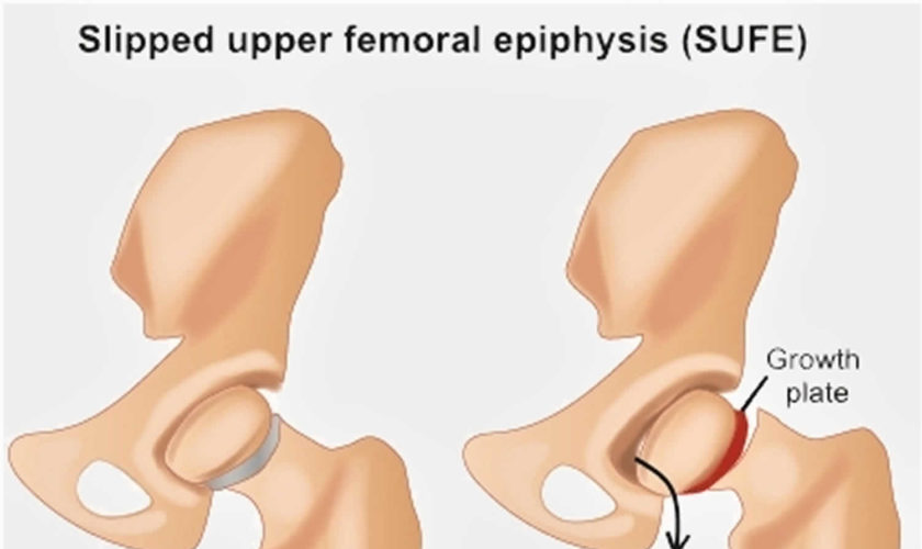
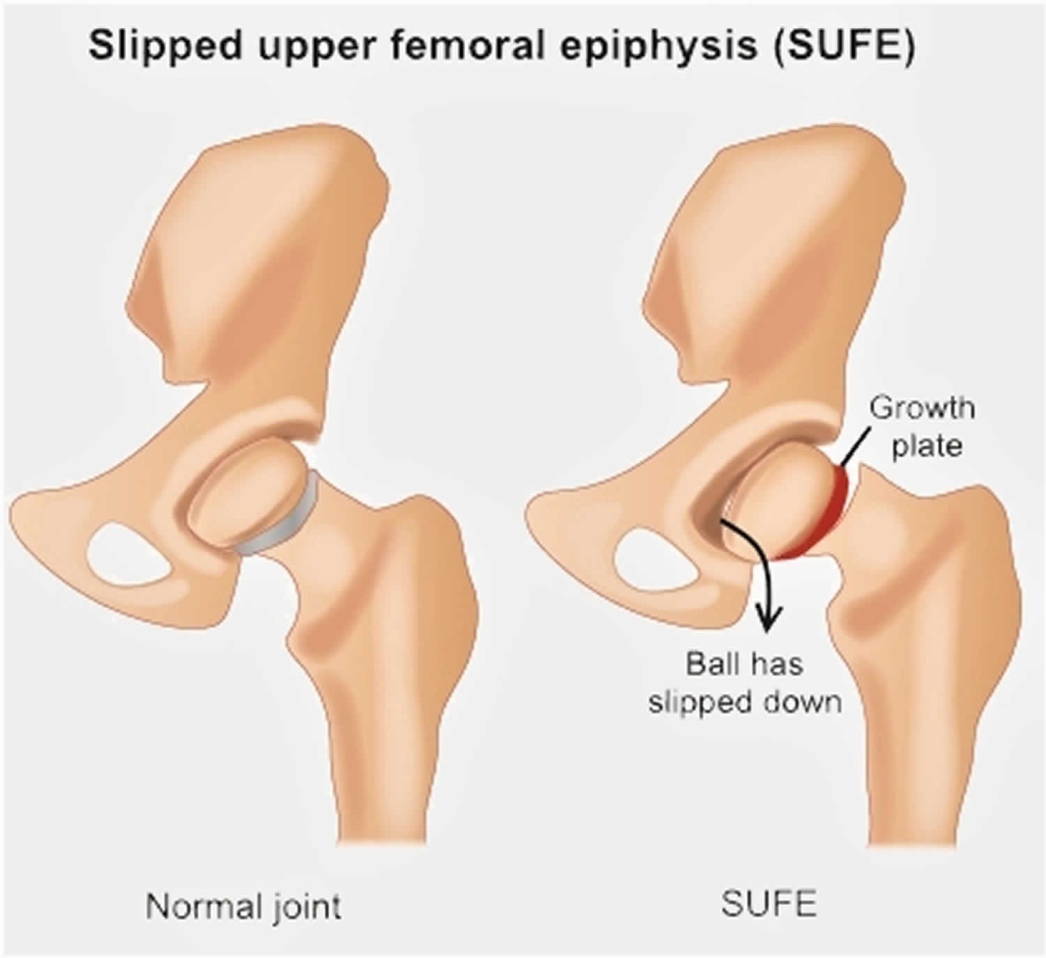
Slipped capital femoral epiphysis (SCFE) also called slipped upper femoral epiphysis (SUFE), is one of the most common developmental conditions of the hip joint that occurs in teens and pre-teens who are still growing 1). For reasons that are not well understood, in slipped capital femoral epiphysis (slipped upper femoral epiphysis), a weakness of the growth plate (physis, the area at the end of the bone responsible for bone growth) in the upper end of the thigh bone (femur) causes the head, or “ball,” of the thigh bone (femoral head, epiphysis) to slip off downwards and backwards off the neck of the thigh bone, much as a scoop of ice cream can slip off the top of a cone. This causes pain, stiffness, and instability in the affected hip. Slipped capital femoral epiphysis usually develops gradually over time and is more common in boys than girls. Slipped capital femoral epiphysis is usually an emergency and must be diagnosed and treated early.

Slipped capital femoral epiphysis usually develops during periods of rapid growth, shortly after the onset of puberty. In boys, this most commonly occurs between the ages of 12 and 16; in girls, between the ages of 10 and 14.

Sometimes slipped capital femoral epiphysis occurs suddenly after a minor fall or trauma. More often, however, the condition develops gradually over several weeks or months, with no previous injury.

Slipped capital femoral epiphysis usually occurs on only one side; however, in up to 40 percent of patients (particularly those younger than age 10) slipped capital femoral epiphysis will occur on the opposite side, as well—usually within 18 months. If only one hip is affected, the other hip will eventually slip 30 to 60 percent of the time.

A slipped capital femoral epiphysis is actually a fracture of the growth plate. The fracture is usually a fairly stable one, and the slippage occurs very slowly. Occasionally, the gradual slippage can become very unstable and the ball can completely slip, leading to severe deformity and even blood supply problems to the “ball.” For this reason, every hip with slipped capital femoral epiphysis should be treated immediately to prevent unstable slipped capital femoral epiphysis.

The signs and symptoms of slipped capital femoral epiphysis (SCFE) include:

  • Pain in the groin, thigh, or knee. Knee pain may be the only symptom present and can lead to a delayed diagnosis as there is no problem with the knee. The pain in the knee is a ‘referred pain’ from the hip joint.
  • A limp or a turned out leg are usually present. The limp may be painless, but there is usually pain associated with the limp.
  • Leg length discrepancy: The affected leg may also appear shorter than the unaffected side.
  • Pain in the hip that’s aggravated by activity and that may subside with rest
  • Walking with a limp, trouble walking, or feeling like the leg is “giving way”
  • Walking with a leg turned outward (unilateral slip)
  • Walking with a waddle (bilateral slip)
  • Inability to sit with knees straight ahead (knees tend to turn outward)

Slipped capital femoral epiphysis (SCFE) is rare condition that is slightly more likely to occur in boys than girls. SCFE occurs in about one per 1,000 to one per 10,000 children and teens; children ages 12 to 14 years are most at risk. SCFE is more prevalent in the northeast region of the U.S. than in the southwest and is more prevalent among African-Americans.

Treatment for slipped capital femoral epiphysis involves surgery to stop the head of the femur from slipping any further. To achieve the best outcome, it is important to be diagnosed as quickly as possible. Without early detection and proper treatment, slipped capital femoral epiphysis can lead to potentially serious complications, including painful arthritis in the hip joint.

Hip anatomy

The hip is a ball-and-socket joint. The socket is formed by the acetabulum, which is part of the large pelvis bone. The ball is the femoral head, which is the upper end of the femur (thighbone).

Like the other long bones in the body, the femur does not grow from the center outward. Instead, growth occurs at each end of the bone around an area of developing cartilage called the growth plate (physis).

Growth plates are located between the widened part of the shaft of the bone (metaphysis) and the end of the bone (epiphysis). The epiphysis at the upper end of the femur is the growth center that eventually becomes the femoral head.

Figure 1. Normal anatomy of the hip

Normal anatomy of the hip

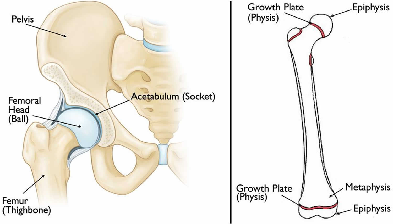
Footnote: (Left) Normal anatomy of the hip. (Right) The location of the growth plates and epiphyses at the ends of the femur (thighbone). The epiphysis at the upper end of the bone eventually becomes the femoral head.

Figure 2. Slipped capital femoral epiphysis (SCFE)

Slipped capital femoral epiphysis

Slipped capital femoral epiphysis types

Slipped capital femoral epiphysis is often described based on whether the patient is able to bear weight on the affected hip. Knowing the type of slipped capital femoral epiphysis will help your doctor determine treatment.

Types of slipped capital femoral epiphysis include:

  • Stable slipped capital femoral epiphysis. In stable slipped capital femoral epiphysis, the patient is able to walk or bear weight on the affected hip, either with or without crutches. Most cases of slipped capital femoral epiphysis are stable slips.
  • Unstable slipped capital femoral epiphysis. This is a more severe slip. The patient cannot walk or bear weight, even with crutches. Unstable slipped capital femoral epiphysis requires urgent treatment. Complications associated with slipped capital femoral epiphysis are much more common in patients with unstable slips.

Slipped capital femoral epiphysis causes

The cause of slipped capital femoral epiphysis is not known. Slipped capital femoral epiphysis is more likely to occur during a growth spurt and is more common in boys than girls.

Risk factors that make someone more likely to develop slipped capital femoral epiphysis include:

  • Excessive weight or obesity—most patients are above the 95th percentile for weight. This increases the stress across the growth plate of the hip bone which is the site where the slip occurs.
  • Family history of slipped capital femoral epiphysis
  • Trauma: Occasionally a child may have a fall at the at the time the problem started, however there may have been an underlying degree of SUFE that was aggravated by the fall.
  • An endocrine or metabolic disorder, such as hypothyroidism, growth hormone deficiency, and hypogonadism—this is more likely to be a factor for patients who are older or younger than the typical age range for slipped capital femoral epiphysis (10 to 16 years of age). Slipped capital femoral epiphysis develops during puberty, a time of many hormonal changes. Rapid growth occurs in response to increased levels of growth hormone. This rapid growth is associated with an increased size in the growth plate of the hip bone and this may contribute to the decreased strength seen at puberty.
  • Bone problems related to kidney disease
  • Treatments for disease, like radiation and chemotherapy for cancer or taking certain medicines, including steroids

Slipped capital femoral epiphysis symptoms

Symptoms of slipped capital femoral epiphysis vary, depending upon the severity of the condition.

A patient with mild or stable slipped capital femoral epiphysis will usually have intermittent pain in the groin, hip, knee and/or thigh for several weeks or months. This pain usually worsens with activity. The patient may walk or run with a limp after a period of activity.

In more severe or unstable slipped capital femoral epiphysis, symptoms may include:

  • Sudden onset of pain, often after a fall or injury
  • Inability to walk or bear weight on the affected leg
  • Outward turning (external rotation) of the affected leg
  • Discrepancy in leg length—the affected leg may appear shorter than the opposite leg

Slipped capital femoral epiphysis complications

Although early detection and proper treatment of slipped capital femoral epiphysis will help decrease the chance of complications, some patients will still experience problems.

The most common complications following slipped capital femoral epiphysis are avascular necrosis and chondrolysis.

Avascular necrosis (osteonecrosis)

If severe cases, slipped capital femoral epiphysis causes the blood supply to the femoral head to become limited. This can lead to a gradual and very painful collapse of the bone, a condition called avascular necrosis or osteonecrosis. Avascular necrosis of the femoral head is thought to result from vascular damage during the time of the initial traumatic event, but it may result from forceful reduction during the time of surgery. The amount of energy, magnitude of epiphyseal damage and displacement, level of increased intra-articular pressure, and degree of vascular occlusion have been implicated in this process. The risk of avascular necrosis is up to 47% with an unstable slipped capital femoral epiphysis.

When the bone collapses, the articular cartilage covering the bone also collapses. Without this smooth cartilage, bone rubs against bone, leading to painful arthritis in the joint. For some patients with avascular necrosis, further surgery may be needed to reconstruct the hip.

Avascular necrosis is more likely to occur in patients with unstable slipped capital femoral epiphysis. Because evidence of osteonecrosis may not be seen on x-ray for up to 12 months following surgery, the patient will be monitored with x-rays during this period of time.

Treatment options are limited (eg, bone grafting, osteotomy to change the position of the femoral head), but often these patients will eventually need a total hip replacement.

Chondrolysis

Chondrolysis is a rare but serious complication of slipped capital femoral epiphysis. In chondrolysis, the articular cartilage on the surface of the hip joint degenerates very rapidly, leading to pain, deformity, and permanent loss of motion in the affected hip.

Although the cause of chondrolysis is not yet fully understood by doctors, it is believed that it may result from inflammation in the hip joint.

Aggressive physical therapy and anti-inflammatory medications may be prescribed for patients who develop chondrolysis. Over time, there may be some gradual return of motion in the hip.

Osteoarthritis

Osteoarthritis is a late complication. There is evidence that increased risk of early degenerative change may result from avascular necrosis, chondrolysis, or alterations of the hip biomechanics following slippage. In general, the more severe the deformity and/or slipped capital femoral epiphysis, the higher risk of developing arthritis. Mild deformities may have few consequences.

Leg-length inequality

Leg-length inequality may result from incomplete reduction, avascular necrosis, chondrolysis, or secondary coxa vara.

Hardware failure and “outgrowing” hardware may cause loss of fixation. Although rare, postoperative infection may occur.

Slipped capital femoral epiphysis diagnosis

During the examination, your doctor will ask about your child’s general health and medical history. He or she will then talk with you about your child’s symptoms and ask when the symptoms began.

While your child is lying down, the doctor will perform a careful examination of the affected hip and leg, looking for:

  • Pain with extremes of motion
  • Limited range of motion in the hip–especially limited internal rotation
  • Involuntary muscle guarding and muscle spasms

Your doctor will also observe your child’s gait (the way he or she walks). A child with slipped capital femoral epiphysis may limp or have an abnormal gait.

X-rays

This type of study provides images of dense structures, such as bone. Your doctor will order x-rays of the pelvis, hip, and thigh from several different angles to help confirm the diagnosis.

In a patient with slipped capital femoral epiphysis, an x-ray will show that the head of the thighbone appears to be slipping off the neck of the bone.

An MRI or CT scan is rarely required.

Slipped capital femoral epiphysis treatment

Treatment of slipped capital femoral epiphysis requires surgery to correct the problem. The aim of surgery is to stop the hip bone from slipping further and make sure any slip that has occurred is corrected. The earlier the treatment, the better the outcomes and for this reason a quick diagnosis is very important. The treatment for a stable slipped capital femoral epiphysis (when the child can walk on the affected side) is slightly different to the treatment of unstable slipped capital femoral epiphysis (when the child is unable to walk on the affected side).

Early diagnosis of slipped capital femoral epiphysis provides the best chance of stabilizing the hip and avoiding complications. When treated early and appropriately, long-term hip function can be expected to be very good.

Once slipped capital femoral epiphysis is confirmed, your child will not be allowed to bear weight on his or her hip and will probably be admitted to the hospital. In most cases, surgery is performed within 24 to 48 hours.

Stable slipped capital femoral epiphysis

The treatment for stable slipped capital femoral epiphysis is usually to insert a single screw into the the thigh bone across the place where the bone is slipping. This holds the bone together and prevents any further slip. In some cases, the surgeon may decide to perform operations that include grafting bone or changing the angle of the bone but usually a single screw is all that is needed.

After the operation, crutches are recommended for several days. After this, the child is recovered to walk on the affected side to ensure the bone heals normally.

Unstable slipped capital femoral epiphysis

Usually, unstable slipped capital femoral epiphysis is considered a more urgent condition and operations will usually be carried out within a short time of the diagnosis. Before the operation, the joint may be drained of any fluid that may have accumulated due to the inflammation caused by the slip. The operation involves first lining the bones up by the use of traction and then placing one or two screws across the place where the bone has slipped.

Following the operation, crutches must be used for 6-8 weeks in order to prevent any further slip. Physiotherapy rehabilitation may be required to ensure the hip is moving well and that the surrounding muscles are strong.

SCFE surgery

The surgical procedure your doctor recommends will depend upon the severity of the slip. Procedures used to treat slipped capital femoral epiphysis include:

In situ fixation

This is the procedure used most often for patients with stable or mild slipped capital femoral epiphysis. The doctor makes a small incision near the hip, then inserts a metal screw across the growth plate to maintain the position of the femoral head and prevent any further slippage.

Over time, the growth plate will close, or fuse. Once the growth plate is closed, no further slippage can occur.

Open reduction

In patients with unstable slipped capital femoral epiphysis, the doctor may first make an open incision in the hip, then gently manipulate (reduce) the head of the femur back into its normal anatomic position.

The doctor will then insert one or two metal screws to hold the bone in place until the growth plate closes. This is a more extensive procedure and requires a longer recovery time.

In situ fixation in the opposite hip

Some patients are at higher risk for slipped capital femoral epiphysis occurring on the opposite side. If this is the case with your child, your doctor may recommend inserting a screw into his or her unaffected hip at the same time to reduce the risk of slipped capital femoral epiphysis. Your doctor will talk with you about whether this is appropriate for your child.

Recovery

Weight bearing

After surgery, your child will be on crutches with protected weight bearing for 6-8 weeks. Your child’s doctor will give you specific instructions about when full weight bearing can begin. To prevent further injury, it is important to closely follow your doctor’s instructions.

Physical therapy

A physical therapist will provide specific exercises to help strengthen the hip and leg muscles and improve range of motion. Physical therapy for strengthening, proprioception, balance, and endurance training may be helpful. Most children can then return to full activity once they are pain free with full strength. However, some literature advocates for not allowing a return to contact sports until the physis has closed.

Sports and other activities

For a period of time after surgery, your child will be restricted from participating in vigorous sports and activities. This will help minimize the chance of complications and enable healing to take place. Your doctor will tell you when your child can safely resume his or her normal activities.

Follow-up care

Your child will return to the doctor for follow-up visits for 18 to 24 months after surgery. These visits may include x-rays every 3 to 4 months to ensure that the growth plate has closed and that no complications have developed.

Depending upon the patient’s age and other factors, a team approach that includes a general pediatrician, endocrinologist, and/or dietician may be necessary for comprehensive care in the long run.

Slipped capital femoral epiphysis prognosis

Most patients with slipped capital femoral epiphysis who are treated with urgent in situ fixation do well. However, in those cases with severe slippage and resultant deformity, long-term sequelae may result (eg, avascular necrosis, chondrolysis, leg-length discrepancy, stiffness, osteoarthritis). Although conservative modalities (eg, therapy, analgesics, orthotics, assistive aids) are used initially for symptomatic relief, urgent operative intervention is indicated. Young patients with unremitting pain, loss of motion, and stiffness secondary to chondrolysis, avascular necrosis, or osteoarthritis may require salvage hip arthrodeses. In hips that are incompletely damaged, proximal osteotomies may aid in redirecting the joint forces to less damaged areas of the articular femoral head.

References   [ + ]

read more

Shaken baby syndrome

shaken baby syndrome

Shaken baby syndrome

Shaken baby syndrome also known as abusive head trauma, shaken impact syndrome, inflicted head injury or whiplash shake syndrome, is a serious brain, head or neck injury from physical child abuse. Shaken baby syndrome happens when someone forcefully shakes a baby or hits the baby against something hard. Most cases happen when a parent or caregiver is angry, tired, or upset because a baby won’t stop crying or the child can’t do something they expect, like toilet train. People should never shake a baby for any reason.

Shaken baby syndrome destroys a child’s brain cells and prevents his or her brain from getting enough oxygen. Shaken baby syndrome is a form of child abuse that can result in permanent brain damage or death. At least one of every four babies who are violently shaken dies from shaken baby syndrome 1).

Shaken baby syndrome is preventable. Help is available for parents who are at risk of harming a child. Parents also can educate other caregivers about the dangers of shaken baby syndrome.

All babies cry and do things that can frustrate caregivers; however, not all caregivers are prepared to care for a baby.

Babies especially babies ages 2 to 4 months, newborn to one year and toddlers younger than 2 years old, are at greatest risk of injury from shaking. Rarely, it can happen in children up to 5 years old. It can happen to boys or girls in any family. At special risk for abuse are children who have a lot of special needs or health problems that make them cry a lot, like colic and gastroesophageal reflux.

Shaking them violently can trigger a “whiplash” effect that can lead to internal injuries, including bleeding in the brain or in the eyes. Often there are no obvious external physical signs, such as bruising or bleeding, to indicate an injury.

In more severe cases of shaken baby syndrome, babies may exhibit the following 2):

  • Unresponsiveness
  • Loss of consciousness
  • Breathing problems (irregular breathing or not breathing)
  • No pulse

Babies suffering lesser damage from shaken baby syndrome may exhibit some of the following 3):

  • Change in sleeping pattern or inability to be awakened
  • Vomiting
  • Convulsions or seizures
  • Irritability
  • Uncontrollable crying
  • Inability to be consoled
  • Inability to nurse or eat

Shaken baby syndrome can potentially result in the following consequences:

  • Death
  • Blindness
  • Mental retardation or developmental delays (any significant lags in a child’s physical, cognitive, behavioral, emotional, or social development, in comparison with norms) 4) and learning disabilities
  • Cerebral palsy
  • Severe motor dysfunction (muscle weakness or paralysis)
  • Spasticity (a condition in which certain muscles are continuously contracted—this contraction causes stiffness or tightness of the muscles and may interfere with movement, speech, and manner of walking) 5)
  • Seizures

Emergency treatment for a baby who has been shaken usually includes life-sustaining measures such as respiratory support and surgery to stop internal bleeding and bleeding in the brain. Doctors may use brain scans, such as MRI and CT, to make a more definite diagnosis.

In comparison with accidental traumatic brain injury in infants, shaken baby injuries have a much worse prognosis. Damage to the retina of the eye can cause blindness. The majority of infants who survive severe shaking will have some form of neurological or mental disability, such as cerebral palsy or cognitive impairment, which may not be fully apparent before 6 years of age. Children with shaken baby syndrome may require lifelong medical care.

When to see a doctor

Seek immediate help if you suspect your child has been injured by violent shaking. Contact your child’s doctor or take your child to the nearest emergency room. Getting medical care right away may save your child’s life or prevent serious health problems.

Health care professionals are legally required to report all suspected cases of child abuse to state authorities.

Can tossing my baby in the air or rough play cause shaken baby syndrome?

Shaken baby syndrome is a form of child abuse that happens when an infant or small child is violently shaken. Shaken injuries are not caused by:

  • Bouncing a baby on your knee.
  • Tossing a baby in the air.
  • Jogging or bicycling with your baby.
  • Falls off a couch or other furniture.
  • Sudden stops in a car or driving over bumps.

Although the activities listed above can be dangerous and are not recommended, they will likely not cause shaken baby syndrome injuries.

Why is shaking a baby dangerous?

Violent shaking for just a few seconds has the potential to cause severe injuries. While shaking may cause injury to children of any age, children are most susceptible to being injured during their first year of life. Factors that contribute to a baby’s vulnerability include:

  • Babies heads are heavy and large in proportion to their body size.
  • Babies have weak neck muscles.
  • Babies have fragile, undeveloped brains.
  • There is a large size and strength difference between the victim and the perpetrator.

How much force is necessary to cause injuries in shaken baby syndrome or abusive head trauma? How many times do you have to shake an infant or young child to cause damage?

The injuries seen in cases of shaken baby syndrome or abusive head trauma are caused by violent shaking and, in some cases, impact. This is due to the rapid and repeated acceleration and deceleration of the victim’s head whipping back and forth and side to side. Shaking injuries are not caused by casual or accidental handling of children. Shaking injuries require massive, violent force. One shake is all it takes to cause traumatic brain injuries in an infant.

Shaken baby syndrome facts

Most cases of shaken baby syndrome or abusive head trauma happen to babies. However, it is difficult to know the exact number of shaken baby syndrome cases per year because many cases of shaken baby syndrome are underreported and/or never receive a diagnosis. However, a study of North Carolina shaken baby syndrome cases suggests that as many as three to four children a day experience severe or fatal head injury from child abuse in the United States 6).

  • Shaken baby syndrome or abusive head trauma is the leading cause of physical child abuse deaths in the U.S.
  • There are approximately 1,300 reported cases of shaken baby syndrome or abusive head trauma in the U.S. each year 7).
  • Babies less than 1 year of age (with the highest risk period at 2 to 4 months) 8) are at greatest risk for shaken baby syndrome because they cry longer and more frequently, and are easier to shake than older and larger children 9).
  • Shaken baby syndrome injuries have been reported in children up to age 5 10).
  • Shaken baby syndrome is the result of violent shaking that leads to a brain injury, which is much like an adult may sustain in repeated car crashes. It is child abuse, not play. This is why claims by perpetrators that the highly traumatic internal injuries that characterize shaken baby syndrome resulted from merely “playing with the baby” are false. While jogging an infant on your knee or tossing him or her in the air can be very risky, the injuries that result from shaken baby syndrome are not caused by these types of activities 11).
  • The most common trigger for shaking a baby is inconsolable or excessive crying—a normal phase in infant development 12).
  • Parents and their partners account for the majority of perpetrators. Biological fathers, stepfathers, and mothers’ boyfriends are responsible for the majority of cases, followed by mothers 13).
  • In most shaken baby syndrome cases there is evidence of some form of prior physical abuse, including prior shaking 14).
  • Upwards of 80% of surviving victims of shaken baby syndrome or abusive head trauma suffer lifelong disabilities.
  • Approximately 25% of victims of shaken baby syndrome or abusive head trauma die.

Shaken baby syndrome causes

Shaken baby syndrome happens when someone:

  • uses force to shake a child
  • uses force to throw or drop a child on purpose
  • hits the child’s head or neck against an object, like the floor or furniture, or hits the child’s head or neck with an object

Shaking a baby is so harmful because:

  • Infants have poor neck strength and their heads are large compared with the size of their bodies. This lets the head move around a lot when shaken.
  • When the head moves around, the baby or child’s brain moves back and forth inside the skull. This can tear blood vessels and nerves inside the brain, causing bleeding and nerve damage.
  • The brain may hit against the inside of the skull, causing brain bruising and bleeding on the outside of the brain.
  • Brain swelling builds pressure in the skull. This pressure makes it hard for blood, carrying oxygen and nutrients, to reach the brain, further harming it.

Babies have weak neck muscles and often struggle to support their heavy heads. If a baby is forcefully shaken, his or her fragile brain moves back and forth inside the skull. This causes bruising, swelling and bleeding.

Shaken baby syndrome usually occurs when a parent or caregiver severely shakes a baby or toddler due to frustration or anger — often because the child won’t stop crying.

Shaken baby syndrome isn’t usually caused by bouncing a child on your knee, minor falls or even rough play.

Risk factors for shaken baby syndrome

The following things may make parents or caregivers more likely to forcefully shake a baby and cause shaken baby syndrome:

  • Unrealistic expectations of babies
  • Young or single parenthood
  • Stress
  • Domestic violence
  • Alcohol or substance abuse
  • Unstable family situations
  • Depression
  • A history of mistreatment as a child

Also, men are more likely to cause shaken baby syndrome than are women.

Shaken baby syndrome prevention

New parent education classes can help parents better understand the dangers of violent shaking and may provide tips to soothe a crying baby and manage stress.

When your crying baby can’t be calmed, you may be tempted to try anything to get the tears to stop — but it’s important to always treat your child gently. Nothing justifies shaking a child.

If you’re having trouble managing your emotions or the stress of parenthood, seek help. Your child’s doctor may offer a referral to a counselor or other mental health provider.

If other people help take care of your child — whether a hired caregiver, sibling or grandparent — make sure they know the dangers of shaken baby syndrome.

  • Tell people caring for your baby to never shake the baby.
  • Talk about normal crying so a caregiver is less likely to get upset.
  • Talk about safe ways to calm a baby, such as swaddling, rocking, or singing.
  • Let caregivers know it’s OK to put the baby or child in a safe place, walk away and take a break.

What to do when babies cry

Why do babies cry?

All newborns cry and get fussy sometimes. It’s normal for a baby to cry for 2–3 hours a day for the first 6 weeks. During the first 3 months of life, they cry more than at any other time.

New parents often are low on sleep and getting used to life with their little one. They’ll quickly learn to find out if their crying baby:

  • is hungry
  • is tired
  • needs to be burped
  • is overstimulated
  • has a wet or dirty diaper
  • is too hot or cold

Often, taking care of a baby’s needs is enough to soothe a baby. But sometimes, the crying goes on longer.

What is colic?

Some babies cry a lot more than others. A baby who cries more than 3 hours a day, more than 3 days a week, for at least 3 weeks might have colic. Usually, it starts when a baby is 2–5 weeks old and ends by the time the baby is 3–4 months old.

Colic happens to a lot of newborns. It’s hard to see your baby cry so much, but colic isn’t caused by anything a parent did or didn’t do. The good news is babies outgrow colic.

What can help a crying baby?

You can’t spoil your baby with too much attention. To soothe a crying baby:

  • First, make sure your baby doesn’t have a fever. In a baby, a fever is a temperature of 100.4°F (38°C). Call the doctor right away if your baby does have a fever.
  • Make sure your baby isn’t hungry and has a clean diaper.
  • Rock or walk with the baby.
  • Sing or talk to your baby.
  • Offer the baby a pacifier.
  • Take the baby for a ride in a stroller.
  • Hold your baby close against your body and take calm, slow breaths.
  • Give the baby a warm bath.
  • Pat or rub the baby’s back.
  • Place your baby across your lap on his or her belly and rub your baby’s back.
  • Put your baby in a swing or vibrating seat. The motion may be soothing.
  • Put your baby in an infant car seat in the back of the car and go for a ride. Often, the vibration and movement of the car are calming.
  • Play music — some babies respond to sound as well as movement.

Some babies need less stimulation. Babies 2 months and younger may do well swaddled, lying on their back in the crib with the lights very dim or dark. Make sure the swaddle isn’t too tight. Stop swaddling when the baby is starting to be able to roll over.

When a baby won’t stop crying

If a baby in your care won’t stop crying:

  • Call a friend or relative for support or to take care of the baby while you take a break.
  • If nothing else works, put the baby on their back in an empty crib (without loose blankets or stuffed animals), close the door, and check on the baby in 10 minutes. During that 10 minutes, do something to try to relax and calm down. Try washing your face, breathing deeply, or listening to music.

Call your doctor if nothing seems to be helping the baby, in case there is a medical reason for the fussiness.

Shaken baby syndrome signs and symptoms

In the most severe shaken baby syndrome cases, babies and children may come to the emergency room, hospital, or doctor’s office not awake, having seizures, or in shock.

In less severe shaken baby syndrome cases, symptoms and signs may include:

  • Extreme fussiness, irritability or cranky and hard to comfort
  • Move less than usual
  • Not smile or coo
  • Difficulty staying awake
  • Grab-type bruises on arms or chest
  • Trouble breathing
  • Poor eating or eat less than usual
  • Vomiting
  • Have trouble sucking or swallowing
  • Pale or bluish skin
  • Seem stiff
  • Have seizures
  • Paralysis
  • Have pupils (the dark spots in center of the eyes) that aren’t the same size
  • Be unable to lift their head
  • Have trouble focusing their eyes or tracking movement
  • Head or forehead appears larger than usual
  • Soft spot on head appears to be bulging
  • Decreased level of consciousness
  • Coma

You may not see any signs of physical injury to the child’s outer body. Sometimes, the face is bruised. Injuries that might not be immediately seen include bleeding in the brain and eyes, spinal cord damage, and fractures of the ribs, skull, legs and other bones. Many children with shaken baby syndrome show signs and symptoms of prior child abuse.

In mild cases of shaken baby syndrome, a child may appear normal after being shaken, but over time he or she may develop health or behavioral problems.

Shaken baby syndrome symptoms later in life

Even brief shaking of an infant can cause irreversible brain damage. Many children affected by shaken baby syndrome die.

Survivors of shaken baby syndrome may require lifelong medical care for conditions such as:

  • Visual disabilities or blindness
  • Mental retardation or developmental delays (any significant lags in a child’s physical, cognitive, behavioral, emotional, or social development, in comparison with norms) 15) and learning disabilities
  • Cerebral palsy
  • Severe motor dysfunction (muscle weakness or paralysis)
  • Spasticity (a condition in which certain muscles are continuously contracted—this contraction causes stiffness or tightness of the muscles and may interfere with movement, speech, and manner of walking) 16)
  • Seizures
  • Learning disabilities
  • Physical disabilities
  • Hearing impairment
  • Speech disabilities
  • Behavior disorders
  • Cognitive impairment
  • Death

Shaken baby syndrome diagnosis

Parents or caregivers often won’t say that the child was shaken or hit, so doctors may not know to check for head injury. Many signs of abusive head trauma, like fussiness and throwing up, are common in routine childhood illnesses. So it can be hard for doctors to figure out that a baby was harmed.

A child who’s been forcefully shaken may need to be examined by many different medical specialists, as well as an expert in child abuse.

If abusive head trauma is suspected, doctors will:

  • Do an eye exam to look for bleeding inside the eyes.
  • Order X-rays of all the bones to look for new or healing breaks, which happen most in the arms, legs, skull, and ribs.
  • Order a CT or MRI of the head to look for:
    • broken bones in the head (skull fractures)
    • brain swelling
    • brain bleeding

Shaken baby syndrome treatment

Depending on the extent of the injuries, the baby may need to be monitored in a pediatric intensive care unit.

Emergency treatment for a child who has been shaken may include breathing support and surgery to stop bleeding in the brain.

After shaken baby syndrome or abusive head trauma,a child may need long-term care from a team of health experts, such as:

  • brain doctors (neurologist)
  • brain surgeons (neurosurgeon)
  • eye doctors (ophthalmologist)
  • hormone doctors (endocrinologist)

They also need a pediatrician who can manage their ongoing complex care. They also might need support from therapists, such as:

  • rehab medicine
  • speech-language therapy
  • physical therapy (PT)
  • occupational therapy (OT)

Before age 3, a child can receive free speech therapy or physical therapy through state-run programs. After age 3, the child’s school district’s provides any needed special educational services.

As kids get older, they may need special schooling and ongoing help to build language and daily living skills, like dressing.

Long term effects of shaken baby syndrome

Shaken baby syndrome or abusive head trauma often causes life-long harm to the brain and, sometimes, death.

Babies and children who survive may have:

  • poor eyesight, partial or total blindness
  • hearing loss
  • seizures
  • delayed development
  • intellectual disability
  • behavior issues
  • problems with speech and learning
  • problems with memory and focus
  • cerebral palsy
  • weakness or problems moving parts of the body
  • problems with hormones controlled by the brain

If a child’s problems are mild, they might not be noticed until the child starts school and has problems with learning, focus, or behavior.

References   [ + ]

read more

Developmental dysplasia of the hip

developmental dysplasia of the hip

Developmental dysplasia of the hip

Developmental dysplasia of the hip (DDH) or developmental dislocation of the hip used to be called congenital dislocation of the hip, is an abnormal development of the hip joint. The hip is a “ball-and-socket” joint. In a normal hip, the ball at the upper end of the thighbone (femur) fits firmly into the socket, which is part of the large pelvis bone. In babies and children with developmental dysplasia of the hip, the hip joint has not formed normally. The ball is loose in the socket and may be easy to dislocate. Sometimes, the hips can dislocate early in life and this may not be noticed until your child starts to walk. Pediatricians screen for developmental dysplasia of the hip at a newborn’s first examination and at every well-baby checkup thereafter.

In all cases of developmental dysplasia of the hip (developmental dislocation of the hip), the socket (acetabulum) is shallow, meaning that the ball of the thighbone (head of the femur) cannot firmly fit into the socket. Sometimes, the ligaments that help to hold the joint in place may also be loose. The degree of hip looseness, or instability, varies among children with developmental dysplasia of the hip.

  • Dislocated. In the most severe cases of developmental dysplasia of the hip, the head of the femur is completely out of the socket.
  • Dislocatable. In these cases, the head of the femur lies within the acetabulum, but can easily be pushed out of the socket during a physical examination.
  • Subluxatable. In mild cases of developmental dysplasia of the hip, the head of the femur is simply loose in the socket. During a physical examination, the bone can be moved within the socket, but it will not dislocate.

Developmental dysplasia of the hip affects 1-7% of infants 1). In the United States, approximately 1 to 2 babies per 1,000 are born with developmental dysplasia of the hip. Well known risk factors for developmental dysplasia of the hip include family history of developmental dysplasia of the hip (parents or siblings), female sex, and breech positioning. Recent evidence suggests higher birth weight is a risk, whereas prematurity may be protective. Screening includes physical examination of all infant hips and imaging when abnormal findings or risk factors are present.

Although developmental dysplasia of the hip is most often present at birth, it may also develop during a child’s first year of life. Recent research shows that babies whose legs are swaddled tightly with the hips and knees straight are at a notably higher risk for developing developmental dysplasia of the hip after birth. As swaddling becomes increasingly popular, it is important for parents to learn how to swaddle their infants safely, and to understand that when done improperly, swaddling may lead to problems like developmental dysplasia of the hip.

It is important to check for and treat developmental dysplasia of the hip as early as possible. If developmental dysplasia of the hip is not treated, your child may develop a painless limp when walking, they may walk on their toes rather than in a heel-and-toe action, or they may develop a ‘waddling’ walk. In time, arthritis will develop in the untreated hip joint, which will become painful and may ultimately need a hip replacement.

Your doctor may ask for an ultrasound or X-ray of the hip joint to diagnose developmental dysplasia of the hip.

Developmental dysplasia of the hip treatment may involve use of a brace, a non-surgical procedure under sedation, or an operation to correct the dysplasia and hip position.

When developmental dysplasia of the hip is detected at birth, it can usually be corrected with the use of a harness or brace. If the hip is not dislocated at birth, the condition may not be noticed until the child begins walking. At this time, treatment is more complicated, with less predictable results.

Developmental dysplasia of the hip key points to remember

  • Treatment for developmental dysplasia of the hip varies between children and depends on how bad the condition is.
  • Treatment may include a brace, a plaster cast called a hip spica, movement of the hip into position under anaesthetic, or surgery to the ligaments around the joint.
  • Children will often need to wear a brace or cast for several months.
  • If developmental dysplasia of the hip is not treated, your child may develop a painless limp. Over time, painful arthritis will develop in the untreated hip joint.

Figure 1. Developmental dysplasia of the hip

developmental dysplasia of the hip

Figure 2. Hip joint anatomy

Hip joint anatomy

Developmental dysplasia of the hip causes

Pregnant women release hormones in their bloodstream that allow their ligaments to relax. These hormones help the delivery of the baby through the mother’s pelvis. Some of these hormones enter the baby’s blood, which can make the baby’s ligaments relaxed as well. This can make the hip joint loose in the socket. The way the baby lies in the uterus can also cause the hip joint to dislocate or become loose.

Developmental dysplasia of the hip tends to run in families where a parent/sibling has had a dislocated hip joint. It can be present in either hip and in any individual. It usually affects the left hip and is predominant in:

  • Girls
  • Firstborn children
  • Babies born in the breech position (especially with feet up by the shoulders). The American Academy of Pediatrics now recommends ultrasound developmental dysplasia of the hip screening of all female breech babies.
  • Family history of developmental dysplasia of the hip (parents or siblings)
  • Oligohydramnios (low levels of amniotic fluid)

Developmental dysplasia of the hip symptoms

Some babies born with a dislocated hip will show no outward signs and sometimes the signs of developmental dysplasia of the hip are hard to see, even by a doctor.

Contact your pediatrician if your baby has:

  • Legs of different lengths
  • Uneven skin folds on their groin or thigh (front or back of the upper leg)
  • A stiff hip joint
  • Lean to the affected side when standing
  • An outward-turning leg on the affected side
  • Less mobility or flexibility on one side
  • Limping, toe walking, or a waddling gait

Developmental dysplasia of the hip diagnosis

In addition to visual clues, your doctor will perform a careful physical examination to check for developmental dysplasia of the hip, such as listening and feeling for “clunks” as the hip is put in different positions. Your doctor will use specific maneuvers to determine if the hip can be dislocated and/or put back into proper position.

Newborns identified as at higher risk for developmental dysplasia of the hip are often tested using ultrasound, which can create images of the hip bones. For older infants and children, x-rays of the hip may be taken to provide detailed pictures of the hip joint.

Developmental dysplasia of the hip treatment

Treatment methods depend on a child’s age. Treatment in the first six months consists of a harness, with 70–95% success 2). Failure risk factors include femoral nerve palsy, static bracing, irreducible hips, initiation after seven weeks of age, right hip dislocation, Graf-IV hips, and male sex. Rigid bracing may be trialed if reduction with a harness fails and closed reduction is indicated after failed bracing. If the hip is still irreducible, nonconcentric, or unstable, open reduction may be required following closed reduction. Evidence does not support delaying hip reduction until the ossific nucleus is present.

Nonsurgical treatment

Newborns

The baby is placed in a soft positioning device, called a Pavlik harness, for 1 to 2 months to keep the thighbone in the socket (see Figure 3). This special brace is designed to hold the hip in the proper position while allowing free movement of the legs and easy diaper care. The Pavlik harness helps tighten the ligaments around the hip joint and promotes normal hip socket formation.

Parents play an essential role in ensuring the harness is effective. Your doctor and healthcare team will teach you how to safely perform daily care tasks, such as diapering, bathing, feeding, and dressing.

Figure 3. Pavlik harness

Pavlik harness

1 month to 6 months

Similar to newborn treatment, a baby’s thighbone is repositioned in the socket using a harness or similar device. This method is usually successful, even with hips that are initially dislocated.

How long the baby will require the harness varies. It is usually worn full-time for at least 6 weeks, and then part-time for an additional 6 weeks.

If the hip will not stay in position using a harness, your doctor may try an abduction brace made of firmer material that will keep your baby’s legs in position.

In some cases, a closed reduction procedure is required. Your doctor will gently move your baby’s thighbone into proper position, and then apply a spica cast (a plaster cast that covers your child’s body from the knees to the waist) to hold the bones in place. This procedure is done while the baby is under anesthesia.

Caring for a baby in a spica cast requires specific instruction. Your doctor and healthcare team will teach you how to perform daily activities, maintain the cast, and identify any problems.

6 months to 2 years. Older babies are also treated with closed reduction and spica casting. In most cases, skin traction may be used for a few weeks prior to repositioning the thighbone. Skin traction prepares the soft tissues around the hip for the change in bone positioning. It may be done at home or in the hospital.

Figure 4. Spica cast

spica cast

Pavlik harness care

Your doctor will let you know if your baby needs to wear the Pavlik harness 24 hours a day without removing it at all, or if the harness can be removed for bathing.

Key points to remember:

  • Monitor your baby’s skin daily. Take care to clean your baby’s skin and if you notice any skin irritation or redness, contact the orthotist.
  • Always try to keep your baby’s legs apart, especially when bathing with the harness off.
  • Babies may cry a little or seem unsettled for the first couple of nights after the harness is fitted. This should settle down within a couple of days.
  • The Pavlik Harness should be washed only if absolutely necessary and do not tumble dry.
  • Make sure you vary the position of your sleeping baby to avoid plagiocephaly (misshapen head).

What should I do if my baby gets a rash under the harness?

It is important to monitor your baby’s skin each day, and contact the orthotist if a rash appears. If mild skin irritation occurs, a barrier ointment (like Vaseline) can be helpful, but discuss this with your child’s orthotist.

My baby seems very reluctant to spend time on her tummy. How should I deal with this?

Tummy time is difficult to achieve when your child is placed in any kind of orthosis (e.g. harness, spica, plaster), though you can try encouraging tummy time during orthosis-free time, if allowed. Fortunately, in the long-term, your child’s developmental ability will not suffer by missing out on tummy-time for this short period in their life.

Care at home

Putting the Pavlik harness on

Your baby’s orthotist will make sure the harness fits your baby correctly, and will show you how to put it on and how to check that it is positioned properly.

  • The chest strap should be firm but you should be able to fit two fingers underneath it. This allows the chest to expand properly when your child is breathing.
  • The ankle and lower leg straps should be firm to hold the foot, but not too tight.

Getting used to the harness

It takes some babies a couple of days to get use to the Pavlik harness. Some babies may cry a little or seem unsettled for the first couple of nights. This should settle down after a few days.

Hygiene and skin care

Monitor your baby’s skin daily. Take care to clean your baby’s skin and if you notice any skin irritation or redness, contact the orthotist.

  • Try to keep the harness dry at all times.
  • If your doctor says the Pavlik harness must be on 24 hours a day, it cannot be removed for bathing. In this case, your baby will need to have sponge baths. The orthotist will show you how to do this.
  • If you are allowed to remove the harness for bathing, then only undo the Velcro straps to remove it. Do not adjust or undo the metal buckles.
  • When bathing your baby, pay particular attention to the creases behind the knees and hip creases. Dry the skin well with a towel before reapplying the harness.
  • Always try to keep your baby’s legs apart if bathing them with the harness off.

Nappies and clothing

Your baby can wear normal nappies under the Pavlik Harness. When changing the nappy, do not hold your baby’s feet together as this will move the hips from the correct position. Loose-fitting clothes that do not pull the knees together should be worn over the harness.

Feeding

You will be able to continue breastfeeding when using the Pavlik Harness. You might need to try some different positions until you find one that is comfortable for both you and your baby.

Positioning your baby

Your baby will be sleeping on their back with the harness on.

It is important to regularly change your baby’s head’s position while they are asleep to avoid a flat spot developing on the back of the head. The skull bones are very soft and the pressure of being in one position for too long can affect the shape of their head. See our fact sheet Plagiocephaly.

Supervised tummy time for your baby will decrease the risk of developing a flat spot. It will also promote body stability, limb coordination and head control in your baby. Tummy time is important, even when your baby is wearing a Pavlik Harness.

Cleaning the harness

The Pavlik Harness should be washed only if absolutely necessary.

  • Remove the soiled section of the brace (remember how it is attached), wash in cold water with soap and gently clean with a nail brush.
  • Blot dry with a towel, or use a hair dryer on low heat.
  • Do not tumble dry the harness (it may shrink).

Potential problems

Femoral nerve palsy is a very rare problem that can happen when using a Pavlik harness. If you notice that your baby stops kicking, contact the orthotist as soon as possible.

Follow-up

Your orthotist will usually arrange regular reviews to monitor the progress of your baby’s growth and adjust the orthosis as required. The review with the orthotist will be linked with your doctor’s appointment. If your doctor’s appointment is changed, you will also need to reschedule your orthotist’s appointment.

Contact your orthotist if you have any questions or concerns regarding your child’s treatment with a Pavlik Harness.

Hip spica plaster care

Key points to remember

  • Children in hip spicas need special care. Nurses will teach you how to care for your child once they go home.
  • Hip spicas are not waterproof and should be kept dry.
  • Nappies need to be changed as soon as they are wet or dirty to help keep the plaster clean and dry.
  • Your child’s position should be changed every two to four hours, and skin should be checked every day.
  • If you notice an odor (not from urine or feces) coming from the plaster, contact your hospital or visit your nearest hospital emergency department.
When to see a doctor

See your child’s doctor if:

  • you notice any sores or blisters on the skin under the edges of the cast
  • your child has a high temperature that cannot be explained by a cold, ear infection or other illness
  • there are cracks, breaks or softening of the plaster
  • your child’s toes are bluish, reddened, swollen, very hot or very cold
  • if there is an unusual odour (bad smell) coming from the plaster which cannot be explained by soiling (poo or wee)
  • if the cast has become too tight.

Care at home

Toileting and nappies

Care of the toileting area of your child’s cast is important, and needed to make sure your child is comfortable and to keep the cast dry.

  • Nappies need to be checked and changed often (at least every two hours during the day and every three hours during the night).
  • Lift older children onto the toilet, making sure they are sitting as upright as possible.
  • Older children can also use a urine bottle or pan. If your child is using a bedpan, they need to be positioned with their head elevated above hip level. This will help prevent urine and faeces coming into contact with the plaster, and urine from running back inside the plaster.
  • If your child wets the bed at night, or if they have special needs, ask the nurses to show you how to protect the plaster using nappies and sanitary pads. These will need to be changed as soon as they are wet to avoid moisture being absorbed by the plaster.
  • If your child has loose bowel motions or if you are having trouble keeping the urine from running under the plaster, cotton padding can be used around the toileting area to help stop this. This will also need to be changed regularly.
  • If the hip spica does get wet, try leaving the toileting area uncovered so it can dry in the air, or use a hair dryer (on a cool setting). Positioning your child on their stomach with their nappy off will help to dry the back of the plaster.
  • A hip spica will rarely be changed if it smells because of soiling and urine staining. A few drops of lavender or eucalyptus oil or Nil-Odour can be used on the plaster if a smell of urine or feces develops. Use only a very small amount so the plaster doesn’t soften.

Bathing and hair washing

It’s important to keep the hip spica dry when bathing and washing hair. Washing your child in a hip spica is done by using a bowl of water and a face washer (a sponge bath).

  • Wash hair over the edge of a sink or bath with a jug. You will need the help of another person. One person holds the child over the bath or sink, while the other washes the hair.
  • A larger child may be washed in the same way but positioned on the bed, floor or a Perthes trolley. A large dish may be placed under your child’s head to allow for hair washing with a cup or jug. Or, one adult can support the child on their knees while seated beside the bath, while the other adult washes the child’s hair.

Lifting

When lifting your child, it is important to support them and the weight of the plaster. Do not lift them under the arms without supporting the plaster as well.

  • Keep your child as close as possible to your own body when picking them up. This helps prevent straining your back and helps make your child feel secure.
  • Older children in hip spicas can be very heavy and an OT may be able to suggest equipment to help with lifting (e.g. a hoist). If you have any concerns about lifting your child or concerns about the amount of support you have at home, please discuss these with your child’s nurse.
  • If there is a bar across the legs, this can be used for lifting after the plaster has a fibreglass outer layer applied (scotching).

Positioning

Children in a hip spica cannot move easily, so you will need to change their position often to avoid sores developing under the plaster.

  • Position changes should be made every two to four hours, during the day and night. You can do the changes during nappy checks.
  • Positions include laying on their back or on either side using pillows or rolled up towels for support. Children can also be positioned on their stomach, supported by pillows, for short periods of time under direct supervision of an adult.
  • Every time you change your child’s position, check that the plaster is not digging in and is not too tight around the edges (tummy, ankles, groin and knees). Also check this when placing your child in the car.
  • Make sure your child’s heels, feet and toes can move freely after each position change. Make sure their feet are not pressed into the mattress or chair, as this could cause pressure sores, especially when positioned on the stomach.
  • If your child develops a reddened area on their spine, they may need to spend more time on their stomach.

Feeding/diet

  • If your baby is breastfed, experiment with different positions to find one that suits you best.
  • If your child is eating solids, you may need to feed them smaller meals more often because the plaster is tighter around the stomach.
  • Sit your child as upright as possible when feeding. It is a good idea to do this in their pram or wheelchair, ensuring your child is well secured.
  • Encourage your child to eat plenty of fruit and vegetables and to drink lots fluids to help prevent constipation and to promote healing.

Clothing

  • Dress your child in larger clothing so it can fit over a hip spica.
  • Smaller children may only need a T-shirt or jumper and socks. Pull socks up over the plaster so they are not tight around the ankles.
  • Underwear for older children can be altered with Velcro, press-studs or ties on the sides. Where there is a bar between the knees, it may be easier to place the Velcro or press-studs on the inside of the legs.

Skin care

Skin around the edges of the plaster should be checked every day for redness, blisters, pressure areas or skin irritations.

  • Your child will be growing, so check regularly to make sure the plaster is not too tight.
  • Powders and creams should only be used on skin that you can see. Do not put any powder or cream under the plaster because this can cause skin irritation.
  • Be sure your child does not poke things down the plaster, even if itchy. Items poked down the plaster can cause sores and may become stuck.

Entertainment

  • Your child will adjust to being in a hip spica very quickly and should continue with their regular routines as much as possible.
  • Toys should be placed within easy reach.

Follow-up

You will be advised when your child needs to have a follow-up appointment with a doctor – this is usually six weeks after surgery. At this appointment an X-ray will be taken to see if the hip spica is ready to be removed.

Surgical treatment

Closed reduction procedure

If splinting does not work, your child may need a procedure called a closed reduction. Closed reduction means the hip joint is repaired without surgery. The hip joint is moved into the correct position while your child is asleep under anesthetic.

Open reduction surgery

Sometimes, when the above treatments do not work or developmental dysplasia of the hip is diagnosed later than six months of age, your child may need open reduction surgery (when surgery is done through a cut in the body).

For developmental dysplasia of the hip open reduction surgery, the hip joint is moved into the correct position while your child is asleep under anaesthetic. The hip joint is made more stable by operating on the surrounding ligaments. This is all done through a small cut near the groin.

After open reduction surgery (and sometimes after closed reduction surgery) your child will need a hip spica – a plaster cast that covers your child’s body from the knees to the waist. Hip spicas may need to be worn for several months. Children may then need to wear different splints or braces to make sure the hip joint remains stable and in the right position.

Osteotomy

Occasionally, when developmental dysplasia of the hip is diagnosed late, more surgery to the thigh or pelvic bones may be needed to make sure the hip joint stays in place. This surgery is called an osteotomy.

6 months to 2 years

If a closed reduction procedure is not successful in putting the thighbone is proper position, open surgery is necessary. In this procedure, an incision is made at the baby’s hip that allows the surgeon to clearly see the bones and soft tissues.

In some cases, the thighbone will be shortened in order to properly fit the bone into the socket. X-rays are taken during the operation to confirm that the bones are in position. Afterwards, the child is placed in a spica cast to maintain the proper hip position.

Older than 2 years

In some children, the looseness worsens as the child grows and becomes more active. Open surgery is typically necessary to realign the hip. A spica cast is usually applied to maintain the hip in the socket.

Recovery

In many children with developmental dysplasia of the hip, a body cast and/or brace is required to keep the hip bone in the joint during healing. The cast may be needed for 2 to 3 months. Your doctor may change the cast during this time period.

X-rays and other regular follow-up monitoring are needed after developmental dysplasia of the hip treatment until the child’s growth is complete.

Complications

Children treated with spica casting may have a delay in walking. However, when the cast is removed, walking development proceeds normally.

The Pavlik harness and other positioning devices may cause skin irritation around the straps, and a difference in leg length may remain. Growth disturbances of the upper thighbone are rare, but may occur due to a disturbance in the blood supply to the growth area in the thighbone.

Even after proper treatment, a shallow hip socket may still persist, and surgery may be necessary in early childhood to restore the normal anatomy of the hip joint.

Developmental dysplasia of the hip prognosis

Developmental dysplasia of the hip if diagnosed early and treated successfully, children are able to develop a normal hip joint and should have no limitation in function. Left untreated, developmental dysplasia of the hip can lead to pain and osteoarthritis by early adulthood. It may produce a difference in leg length or decreased agility.

Even with appropriate treatment, hip deformity and osteoarthritis may develop later in life. This is especially true when treatment begins after the age of 2 years.

References   [ + ]

read more

Little league elbow

Little league elbow

Little league elbow

Little League elbow also called medial apophysitis, is an overuse injury to the elbow or growth plate injury to the inner part of the elbow that occurs as a result of repetitive throwing motions. By definition, Little League elbow is a repetitive traction injury to the medial epicondylar apophysis (Figures 1 and 2) 1). Most cases happen in pitchers, but any young athlete who throws a lot (catchers, infielders, and outfielders) can get the condition. Little League elbow happens to young athletes who are still growing. Most cases are in kids and teens 8–15 years old. They’re still growing, so their bones still have growth plates. Bones that are done growing don’t have growth plates. Elbow pain after this age likely is not Little League elbow.

little league elbow is a growth plate injury to the medial (inner) part of the elbow that occurs as a result of repetitive throwing motions. The growth plate is the attachment site for the group of muscles that flex the wrist and rotate the forearm.

Little league elbow is most often the result of repetitive throwing motions, which can create an overload or overstress injury to the medial elbow. During the throwing motion, a large amount of tension is placed on the medial elbow structures that can cause injury to the growth plate of the medial (inside) of the elbow.

If left untreated, little league elbow can become more severe, causing ligaments and tendons tears. In addition, tearing may cause tiny bone fragments to break off and travel to other areas of the elbow joint, disrupting normal bone growth, resulting in deformity.

Patients suffering from little league elbow often report a gradual increase of medial elbow pain and stiffness, particularly while throwing. As the condition progresses, the child will often experience a decrease in throwing velocity and effectiveness.

Diagnosis of Little League elbow is most often made after reports of persistent arm pain and loss of function in youth baseball pitchers between the ages of 11 and 15 years (Table 1) 2).

Most patients are able to return to their favorite sport pain-free after a period of rest and conservative treatments.

Figure 1. Little League elbow

little league elbow

Figure 2. Little League elbow X-ray

Radiograph of little league elbow

Footnote: Radiograph of a 13-year-old male baseball player with Little League elbow

[Source 3) ]

Little league elbow causes

Little League elbow is an overuse injury also called a repetitive stress injury. Overuse injuries happen because the same motion is repeated over and over again.

In Little League elbow, repeated throwing injures a growth plate in the elbow. A growth plate is a layer of cartilage near the end of a bone where most of the bone’s growth happens. It’s weaker and more at risk for injury than the rest of the bone.

Risk Factors for developing Little League elbow

Adolescent pitchers, and other adolescent players who throw repetitively, have a high risk of developing little league elbow. If left untreated, little league elbow can lead to major complications and jeopardize a child’s ability to remain active in a sport, such as softball. Other factors that contribute to the development of little league elbow and the increased injury rates seen in pediatric athlete include:

  • Increased single-sport participation with year-round training
  • Participation in higher intensity sports at younger ages
  • Longer competitive seasons
  • Conditioning and training errors

Little league elbow prevention

The best way to prevent Little Leaguer’s elbow is to limit how much your child throws. Children should not play through pain. If your child’s arm hurts, your child should stop throwing.

Overuse injuries, such as little league elbow, can often be prevented. Prevention techniques include:

  • Proper warm-up (including stretching, running, easy and gradual throwing)
  • Rotate positions while on the field
  • Concentrate on age-appropriate pitching
  • Adhere to pitching count guidelines
  • Avoid pitching on multiple teams with overlapping seasons
  • In the event of elbow pain, do not pitch
  • Communicate regularly about how your arm feels
  • Emphasize control, accuracy, and good mechanics

To help prevent Little League elbow, young athletes should:

  • Take a break from throwing for 3–6 months a year. They can play another sport that doesn’t involve throwing, like soccer or swimming.
  • Make sure they throw correctly.

They also should follow pitching guidelines that specify:

  • how many pitches are OK for each age
  • what kind of pitches are OK for each age
  • how much rest is needed between practices and games

You can find information online about pitching guidelines at:

If your child is a baseball pitcher, be sure to follow the guidelines for how many pitches or innings a child can throw in a week. In general:

  • A child 9 through 12 years old should pitch no more than 6 innings a week and should throw no more than 250 pitches.
  • A child 13 through 15 years old should pitch no more than 9 innings a week and should throw no more than 350 pitches.

When your child is not pitching, he or she needs to be sure they are not throwing hard while playing another position (like shortstop), practicing, or playing other sports. It’s also very important for your child to learn proper pitching technique.

Regular Season Pitching Rules

The eligibility of a player to pitch in a Little League® Baseball game is governed by a tiered pitch count that is tied to the number of pitches throw in a game (https://www.littleleague.org/playing-rules/pitch-count). The pitch count determines how many days of rest are required before said player may pitch again in a Little League game. Below you will find a break down of the regular season pitching rules for Baseball and Softball.

Regular Season Pitching Rules – Baseball

VI – PITCHERS

(a) Any player on a regular season team may pitch. (NOTE: There is no limit to the number of pitchers a team may use in a game.) Exception: Any player who has played the position of catcher in four (4) or more innings in a game is not eligible to pitch on that calendar day.

(b) A pitcher once removed from the mound cannot return as a pitcher. Intermediate (50-70), Junior, and Senior League Divisions only: A pitcher remaining in the game, but moving to a different position, can return as a pitcher anytime in the remainder of the game, but only once per game.

A player who played the position of catcher for three (3) innings or less, moves to the pitcher position, and delivers 21 pitches or more (15- and 16-year-olds: 31 pitches or more) in the same day, may not return to the catcher position on that calendar day. EXCEPTION: If the pitcher reaches the 20-pitch limit (15- and 16-year-olds: 30-pitch limit) while facing a batter, the pitcher may continue to pitch, and maintain their eligibility to return to the catcher position, until any one of the following conditions occur:

  1. that batter reaches base;
  2. that batter is retired; or
  3. the third out is made to complete the half-inning or the game.

(c) The manager must remove the pitcher when said pitcher reaches the limit for his/her age group as noted below, but the pitcher may remain in the game at another position:

League Age:

  • 13 to 16 years of age – 95 pitches per day
  • 11 to 12 years of age– 85 pitches per day
  • 9 to 10 years of age– 75 pitches per day
  • 7 to 8 years of age– 50 pitches per day

Exception: If a pitcher reaches the limit imposed in Regulation VI (c) for his/her league age while facing a batter, the pitcher may continue to pitch until any one of the following conditions occurs:

  1. That batter reaches base;
  2. That batter is put out;
  3. The third out is made to complete the half-inning.

NOTE: If a pitcher reaches 40 pitches while facing a batter, the pitcher may continue to pitch, and maintain their eligibility to play the position of catcher for the remainder of that day, until any one of the following conditions occurs:

  1. that batter reaches base;
  2. that batter is retired; or
  3. the third out is made to complete the half-inning or the game.

The pitcher would be allowed to play the catcher position provided that pitcher is moved, removed, or the game is completed before delivering a pitch to another batter. If a player delivers 41 or more pitches, and is not covered under the threshold exception, the player may not play the position of catcher for the remainder of that day.

(d) Pitchers league age 14 and under must adhere to the following rest requirements:

  • If a player pitches 66 or more pitches in a day, four (4) calendar days of rest must be observed.
  • If a player pitches 51-65 pitches in a day, three (3) calendar days of rest must be observed.
  • If a player pitches 36-50 pitches in a day, two (2) calendar days of rest must be observed.
  • If a player pitches 21-35 pitches in a day, one (1) calendar days of rest must be observed.
  • If a player pitches 1-20 pitches in a day, no (0) calendar day of rest is required.

Exception: If a pitcher reaches a day(s) of rest threshold while facing a batter, the pitcher may continue to pitch until any one of the following conditions occurs: (1) that batter reaches base; (2) that batter is retired; or (3) the third out is made to complete the half-inning or the game. The pitcher will only be required to observe the calendar day(s) of rest for the threshold he/she reached during that at-bat, provided that pitcher is removed or the game is completed before delivering a pitch to another batter.”

NOTE: If a pitcher reaches 30 pitches while facing a batter in the first game, the pitcher may continue to pitch, and maintain their eligibility to pitch in the second game on that day, until any one of the following conditions occurs: (1) that batter reaches base; (2) that batter is retired; or (3) the third out is made to complete the half-inning or the game. The pitcher would be allowed to pitch in a second game provided that pitcher is moved, removed, or the game is completed before delivering a pitch to another batter. If a player delivers 31 or more pitches in the first game, and is not covered under the threshold exception, the player may not pitch in the second game that day);

(d) Pitchers league age 15-16 must adhere to the following rest requirements:

  • If a player pitches 76 or more pitches in a day, four (4) calendar days of rest must be observed.
  • If a player pitches 61-75 pitches in a day, three (3) calendar days of rest must be observed.
  • If a player pitches 46-60 pitches in a day, two (2) calendar days of rest must be observed.
  • If a player pitches 31-45 pitches in a day, one (1) calendar days of rest must be observed.
  • If a player pitches 1-30 pitches in a day, no (0) calendar day of rest is required.

Exception: If a pitcher reaches a day(s) of rest threshold while facing a batter, the pitcher may continue to pitch until any one of the following conditions occurs: (1) that batter reaches base; (2) that batter is retired; or (3) the third out is made to complete the half-inning or the game. The pitcher will only be required to observe the calendar day(s) of rest for the threshold he/she reached during that at-bat, provided that pitcher is removed or the game is completed before delivering a pitch to another batter.

(e) Each league must designate the scorekeeper or another game official as the official pitch count recorder.

(f) The pitch count recorder must provide the current pitch count for any pitcher when requested by either manager or any umpire. However, the manager is responsible for knowing when his/her pitcher must be removed.

(g) The official pitch count recorder should inform the umpire-in-chief when a pitcher has delivered his/her maximum limit of pitches for the game, as noted in Regulation VI (c). The umpire-in-chief will inform the pitcher’s manager that the pitcher must be removed in accordance with Regulation VI (c). However, the failure by the pitch count recorder to notify the umpire-in-chief, and/or the failure of the umpire-in- chief to notify the manager, does not relieve the manager of his/her responsibility to remove a pitcher when that pitcher is no longer eligible.

(h) Violation of any section of this regulation can result in protest of the game in which it occurs. Protest shall be made in accordance with Playing Rule 4.19.

(j) A player who has attained the league age of twelve (12) is not eligible to pitch in the Minor League. (See Regulation V – Selection of Players)

(k) A player may not pitch in more than one game in a day.

NOTES:

  1. The withdrawal of an ineligible pitcher after that pitcher is announced, or after a warm-up pitch is delivered, but before that player has pitched a ball to a batter, shall not be considered a violation. Little League officials are urged to take precautions to prevent protests. When a protest situation is imminent, the potential offender should be notified immediately.
  2. Pitches delivered in games declared “Regulation Tie Games” or “Suspended Games” shall be charged against pitcher’s eligibility.
  3. In suspended games resumed on another day, the pitchers of record at the time the game was halted may continue to pitch to the extent of their eligibility for that day, provided said pitcher has observed the required days of rest.

Example 1: A league age 12 pitcher delivers 70 pitches in a game on Monday when the game is suspended. The game resumes on the following Thursday. The pitcher is not eligible to pitch in the resumption of the game because he/she has not observed the required days of rest.

Example 2: A league age 12 pitcher delivers 70 pitches in a game on Monday when the game is suspended. The game resumes on Saturday. The pitcher is eligible to pitch up to 85 more pitches in the resumption of the game because he/she has observed the required days of rest.

Example 3: A league age 12 pitcher delivers 70 pitches in a game on Monday when the game is suspended. The game resumes two weeks later. The pitcher is eligible to pitch up to 85 more pitches in the resumption of the game, provided he/she is eligible based on his/her pitching record during the previous four days.

(EXCEPTION: Junior and Senior League: If a pitcher reaches 30 pitches while facing a batter in the first game, the pitcher may continue to pitch, and maintain their eligibility to pitch in the second game on that day, until any one of the following conditions occurs: (1) that batter reaches base; (2) that batter is retired; or (3) the third out is made to complete the half-inning or the game. The pitcher would be allowed to pitch in a second game provided that pitcher is moved, removed, or the game is completed before delivering a pitch to another batter. If a player delivers 31 or more pitches in the first game, and is not covered under the threshold exception, the player may not pitch in the second game that day).

Note: The use of this regulation negates the concept of the “calendar week” with regard to pitching eligibility

Regular Season Pitching Rules – Softball

Regulation VI – PITCHERS

  • (a) Any player on the team roster may pitch. EXCEPTION: A player who has attained a league age of twelve (12) is not eligible to pitch in the Minor League.
  • (b) Minors/Little League (Majors): A player may pitch in a maximum of twelve (12) innings in a day. If a player pitches in seven (7) or more innings in a day, one calendar day of rest is mandatory. Delivery of a single pitch constitutes having pitched in an inning.

Little League (Majors) and Minor League example

If a player pitched in seven (7) or more innings on (Column A), that player can pitch again on (Column B):

Column A Column B
Sunday Tuesday
Monday Wednesday
Tuesday Thursday
Wednesday Friday
Thursday Saturday
Friday Sunday
Saturday Monday

Junior/Senior League: No pitching restrictions apply.

NOTE: The local league Board of Directors or District may impose additional pitching limitations during the Regular Season and interleague.

Pitching Restrictions for 12 year olds participating in Majors and Junior League

For a 12-year-old participating in the Major and Junior League Divisions as permitted under Regulation IV(a), the pitching rules and regulations regarding days of rest that are pertinent to the division in which the pitcher is used will apply to that game. Innings pitched previously in both divisions are taken into account when determining the eligibility of the pitcher for a particular game, with respect to days of rest and number of innings available.

Example 1 – A player pitches seven innings in a Junior Division game on Sunday. On Monday, she has a scheduled game in the Major Division. She would not be eligible to pitch in that game because the Major Division regulations require her to have one calendar day of rest, as a result of pitching in more than six innings on the previous day.

Example 2 – A player pitches nine innings in a Major Division game on Sunday. On Monday, she has a scheduled game in the Junior Division, and she would be eligible to pitch in that game because the Junior Division has no pitching restrictions.

Example 3 – A player pitches in seven innings in a Junior Division game played on Sunday and has a Major Division game later that same day. The player would be limited to five more innings for the Major Division game (for a total of 12 innings in a day in the Major Division).

  • (c) Minor/Major: A pitcher remaining in the game, but moving to a different position, can return as a pitcher anytime in the remainder of the game but only once in the same inning as he/she was removed. A pitcher, withdrawn from the game offensively or defensively for a substitute, may not re-enter the game as a pitcher. Exception: See Rule 3.03(c). Junior/Senior League: A pitcher may be withdrawn from the game, offensively or defensively, and return as pitcher only once per inning provided the return does not violate the substitution, visits per pitcher, or mandatory play rule(s).
  • (d) Little League (Major) Division/Junior/Senior League: Not more than five (5) pitchers per team shall be used in one game.
  • (e) Violation of any section of this regulation can result in protest of the game in which it occurs. Protest shall be made in accordance with Playing Rule 4.19.

NOTES:

  1. The withdrawal of an ineligible pitcher after that pitcher is announced, or after a warm-up pitch is delivered, but before that player has pitched a ball to a batter, shall not be considered a violation. Little League officials are urged to take precautions to prevent protests. When a protest situation is imminent, the potential offender should be notified immediately.
  2. Innings pitched in games declared “Regulation Tie Games” or “Suspended Games” shall be charged against pitcher’s eligibility. NOTE 1: In suspended games resumed on a subsequent day, the pitchers of record at the time the game was halted may continue to pitch to the extent of their remaining eligibility for that day.
  3. Minors/Little League (Majors): If doubleheaders are played, the limitation of twelve (12) innings in a calendar day would apply to each pitcher. A pitcher who pitches in the first game may pitch in the second game provided that pitcher has eligibility remaining.
  4. There is no limit to the number of pitchers of a particular league age group on a team that can be used. EXCEPTION: A player who has attained a league age of twelve (12) is not eligible to pitch in the Minor League.

Little league elbow symptoms

Kids with Little League elbow have pain on the inner part of their elbow. At first, the elbow may hurt only during or right after throwing. But without treatment, the elbow can start hurting all the time. The pain usually starts gradually, but can happen after one throw if the athlete has been making the same motions often.

Little league elbow diagnosis

Doctors diagnose Little League elbow by:

  • asking about sports and activities
  • doing an exam of the elbow, observing range of motion and doing strength tests
  • getting X-rays (X-rays can be normal in Little League elbow but can show other problems in the elbow)
  • comparing the affected elbow with the unaffected elbow

Little league elbow treatment

If caught early enough and treated properly by a pediatric orthopaedic physician, Little League elbow will heal completely and not cause any permanent elbow damage. To ensure a proper diagnosis, the orthopedic physician will review the patient’s symptoms, clinical examination results, and x-rays.

Little league elbow treatment options are dependent on the extent of the growth plate injury. Left untreated, throwing injuries in the elbow can be very complex. However, younger children tend to respond better to non-surgical treatments.

Kids with Little League elbow must take a break from all throwing for about 6 weeks. For pain and swelling, they can:

  • Put ice or a cold pack on the elbow every 1–2 hours for 20 minutes at a time to bring down any swelling. Put a thin towel between the ice and the skin to protect it from the cold.
  • Take non-steroidal anti-inflammatory drugs (NSAIDS) such as ibuprofen (Advil, Motrin, or store brand) or naproxen (Aleve, Naprosyn, or store brand) if the health care provider says it’s OK. Follow the package directions for how much to give and how often to give it. Kids should always take the medicines with food.

When pain and swelling ease, health care providers usually recommend physical therapy or another exercise program. Before returning to play, it might help some kids to work on how they throw with a pitching coach or physical therapist.

If pain persists after a few days of complete rest of the affected arm, or if pain recurs when throwing is resumed, it is recommended that the child stop the activity until cleared by a pediatric orthopaedic physician. Based on the severity of the injury, a 6-week period of rest may be recommended. Upon approval from the physician, a slow progressive throwing program may be instituted over the next 6-8 weeks. While rare, surgery or casting is occasionally necessary to relieve pain symptoms.

How can parents help a child with Little league elbow

To help kids with Little League elbow:

  • Make sure they follow the health care provider’s recommendations for rest and exercises.
  • Make sure they don’t go back to throwing until the doctor says it’s OK. Throwing too soon can cause permanent elbow damage.
  • When they’re back to throwing, be sure the pitching guidelines are followed. Someone from the team should keep track of pitches. If no one does, you may need to do it yourself.
  • Teach your child that if something hurts during training or a game, they should stop playing right away. If the pain continues, your child needs to get checked by a coach, trainer, and health care provider before returning to play.

Little league elbow recovery time

After a rest period and physical therapy or another exercise program, athletes with Little League elbow can slowly return to pitching if they:

  • do not have any elbow pain
  • have full strength in their arm
  • can bend and straighten their elbow fully

Athletes with Little League elbow need to work with their health care provider and coach to create a return to pitching program. The program should:

  • slowly increase the number, distance, and intensity of pitches over 6–8 weeks
  • say how many pitches can be thrown a day
  • say what distance it is OK to throw

They shouldn’t go back to throwing until their health care provider says it’s OK. Going back too early can permanently damage the elbow.

References   [ + ]

read more

Polydactyly

polydactyly

What is polydactyly

Polydactyly is a condition in which a person has more than 5 fingers per hand or 5 toes per foot. Polydactyly is the most common birth defect of the hand and foot. Polydactyly can occur as an isolated finding such that the person has no other physical anomalies or intellectual impairment. However, polydactyly can occur in association with other birth defects and cognitive abnormalities as part of a genetic syndrome.

Polydactyly can be broadly classified as 1):

  • Pre-axial polydactyly: extra digit(s) towards the thumb/hallux (radially) – extra first digit
  • Post-axial polydactyly: extra digit(s) towards little finger/toe (ulnar) – extra fifth digit
  • Central polydactyly: central polydactyly involves duplication of the second, third, or fourth digit.

Estimated incidence is different for pre and post-axial polydactyly 2):

  • Post-axial polydactyly: ~1 in 3000
  • Pre-axial polydactyly: ~1 in 7000
  • Central polydactyly is the rarest encountered.

The association of hand and foot polydactyly is uncommon. If hand and foot polydactyly does occur, it may be a case of “crossed polydactyly”—when preaxial involement of the hand and postaxial involvement of the foot occurs or vice versa.

In addition, there may be a greater prevalence in individuals of African descent (particularly for post-axial polydactyly) 3).

Polydactyly or having extra fingers or toes (6 or more) can occur on its own. There may not be any other symptoms or disease present. Polydactyly may be passed down in families. This trait involves only one gene that can cause several variations.

African Americans, more than other ethnic groups, can inherit a 6th finger. In most cases, this is not caused by a genetic disease.

Polydactyly can also occur with some genetic diseases. If polydactyly is an isolated anomaly it is incidental and not of concern but if polydactyly is associated with another anomaly it then carries a vastly variable prognosis dependent on the rest of the syndrome.

Extra digits may be poorly developed and attached by a small stalk. This most often occurs on the little finger side of the hand. Poorly formed digits are usually removed. Simply tying a tight string around the stalk can cause it to fall off in time if there are no bones in the digit.

In some cases, the extra digits may be well-formed and can even function.

In some cases, the extra digits may be well-formed and functional. Surgery may be considered especially for poorly formed digits or very large extra digits. Surgical management depends greatly on the complexity of the deformity 4). Larger digits may need surgery to be removed.

Figure 1. Polydactyly

polydactyly

Footnote: A 39-year-old man presented for evaluation of his acne. He had no notable medical history and was taking no medications. Physical examination revealed an incidental finding of six functional digits on his hands and feet. He had no family history of similar findings. Bilateral polydactyly of the hands and feet is rare. The extra digit can be postaxial (occurring along the ulnar aspect of the hand or fibular aspect of the foot), preaxial (occurring along the radial aspect of the hand or tibial aspect of the foot), or central (occurring in the middle of the hand or foot). Polydactyly can occur by itself, typically as a manifestation of autosomal dominant mutations, or in conjunction with a syndrome of congenital anomalies. This patient did not have any additional congenital anomalies associated with the bilateral postaxial polydactyly. No treatment is required for the condition, but surgical removal for cosmesis and comfort is feasible.

[Source 5) ]

Preaxial polydactyly

Pre-axial polydactyly refers to polydactyly where the additional digit is towards the first digit of the hand (radial side) or foot (medially).

Pre-axial polydactyly is less common than post-axial polydactyly, with an estimated incidence of 1 in 7000.

Pre-axial polydactyly associations

Pre-axial polydactyly can be associated with:

  • Down syndrome
  • VATER association
  • Holt-Oram syndrome
  • Greig cephalopolysyndactyly syndrome
  • Carpenter syndrome
  • Laurin-Sandrow syndrome
  • Fanconi anemia

Post-axial polydactyly

Post-axial polydactyly refers to polydactyly where the additional digit is on the ulnar margin of the hand, or lateral to the 5th toe.

Post-axial polydactyly is more common than pre-axial polydactyly, with an estimated incidence of 1 in 3000.

Postaxial hand polydactyly is a common isolated disorder in black children and autosomal dominant transmission is suspected 6). In contrast, postaxial polydactyly seen in white children is usually syndromic and associated with an autosomal recessive transmission. Postaxial polydactyly is approximately 10 times more frequent in blacks than in whites and is more frequent in male children 7).

Other factors associated with postaxial hand polydactyly include twinning, low maternal education, parental consanguinity, occurrence in first-degree relatives, and maternal bleeding in the first trimester 8).

Post-axial polydactyly has been defined by Temtamy and McKusick as 9):

  • Type A: additional digit at the metacarpophalangeal joint (or more proximally at the carpometacarpal joint)
  • Type B: small nubbin of non-functioning tissue or additional soft tissue on a pedicle

Or, you can use a separate three-part classification system:

  • Type 1: nubbin or floating digit
  • Type 2: duplications at the metacarpophalangeal joint
  • Type 3: duplication of the entire ray

Post-axial polydactyly associations

Post-axial polydactyly can be associated with:

  • Patau syndrome (trisomy 13)
  • Bardet-Biedl syndrome
  • Meckel Gruber syndrome
  • McKusick-Kaufman syndrome
  • Following oral-facial-digital syndromes (OFDS)
    • Oral-facial-digital syndrome (OFDS) type 2 – Mohr syndrome
    • Oral-facial-digital syndrome (OFDS) type 6
  • Certain skeletal dysplasias
    • Chondroectodermal dysplasia – Ellis-van Creveld syndrome
    • Asphyxiating thoracic dysplasia – Jeune syndrome
  • Smith Lemli Opitz syndrome
  • Certain short rib polydactyly syndromes: e.g types 1 and 3

Polydactyly causes

Polydactyly is the most common congenital digital anomaly of the hand and foot. Polydactyly may appear in isolation or in association with other birth defects. Isolated polydactyly is often autosomal dominant, while syndromic polydactyly is often autosomal recessive 10).

Current theories on polydactyly focus on mutations in specific genetic locations that cause limb development to go awry 11).

Many specific mutations have been linked to polydactyly; however, a molecular etiology has not been found in a third of human disorders associated with polydactyly 12).

Mammals have been shown to have genetic clusters identified as homeobox or Hox genes corresponding to five domains across the limb bud. According to Muragaki et al 13), mutations in the HOXD13 gene are associated with synpolydactyly.

The steps in limb development and outgrowth are controlled by at least two described signal centers, as follows 14):

  • Zone of polarizing activity (ZPA) – Sonic Hedgehog is a molecule found to mediate ZPA activity
  • Apical ectodermal ridge (AER) – Expresses fibroblast growth factors

As limb growth in utero progresses along a preset timeline, elongation of the limb, development of soft tissue, and differentiation of digits progresses.

Polydactyly causes may include:

  • Asphyxiating thoracic dystrophy
  • Carpenter syndrome
  • Ellis-van Creveld syndrome (chondroectodermal dysplasia)
  • Familial polydactyly
  • Laurence-Moon-Biedl syndrome
  • Rubinstein-Taybi syndrome
  • Smith-Lemli-Opitz syndrome
  • Trisomy 13 (Patau syndrome)

Polydactyly genetics

A large proportion of polydactyly is isolated although they can be associated with an immense amount of anomalies which include:

  • Aneupliodic syndromic
    • Trisomy 13 (Patau syndrome): tends to give post-axial polydactyly
  • Non-aneuploidic syndromic
    • Bardet-Biedl syndrome: often post-axial
    • Carpenter syndrome
    • Fetal valproate syndrome
    • Hydrolethalus syndrome
    • Joubert syndrome
    • Juberg-Hayward syndrome
    • Lhermitte duclos disease
    • Meckel Gruber syndrome: tends to be post-axial
    • McKusick-Kaufman syndrome: post-axial 15)
    • Megalencephaly, polymicrogyria, polydactyly, and hydrocephalus (MPPH) syndrome 16)
    • Oral-facial-digital syndromes
      • Oral-facial-digital syndrome (OFDS) type 2 – Mohr syndrome: post-axial
      • Oral-facial-digital syndrome (OFDS) type 6: post-axial 17)
    • Pallister-Hall syndrome
    • Short rib polydactyly syndrome(s) 18)
    • Skeletal dysplasias
      • Asphyxiating thoracic dysplasia 19)
      • Ellis-van Creveld syndrome 20) – chondroectodermal dysplasia
    • Smith-Lemli-Opitz syndrome
    • VACTERL association
  • Non-aneuploidic, non-syndromic
    • Macrodystrophia lipomatosa
    • Syndactyly: most common associated limb anomaly, it is then termed polysyndactyly

Table 1. Clinical Entities That Manifest Polydactyly (abbreviations: D = Autosomal dominant inheritance, R = Autosomal recessive inheritance, X = Sex-linked Inheritance, M = Multiple modes of inheritance, U = Unknown inheritance. Highlighted clinical entities are non-syndromic.)

Inheritance Clinical Entity Gene
D Aase Smith Syndrome Type I
R Achondrogenesis, Type II COL2A1
R Acro-Cranio-Facial Dysostosis
R Acrocallosal Syndrome GLI3
R Acrocephalopolysyndactylous Dysplasia
R Acrocephalosyndactyly Type I
U Acrofacial Dysostosis
R Acrofacial Dysostosis-Type Rodriguez
R Acrofrontofacionasal Dysostosis Type 1
R Acrofrontofacionasal Dysostosis Type 2
D Acromelic Frontonasal Dysostosis
D Acropectoral Syndrome
D Acropectorovertebral Dysplasia, F-form of
U Acrorenal
R Acrorenal-Mandibular Syndrome
U Adamsbaum (1991) Syndrome
U Aminopterin Syndrome sine Aminopterin
U Anandan (2008) Sternal Defects – Aplasia Cutis Congenita-Polydactyly
D Apert Syndrome FGFR2
U Arens (1991) Acrofacial Dysostosis
R Arimia Syndrome
R Asphyxiating Thoracic Dystrophy IFT80, DYNC2H1
D Atelosteogenesis Type III FLNB
U Baisch-Polydactyly; Absent Nails
R Baller-Gerold Syndrome RECQL4
U Baraitser-Brachyphalangy-Polydactyly-Absent Tibiae
U Barakat (1996) Polydactyly-Osteopenia-Hypoplastic Kidney
R Bardet-Biedl Syndrome BBS1, BBS2, ARL6, BBS4, BBS5, MKKS, BBS7, TTC8, BBS9, BBS10, BBS11, BBS12
D Basal Cell Nevus Syndrome PTCH1
U Bates (2002) Acrofacial Dysostosis-Digital Anomalies-Renal Agenesis
D Beckwith-Wiedemann Syndrome CDKN1C
R Biemond Syndrome II
U Bitoun (1994)-Glaucoma-Thumb Anomalies-Joint Dislocations
R Blair (2000) Autosomal Recessive Craniosynostosis Syndrome
R Bloom Syndrome RECQL3
R Bonneau Syndrome
D Brachydactyly (Preaxial) Hallux Varus-Thumb Abduction
D Brachydactyly Type B ROR2
D Brachydactyly Type C GDF5
D Brachyphalangy, Polydactyly, and Tibial Aplasia/Hypoplasia
U Braddock (2003) Laryngeal Webs; CHD; Vertebral Defects
D Branchiooculofacial Syndrome TFAP2A
R Braun (1962) Nephrosis; Deafness; Brachytelephalangy
X BRESHECK
U Brunoni (1984) Radial Aplasia; Short Stature; Unusual Face
D C Syndrome CD96
D C-LIKE Syndrome CD96
R Camptobrachydactyly
R Carpenter Syndrome RAB23
U Carpenter-Hunter-Micromelia; Polysyndactyly; Encephalocele; Fragile Bones
U Cerebro-fronto-facial Syndrome Type III
R Cerebrofaciothoracic Dysplasia
D Cerebrooculonasal Syndrome
D CHAR Syndrome TFAP2B
D CHARGE Syndrome CHD7, SEMA3E
U Chitayat (1993) Hyperphalangism; Hallux Valgus; Bronchomalacia
R Chitty (1993) Bowed Tibiae; Radial Anomalies; Osteopenia; Fractures
U Chondrodysplasia-Situs Inversus-Cystic Pancreatic Dysplasia
R Chondrodysplasia, Grebe Type GDF5
X Chonrdodysplasia Punctata 2, X-Linked Dominant EBP
R Cleft Lip/Palate with Characteristic Facies, Intestinal Malrotation, and Lethal Congenital Heart Disease
U COH Syndrome-Craniosynostosis; Bifid Thumb; Micropenis
D Colobomas-Brachydactyly (Type Sorsby)
U Craniofacial Malformations, Asymmetric, with Polysyndactyly and Abnormal Skin and Gut Development
X Craniofrontonasal Syndrome EFNB1
D Culler (1984) Hypopituitarism; Postaxial Polydactyly
U Cutis Aplasia-Blue Sclerae-Hypertelorism-Polydactyly-Hypoplastic Nipples
U Cutis Marmorata Telangiectasia Congenita
R Dandy-Walker Malformation with Postaxial Polydactyly
D De Smet Complex Synpolydactyly FBLN1
D Deafness, Congenital and Onychodystrophy, Autosomal Dominant
U Deafness, Onychodystrophy, Triphalangeal Thumbs
R Desbuquois (1966)-Chondrodystrophy; Advanced Bone Age CANT1
R Diamond-Blackfan RPS19, RPL5, RPL11
U Disorganization, Mouse, Homolog of
U Donnai (1988) Meckel-like Syndrome
R DOOR
U Double Nail For Fifth Toe
D Duane-Radial Ray Syndrome SALL4
U Duplication of Lower Limb-Plus
U Ectodermal Dysplasia Syndrome with Distinctive Facial Appearance and Preaxial Polydactyly of Feet
M Ectrodactyly-Autosomal Dominant WNT10A
R Ectrodactyly-Polydactyly
R Eiken (1984) Retarded Ossification; Abnormal Modeling of Bones PTHR1
R Ellis-Van Creveld Syndrome EVC, EVC2
R Encephalocele-Radial, Cardiac, Gastrointestinal, Anal/renal Anomalies
R Endocrine-Cerebrosteodysplasia ICK
R Engelhard (1979) Pre- and Postaxial Polysyndactyly; Ptosis
R Faciocardiomelic Syndrome
R Fanconi Pancytopenia Syndrome FANCA, FANCB, BANCC, FANCD1, FANCD2, FANCE, FANCF, XRCC9, FANCI, FANCJ, PHF9, FANCM, FANCN, FANCO
R Fibular Aplasia or Hypoplasia, Femoral Bowing and Poly-, Syn-, and Oligodactyly WNT7A
U Femoral-Facial Syndrome
U Ferda Percin-Yilmaz-Syn-Poly-Oligo-Brachydactyly
U Fibular Aplasia-Anonychia-Finger Polydactyly-Toe Oligodactyly
U Fibular Hemimelia-Polysyndactyly
D Floating-Harbor-Short Stature-Delayed Bone Age-Broad Nose
U Focal Dermal Hypoplasia, Morning Glory Anomaly, and Polymicrogyria
X Focal Dermal Hypoplasia PORCN
U Franceschini (1994) Mask-Like Face; Ear Anomalies; Digital Malformations
D Frias Syndrome
M Frontonasal Dysplasia ALX3
U Fryns-Aftimos
U Fryns-Lagae-Rizzo-Polydactyly-Growth Retardation-Spasticity-Urogenital
U Gandhi (2008)
U Garrett-Tripp-MR; Polydactyly; Hair Absence; Dermatitis; Perthe’s Disease
U Giant Diencephalic Hamartoma-Facial Cleft-Ear and Eye Anomalies
U Goiter, Multinodular, Cystic Renal Disease, and Digital Anomalies
R Goossens (2006) Congenital Heart Disease-Polydactyly-Ectopic Neuropituitary
D Greig Cephalopolysyndactyly Syndrome GLI3
U Growth and Mental Retardation, Mandibulofacial Dysostosis, Microcephaly, and Cleft Palate
U Gül (2000)-Craniofacial Anomalies
R Guschmann (2001) Mesomelic Campomelia-Polydactyly-Dandy-Walker
R Hallux Varus and Preaxial Polysyndactyly
U Hameed (1999) Acrocraniofacial Syndrome
U Happle-Tinschert Syndrome
U Hartsfield (1984) Holoprosencephaly; Ectrodactyly; Cleft Face
R Heart-hand-Type 4-with Mesoaxial Hexadactyly
U Hemifacial Microsomia-Radial Defects
U Hemihypertrophy-Hemimegalencephaly-Polydactyly
R Hernandez (1985) Cortical Blindness; Polydactyly; Mental Retardation
R Hirschsprung Disease with Heart Defects, Laryngeal Anomalies, and Preaxial Polydactyly
R Hirschsprung Disease with Polydactyly, Renal Agenesis, and Deafness
R Hirschsprung Disease with Ulnar Polydactyly, Polysyndactyly of Big Toes, and Ventricular Septal Defect
U Ho (1975) Cleft Palate; Congenital Heart Disease; Absent Tibia; Polydactyly
U Holmes-Schimke-Microcephaly; CHD; Skeletal Abnormalities
D Holprosencephaly 9 GLI2
D Holt-Oram Syndrome TBX5
R Holzgreve Syndrome
R Huang (1999) Hirschsprung, Congenital Heart Defect, Laryngeal Anomalies
R Hydrolethalus Syndrome 1 HYLS1
R Hydrops-Ectopic Calcification-Moth-Eaten Skeletal Dysplasia LBR
D Hypomelia with Müllerian Duct Anomalies
M Hypoplastic Left Heart-Postaxial Polydactyly
U Johnson (1974) Glossopalatine Ankylosis; Cataracts; Abnormal Digits
U Johnson Neuroectodermal Syndrome
M Joubert Syndrome; JBTS CXORF5, INPP5E, TMEM216, RPGRIP1L
U Kantaputra (2003) Symphalangism; Multiple Frenula; Polydactyly; Dental Anomalies
D Klippel-Trenaunay-Weber Syndrome
R Kondoh (2001) Microcephaly-growth Retardation-Atopic Dermatitis-Mental Retardation
R Kozlowski-Krajewska-Polydactyly-Brachydactyly-Uncombable Hair-Mental Retardation
D Lacrimoauriculodentodigital Syndrome FGFR2, FGFR3, FGF10
R Lambotte (1990) Microcephaly, Large Ears, Polydactyly, Hypoplastic Thumb
U Lampert (1984)-Craniostenosis; Polydactyly
U Landy-Donnai-Split Hand; Hydrops; Renal Anomalies
U Laryngomalacia-Plus
R Lathosterolosis SC5DL
D Laurin-Sandrow Syndrome
X Lenz Microphthalmia
U Linear Nevus Sebaceous Syndrome
R Liver Fibrocystic Disease and Polydactyly
U Macrocephaly-Capillary Malformation
U Mandibulofacial Dysostosis-Type Guion-Almeida
U Mardini (1985) Lung Agenesis; Congenital Heart Disease; Thumb Anomalies
D Martin-Gorski-Ocular Malformations-polydactyly-membranous Bone Abnormal
D Martinez-Frias (1995) Distal Aphalangia; Syndactyly; Microcephaly
U Martsolf (1977) Skeletal Dysplasia; Polydactyly; Pierre Robin
U McGaughran (1998) Micrognathia-Syndactyly
R McKusick (1968) Cataract; Unilateral Limb Defects
R McKusick-Kaufman Syndrome MKKS
R Meckel Syndrome CC2D2A, CEP290, MKS1, TMEM67, RPGRIP1L
R Megalencephaly Polymicrogyria-Polydactyly Hydrocephalus Syndrome
R Megalocornea-Mental Retardation Syndrome
U Meningocoele-Renal Dysplasia-Postaxial Polydactyly
R Mental Retardation, Congenital Heart Disease, Blepharophimosis, Blepharoptosis and Hypoplastic Teeth
R Mental Retardation, Truncal Obesity Retinal Dystrophy, and Micropenis INPP5E
R Microcephaly with Mental Retardation and Digital Anomalies
U Microcephaly, Corpus Callosum Dysgenesis, and Cleft Lip/Palate
R Microphthalmia with Limb Anomalies
D Microphthalmia, Syndromic 6 BMP4
R Mildenberger (1998) Diffuse Mesangial Sclerosis-Dandy-Walker-polydactyly
U Mirror-Image Polydactyly MIPOL1
M Moebius
R MOHR Syndrome
U Morava (2004) Focal Skin Defect-Microphthalmia-Limb Defects
R Morton (1998) Lethal Skeletal Dysplasia-Ectopic Digits
R Müllerian Derivatives Persistence of, with Lymphangiectasia and Postaxial Polydactyly
R Multiple Maternal Hypomethylation Syndrome
X Multiple Pterygium Syndrome, X-Linked
U MURCS
D Nager Acrofacial Dysostosis
U Neural Tube Defect-Preaxial Polydactyly-Vertebral Anomalies
R Neuro-Facio-Digito-Renal Syndrome
U NevusComedonicus Syndrome
R Nijmegen Immunodeficiency Syndrome NBN
U Occult Toe
D Oculo-dento-digital Syndrome GJA1
R Odontotrichomelic Syndrome
U OEIS
R Oliver Syndrome
D Onychonychia-Acral Defects-Cooks SOX9
U Opitz (1989) Mandibulofacial Dysostosis; Hexadactyly; Lymphoedema
U Oral-Facial-Digital Syndrome-Cerebral Dysgenesis
U Oral-Facial-Digital Syndrome-Type Gabrielli
U Oral-Facial-Digital Syndrome-Type Stenram
U Oral-Facial-Digital Syndrome-Type XII
X Oral-Facial-Digital Syndrome Syndrome I; OFD1 CXORF5
R Oral-Facial-Digital Syndrome Syndrome III; OFD3
R Oral-Facial-Digital Syndrome Syndrome IV; OFD4
R Oral-Facial-Digital Syndrome Syndrome IX; OFD9
D Oral-Facial-Digital Syndrome Syndrome V; OFD5
R Oral-Facial-Digital Syndrome Syndrome VI; OFD6
X Oral-Facial-Digital Syndrome Syndrome VIII; OFD8
D Oral-Facial-Digital Syndrome Syndrome X; OFD10
R Orstavik (1992) Oral-Cardiac-Digital Syndrome
U Osteochondrodysplasia-Osteopenia-Preaxial Polydactyly
X Otopalatodigital Syndrome, Type II FLNA
D Pallister-Hall Syndrome; PHS GLI3
R Panigrahi (2002) Ptosis Polydactyly Mental Retardation
U Parentin-Perissutti Single Incisor-Duane-Bifid Thumb
U Pavone (1991) Connective Tissue Disorder; Polydactyly
U Percin and Percin (2003) An Unusual Syndactyly
R Peters’ Anomaly-Microphthalmia-Arhinia
U Pfeiffer-Angerstein-Bowing-Bone Dysplasia
R Pfeiffer-Mayer-Coloboma; Polydactyly; Mental Retardation
R Phadke (1999) Complex Camptopolydactyly
U Piepkorn (1977) Short Ribs; Polydactyly
U Polydactyly-Colobomata-Hypopituitarism-Cleft Palate
U Polydactyly-Palmar Type
U Polydactyly-Panfollicular Nevus
U Polydactyly-Renal Vascular Malformation
U Polydactyly-Obstructive Uropathy
M Polydactyly, Postaxial GLI3
U Polydactyly, Postaxial, with Dental and Vertebral Anomalies
D Polydactyly, Postaxial, with Progressive Myopia
D Polydactyly, Preaxial I
D Polydactyly, Preaxial II; PPD2 SHH ZRS
D Polydactyly, Preaxial III
D Polydactyly, Preaxial IV GLI3
U Polymetatarsia-Without Polydactyly.
U Polysyndactyly-Delta Phalanges
R Polysyndactyly with Cardiac Malformation
D Polysyndactyly, Crossed
R Postaxial Acrofacial Dysostosis DHODH
D Postaxial Oligodactyly, Tetramelic
U Postaxial Polydactyly-Atrium Anomaly
U Postaxial Polydactyly-Single Atrium-Mental Retardation
D Preaxial Deficiency Postaxial Polydactyly, and Hypospadias HOXA13
R Pseudotrisomy 13 Syndrome
D Radial Defects-Deafness (IVIC)
X Radioulnar Synostosis-Radial Ray Anomalies
R Reinitis Pgmentosa 41 PROML1
R Renal Dysplasia; Limb Defects
R Renal-Hepatic-Pancreatic Dysplasia NPHP3
M Rhombencephalosynapsis
U Rippberger (1976) BBB-like Syndrome with Brachydactyly
D Robinow Syndrome ROR2
D Robinow-Sorauf Syndrome TWIST
D Rubinstein-Taybi Syndrome CBP
U Sakati-Nyhan-Acrocephalopolysyndactyly Type III
U Say (1987) Clover-leaf Skull; Skeletal Dysplasia
U Scalp Defects and Postaxial Polydactyly
R Schinzel-Giedion Midface-Retraction Syndrome SETBP1
D Schmitt Hypoplastic Radius, Hypospadias, Maxillary Diastema
R Sener (1990) Synpolydactyly
U Shepherd (1989) Noonan-like Syndrome
U Short Rib-polydactyly Syndrome (Kannu-Aftimos)
R Short Rib-Polydactyly Syndrome, Type I
R Short Rib-Polydactyly Syndrome, Type II
R Short Rib-Polydactyly Syndrome, Type III DYNC2H1
R Short Rib-Polydactyly Syndrome, Type IV
X Siderius X-Linked Mental Retardation Syndrome PHF8
X Simpspn-Golabi-Behmel Syndrome, Type I GPC3
D Sinha-Verma-Postaxial and Mesoaxial Polydactyly
U Situs Inversus-Polydactyly-Broad Thumbs
R Sjogren-Larsson-Like Syndrome with Bone Dysplasia
R Smith-Lemli-Opitz Syndrome DHCR7
D Split-Hand/Foot Malformation
R Spondylocaroptarsal Synostosis Syndrome FLNB
U Spondylocostal Dysostosis-Preaxial Polydactyly
R Spondylocostal Dysostosis DLL3
U Stoll-Gasser-Hepatic Ductal Plate Anomalies; Digital Anomalies; Congenital Heart Disease
U Sugiura-Lenz (1999) Polysyndactyly
U Sulko (2010) Tibial Hemimelia-Preaxial Polydactyly-Heart Defects
D Syndactyly-Polydactyly-Earlobe Syndrome
D Syndactyly, Type IV SHH ZRS
D Synpolydactyly 1; SPD1 HOXD13
D Synpolydactyly 2; SPD2 FBLN1
D Tabatznik Syndrome
U Tarhan (2004) Mental Retardation, Polysyndactyly, Deafness, Facial Dysmorphism
U Thai Symphalangism Syndrome
D Thanatophoric Dysplasia, Type I FGFR3
U Thumb Hypoplasia-Preaxial Toe Polydactyly
R Tibia, Absence of Hypoplasia of, with Polydactyly, Retrocerebellar Arachnoid Cyst, and Other Anomalies
U Tibia, Absence of, with Polydactyly
D Tibia, Hypoplasia of, with Polydactyly
D Tibial Aplasia Ectrodactyly Syndrome
M Tibial Hemimelia
D Tibial Hemimelia-Polydactyly-Club Foot PITX1
U Tollner (1981) Polydactyly; Visceral Anomalies
D Townes-Brocks Syndrome SALL1
U Triangular Thumb Epiphysis-Angulation Deformity
D Triphalangeal Thumb, Nonopposable
D Triphalangeal Thumbs and Dislocation of Patella
D Triphalangeal Thumbs with Brachyectrodactyly Ulna and Fibula, Absence of, with Severe Limb
R Deficiency WNT7A
R Ulnar Hypoplasia with Mental Retardation
M Ulnar Ray Dysgenesis with Postaxial Polydactyly and Renal Cystic Dysplasia
D Ulnar-Mammary Syndrome TBX3
R Urbach (1986) Skeletal Dysplasia; Rhizomelia of Humeri
R Urioste (1996) Limb Deficiency-Vertebral Hypersegmentation-Absent Thymus
U VACTERL Association-Tibial Aplasia
U VATER Association
R VATER-Like Defects with Pulmonary Hypertension, Laryngeal Webs, and Growth Deficiency
U Weaver Syndrome NSD1
D Weyers Acrofacial Dysostosis EVC
U Wieczorek (2002) Thumb Aplasia-Radial Aplasia-Microcephaly ZBTB16
U Wiedemann (1985c) Macrocephaly; Polydactyly of Toes; Tibial Defect
U Wolter (1971) Papilla Nigra; Cleft Palate; Extra Thumb
D WT Syndrome-Pancytopenia-Hand Defects
D Zannolli (2008) Polydactyly-Ectodermal Dysplasia
[Source 21) ]

Table 2. Genes that, when mutated, can cause more than one clinically distinct phenotype in humans

Gene Clinical Entity
CD96 C syndrome
C-like syndrome
CXORF5 Joubert syndrome*
Oral-facial-digital syndrome type I
DYNC2H1 Asphyxiating thoracic dystrophy type 3
Short-Rib polydactyly syndrome
EVC Ellis-Van Creveld syndrome
Weyers acrofacial dysostosis
FBLN1 De Smet complex synpolydactyly
Synpolydactyly*
FGFR2 Apert syndrome
Lacrimoauriculodentodigital syndrome
FLNB Atelosteogenesis type III
Spondylocarpotarsal dysostosis
GDF5 Brachydactyly type C
Chondrodysplasia, Grebe type
GLI3 Greig cephalopolysyndactyly syndrome
Pallister-Hall syndrome
Acrocallosal syndrome
Polydactyly, postaxial
Polydactyly, preaxial*
INPP5B Joubert syndrome*
Mental retardation, truncal obesity, retinal dystrophy, and micropenis
MKKS Bardet-Biedl syndrome
McKusick-Kaufman syndrome
ROR2 Brachydactyly type B
Robinow syndrome
RPGRIP1L Joubert syndrome*
Meckel Syndrome
SHH** Polydactyly, preaxial*
Synpolydactyly*
WNT7A Fibular a/hypoplasia, femoral bowing and poly-, syn-, and oligodactyly
Ulna and fibula, absence of, with severe limb deficiency

Footnotes:

* These phenotypes can be caused by mutations in more than one of the genes listed in the left column.
**These mutations are in the SHH ZRS (ZPA regulatory sequence) and not in the SHH gene proper

[Source 22) ]

Polydactyly symptoms

Most of the time, polydactyly is discovered at birth when the baby is still in the hospital. Polydactyly may be present as an isolated finding, or they may occur as part of a syndrome, in which case other abnormalities are usually present.

Polydactyly diagnosis

Your health care provider will diagnose polydactyly based on your family history, medical history, and a physical exam.

Medical history questions may include:

  • Have any other family members been born with extra fingers or toes?
  • Is there a known family history of any of the disorders linked to polydactyly?
  • Are there any other symptoms or problems?

Tests used to diagnose polydactyly:

  • Chromosome studies
  • Enzyme tests
  • X-rays
  • Metabolic studies

You may want to make a note of polydactyly in your personal medical record.

Extra digits may be discovered the first 3 months of pregnancy with ultrasound or a more advanced test called embryofetoscopy.

Prenatal ultrasound

Fetal finger buds can be seen using transvaginal ultrasound as early as 9 weeks and reliably by 13 weeks of pregnancy. Once polydactyly is established, a thorough ultrasound evaluation, especially of the heart, nervous system, limbs, and kidneys, to identify an associated syndrome (e.g., Meckel-Gruber syndrome, trisomy 13) should be performed. Follow-up ultrasound between 17 and 34 weeks with biometric profile is recommended to establish the diagnosis of isolated polydactyly 23).

Radiographs

Radiographs of the affected limb are recommended to show whether the rudimentary digit contains skeletal elements. The degree of deviation of the digit and the size of the articulating metacarpal or metatarsal also may be helpful in surgical planning.

Polydactyly treatment

A child with distal extremity anomalies experiences emotional stress 24). By age 3 years, the child has become aware of the anomaly. By age 7 years, the child has begun to experience the close scrutiny of his peers at school. For these reasons, along with others, surgical correction should be initiated early in life.

Polydactyly surgery

Surgical management depends greatly on the complexity and location of the deformity. Traditionally, postaxial polydactyly was managed by pediatricians with suture ligation and only syndactyly was treated in the operating room. However, the increased risk of painful neuromas when using suture ligation has led to the use of sharp excision for postaxial polydactyly. The accessory digital nerve in postaxial polydactyly needs to be identified and transected as far proximally as is safe in order to decrease the risk of neuroma. Soft tissue then covers the end of the divided nerve. In cases of preaxial and central polydactyly, treatment is variable and the surgeon looks to find a balance between aesthetics and functionality. Waiting until age 9-12 months is advisable to decrease anesthesia risk 25).

Conversely, if an infant has postaxial type B polydactyly (rudimentary extra digit attached to the ulnar side by a soft tissue stalk), excision in the neonatal nursery is a safe and simple procedure with consistently positive outcomes. Excision is done with a single swipe of a scalpel after the use of topical anesthesia 26).

Polydactyly surgery complications

Complications in the perioperative timeframe include those secondary to bleeding and anesthesia.

Later, decreased function due to ligamentous laxity or contracture can occur. Painful neuromas can also occur at the treatment site, especially when using suture ligation as therapy 27).

References   [ + ]

read more

Sever’s disease

sever’s disease

Sever’s disease

Sever’s disease also known as calcaneal apophysitis, is one of the most common causes of heel pain, particularly in growing children and adolescents between 9 and 12 years of age who are physically active. Sever’s disease is an inflammation of the growth plate in the calcaneus (heel). Sever’s disease usually develops around puberty. Boys are slightly more likely to have Sever’s disease than girls. Sever disease occurs more commonly in males, presenting most frequently at a median age of 12 years for males and 11 years for females. Symptoms can be unilateral, but up to 60% of cases can present with bilateral pain. The most commonly involved sports in these cases require repetitive running and jumping such as in basketball, soccer, track, cross-country, and gymnastics. Risk factors for Sever disease include general risk factors for overuse injury such as long or year-round activities, poorly fitting or worn-out footwear, or poor training mechanics. Additional biomechanical factors such as poor heel cord flexibility, pes cavus, pes planus, genu varum, or forefoot varus can predispose patients to the development of Sever’s disease 1).

Sever’s disease is caused by repetitive stress to the heel, and most often occurs during growth spurts, increased body weight, when bones, muscles, tendons, and other structures are changing rapidly. Children and adolescents who participate in athletics—especially running and jumping sports—are at an increased risk for Sever’s disease. However, less active adolescents may also experience Sever’s disease, especially if they wear very flat shoes.

The classic findings from a thorough history taking during the clinical evaluation will often include resolution of pain during periods of rest or inactivity 2). The clinical exam is notable for tenderness over calcaneal insertion of Achilles tendon and a positive squeeze test. Sever’s disease diagnosis is clinical and does not require imaging studies.

Sever’s disease is a self-limiting condition. This means that symptoms often ease with time. Sever’s disease management includes activity modification or relative rest as guided by pain. Symptoms may be managed with ice, anti-inflammatory medications, heel cups or heel lifts, and in severe cases, immobilization. A rehabilitation regimen focusing on heel cord stretching and strengthening should be included in the plan of care to both improve symptoms and correct predisposing underlying biomechanical factors. In most cases of Sever’s disease, simple measures like rest, over-the-counter medication, a change in footwear, and stretching and strengthening exercises will relieve pain and allow a return to daily activities.

Figure 1. Sever’s disease

Sever's disease

Sever’s disease cause

Sever’s disease is an overuse injury due to repetitive strain and microtrauma caused by the force of the strong Achilles tendon and resulting in irritation and potential partial avulsion of the relatively soft calcaneal apophysis. The force is increased after periods of rapid growth and increased activity. Rarely, trauma may lead to a full avulsion fracture. Contributing factors include increased or excessive sports activity (especially sports requiring repetitive running and jumping), heel cord tightness, weak ankle dorsiflexion, poorly cushioned or worn-out athletic shoes, and running on hard surfaces. Additional biomechanical factors contributing to poor shock absorption such as genu varum, forefoot varus, pes cavus, or pes planus can predispose one to Sever’s disease 3).

The bones of children and adolescents possess a special area where the bone is growing called the growth plate. Growth plates are areas of cartilage located near the ends of bones.

When a child is fully grown, the growth plates close and are replaced by solid bone. Until this occurs, the growth plates are weaker than the nearby tendons and ligaments and are vulnerable to trauma.

Sever’s disease affects the part of the growth plate at the back of the heel where bone growth occurs. This growth area serves as the attachment point for the Achilles tendon—the strong band of tissue that connects the calf muscles at the back of the leg to the heel bone.

Repetitive stress from running, jumping, and other high-impact activities can cause pain and inflammation in this growth area of the heel. Additional stress from the pulling of the Achilles tendon at its attachment point can sometimes further irritate the area.

Risk factors for developing Sever’s disease

Sever’s disease is age- and activity-related. It usually starts in pre-teens, and may be more common in pre-teens who are physically active. It occurs when the calcaneal (heel) apophysis is open and active.

Factors that may contribute to Sever’s disease in pre-teens include changes in:

  • height and weight
  • how much physical activity they are doing – this may be an increase in volume, intensity or frequency of activity. This commonly occurs:
    • as one sports season ends and another starts
    • where there is crossover in sport
    • when a child starts to train and play for a team (the volume of activity increases with multiple weekly training sessions and a game)
    • when they are involved in a sports carnival which involves playing multiple games in one day or over a number of days
  • frequency of physical activity
  • the type of physical activity – such as starting a different activity, or returning to a physical activity after a break. Sever’s disease is most commonly associated with sports and activities that are weight bearing, such as sports that involve running or jumping or both (for example, football, netball, running and gymnastics)
  • equipment or external factors – such as changing to shoes with a low heel (for example, football boots or some types of running shoes; the lower heel adds extra load to the apophysis, because it places the Achilles tendon on increased stretch), doing a sport in bare feet, or even walking at the beach in thongs/flip flops.

Physical attributes that may contribute to developing Sever’s disease include:

  • foot posture – active children who have a flat foot posture may be slightly more predisposed to Sever’s disease
  • increased body weight, or
  • a high BMI (body mass index).

Sever’s disease pathophysiology

The posterior calcaneus develops as a secondary ossification center that provides attachment for the Achilles tendon. During the early adolescent growth spurt, bone growth exceeds the ability of the muscle-tendon unit to stretch sufficiently to maintain previous flexibility which in turns leads to increased tension across the unossified or incompletely ossified apophysis. The apophysis is the weakest point in the muscle-tendon-bone-attachment (as opposed to the tendon in an adult), and therefore it is at risk for overuse injury from repetitive stress. Excessive and repetitive traction from the strong Achilles tendon results in microtrauma and chronic irritation causing thickening and pain at the apophysis 4).

Sever’s disease prevention

Preventative measures include general counseling to avoid overuse injuries. Patients should be encouraged to maintain adequate hydration, diet, and sleep and avoid increasing activity level > 10% per week.

Ensure the use of proper equipment and techniques, encourage stretching to maintain flexibility, and consider recommending against early single sport specialization.

The decision to avoid or limit activity should be shared between provider, patient, and parent and include a discussion of short term and long term goals and primarily be driven by the degree of pain.

As patients generally are very active in multiple arenas (multiple sports or multiple teams in same sport) in the same season, consider eliminating one team/sport as opposed to complete cessation of activity which can be difficult to achieve patient buy-in 5).

Sever’s disease symptoms

A few signs and symptoms point to Sever’s disease, which may affect one or both heels, although one heel may be worse than the other. Symptoms may include:

  • heel pain during physical exercise and other sports-related activities, especially activities that require running or jumping
  • heel pain and tenderness underneath the heel
  • worsening of pain after exercise
  • mild swelling at the heel
  • limping – often in the morning, or during or after sport
  • a tendency to tiptoe (Sever sign).

Typical presentation includes an active adolescent with unilateral or bilateral heel pain that is worse during and after activity, especially running and jumping, and often in the setting of a recent growth spurt or starting a new sport/training. There is usually no preceding trauma. Pain improves with rest and typically is absent in the morning. Over time, the pain may progress in severity enough to limit activity. The physical exam should be negative for erythema or ecchymosis, but tenderness and mild swelling may be present at the Achilles insertion on the heel. The exam may also reveal pain with passive ankle dorsiflexion. Pain is reproduced with compression of the posterior calcaneus (squeeze test) and aggravated by standing on tiptoes (Sever sign). Poor heel cord flexibility or weakness with dorsiflexion may be present as predisposing factors 6).

Sever’s disease diagnosis

Sever’s disease is a clinical diagnosis, and imaging is usually not necessary. A doctor can diagnose Sever’s disease by asking the young person to describe their symptoms and by conducting a physical examination. If the presentation is atypical, severe, or persistent, medical imaging may be required to evaluate and rule out infection, neoplasm, or occult fracture. Plain radiographs may show fragmentation, sclerosis, or increased density of the calcaneal apophysis.; however, these changes also can be seen in normal variants. If ordering radiographs, consider bilateral imaging to delineate osseous abnormality versus normal variants in the individual patient 7).

When an apophysis is active it is changing from cartilage to bone. During this phase, the normal x-ray appearance will vary from no bony tissue to small deposits of bone to a fully united bony tendon attachment.

However, there is usually no difference in what can be seen in a heel x-ray of a child experiencing Sever’s disease-related pain, and that of another child of the same age who is pain free. For this reason, x-rays are generally not used to diagnose Sever’s disease.

Sever’s disease differential diagnosis

Causes of heel pain in pre-teens, other than Sever’s disease, include:

  • bursitis – bursae are small sacs that contain fluid to lubricate moving parts such as joints and muscles. Common causes of bursitis at the back of the heel include injury, overuse and tight shoes
  • posterior ankle impingement (not common in this age group) – can occur after an ankle sprain and in activities such as dance, gymnastics and football where participants spend a lot of time on their toes
  • stress fracture (not common in this age group) – can result when loading on the bone leads to weakening of the bone
  • heel fracture – can occur with a fall from a height directly onto the heel
  • juvenile rheumatoid arthritis – causes persistent joint pain, swelling and stiffness
  • tumor – this is a less common cause of heel pain, but is important to consider and rule out.

Sever’s disease treatment

No one treatment method has been proven to be better than others in the long-term management of pain from Sever’s disease. Sever’s disease is a self-limiting condition (will get better on its own) and treatment depends on the how much pain is present.

Treatment for Sever’s disease focuses on reducing pain and swelling. This typically requires limiting exercise activity until your child can enjoy activity without discomfort or significant pain afterwards. In some cases, rest from activity is required for several months, followed by a strength conditioning program. However, if your child does not have a large amount of pain or a limp, participation in sports may be safe to continue.

Your doctor may recommend additional treatment methods, including:

  • Heel pads. Heel cushions inserted in sports shoes can help absorb impact and relieve stress on the heel and ankle.
  • Wearing shoes with a slightly elevated heel. Elevating the heel may relieve some of the pressure on the growth plate.
  • Stretching exercises. Stretches for the Achilles tendon can reduce stress on the heel, help relieve pain, and hopefully prevent the disease from returning.
  • Nonsteroidal anti-inflammatory medication. Drugs like ibuprofen and naproxen can help reduce pain and swelling.

In cases where the pain is bad enough to interfere with walking, a “walker boot” might be required to immobilize the foot while it heals.

A rehabilitation regimen is essential and should include heel cord stretching in addition to dorsiflexor strengthening. If the pain does not respond to conservative measures, a walking boot or short leg cast may be used for short-term immobilization. Symptoms are usually self-limited with improvement within 6 to 12 months and a complete resolution with apophyseal closure. There is no role for injection therapy or surgical intervention in the treatment of Sever disease. There are no long-term complications, and the prognosis is excellent 8).

Figure 2. Sever’s disease stretching exercises

Sever’s disease stretching exercises

Sever’s disease prognosis

It is not unusual for Sever’s disease to recur. This typically happens when a child once again increases sports activities. Wearing sports shoes that provide good support to the foot and heel may help prevent recurrence.

Sever’s disease will not return once a child is fully grown and the growth plate in the heel has matured into solid bone.

References   [ + ]

read more

Myelomeningocele

myelomeningocele

Myelomeningocele

Myelomeningocele also called spina bifida cystica or open spina bifida, is a type of spina bifida. Spina bifida happens when a baby’s backbone (spine) does not form normally during pregnancy. The baby is born with a gap in the bones of the spine. Myelomeningocele is the most severe and the most common form of spina bifida. Myelomeningocele refers to the meninges and spinal cord protruding through a cleft. The spinal cord is usually damaged and has not developed properly. The spinal canal is open along several vertebrae in the lower or middle back. The membranes and spinal nerves push through this opening at birth, forming a sac on the baby’s back, typically exposing tissues and nerves. This makes the baby prone to life-threatening infections. This is usually associated with paralysis and loss of sensation below the level of the lesion. Babies with myelomeningocele need surgery before birth or within the first few days of life. During surgery, a surgeon tucks the spinal cord and nerves back into the spine and covers them with muscle and skin. This can help prevent new nerve damage and infection. But the surgery can’t undo any damage that’s already happened. Even with surgery, babies with myelomeningocele have lasting disabilities, like problems walking and going to the bathroom.

A myelomeningocele is a sac that contains:

  • part of the spinal cord
  • its covering (called the meninges)
  • spinal fluid

Myelomeningocele pushes through the gap in the spine and the skin. It can be seen on the baby’s back.

Figure 1. Spina bifida

Spina bifida

What is spina bifida

Spina bifida is a neural tube defect. The neural tube is that part of the very young fetus which later develops into the central nervous system and the structures surrounding it. The neural tube comprises the brain and spinal cord, their three layers of lining called the meninges as well as the vertebrae (backbone) and skull. The baby’s brain and spine develop from a neural tube in the first four weeks of pregnancy. Spina bifida is caused when the neural tube does not fully develop, leaving a gap or split in the spine. Spina bifida can affect how your baby’s brain, spine, spinal cord and meninges develop. Meninges are the tissues that cover and protect the brain and the spinal cord. Most neural tube defects can be prevented by taking folic acid before and after conception.

The total incidence of neural tube defects is approximately 1 in 700 live births in Caucasian people. The incidence amongst African-Americans is less than 1 in 3000 live births. There is no particular predominance for male or female newborns.

Spina bifida can range from mild to severe, depending on the type of defect, size, location and complications. When early treatment for spina bifida is necessary, it’s done surgically, although such treatment doesn’t always completely resolve the problem.

Figure 2. Neural tube development

Neural tube development

Around the third week of pregnancy a flat sheet of cells called the neural plate starts to change shape and forms a groove. The process continues until a tube is formed. This tube is called the neural tube. If the process does not complete and an opening is left somewhere along the length of the neural tube then the result is what is called a neural tube defect. If the opening is in the top part of the tube which later forms the brain and skull, the defect is called anencephaly and it is always fatal.

If the opening is in the bottom part of the tube the defect is spina bifida. Because the bottom part of the neural tube develops into the spinal cord, meninges (linings) and vertebrae, any or all of these structures can be involved in the spina bifida.

Risk Factors for developing spina bifida

  • Maternal folic acid deficiency has a strong correlation with neural tube defects.
  • Maternal use of valproate in the 1st trimester may be associated with spina bifida, however the risk of epilepsy in pregnancy outweighs not using medication in most cases.
  • Previous children with spina bifida is associated with an increased risk of spia bifida – this suggests there may be some genetic component to this disease (in the absence of ongoing environmental factors).
  • It should be noted that over ninety percent of cases of spina bifida, the pregnancy is classified ‘low risk’.

Where the spinal cord and meninges (linings) protrude into a sac on the back which is exposed, it is called meningomyelocele or sometimes myelomeningocoele.

Where the meninges, but not the spinal cord, protrude through the opening in the vertebrae into an exposed sac, the condition is called meningocele. The sac is exposed (not covered by skin) when the baby is born so these forms of spina bifida are very obvious at birth. They are also usually detected by ultrasound when scans are taken in the 18th week of pregnancy.

Where there is no exposed sac and the lesion is covered by skin the condition is called spina bifida occulta. The spinal cord and meninges are usually not affected. However the skin may show some unusual signs of underlying problems and this will be discussed later. Spina bifida occulta almost always occurs at the bottom of the spine – in the lumbar or sacral area.

Spina bifida can result in varying degrees of paralysis, loss of sensation, incontinence, spine and limb problems and in some cases cognitive impairment. However it should be noted that more than eighty percent of patients with spina bifida have a normal intelligence.

Early problems that may occur predominantly involve infection at the source of the lesion. The infection may spread to the central nervous system if left untreated. The spinal cord may also tether causing further neurological deterioration. Treatment may involve surgery, but it depends on the severity of the condition.

Types of spina bifida

Spina bifida can occur in different forms:

  • spina bifida occulta,
  • meningocele
  • myelomeningocele (spina bifida cystica).

The severity of spina bifida depends on the type, size, location and complications.

Spina bifida occulta

The term spina bifida occulta covers two different conditions and this can be very confusing. One of them is relatively harmless and common – an anatomical anomaly rather than a medical condition. The other condition is much less common but it can cause significant problems. It is similar to a mild form of spina bifida.

Spina bifida is Latin for split spine and occulta means hidden. So spina bifida occulta is a split in the spine hidden by skin. What this means is that at least one vertebra is malformed or has not developed fully. Nobody knows for sure but it is assumed that spina bifida occulta develops in a similar way to spina bifida.

Spina bifida occulta – the common form

Spina bifida occulta where the spinal cord or meninges are not involved is quite common. A few recent medical studies show that about 22% of people have spina bifida occulta. In this form of spina bifida occulta it is usually only one vertebra which has not formed completely and the opening is usually very narrow.

In theory, because the spinal cord and meninges are not involved in any way, this form of spina bifida should cause no problems. However, there is some medical evidence that older children with urinary problems and adults with spondylolysis are over-represented in the group of people with this type of spina bifida occulta. Spondyloysis is a crack or stress fracture of the back part of one of the vertebra and it is reasonably common.

The association between spina bifida occulta and these conditions does not necessarily mean that the spina bifida occulta causes urinary problems or spondylolysis. It might mean that some of the things which caused the neural tube to not close fully have also caused other things to happen and these have led to the other conditions. This is one of the areas regarding spina bifida occulta that medical research has yet to provide an answer.

Spina bifida occulta – a mild spina bifida type

This type of spina bifida occulta is sometimes called occult spinal dysraphism or closed spina bifida. For clarity, this type will be referred to as occult spinal dysraphism from this point on.

Occult spinal dysraphism differs from the common type of spina bifida occulta in that the spinal nerves and or meninges are mixed up in some way with their surrounding structures and this involvement causes complications. Unfortunately, it is not always easy to say with any certainty which type a person has. There seems to be a range of lesions with very obvious cases of harmless spina bifida occulta at one end and very obvious cases of occult spinal dysraphism at the other and a grey area in the middle. While MRI technology has gone a long way to shed light on what is happening at the site, it seems that there are a number of people with no apparent involvement of nerve tissue who do have complications.

While spina bifida occulta is very common, there are much fewer people who have occult spinal dysraphism. Estimations of the incidence vary from 1 in 250 to 1 in 5,000 of the general population. However, these figures may change as medical technology improves and the ability to detect what is happening at the lesion site improves. The condition may also be undiagnosed in many people. The incidence of occult spinal dysraphism may be found to be similar to the incidence of spina bifida in the way that it varies from one geographic area to another and from one racial group to another. It is usually just one vertebra that is involved with the common spina bifida occulta and if there are more involved then the diagnosis is more likely to be occult spinal dysraphism. Apart from the size of the defect and the number of vertebrae involved, there can be some telltale signs on the skin which are visible at birth which might give a clue that something unusual is going on underneath.

Spina bifida is caused by both environmental and genetic factors. Even though 9 out of 10 children with spina bifida are born to parents with no family history of spina bifida, there is a higher risk of a pregnancy being affected by a neural tube defect (anencephaly or spina bifida) where there is a family history. The risk is higher when the family relationship is closer. The same seems to be true for occult spinal dysraphism.

In a family where one child has some form of occult spinal dysraphism, any future children seem to have a similar risk (3.5%) of developing a neural tube defect. It is not clear whether the risk is only for occult spinal dysraphism or any type of neural tube defect.

As many as 80% of people with occult spinal dysraphism have at least one of these outward signs or herald marks. The signs include:

  • A hairy patch in the middle of the lower back
  • A fatty lump over the bottom of the spine
  • A stork bite or hemangioma (a reddish or purple spot) on the skin
  • A dimple or sinus (hole) above the level of the crease in the buttocks (Dimples below the level of the crease are common in newborns and are usually no cause for alarm)
  • A pigmented area or birthmark over the bottom of the spine.
  • A small tail

Occult spinal dysraphism can be very complex because it is not just one condition. It represents a number of conditions which can occur separately or in combination. Some of these conditions are:

  • A tethered spinal cord where the lower end of the spinal cord is stuck or attached to surrounding bone or other structures. The spinal cord is usually free (to some extent) to move up and down within the spinal canal.
  • A lipoma which is a fatty lump whose tissues are often interwoven with those of the spinal cord, making them very difficult to separate. Lipomas can also tether the spinal cord.
  • Diastematomyelia where the spinal cord is split in two usually by a piece of abnormal bone or cartilage. This can also tether the spinal cord.
  • A dermal sinus which is a connection between the spinal canal and the skin of the back.

All of these conditions can affect the functioning of the spinal cord i.e. its ability to send messages to and from the brain. The cord can become stretched which causes pain and the blood supply to the cells in the spinal cord can be affected with the result that the nerves lose their ability to function properly.

Complications of occult spinal dysraphism

Not everyone with occult spinal dysraphism will have complications. Sometimes the onset of signs and symptoms will be so gradual that they may not appear until adulthood. For most though, there will be some indications early in the person’s life that the nerves in the spine are not working as they should.
Some of these are:

  • Foot deformity
  • Weakness in the legs
  • Reduced feeling or numbness in the legs or feet
  • Back or leg pain
  • Bladder infections
  • Bladder incontinence
  • Constipation
  • Scoliosis or other orthopedic deformities.

All of these symptoms can be caused by conditions other than occult spinal dysraphism so it is important to see your doctor for thorough testing and an accurate diagnosis.

It is especially important to seek medical advice where the symptoms are progressing or getting worse. These changes may indicate that the spinal cord is tethered and an operation to untether the cord might be required.

Prevention

It has been known for a few decades that folic acid (vitamin B-9) when taken for a month prior to conception and for 3 months afterwards can reduce the risk of the baby developing a neural tube defect by up to 70%. For women with no family history of a neural tube defect the recommended dose is 600 micrograms (600 mcg) a day. For women with a family history, including them or their partner, the recommended dose is 4 milligrams a day.

Folic acid (called folate in its natural form) is available naturally in leafy green vegetables and some other foods. The levels consumed naturally do not always reach the recommended level for lowering the risk of a neural tube defect in affected pregnancy, so it is important for women of child bearing age to take a daily folic acid supplement. Because about half of all pregnancies in the US are unplanned, the recommendation is for all women of child bearing age to supplement their diet with a folic acid tablet.

Medical treatment

If is felt that medical intervention is required then treatment may involve untethering of the spinal cord. This procedure is performed by a neurosurgeon. Because of the way that the nerves are arranged in the lower spinal cord, it is very complex surgery and there is always a risk that some nerves or the spinal cord itself may be damaged. Because of this risk and the fact that there is no guarantee that the operation will be successful in removing the symptoms or even reducing them, many neurosurgeons will wait until the symptoms become relatively serious before operating. On the other hand some neurosurgeons will operate early to try to prevent symptoms from progressing. At the moment there does not seem to be any clear evidence that one approach is better than the other.

Depending on the type or severity of complications, it may be necessary to be treated by another specialist such as a urologist or orthopedic surgeon.

The procedure to untether the spinal cord creates scar tissue at the site which increases the risk for further tethering. There are a number of ways neurosurgeons try to avoid this, but it is a complicating factor that you and your neurosurgeon must take into consideration.

Meningocele

This is the rarest form of spina bifida. In this form of spina bifida called meningocele, the protective membranes around the spinal cord (meninges) push out through the opening in the vertebrae, forming a sac filled with fluid. But this sac doesn’t include the spinal cord, so nerve damage is less likely, but some babies may have problems controlling their bladder and bowels and later complications are possible. Surgery can remove the meningocele.

Spina bifida cystica (myelomeningocele)

This is the most severe and the most common form of spina bifida. Spina bifida cystica also known as myelomeningocele or open spina bifida, is the most severe form of spina bifida and it refers to the meninges and spinal cord protruding through a cleft. The spinal cord is usually damaged and has not developed properly. The spinal canal is open along several vertebrae in the lower or middle back. The membranes and spinal nerves push through this opening at birth, forming a sac on the baby’s back, typically exposing tissues and nerves. This makes the baby prone to life-threatening infections. This is usually associated with paralysis and loss of sensation below the level of the lesion. Babies with this condition need surgery before birth or within the first few days of life. During surgery, a surgeon tucks the spinal cord and nerves back into the spine and covers them with muscle and skin. This can help prevent new nerve damage and infection. But the surgery can’t undo any damage that’s already happened. Even with surgery, babies with this condition have lasting disabilities, like problems walking and going to the bathroom.

This is the most severe and the most common form of spina bifida. Spina bifida cystica also known as myelomeningocele or open spina bifida, is the most severe form of spina bifida and it refers to the meninges and spinal cord protruding through a cleft. The spinal cord is usually damaged and has not developed properly. The spinal canal is open along several vertebrae in the lower or middle back. The membranes and spinal nerves push through this opening at birth, forming a sac on the baby’s back, typically exposing tissues and nerves. This makes the baby prone to life-threatening infections. This is usually associated with paralysis and loss of sensation below the level of the lesion. Babies with myelomeningocele need surgery before birth or within the first few days of life. During surgery, a surgeon tucks the spinal cord and nerves back into the spine and covers them with muscle and skin. This can help prevent new nerve damage and infection. But the surgery can’t undo any damage that’s already happened. Even with surgery, babies with myelomeningocele have lasting disabilities, like problems walking and going to the bathroom.

Figure 3. Spina bifida cystica (myelomeningocele)

myelomeningocele

Spina bifida complications

Spina bifida may cause minimal symptoms or only minor physical disabilities. If the spina bifida is severe, sometimes it leads to more significant physical disabilities. Severity is affected by:

  • The size and location of the neural tube defect
  • Whether skin covers the affected area
  • Which spinal nerves come out of the affected area of the spinal cord

This list of possible complications may seem overwhelming, but not all children with spina bifida get all these complications. And these conditions can be treated.

  • Walking and mobility problems. The nerves that control the leg muscles don’t work properly below the area of the spina bifida defect, causing muscle weakness of the legs, sometimes involving paralysis. Whether a child can walk typically depends on where the defect is, its size, and the care received before and after birth.
  • Paralysis. People with spina bifida higher on the spine may have paralyzed legs or feet and need to use wheelchairs. Those with spina bifida lower on the spine (near the hips) may have more use of their legs. They may be able to walk on their own or use crutches, braces or walkers to help them walk. Some babies can start exercises for the legs and feet at an early age to help them walk with braces or crutches when they get older.
  • Orthopedic complications. Children with myelomeningocele can have a variety of problems in the legs and spine because of weak muscles in the legs and back. The types of problems depend on the level of the defect. Possible problems include a curved spine (scoliosis), abnormal growth or dislocation of the hip, bone and joint deformities, muscle contractures and other orthopedic concerns.
  • Bowel and bladder problems. Nerves that supply the bladder and bowels usually don’t work properly when children have myelomeningocele. This is because the nerves that supply the bowel and bladder come from the lowest level of the spinal cord.
  • Accumulation of fluid in the brain (hydrocephalus). Babies born with myelomeningocele commonly experience accumulation of fluid in the brain, a condition known as hydrocephalus. Extra fluid can cause the head to swell and put pressure on the brain. Hydrocephalus can cause intellectual and developmental disabilities. These are problems with how the brain works that can cause a person to have trouble or delays in physical development, learning, communicating, taking care of himself or getting along with others. In some cases, a surgeon needs to drain the extra fluid from a baby’s brain.
  • Shunt malfunction. Shunts can stop working or become infected. Warning signs may vary. Some of the warning signs of a shunt that isn’t working include headaches, vomiting, sleepiness, irritability, swelling or redness along the shunt, confusion, changes in the eyes (fixed downward gaze), trouble feeding, or seizures.
  • Chiari malformation type II. Chiari malformation type II is a common brain abnormality in children with the myelomeningocele form of spina bifida. The brainstem, or lowest part of the brain above the spinal cord, is elongated and positioned lower than usual. This can cause problems with breathing and swallowing. Rarely, compression on this area of the brain occurs and surgery is needed to relieve the pressure.
  • Infection in the tissues surrounding the brain (meningitis). Some babies with myelomeningocele may develop meningitis, an infection in the tissues surrounding the brain. This potentially life-threatening infection may cause brain injury.
  • Tethered spinal cord. Tethered spinal cord results when the spinal nerves become bound to the scar where the defect was closed surgically, making the spinal cord less able to grow as the child grows. This progressive tethering can cause loss of muscle function to the legs, bowel or bladder. Surgery can limit the degree of disability.
  • Sleep-disordered breathing. Both children and adults with spina bifida, particularly myelomeningocele, may have sleep apnea or other sleep disorders. Assessment for a sleep disorder in those with myelomeningocele helps detect sleep-disordered breathing, such as sleep apnea, which warrants treatment to improve health and quality of life.
  • Skin problems. Children with spina bifida may get wounds on their feet, legs, buttocks or back. They can’t feel when they get a blister or sore. Sores or blisters can turn into deep wounds or foot infections that are hard to treat. Children with myelomeningocele have a higher risk of wound problems in casts.
  • Latex allergy. Children with spina bifida have a higher risk of latex allergy, an allergic reaction to natural rubber or latex products. Latex allergy may cause rash, sneezing, itching, watery eyes and a runny nose. It can also cause anaphylaxis, a potentially life-threatening condition in which swelling of the face and airways can make breathing difficult. So it’s best to use latex-free gloves and equipment at delivery time and when caring for a child with spina bifida.
  • Other complications. More problems may arise as children with spina bifida get older, such as urinary tract infections, gastrointestinal (GI) disorders and depression. Children with myelomeningocele may develop learning disabilities, such as problems paying attention, and difficulty learning reading and math.
  • Skin problems. People with spina bifida can develop sores, calluses, blisters and burns on their feet, ankles and hips. But they may not know they have these problems because they may not be able to feel certain parts of their body. Your baby’s provider can recommend ways to help prevent skin problems.
  • Tethered spinal cord. This condition happens when the spinal cord is held tightly in place, causing the cord to stretch as your baby grows. The stretching can cause nerve damage in the spine. Babies with tethered spinal cord may have problems like back pain and a curved spine (also called scoliosis). Tethered spinal cord can be treated with surgery. This condition affects babies with myelomeningocele, meningocele and spina bifida occulta.
  • Urinary tract infections (also called UTIs). The urinary tract is the system of organs (including the kidneys and bladder) that helps your body get rid of waste and extra fluids in the urine. Babies with spina bifida often can’t control when they go to the bathroom because the nerves that help a baby’s bladder and bowels work are damaged. If your baby has problems emptying the bladder completely, this can cause UTIs and kidney problems. Your baby’s health care provider can teach you how to use a plastic tube called a catheter to empty your baby’s bladder.
  • Other conditions. Some people with spina bifida have problems with:
    • Obesity (being very overweight)
    • Digestion, the process of how your body breaks down food after you eat it
    • Having sex
    • Social and emotional conditions, including depression
    • Vision

Spina bifida life expectancy

There is no cure for spina bifida. The prognosis of spina bifida has greatly improved thanks to advances in multidiscplinary care. Depending on the degree of disability, including paralysis, incontinence and the loss of sensation, spina bifida can be effectively managed in many cases.

Spina bifida in adults

  • Many adults living with Spina Bifida lead happy, productive and independent lives. There are just a few things that need attention.
  • Find a good doctor that you can talk to. Link in to a Spina Bifida Service or Rehabilitation Specialist. The Spina Bifida Adult Resource team are a great resource in the community for people over 18. You can self-refer to them at Northcott. Try to have an annual review to stay ahead of any issues related to your Spina Bifida.
  • Monitor your mobility. Be open to changing your mobility equipment to support your body, this may mean that you are able to be more independent for longer.
  • See medical specialists urgently if you experience worsening spasms or loss of strength in legs or arms, and it is affecting your ability to walk or transfer.
  • Check your skin but in particular your feet and bottom daily. Use a pressure cushion and wear shoes. If you develop pressure injuries or swelling on the legs see your doctor and follow up with your rehabilitation specialist. You should attend daily to self-skin checks and take good care with your hygiene. Use a podiatrist for managing your foot care.
  • If you have orthoses remember to check them on a daily basis. They should be reviewed by an orthotist at least once a year. They need to be cleaned regularly & inspected for any signs of wear & tear. Although rare, sometimes the plastic or componentry of the orthoses may break and this could result in a fall. If ill fitting, they could contribute to pressure injury also.
  • Have a renal ultrasound every year. Keep up your clean intermittent self-catheterisation program. You might require repeat urodynamic testing and a cystoscopy if you have had an indwelling catheter for more than 15 years. The frequency of testing will be decided by your treating urologist.
  • Eat a balanced diet with sufficient fibre (eg. cereals, grain, fruit & vegetables). Remember that you will gain weight more easily so dietary advice may be necessary if you are gaining weight. Exercise regularly to maintain a healthy body weight.
  • Ensure you have a regular shunt review by either a neurosurgeon or the Spina Bifida Service.
  • Minimise alcohol consumption, recreational drugs and don’t smoke cigarettes.
  • Know the reason why you are taking different medications and keep track of any changes.
  • For women, be aware of the normal look and feel of your breast and see your doctor immediately if you notice any new or unusual changes.
  • Women over 18 need second yearly pap smear and gynaecological review. Men need regular prostate cancer and related screening. It is important not to neglect aspects of your sexual health.
  • Important general health reviews as you age include blood pressure and cholesterol checks, eye tests and blood sugar tests.
  • It is important to have regular 6 monthly or 12 monthly dental reviews. In between dental visits good oral hygiene practices need to be maintained.
  • Try to find social activities that you enjoy. Get out and about and have lots of fun.

Things to remember:

  • Living well with Spina Bifida includes weight control, exercise, regular health and equipment reviews and vigilance to any warning symptoms.
  • Know your body and be proactive in managing any changes.
  • Make sure you tell new health care professionals about any allergies you may have.
  • Use services and supports available to you to maintain your independence.
  • Review your equipment on a daily basis. Remember to clean your equipment and keep up with maintenance needs.

Myelomeningocele causes

Doctors don’t know exactly why some babies get a myelomeningocele or spina bifida. It can happen if a woman does not get enough of the vitamin folic acid early in her pregnancy. A woman also might be more likely to have a baby with a myelomeningocele if she:

  • takes certain seizure medicines during pregnancy
  • already has had a baby with spina bifida
  • has diabetes

All types of spina bifida happen in the first month of pregnancy. At first, a fetus’ spinal cord is flat. It then closes into a tube called a neural tube. If this tube does not fully close, the baby is born with spina bifida. In myelomeningocele, a sac containing part of the spinal cord, meninges, and spinal fluid push through the gap in the spine and the skin.

Risk factors for spina bifida

Spina bifida is more common among whites and Hispanics, and females are affected more often than males. Although doctors and researchers don’t know for sure why spina bifida occurs, they have identified some risk factors:

  • Folate deficiency. Folate (vitamin B-9) is important to the healthy development of a baby. Folate is the natural form of vitamin B-9. The synthetic form, found in supplements and fortified foods, is called folic acid. A folate deficiency increases the risk of spina bifida and other neural tube defects. During pregnancy, take a prenatal vitamin each day that has 600 micrograms of folic acid in it.
  • Family history of neural tube defects. Couples who’ve had one child with a neural tube defect have a slightly higher chance of having another baby with the same defect. That risk increases if two previous children have been affected by the condition. In addition, a woman who was born with a neural tube defect has a greater chance of giving birth to a child with spina bifida. However, most babies with spina bifida are born to parents with no known family history of the condition.
    • You may be more likely than others to have a baby with spina bifida if:
      • You or your partner has spina bifida. When one parent has spina bifida, there’s a 1 in 25 (4 percent) chance of passing spina bifida to your baby.
      • You already have a child with spina bifida. In this case, there’s a 1 in 25 (4 percent) chance of having another baby with spina bifida.
    • In these cases, you may want to see a genetic counselor. This is a person who is trained to help you understand how genes, birth defects and other medical conditions run in families, and how they can affect your health and your baby’s health. In most cases, spina bifida happens without any family history of the condition. This means no one in your family or your partner’s family has spina bifida.
  • Some medications. For example, anti-seizure medications, such as valproic acid (Depakene) and and carbamazepine, seem to cause neural tube defects when taken during pregnancy, possibly because they interfere with the body’s ability to use folate and folic acid. Doctors will try to avoid prescribing these medications if there’s a chance you could get pregnant while taking them, but they may be needed if the alternatives aren’t effective. It’s advisable to use a reliable form of contraception if you need to take one of these medications and aren’t trying to get pregnant. Tell your doctor if you’re thinking about trying for a baby and you need to take one of these medications. They may be able to lower the dose and prescribe folic acid supplements at a higher than normal dose, to reduce the risk of problems.
  • Diabetes. Women with diabetes who don’t control their blood sugar well have a higher risk of having a baby with spina bifida.
  • Obesity. Pre-pregnancy obesity is associated with an increased risk of neural tube birth defects, including spina bifida.
  • Increased body temperature. Some evidence suggests that increased body temperature (hyperthermia) in the early weeks of pregnancy may increase the risk of spina bifida. Elevating your core body temperature, due to fever or the use of saunas or hot tubs, has been associated with a possible slight increased risk of spina bifida. If you have a fever, take acetaminophen (Tylenol®) right away and call your provider.
  • Genetic conditions:  Very rarely, spina bifida can occur alongside a genetic condition such as Patau’s syndrome, Edwards’ syndrome or Down’s syndrome. If your baby is found to have spina bifida and it’s thought they may also have one of these syndromes, you’ll be offered a diagnostic test, such as amniocentesis or chorionic villus sampling that can tell for certain if your baby has one of these genetic conditions.

If you have known risk factors for spina bifida, talk with your doctor to determine if you need a larger dose or prescription dose of folic acid, even before a pregnancy begins.

If you take medications, tell your doctor. Some medications can be adjusted to diminish the potential risk of spina bifida, if plans are made ahead of time.

Myelomeningocele prevention

Folic acid, taken in supplement form starting at least one month before conception and continuing through the first trimester of pregnancy, greatly reduces the risk of spina bifida and other neural tube defects.

Due to folic acid role in the synthesis of DNA and other critical cell components, folate is especially important during phases of rapid cell growth 1). Clear clinical trial evidence shows that when women take folic acid periconceptionally, a substantial proportion of neural tube defects is prevented 2). Scientists estimate that periconceptional folic acid use could reduce neural tube defects by 50% to 60% 3).

Since 1998, when the mandatory folic acid fortification program took effect in the United States, neural tube defect rates have declined by 25% to 30% 4). However, significant racial and ethnic disparities exist. Spina bifida and anencephaly rates have declined significantly among Hispanic and non-Hispanic white births in the United States, but not among non-Hispanic black births 5). Differences in dietary habits and supplement-taking practices could be a factor in these disparities 6). In addition, factors other than folate status—such as maternal diabetes, obesity, and intake of other nutrients such as vitamin B12—are believed to affect the risk of neural tube defects 7).

Because approximately 50% of pregnancies in the United States are unplanned, adequate folate status is especially important during the periconceptual period before a woman might be aware that she is pregnant. The Food and Nutrition Board at the Institute of Medicine advises women capable of becoming pregnant to “consume 400 mcg of folate daily from supplements, fortified foods, or both in addition to consuming food folate from a varied diet”8). The U.S. Public Health Service and the Centers for Disease Control and Prevention have published similar recommendations 9).

The Food and Nutrition Board at the Institute of Medicine has not issued recommendations for women who have had a previous neural tube defect and are planning to become pregnant again. However, other experts recommend that women obtain 4,000 to 5,000 mcg (4 – 5 mg) supplemental folic acid daily starting at least 1 to 3 months prior to conception and continuing for 2½ to 3 months after conception 10). These doses exceed the Tolerable Upper Intake Level (maximum daily intake unlikely to cause adverse health effects) and should be taken only under the supervision of a physician 11).

Get folic acid first

It’s critical to have enough folic acid in your system by the early weeks of pregnancy to prevent spina bifida. Because many women don’t discover that they’re pregnant until this time, experts recommend that all women of childbearing age take a daily supplement of folic acid. During pregnancy, demands for folate increase due to its role in nucleic acid synthesis 12). To accommodate this need, the Food and Nutrition Board at the Institute of Medicine increased the folate Recommended Dietary Allowance (RDA) from 400 mcg/day for nonpregnant women to 600 mcg/day during pregnancy 13). This level of intake might be difficult for many women to achieve through diet alone. The American College of Obstetricians and Gynecologists recommends a prenatal vitamin supplement for most pregnant women to ensure that they obtain adequate amounts of folic acid and other nutrients 14).

Several foods, including enriched bread, pasta, rice and some breakfast cereals, are fortified with 400 mcg of folic acid per serving. Folic acid may be listed on food packages as folate, which is the natural form of folic acid found in foods.

Planning pregnancy

If you’re actively trying to conceive, most pregnancy experts believe supplementation of at least 400 mcg of folic acid a day is the best approach for women planning pregnancy.

Your body doesn’t absorb folate as easily as it absorbs synthetic folic acid, and most people don’t get the recommended amount of folate through diet alone, so vitamin supplements are necessary to prevent spina bifida. And, it’s possible that folic acid will also help reduce the risk of other birth defects, including cleft lip, cleft palate and some congenital heart defects.

It’s also a good idea to eat a healthy diet, including foods rich in folate or enriched with folic acid. This vitamin is present naturally in many foods, including:

  • Beans
  • Citrus fruits and juices
  • Egg yolks
  • Dark green vegetables, such as broccoli and spinach

When higher doses are needed

If you have spina bifida or if you’ve given birth to a child with spina bifida, you’ll need extra folic acid before you become pregnant. If you’re taking anti-seizure medications or you have diabetes, you may also benefit from a higher dose of this B-9 vitamin. But check with your doctor before taking additional folic acid supplements.

Myelomeningocele symptoms

The signs and symptoms of a myelomeningocele depend on where it is. A myelomeningocele can lead to:

  • weakness, loss of feeling, or trouble moving body parts below the level of the myelomeningocele
  • problems with bladder (pee) and bowel (poop) control
  • too much spinal fluid in the brain (hydrocephalus)
  • problem with how the brain is formed (Chiari malformation)
  • learning problems
  • seizures

Sometimes babies with a myelomeningocele are born with other medical problems like clubfoot, curvature of the spine, hip problems, heart problems, or kidney problems.

Movement problems

The brain controls all the muscles in the body with the nerves that run through the spinal cord. Any damage to the nerves can result in problems controlling the muscles.

Most children with spina bifida have some degree of weakness or paralysis in their lower limbs. They may need to use ankle supports or crutches to help them move around. If they have severe paralysis, they’ll need a wheelchair.

Paralysis can also cause other, associated problems. For example, as the muscles in the legs aren’t being used regularly, they can become very weak.

As the muscles support the bones, muscle weakness can affect bone development. This can cause dislocated or deformed joints, bone fractures, misshapen bones and an abnormal curvature of the spine (scoliosis).

Bladder problems

Many people with spina bifida have problems storing and passing urine. This is caused by the nerves that control the bladder not forming properly. It can lead to problems such as:

  • urinary incontinence
  • urinary tract infections (UTIs)
  • hydronephrosis – where one or both kidneys become stretched and swollen due to a build-up of urine inside them
  • kidney scarring
  • kidney stones

Due to the risk of infection, the bladder and kidneys will need to be regularly monitored. Ultrasound scans may be needed, as well as tests to measure the bladder’s volume and the pressure inside it.

Bowel problems

The nerves that run through the spinal cord also control the bowel and the sphincter muscles that keep stools in the bowel.

Many people with spina bifida have limited or no control over their sphincter muscles and have bowel incontinence.

Bowel incontinence often leads to periods of constipation followed by episodes of diarrhoea or soiling.

Hydrocephalus

Some babies with spina bifida have hydrocephalus (excess fluid on the brain), which can damage the brain and cause further problems.

Many people with spina bifida and hydrocephalus will have normal intelligence, although some will have learning difficulties, such as:

  • a short attention span
  • difficulty solving problems
  • difficulty reading
  • difficulty understanding some spoken language – particularly fast conversations between a group of people
  • difficulty organising activities or making detailed plans

They may also have problems with visual and physical co-ordination – for example, tasks such as tying shoelaces or fastening buttons.

Some babies have a problem where the lower parts of the brain are pushed downwards towards the spinal cord. This is known as type 2 Arnold-Chiari malformation and is linked to hydrocephalus.

Hydrocephalus can cause additional symptoms soon after birth, such as irritability, seizures, drowsiness, vomiting and poor feeding.

Other problems

Other problems associated with spina bifida include:

  • skin problems – reduced sensation can make it difficult to tell when the skin on the legs has been damaged – for example, if the skin gets burnt on a radiator; if a person with spina bifida injures their legs without realising, the skin could become infected or an ulcer could develop; it’s important to check the skin regularly for signs of injury
  • latex allergy – people with spina bifida can develop an allergy to latex; symptoms can range from a mild allergic reaction – watery eyes and skin rashes – to a severe allergic reaction, known as anaphylactic shock, which requires an immediate injection of adrenalin; tell medical staff if you or your child is allergic to latex

Myelomeningocele diagnosis

Spina bifida can be diagnosed during pregnancy or after your baby is born. If you’re pregnant, you’ll be offered prenatal screening tests to check for spina bifida and other birth defects. The tests aren’t perfect. Some mothers who have positive blood tests have normal babies. Even if the results are negative, there’s still a small chance that spina bifida is present. Talk to your doctor about prenatal testing, its risks and how you might handle the results.

During pregnancy, a blood test called alpha feta protein (AFP) can tell if a woman has a higher risk of having a baby with a myelomeningocele. A prenatal ultrasound or fetal MRI can show whether a baby has one.

A myelomeningocele that’s not diagnosed during pregnancy is seen when the baby is born.

Blood tests

Your doctor will most likely check for spina bifida by first performing these tests:

  • Maternal serum alpha-fetoprotein (MSAFP) test. For the maternal serum alpha-fetoprotein test, a sample of the mother’s blood is drawn and tested for alpha-fetoprotein (AFP) — a protein produced by the baby. The test is done at 15 to 20 weeks of pregnancy. It’s normal for a small amount of AFP to cross the placenta and enter the mother’s bloodstream. But abnormally high levels of AFP suggest that the baby has a neural tube defect, such as spina bifida, though some spina bifida cases don’t produce high levels of AFP.
  • Test to confirm high AFP levels. Varying levels of AFP can be caused by other factors — including a miscalculation in fetal age or multiple babies — so your doctor may order a follow-up blood test for confirmation. If the results are still high, you’ll need further evaluation, including an ultrasound exam.
  • Other blood tests. Your doctor may perform the maternal serum alpha-fetoprotein test with two or three other blood tests. These tests are commonly done with the maternal serum alpha-fetoprotein test, but their objective is to screen for other abnormalities, such as trisomy 21 (Down syndrome), not neural tube defects.

Ultrasound

Many obstetricians rely on ultrasonography to screen for spina bifida. If blood tests indicate high AFP levels, your doctor will suggest an ultrasound exam to help determine why. The most common ultrasound exams bounce high-frequency sound waves off tissues in your body to form images on a video monitor.

The information these images provide can help establish whether there’s more than one baby and can help confirm gestational age, two factors that can affect AFP levels. An advanced ultrasound also can detect signs of spina bifida, such as an open spine or particular features in your baby’s brain that indicate spina bifida.

Amniocentesis

If a blood test shows high levels of AFP in your blood but the ultrasound is normal, your doctor may offer amniocentesis. During amniocentesis, your doctor uses a needle to remove a sample of fluid from the amniotic sac that surrounds the baby.

An analysis of the sample indicates the level of AFP present in the amniotic fluid. A small amount of AFP is normally found in amniotic fluid. However, when an open neural tube defect is present, the amniotic fluid contains an elevated amount of AFP because the skin surrounding the baby’s spine is gone and AFP leaks into the amniotic sac.

Discuss the risks of amniocentesis, including a slight risk of loss of the pregnancy, with your doctor.

After your baby is born

In some cases, providers diagnose a baby’s spina bifida after birth. A hairy patch of skin or a dimple on your baby’s back may be the first sign of spina bifida. If your provider thinks your baby has spina bifida, she may use one of these tests to get a clearer view of your baby’s spine:

  • Computed tomography (also called CT or CAT scan). CT scans use special X-ray equipment and powerful computers to make pictures of the inside of your body.
  • Magnetic resonance imaging (also called MRI). MRI is a medical test that makes a detailed picture of the inside of your body.
  • X-ray. X-rays use radiation to make a picture of your baby’s body on film.

Myelomeningocele treatment

After delivery, a baby born with a myelomeningocele will need:

  • surgery to close the skin over the myelomeningocele (usually within 3 days)
  • testing for Chiari malformation and hydrocephalus with an ultrasound, CT scan, or MRI
  • regular checks of head size to see if hydrocephalus develops
  • regular checks of movements to see how the spinal cord and nerves are working

Other medical care will depend on a child’s needs. Treatments can include:

  • a shunt for hydrocephalus (the shunt drains the spinal fluid into the belly so it doesn’t build up)
  • leg braces to help walking
  • a wheelchair
  • a tube (called a catheter) to help empty the bladder
  • surgery on the spine or legs
  • surgery for Chiari malformations
  • physical therapy (PT)
  • occupational therapy (OT)
  • special help at school

Some myelomeningoceles found before birth are treated with surgery while the baby is still in the womb. Sometimes this can lower the chances of the baby getting hydrocephalus later. And it might increase the strength of the child’s legs. There are risks to the mom and baby from this surgery, so doctors and families have to decide together if the surgery is right for them.

Surgery before birth

Nerve function in babies with spina bifida can worsen after birth if it’s not treated. Prenatal surgery for spina bifida (fetal surgery) takes place before the 26th week of pregnancy. Surgeons expose a pregnant mother’s uterus surgically, open the uterus and repair the baby’s spinal cord.

Research suggests that children with spina bifida who had fetal surgery may have reduced disability and be less likely to need crutches or other walking devices. In addition, fetal surgery may reduce the risk of hydrocephalus. Ask your doctor whether this procedure may be appropriate for you. Discuss the risks, such as possible premature delivery and other complications, and potential benefits for you and your baby.

It’s important to have a comprehensive evaluation to determine whether fetal surgery is feasible. This specialized surgery should only be done at a health care facility that has experienced fetal surgery experts, a multispecialty team approach and neonatal intensive care. Typically the team includes a fetal surgeon, pediatric neurosurgeon, maternal-fetal medicine specialist, fetal cardiologist and neonatologist.

Cesarean birth

Many babies with myelomeningocele tend to be in a feet-first (breech) position. If your baby is in this position or if your doctor has detected a large cyst or sac, cesarean birth may be a safer way to deliver your baby.

Surgery after birth

Meningocele involves surgery to put the meninges back in place and close the opening in the vertebrae. Because the spinal cord develops normally in babies with meningocele, these membranes often can be removed by surgery with little or no damage to nerve pathways.

Myelomeningocele also requires surgery. Performing the surgery early can help minimize risk of infection that’s associated with the exposed nerves and may also help protect the spinal cord from more trauma.

During the procedure, a neurosurgeon places the spinal cord and exposed tissue inside the baby’s body and covers them with muscle and skin. Sometimes a shunt to control hydrocephalus in the baby’s brain is placed during the operation on the spinal cord.

Treatment for complications

In babies with myelomeningocele, irreparable nerve damage has likely already occurred and ongoing care from a multispecialty team of surgeons, physicians and therapists is usually needed. Babies with myelomeningocele may need more surgery for a variety of complications. Treatment for complications — such as weak legs, bladder and bowel problems or hydrocephalus — typically begins soon after birth.

Depending on the severity of spina bifida and the complications, treatment may include, for example:

  • Walking and mobility aids. Some babies may start exercises to prepare their legs for walking with braces or crutches when they’re older. Some children may need walkers or a wheelchair. Mobility aids, along with regular physical therapy, can help a child become independent.
  • Bowel and bladder management. Routine bowel and bladder evaluations and management plans help reduce the risk of organ damage and illness. Evaluations include X-rays, kidney scans, ultrasounds, blood tests and bladder function studies. These evaluations will be more frequent in the first few years of life, but less often as children grow.
    • Bowel management may include oral medications, suppositories, enemas, surgery, or a combination of these approaches.
    • Bladder management may include medications, using catheters to empty the bladder, surgery, or a combination.
    • For children, a specialist in pediatric urology with experience in evaluating and performing surgery on children with spina bifida is the best choice.
  • Surgery for hydrocephalus. Most babies with myelomeningocele will need a ventricular shunt — a surgically placed tube that allows fluid in the brain to drain into the abdomen. This tube might be placed just after birth, during the surgery to close the sac on the lower back or later as fluid accumulates. A less invasive procedure, called endoscopic third ventriculostomy, may be used, but candidates must be carefully chosen and meet certain criteria. The surgeon uses a small video camera to see inside the brain and makes a hole in the bottom of or between the ventricles so cerebrospinal fluid can flow out of the brain.
  • Treatment and management of other complications. To help with functioning, special equipment such as bath chairs, commode chairs and standing frames may be needed. Whatever the issue — orthopedic complications, tethered spinal cord, GI issues, skin problems, or others — most spina bifida complications can be treated or at least managed to improve quality of life.

What can parents do to help their child

Most children with myelomeningocele will need ongoing medical care. To help your child stay as healthy as possible:

  • Take your child to all medical appointments.
  • Follow all treatment recommendations, such as giving medicines and going to PT and OT.

See your child’s doctor right away if your child has:

  • new weakness
  • worsening bladder or bowel control
  • headaches
  • vomiting
  • tiredness
  • back pain or pain at the myelomeningocele site
  • neck pain, trouble swallowing, or voice changes

Having a child with a serious medical condition can feel overwhelming for any family. But you don’t have to go it alone. Talk to anyone on the care team about ways to find support. You also can visit online sites for more information and support, such as:

Ongoing care

Children with spina bifida need close follow-up care and observation. Their primary care doctors follow growth, the need for immunizations and general medical issues. They coordinate your child’s medical care.

Parents and other caregivers are a key part of the team, learning how to help manage their child’s condition and how to encourage and support their child emotionally and socially. Keep in mind that children with spina bifida can go on to college, hold jobs and have families. Special accommodations may be necessary along the way, but encourage your child to be as independent as possible.

Children with spina bifida also often need treatment and ongoing care from:

  • Physical medicine and rehabilitation
  • Neurology
  • Neurosurgery
  • Urology
  • Orthopedics
  • Physical therapy
  • Occupational therapy
  • Special education teachers
  • Social workers
  • Dietitians

Physiotherapy

Physiotherapy is an important way of helping someone with spina bifida to become as independent as possible. The main aim is to help with movement, prevent deformity, and stop the leg muscles weakening further.

This may involve daily exercises to help maintain strength in the leg muscles, as well as wearing special splints to support the legs.

Occupational therapy

Occupational therapy can help people find ways to carry out everyday activities and become more independent.

An occupational therapist can help work out practical solutions to problem areas such as getting dressed. They may for example provide equipment, such as handrails, to make the activity easier.

Mobility aids

People who are unable to use their legs at all will usually require a wheelchair. Electric wheelchairs are available, but using a manual wheelchair can help to maintain good upper body strength.

Leg braces, splints and other walking aids can be used by people who have weak leg muscles.

Treating bone and joint problems

Further corrective surgery may be needed if there are problems with bone development, such as hip dislocation or club foot (a deformity of the foot and ankle). This type of surgery is known as orthopaedic surgery.

Treating bladder problems

Many people with spina bifida have problems controlling their bladder.

Treatments for bladder problems include:

  • antibiotics – lifelong antibiotics are sometimes needed to help prevent kidney and urinary infections
  • medicines – that help relax the bladder so it can store more urine
  • urinary catheterisation – an intermittent urinary catheter is usually needed to drain urine from the bladder several times a day to help prevent infection
  • bladder surgery – may involve enlarging the bladder so it can hold more urine, or connecting the appendix to the bladder and making an opening in the belly so that a catheter can be used more easily

Treating bowel problems

Bowel problems, particularly constipation, are often a problem for people with spina bifida.

Treatments for bowel problems include:

  • laxatives – a type of medicine to help empty the bowels
  • suppositories and enemas – medicines put into the bottom to help stimulate the bowels and relieve constipation
  • anal irrigation – where using special equipment, you pump water through a tube into your bottom to clean out your bowels; this can be done at home once you’ve been trained in using the equipment
  • antegrade continence enema – an operation to create a channel between the bowel and a small opening (stoma) on the surface of the tummy; this means liquids can be passed through the opening in the tummy to flush stools out of the bottom
  • colostomy – surgery to divert one end of the large bowel through an opening in the tummy; a pouch is placed over the opening to collect stools; a colostomy may be recommended if other treatments don’t work

Coping and support

News that your newborn child has a condition such as spina bifida can naturally cause you to feel grief, anger, frustration, fear and sadness. There’s good reason to hope, however, because most people with spina bifida live active, productive and full lives — especially with encouragement and support from loved ones.

Independent mobility is an important and appropriate goal for all children with spina bifida. This may mean walking with or without braces, using walking aids (such as canes or crutches) or exclusively using a wheelchair. They benefit from encouragement to participate in activities with their peers, and caregivers can help adjust activities to accommodate physical limitations.

Many children with spina bifida have normal intelligence, but some may need educational intervention for learning problems. Some children experience difficulty with attention, concentration or language that requires treatment from professionals outside of school.

As for any child with a chronic medical condition, children with spina bifida may benefit from meeting with mental health professionals, such as a child psychologist, to assist with adjustment and coping. Most children with spina bifida are resilient and adapt to their challenges with support from their parents, teachers and other caregivers.

If your child has spina bifida, you may benefit from finding a support group of other parents who are dealing with the condition. Talking with others who understand the challenges of living with spina bifida can be helpful.

Support at school

Most children with spina bifida have a normal level of intelligence and are often be able to attend a mainstream school.

However, they may need support to help with any learning disabilities they have, as well as any physical problems, such as incontinence.

If you think your child may need extra support at school or nursery, talk to their teacher or the special educational needs co-ordinator.

References   [ + ]

read more

Nursemaid’s elbow

nursemaid’s elbow

Nursemaid’s elbow

Nursemaid’s elbow also called pulled elbow, is a common injury among children under the age of five. A nursemaid’s elbow is a result of the lower arm (radius bone) becoming partially dislocated (slipping out) of its normal position at the elbow joint 1). The medical term for the injury is “radial head subluxation.” Nursemaid’s elbow is caused by a sudden pull on a child’s lower arm or wrist, for example when a child is lifted up by one arm. It can also happen when a child falls.

A strong, stretchy band called annular ligament normally holds the radius bone in place, but after a fall or a sudden pull, the ligament can be overstretched and the bone partially slips out from underneath the ligament. Because a young child’s bones and muscles are still developing, it typically takes very little force to pull the bones of the elbow partially out of place, making this injury very common. A nursemaid’s elbow occurs most often in children 1 to 4 years of age and represents more than 20% of upper extremity injuries in children, but can happen any time from birth up to age 6 or 7 years old. A nursemaid’s elbow occurs less commonly in children older than the age of 5 because the annular ligament strengthens with age. There is a slight female predominance, and the left arm is more commonly affected than the right. Recurrence rate is approximately 20% 2).

Although the injury may cause initial pain, a doctor or other healthcare professional can easily reset the elbow, quickly relieving any discomfort and restoring arm movement 3). It will not cause any long-term damage to your child.

Pulled elbow key points to remember

  • A nursemaid’s elbow or pulled elbow is caused by a sudden yank or pull on a child’s lower arm or wrist, or by a fall.
  • Sometimes the elbow will slip back into place on its own. Even then, it is best for the child to see a doctor.
  • DO NOT try to straighten the arm or change its position. Apply an ice pack to the elbow. Keep the areas above and below the injured elbow (including the shoulder and wrist) from moving, if possible.
  • Take the child to your doctor’s office or an emergency room.
  • Seek immediate medical assistance, because the longer the elbow has been out of place, the more painful and difficult it is to put back into place.
  • Your doctor will fix the dislocation by gently flexing the elbow and rotating the forearm so that the palm faces upward. DO NOT try to do this yourself because you may harm yor child.
  • When nursemaid’s elbow returns several times, your doctor may teach you how to correct the problem yourself.
  • A nursemaid’s elbow will not cause any long-term damage to your child.
  • Don’t pick your child up by the lower arms or wrists and teach others the correct way to pick up your child.

Figure 1. Elbow bones and ligaments

Elbow bones and ligaments

Footnote: (Left) The bones of the elbow and forearm shown with the palm facing forward. (Right) The ligaments of the elbow. In young children, the annular ligament may be weak, making it easier for the radius to slip out of place.

When to see a doctor

If you think your child has a nursemaid’s elbow or pulled elbow, you should seek immediate medical treatment from a family doctor or at a hospital emergency department. The longer the elbow has been out of place, the more painful and difficult it is to put back into place, and the longer it takes to fully recover.

Nursemaid’s elbow causes

The annular ligament encircles the radial head and holds it against the ulna (see Figure 1). Axial traction on a pronated forearm and extended elbow causes the annular ligament to slip over the head of the radius and become trapped in the radiohumeral joint between the radial head and capitellum.

Pulled elbow or nursemaid’s elbow is a common condition in young children, especially under age 5. The injury occurs when a child is pulled up too hard by their hand or wrist. It is often seen after someone lifts a child up by one arm. This might occur, for example when trying to lift the child over a curb or high step.

Other ways nursemaid’s elbow may happen include:

  • Stopping a fall with the arm
  • Rolling over in an unusual way
  • Swinging a young child from their arms while playing

Once the elbow dislocates, it is likely to do so again, especially in the 3 or 4 weeks after the injury.

Pulled elbow or nursemaid’s elbow does not usually occur after age 5. By this time, a child’s joints and the structures around it are stronger. Also, the child is less likely to be in a situation where this injury might occur. In some cases, the injury can happen in older children or adults, usually with a fracture of the forearm.

Nursemaid’s elbow prevention

Some children are more likely than others to get a nursemaid’s elbow. It can happen more than once, and it may occur several times in children who have particularly loose joints.

To prevent a nursemaid’s elbow, make sure you don’t pick your child up by the lower arms or wrists – lift them up using their armpits instead. Teach others who care for your child, such as grandparents and child care workers, the correct way to pick up your child.

DO NOT swing children by their hands or forearms. To swing a young child in circles, provide support under their arms and hold their upper body next to yours.

It is unusual for children over five years old to get a pulled elbow, as their joints are a lot stronger.

Nursemaid’s elbow symptoms

In most cases, children with a nursemaid’s elbow will cry immediately after the sudden pull, and not use the injured arm at all. Their arm may simply hang by their side.

When nursemaid’s elbow occurs:

  • The child usually begins crying right away and refuses to use the arm because of elbow pain.
  • The child may hold the arm slightly bent (flexed) at the elbow and pressed up against their belly (abdominal) area.
  • The child will move the shoulder, but not the elbow. Some children stop crying as the first pain goes away, but continue to refuse to move their elbow.

Nursemaid’s elbow possible complications

In some cases, children may have problems that limit movement of the arm.

Nursemaid’s elbow diagnosis

The diagnosis of nursemaid’s elbow or radial head subluxation can typically be made clinically. Imaging should be performed if there is suspicion for fracture, elbow dislocation, or if edema or deformity are present on exam. Imaging is also indicated if the mechanism did not involve the typical axial traction of the arm or nonaccidental trauma is a concern. Radiographs are typically normal in radial head subluxation, but the displacement of the radiocapitellar line may be seen on plain film x-rays 4).

Nursemaid’s elbow treatment

The partial dislocation will be reduced (manipulated back into place) by a doctor in a few seconds. This procedure is painful and distressing, but it only lasts a short moment and is over when the radial bone pops back into place. An X-ray is not necessary to diagnose a pulled elbow.

Hyperpronation and supination/flexion are two common techniques preferred for reduction of a subluxed radial head. The hyperpronation technique has a higher reported first attempt success rate than the supination/flexion technique. Studies have also suggested that the hyperpronation technique may be less painful than the supination/flexion technique. If it is unsuccessful, the supination/flexion technique may be attempted.

To perform the hyperpronation method, moderate pressure should be applied to the radial head while the child’s elbow is supported with the same hand. The forearm should be hyperpronated by applying force to the wrist with the opposite hand. A click is usually felt over the radial head which indicates the maneuver was successful.

To perform the supination/flexion method, slight pressure should be applied to the radial head with the physician’s thumb while supporting the elbow with the same hand. The other hand should grasp the patient’s distal forearm. The patient’s forearm should then be supinated and fully flexed with gentle traction applied. A click may be felt or heard if the maneuver is successful.

A successful reduction should result in immediate cessation of pain. Most children will begin to use their arm within 5-10 minutes, and within 30 minutes 90% of children will be asymptomatic. It may take a few minutes for the children to realize that it is no longer painful to move the arm. If the patient does not regain function of the arm, imaging studies to assess for fracture or orthopedic consult may be warranted. If the patient refuses to use the arm after several minutes and imaging are normal, the arm should be placed in a sling, and the patient should be referred to an orthopedic surgeon.

If reduction was successful, no splinting or sling is necessary, and the prognosis is excellent. Because of possible recurrence, parents should be instructed to avoid activities that cause axial traction to the arm such as lifting, jerking or swinging the child by the hands, wrists or forearms.

Your child will be observed for a short while to check that they are using their arm without any problems or pain. They may be able to use their arm normally almost immediately after the elbow is reduced, or it might take a bit longer.

If a nursemaid’s elbow is not able to be put back into place, or your child is still not using the injured arm, an X-ray may be ordered to check for other possible injuries such as a fracture. You will be advised if this is necessary.

Care at home

Once a nursemaid’s elbow or pulled elbow has been treated, your child should be able to return to normal activities. However, if the elbow was partially dislocated for quite a while, then your child may need some pain medicine for a day or two. Follow the advice of the nurse or doctor, or see our fact sheet Pain relief for children.

If your child is not moving their arm fully by the next day, take them back to the doctor so that their arm can be evaluated again.

A nursemaid’s elbow will not cause any long-term damage to your child if treated promptly and appropriately.

Nursemaid’s elbow prognosis

If pulled elbow or nursemaid’s elbow is not treated, the child may be permanently unable to fully move the elbow. With treatment, there is usually no permanent damage.

References   [ + ]

read more

Osteomyelitis

osteomyelitis

What is osteomyelitis

Osteomyelitis is the medical term for an infection in a bone, often caused by bacteria called Staphylococcus aureus. Depending on how the bone becomes infected and the age of the person, other types of bacteria can cause it, too. Osteomyelitis is usually caused by bacteria that enters the body through a wound or spreads from an infection elsewhere in the body.

Osteomyelitis is most common in young kids under age 5. But it can happen at any age. Boys get it almost twice as often as girls do.

Although any bone in the body can be affected, the long bones of the arms and legs are most commonly infected in children, while the feet, spine bones, and hips are primarily affected in adults.

Bacteria can infect bones in a number of ways. Bacteria can travel into the bone through the bloodstream from other infected areas in the body. This is called hematogenous (hema refers to the blood) osteomyelitis and is the most common way that people get bone infections.

Another way is by direct infection, when bacteria enter the body’s tissues through a wound and travel to the bone (like after an injury). Open fractures — breaks in the bone with the skin also open — are the injuries that most often develop osteomyelitis.

A bone also can become infected when the blood supply to that area of the bone is disrupted. This can happen in older people with atherosclerosis, which is a narrowing of the blood vessels, or in association with diabetes. Most infections of this kind occur in the toes or feet.

Osteomyelitis is generally categorized as acute or chronic based on histopathologic findings, rather than duration of the infection. Acute osteomyelitis is associated with inflammatory bone changes caused by pathogenic bacteria, and symptoms typically present within two weeks after infection. Necrotic bone is present in chronic osteomyelitis, and symptoms may not occur until six weeks after the onset of infection 1). Further classification of osteomyelitis is based on the presumed mechanism of infection (e.g., hematogenous or direct inoculation of bacteria into bone from contiguous soft tissue infection or a chronic overlying open wound) 2). The more complex Cierny-Mader classification system was developed to help guide surgical management, but is generally not used in primary care 3).

The incidence of chronic osteomyelitis is increasing because of the prevalence of predisposing conditions such as diabetes mellitus and peripheral vascular disease.

Acute hematogenous osteomyelitis results from bacteremic seeding of bone. Children are most often affected because the metaphyseal (growing) regions of the long bones are highly vascular and susceptible to even minor trauma. More than one-half of cases of acute hematogenous osteomyelitis in children occur in patients younger than five years 4). Children typically present within two weeks of disease onset with systemic symptoms, including fever and irritability, as well as local erythema, swelling, and tenderness over the involved bone 5). Chronic osteomyelitis in children is uncommon 6).

Chronic osteomyelitis is generally secondary to open fractures, bacteremia, or contiguous soft issue infection. The incidence of significant infection within three months after an open fracture has been reported to be as high as 27 percent 7). The incidence appears to be independent of the length of time from the injury to surgery 8). Only 1 to 2 percent of prosthetic joints become infected 9).

Hematogenous osteomyelitis is much less common in adults than in children. It typically involves the vertebrae, but can occur in the long bones, pelvis, or clavicle. Patients with vertebral osteomyelitis often have underlying medical conditions (e.g., diabetes mellitus, cancer, chronic renal disease) or a history of intravenous drug use 10). Back pain is the primary presenting symptom.

Chronic osteomyelitis from contiguous soft tissue infection is becoming more common because of the increasing prevalence of diabetic foot infections and peripheral vascular disease. Up to one-half of patients with diabetes develop peripheral neuropathy, which may reduce their awareness of wounds and increase the risk of unrecognized infections 11). Peripheral vascular disease, which is also common in patients with diabetes, reduces the body’s healing response and contributes to chronically open wounds and subsequent soft tissue infection. These conditions may act synergistically to significantly increase the risk of osteomyelitis in these patients 12).

Clinical symptoms of osteomyelitis can be nonspecific and difficult to recognize. They include chronic pain, persistent sinus tract or wound drainage, poor wound healing, malaise, and sometimes fever.

If your child has a fever and bone pain, visit the doctor right away. Osteomyelitis can get worse within hours or days and become much harder to treat.

Osteomyelitis needs to be treated early to get rid of the infection and prevent damage to the bone.

Treating osteomyelitis depends on:

  • the age and general health of the child
  • how severe the infection is
  • whether the infection is acute (recent) or chronic (has been going on for a longer time)

Treatment includes antibiotics for the infection and medicine for pain relief. Most kids with osteomyelitis have a brief stay in the hospital to get IV (given in a vein) antibiotics to fight the infection. They can go home when they feel better, but might need to continue IV or oral antibiotics for several more weeks.

Sometimes surgery is needed to clean out an infected bone. If a cavity or hole developed in the bone and is filled with pus (a collection of bacteria and white blood cells), a doctor will do a debridement. In this procedure, the doctor cleans the wound, removes dead tissue, and drains pus out of the bone so that it can heal.

Is osteomyelitis contagious?

No, bones infections aren’t contagious. But the germs that cause osteomyelitis can sometimes pass from one person to another.

Outlook (Prognosis) of osteomyelitis

With treatment, the outcome for acute osteomyelitis is often good.

In severe cases of osteomyelitis, the infection can be very destructive to the bone, surrounding muscles, tendons, and blood vessels, resulting in long-term or chronic infection. The outlook is worse for those with long-term (chronic) osteomyelitis. Symptoms may come and go for years, even with surgery. Amputation may be needed, especially in people with diabetes or poor blood circulation.

The outlook for people with an infection of a prosthesis depends partly on:

  • The person’s health
  • The type of infection
  • Whether the infected prosthesis can be safely removed

Complications of Osteomyelitis

Osteomyelitis complications may include:

  • Bone death (osteonecrosis). An infection in your bone can impede blood circulation within the bone, leading to bone death. Your bone can heal after surgery to remove small sections of dead bone. If a large section of your bone has died, however, you may need to have that limb surgically removed (amputated) to prevent spread of the infection.
  • Septic arthritis. In some cases, infection within bones can spread into a nearby joint.
  • Impaired growth. In children, the most common location for osteomyelitis is in the softer areas, called growth plates, at either end of the long bones of the arms and legs. Normal growth may be interrupted in infected bones.
  • Skin cancer. If your osteomyelitis has resulted in an open sore that is draining pus, the surrounding skin is at higher risk of developing squamous cell cancer.

Jaw osteomyelitis

An overview of the literature on osteomyelitis of the jaw reveals a wide variety of proposed classifications based on different aspects such as clinical course, radiological features, pathogenesis, and etiology. However, most agree that osteomyelitis can be classified as acute or chronic. Acute osteomyelitis differs from chronic osteomyelitis, which has duration of four weeks after the onset of clinical symptoms 13), 14). Many authors advocate that chronic osteomyelitis involving the jawbone can be further divided into two major categories: suppurative and nonsuppurative forms 15). The main cause of chronic suppurative osteomyelitis of the jaws is odontogenic infections, which might occur as a complication of dental extractions, maxillofacial trauma, or irradiation of the facial skeleton 16). The clinical findings of this disease were characterized by the presence of pus, fistula, and sequestration 17). Odontogenic microorganisms such as Staphylococcus aureus, Staphylococcus epidermidis, and Actinomyces usually contribute to the pathogenesis of osteomyelitis of the jaws 18).

The term nonsuppurative osteomyelitis is characterized by the absence of the formation of pus, fistula, and sequestration 19). These forms of osteomyelitis of the jaw include osteoradionecrosis, bisphosphonate-related osteonecrosis of the jaws, chronic recurrent multifocal osteomyelitis of children, and chronic sclerosing osteomyelitis 20). Diffuse Sclerosing Osteomyelitis (DSO) is a radiographic term that has been used to describe the radiographic pattern associated with both primary chronic osteomyelitis and chronic suppurative osteomyelitis 21). Chronic recurrent multifocal osteomyelitis (CRMO) is a nonautoimmune disorder that mostly affects children and is characterized by periods of exacerbations and remissions over many years 22). It causes periodic bone pain, fever, and the appearance of multiple bone lesions that can occur in any skeletal site. SAPHO [synovitis, acne, pustulosis, hyperostosis, osteitis] syndrome is the adult version of chronic recurrent multifocal osteomyelitis, which is associated with synovitis, acne, pustulosis, hyperostosis, and osteitis 23).

Primary Chronic Osteomyelitis is a nonodontogenic and nonsuppurative chronic inflammatory condition of unknown origin 24). Several authors have pointed out a possible association between primary chronic osteomyelitis of the jaw and other syndromes, such as CRMO [chronic recurrent multifocal osteomyelitis], DSO [Diffuse Sclerosing Osteomyelitis], and SAPHO [synovitis, acne, pustulosis, hyperostosis, osteitis] 25), 26).

Primary chronic osteomyelitis is a nonbacterial chronic inflammatory disease of unknown cause, which can also be associated with other conditions, including autoimmune diseases and syndromes such as “SAPHO (Synovitis, Acne, Pustulosis, Hyperostosis, and Osteitis) syndrome,” Majeed syndrome, and cherubism (a disorder characterized by abnormal bone tissue in the lower part of the face) 27). Hematogenous spread of inflammation has also been mentioned in the literature, especially in osteomyelitis of the long bones of infants and children 28).

Primary chronic osteomyelitis of the jaw has been reported in children of both sexes with a peak onset between 10 and 20 years 29). In the maxillofacial region, the most affected site is the mandible 30).

The bacterial contamination of bone tissue is best determined by bone biopsy under radiographic guide. The most frequently bacteria associated with osteomyelitis are Staphylococcus aureus, Gram negatives (Pseudomonas aeruginosa), and anaerobe bacteria (Bacteroides fragilis) 31).

Secondary chronic osteomyelitis of the jaw is usually caused by bacterial infection of dental origin (pulpal disease, posttooth extraction, or foreign bodies) and is much more common than primary chronic osteomyelitis 32).

Treatment of primary chronic osteomyelitis of the jaw includes anti-inflammatory drug therapy such as corticosteroids in combination with surgical removal of inflammatory or necrotic bone tissue 33). Nonsteroidal anti-inflammatory drugs (NSAID) are beneficial and considerably improve the patient’s quality of life 34). Antibiotics have been given most of preventive reasons and hyperbaric oxygen therapy has been used with varying results 35).

Osteomyelitis causes

Many different types of bacteria can cause osteomyelitis. The most common type of bacteria is called Staphylococcus aureus. Fungi can also cause osteomyelitis. The bacteria and fungi can enter the body in a variety of ways including, but not limited to, the following:

  • Infected wounds
  • Open fractures in which broken bones penetrate through the skin
  • Foreign object penetrating the skin
  • Infected joints
  • Infection that spreads from another source inside the body, such as ear infections
  • Trauma

In most cases, osteomyelitis is caused by a type of bacteria found on the skin, the staphylococcus bacteria. But it can also be caused by fungi or other germs. The bone may become infected after an injury, such as a bone fracture, or surgery.

The most common pathogens in osteomyelitis depend on the patient’s age. Staphylococcus aureus is the most common cause of acute and chronic hematogenous osteomyelitis in adults and children. Group A streptococcus, Streptococcus pneumoniae, and Kingella kingae are the next most common pathogens in children. Group B streptococcal infection occurs primarily in newborns 36). In adults, staphylococcus aureus is the most common pathogen in bone and prosthetic joint infections. Increasingly, methicillin-resistant staphylococcus aureus (MRSA) is isolated from patients with osteomyelitis. In some studies, MRSA accounted for more than one-third of staphylococcal isolates 37). In more chronic cases that may be caused by contiguous infection, Staphylococcus epidermidis, Pseudomonas aeruginosa, Serratia marcescens, and Escherichia coli may be isolated. Fungal and mycobacterial infections have been reported in patients with osteomyelitis, but these are uncommon and are generally found in patients with impaired immune function 38).

Germs can enter a bone in a variety of ways, including:

  • Bacteria or other germs may spread to a bone from infected skin, muscles, or tendons next to the bone. This may occur under a skin sore.
  • The infection can start in another part of the body and spread to the bone through the blood. Infection such as a urinary tract infection or pneumonia, spreads through the blood to the bone.
  • The infection can also start after bone surgery. This is more likely if the surgery is done after an injury or if metal rods or plates are placed in the bone.
  • Infected tissue or an infected prosthetic joint. Severe puncture wounds can carry germs deep inside your body. If such an injury becomes infected, the germs can spread into a nearby bone.
  • Open wounds. Germs can enter the body if you have broken a bone so severely that part of it is sticking out through your skin. Direct contamination can also occur during surgeries to replace joints or repair fractures.

Risk factors for osteomyelitis

Your bones are normally resistant to infection. For osteomyelitis to occur, a situation that makes your bones vulnerable must be present.

Risk factors for osteomyelitis include:

  • diabetes,
  • hemodialysis,
  • poor blood supply,
  • recent injury,
  • intravenous drug abuse,
  • surgery involving the bones, and
  • a weakened immune system.

Recent injury or orthopedic surgery

A severe bone fracture or a deep puncture wound gives infections a route to enter your bone or nearby tissue. Surgery to repair broken bones or replace worn joints also can accidentally open a path for germs to enter a bone.

Implanted orthopedic hardware is a risk factor for infection. Deep animal bites also can provide a pathway for infection.

Circulation disorders

When blood vessels are damaged or blocked, your body has trouble distributing the infection-fighting cells needed to keep a small infection from growing larger. What begins as a small cut can progress to a deep ulcer that may expose deep tissue and bone to infection.

Diseases that impair blood circulation include:

  • Poorly controlled diabetes
  • Peripheral arterial disease, often related to smoking
  • Sickle cell disease

Problems requiring intravenous lines or catheters

There are a number of conditions that require the use of medical tubing to connect the outside world with your internal organs. However, this tubing can also serve as a way for germs to get into your body, increasing your risk of an infection in general, which can lead to osteomyelitis.

Examples of when this type of tubing might be used include:

  • Dialysis machine tubing
  • Urinary catheters
  • Long-term intravenous tubing, sometimes called central lines

Conditions that impair the immune system

If your immune system is affected by a medical condition or medication, you have a greater risk of osteomyelitis. Factors that may suppress your immune system include:

  • Chemotherapy
  • Poorly controlled diabetes
  • Needing to take corticosteroids or drugs called tumor necrosis factor (TNF) inhibitors

Illicit drugs

People who inject illicit drugs are more likely to develop osteomyelitis because they typically use nonsterile needles and don’t sterilize their skin before injections.

Osteomyelitis Prevention

If you’ve been told that you have an increased risk of infection, talk to your doctor about ways to prevent infections from occurring. Reducing your risk of infection will also reduce your risk of developing osteomyelitis.

In general, take precautions to avoid cuts and scrapes, which give germs easy access to your body. If you do get any cuts and scrapes, clean the area immediately and apply a clean bandage. Check wounds frequently for signs of infection.

Osteomyelitis signs and symptoms

The following are the most common symptoms of osteomyelitis. However, each child may experience symptoms differently. Symptoms may include:

  • Tenderness or pain in the infected area
  • The child may have limited use or may not use the infected extremity at all
  • The child typically will guard or protect this area from being touched or seen
  • Swelling in the infected area
  • Redness in the infected area
  • Warmth around the infected area
  • Fever
  • Ill feeling (malaise)
  • General discomfort
  • An open wound that may show pus

It is important to seek medical attention immediately if osteomyelitis is suspected. The symptoms of osteomyelitis may resemble other conditions or medical problems. Always consult your child’s doctor for a diagnosis.

People with bone infection (osteomyelitis) often feel severe pain in the infected bone. They might have fever and chills, feel tired or nauseated, or have a general feeling of not being well. The skin above the infected bone may be sore, red, and swollen. It’s sometimes difficult to diagnose osteomyelitis in infants and young children because they don’t always show pain or specific symptoms in the area of the infection. Also, older people with diabetes or another problem with their blood vessels don’t always show signs of fever or pain. For teenagers, it’s frequently a preceding accident or injury that leads to the infection.

If the osteomyelitis developed after an injury, the injured area may begin to hurt again after initially seeming to get better.

Osteomyelitis diagnosis

Your doctor may diagnose osteomyelitis by examining you and ask questions about recent injuries to the painful area. Your doctor will also do a blood test to can check for an increased white blood cell count (a sign of infection) and other signs of possible inflammation or infection. Although a blood test cannot tell whether you have osteomyelitis, it may be used to detect signs of infection, including the bacteria causing the infection.

An X-ray may be ordered although X-rays don’t always show signs of infection in a bone in the early stages. Your doctor might suggest a bone scan to get a more detailed look at the bone. The doctor might also recommend an MRI, which gives much more detailed images than X-rays. MRIs not only can diagnose osteomyelitis, but can help establish how long the bone has been infected.

Sometimes your doctor will take a bone biopsy, where a small piece of bone is removed for testing. This lets the doctor find out which bacteria caused the infection. It also can help the doctor decide which antibiotic would best treat the infection.

Blood tests

Blood tests may reveal elevated levels of white blood cells and other factors that may indicate that your body is fighting an infection. If your osteomyelitis was caused by an infection in the blood, tests may reveal what germs are to blame.

No blood test exists that tells your doctor whether you do or do not have osteomyelitis. However, blood tests do give clues that your doctor uses to decide what further tests and procedures you may need.

Imaging tests

  • X-rays. X-rays can reveal damage to your bone. However, damage may not be visible until osteomyelitis has been present for several weeks. More-detailed imaging tests may be necessary if your osteomyelitis has developed more recently.
  • Computerized tomography (CT) scan. A CT scan combines X-ray images taken from many different angles, creating detailed cross-sectional views of a person’s internal structures.
  • Magnetic resonance imaging (MRI). Using radio waves and a strong magnetic field, MRI scans can produce exceptionally detailed images of bones and the soft tissues that surround them.

Imaging is useful to characterize the infection and to rule out other potential causes of symptoms. Plain radiography, technetium-99 bone scintigraphy, and magnetic resonance imaging (MRI) are the most useful modalities (Table 1) 39). Plain radiography usually does not show abnormalities caused by osteomyelitis until about two weeks after the initial infection, when nearly 50 percent of the bone mineral content has been lost 40). Typical findings include non-specific periosteal reaction and osteolysis (Figure 1). Plain radiography is a useful first step that may reveal other diagnoses, such as metastases or osteoporotic fractures. It generally complements information provided by other modalities and should not be omitted, even if more advanced imaging is planned.

The role of computed tomography in the diagnosis of osteomyelitis is limited. Although computed tomography is superior to MRI in detecting necrotic fragments of bone, its overall value is generally less than that of other imaging modalities. Computed tomography should be used only to determine the extent of bony destruction (especially in the spine), to guide biopsies, or in patients with contraindications to MRI 41).

MRI provides better information for early detection of osteomyelitis than do other imaging modalities (Figure 1). MRI can detect osteomyelitis within three to five days of disease onset 42). Most studies of the diagnostic accuracy of MRI in detecting osteomyelitis included patients with diabetic foot ulcers 43). The sensitivity and specificity of MRI in the diagnosis of osteomyelitis may be as high as 90 percent 44). Because MRI can also detect necrotic bone, sinus tracts, or abscesses, it is superior to bone scintigraphy in diagnosing and characterizing osteomyelitis 45). Its use can be limited, however, if surgical hardware is present.

Nuclear imaging can be helpful in diagnosing osteomyelitis (Figure 2). Three-phase technetium-99 bone scintigraphy and leukocyte scintigraphy are usually positive within a few days of the onset of symptoms 46). The sensitivity of bone scintigraphy is comparable to MRI, but the specificity is poor. Leukocyte scintigraphy also has poor specificity, but when combined with three-phase bone scintigraphy, sensitivity and specificity are improved 47). Bone and leukocyte scintigraphy can provide valuable information if MRI is contraindicated or unavailable 48).

Other imaging modalities seem promising for the diagnosis of osteomyelitis, but they are not routinely used. Positron emission tomography has the highest sensitivity and specificity—more than 90 percent—but it is expensive and not as widely available as other modalities 49). The role of musculoskeletal ultrasonography in the diagnosis of osteomyelitis is evolving. Some studies suggest that in some patients, such as those with sickle cell disease, detection of subperiosteal fluid collections can be useful or even diagnostic; however, reliable estimates of sensitivity and specificity are lacking 50).

Figure 1. Magnetic resonance image (MRI) of foot osteomyelitis

foot osteomyelitis - mri scan

Note: Magnetic resonance image demonstrating abnormal T1-weighted signal within the calcaneus (long arrow), consistent with osteomyelitis. Inferior cortical disruption and contiguous soft tissue fluid and edema are also present (short arrow).

Figure 2. Bone scintigraphy images demonstrating localized increased radioactive tracer uptake within the left calcaneus, consistent with osteomyelitis.

foot osteomyelitis - bone scan

Table 1. Diagnostic Imaging Studies for Osteomyelitis

Imaging modality Sensitivity (%) Specificity (%) Comments

Computed tomography

67

50

Generally should not be used in osteomyelitis evaluation

Leukocyte scintigraphy

61 to 84

60 to 68

Combining with technetium-99 bone scintigraphy can increase specificity

Magnetic resonance imaging

78 to 90

60 to 90

Useful to distinguish between soft tissue and bone infection, and to determine extent of infection; less useful in locations of surgical hardware because of image distortion

Plain radiography(anteroposterior, lateral, and oblique views)

14 to 54

68 to 70

Preferred imaging modality; useful to rule out other pathology

Positron emission tomography

96

91

Expensive; limited availability

Technetium-99 bone scintigraphy

82

25

Low specificity, especially if patient has had recent trauma or surgery; useful to differentiate osteomyelitis from cellulitis, and in patients in whom magnetic resonance imaging is contraindicated

[Source 51)]

Bone biopsy

A bone biopsy is the gold standard for diagnosing osteomyelitis, because it can also reveal what particular type of germ has infected your bone. Knowing the type of germ allows your doctor to choose an antibiotic that works particularly well for that type of infection.

An open biopsy requires anesthesia and surgery to access the bone. In some situations, a surgeon inserts a long needle through your skin and into your bone to take a biopsy. This procedure requires local anesthetics to numb the area where the needle is inserted. X-ray or other imaging scans may be used for guidance.

Osteomyelitis treatment

Treatment of osteomyelitis depends on appropriate antibiotic therapy and often requires surgical removal of infected and necrotic tissue. Choice of antibiotic therapy should be determined by culture and susceptibility results, if possible (Table 1) 52). In the absence of such information, broad-spectrum, empiric antibiotics should be administered. False-negative blood or biopsy cultures are common in patients who have begun antibiotic therapy. If clinically possible, delaying antibiotics is recommended until microbial culture and sensitivity results are available. Indications for surgery include antibiotic failure, infected surgical hardware, and chronic osteomyelitis with necrotic bone and soft tissue 53).

Acute osteomyelitis is usually treated with antibiotics for at least four to six weeks. The antibiotics are first given intravenously (through a vein), then as tablets once symptoms improve.

Acute hematogenous osteomyelitis in children typically requires a much shorter course of antibiotic therapy than does chronic osteomyelitis in adults. Although randomized controlled trials are lacking, therapy with four days of parenteral antibiotics followed by oral antibiotics for a total of four weeks seems to prevent recurrence in children who have no serious underlying pathology 54). In immunocompromised children, the transition to oral antibiotics should be delayed, and treatment should continue for at least six weeks based on clinical response 55). Recurrence rates are typically higher in this population. Surgical treatment in immunocompetent children is rare.

Despite the use of surgical debridement and long-term antibiotic therapy, the recurrence rate of chronic osteomyelitis in adults is about 30 percent at 12 months 56). Recurrence rates in cases involving P. aeruginosa are even higher, nearing 50 percent. The optimal duration of antibiotic treatment and route of delivery are unclear 57). For chronic osteomyelitis, parenteral antibiotic therapy for two to six weeks is generally recommended, with a transition to oral antibiotics for a total treatment period of four to eight weeks 58). Long-term parenteral therapy is likely as effective as transitioning to oral medications, but has similar recurrence rates with increased adverse effects 59). In some cases, surgery is necessary to preserve viable tissue and prevent recurrent systemic infection.

Antibiotic regimens for the empiric treatment of acute osteomyelitis, particularly in children, should include an agent directed against Staphylococcus aureus. Betalactam antibiotics are first-line options unless MRSA (methicillin-resistant Staphylococcus aureus) is suspected. If methicillin resistance among community isolates of Staphylococcus is greater than 10 percent, MRSA (methicillin-resistant Staphylococcus aureus) should be considered in initial antibiotic coverage 60). Intravenous vancomycin is the first-line choice. In patients with diabetic foot infections or penicillin allergies, fluoroquinolones are an alternate option for staphylococcal infections; these agents seem to be as effective as beta-lactams 61). Fluoroquinolones also cover quinolone-sensitive enterobacteria and other gram-negative rods.

Table 2. Initial Antibiotic Therapy for Treatment of Osteomyelitis in Adults

Organism Preferred regimens Alternative regimens

Anaerobes

Clindamycin, 600 mg IV every 6 hours
Ticarcillin/clavulanate (Timentin), 3.1 g IV every 4 hours
Cefotetan (Cefotan), 2 g IV every 12 hours
Metronidazole, 500 mg IV every 6 hours

Enterobacteriaceae (e.g., Escherichia coli), quinolone-resistant

Ticarcillin/clavulanate, 3.1 g IV every 4 hours
Piperacillin/tazobactam (Zosyn), 3.375 g IV every 6 hours
Ceftriaxone, 2 g IV every 24 hours

Enterobacteriaceae, quinolone-sensitive

Fluoroquinolone (e.g., ciprofloxacin [Cipro], 400 mg IV every 8 to 12 hours)
Ceftriaxone, 2 g IV every 24 hours

Pseudomonas aeruginosa

Cefepime, 2 g IV every 8 to 12 hours, plus ciprofloxacin, 400 mg IV every 8 to 12 hours
Piperacillin/tazobactam, 3.375 g IV every 6 hours, plus ciprofloxacin, 400 mg IV every 12 hours
Imipenem/cilastatin (Primaxin), 1 g IV every 8 hours, plus aminoglycoside

Staphylococcus aureus, methicillin-resistant

Vancomycin, 1 g IV every 12 hours
For patients allergic to vancomycin: Linezolid (Zyvox), 600 mg IV every 12 hours
Trimethoprim/sulfamethoxazole (Bactrim, Septra), 1 double-strength tablet every 12 hours
Minocycline (Minocin), 200 mg orally initially, then 100 mg daily
Fluoroquinolone (e.g., levofloxacin[Levaquin], 750 mg) IV daily plus rifampin, 600 mg IV every 12 hours

S. aureus, methicillin sensitive

Nafcillin or oxacillin, 1 to 2 g IV every 4 hours
Cefazolin, 1 to 1.5 g IV every 6 hours
Ceftriaxone, 2 g IV every 24 hours
Vancomycin, 1 g IV every 12 hours

Streptococcus species

Penicillin G, 2 to 4 million units IV every 4 hours
Ceftriaxone, 2 g IV every 24 hours
Clindamycin, 600 mg IV every 6 hours

IV = intravenously
[Source 62)]

In more severe cases, and for chronic osteomyelitis, you may also need surgery to remove damaged bone or tissue.

Surgery

Depending on the severity of the infection, osteomyelitis surgery may include one or more of the following procedures:

  • Drain the infected area. Opening up the area around your infected bone allows your surgeon to drain any pus or fluid that has accumulated in response to the infection.
  • Remove diseased bone and tissue. In a procedure called debridement, the surgeon removes as much of the diseased bone as possible, and takes a small margin of healthy bone to ensure that all the infected areas have been removed. Surrounding tissue that shows signs of infection also may be removed.
  • Restore blood flow to the bone. Your surgeon may fill any empty space left by the debridement procedure with a piece of bone or other tissue, such as skin or muscle, from another part of your body. Sometimes temporary fillers are placed in the pocket until you’re healthy enough to undergo a bone graft or tissue graft. The graft helps your body repair damaged blood vessels and form new bone.
  • Remove any foreign objects. In some cases, foreign objects, such as surgical plates or screws placed during a previous surgery, may have to be removed.
  • Amputate the limb. As a last resort, surgeons may amputate the affected limb to stop the infection from spreading further.

Osteomyelitis can be successfully treated. However, it is important to prevent it from happening again. Your doctor will advise you on the steps you should take.

References   [ + ]

read more

Torticollis in babies

infant torticollis

Infant torticollis

Infant torticollis is also called congenital muscular torticollis, is when a muscle of the neck, called the sternocleidomastoid muscle, is shorter on one side of the neck than the other and it is present at birth or develops soon after (see Figure 3). The tight sternocleidomastoid muscle causes the head to tilt toward the side of the neck with the shortened muscle and the head to be turned away from that side. The baby tends to look away from the tight muscle.

In newborns, torticollis can happen due to the baby’s position in the womb or after a difficult childbirth.

Infant torticollis or congenital muscular torticollis  is usually discovered in the first 6 to 8 weeks of life, when a newborn begins to gain more control over the head and neck.

Some babies with congenital torticollis also have developmental dysplasia of the hip—a condition in which the head of the thighbone is not held firmly in the hip socket.

It can be upsetting to see that your baby has a tilted head or trouble turning his or her neck. But most with babies don’t feel any pain from torticollis. And the problem usually gets better with simple position changes or stretching exercises done at home.

Torticollis can also develop later in infancy and childhood and even in adults. This type of torticollis is referred to as “acquired” torticollis and may be associated with a variety of conditions that require specialized treatment. Acquired torticollis is not discussed in this post.

Infant torticollis key points

  • Torticollis is when the muscle on one side of the neck is shorter than on the other side. It causes the head to tilt to one side. The baby tends to look away from the tight muscle.
  • If your baby only looks in one direction, try to encourage them to look to the less preferred side. A physiotherapist or occupational therapist may need to prescribe specific stretches.
  • Torticollis is sometimes associated with a condition called positional plagiocephaly (flattened head syndrome), which is when the skull becomes flattened when a baby lays on their back or looks in one direction too long (see Figure 2).
  • During the baby’s first few weeks, a soft lump or bump, which is similar to a “knot” in a tense muscle, may be felt in the affected neck muscle. This lump is not painful and gradually goes away before the baby reaches 6 months of age.
  • Both of the tense muscle lump and positional plagiocephaly tend to go away as the torticollis gets better.

Figure 1. Infant torticollis

torticollis in babies

congenital muscular torticollis

Figure 2. Flattened head syndrome (positional plagiocephaly)

flattened head syndrome

Figure 3. Sternocleidomastoid muscle

sternocleidomastoid muscle

When to seek medical attention

If you feel your baby has limited neck movement, see your doctor to learn about other help available. They may refer you to a physiotherapist.

After assessing your child’s head and neck, the therapist will design a home program for your baby. You may be given exercises and other recommendations.

Should I be concerned about the lump on my baby’s neck?

No. The lump that you may be feeling is scar tissue. This is a normal result of the healing process. It is not painful to your child. With specific stretching exercises given to you by a physiotherapist, it should go away in a few months.

Why does my baby prefer to look in one direction?

A baby with torticollis may tend to look in only one direction. The shortened neck muscle causes the head to be tilted towards it. The chin turns away from it. This is why your child prefers to look away from the tight muscle.

If your baby is always on their back or prefers to looks in one direction, part of their skull may become flat. This condition is called positional plagiocephaly. Positional plagiocephaly means flattening of the skull. Torticollis and plagiocephaly are closely associated with one another.

What should I do if my child only looks in one direction?

If your baby prefers to look in one direction, you should encourage them to look to the less-preferred side until they look equally in both directions. Your baby may have a tight muscle in their neck and they may need specific stretches. You should speak to your doctor or to a physiotherapist for more information.

In the meantime, here are some things you can do:

  • During playtime, use mobiles or brightly coloured toys to encourage your baby to look in the less-preferred direction.
  • When you are holding your baby, hold them in a way to encourage them to look in the less-preferred direction.
  • If your baby’s crib is against the wall, put them at opposite ends of the crib each night. Babies prefer to look out into the room.
  • If your baby’s crib is not against a wall, move a brightly coloured crib-safe toy to encourage them to look in a different direction each night.

Infant torticollis causes

Torticollis is fairly common in newborns. Boys and girls are equally likely to develop the head tilt. It can be present at birth or take up to 3 months to happen.

Doctors aren’t sure why some babies get torticollis and others don’t. It might happen if a fetus is cramped inside the uterus or in an unusual position (such as being in the breech position, where the baby’s buttocks face the birth canal). This results in an injury to the neck muscle that scars as it heals. The amount of scar in the muscle determines how tight the muscle is. The use of forceps or vacuum devices to deliver a baby during childbirth also makes a baby more likely to develop congenital muscular torticollis.

Having tighter space in the uterus is more common for first-born children, who are more likely to have torticollis, as well as hip dysplasia.

These things put pressure on a baby’s sternocleidomastoid muscle. This large, rope-like muscle runs on both sides of the neck from the back of the ears to the collarbone. Extra pressure on one side of the sternocleidomastoid muscle can make it tighten, which makes it hard for a baby to turn his or her neck.

Some babies with torticollis also have developmental dysplasia of the hip, another condition caused by an unusual position in the womb or a tough childbirth.

There is no known prevention of congenital muscular torticollis.

Congenital muscular torticollis

By far the most common cause of head tilt among children under age five, this condition is the result of injury to the muscle that connects the breastbone, head, and neck (the ster-nocleidomastoid muscle). The injury may occur during birth (particularly breech and difficult first-time deliveries), but it also can occur while the baby is still in the womb. Whatever the cause, this condition usually is detected in the first six to eight weeks of life, when the pediatrician notices a small lump on the side of the baby’s neck in the area of the damaged muscle. Later the muscle contracts and causes the head to tilt to one side and look toward the opposite side.

Klippel-Feil syndrome

Klippel-Feil syndrome, which is present at birth, the tilt of the neck is caused by a fusion or bony connection between two or more bones in the neck. Children with Klippel-Feil syndrome may have a short, broad neck, low hairline, and very restricted neck movement.

Torticollis due to injury or inflammation

This is more likely to occur in older children, up to the age of nine or ten. This type of torticollis results from an inflammation of the throat caused by an upper respiratory infection, a sore throat, an injury, or some unknown factor. The swelling, for reasons still not known, causes the tissue surrounding the upper spine to loosen, allowing the vertebral bones to move out of normal position. When this happens, the neck muscles go into spasm, causing the head to tilt to one side.

Infant torticollis signs and symptoms

Babies with torticollis will act like most other babies except when it comes to activities that involve turning. A baby with torticollis might:

  • tilt the head in one direction (this can be hard to notice in very young infants)
  • prefer looking at you over one shoulder instead of turning to follow you with his or her eyes
  • if breastfed, have trouble breastfeeding on one side (or prefers one breast only)
  • work hard to turn toward you and get frustrated when unable turn his or her head completely

In 75% of babies with torticollis, the muscle on the right side of the neck is affected. Limited range of motion in the neck makes it difficult for the baby to turn the head side to the side, and up and down.

Some babies with torticollis develop a flat head (positional plagiocephaly) on one or both sides from lying in one direction all the time. During the baby’s first few weeks, a soft lump or bump, which is similar to a “knot” in a tense muscle, may be felt in the affected neck muscle. This lump is not painful and gradually goes away before the baby reaches 6 months of age. Both of these conditions tend to go away as the torticollis gets better.

Possible results of untreated torticollis

  • Plagiocephaly – This is the most common consequence of untreated Torticollis. Plagiocephaly is the mishapening of the bones on the skull, usually resulting in a large flat spot on one side of the back of the head and facial assymetries. Early diagnosis and conservative treatment can be successful in decreasing the severity of the Plagiocephaly. However, late diagnosis must be treated by a helmet or craniofacial surgery.
  • Cervical spine contractures into the preferred head rotation and sidebend – These contractures can become ossified over time, significantly impacting functional mobility and ability to interact with peers. Once a contracture is ossified, surgery is required to lengthen the muscle, followed by several sessions of physical therapy to regain full cervical spine range of motion.
  • Limited shoulder mobility – Decreased active movement into non-preferred rotation and sidebend can also result in shoulder elevation. This in turn impacts the child’s ability for upper extremity weight-bearing and reaching toward midline with hand.
  • Cervical scoliosis – Persistent head tilt in the absence of shoulder elevation can result in a lateral shift of the cervical spine, which leads to cervical scoliosis.

Congenital torticollis complications

If the problem of wry-neck is not corrected, some of the complications that will appear as the child grows include:

  • Asymmetry of skull and face – the face of the affected side may remain “flattened”. This is only reversible if the torticollis is corrected before age 1.
  • Permanent limtations in head and neck movements.

Infant torticollis diagnosis

If you notice that your child holds his or her head tilted to one side, consult your pediatrician. He or she will discuss your child’s general health, and will ask specific questions about the torticollis symptoms.

Your baby’s doctor will perform a comprehensive physical examination to see how far your baby can turn their head and check for other conditions that can cause torticollis symptoms. Imaging tests, such as x-rays and ultrasound scans, may be taken of your child’s neck and/or hips.

There is a 20% incidence of hip dysplasia children with muscular torticollis. So your doctor will perform an ultrasound exam of the hips in the first 4 to 6 weeks of life to rule that out.

Infant torticollis treatment

If your baby does have congenital muscular torticollis, your baby’s doctor might teach you neck stretching exercises to practice at home. These help loosen the tight sternocleidomastoid muscle and strengthen the weaker one on the opposite side (which is weaker due to underuse). This will help to straighten out your baby’s neck.

Sometimes, doctors suggest taking a baby to a physical therapist for more treatment.

After treatment starts, the doctor may check your baby every 2 to 4 weeks to see if the torticollis is getting better.

Most babies with torticollis get better through position changes and stretching exercises. It might take up to 6 months to go away completely, and in some cases can take a year or longer.

Stretching exercises to treat torticollis work best if started when a baby is 3–6 months old. If you find that your baby’s torticollis is not improving with stretching, talk to your doctor. Your baby may be a candidate for muscle-release surgery, a procedure that cures most cases of torticollis that don’t improve.

If your child’s head tilt is caused by something other than congenital muscular torticollis, and the X-rays show no spinal abnormality, other treatment involving rest, a special collar, traction, application of heat to the area, medication, or, rarely, surgery may be necessary. To treat Klippel-Feil syndrome, a specialist may recommend treatments ranging from physical therapy to an operation. For treating torticollis due to injury or inflammation, your doctor may recommend applying heat, as well as using massage and stretching to ease head and neck pain. Your pediatrician can refer you to a specialist for a definitive diagnosis and treatment program.

Helping your baby at home

Encourage your baby to turn the head in both directions. This helps loosen tense neck muscles and tighten the loose ones. Babies cannot hurt themselves by turning their heads on their own.

There are other things that you can do at home to help:

  • Place toys where your baby must turn his or her head to see them.
  • Carry your child so that he or she looks away from the limited side.
  • Position the crib and changing table so that your child must look away from the limited side to see you.
  • Lay your baby on his or her stomach for brief periods when awake (“tummy time”) to help strengthen the neck muscles.

Torticollis baby exercises

The standard treatment for congenital muscular torticollis consists of an exercise program to stretch the sternocleidomastoid muscle.

Stretching exercises include turning the baby’s neck side to side so that the chin touches each shoulder, and gently tilting the head to bring the ear on the unaffected side down to the shoulder.

These exercises must be done several times a day. Your doctor or a physical therapist will teach you how to perform the exercises.

Here are some exercises to try:

  • When your baby wants to eat, offer the bottle or your breast in a way that encourages your baby to turn away from the favored side.
  • When putting your baby down to sleep, position them to face the wall. Since babies prefer to look out onto the room, your baby will actively turn away from the wall and this will stretch the tightened muscles of the neck. Remember — always put babies down to sleep on their back to help prevent Sudden Infant Death Syndrome (SIDS).
  • During play, draw your baby’s attention with toys and sounds to make him or her turn in both directions.

Don’t forget “Tummy Time”

Laying your baby on the stomach for brief periods while awake (known as “tummy time”) is an important exercise. It helps strengthen neck and shoulder muscles and prepares your baby for crawling.

This exercise is especially useful for a baby with torticollis and a flat head, and can help treat both problems at once. Here’s how to do it:

  • Lay your baby on your lap for tummy time. Position your baby so that his or her head is turned away from you. Then, talk or sing to your baby and encourage him or her to turn and face you. Practice this exercise for 10 to 15 minutes.

For stretching and positioning RIGHT sternomastoid torticollis

Stretching

  1. For the following stretching exercises, the parent sits with the back against the wall and knees bent.
  2. Place the child in your lap, with the child on her back and knees tucked.

Sidebending

  1. Hold the child’s RIGHT shoulder down with your LEFT hand.
  2. Place your RIGHT hand on top of the RIGHT side of the child’s head, and slowly bend her head towards her LEFT shoulder.
  3. Hold the position for 10 seconds. Repeat 15 times, 4 to 6 times a day.

Rotation

  1. Place your RIGHT forearm against the child’s LEFT shoulder, and cup the child’s head with the same hand.
  2. Use your LEFT hand to hold the child’s chin.
  3. Slowly rotate the child’s face to her RIGHT.

Hold the position for 10 seconds. Repeat 15 times, 4 to 6 times a day.

Positioning

  • Playing on stomach: When the child is on her stomach, position all toys in the crib so that the child has to turn her face to the RIGHT.

Carrying

  1. Hold the child facing away from you, in a side-lying position, with the child’s RIGHT ear resting against your RIGHT forearm.
  2. Place your LEFT arm between the child’s legs and support the child’s body.
  3. Carry the child in this position as much as possible.

Other suggestions

  1. Hold toys so that the child has to look up and out to her RIGHT.
  2. Position child in crib so that activities in the room encourage her to look RIGHT.
  3. While bottle feeding the child, position her to face RIGHT.
  4. While holding the baby across the shoulder, position her to face RIGHT.

For stretching and positioning LEFT sternomastoid torticollis

Stretching

  1. For the following stretching exercises, the parent sits with the back against the wall and knees bent.
  2. Place the child in your lap, with the child on her back and knees tucked.

Sidebending

  1. Hold the child’s LEFT shoulder down with your RIGHT hand.
  2. Place your LEFT hand on top of the LEFT side of the child’s head, and slowly bend her head towards her RIGHT shoulder.
  3. Hold the position for 10 seconds. Repeat 15 times, 4 to 6 times a day.

Rotation

  1. Place your LEFT forearm against the child’s RIGHT shoulder, and cup the child’s head with the same hand.
  2. Use your RIGHT hand to hold the child’s chin.
  3. Slowly rotate the child’s face to her LEFT.
  4. Hold the position for 10 seconds. Repeat 15 times, 4 to 6 times a day.

Positioning

  • Playing on stomach: When the child is on her stomach, position all toys in the crib so that the child has to turn her face to the LEFT.

Carrying

  1. Hold the child facing away from you, in a side-lying position, with the child’s LEFT ear resting against your LEFT forearm.
  2. Place your RIGHT arm between the child’s legs and support the child’s body.
  3. Carry the child in this position as much as possible.

Other suggestions

  1. Hold toys so that the child has to look up and out to her LEFT.
  2. Position child in crib so that activities in the room encourage her to look LEFT.
  3. While bottle feeding the child, position her to face LEFT.
  4. While holding the baby across the shoulder, position her to face LEFT.

Surgery for torticollis

If nonsurgical options do not correct the torticollis, your doctor may suggest surgery.

Approximately 10% of children with congenital muscular torticollis require surgery. The operation is typically scheduled once the child reaches preschool years. The procedure will lengthen the short sternocleidomastoid muscle, and may be done as an outpatient surgery, meaning your child could go home the same day.

read more