Uncategorized

Encopresis

encopresis

What is encopresis

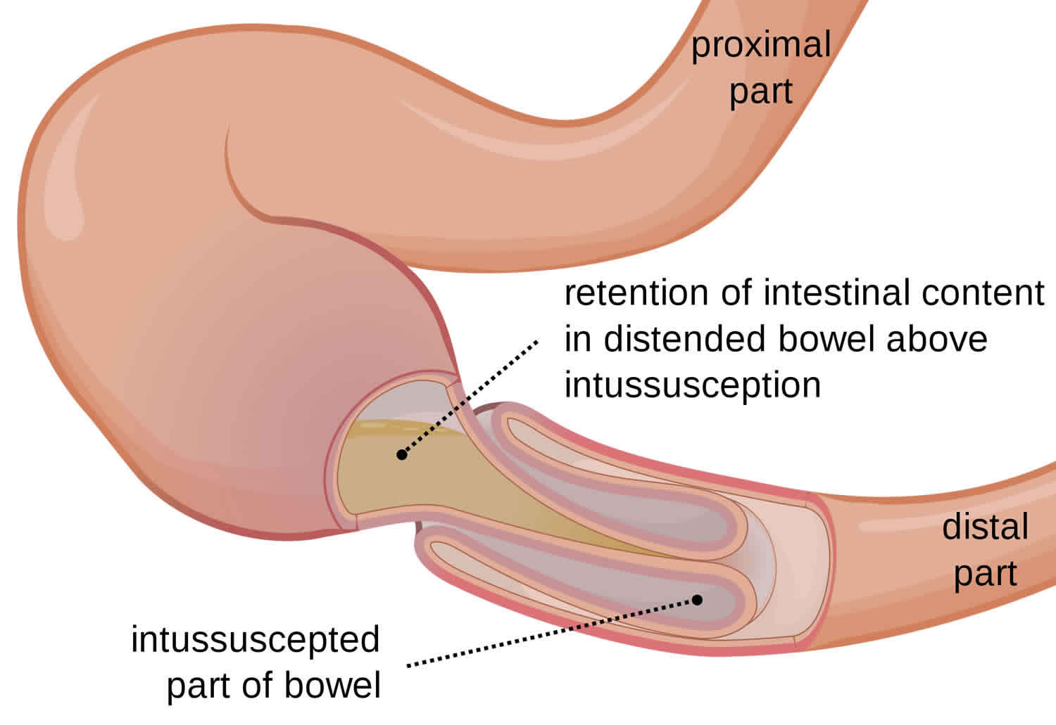
If a child over 4 years of age has been toilet trained, and still passes stool and soils clothes, it is called encopresis. Encopresis is another name for fecal soiling — or an accidental bowel movement. Encopresis isn’t a disease, but rather a symptom that may have different causes. Encopresis is the repeated passing of stool (usually involuntarily) into clothing. The child may or may not be doing this on purpose. Typically encopresis happens when impacted stool collects in the colon and rectum: the colon becomes too full and liquid stool leaks around the retained stool, staining underwear. Eventually, stool retention can cause swelling (distention) of the bowels and loss of control over bowel movements.

It’s difficult to say for certain how many children suffer from encopresis because many cases of encopresis are not reported. There seems to be a stigma attached to this condition that prevents many parents from reaching out and talking to other parents whose children may have had the same problem. It is believed encopresis affects only about 1% to 2% of kids under age 10, but problems with encopresis and constipation account for more than 25% of all visits to pediatric gastroenterologists (doctors who specialize in disorders of the stomach and intestines).

Doctors estimate that between one and three percent of kids have this problem at one time or another in childhood. The problem is much more common in boys than in girls.

There are two main causes of encopresis in a child:

  • Long-term constipation — Your toilet-trained child becomes constipated which stretches his intestine and rectum until he cannot effectively hold the stool and it leaks out.
  • Toilet refusal (much less common) — Your child has never been toilet trained and refuses to have a bowel movement in the toilet, which leads to constipation and encopresis.

While encopresis itself isn’t usually painful (unless the leaking stool leads to a rash on your child’s skin), the constipation that leads to it may be.

Constipated children have fewer bowel movements than normal, and their bowel movements can become hard, dry, difficult to pass and so large that they can often even block up the toilet. Here’s a common scenario:

  1. Your child’s stool can become impacted (packed into her rectum and large intestine).
  2. Your child’s rectum and intestine become enlarged due to the retained stool.
  3. Eventually, her/his rectum and intestine have problems sensing the presence of stool, and the anal sphincter (the muscle at the end of the digestive tract that helps hold stool in) becomes dilated, losing its strength.
  4. Stool can start to leak around the impacted stool, soiling your child’s clothing.
  5. As more and more stool collects, your child will be less and less able to hold it in, leading to accidents. Because of decreased sensitivity in your child’s rectum due to its larger size, she may not even be aware she’s had an accident until after it has occurred.

Because of decreased sensitivity in your child’s rectum, she may not even be aware she’s had an accident until after it has occurred.

In kids who haven’t yet been toilet trained, struggling to hold in excess stool or the constipation that arises from refusing to have a bowel movement (on the toilet) can also lead to encopresis.

Therefore, any child with chronic constipation may develop encopresis. Some situations that may contribute to your child’s constipation include:

  • eating a “junk-food” diet that is low in fiber
  • painful bowel movements
  • lack of exercise
  • stress in the family, with friends or at school
  • change in bathroom routine, such as when a child starts a new school year and bathroom breaks are less frequent
  • being too busy to take time to use the bathroom

For children who have never been toilet trained and who refuse to have a bowel movement on the toilet, additional concerns apply including:

  • reluctance to use bathrooms at home or in public
  • anxiety about using the toilet

In kids who haven’t yet been toilet trained, struggling to hold in excess stool, or constipation that arises from refusing to have a bowel movement on the toilet, can also lead to encopresis.

Many people mistakenly believe that encopresis is a behavioral issue — a simple lack of self-control. But punishing or humiliating a child with encopresis will only make matters worse. Instead, talk to your doctor, who can help you and your child through this challenging but treatable problem.

Encopresis can cause your child to have both physical and emotional problems.

Physical problems

  • Impacted (backed up) stool in her intestine can cause abdominal pain, a loss of appetite and stool accidents.
  • Some children, especially girls, develop urinary tract and/or bladder infections.
  • The enlarged bowel can push on the bladder causing urine accidents during the day or night.
  • Rarely, other health problems may cause the chronic constipation leading to encopresis, including:
    • Hypothyroidism
    • Hirschsprung’s Disease
    • Celiac disease

Emotional problems

Encopresis can be embarrassing, and can prompt teasing from siblings and peers. Watch out for depression and low self-esteem as a result of this condition.

Your child might feel emotionally upset by soiling her clothes, leading to feelings of shame and embarrassment.

You, too, might feel guilt, shame and anger because of your child’s encopresis. It’s very important to try not to communicate this to your child, as this may worsen her emotional state.

Encopresis can be frustrating for parents — and embarrassing for the child. However, with patience and positive reinforcement, treatment for encopresis is usually successful.

Enuresis and encopresis

Enuresis also known as bedwetting or nocturnal enuresis, is when a child of five or older empties their urinary bladder while they are asleep. This can happen every so often, or every night. Soggy sheets and pajamas and an embarrassed child — are a familiar scene in many homes. But don’t despair. Bed-wetting isn’t a sign of toilet training gone bad. It’s often just a normal part of a child’s development.

Enuresis is common. About one in every five children in the US wets their bed. Generally, bed-wetting before age 7 isn’t a concern. At this age, your child may still be developing nighttime bladder control. Enuresis can run in families. It is more common in boys than girls before the age of nine. It can be upsetting for the child and stressful for the whole family.

There are two kinds of enuresis: primary and secondary.

  • Someone with primary nocturnal enuresis has wet the bed since he or she was a baby (primary nocturnal enuresis is the most common form).
  • Secondary enuresis is a condition that develops at least 6 months — or even several years — after a person has learned to control his or her bladder.

The bladder is a muscular receptacle, or holding container, for pee (urine). It expands (gets bigger) as urine enters and then contracts (gets smaller) to push the urine out.

In a person with normal bladder control, nerves in the bladder wall send a message to the brain when the bladder is full; the brain then sends a message back to the bladder to keep it from automatically emptying until the person is ready to go to the bathroom. But people with nocturnal enuresis have a problem that causes them to pee involuntarily at night.

Some children who wet the bed at night also have problems with how their bladder works during the day.

They may:

  • go to the toilet too often
  • not go to the toilet often enough need to rush to the toilet in a hurry have damp underwear
  • have trouble emptying all of the urine from their bladder have bowel problems, including constipation.

The good news is that it’s likely that bedwetting will go away on its own. In fact, 15 out of 100 kids who wet the bed will stop every year without any treatment at all. If enuresis continues, treat the problem with patience and understanding. Lifestyle changes, bladder training, moisture alarms and sometimes medication may help reduce bed-wetting.

Children don’t wet the bed to irritate their parents. Try to be patient as you and your child work through the problem together. Effective treatment may include several strategies and may take time to be successful.

  • Be sensitive to your child’s feelings. If your child is stressed or anxious, encourage him or her to express those feelings. Offer support and encouragement. When your child feels calm and secure, bed-wetting may become less problematic. If needed, talk to your pediatrician about additional strategies for dealing with stress.
  • Plan for easy cleanup. Cover your child’s mattress with a plastic cover. Use thick, absorbent underwear at night to help contain the urine. Keep extra bedding and pajamas handy. However, avoid the long-term use of diapers or disposable pull-up underwear.
  • Enlist your child’s help. If age-appropriate, consider asking your child to rinse his or her wet underwear and pajamas or place these items in a specific container for washing. Taking responsibility for bed-wetting may help your child feel more control over the situation.
  • Celebrate effort. Bed-wetting is involuntary, so it doesn’t make sense to punish or tease your child for wetting the bed. Also, discourage siblings from teasing the child who wets the bed. Instead, praise your child for following the bedtime routine and helping clean up after accidents. Use a sticker reward system if you think this might help motivate your child.

With reassurance, support and understanding, your child can look forward to the dry nights ahead.

When to see a doctor

Most children outgrow bed-wetting on their own — but some need a little help. In other cases, bed-wetting may be a sign of an underlying condition that needs medical attention.

Consult your child’s doctor if:

  • Your child still wets the bed after age 7
  • Your child starts to wet the bed after a few months of being dry at night
  • Bed-wetting is accompanied by painful urination, unusual thirst, pink or red urine, hard stools, or snoring

Any child who is toilet trained and starts new daytime wetting should see a doctor. If the child has daytime symptoms as well as bedwetting, these will be treated before the bedwetting.

Many children do stop wetting in their own time with no help. After age eight or nine, if the child is still wetting often, the problem usually does not get better by itself.

What causes enuresis (bedwetting)?

Doctors don’t always know the exact cause of nocturnal enuresis. They do have some theories, though, on what may contribute to someone developing the condition:

  • Hormonal problems. A hormone called antidiuretic hormone, or ADH, causes the body to produce less urine at night. But some people’s bodies don’t make enough ADH, which means their bodies may produce too much urine while they’re sleeping.
  • Bladder problems. In some people with enuresis, too many muscle spasms can prevent the bladder from holding a normal amount of urine. Some teens and adults also have relatively small bladders that can’t hold a large volume of urine.
  • Inability to recognize a full bladder. If the nerves that control the bladder are slow to mature, a full bladder may not wake your child — especially if your child is a deep sleeper.
  • A small bladder. Your child’s bladder may not be developed enough to hold urine produced during the night.
  • Urinary tract infection. This infection can make it difficult for your child to control urination. Signs and symptoms may include bed-wetting, daytime accidents, frequent urination, red or pink urine, and pain during urination.
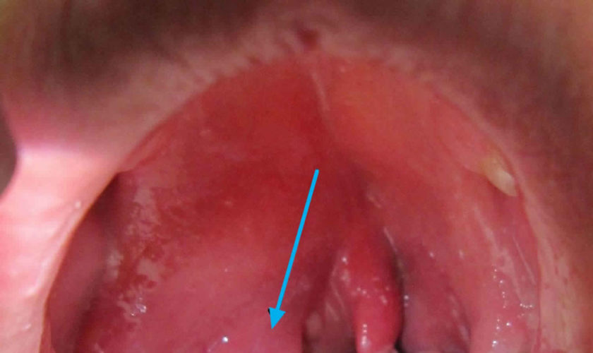
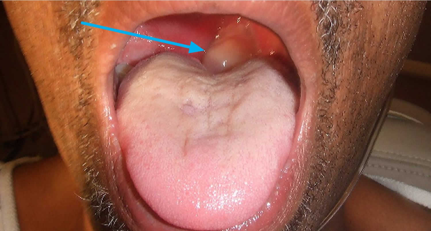
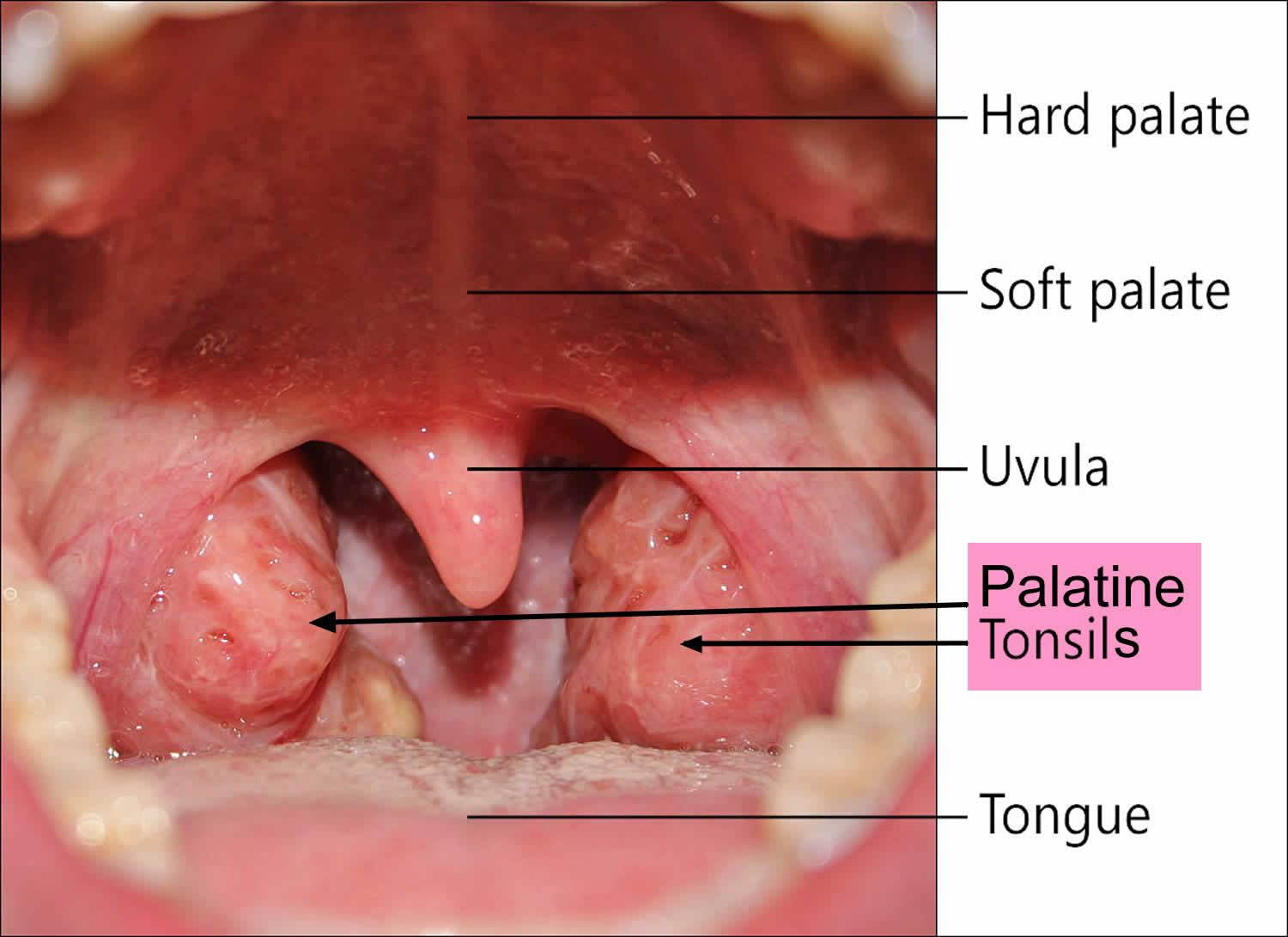
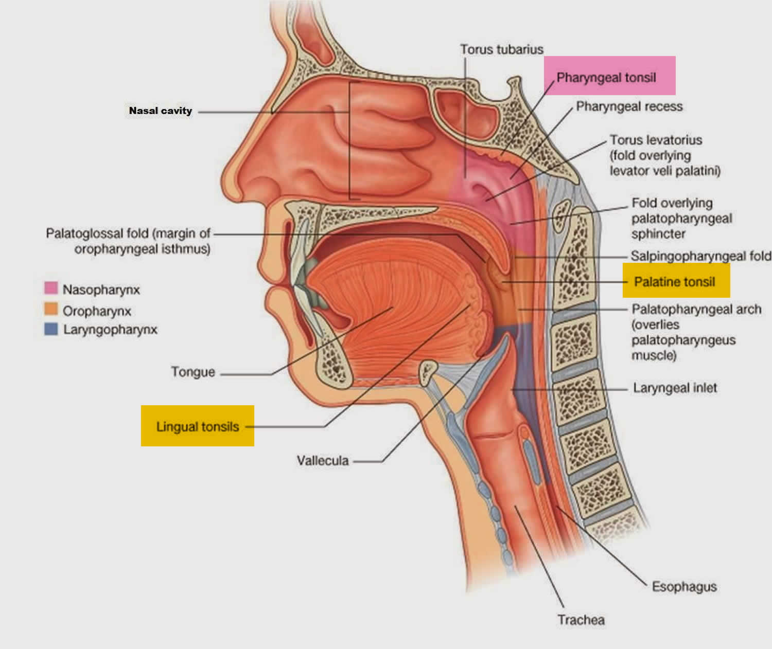
  • Sleep apnea. Sometimes bed-wetting is a sign of obstructive sleep apnea, a condition in which the child’s breathing is interrupted during sleep — often due to inflamed or enlarged tonsils or adenoids. Other signs and symptoms may include snoring and daytime drowsiness.
  • Diabetes. For a child who’s usually dry at night, bed-wetting may be the first sign of diabetes. Other signs and symptoms may include passing large amounts of urine at once, increased thirst, fatigue and weight loss in spite of a good appetite.
  • Chronic constipation. The same muscles are used to control urine and stool elimination. When constipation is long term, these muscles can become dysfunctional and contribute to bed-wetting at night.
    A structural problem in the urinary tract or nervous system. Rarely, bed-wetting is related to a defect in the child’s neurological system or urinary system.
  • Genetics. Teens with enuresis often have a parent who had the same problem at about the same age. Scientists have identified specific genes that cause enuresis.
  • Sleep problems. Some teens may sleep so deeply that they don’t wake up when they need to pee.
  • Caffeine. Using caffeine causes a person to urinate (pee) more.
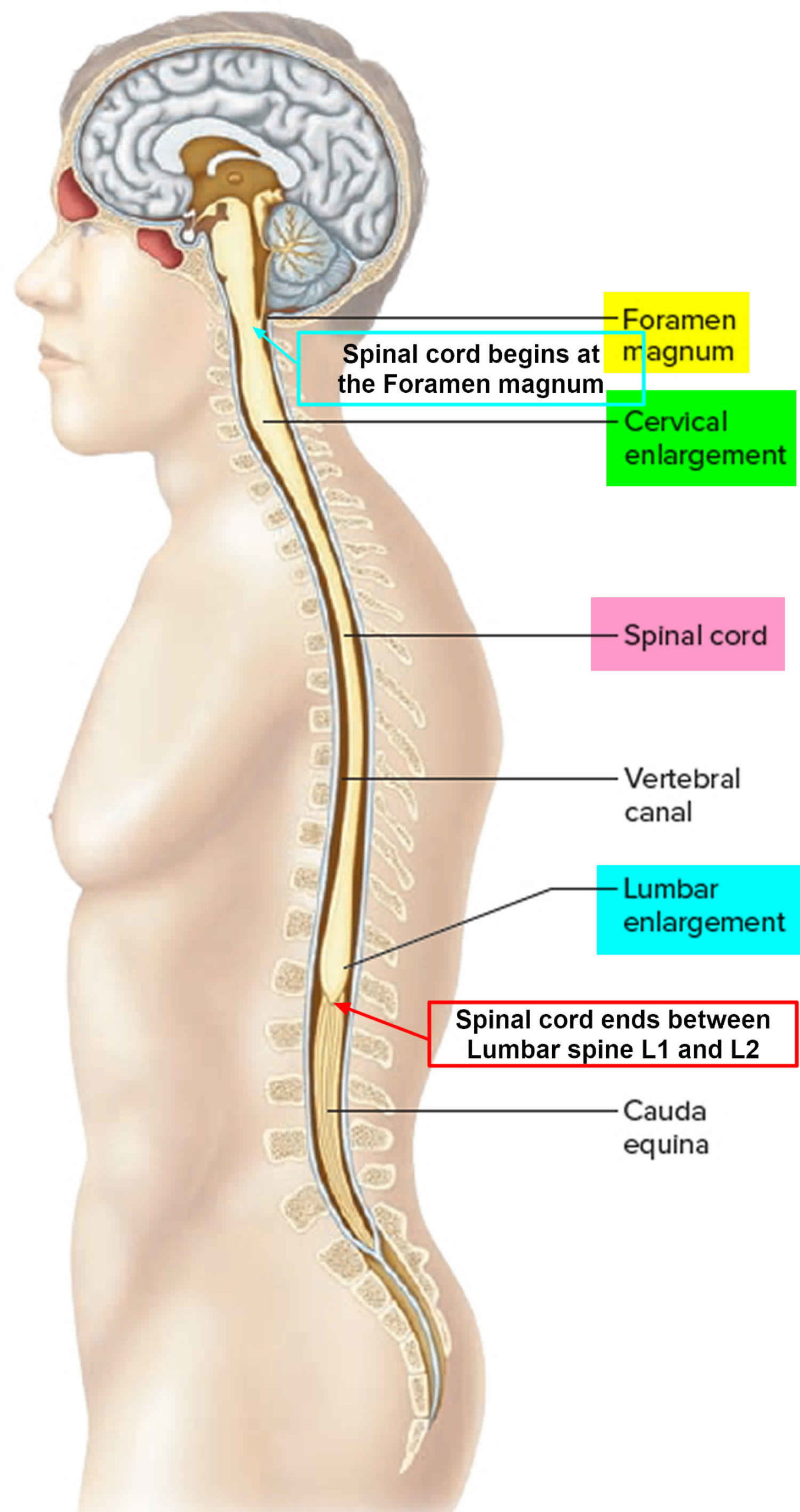
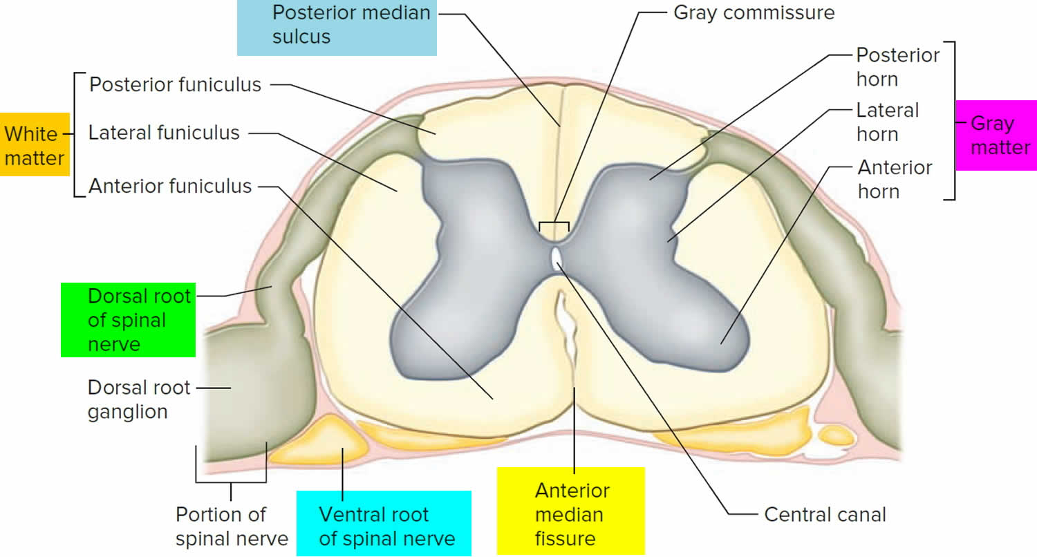
  • Medical conditions. Medical conditions that can trigger secondary enuresis include diabetes, urinary tract abnormalities (problems with the structure of a person’s urinary tract), constipation, and urinary tract infections (UTIs). Spinal cord trauma, such as severe stretching of the spinal cord resulting from a fall, sports injury, auto accident, or other event may also play a role in enuresis, although this is rare.
  • Psychological problems. Some experts believe that stress can be associated with enuresis. It’s not uncommon to feel stressed out during the teenage years, and things such as divorce, the death of a friend or family member, a move to a new town and adapting to a new school and social environment, or family tension can feel overwhelming.

Nocturnal enuresis can be caused by a mix of three things:

  • The body making a large amount of urine through the night.
  • Having a bladder that can only store a small amount of urine at night.
  • Not being able to fully wake up from sleep.

Children who wet the bed are not lazy or being naughty. Some illnesses are linked with bedwetting. But most children who wet the bed do not have major health problems.

Daytime control of the bladder comes before night-time dryness. Most children are dry through the day by three years and dry at night by school age. Any child may still wet every now and then, day or night, up until they are seven or eight years.

Doctors don’t know exactly why, but more than twice as many boys as girls have enuresis. It is often seen in combination with attention-deficit/ hyperactivity disorder (ADHD) 1).

Risk factors for enuresis

Bed-wetting can affect anyone, but it’s twice as common in boys as in girls. Several factors have been associated with an increased risk of bed-wetting, including:

  • Stress and anxiety. Stressful events — such as becoming a big brother or sister, starting a new school, or sleeping away from home — may trigger bed-wetting.
  • Family history. If one or both of a child’s parents wet the bed as children, their child has a significant chance of wetting the bed, too.
  • Attention-deficit/hyperactivity disorder (ADHD). Bed-wetting is more common in children who have ADHD.

Enuresis complications

Although frustrating, bed-wetting without a physical cause doesn’t pose any health risks. However, bed-wetting can create some issues for your child, including:

  • Guilt and embarrassment, which can lead to low self-esteem
  • Loss of opportunities for social activities, such as sleepovers and camp
  • Rashes on the child’s bottom and genital area — especially if your child sleeps in wet underwear

Enuresis diagnosis

If your child is having trouble controlling his/her urine at night, talk to your doctor to learn more about nocturnal enuresis and to rule out the possibility of a medical problem.

Depending on the circumstances, your doctor may recommend the following to identify any underlying cause of bed-wetting and help determine treatment:

  • Physical exam
  • Discussion of symptoms, fluid intake, family history, bowel and bladder habits, and problems associated with bed-wetting
  • Urine tests to check for signs of an infection or diabetes
  • X-rays or other imaging tests of the kidneys or bladder to look at the structure of the urinary tract
  • Other types of urinary tract tests or assessments, as needed

Enuresis treatment

Most children outgrow bed-wetting on their own. If treatment is needed, it can be based on a discussion of options with your doctor and identifying what will work best for your situation.

If your child isn’t especially bothered or embarrassed by an occasional wet night, lifestyle changes — such as avoiding caffeine entirely and limiting fluid intake in the evening — may work well. However, if lifestyle changes aren’t successful or if your grade schooler is terrified about wetting the bed, he or she may be helped by additional treatments.

If found, underlying causes of bed-wetting, such as constipation or sleep apnea, should be addressed before other treatment.

Options for treating bed-wetting may include moisture alarms and medication.

Moisture alarms

These small, battery-operated devices — available without a prescription at most pharmacies — connect to a moisture-sensitive pad on your child’s pajamas or bedding. When the pad senses wetness, the alarm goes off.

Ideally, the moisture alarm sounds just as your child begins to urinate — in time to help your child wake, stop the urine stream and get to the toilet. If your child is a heavy sleeper, another person may need to listen for the alarm and wake the child.

If you try a moisture alarm, give it plenty of time. It often takes one to three months to see any type of response and up to 16 weeks to achieve dry nights. Moisture alarms are effective for many children, carry a low risk of relapse or side effects, and may provide a better long-term solution than medication does. These devices are not typically covered by insurance.

Medication

As a last resort, your child’s doctor may prescribe medication for a short period of time to stop bed-wetting. Certain types of medication can:

  • Slow nighttime urine production. The drug desmopressin (DDAVP) reduces urine production at night. But drinking too much liquid with the medication can cause problems, and desmopressin should be avoided if your child has symptoms such as a fever, diarrhea or nausea. Be sure to carefully follow instructions for using this drug. Desmopressin is given orally as a tablet and is only for children over 5 years old. According to the Food and Drug Administration, nasal spray formulations of desmopressin (Noctiva, others) are no longer recommended for treatment of bed-wetting due to the risk of serious side effects.
  • Calm the bladder. If your child has a small bladder, an anticholinergic drug such as oxybutynin (Ditropan XL) may help reduce bladder contractions and increase bladder capacity, especially if daytime wetting also occurs. This drug is usually used along with other medications and is generally recommended when other treatments have failed.

Sometimes a combination of medications is most effective. There are no guarantees, however, and medication doesn’t cure the problem. Bed-wetting typically resumes when medication is stopped, until it resolves on its own at an age that varies from child to child.

Home remedies

Here are changes you can make at home that may help:

  • Limit fluids in the evening. It’s important to get enough fluids, so there’s no need to limit how much your child drinks in a day. However, encourage drinking liquids in the morning and early afternoon, which may reduce thirst in the evening. But don’t limit evening fluids if your child participates in sports practice or games in the evenings.
  • Avoid beverages and foods with caffeine. Beverages with caffeine are discouraged for children at any time of day. Because caffeine may stimulate the bladder, it’s especially discouraged in the evening.
  • Encourage double voiding before bed. Double voiding is urinating at the beginning of the bedtime routine and then again just before falling asleep. Remind your child that it’s OK to use the toilet during the night if needed. Use small night lights, so your child can easily find the way between the bedroom and bathroom.
  • Encourage regular toilet use throughout the day. During the day and evening, suggest that your child urinate every two hours or so, or at least often enough to avoid a feeling of urgency.
  • Prevent rashes. To prevent a rash caused by wet underwear, help your child rinse his or her bottom and genital area every morning. It also may help to cover the affected area with a protective moisture barrier ointment or cream at bedtime. Ask your pediatrician for product recommendations.

What can parents do

  • Seek help from a health professional with special training in children’s bladder problems, such as a doctor, continence physiotherapist or continence nurse advisor.
  • Make sure there is enough light at night. This makes it easy to get to the toilet.
  • Watch for constipation as this can make bladder problems worse. Seek medical help if constipation is an ongoing problem.
  • If your child is using a bedwetting alarm, you can help by:
    • getting up when it goes off
    • waking them up
    • helping them change their clothes or sheets.
  • Attend review appointments with your child’s continence professional

There are some things which DO NOT help:

  • Do not punish the child for wet beds.
  • Do not shame the child in front of friends or family.
  • Do not lift the child at night to toilet them. This may cut down on some wet beds but it does not help the child learn to be dry.
  • Do not try to fix bedwetting at a stressful time.

Encopresis complications

A child who has encopresis may experience a range of emotions, including embarrassment, frustration, shame and anger. If your child is teased by friends or criticized or punished by adults, he or she may feel depressed or have low self-esteem.

Encopresis causes

There are several causes of encopresis, including constipation and emotional issues. Some kids may develop chronic constipation after stressful life events such as a divorce or the death of a close relative.

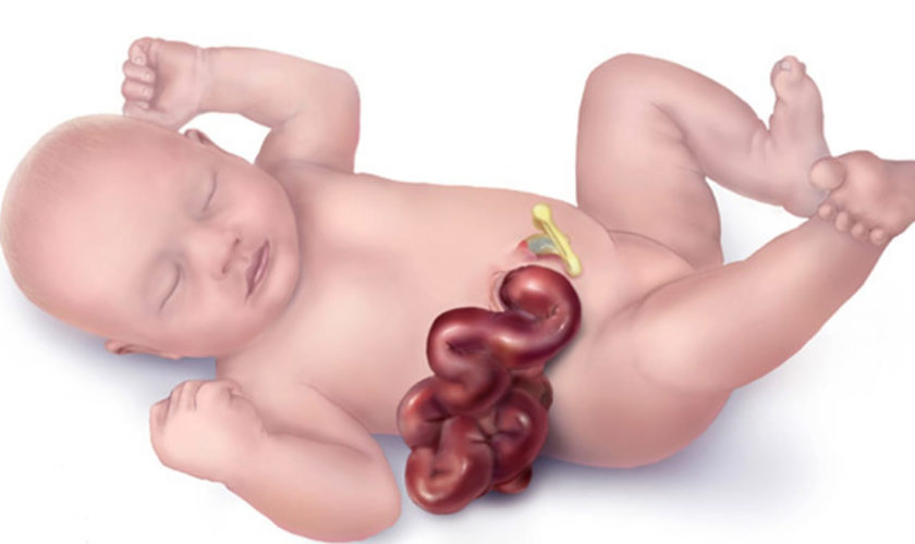
Although rectal surgery or birth defects such as Hirschsprung disease and spina bifida can cause constipation or encopresis without constipation, this is uncommon.

Constipation

Most cases of encopresis are the result of chronic constipation. In constipation, the child’s stool is hard, dry and may be painful to pass. As a result, the child avoids going to the toilet — making the problem worse.

The longer the stool remains in the colon, the more difficult it is for the child to push stool out. This is because the colon’s job is to remove water from the stool before it’s passed. The longer the poop is stuck there, the more water is removed — and the harder it is to push the large, dry poop out. The large poop also stretches out the colon, weakening the muscles there and affecting the nerves signal that tell a child when it’s time to go to the bathroom.

Then, the colon can’t easily push the hard poop out, and it’s painful to pass. So the child continues to avoid having a bowel movement, often by dancing, crossing the legs, making faces, or walking on tiptoes.

Eventually, the rectum and lower part of the colon becomes so full that it’s hard for the sphincter (the muscular valve that controls the passage of feces out of the anus) to hold the poop in. Partial bowel movements may pass through, causing the child to soil his or her pants. Softer stool may also leak out around the large mass of feces and stain the child’s underwear when the sphincter relaxes. Kids can’t prevent this soiling — nor do they have any idea it’s happening — because the nerves aren’t sending the signals that regulate pooping.

Some causes of constipation include:

  • Withholding stool due to fear of using the toilet (especially when away from home) or because stools are painful
  • Not wanting to interrupt play or other activities
  • Eating too little fiber
  • Not drinking enough fluids
  • Drinking too much cow’s milk or, rarely, an intolerance to cow’s milk — though research results conflict on these issues

Emotional issues

Emotional stress may trigger encopresis. A child may experience stress from:

  • Premature, difficult or conflict-filled toilet training
  • Changes in the child’s life, such as dietary changes, toilet training, starting school or schedule changes
  • Emotional stressors, for example, the divorce of a parent or the birth of a sibling

Encopresis can also be associated with emotional and developmental difficulties that express themselves in an unwillingness or inability to have regular bowel movements. Some children experience particularly negative reactions to toilet training, aggravated by a variety of parental responses. Others develop encopresis in reaction to changes in social circomstances, such as when shifting from private to shared bathrooms at the beginning of preschool or kindergarten. Stress resulting from a move, parents divorcing and other major changes to a child’s environment can all trigger constipation that may eventually result in the cycle that leads to encopresis.

Children with encopresis associated with developmental disorders may have never been fully toilet trained, whereas children with other types of encopresis may have been toilet trained but developed the disorder in response to apparent environmental stressors.

Risk factors for encopresis

Encopresis is more common in boys. These risk factors may increase the chances of having encopresis:

  • Using medications that may cause constipation, such as cough suppressants
  • Attention-deficit/hyperactivity disorder (ADHD)
  • Autism spectrum disorder
  • Anxiety or depression

Encopresis prevention

Below are some strategies that can help prevent encopresis and its complications.

Avoid constipation

Help your child avoid constipation by providing a balanced diet that’s high in fiber and encouraging your child to drink enough water.

Learn about effective toilet training techniques

Educate yourself on effective toilet training techniques. Avoid starting too early or being too forceful in your methods. Wait until your child is ready, and then use positive reinforcement and encouragement to help make progress. Ask your doctor about resources on toilet training.

Get early treatment for encopresis

Early treatment, including guidance from your child’s doctor or mental health professional, can help prevent the social and emotional impact of encopresis. Regular follow-up visits with your doctor can help identify ongoing or recurring problems so that adjustments in treatment can be made as needed.

Long-Term outlook for children with encopresis

Though it may seem as though your child will suffer from encopresis forever, this isn’t the case. The vast majority of kids (the possible exceptions being those who have an underlying medical issue) will stop having stool accidents and have regular bowel movements on the toilet.

The end result of treatment is the same for both causes of encopresis, but the way you get there is different.

  • Long-term constipation— Your doctor will help your child pass the impacted stool and then help keep stool soft so that it passes easily and doesn’t get backed up again. After about six months, your child’s intestine and rectum will shrink to their normal size and your child should be able to have normal bowel movements on her own without any medication or prompting.
  • Toilet refusal— These children will get a combination of medical (laxatives, stool softeners) and behavioral treatments to help them become more comfortable using the toilet for bowel movements.

Encopresis symptoms

At first, parents may think their child has a simple case of diarrhea. But after repeated episodes, it becomes clear that there’s another problem — especially because the soiling happens when the child isn’t sick.

As the colon is stretched by the buildup of stool, the nerves’ have trouble telling the brain that it’s time for a bowel movement. If untreated, the soiling will get worse and kids may lose their appetites or complain of stomach pain.

A large, hard poop may also cause a tear in the skin around the anus that will leave blood on the stools, the toilet paper, or in the toilet.

Parents are often frustrated by the fact that their child seems unfazed by the poop accidents, which happen mostly during waking hours. Denial may be one reason for a child seeming calm — kids just can’t face the shame and guilt associated with the condition (some even try to hide their soiled underpants from their parents).

Another reason may be more scientific: Because the brain eventually gets used to the smell of poop, the child may no longer notice the odor.

Signs and symptoms of encopresis may include:

  • Leakage of stool or liquid stool on underwear, which can be mistaken for diarrhea
  • Constipation with dry, hard stool
  • Passage of large stool that clogs or almost clogs the toilet
  • Avoidance of bowel movements
  • Long periods of time between bowel movements
  • Lack of appetite
  • Abdominal pain
  • Problems with daytime wetting or bedwetting (enuresis)
  • Repeated bladder infections, typically in girls

Encopresis diagnosis

To be diagnosed with encopresis, a child must be at least the age of 4 and experience the repeated leakage or passage of feces into inappropriate places, such as the floor or her underwear, at least once a month for at least three months. Before making a diagnosis of encopresis, a doctor will rule out possible physiological factors such as food allergies or medicines that act as laxatives.

To diagnose encopresis, your child’s doctor may:

  • Conduct a physical exam and discuss symptoms, bowel movements and eating habits to rule out physical causes for constipation or soiling
  • Do a digital rectal exam to check for impacted stool by inserting a lubricated, gloved finger into your child’s rectum while pressing on his or her abdomen with the other hand
  • Recommend an abdominal X-ray to confirm the presence of impacted stool
  • Suggest that a psychological evaluation be done if emotional issues are contributing to your child’s symptoms.

Encopresis treatment

How to help a child with encopresis

Generally, the earlier that treatment begins for encopresis, the better.

The first step involves clearing the colon of retained, impacted stool. After that, treatment focuses on encouraging healthy bowel movements. In some cases, psychotherapy may be a helpful addition to treatment.

Step 1. Clearing the colon of impacted stool

There are several methods for clearing the colon and relieving constipation. Your child’s doctor will likely recommend one or more of the following:

  • Certain laxatives
  • Rectal suppositories
  • Enemas

Your child’s doctor may recommend close follow-up to check the progress of the colon clearing.

  • Laxatives and enemas should be given only under the supervision of a doctor; never give these treatments at home without first checking with your doctor.

After your child passes the hard stool, it’s important to develop a good routine to ensure that stool does not get backed up again. Because your child’s intestine and rectum will remain stretched (they go back to normal after about six months), your child may still have problems with leakage.

To reduce the number of accidental bowel movements, or fecal soilings, have your child sit on the toilet two to three times a day for five to ten minutes, preferably shortly after a meal.

Step 2. Encouraging healthy bowel movements

Once the colon is cleared, it’s important to encourage your child to have regular bowel movements. Your child’s doctor may recommend:

  • Dietary changes that include more fiber and drinking adequate fluids
  • Laxatives, gradually discontinuing them once the bowel returns to normal function. It’s important to continue using the stool softener to give the bowels a chance to shrink back to normal size (the muscles of the intestines have been stretched out, so they need time to recover).
  • Training your child to go to the toilet as soon as possible when the urge to have a bowel movement occurs
  • A short trial of going off cow’s milk or checking for cow’s milk intolerance, if indicated
  • Parents also will be asked to schedule potty times twice daily after meals (when the bowels are naturally stimulated). The child will sit on the toilet for about 5 to 10 minutes. This helps kids learn to pay attention to the urges to go.

Keep in mind that relapses are normal, so don’t get discouraged. Your child might get constipated again or soil his or her pants during treatment, especially when being weaned off of the stool softeners.

A good way to track your child’s progress is by keeping a daily poop calendar. Make sure to note the frequency, consistency (hard, soft, dry), and size (large, small) of the bowel movements.

Patience is the key to treating encopresis. It may take anywhere from several months to a year for the stretched-out colon to return to its normal size and for the nerves in the colon to become effective again.

Some parents find that positive reinforcement helps to encourage the child throughout treatment. For instance, put a star or sticker on the poop calendar for having a bowel movement (or even just for trying to), sitting on the toilet, or taking medicines.

Whatever you do, don’t blame or yell — it will only make your child feel bad and it won’t help manage the condition. With lots of love, support, and reassurance that he or she isn’t the only one in the world with this problem, your child can overcome encopresis.

Step 3. Behavior modification

Your child’s doctor or mental health professional can discuss techniques for teaching your child to have regular bowel movements. This is sometimes called behavior modification or bowel retraining.

Your child’s doctor may recommend psychotherapy with a mental health professional if the encopresis may be related to emotional issues. Psychotherapy may also be helpful if your child feels shame, guilt, depression or low self-esteem related to encopresis.

Encopresis treatment at home

  • Avoid using enemas or laxatives — including herbal or homeopathic products — without first talking to your child’s doctor.

Once your child has been treated for encopresis, it’s important that you encourage regular bowel movements. These tips can help:

  • Focus on fiber. Feed your child a healthy balanced diet that includes plenty of fruits, vegetables, whole grains and other foods high in fiber and avoid constipation-causing foods, which can help form soft stools.
  • Encourage your child to drink water. Drinking enough water helps keep stool from hardening. Other fluids may help, but watch the calories.
  • Limit cow’s milk if that’s what the doctor recommends. In some cases, cow’s milk may contribute to constipation, but dairy products also contain important nutrients, so ask the doctor how much dairy your child needs each day.
  • Arrange toilet time. Have your child sit on the toilet for five to 10 minutes at a regularly scheduled time every day. This is best done after meals because the bowel becomes more active after eating. Praise your child for sitting on the toilet as requested and trying.
  • Put a footstool near the toilet. This may make your child more comfortable, and changing the position of his or her legs can put more pressure on the abdomen, making a bowel movement easier.
  • Stick with the program. It may take months to resume normal bowel sensation and function and develop new habits. Sticking with the program can also reduce relapses.
  • Be encouraging and positive. As you help your child overcome encopresis, be patient and use positive reinforcement. Don’t blame, criticize or punish your child if he or she has an accident. Instead, offer your unconditional love and support.

Over time, with the proper guidance, children can redevelop a positive association with regular, healthy bowel movements.

Encopresis can be embarrassing, and can prompt teasing from siblings and peers. Watch out for depression and low self-esteem as a result of this condition.

If a child has experienced feelings of shame, guilt or depression as a result of having encopresis, a knowledgeable psychotherapist can help her both understand those feelings and develop techniques for having regular healthy bowel movements.

Diet and Exercise

Diet and exercise are extremely important in keeping stools soft and bowel movements regular. Also, make sure your child gets plenty of fiber-rich foods such as fresh fruits, dried fruits like prunes and raisins, dried beans, vegetables, and high-fiber bread and cereal.

Try these creative ways to add it to your child’s diet:

  • Bake cookies or muffins using whole-wheat flour instead of regular flour. Add raisins, chopped or pureed apples, or prunes to the mix.
  • Add bran to baking items such as cookies and muffins, or to meatloaf or burgers, or sprinkled on cereal. (The trick is not to add too much bran or the food will taste like sawdust.)
  • Serve apples topped with peanut butter.
  • Create tasty treats with peanut butter and whole-wheat crackers.
  • Top ice cream, frozen yogurt, or regular yogurt with high-fiber cereal for some added crunch.
  • Serve bran waffles topped with fruit.
  • Make pancakes with whole-grain pancake mix and top with peaches, apricots, or grapes.
  • Top high-fiber cereal with fruit.
  • Sneak some raisins or pureed prunes or zucchini into whole-wheat pancakes.
  • Add shredded carrots or pureed zucchini to spaghetti sauce or macaroni and cheese.
  • Add lentils to soup.
  • Make bean burritos with whole-grain soft-taco shells.

And don’t forget to have your child drink plenty of fluids each day, especially water. Diluted 100% fruit juice (like pear, peach, or prune) is an option if your child is not drinking enough water. Also, limiting your child’s daily dairy intake (including milk, cheese, and yogurt) may help.

References   [ + ]

read more

Peritonsillar abscess

peritonsillar abscess

What is peritonsillar abscess

Peritonsillar abscess, also known as quinsy, is the localized collection of pus in peritonsillar space at the back of the mouth, next to one of the tonsils (between the tonsillar capsule and superior constrictor muscle) 1). Peritonsillar abscess can be very painful and can make it difficult to open your mouth. Peritonsillar abscess can also cause swelling that can push the tonsil toward the uvula (the dangling fleshy object at the back of the mouth). This can block the throat, making it hard to swallow, speak, and sometimes even breathe. Peritonsillar abscess is usually caused by a complication of tonsillitis. Peritonsillar abscess most often occurs in older children, adolescents, and young adults. But this infection can occur in all age groups, with the highest incidence occurring in adults 20 to 40 years of age 2). Peritonsillar abscess is rare below five years of age 3). Peritonsillar abscess is rare now that antibiotics are used to treat tonsillitis.

The symptoms of a peritonsillar abscess include:

  • Severe sore throat that is worse on one side
  • Fever and chills
  • Swollen lymph glands
  • Trouble swallowing
  • Pain when you speak

Peritonsillar abscess is a common infection of the head and neck region. With an incidence of approximately 1 in 10,000, it is the most common deep head and neck space infection that presents in the emergency department 4). There is no sexual or racial predilection. In the United States, the incidence is 30 per 100,000 among patients who are 5 to 59 years of age.

The prognosis of peritonsillar abscess in most patients is excellent. However, if there are airway compromise and delay in treatment, death can occur.

If you think you have an abscess in the back of your throat, you need to see a doctor. A peritonsillar abscess that isn’t treated can lead to more serious health problems.

The usual treatment for a peritonsillar abscess involves having a doctor drain the abscess. The doctor does this either by withdrawing the pus with a needle (called aspiration) or making a small cut in the abscess with a scalpel so the pus can drain out.

If this doesn’t work, a patient’s tonsils might have to be removed in a procedure called a tonsillectomy. This is especially true for people who have had tonsillitis a lot or who have had a peritonsillar abscess in the past.

If it’s hard to eat or drink, patients may need IV (intravenous, given into a vein) fluids for hydration. A doctor also will prescribe painkillers and antibiotics. Whenever you take antibiotics, always finish the full course of the medicine as prescribed, even if you feel better after a few days. Otherwise, the infection could come back.

People who have a tonsillectomy may need a brief stay in the hospital. That way, doctors can keep an eye on them to make sure everything went as planned.

When to see a doctor

See your doctor right away if you have had tonsillitis and you develop symptoms of peritonsillar abscess. Call your doctor right away if you have trouble breathing, swallowing, talking, or if you start to drool. These are symptoms of a more serious abscess and need immediate medical attention.

See your doctor if you have:

  • Breathing problems
  • Trouble swallowing
  • Pain in the chest
  • Persistent fever
  • Symptoms that get worse

Peritonsillar abscess complications

Rare complications of peritonsillar abscess include:

  • Parapharyngeal abscess
  • Retropharyngeal abscess
  • Laryngeal edema leading to airway compromise
  • Rarely pneumonia or lung abscess following aspiration of a ruptured abscess.
  • Sepsis (infection in the blood) 5)
  • Airway obstruction
  • Extension of infection into the deep tissues of the neck or superior mediastinum with cellulitis of the jaw, neck, or chest
  • Endocarditis (rare)
  • Fluid around the lungs (pleural effusion)
  • Inflammation around the heart (pericarditis)
  • Life-threatening hemorrhage secondary to erosion or septic necrosis into carotid sheath
  • Poststreptococcal complications, such as glomerulonephritis and rheumatic fever, when infection is caused by group A Streptococcus

Figure 1. Peritonsillar abscess

peritonsillar abscess

Tonsils anatomy

The two palatine tonsils are the oval-shaped areas of pink tissue on each side at the back of your throat. The tonsils are formed during the last months of gestation and grow irregularly, reaching their largest size by the time a child is six to seven years of age 6). The tonsils typically begin to involute gradually at puberty, and after 65 years of age, little tonsillar tissue remains 7). Each tonsil has a number of crypts on its surface and is surrounded by a capsule between it and the adjacent constrictor muscle through which blood vessels and nerves pass.

Peritonsillar space consists of loose connective tissue between the fibrous capsule of palatine tonsils medially and superior constrictor muscle laterally. The anterior tonsillar pillar (palatoglossal arch) and the posterior tonsillar pillar (palatopharyngeal arch) contribute to anterior and posterior limits, respectively. Superiorly, this space is related to torus tubarius, while pyriform sinus forms the inferior limit. Since this space is composed of loose connective tissue, it is highly susceptible to abscess formation following infection 8).

Peritonsillar abscess is a localized infection where pus accumulates between the fibrous capsule of the tonsil and the superior pharyngeal constrictor muscle 9).

Figure 2. Normal palatine tonsils

Normal palatine tonsils


Peritonsillar abscess causes

Most peritonsillar abscesses are caused by the same bacteria that cause Strep throat (group A beta-hemolytic streptococcus). Sometimes, other types of bacteria are involved. Peritonsillar abscess usually occurs as a complication of acute tonsillitis. Infectious mononucleosis can also result in abscess formation. Rarely, it may occur de novo without any prior history of a sore throat. If the infection breaks out of a tonsil and gets into the space around it, an abscess can form. Luckily, peritonsillar abscesses aren’t that common these days because doctors use antibiotics to treat tonsillitis.

Tooth and gum disease can increase the chances of a peritonsillar abscess, as can smoking — more good reasons to brush your teeth and not smoke 10).

Cultures most commonly reveal Group A beta-hemolytic streptococcus. The next most commonly found organisms include staphylococcal, pneumococcal, and hemophilic organisms. Rarely, Lactobacillus and filamentous forms like Actinomyces and Micrococcus may be present. Most of the time, the growth is mixed, with both aerobic and anaerobic organisms.

The exact pathophysiology of peritonsillar abscess formation remains unknown to date. The most accepted theory is that an infection develops in crypta magna that then spreads beyond the confines of the tonsillar capsule, initially causing peritonsillitis and then developing into a peritonsillar abscess 11).

Another proposed mechanism is necrosis and pus formation in the capsular area which then obstructs the Webers glands 12), resulting in abscess formation. These are minor salivary glands in peritonsillar space, located in the space just superior to the tonsil in the soft palate and is connected by a duct to the surface of the tonsil, which are responsible for clearing debris from the tonsillar area and assist with digesting food particles trapped in the tonsillar crypts 13). If Weber glands become inflamed, local cellulitis can develop. As the infection progresses, the duct to the surface of the tonsil becomes obstructed from surrounding inflammation. The resulting tissue necrosis and pus formation produces the classic signs and symptoms of peritonsillar abscess 14). These abscesses are generally formed within the soft palate, just above the superior pole of the tonsil, which coincides with the typical location of a peritonsillar abscess 15) The lack of these abscesses in patients who have undergone tonsillectomy supports the theory that Weber glands may contribute to the pathogenesis of peritonsillar abscesses 16).

Other clinical variables associated with the formation of peritonsillar abscesses include significant periodontal disease and smoking 17).

Is peritonsillar abscess contagious?

Yes. The germs that cause viral and bacterial peritonsillar abscess are contagious.

To prevent the spread of a bacterial or viral infection to others:

  • Stay at home when you are ill
  • Ask your doctor when it’s all right for you to return to work or school
  • Cough or sneeze into a tissue.
  • Wash your hands after sneezing or coughing

Peritonsillar abscess prevention

You can take a few precautions to lower your risk of getting an abscess in your tonsils — like not smoking and making sure you keep your teeth and mouth clean.

But sometimes a peritonsillar abscess is beyond your control. If you think you have an abscess, call your doctor right away. The earlier a doctor diagnoses it, the easier treatment is likely to be.

The best prevention is to practice good hygiene:

  • Wash your hands thoroughly and frequently, especially after using the toilet and before eating
  • Avoid sharing food, drinking glasses, water bottles or utensils
  • Replace your toothbrush after being diagnosed with tonsillitis

Peritonsillar abscess signs and symptoms

One or both tonsils become infected. The infection most often spreads to behind the tonsil. It can then spread down into the neck and chest. Swollen tissues can block the airway. This is a life-threatening medical emergency.

The abscess can break open (rupture) into the throat. The content of the abscess can travel into the lungs and cause pneumonia.

Often, the first sign of a peritonsillar abscess is a sore throat. As the abscess develops, other symptoms start, such as:

  • Red, swollen tonsils
  • A tonsil that’s pushing against the uvula
  • Tender, swollen glands (lymph nodes) on one side of the neck
  • Severe pain on one side of the throat
  • Difficulty and pain when swallowing (odynophagia) or opening the mouth
  • Fever and chills
  • Severe throat pain that is usually on one side
  • Ear pain on the side of the abscess
  • Difficulty opening the mouth, and pain with opening the mouth
  • Swallowing problems
  • Drooling or inability to swallow saliva
  • Facial or neck swelling
  • Earache
  • Headache
  • Muffled or hoarse voice
  • Tender glands of the jaw and throat

Patients with peritonsillar abscess appear ill and report malaise, fever, progressively worsening throat pain, and dysphagia 18). The associated sore throat is markedly more severe on the affected side and is often referred to the ear on the same side as the peritonsillar abscess. Physical examination usually reveals trismus (reduced opening of the mouth), with difficulty opening the mouth secondary to inflammation and spasm of masticator muscles 19). Swallowing can be difficult and painful 20). The combination of painful swallowing (odynophagia) and difficulty in swallowing (dysphagia) often leads to the pooling of saliva and subsequent drooling. Patients often speak in a muffled or “hot potato” voice. Marked tender cervical lymphadenitis may be palpated on the affected side. Inspection of the oropharynx reveals tense swelling and erythema of the anterior tonsillar pillar and soft palate overlying the infected tonsil. The tonsil is generally displaced inferiorly and medially with deviation of the uvula to the opposite side (see Figure 1).

An peritonsillar abscess that’s not treated quickly can lead to serious problems — for example, the infection may go into the jaw and neck. If the abscess pops, the infection may spread to the chest and lead to pneumonia.

Peritonsillar abscess diagnosis

See your doctor if you have a sore throat with a fever or any of the other problems that can be caused by a peritonsillar abscess. It’s rare that an abscess will get in the way of your breathing, but if it does, you may need to go to the emergency room right away.

The doctor will examine your mouth, throat, and neck. He or she also may take a throat culture and a blood test. On rare occasions, a doctor may order a CT scan or ultrasound.

The most common history and physical examination findings in patients with peritonsillar abscess are:

  • Cervical lymphadenitis
  • Drooling
  • Erythematous, swollen soft palate with uvula deviation to contralateral side and enlarged tonsil
  • Muffled voice (“hot potato” voice)
  • Rancid or foul-smelling breath (fetor)
  • Trismus

In several retrospective studies, infectious mononucleosis has been reported as a coinfection in 1.5% to 6% of peritonsillar abscess cases 21), making it a possible alternative diagnosis and comorbidity. This is particularly true in adolescents and young adults. Testing for infectious mononucleosis should be based on the patient history, examination findings (e.g., splenomegaly, lymphadenopathy, bilateral tonsillar infection), and clinical suspicion. If infectious mononucleosis is confirmed, amoxicillin use should be avoided secondary to the associated drug-induced rash 22).

The following tests may be done:

  • Aspiration of the abscess using a needle
  • CT scan
  • Fiber optic endoscopy to check if the airway is blocked

Patients with peritonsillar cellulitis often present with symptoms similar to peritonsillar abscess, making it difficult to differentiate between the conditions. In peritonsillar cellulitis, the area between the tonsil and its capsule is erythematous and edematous, without an obvious area of fluctuance or pus formation. Often, these two conditions are distinguished by the absence of pus on needle aspiration, which indicates cellulitis. If the presence of an abscess remains uncertain after needle aspiration, radiologic testing may be helpful. Computed tomography (CT) with contrast media enhancement can be used to demonstrate the presence and extent of an abscess. Alternatively, several small studies have shown that intraoral ultrasonography, if available, can accurately identify and distinguish abscess from cellulitis 23).

If there is suspicion that infection has spread beyond the peritonsillar space or if there are complications involving the lateral neck space, CT or magnetic resonance imaging (MRI) is required 24). Lateral neck infections should be suspected if there is swelling or induration below the angle of the mandible or medial bulging of the pharyngeal wall. Besides accurately diagnosing peritonsillar abscess, CT can detect potential airway compromise and demonstrate the spread of infection to the contiguous deep neck spaces. MRI is superior to CT for soft-tissue definition and is therefore better at detecting complications from deep neck infections, such as internal jugular vein thrombosis or erosion of the abscess into the carotid sheath 25). Disadvantages of MRI include longer scanning times, higher cost, and the potential for claustrophobia 26).

Peritonsillar abscess treatment

Drainage, antibiotic therapy, and supportive therapy for maintaining hydration and pain control are the cornerstones of treatment for peritonsillar abscess 27). Figure 3 outlines the basic treatment approach to patients presenting with a peritonsillar abscess.

Your doctor will need to remove the pus from the abscess. Your doctor will numb the skin around the abscess. He or she will either take the pus out with a needle or make a small cut in the abscess so the pus can drain out. Surgery to remove your tonsils (called a tonsillectomy) is also an option. You will probably only need surgery if you have had many tonsil infections or abscesses before.

  • Aspiration with a wide-bore needle serves both diagnostic and therapeutic purposes. The aspirated pus can be sent for culture sensitivity, and in some cases, further incision and drainage may not be required.
  • Intraoral incision and drainage are carried out in a sitting position to prevent aspiration of pus. Oral and laryngeal mucosa is anesthetized with lidocaine 10% spray. The incision is given at the point of the maximum bulge above the upper pole of the tonsil. Another alternative site for incision is lateral to the point of junction of the anterior pillar with a line drawn through the base of the uvula. Quinsy forceps or No. 11 guarded blade and then sinus forceps are inserted to break the loculi. The opening created is left open to drain, and the patient is asked to gargle with sodium chloride solution. This helps in self-drainage of accumulated material.
  • In uncooperative, young patients or those affected in an unusual location, the procedure might have to be done under general anesthesia.

Your pain and symptoms should get better after the pus is drained. Your doctor will likely prescribe antibiotics to make sure the infection goes away completely. He or she may also give you medicine to help relieve the pain.

Although no longer routinely performed, immediate tonsillectomy should be considered in patients who have strong indications for it, especially those with a history of recurrent tonsillitis, because there is a recurrence rate up to 40% in these patients compared with 10% to 15% for the average patient 28). Tonsillectomy may also be favored in children because they are likely to have recurrent episodes of tonsillitis and may be intolerant of drainage procedures under local anesthesia 29).

Figure 3. Peritonsillar abscess treatment algorithm

Peritonsillar abscess treatment algorithm

[Source 30) ]

Drainage

Some type of drainage procedure is appropriate for most patients who present with a peritonsillar abscess 31). Exceptions include small abscesses (less than 1 cm) without muffled voice, drooling, or trismus. The main procedures include needle aspiration, incision and drainage, or immediate tonsillectomy (at time of presentation or shortly thereafter). Most studies comparing different surgical methods have found that all were equally effective for the treatment of peritonsillar abscess, and there were no statistically significant differences in patient outcomes 32). The acute surgical management of peritonsillar abscess has evolved from immediate tonsillectomy to primarily incision and drainage or needle aspiration 33). Peritonsillar aspiration is a technique well suited for family physicians with proper training. Drainage or aspiration should be performed in a setting where possible airway complications can be managed and the patient can be observed for a few hours afterward to ensure adequate oral fluid intake 34). Table 1 describes the technique for needle aspiration of a peritonsillar abscess 35). Physicians must be aware of important anatomic relationships when performing needle aspiration (Figure 4). If a physician is not comfortable aspirating the abscess, appropriate antibiotics and intravenous fluids should be administered while awaiting otolaryngology consultation.

Table 1. Technique for Needle Aspiration of Peritonsillar Abscess

Make sure the setting is suitable for managing airway complications.
Check that adequate lighting and suction are available.
Ask the patient to sit slightly forward and at eye level to the clinician.
Gently palpate soft palate to localize fluctuant area.
Apply topical anesthetic using Cetacaine spray.
Wait a few minutes for topical anesthetic to take effect, then draw up 6 to 10 mL of 1% to 2% lidocaine with epinephrine.
Use a 25-gauge 1 ½-inch needle to inject local anesthesia into the mucosa overlying the fluctuant area.
Retract the tongue using a tongue depressor.
Insert an 18-gauge spinal needle attached to a 10-mL syringe into area of maximum fluctuance and aspirate.
Do not insert the needle more than 8 mm.
If positive for pus, aspirate until no pus returns.
If negative for pus, withdraw needle and redirect slightly inferior; be aware that the carotid artery is 2 cm posterior and lateral to tonsillar pillar, and the risk of puncture increases the more inferior the needle is directed.
If aspiration is unsuccessful, perform imaging to confirm the presence of abscess; arrange otolaryngology consultation for possible incision and drainage as appropriate.
[Source 36) ]

Figure 4. Peritonsillar abscess needle aspiration important anatomic relationships

Peritonsillar abscess needle aspiration important anatomic relationships

Footnote: When performing needle aspiration for peritonsillar abscess, the physician should be aware of important anatomic relationships, particularly the carotid artery, which lies posterior and lateral to the tonsil. To avoid this structure, the insertion should be at the superior pole of tonsil, not too lateral, and at a depth of 8 mm or less.

[Source 37) ]

Peritonsillar abscess antibiotics

Peritonsillar abscesses are a polymicrobial mixture of aerobic and anaerobic bacteria. Group A streptococcus and Streptococcus milleri group (a subgroup of viridans streptococci) are the most commonly isolated aerobes recovered from culture, whereas Fusobacterium necrophorum is the predominant anaerobe 38). Common organisms associated with peritonsillar abscess are listed in Table 2. Initial empiric antibiotic therapy should include antimicrobials effective against streptococcus and oral anaerobes 39). There is almost universal sensitivity of streptococcus species to penicillin, and several studies show the clinical effectiveness of intravenous penicillin alone after adequate drainage of the abscess 40). However, there are growing concerns about the polymicrobial nature of peritonsillar abscesses. Culture reports demonstrate a greater than 50% penicillin-resistance rate among pathogens other than streptococcus found in peritonsillar abscess, which has led to the routine use of broad-spectrum antibiotics as first-line therapy 41). Macrolides should be avoided secondary to Fusobacterium resistance 42). Table 3 lists suggested antimicrobial regimens 43).

Intravenous fluids are started, as the patient is usually dehydrated 44).

A suitable intravenous antibiotic is started. The antibacterial spectrum should include gram-positive, gram-negative, and anaerobes. Commonly used empirical antibiotics are penicillins like ampicillin/amoxicillin in combination with metronidazole or clindamycin. (Ideally, antibiotic therapy should be started as per culture sensitivity reports). A patient is shifted to oral antibiotics once he improves and can tolerate orally.

Analgesics and antipyretics are given to relieve pain and fever.

The role of steroids is controversial. A study 45) shows that a single dose of intravenous (IV) dexamethasone reduces the hospital stay and severity of symptoms.

These conservative measures can cure peritonsillitis; however, for peritonsillar abscess, drainage is a must along with medical management.

Table 2. Common Organisms Associated with Peritonsillar Abscess

Aerobic bacteria
Corynebacterium
Group A streptococcus
Staphylococcus aureus
Streptococcus milleri group (Streptococcus intermedius, Streptococcus anginosus, Streptococcus constellatus)
Anaerobic bacteria
Bacteroides
Fusobacterium
Peptostreptococcus
Prevotella
[Source 46) ]

Table 3. Suggested Antimicrobial Regimens for the Treatment of Peritonsillar Abscess

Intravenous antibiotic therapy
Penicillin G, 10 million units every 6 hours, plus metronidazole (Flagyl), 500 mg every 6 hours
Ampicillin/sulbactam (Unasyn), 3 g every 6 hours
Third-generation cephalosporin (e.g., ceftriaxone, 1 g every 12 hours) plus metronidazole, 500 mg every 6 hours
Piperacillin/tazobactam (Zosyn), 3.375 g every 6 hours (maximum daily dosage of 18 g)
If penicillin allergic, then clindamycin, 900 mg every 8 hours
If MRSA is a concern, then vancomycin, 1 g every 12 hours, plus metronidazole, 500 mg every 6 hours
Oral antibiotic therapy
Penicillin VK, 500 mg every 6 hours, plus metronidazole, 500 mg every 6 hours
Amoxicillin/clavulanate (Augmentin), 875 mg every 12 hours
Third-generation cephalosporin (e.g., cefdinir [Omnicef], 300 mg every 12 hours) plus metronidazole, 500 mg every 6 hours
Clindamycin, 300 to 450 mg every 8 hours
If MRSA is a concern, then linezolid (Zyvox), 600 mg every 12 hours, plus metronidazole, 500 mg every 6 hours
[Source 47) ]

Inpatient versus outpatient management

Patients with a peritonsillar abscess can be treated as outpatients, but a small percentage may require hospitalization 48). The most common reasons for admission are dehydration, inability to manage oral fluid intake, airway concerns (kissing tonsils), and failure of outpatient management 49). Other comorbid conditions that warrant inpatient management include diabetes mellitus, immunosuppressive disease, chronic immunosuppressive medication use (including prolonged corticosteroid use), or signs of sepsis 50). Complication rates are higher in patients 40 years or older compared with younger patients.24,25 Hospital stays averaged two to four days for all patients 51). If the decision is made to pursue outpatient management, patients should be observed for a few hours after drainage of the abscess to ensure they can tolerate oral fluids, antibiotics, and pain medications. Patients should continue to be monitored closely, with a follow-up appointment scheduled within 24 to 36 hours.

Adjuvant corticosteroid therapy

The acute symptoms of peritonsillar abscess result from inflammation and soft palate edema. Although corticosteroids have been used to treat edema and inflammation in other otolaryngologic diseases, their use as part of a treatment regimen for peritonsillar abscess has not been extensively studied. Two small studies investigated whether the addition of a single corticosteroid dose administered intramuscularly or intravenously (methylprednisolone, 2 to 3 mg per kg up to 250 mg, or dexamethasone, 10 mg) would speed recovery 52), 53). Patients who received the corticosteroids reported decreased pain and improved oral fluid intake within 12 to 24 hours compared with patients who did not receive corticosteroids. These differences seemed to disappear after 48 hours. The empiric use of corticosteroids for the treatment of peritonsillar abscess appears to speed recovery as demonstrated by shorter hospital stays and quicker resolution of pain. However, additional studies are needed before the routine use of corticosteroids is included in treatment protocols 54).

References   [ + ]

read more

Intussusception

intussusception

What is intussusception

Intussusception is a medical emergency in which part of the intestine slides into an adjacent part of the intestine. This “telescoping” often blocks food or fluid from passing through. Intussusception also cuts off the blood supply to the part of the intestine that’s affected, which can lead to a tear in the bowel (perforation), infection and death of bowel tissue.

Intussusception is an important cause of an intestinal obstruction in children younger than 3 and merits timely ultrasound examination and reduction to preclude significant sequelae including bowel necrosis. The vast majority of intussusceptions occur in children (95%), usually after the first three months of life 1). The cause of most cases of intussusception in children is unknown.

Intussusception can occur essentially anywhere. In adults no pattern of distribution is present as such as in the vast majority of cases a lead point lesion is present, and thus the location will depend on the location of that lesion. In children there is a strong predilection for the ileocolic region:

  • ileocolic: most common (75-95%), presumably due to the abundance of lymphoid tissue related to the terminal ileum and the anatomy of the ileocaecal region
  • ileoileocolic: second most common
  • ileoileal and colocolic: uncommon
  • gastric intussusception: rare, but documented 2)

Though rare in adults, most cases of adult intussusception are the result of an underlying medical condition, such as a tumor, where it caused a focal lesion acting as a lead point.

In children, the intestines can usually be pushed back into position with an X-ray procedure. In adults, surgery is often required to correct the problem.

Figure 1. Small and large intestines

Small and large intestines

Figure 2. Intussusception

intussusception

Figure 3. Intussusception of distal small intestine into the cecum (large intestine)

Intussusception

Intussusception in infants

Intussusception is the most common abdominal emergency affecting children under 2 years old. Intussusception happens when one portion of the bowel slides into the next, much like the pieces of a telescope.

When this “telescoping” happens, the flow of fluids and food through the bowel can become blocked, the intestine can swell and bleed, and the blood supply to the affected part of the intestine can get cut off. Eventually, this can cause part of the bowel to die.

Intussusception happens in 1 to 4 out of every 1,000 infants and is most common in babies 5 to 9 months old, though older children also can have it. Boys get intussusception more often than girls.

Intussusception in infants signs and symptoms

Babies and children with intussusception have intense abdominal pain, which often begins suddenly and causes the child to draw the knees up toward the chest. The pain often makes the child cry very loudly. As it eases, the child may stop crying for a while and may seem to feel better. The pain usually comes and goes like this, but can become very strong when it returns.

The pain of intussusception comes and goes, usually every 15 to 20 minutes at first. These painful episodes last longer and happen more often as time passes.

Symptoms also can include:

  • abdominal swelling
  • vomiting
  • vomiting up bile, a bitter-tasting yellowish-green fluid
  • passing stools (poop) mixed with blood and mucus, known as currant jelly stool
  • grunting due to pain
  • fever

Not everyone has all of the symptoms. Some infants have no obvious pain, and some children don’t pass blood or have a lump in the abdomen. Some older children have pain but no other symptoms.

As the illness continues, the child may gradually become weaker. He or she may develop a fever and appear to go into shock, a life-threatening medical problem in which lack of blood flow to the body’s organs causes the heart to beat quickly and blood pressure to drop.

Some babies with intussusception may just appear drowsy without vomiting, have stool changes, or have abdominal swelling.

Intussusception in infants causes

Most of the time, doctors don’t know what causes intussusception. In some cases, it might follow a recent attack of gastroenteritis (or “stomach flu”). Bacterial or viral gastrointestinal infections may cause swelling of the infection-fighting lymph tissue that lines the intestine, which may result in one part of the intestine being pulled into the other.

In kids younger than 3 months of age or older than 5, intussusception is more likely to be caused by an underlying condition like enlarged lymph nodes, a tumor, or a blood vessel abnormality in the intestines.

Intussusception in infants diagnosis and treatment

Doctors usually check for intussusception if a child is having repeat episodes of pain, drawing up the legs, vomiting, feeling drowsy, or passing stools with blood and mucous.

During the visit, the doctor will ask about the child’s overall health, family health, any medications the child is taking, and any allergies the child may have. Next, the doctor will examine the child, paying special attention to the abdomen, which may be swollen or tender to the touch. Sometimes the doctor can feel the part of the intestine that’s involved.

If the doctor suspects intussusception, the child may be sent to an emergency room (ER). Usually, doctors there will ask a pediatric surgeon to see the child right away. The ER doctor might order an abdominal ultrasound or X-ray, which can sometimes show a blockage in the intestines. If the child looks very sick, suggesting damage to the intestine, the surgeon may take the child to the operating room right away to correct the bowel obstruction.

Two kinds of enemas (an air enema or a barium enema) often can diagnose and treat intussusception at the same time.

For an air enema, a small soft tube is placed in the rectum and air is passed through the tube. The air travels into the intestines and outlines the bowels on the X-rays. If intussusception is present it shows the doctors the telescoping piece in the intestine. At the same time, the pressure of the air unfolds the bowel that has been turned inside out and cures the blockage. Barium, a liquid mixture, is sometimes used in place of air to fix the blockage in the same way.

Both types of enema are very safe, and children usually do very well. However, it’s important to remember that the intussusception can return in 1 out of 10 cases. This usually happens within 72 hours following the procedure.

If the intestine is torn, an enema doesn’t work, or the child is too sick to try an enema, the child will need surgery. This is often the case in older children. Surgeons will try to fix the obstruction, but if too much damage has been done, that part of the bowel will be removed.

After treatment, the child will stay in the hospital and get intravenous (IV) feedings through a vein until he or she can eat and normal bowel function returns. Doctors will watch the child closely to make sure that the intussusception does not return. Some babies may also need antibiotics to prevent infection.

Intussusception in children

Intussusception is the sliding of one part of the intestine into another.

Intussusception in children causes

Intussusception is caused by part of the intestine being pulled inward into itself.

The pressure created by the walls of the intestine pressing together causes:

  • Decreased blood flow
  • Irritation
  • Swelling

Intussusception can block the passage of food through the intestine. If the blood supply is cut off, the segment of intestine pulled inside can die. Heavy bleeding may also occur. If a hole develops, infection, shock, and dehydration can take place very rapidly.

The cause of intussusception is not known. Conditions that may lead to the problem include:

  • Viral infection
  • Englarged lymph node in the intestine
  • Polyp or tumor

The reason for the problem is more likely to be found in older children.

Intussusception can affect both children and adults. However, most cases occur in children ages 6 months to 2 years. It affects boys four times as often as girls.

Intussusception in children prognosis

The outcome is good with early treatment. There is a risk this problem will come back.

When a hole or tear in the bowel occurs, it must be treated right away. If not treated, intussusception is almost always fatal for infants and young children.

Intussusception in children symptoms

The first sign of intussusception is very often sudden, loud crying caused by abdominal pain. The pain is colicky and not continuous (intermittent), but it comes back often. The pain will get stronger and last longer each time it returns.

An infant with severe abdominal pain may draw the knees to the chest while crying.

Other symptoms include:

  • Bloody, mucus-like bowel movement, sometimes called a “currant jelly” stool
  • Fever
  • Shock (pale color, lethargy, sweating)
  • Stool mixed with blood and mucus
  • Vomiting

Intussusception in children diagnosis

Your doctor will perform a thorough exam, which may reveal a mass in the abdomen. There may also be signs of dehydration or shock.

Tests may include:

  • Abdominal ultrasound
  • Abdominal x-ray
  • Air or contrast enema

Intussusception in children treatment

The child will first be stabilized. A tube will be passed into the stomach through the nose (nasogastric tube). An intravenous (IV) line will be placed in the arm, and fluids will be given to prevent dehydration.

In some cases, the bowel blockage can be treated with an air or contrast enema. This is done by a radiologist skilled with the procedure. There is a risk of bowel tearing (perforation) with this procedure.

The child will need surgery if these treatments do not work. The bowel tissue can very often be saved. Dead tissue will be removed.

Antibiotics may be needed to treat any infection.

Intravenous feeding and fluids will be continued until the child has a normal bowel movement.

Intussusception in adults

Bowel intussusception in adults is considered a rare condition, accounting for 5% of all cases of intussusceptions and almost 1%-5% of bowel obstruction 3). Eight to twenty percent of cases are idiopathic, without a lead point lesion. Secondary intussusception is caused by organic lesions, such as inflammatory bowel disease, postoperative adhesions, Meckel’s diverticulum, benign and malignant lesions, metastatic neoplasms or even iatrogenically, due to the presence of intestinal tubes, jejunostomy feeding tubes or after gastric surgery. Computed tomography is the most sensitive diagnostic modality and can distinguish between intussusceptions with and without a lead point. Surgery is the definitive treatment of adult intussusceptions.

Adult intussusception is distinct from pediatric intussusception in various aspects. In children, it is usually primary and benign, and pneumatic or hydrostatic (air contrast enemas) reduction of the intussusception is sufficient to treat the condition in 80% of the patients. In contrast, almost 90% of the cases of intussusception in adults are secondary to a pathologic condition that serves as a lead point, such as carcinomas, polyps, Meckel’s diverticulum, colonic diverticulum, strictures or benign neoplasms, which are usually discovered intraoperatively 4). Due to a significant risk of associated malignancy, which approximates 65% 5), radiologic decompression is not addressed preoperatively in adults. Therefore, 70 to 90% of adult cases of intussusception require definite treatment, of which surgical resection is, most often, the treatment of choice 6).

Intussusceptions have been classified according to their locations into four categories 7):

  1. Entero-enteric, confined to the small bowel,
  2. Colo-colic, involving the large bowel only,
  3. Ileo-colic, defined as the prolapse of the terminal ileum within the ascending colon and
  4. Ileo-cecal, where the ileo-cecal valve is the leading point of the intussusception and that is distinguished with some difficulty from the ileo-colic variant.

Intussusceptions have also been classified according to cause (benign, malignant or idiopathic). In the small intestine, an intussusception can be secondary either to the presence of intra- or extra-luminal lesions (inflammatory lesions, Meckel’s diverticulum, postoperative adhesions, lipoma, adenomatous polyps, lymphoma and metastases) or iatrogenic, e.g. due to the presence of an intestinal tube 8) or even in patients with a gastrojejunostomy 9). Malignancy (adenocarcinoma) accounts for up to 30% of cases of intussusception occurring in the small intestine 10).

Adult bowel intussusception is a rare but challenging condition for the surgeon. Preoperative diagnosis is usually missed or delayed because of nonspecific and often subacute symptoms, without the pathognomonic clinical picture associated with intussusception in children. Abdominal CT is considered as the most sensitive imaging modality in the diagnosis of intussusception and distinguishes the presence or absence of a lead point. Due to the fact that adult intussusception is often frequently associated with malignant organic lesions, surgical intervention is necessary. Treatment usually requires formal resection of the involved bowel segment. Reduction can be attempted in small bowel intussusceptions provided that the segment involved is viable or a malignancy is not suspected.

Intussusception in adults causes

Intussusception in adults up to 90% of cases having a lead point, which is usually malignant in the large bowel and benign in the small bowel 11).

Intussusception in adults symptoms

The clinical presentation of adult intussusception varies considerably. The presenting symptoms are nonspecific and the majority of cases in adults have been reported as chronic, consistent with partial obstruction 12). The classic pediatric presentation of acute intussusception (a triad of cramping abdominal pain, bloody diarrhea and a palpable tender mass) is rare in adults. Nausea, vomiting, gastrointestinal bleeding, change in bowel habits, constipation or abdominal distension are the nonspecific symptoms and signs of intussusception 13).

Intussusception in adults can be further classified according to the presence of a lead point or not 14): transient non-obstructing intussusception without a lead point has been described in patients with celiac 15) or Crohn’s disease 16), but is more frequently idiopathic and resolves spontaneously without any specific treatment. On the other hand, intussusception with an organic lesion as the lead point usually presents as a bowel obstruction, persistent or relapsing, necessitating, however, a definite surgical therapy.

Intussusception in adults diagnosis

Variability in clinical presentation and imaging features often make the preoperative diagnosis of intussusception a challenging and difficult task. Reijnen et al 17) reported a preoperative diagnostic rate of 50%, while Eisen et al 18) reported a lower rate of 40.7%.

Plain abdominal films are typically the first diagnostic tool, since in most cases the obstructive symptoms dominate the clinical picture. Such films usually demonstrate signs of intestinal obstruction and may provide information regarding the site of obstruction 19). Upper gastrointestinal contrast series may show a “stacked coin” or “coil-spring” appearance, while a barium enema examination may be useful in patients with colo-colic or ileo-colic intussusception, during which a “cup-shaped” filling defect or “spiral” or “coil-spring” appearances are characteristically demonstrated 20).

Ultrasonography is considered a useful tool for the diagnosis of intussusception, both in children and in adults 21). The classical imaging features include the “target” or “doughnut” signs on the transverse view and the “pseudo-kidney” sign or “hay-fork” sign in the longitudinal view 22). Undoubtedly, this procedure requires handling and interpretation by an experienced radiologist, in order to confirm the diagnosis. However, obesity and the presence of massive air in the distended bowel loops limit the image quality and the subsequent diagnostic accuracy.

Abdominal computed tomography (CT) is currently considered as the most sensitive radiologic method to confirm intussusception, with a reported diagnostic accuracy of 58%-100% 23). The characteristic features of CT scan include an unhomogeneous “target” or “sausage”- shaped soft- tissue mass with a layering effect (see Figures 4 and 5 below); mesenteric vessels within the bowel lumen are also typical 24). A CT scan may define the location, the nature of the mass, its relationship to surrounding tissues and, additionally, it may help staging the patient with suspected malignancy causing the intussusception 25). In a recent interesting report by Kim et al 26), abdominal CT was able to distinguish between intussusception without a lead point (features: no signs of proximal bowel obstruction, target-like or sausage-shaped mass, layering effect) from that with a lead point (features: signs of bowel obstruction, bowel wall edema with loss of the classic three-layer appearance due to impaired mesenteric circulation and demonstration of the lead mass), and this may help reducing the number of unnecessary surgical interventions.

Intussusception in adults treatment

Due to the fact that adults present with acute, subacute, or chronic nonspecific symptoms 27), the initial diagnosis is missed or delayed and is established only when the patient is on the operating table. Most surgeons accept that adult intussusception requires surgical intervention because of the large proportion of structural anomalies and the high incidence of occurring malignancy. However, the extent of bowel resection and the manipulation of the intussuscepted bowel during reduction remain controversial 28). In contrast to pediatric patients, where intussusception is primary and benign, preoperative reduction with barium or air is not suggested as a definite treatment for adults 29).

The theoretical risks of preliminary manipulation and reduction of an intussuscepted bowel include:

  • (1) intraluminal seeding and venous tumor dissemination,
  • (2) perforation and seeding of microorganisms and tumor cells to the peritoneal cavity and
  • (3) increased risk of anastomotic complications of the manipulated friable and edematous bowel tissue 30).

Moreover, reduction should not be attempted if there are signs of inflammation or ischemia of the bowel wall 31). Therefore, in patients with ileo-colic, ileo-cecal and colo-colic intussusceptions, especially those more than 60 years of age, due to the high incidence of bowel malignancy as the underlying etiologic factor, formal resections using appropriate oncologic techniques are recommended, with the construction of a primary anastomosis between healthy and viable tissue 32). Azar et al 33) report that, for right-sided colonic intussusceptions, resection and primary anastomosis can be carried out even in unprepared bowels, while for left-sided or rectosigmoid cases resection with construction of a colostomy and a Hartmann’s pouch with re-anastomosis at a second stage is considered safer, especially in the emergency setting.

However, when a preoperative diagnosis of a benign lesion is safely established, the surgeon may reduce the intussusception by milking it out in a distal to proximal direction 34), allowing for a limited resection. Wang et al 35) report that for enteric intussusceptions due to benign lesions, reduction and limited resection resulted in non-recurrence of intussusception. In patients with a risk of a short bowel syndrome due to multiple small intestinal polyps causing intussusception, such as Peutz-Jeghers syndrome, a combined approach with limited intestinal resections and multiple snare polypectomies should be mandatory 36). Moreover, in patients complicated with postoperative bowel obstruction due to an intussusception, reduction is also recommended, provided that the bowel appears non-ischemic and viable 37).

Finally, several reports have been published regarding the laparoscopic approach of adult intussusception, due to benign and malignant lesions of the small and large bowel 38). Laparoscopy has been used successfully in selected cases, depending on patients’ general status and availability of surgeons with sufficient laparoscopic expertise. After establishing the diagnosis of intussusception and the underlying disease laparoscopically, reduction and/or en bloc resection can be performed with the same method.

When to see a doctor

Intussusception requires emergency medical care. If you or your child develops the signs or symptoms listed above, seek medical help right away.

In infants, remember that signs of abdominal pain may include recurrent bouts of pulling the knees to the chest and crying.

Intussusception complications

Intussusception can cut off the blood supply to the affected portion of the intestine. If left untreated, lack of blood causes tissue of the intestinal wall to die. Tissue death can lead to a tear (perforation) in the intestinal wall, which can cause an infection of the lining of the abdominal cavity (peritonitis).

Peritonitis is a life-threatening condition that requires immediate medical attention. Signs and symptoms of peritonitis include:

  • Abdominal pain
  • Abdominal swelling
  • Fever

Peritonitis may cause your child to go into shock. Signs and symptoms of shock include:

  • Cool, clammy skin that may be pale or gray
  • A weak and rapid pulse
  • Abnormal breathing that may be either slow and shallow or very rapid
  • Anxiety or agitation
  • Profound listlessness

A child who is in shock may be conscious or unconscious. If you suspect your child is in shock, seek emergency medical care right away.

Intussusception causes

Your intestine is shaped like a long tube. In intussusception, one part of your intestine — usually the small intestine — slides inside an adjacent part. This is sometimes called telescoping because it’s similar to the way a collapsible telescope folds together.

In some cases, the telescoping is caused by an abnormal growth in the intestine, such as a polyp or a tumor (called a lead point). The normal wave-like contractions of the intestine grab this lead point and pull it and the lining of the intestine into the bowel ahead of it. In most cases, however, no cause can be identified for intussusception.

In children, a lead point is not identified in 90% of cases, and this is most frequently thought to relate to hypertrophic lymphoid tissue following an infection. This potentially explains the relative rarity of the condition in the first three months of life, when passive immunity is still paramount.

In infants and adults, a lead point is more frequently identified with up to 90% of adult cases having a lead point (which is usually malignant in the large bowel and benign in the small bowel).

Lead points are numerous and include 39), 40), 41):

  • gastrointestinal malignancy (most common cause in adults, accounting for 65% of cases) 42)
    • colorectal carcinoma (most common)
    • metastases, e.g. malignant melanoma, breast cancer, lung cancer
    • small bowel lymphoma/Burkitt lymphoma
  • benign neoplasms
    • gastrointestinal stromal tumor (GIST)
    • intestinal polyps
    • intestinal lipoma
    • polypoid hemangioma
  • congenital
    • Meckel diverticulum
    • duplication cyst
    • ectopic pancreas
  • inflammatory
    • periappendicitis
  • trauma
    • mural hematoma

Children

In the vast majority of cases of intussusception in children, the cause is unknown. Because intussusception seems to occur more often in the fall and winter and because many children with the problem also have flu-like symptoms, some suspect a virus may play a role in the condition. Sometimes, a lead point can be identified as the cause of the condition — most frequently the lead point is a Meckel’s diverticulum (a pouch in the lining of the small intestine).

Adults

In adults, intussusception is usually the result of a medical condition or procedure, including:

  • A polyp or tumor
  • Scar-like tissue in the intestine (adhesions)
  • Weight-loss surgery (gastric bypass) or other surgery on the intestinal tract
  • Inflammation due to diseases such as Crohn’s disease

Risk factors for intussusception

Risk factors for intussusception include:

  • Age. Children — especially young children — are much more likely to develop intussusception than adults are. It’s the most common cause of bowel obstruction in children between the ages of 6 months and 3 years.
  • Sex. Intussusception more often affects boys.
  • Abnormal intestinal formation at birth. Intestinal malrotation is a condition in which the intestine doesn’t develop or rotate correctly, and it increases the risk for intussusception.
  • A prior history of intussusception. Once you’ve had intussusception, you’re at increased risk of developing it again.
  • A family history. Siblings of someone who’s had an intussusception are at a much higher risk of the disorder.

Intussusception symptoms

Symptoms in infants and children

Babies and children with intussusception have intense abdominal pain, which often begins suddenly and causes the child to draw the knees up toward the chest. The pain often makes the child cry very loudly. As it eases, the child may stop crying for a while and may seem to feel better. The pain usually comes and goes like this, but can become very strong when it returns.

The pain of intussusception comes and goes, usually every 15 to 20 minutes at first. These painful episodes last longer and happen more often as time passes.

Symptoms also can include:

  • abdominal swelling
  • vomiting
  • vomiting up bile, a bitter-tasting yellowish-green fluid
  • passing stools (poop) mixed with blood and mucus, known as currant jelly stool
  • grunting due to pain
  • fever

Not everyone has all of the symptoms. Some infants have no obvious pain, and some children don’t pass blood or have a lump in the abdomen. Some older children have pain but no other symptoms.

As the illness continues, the child may gradually become weaker. He or she may develop a fever and appear to go into shock, a life-threatening medical problem in which lack of blood flow to the body’s organs causes the heart to beat quickly and blood pressure to drop.

Some babies with intussusception may just appear drowsy without vomiting, have stool changes, or have abdominal swelling.

Symptoms in adults

Because intussusception is rare in adults and symptoms of the disorder often overlap with the symptoms of other disorders, it’s more challenging to identify. The most common symptom is abdominal pain that comes and goes. Nausea and vomiting may also occur. Adults sometimes have symptoms for weeks before seeking medical attention.

Intussusception diagnosis

Your or your child’s doctor will start by getting a history of the symptoms of the problem. He or she may be able to feel a sausage-shaped lump in the abdomen. To confirm the diagnosis, your doctor may order:

  • Ultrasound or other abdominal imaging. An ultrasound, X-ray or computerized tomography (CT) scan may reveal intestinal obstruction caused by intussusception. Imaging will typically show a “bull’s-eye,” representing the intestine coiled within the intestine. Abdominal imaging also can show if the intestine has been torn (perforated).
  • Air or barium enema. An air or barium enema is basically enhanced imaging of the colon. During the procedure, the doctor will insert air or liquid barium into the colon through the rectum. In addition, an air or barium enema can actually fix intussusception 90 percent of the time in children, and no further treatment is needed. A barium enema can’t be used if the intestine is torn.

Figure 4. Intussusception ultrasound

Intussusception ultrasound

Figure 5. Intussusception ultrasound (target sign)

Intussusception ultrasound

Intussusception treatment

Treatment of intussusception typically happens as a medical emergency. Emergency medical care is required to avoid severe dehydration and shock, as well as prevent infection that can occur when a portion of intestine dies due to lack of blood.

In some cases, intussusception may be temporary and go away without treatment.

Initial care

When your child arrives at the hospital, the doctors will first stabilize his or her medical condition. This includes:

  • Giving your child fluids through an intravenous (IV) line
  • Helping the intestines decompress by putting a tube through the child’s nose and into the stomach (nasogastric tube)

Correcting the intussusception

To treat the problem, your doctor may recommend:

  • A barium or air enema. This is both a diagnostic procedure and a treatment. If an enema works, further treatment is usually not necessary. This treatment is highly effective in children, but rarely used in adults. Intussusception recurs as often as 10 percent of the time and the treatment will have to be repeated.

Intussusception surgery

If the intestine is torn, if an enema is unsuccessful in correcting the problem or if a lead point is the cause, surgery is necessary. The surgeon will free the portion of the intestine that is trapped, clear the obstruction and, if necessary, remove any of the intestinal tissue that has died. Surgery is the main treatment for adults and for people who are acutely ill.

References   [ + ]

read more

Cleft palate

cleft-lip-and-cleft-palate

What is a cleft palate

A cleft palate happens if the tissue that makes up the roof of the mouth does not join together completely during pregnancy (see Figures 1 and 2). For some babies, both the front and back parts of the palate are open. For other babies, only part of the palate is open. Cleft lip and cleft palate comprise the most common birth defect in the United States. One of every 600 newborns is affected by cleft lip and/or cleft palate. Centers for Disease Control and Prevention 1) recently estimated that, each year in the United States, about 2,650 babies are born with a cleft palate and 4,440 babies are born with a cleft lip with or without a cleft palate 2). Isolated orofacial clefts, or clefts that occur with no other major birth defects, are one of the most common types of birth defects in the United States 3). Depending on the cleft type, the rate of isolated orofacial clefts can vary from 50% to 80% of all clefts 4).

Usually the palatine processes of the maxillary bones unite during weeks 10 to 12 of embryonic development (see Figure 1). Failure to do so can result in one type of cleft palate. The condition may also involve incomplete fusion of the horizontal plates of the palatine bones (see Figure 1). Another form of this condition, called cleft lip, involves a split in the upper lip. Cleft lip and cleft palate often occur together.

Cleft lip and cleft palate are birth defects that occur when a baby’s lip or mouth do not form properly. They happen early during pregnancy. A baby can have a cleft lip, a cleft palate, or both.

  • A cleft lip is a separation of the two sides of the lip. The separation often includes the bones of the upper jaw and/or upper gum.
  • A cleft palate is an opening in the roof of the mouth in which the two sides of the palate did not fuse, or join together, as the unborn baby was developing. Cleft lip and cleft palate can occur on one side (unilateral cleft lip and/or palate), or on both sides (bilateral cleft lip and/or palate) or a cleft palate may go the full length of the palate. Because the lip and the palate develop separately, it is possible for the child to have a cleft lip, a cleft palate, or both cleft lip and cleft palate.

With cleft lip and palate, the majority of babies (91%) are diagnosed with a cleft lip before birth using a 20 week ultrasound scan, so most parents have a chance to learn more about cleft and prepare for the possibility that their child will have a cleft palate as well (see Cleft Lip Ultrasound Scan below). However for some parents, this isn’t the case, and the birth of their child may be the first time they’ve ever heard of the condition.

A cleft lip and palate can:

  • Affect the appearance of the face
  • Lead to problems with feeding and speech
  • Lead to ear infections

Symptoms

  • A child may have one or more birth defects.
  • A cleft lip may be just a small notch in the lip. It may also be a complete split in the lip that goes all the way to the base of the nose.
  • A cleft palate can be on one or both sides of the roof of the mouth. It may go the full length of the palate.

Other symptoms include:

  • Change in nose shape (how much the shape changes varies)
  • Poorly aligned teeth

Problems that may be present because of a cleft lip or palate are:

  • Failure to gain weight
  • Feeding problems
  • Flow of milk through nasal passages during feeding
  • Poor growth
  • Repeated ear infections
  • Speech difficulties

Depending on the extent and position of the cleft palate, speech and swallowing may be affected. In addition, children with cleft palate tend to have many ear infections, which can lead to hearing loss. Facial and oral surgeons recommend closure of cleft lip during the first few weeks following birth, and surgical results are excellent. Repair of cleft palate typically is completed between 12 and 18 months of age, ideally before the child begins to talk. Because the palate is important for pronouncing consonants, speech therapy may be required, and orthodontic therapy may be needed to align the teeth. Recent research strongly suggests that supplementation with folic acid (one of the B vitamins) during early pregnancy decreases the incidence of cleft palate and cleft lip. The mechanism behind this is not yet understood.

Babies are more likely to be born with a cleft lip and palate if they have a family history of these conditions or other birth defects.

With treatment, most children with orofacial clefts do well and lead a healthy life. Some children with orofacial clefts may have issues with self-esteem if they are concerned with visible differences between themselves and other children. Parent-to-parent support groups can prove to be useful for families of babies with birth defects of the head and face, such as orofacial clefts.

Many children with cleft palate need the help of a speech pathologist, and some many need an additional operation to improve speech. The most common problem these children experience is an overly nasal tone to their speech.


Figure 1. Inferior view of the skull

Inferior view of the skull

Figure 2. Cleft palate

Cleft palate

Cleft lip and cleft palate

Cleft lip and cleft palate are birth defects that occur when a baby’s lip or mouth do not form properly during pregnancy. Together, these birth defects commonly are called “orofacial clefts”.

Figure 3. Cleft lip and cleft palate

Cleft lip and cleft palate

Submucous cleft palate

In the submucous cleft palate, the separation of the palate is not complete. In submucous cleft palate there is a cleft in the muscles but it is covered by the lining at the top of the mouth, so it can’t usually been seen by the naked eye but can still have an impact on feeding, speech and hearing. The characteristic in the appearance of the submucous cleft palate is the bifid (double) uvula (see Figure 4 below). The cleft is referred mainly to the muscle layer of the palate, which is separated, and a notch at the back of the hard palate.

The functional problems in the submucous cleft are the same that are observed in the complete types of cleft palate.

  • Nasal speech (rinolalia)
  • Frequent episodes of otitis media (middle ear infection)

It should be noted that these problems are usually mild or may not exist at all, so it is possible not to be diagnosed.

The diagnosis is made solely with a clinical examination, in which the bifid uvula is observed. By gentle palpation of the palate it is detected a notch (deficit) at the back of in the hard palate.

The doctor starts to suspect the existence of submucous cleft palate when the child has unexplained rinolalia or frequent episodes of otitis media.

The surgical repair in submucous cleft should be performed only in the case that it is detected rinolalia (nasal speech) or frequent episodes of otitis.

Though, it should be noted that the results after the surgical repair for improving the speech or reducing the frequency of otitis media episodes are not always completely satisfactory. They may be less satisfactory than the cases of complete cleft palate.

Figure 4. Submucous cleft palate

Submucous cleft palate

What causes cleft palate

There are many causes of cleft lip and palate. Problems with genes passed down from 1 or both parents, drugs, viruses, or other toxins can all cause these birth defects. Cleft lip and palate may occur along with other syndromes or birth defects.

Research tells us it’s often caused by a combination of different genetic and environmental factors, but because of the huge number of factors involved it can be very difficult to narrow these down.

Genetics is all about things inherited from family members, like eye and hair color. Sometimes there is a clear family link, other times it just happens as a ‘one off’.

Environmental factors mean things that happen just before or during pregnancy, like taking a certain medicine or how the baby starts growing in the womb.

Most of the time, a cleft is caused by genetic and environmental factors coming together in a way which can’t be predicted or prevented.

Recently, the Centers for Disease Control and Prevention reported on important findings from research studies about some factors that increase the chance of having a baby with an orofacial cleft:

  • Smoking―Women who smoke during pregnancy are more likely to have a baby with an orofacial cleft than women who do not smoke 5), 6).
  • Diabetes―Women with diabetes diagnosed before pregnancy have an increased risk of having a child with a cleft lip with or without cleft palate, compared to women who did not have diabetes 7).
  • Use of certain medicines―Women who used certain medicines to treat epilepsy, such as topiramate or valproic acid, during the first trimester (the first 3 months) of pregnancy have an increased risk of having a baby with cleft lip with or without cleft palate, compared to women who didn’t take these medicines 8), 9).

The Centers for Disease Control and Prevention continues to study birth defects, such as cleft lip and cleft palate, and how to prevent them. If you are pregnant or thinking about becoming pregnant, talk with your doctor about ways to increase your chances of having a healthy baby.

Cleft lip

The lip forms between the fourth and seventh weeks of pregnancy. As a baby develops during pregnancy, body tissue and special cells from each side of the head grow toward the center of the face and join together to make the face. This joining of tissue forms the facial features, like the lips and mouth. A cleft lip happens if the tissue that makes up the lip does not join completely before birth. This results in an opening in the upper lip. The opening in the lip can be a small slit or it can be a large opening that goes through the lip into the nose. A cleft lip can be on one or both sides of the lip or in the middle of the lip, which occurs very rarely. Children with a cleft lip also can have a cleft palate.

Bilateral and Unilateral Cleft Lip: Your baby’s cleft may have been described as ‘bilateral’ or ‘unilateral’. This is just whether or not the cleft affects one (unilateral) or two (bilateral) sides of the lip.

Complete and Incomplete: This describes whether or not the cleft goes all the way up into the nose or not.

Children with a cleft lip or a cleft palate often have problems with feeding and talking. They also might have ear infections, hearing loss, and problems with their teeth.

Often, surgery can close the lip and palate. Cleft lip surgery is usually done before age 12 months, and cleft palate surgery is done before 18 months. Many children have other complications. They may need additional surgeries, dental and orthodontic care, and speech therapy as they get older. With treatment, most children with clefts do well and lead a healthy life.

Figure 5. Cleft lip

Cleft lip

Cleft lip causes

There are many causes of cleft lip and palate. Problems with genes passed down from 1 or both parents, drugs, viruses, or other toxins can all cause these birth defects. Cleft lip and palate may occur along with other syndromes or birth defects.

Research tells us it’s often caused by a combination of different genetic and environmental factors, but because of the huge number of factors involved it can be very difficult to narrow these down.

Genetics is all about things inherited from family members, like eye and hair color. Sometimes there is a clear family link, other times it just happens as a ‘one off’.

Environmental factors mean things that happen just before or during pregnancy, like taking a certain medicine or how the baby starts growing in the womb.

Most of the time, a cleft is caused by genetic and environmental factors coming together in a way which can’t be predicted or prevented.

Recently, the Centers for Disease Control and Prevention reported on important findings from research studies about some factors that increase the chance of having a baby with an orofacial cleft:

  • Smoking―Women who smoke during pregnancy are more likely to have a baby with an orofacial cleft than women who do not smoke 10), 11).
  • Diabetes―Women with diabetes diagnosed before pregnancy have an increased risk of having a child with a cleft lip with or without cleft palate, compared to women who did not have diabetes 12).
  • Use of certain medicines―Women who used certain medicines to treat epilepsy, such as topiramate or valproic acid, during the first trimester (the first 3 months) of pregnancy have an increased risk of having a baby with cleft lip with or without cleft palate, compared to women who didn’t take these medicines 13), 14).

The Centers for Disease Control and Prevention continues to study birth defects, such as cleft lip and cleft palate, and how to prevent them. If you are pregnant or thinking about becoming pregnant, talk with your doctor about ways to increase your chances of having a healthy baby.

Cleft lip ultrasound

Orofacial clefts, especially cleft lip with or without cleft palate, can be diagnosed during pregnancy by a routine ultrasound. Around 81% of cleft lips are diagnosed before birth, usually at the 20-week anomaly scan where parents can find out their baby’s gender. They can also be diagnosed after the baby is born, especially cleft palate. However, sometimes certain types of cleft palate (for example, submucous cleft palate and bifid uvula) might not be diagnosed until later in life.

Most scans only ever pick up a cleft lip and sometimes a cleft gum (sometimes called a cleft alveolus, which is the bony bit of the gum). A regular ultrasound scan cannot reliably identify a cleft palate, either by itself or along with a cleft lip. If you have received a diagnosis of ‘cleft lip and palate’ from a regular ultrasound scan, it’s very likely that what was actually found was a cleft lip and gum. However, as around 60% of babies with a cleft lip will also have a cleft palate, expectant parents are often given a diagnosis of both. Only a very small number (1%) of cleft palates are actually diagnosed before birth.

If you’ve had an antenatal diagnosis of cleft lip you may be able to get a 3D/4D scan to see your baby in more detail.

What is a 3D/4D Scan?

3D/4D scans can provide more detail than a regular 2D ultrasound such as the one you would have had when the cleft was diagnosed. This can help expectant parents to get an idea of what their child may look like when he or she is born, and being able to see the ‘whole baby’ in this way can help with easing some of the distress that may be experienced after a diagnosis.

Which type should I get?

The only difference between these is that 3D scans take still images, and 4D scans show moving images which you may be able to take home as a DVD. These images will often be sepia-colored (yellowish) as this provides the sonographer with a better picture.

Diagnosis after Birth

Sometimes a cleft lip is not picked up at any scans before birth and can be a big surprise to parents. This is especially likely to happen (30% of cases) if the baby has a cleft lip without a cleft palate.

A cleft palate by itself (called an ‘isolated cleft palate’) is almost impossible to pick up before birth through regular scans, and can be easily missed when a baby is born if it is not looked for. Midwives and other health professionals should visually examine the baby’s mouth with a torch by pressing down on the baby’s tongue so the whole of the mouth can be seen.

Cleft lip repair

Surgery to close the cleft lip is often done when the child is between 6 weeks and 9 months old. However, most times, cleft lip repair is done when the child is 3 to 6 months old.

A cleft lip repair involves reconstructing the shape of the lip and the nose and joining the tissues that were not joined before birth. There are a number of different techniques which depend on the shape of your baby’s cleft and the surgeon’s preference. If the palate is involved, part of it may be repaired at the same time.

Dental impressions may be taken before or during the operation so the team has an accurate record of your baby’s cleft to compare with the future development of their mouth.

For cleft lip surgery, your child will have general anesthesia (asleep and not feeling pain). The surgeon will trim the tissues and sew the lip together. The stitches will be very small so that the scar is as small as possible. Most of the stitches will be absorbed into the tissue as the scar heals, so they will not have to be removed later. The surgery usually takes from 1-2 hours depending on the cleft, but your baby will likely be away from the ward for 2-4 hours.

Babies will usually stay in hospital for one or two nights after the operation, however if the cleft is quite small some babies may even be discharged on the same day.

Your baby will look quite different after the surgery, and this can be quite distressing to some parents. Many say that they miss their baby’s ‘old’ smile and that it takes a while to get used to how they look following the cleft repair. Prepare yourself for this as best as you can.

Surgery may be needed later in life if the problem has a major effect on the nose area.

Cleft palate repair

The palate (especially the soft palate) is very important for not just eating and swallowing, but also for speech, so a palate repair surgery aims to make sure the palate works as well as possible.

A cleft palate is most often closed within the first year of life so that the child’s speech develops normally. Most times, cleft palate repair is done when the child is older, between 9 months and 1 year old. This allows the palate to change as the baby grows. Doing the repair when the child is this age will help prevent further speech problems as the child develops.

A cleft palate is usually repaired in layers, with the focus on reconstructing the muscle of the soft palate.

Sometimes, a prosthetic device is temporarily used to close the palate so the baby can feed and grow until surgery can be done.

In cleft palate repair, your child will have general anesthesia (asleep and not feeling pain). Tissue from the roof of the mouth may be moved over to cover the soft palate. Sometimes a child will need more than one surgery to close the palate.

During these procedures, the surgeon may also need to repair the tip of your child’s nose. This surgery is called rhinoplasty.

Continued follow-up may be needed with speech therapists and orthodontists.

Alveolar Bone Graft (ABG) Surgery

If there is a gap in your baby’s gums, this will be repaired as his or her adult teeth are coming through with an Alveolar Bone Graft (ABG) operation at around 8-12 years old. The timing of treatment will be discussed with you by the cleft team. If your child’s cleft affects the alveolus (the bone in the gum where the teeth sit), they will probably need surgery to fill the gap in their gum so that their adult teeth can erupt (come through) properly. A ‘bone graft’ involves borrowing a bit of bone from one part of the body to help repair another.

Assessment for this surgery will begin between 7 and 9 years of age, and the surgery itself will usually be completed by age 12.

The orthodontist will often need to widen the area around the cleft to allow the surgeon to access it easier. This can involve a brace being fitted, most commonly a ‘quadhelix’. This takes place 6-9 months before the Alveolar Bone Graft (ABG) surgery. The orthodontist may also want to arrange to take out some of your child’s top baby teeth next to the cleft. This will give them more room to work with inside the mouth.

During the procedure, bone marrow will be taken (usually from the hip, sometimes from the tibia (shin) bone) and grafted into the gap left by the cleft. If the bone is taken from the hip, a small cut around 5 cm long is made low down on the tummy in an area that is usually covered by swimwear, a window is made in the outer surface of the bone and the inner marrow bone is scooped out. This is then closed and stitched up. The marrow will eventually grow back and the hip will be good as new.

The mucosa (red tissue covering the bone of the gum) is then opened up and peeled away from the gap in the bone. The mucosa from each side is then stitched together to make a ‘pocket’, and the bone marrow is packed into this pocket so the entire gap is filled. The gum is then closed over the bone. The ‘floor’ of the nose may also be repaired at this time. This can help the bottom of the nose look straighter, but won’t change the shape of the face.

Most children are admitted onto the ward the day before surgery and are able to go home the day after. Complications are rare and minor, and the scar on the donor site (hip or shin) will fade considerably with time.

After Alveolar Bone Graft (ABG) Surgery

Afterwards, the ‘donor’ site where the bone was taken is often sore, and your child’s mouth will feel quite strange. They will have a ‘drip’ with painkillers to help ease the discomfort.

Your surgeon will be able to advise on when your child will be able to eat and drink again. They’ll have to stick to ‘soft’ foods for a while after they go home until they’re healed.

It’s vital that the mouth is kept very clean for the first 10-14 days after surgery. This is because the bone placed in the gap will have ‘new’ bone growing around it, and at this time it’s very vulnerable to infection. Good, careful brushing and using a mouthwash recommended by your surgeon will help your child recover.

Usually your child will need at least a week of school to make sure they don’t get an infection from anyone and can rest and recover. Any sporting activities should be avoided for at least two weeks, but check with your surgeon about this. It may take several weeks before the swelling of the face goes down and the inside of your child’s mouth starts to feel normal again.

Your child will need to come back a few weeks after the operation so the consultant can make sure everything is healing properly. After 6 months, your child will have an x-ray to see how successful the treatment has been. It may take several months or even a year before the adult canine tooth erupts through the new bone graft. If teeth are crooked once they’ve come through, these can be straightened 3-6 months after the bone graft. Braces are usually not used until all a child’s permanent teeth have erupted into the mouth.

Cleft palate surgery risks

Risks from any anesthesia and any surgery include:

  • Breathing problems
  • Reactions to the medicines
  • Bleeding
  • Infection
  • Need for further surgery

Problems these surgeries may cause are:

  • The bones in the middle of the face may not grow correctly.
  • The connection between the mouth and nose may not be normal.

Before the Procedure

You will meet with a speech therapist or feeding therapist soon after your child is born. The therapist will help you find the best way to feed your child before the surgery. Your child must gain weight and be healthy before surgery.

Your child’s health care provider may:

  • Test your child’s blood (do a complete blood count and “type and cross” to check your child’s blood type)
  • Take a complete medical history of your child
  • Do a complete physical exam of your child

Always tell your child’s provider:

What medicines you are giving your child. Include drugs, herbs, and vitamins you bought without a prescription.

During the days before the surgery:

  • About 10 days before the surgery, you will be asked to stop giving your child aspirin, ibuprofen (Advil, Motrin), warfarin (Coumadin), and any other drugs that make it hard for your child’s blood to clot.
  • Ask which drugs the child should still take on the day of the surgery.

On the day of the surgery:

Most times, your child will not be able to drink or eat anything for several hours before the surgery.

  • Give your child a small sip of water with any drugs your doctor told you to give your child.
  • You will be told when to arrive for the surgery.
  • The provider will make sure your child is healthy before the surgery. If your child is ill, surgery may be delayed.

Dental impressions may be taken before or during the operation so the team has an accurate record of your baby’s cleft to compare with the future development of their mouth.

Sometimes, small incisions are made to the side of the cleft to ‘loosen’ the tissue and give the surgeon more to work with, but these will heal very quickly.

After the Procedure

Your child will probably be in the hospital for 5 to 7 days right after surgery. Complete recovery can take up to 4 weeks.

The surgery wound must be kept very clean as it heals. It must not be stretched or have any pressure put on it for 3 to 4 weeks. Your child’s nurse should show you how to take care of the wound. You will need to clean it with soap and water or a special cleaning liquid, and keep it moist with ointment.

Until the wound heals, your child will be on a liquid diet. Your child will probably have to wear arm cuffs or splints to prevent picking at the wound. It is important for your child not to put hands or toys in their mouth.

Cleft palate surgery outlook (prognosis)

Most babies heal without problems. How your child will look after healing often depends on how serious the defect was. Your child might need another surgery to fix the scar from the surgery wound.

A child who had a cleft palate repair may need to see a dentist or orthodontist. The teeth may need correcting as they come in.

Hearing problems are common in children with cleft lip or cleft palate. Your child should have a hearing test early on, and it should be repeated over time.

Your child may still have problems with speech after the surgery. This is caused by muscle problems in the palate. Speech therapy will help your child.

Many children with cleft palate need the help of a speech pathologist, and some many need an additional operation to improve speech. The most common problem these children experience is an overly nasal tone to their speech.

References   [ + ]

read more

Tonsillectomy

tonsillectomy

What is tonsillectomy

Tonsillectomy is the surgical procedure to remove the tonsils, two oval-shaped pads of lymphatic tissue at the back of the throat — one tonsil on each side. Tonsillectomy completely removes the tonsil, including its capsule, by dissecting the peritonsillar space between the tonsil capsule and the muscular wall. Depending on the context in which it is used, the term may indicate tonsillectomy with adenoidectomy, especially in relation to sleep-disordered breathing.

Your tonsils are two small glands in the back of the throat, on either side of the tongue and closely associated with the palate, are masses of lymphatic tissue called palatine tonsils (see Figures 1 and 2). Like other lymphatic tissues, the two palatine tonsils help protect your body against infection, particularly in childhood.

Tonsillectomy is the third most common surgical procedure (after circumcision and ear tubes) performed on children in the United States, with over 530,000 annual procedures (1 in 7 ambulatory surgeries under age 15 years) 1), 2). When performed in properly selected children, tonsillectomy can reduce throat infections, doctor visits, and antibiotic use, and can improve a child’s quality of life, daytime functioning, and ability to sleep soundly.

Indications for tonsillectomy include recurrent throat infections 3) and sleep-disordered breathing 4), both of which can substantially affect child health status 5) and quality of life 6). Although there are benefits of tonsillectomy, complications of surgery may include throat pain, postoperative nausea and vomiting, delayed feeding, voice changes, hemorrhage, and rarely death.

When to see the doctor or get emergency care

Watch for the following complications that require prompt medical care:

  • Bleeding. You may see small specks of dark blood from the nose or in the saliva, but any bright red blood requires a trip to the emergency room for a prompt evaluation and treatment. Surgery to stop bleeding may be necessary.
  • Fever. Call your doctor if you or your child has a fever of 102° F (38.9° C) or higher.
  • Dehydration. Call your doctor if you observe signs of dehydration, such as reduced urination, thirst, weakness, headache, dizziness or lightheadedness.
  • Common signs of dehydration in children include urinating fewer than two or three times a day or crying with no tears.
  • Breathing problems. Snoring or noisy breathing is common during the first week or so of recovery. However, if you or your child is having difficulty breathing, get emergency care.

A tonsillectomy may be recommended to prevent frequent, recurring episodes of tonsillitis. Frequent is generally defined as:

  • More than seven episodes a year
  • More than five episodes a year in each of the preceding two years
  • More than three episodes a year in each of the preceding three years

The tonsillectomy procedure may also be recommended if:

  • A bacterial infection causing tonsillitis doesn’t improve with antibiotic treatment
  • An infection that results in a collection of pus behind a tonsil (tonsillar abscess) doesn’t improve with drug treatment or a drainage procedure
  • A tonsillectomy may also be used to treat other rare diseases or conditions of the tonsils, such as:
    • Cancerous tissue in one or both tonsils
    • Recurrent bleeding from blood vessels near the surface of the tonsils

Adenoids sit high in the throat behind the nose and the roof of the mouth. Tonsils and adenoids are often removed when they become enlarged and block the upper airway, leading to breathing difficulty. They are also removed when recurrence of tonsil infections or strep throat cannot be successfully treated by antibiotics.

The excision of the adenoids is an adenoidectomy. Both tonsillectomy and adenoidectomy procedures are often performed at the same time; hence the surgery is known as a tonsillectomy and adenoidectomy or T&A 7).

Tonsillectomy and adenoidectomy is an outpatient surgical procedure lasting between 30 and 45 minutes and performed under general anesthesia 8). Normally, you will remain at the hospital or clinic for several hours after surgery for observation. Children with severe obstructive sleep apnea (OSA) and very young children are usually admitted overnight to the hospital for close monitoring of respiratory status. An overnight stay may also be required if there are complications such as excessive bleeding, severe vomiting, or low oxygen saturation.

  • Recovery time for a tonsillectomy is usually at least 10 days to two weeks.
  • On average, the amount paid for a tonsillectomy without complication was $3,832 whereas tonsillectomy with hemorrhage resulted in an average expenditure of $6,388 9).

A tonsillectomy was once a common procedure to treat infection and inflammation of the tonsils (tonsillitis). Today, a tonsillectomy is usually performed for sleep-disordered breathing but may still be a treatment when tonsillitis occurs frequently or doesn’t respond to other treatments.

A tonsillectomy may also be necessary to treat breathing and other problems related to enlarged tonsils and to treat rare diseases of the tonsils.

Figure 1. Normal palatine tonsils

Normal palatine tonsils


Figure 2. Palatine tonsil, Pharyngeal tonsil and Lingual tonsil

Palatine tonsil, Pharyngeal tonsil and Lingual tonsil

Post tonsillectomy

Most children take seven to ten days to recover from the surgery. Some may recover more quickly; others can take up to two weeks for a full recovery. The following guidelines are recommended:

  • Drinking: The most important requirement for recovery is for the patient to drink plenty of fluids..Starting immediately after surgery, children may have fluids such as water or apple juice. Some patients experience nausea and vomiting after the surgery. This usually occurs within the first 24 hours and resolves on its own after the effects of anesthesia wear off. Contact your physician if there are signs of dehydration (urination less than 2-3 times a day or crying without tears).
  • Eating: Generally, there are no food restrictions after surgery, but some physicians will recommend a soft diet during the recovery period. The sooner the child eats and chews, the quicker the recovery. Tonsillectomy patients may be reluctant to eat because of throat pain; consequently, some weight loss may occur, which is gained back after a normal diet is resumed.
  • Fever: A low-grade fever may be observed the night of the surgery and for a day or two afterward. Contact your physician if the fever is greater than 102ºF
  • (38.9° C).
  • Activity: Activity may be increased slowly, with a return to school after normal eating and drinking resumes, pain medication is no longer required, and the child sleeps through the night. Travel on airplanes or far away from a medical facility is not recommended for two weeks following surgery.
  • Breathing: The parent may notice snoring and mouth breathing due to swelling in the throat. Breathing should return to normal when swelling subsides, 10-14 days after surgery.
  • Scabs: A scab will form where the tonsils and adenoids were removed. These scabs are thick, white, and cause bad breath. This is normal. Most scabs fall off in small pieces five to ten days after surgery.
  • Bleeding: With the exception of small specks of blood from the nose or in the saliva, bright red blood should not be seen. If such bleeding occurs, contact your physician immediately or take your child to the emergency room.
  • Pain: Nearly all children undergoing a tonsillectomy or adenoidectomy will have mild to severe pain in the throat after surgery. Some may complain of an earache (so called referred pain) and a few may have pain in the jaw and neck .
  • Pain control: Your physician will prescribe pain medication for the young patient such as acetaminophen or ibuprofen acetaminophen. The pain medication will be in a liquid form or sometimes a rectal suppository will be recommended. Pain medication should be given as prescribed. Contact your physician if side effects are suspected or if pain is not well-controlled. If you are troubled about any phase of your child’s recovery, contact your physician immediately.

Tonsillectomy in Children

American Academy of Otolaryngology — Head and Neck Surgery Clinical Practice Guideline 10)

The panel made recommendations for:

  1. Watchful waiting for recurrent throat infection if there have been fewer than 7 episodes in the past year or fewer than 5 episodes per year in the past 2 years or fewer than 3 episodes per year in the past 3 years;
  2. Assessing the child with recurrent throat infection who does not meet criteria in statement 2 for modifying factors that may nonetheless favor tonsillectomy, which may include but are not limited to multiple antibiotic allergy/intolerance, periodic fever, aphthous stomatitis, pharyngitis and adenitis, or history of peritonsillar abscess;
  3. Asking caregivers of children with sleep-disordered breathing and tonsil hypertrophy about comorbid conditions that might improve after tonsillectomy, including growth retardation, poor school performance, enuresis, and behavioral problems;
  4. Counseling caregivers about tonsillectomy as a means to improve health in children with abnormal polysomnography who also have tonsil hypertrophy and sleep-disordered breathing;
  5. Counseling caregivers that sleep-disordered breathing may persist or recur after tonsillectomy and may require further management;
  6. Advocating for pain management after tonsillectomy and educating caregivers about the importance of managing and reassessing pain; and
  7. Clinicians who perform tonsillectomy should determine their rate of primary and secondary posttonsillectomy hemorrhage at least annually. The panel offered options to recommend tonsillectomy for recurrent throat infection with a frequency of at least 7 episodes in the past year or at least 5 episodes per year for 2 years or at least 3 episodes per year for 3 years with documentation in the medical record for each episode of sore throat and 1 or more of the following: temperature >38.3°C (100.4 °F), cervical adenopathy, tonsillar exudate, or positive test for group A β-hemolytic streptococcus.

Indications for tonsillectomy

The 2 most common indications for tonsillectomy are recurrent throat infections and sleep-disordered breathing. Throat infection is defined as sore throat caused by viral or bacterial infection of the pharynx, palatine tonsils, or both, which may or may not be culture positive for group A streptococcus. This includes the terms strep throat and acute tonsillitis, pharyngitis, adenotonsillitis, or tonsillopharyngitis 11). Throat infections are a common reason to see a primary care physician and often result in antibiotic treatment 12). The cost of outpatient visits and the medications prescribed for sore throats including antibiotics are substantial. Indirect costs associated with throat infections and sleep-disordered breathing are substantial due to missed school and loss of time from work for caregivers.

Treatment of sleep-disordered breathing is associated with an increase in health care utilization and cost. Children with sleep-disordered breathing, compared with controls, have a significantly higher rate of antibiotic use, 40% more hospital visits, and an overall elevation of 215% in health care usage mostly from increased respiratory tract infections 13). Children with tonsillar disease, including children with throat infections and sleep-disordered breathing, also showed significantly lower scores on several quality of life subscales including general health, physical functioning, behavior, bodily pain, and caregiver impact when compared with healthy children 14).

Sleep-disordered breathing represents a spectrum of disorders ranging in severity from primary snoring to OSA (obstructive sleep apnea). The prevalence of OSA (obstructive sleep apnea) in the pediatric population is 1% to 4% 15); as many as 10% of children have primary snoring 16). Up to 30% to 40% of children with clinically diagnosed sleep-disordered breathing exhibit behavioral problems that include enuresis,28 hyperactivity, aggression, anxiety, depression, and somatization 17). OSA (obstructive sleep apnea) is also associated with poor school performance and a decrease in quality of life 18). The Quality of life of children with OSA is similar to children with chronic conditions such as asthma and juvenile rheumatoid arthritis 19).

Controversy persists about the actual benefits of tonsillectomy as compared with observation and medical treatment of throat infections. Although tonsillectomy for recurrent throat infections in severely affected children has been shown in a randomized controlled trial to reduce the frequency and severity of infections in the 2 years following surgery 20), the same cannot be shown for less severe cases or for a period greater than 2 years after surgery 21). Observational studies, however, show improved disease-specific and global quality of life after tonsillectomy for recurrent or chronic sore throat, as measured by validated instruments 22). These children suffered fewer infections after surgery, resulting in fewer antibiotics and physician visits.

A growing body of evidence indicates that tonsillectomy is an effective treatment for sleep-disordered breathing 23), based on the idea that tonsillar hypertrophy is a principal cause. A meta-analysis of case series 24) and a recent study 25) showed that tonsillectomy was effective at improving or resolving sleep-disordered breathing in most children. There is also evidence that behavioral parameters, school performance, and quality of life improve after resolution of this sleep disorder 26).

Harms and Adverse Events of Tonsillectomy

Tonsillectomy is a surgical procedure with an associated morbidity that includes possible hospitalization, risks of anesthesia, prolonged throat pain, and financial costs. A common complication of tonsillectomy is bleeding during or after the surgery. In published reports, the rate of primary hemorrhage (within 24 hours of surgery) has ranged from 0.2% to 2.2% and the rate of secondary hemorrhage (more than 24 hours after surgery) from 0.1% to 3% 27). Hemorrhage after tonsillectomy may result in readmission for observation or in further surgery to control bleeding.

Other complications of tonsillectomy are diverse and have been well described 28). Operative complications include trauma to the teeth, larynx, pharyngeal wall, or soft palate; difficult intubation; laryngospasm; laryngeal edema; aspiration; respiratory compromise; endotracheal tube ignition; and cardiac arrest. Injury to nearby structures has been reported, including lip burn, eye injury, and fracture of the mandibular condyle. Postoperative complications include nausea, vomiting, pain, dehydration, referred otalgia, postobstructive pulmonary edema, velopharyngeal insufficiency, and nasopharyngeal stenosis. Complications are more common in patients with craniofacial disorders, Down syndrome, cerebral palsy, major heart disease, or bleeding diatheses and in children younger than 3 years with polysomnography (PSG)–proven OSA 29), 30), 31).

After tonsillectomy, about 1.3% of patients experience delayed discharge during the initial hospital stay, and up to 3.9% have secondary complications requiring readmission 32). The primary reasons for readmission or prolonged initial stay included pain, vomiting, fever, and tonsillar hemorrhage. In addition to these common causes of morbidity, many unusual and rare complications of tonsillectomy have also been described 33). Among these are reports of vascular injury, subcutaneous emphysema, jugular vein thrombosis, atlantoaxial subluxation (Grisel syndrome), taste disorders (hypogeusia, ageusia, dysgeusia, and phantogeusia), and persistent neck pain (Eagle syndrome).

Mortality rates for tonsillectomy have been estimated at between 1 in 16 000 to 1 in 35 000, based on data from the 1970s 34). There are no current estimates of tonsillectomy mortality, but a prospective audit reported only 1 postoperative death after 33 921 procedures in England and Northern Ireland 35). About one-third of deaths are attributable to bleeding, while the remainder are related to aspiration, cardiopulmonary failure, electrolyte imbalance, or anesthetic complications 36). Similarly, airway compromise is the major cause of death or major injury in malpractice claims after tonsillectomy 37).

Effects of Tonsillitis and Tonsillectomy on Immunity

With chronic or recurrent tonsillitis, the controlled process of antigen transport and presentation is altered due to shedding of the M cells from the tonsil epithelium 38). The direct influx of antigens disproportionately expands the population of mature B-cell clones and, as a result, fewer early memory B cells go on to become J-chain–positive IgA immunocytes. In addition, the tonsillar lymphocytes can become so overwhelmed with persistent antigenic stimulation that they may be unable to respond to other antigens. Once this immunological impairment occurs, the tonsil is no longer able to function adequately in local protection, nor can it appropriately reinforce the secretory immune system of the upper respiratory tract. There would therefore appear to be a therapeutic advantage to removing recurrently or chronically diseased tonsils. On the other hand, some studies demonstrate minor alterations of Ig concentrations in the serum and adjacent tissues following tonsillectomy 39). Nevertheless, there are no studies to date that demonstrate a significant clinical impact of tonsillectomy on the immune system 40).

Adult tonsillectomy

Chronic infection remains the most common indication for adult tonsillectomy, in contrast to the pediatric population 41). Complication rates vary according to the indication for surgery.

Indications for surgery were chronic infection in 207 patients (57%) (71.3% recurrent, 9.2% tonsilliths/halitosis and 19.5% recurrent and tonsilliths/halitosis), upper airway obstruction secondary to tonsillar hypertrophy in 98 patients (27%), and suspected neoplasm in 56 patients (16%) 42). Indications for partial intracapsular tonsillectomies were 77.8% infection (28.6% recurrent, 50.0% tonsilliths/halitosis, and 21.4% recurrent and tonsilliths/halitosis), 19.4% upper airway obstruction, and 2.8% obstruction/infection 43).

Postoperative complications occurred in 54 cases (15%) with hemorrhage in 19 patients (5%), followed by pain and dehydration in 16 patients (4%) and admission for postoperative oxygen desaturations in 11 patients (3%) 44). Hospitalization beyond 24 hours occurred in 18 cases (5%), with emergency room visits for pain and dehydration in 16 patients (4%), and readmission for pain control in 17 cases (5%). Patients who underwent tonsillectomy for upper airway obstruction had an increased incidence of prolonged hospitalization or readmission (19% vs. 6%), while patients who underwent tonsillectomy for infection had an increased incidence of post-operative bleeding (6% vs. 4%) 45).

Conclusion: While recurrent infection remains the most common indication for adult tonsillectomy, there is a growing rate of tonsillectomies performed for upper airway obstruction. Complication rates varied according to indication for surgery and surgical technique (total versus partial intracapsular tonsillectomy); however, neither was found to significantly increase risk of postoperative complications 46).

References   [ + ]

read more

Germ cell tumors

germ cell tumors

Germ cell tumors

Germ cell tumors are tumors that develop in germ cells (fetal cells that give rise to sperm and eggs) in the ovaries (in girls) and the testes (in boys). But germ cells can sometimes be left behind in other parts of the body from when you developed in the womb. So germ cell tumors can develop anywhere in your body where there are germ cells such as in the lower back (common in infancy), the abdomen, the chest, and within the brain. Germ cell tumors that form in the ovaries or testicles are called gonadal germ cell tumors. Germ cell tumors that occur in places other than the testicles and ovaries are called extragonadal germ cell tumors and they are very rare. Germ cell tumors that form in the brain or spinal column are called intracranial or intraspinal germ cell tumors. Germ cell tumors that are extracranial and extragonadal tend to form along the midline of the body, such as in the bottom of the spine, at the back of the belly, between the lungs or on the neck.

Germ cell tumors may be cancerous or noncancerous. Most germ cell tumors that are cancerous occur as cancer of the testicles (testicular cancer) or cancer of the ovaries (ovarian cancer).

Germ cell tumors can be 1 of 3 types:

  1. Mature teratomas are benign tumors that are not likely to become cancerous. They usually occur in the bottom part of the spine in babies, or in the ovaries of girls when they reach puberty. These are the most common type of extracranial germ cell tumors.
  2. Immature teratomas might become cancerous. They usually occur in the bottom part of the spine in babies, or in the ovaries of girls when they reach puberty. They can contain several types of cells, such as bone, hair and muscle.
  3. Malignant germ cell tumors are cancerous. They are divided into germinomas and nongerminomas, depending on the type of hormone they release. Germinomas are the most common type of intracranial germ cell tumor.

Childhood germ cell tumors are rare in children younger than 15 years, accounting for approximately 3% of cancers in this age group 1). The incidence of extracranial germ cell tumors with the onset of puberty represents approximately 14% of cancers in adolescents aged 15 to 19 years. The incidence of extracranial germ cell tumors according to 5-year age group and sex is shown in Table 1.

Table 1. Incidence of extracranial germ cell tumors by age group and sex

0–4 years 5–9 years 10–14 years 15–19 years
Males 7 0.3 1.4 31
Females 5.8 2.4 7.8 25.3

Footnote: Rates are per 1 million children from 1986 to 1995 for the nine Surveillance, Epidemiology, and End Results (SEER) regions plus Los Angeles

[Source 2) ]

Based on the pathology, germ cell tumors are classified into several subtypes:

  • Benign teratoma, requires no chemotherapy
  • Malignant teratoma, may require chemotherapy, depending upon Stage
  • Yolk sac tumor, has a high alpha-fetoprotein (AFP) blood test, which can be used to follow for recurrence
  • Choriocarcinoma, has a high beta human chorionic gonadotropin (bHCG) blood test, which can be used to follow for recurrence
  • Embryonal carcinoma, common in the testes of adolescent boys
  • Germinoma (also called seminoma in boys or dysgerminoma in girls)

In the fetal/neonatal age group, most extracranial germ cell tumors are benign teratomas occurring at midline locations, including the head and neck, sacrococcyx, and retroperitoneum 3). Despite the small percentage of malignant teratomas that occur in this age group, perinatal tumors have a high morbidity rate caused by hydrops fetalis and premature delivery 4).

Germ cell tumors in the testes of an adolescent male commonly present as an enlarging, solid mass, which may be painful. Within the ovaries, germ cell tumors can usually be distinguished from ovarian cysts, which are much more common, using ultrasound. Germ cell tumors can spread to lymph nodes, lung, liver, and brain. Some germ cell tumors secrete hormones that can lead to changes resembling puberty. Germ cell tumors are more common, but still rare, in undescended testes that were not corrected. Abnormal ovaries or testes due to genetic syndromes (such as Turner’s or Klinefelter’s) are also at higher risk.

Germ cell tumors tend to respond to treatment and many can be cured, even when diagnosed at a late stage.

You usually have surgery to remove germ cell cancers. This might be all the treatment you need if the cancer is small and easy to remove.

You might have chemotherapy after surgery if there is a chance of the cancer coming back. Germ cell tumors generally respond very well to chemotherapy and most people are cured. Even cancers that have spread are still very treatable with chemotherapy.

Germ cell tumors in children key points

  • Germ cell tumors may be cancer (malignant) or not cancer (benign).
  • Germ cell tumors usually affect the ovaries or testicles. They may also affect the brain, mediastinum, retroperitoneum, sacrum, or coccyx.
  • Symptoms vary depending on the size and location of the tumor. There may a lump, pain, or other symptoms.
  • Germ cell tumors are diagnosed with blood tests, biopsy, and imaging tests.
  • Treatment may include surgery, chemotherapy, and radiation therapy.

Figure 1.  Extracranial germ cell tumor locations

Extracranial germ cell tumor locations

Figure 1. Extracranial germ cell tumors form in parts of the body other than the brain. This includes the testicles, ovaries, sacrococcyx (usually originating from the coccyx and including the sacrum), mediastinum, and retroperitoneum.

Germ cell tumor types

Testicular germ cell tumors

The most common germ cell tumors are teratomas or seminomas of the testicle in men. Between 40 and 45 out of every 100 testicular cancers (40 to 45%) are pure seminomas. Some testicular tumors have both seminoma cells and non seminoma cells (teratomas). Doctors usually treat these in the same way as non seminomas.

Younger men are more likely to get testicular cancer. Men in their early 30s are the most likely to get it. It then becomes less common as men get older.

  • Children (aged <11 years): During early childhood, both testicular teratomas and malignant testicular germ cell tumors are identified. The malignant tumors are commonly composed of pure yolk sac tumor (also known as endodermal sinus tumor), are generally diploid or tetraploid, and up to approximately 44% contain the isochromosome of the short arm of chromosome 12 (i12p) that characterizes testicular cancer in young adults 5). Deletions of chromosomes 1p, 4q, and 6q and gains of chromosomes 1q, 3, and 20q are reported as recurring chromosomal abnormalities for this group of tumors 6).
  • Adolescents and young adults (aged ≥11 years): Testicular germ cell tumors in the adolescent and young adult population almost always possess an i12p 7) and are aneuploid 8).

Ovarian germ cell tumors

Ovarian germ cell tumors occur primarily in adolescent and young adult females. Women can develop ovarian germ cell tumors. Many of these are non cancerous (benign). But some are cancerous. Only about 1 or 2% of ovarian cancers are this type.

Most ovarian germ cell tumors occur in teenagers or young women, although they also occur in women in their 60s.

Ovarian teratoma is a type of ovarian germ cell tumor. While most ovarian germ cell tumors are benign mature teratomas (dermoid cysts), a heterogeneous group of malignant germ cell tumors, including immature teratomas, dysgerminomas, yolk sac tumors, and mixed germ cell tumors, do occur in females. The malignant ovarian germ cell tumor commonly shows increased copies of the short arm of chromosome 12 9).

Extragonadal germ cell tumor

The medical name for germ cell tumors that develop outside of the gonads (ovaries, testicles) or the brain is extragonadal extracranial germ cell tumor. Cancers that develop from germ cells in other parts of the body are rare.

Germ cell tumors can start in:

  • Germ cell tumors that form in the brain or spinal cord are called CNS (central nervous system) germ cell tumors.
  • The back of the abdomen (retroperitoneal cancer)
  • A part of the chest called the mediastinum (mediastinal germ cell tumors). The mediastinum is the area between the lungs, which contains the heart.

Children (aged <11 years)

Extragonadal germ cell tumors typically present at birth or during early childhood. Most of these tumors are benign teratomas occurring in the sacrococcygeal region, and thus are not included in Surveillance, Epidemiology, and End Results (SEER) data 10). Malignant yolk sac tumor histology occurs in a minority of these tumors; however, they may have cytogenetic abnormalities similar to those observed for tumors occurring in the testes of young males 11). Mediastinal germ cell tumors in children younger than 8 years share the same genetic gains and losses as do sacrococcygeal and testicular tumors in young children 12).

Older children, adolescents, and young adults (aged ≥11 years)

The mediastinum is the most common primary site for extragonadal germ cell tumors in older children and adolescents 13).

Germ cell tumors pathology

Childhood extracranial germ cell tumors are broadly classified as the following:

  • Teratomas
    • Mature teratoma.
    • Immature teratoma.
  • Malignant germ cell tumors
    • Seminomatous germ cell tumor
      • Seminoma (testis).
      • Dysgerminoma (ovary).
      • Germinoma (brain).
    • Nonseminomatous germ cell tumor.
      • Yolk sac tumor (endodermal sinus tumor)
      • Choriocarcinoma.
      • Embryonal carcinoma.
      • Gonadoblastoma.
      • Teratoma and yolk sac tumor.
  • Mixed germ cell tumor (contains at least two of the malignant histologies listed above).

The histologic properties of extracranial germ cell tumors are heterogeneous and vary by primary tumor site and the sex and age of the patient 14). Histologically identical germ cell tumors that arise in younger children have different biological characteristics from those that arise in adolescents and young adults 15).

Mature teratoma

Mature teratomas can occur at gonadal or at extragonadal locations. They are the most common histological subtype of childhood germ cell tumor 16). Mature teratomas usually contain well-differentiated tissues from the ectodermal, mesodermal, and endodermal germ cell layers, and any tissue type may be found within the tumor.

Mature teratomas are benign, although some mature teratomas may secrete enzymes or hormones, including insulin, growth hormone, androgens, and prolactin 17).

Immature teratoma

Immature teratomas contain tissues from the ectodermal, mesodermal, and endodermal germ cell layers, but immature tissues, primarily neuroepithelial, are also present. Immature teratomas are graded from 0 to 3 on the basis of the amount of immature neural tissue found in the tumor specimen 18). Tumors of higher grade are more likely to have foci of yolk sac tumor 19). Immature teratomas can exhibit malignant behavior and metastasize.

Immature teratomas occur primarily in young children at extragonadal sites and in the ovaries of girls near the age of puberty, but there is no correlation between tumor grade and patient age 20). Some immature teratomas may secrete enzymes or hormones such as vasopressin 21).

Malignant germ cell tumors

germ cell tumors contain malignant tissues of germ cell origin and, rarely, tissues of somatic origin. Isolated malignant elements may constitute a small fraction of a predominantly mature or immature teratoma 22).

Malignant germ cell elements of children, adolescents, and young adults can be grouped broadly by location (refer to Table 2). Adolescent and young adult males present with more germinomas (testicular and mediastinal seminomas), and females present with more ovarian dysgerminomas.

Table 2. Histology of malignant germ cell tumors in children, adolescents, and young adults

Malignant Germ Cell Elements Location
Seminoma Testicular
Dysgerminoma Ovarian
Germinoma Extragonadal
Yolk sac tumor (endodermal sinus tumor) Extragonadal, ovarian, testicular
Choriocarcinoma Extragonadal, ovarian, testicular
Embryonal carcinoma Extragonadal, testicular
Mixed germ cell tumors Extragonadal, ovarian, testicular
[Source 23) ]

Germ cell tumors causes

The cause of germ cell tumors isn’t fully known. Some gene defects passed on from parents to children (inherited) may increase the risk for germ cell tumors. Some genetic syndromes can cause abnormal growth of the male and female reproductive systems. Boys born with undescended testicles (cryptorchidism) are believed to have a higher risk of germ cell tumors in the testicles.

Germ cells are special types of cells that are present as the fetus (unborn baby) develops. These cells usually become sperm in the testicles or unfertilized eggs in the ovaries as the child matures. Most germ cell tumors form in the testes or ovaries. Sometimes the germ cells travel to or from other parts of the fetus as it develops and later become germ cell tumors. Germ cell tumors arise from primordial germ cells, which migrate during embryogenesis from the yolk sac through the mesentery to the gonads 24). Childhood extracranial germ cell tumors can be divided into the following two types:

  • Gonadal germ cell tumors
  • Extragonadal germ cell tumors

Most childhood extragonadal germ cell tumors arise in midline sites (i.e., head and neck, sacrococcygeal, mediastinal, and retroperitoneal); the midline location may represent aberrant embryonic migration of the primordial germ cells.

The presence of an abdominal undescended testis (cryptorchidism) has been associated with a risk of developing testicular cancer that is 3.7 to 7.5 times higher than in those with normal testes 25). The presence of Y-chromosome material in an abdominal gonad, as seen in gonadal dysgenesis, also increases the risk of developing a gonadal germ cell tumor, especially gonadoblastoma 26).

There are few data about the potential genetic or environmental risk factors associated with childhood extragonadal extracranial germ cell tumors. Patients with the following syndromes are at an increased risk of extragonadal extracranial germ cell tumors:

  • Klinefelter syndrome—increased risk of mediastinal germ cell tumors 27). Patients with germ cell tumors were identified from the Children’s Oncology Group (COG) Childhood Cancer Research Network. Twenty-nine patients in the study had mediastinal primary tumors, and nine patients (31%) had Klinefelter syndrome. In the Centers for Disease Control and Prevention’s large 2013 WONDER database, 3% of patients with germ cell tumors had Klinefelter syndrome (70% were mediastinal), compared with 0.2% of males in the general population with reported Klinefelter syndrome 28).
  • Swyer syndrome—increased risk of gonadoblastomas and seminomas 29).
  • Turner syndrome—increased risk of gonadoblastomas and dysgerminomas 30).

Germ cell tumors symptoms

Childhood extracranial germ cell tumors develop at many sites, including testicles, ovaries, mediastinum, retroperitoneum, sacrum, coccyx, and head and neck (see Figure 1) 31). The clinical features at presentation are specific for each site.

Symptoms of germ cell tumors depend on where the tumor is, how big it is and whether it produces hormones.

Symptoms of germ cell tumors outside the brain may include:

  • a lump in the abdomen (belly), lower back or testicle
  • a solid testicular mass, with or without associated testicular pain
  • abdominal (belly) pain
  • fever
  • constipation
  • in girls, not having periods or having unusual bleeding from the vagina
  • a solid ovarian mass discovered on ultrasound done for abdominal pain
  • a mass visible in the lower back, near the anus, often in an infant
  • respiratory distress associated with a mass inside the chest
  • development of pubic hair, breast enlargement, or vaginal bleeding at a very young age

Germ cell tumors in the brain may cause symptoms such as:

  • extreme thirst
  • frequent urination
  • headaches
  • nausea or vomiting
  • changes in vision
  • loss of appetite
  • weight loss
  • tiredness
  • early or late puberty.

Benign teratomas and immature teratomas can cause morbidity and mortality through obstruction (i.e., head and neck teratomas). In preterm infants and neonates, head and neck teratomas and immature teratomas can cause significant airway compromise. In a single-institutional report, airway obstruction was overcome by using the ex utero intrapartum treatment procedure 32). Complete resection of a teratoma can be achieved.

Germ cell tumors complications

A child may have complications from the tumor or from treatment. They may include:

  • Damage to the brain or nervous system that causes problems with coordination, muscle strength, speech, or eyesight
  • Problems after surgery, such as infection, bleeding, and problems with general anesthesia
  • Infection and bleeding from chemotherapy
  • Delayed growth and development
  • Learning problems
  • Problems with reproduction (infertility)
  • Return of the cancer
  • Growth of other cancers

Germ cell tumor diagnosis

Diagnostic evaluation of germ cell tumors includes imaging studies and measurement of tumor markers. In suspected cases, tumor markers can suggest the diagnosis before surgery and/or biopsy. This information can be used by the multidisciplinary team to make appropriate treatment choices.

After a careful history and physical examination, your doctor typically obtain:

  • CT scan of chest, abdomen, and pelvis, with oral and intravenous contrast
  • Ultrasound of the testes (if testes tumor) or abdomen (if ovarian)
  • Blood tests for alpha-fetoprotein (AFP) and beta human chorionic gonadotropin (bHCG) (elevated in some germ cell tumors)
  • Blood tests to measure blood counts and liver and kidney function
  • A baseline hearing evaluation
  • Pulmonary function tests, as a baseline lung evaluation
  • A nuclear medicine glomerular filtration rate (GFR), as a baseline kidney evaluation

After completing the diagnostic evaluation, a surgical resection is planned. A testicular or ovarian malignancy will be removed along with the involved normal testes or ovary. Abdominal lymph nodes may be biopsied. Tumors arising in the lower back, the chest, or elsewhere will also be removed surgically if possible – otherwise they will be biopsied. A central line will be placed to allow chemotherapy.

Tumor markers

Yolk sac tumors produce alpha-fetoprotein (AFP), and beta-human chorionic gonadotropin (beta-hCG) is produced by all choriocarcinomas and by some germinomas (seminomas and dysgerminomas) and embryonal carcinomas, resulting in elevated serum levels of these substances. Most children with malignant germ cell tumors will have a component of yolk sac tumor and have elevations of AFP levels 33)., which are serially monitored during treatment to help assess response to therapy 34). Benign teratomas and immature teratomas may produce small elevations of AFP and beta-hCG.

The fetal liver produces AFP, and during the first year of life, infants have elevated serum AFP levels, which are not associated with the presence of a germ cell tumor. Normal ranges have been described 35). The serum half-life of AFP is 5 to 7 days, and the serum half-life of beta-hCG is 1 to 2 days. Even though the data are limited, tumor markers are measured with each cycle of chemotherapy for all pediatric patients with malignant germ cell tumors. After initial chemotherapy, tumor markers may show a transient elevation 36). A Children’s Oncology Group study measured AFP levels in children who received chemotherapy for germ cell tumors. AFP decline was defined as automatically satisfactory if AFP normalized after two cycles of chemotherapy and was calculated satisfactory if the AFP half-life decline was less than or equal to 7 days after the start of chemotherapy. Other decline in AFP was defined as unsatisfactory. The cumulative incidence of relapse was 11% for patients with a satisfactory decline in AFP (n = 117) and 38% for patients with an unsatisfactory decline in AFP (n = 14) 37).

Imaging tests

Imaging tests may include the following:

  • Computed tomography (CT) scan of the chest.
  • CT or magnetic resonance imaging (MRI) of the primary site.
  • Radionuclide bone scan, if clinically indicated.
  • MRI of the brain, if clinically indicated.

Based on the results of the imaging studies and surgery, a clinical stage will be assigned:

  • Stage I tumor completed resected
  • Stage II tumor resected, but a small number of cells were left behind
  • Stage III tumor has spread to lymph nodes
  • Stage IV tumor spread to the lung or liver or other locations

Germ cell tumor staging

As with other childhood solid tumors, stage directly impacts the outcome of patients with malignant germ cell tumors 38). The most commonly used staging systems in the United States are as follows 39):

  • Testicular germ cell tumor Staging (Children’s Oncology Group [COG]).
  • Ovarian germ cell tumor Staging (Children’s Oncology Group).
  • Ovarian germ cell tumor Staging (International Federation of Gynecology and Obstetrics [FIGO]).
  • Extragonadal Extracranial germ cell tumor Staging (Children’s Oncology Group).

Testicular germ cell tumor staging from Children’s Oncology Group (Patients Aged <11 Years)

Table 3. Testicular germ cell tumor Staging From the Children’s Oncology Group AGCT1531 Trial 40)

Stage Extent of Disease
I (1) Ovarian tumor removed without violation of the tumor capsule; (2) no evidence of partial or complete capsular penetration; (3) peritoneal cytology negative for malignant cells; (4) peritoneal surfaces and omentum documented to be free of disease in operative note or biopsied with negative histology if abnormal in appearance; (5) lymph nodes all <1 cm by short-axis diameter on multiplanar imaging or biopsy proven negative. (Note: Nodes 1–2 cm require short-interval follow-up in 4–6 weeks. If nodes are unchanged at 4–6 weeks [1–2 cm], consider biopsy or transfer to chemotherapy arm. If growing, transfer to chemotherapy arm.)
II (1) Ovarian tumor completely removed but with preoperative biopsy, violation of tumor capsule in situ, or presence of partial or complete capsule penetration at histology; (2) tumor >10 cm removed laparoscopically; (3) tumor morcellated for removal so that capsule cannot be assessed for penetration; (4) peritoneal cytology must be negative for malignant cells; (5) lymph nodes, peritoneal surfaces, and omentum documented to be free of disease in operative note or biopsied with negative histology if abnormal in appearance.
III (1) Lymph nodes ≥2 cm or lymph nodes >1 cm but <2 cm on short axis by multiplanar imaging CT that fail to resolve on re-imaging at 4–6 weeks; (2) ovarian tumor biopsied or removal with gross residual; (3) positive peritoneal fluid cytology for malignant cells, including immature teratoma; (4) lymph nodes positive for malignant cells, including immature teratoma; (5) peritoneal implants positive for malignant cells, including immature teratoma.
III–X Patients otherwise stage I or II by Children’s Oncology Group criteria but with the following: (1) Failure to collect peritoneal cytology; (2) failure to biopsy lymph nodes >1 cm on short axis by multiplanar imaging; (3) failure to sample abnormal peritoneal surfaces or omentum; or (4) delayed completion of surgical staging at a second procedure for patients who had only oophorectomy at first procedure.
IV (1) Metastatic disease to the parenchyma of the liver (surface implants are stage III) or metastases outside the peritoneal cavity to any other viscera (bone, lung, or brain) and pleural fluid with positive cytology.

Footnote: a) Males younger than 50 years are eligible for the AGCT1531 trial. b) Children’s Oncology Group trials include patients younger than 15 years with testicular germ cell tumor. Although data are scarce, patients between the ages of 11 years and 15 years might be more appropriately staged according to adult testicular guidelines.

Abbreviations: COG = Children’s Oncology Group; CT = computed tomography; GCT = germ cell tumor.

Testicular germ cell tumor staging (Patients Aged ≥11 Years)

The staging system most often used for testicular cancer is the American Joint Committee on Cancer (AJCC) TNM system, which is based on 4 key pieces of information:

  • The size and extent of the main tumor (T): How large is the tumor? Has it grown into nearby structures or organs?
  • The spread to nearby lymph nodes (N): Has the cancer spread to nearby lymph nodes ? How many, and how big are they?
  • The spread (metastasis) to distant sites (M): Has the cancer spread to distant parts of the body? (The most common sites of spread are distant lymph nodes, the bones, the liver, and the lungs.)
  • The serum (blood) levels of tumor markers (S): Are any tumor marker levels higher than normal? This includes lactate dehydrogenase (LDH), human chorionic gonadotropin (HCG), and alpha-fetoprotein (AFP).

Numbers or letters after T, N, M, and S provide more details about each of these factors. Higher numbers mean the cancer is more advanced. Once a person’s T, N, M, and S categories have been determined, this information is combined in a process called stage grouping to assign an overall stage.

The system described below is the most recent AJCC system, effective as of January 2018. It’s used for germ cell tumors (seminomas and non-seminomas) that occur after puberty, and for sex cord stromal tumors (Leydig cell tumors and Sertoli cell tumors).

Testicular cancer might be given a clinical T category (written as cT) based on the results of a physical exam, biopsy, and imaging tests (as described in Tests for Testicular Cancer). Once surgery is done, the pathologic T category (written as pT) is determined by examining tissue removed during the operation.

Retroperitoneal lymph node dissection has not been required in pediatric germ cell trials to stage disease in males younger than 15 years. Data on adolescent males with testicular germ cell tumors are limited. Retroperitoneal lymph node dissection is used for both staging and treatment in adult testicular germ cell tumor trials 41).

In males older than 15 years, there are only stage I tumors and metastatic tumors. Metastatic tumors are assigned risk according to the International Germ Cell Consensus Classification 42).

Table 4. Stages of testicular cancer

AJCC Stage Stage grouping Stage description*
0 pTis
N0
M0
S0
The cancer is only in the seminiferous tubules (small tubes inside each testicle). It has not grown into other parts of the testicle (pTis). It hasn’t spread to nearby lymph nodes (N0) or to distant parts of the body (M0). All tumor marker levels are within normal limits (S0).
I pT1-pT4
N0
M0
SX
The tumor has grown beyond the seminiferous tubules, and might have grown outside the testicle and into nearby structures (pT1-pT4). The cancer has not spread to nearby lymph nodes (N0) or to distant parts of the body (M0). Tumor marker test results aren’t available, or the tests haven’t been done (SX).
IA pT1
N0
M0
S0
The tumor has grown beyond the seminiferous tubules, but is still within the testicle, and it hasn’t grown into nearby blood vessels or lymph nodes (pT1). The cancer hasn’t spread to nearby lymph nodes (N0) or to distant parts of the body (M0). All tumor marker levels are within normal limits (S0).
IB pT2-pT4
N0
M0
S0
The tumor has grown outside of the testicle and into nearby structures (pT2-pT4). The cancer has not spread to nearby lymph nodes (N0) or to distant parts of the body (M0). All tumor marker levels are within normal limits (S0).
IS Any pT (or TX)
N0
M0

S1-S3
The tumor might or might not have grown outside the testicle (any pT), or the extent of the tumor can’t be assessed for some reason (TX). The cancer has not spread to nearby lymph nodes (N0) or to distant parts of the body (M0). At least one tumor marker level is higher than normal (S1-S3).
II Any pT (or TX)
N1-N3
M0

SX
The tumor might or might not have grown outside the testicle (any pT), or the extent of the tumor can’t be assessed for some reason (TX). The cancer has spread to 1 or more nearby lymph nodes (N1-N3), but it hasn’t spread to distant parts of the body (M0). Tumor marker test results aren’t available, or the tests haven’t been done (SX).
IIA Any pT (or TX)
N1
M0

S0 or S1
The tumor might or might not have grown outside the testicle (any pT), or the extent of the tumor can’t be assessed for some reason (TX). The cancer has spread to at least 1 nearby lymph node (but no more than 5, if checked by surgery), and none of the lymph nodes are larger than 2 centimeters (cm) across (N1). The cancer has not spread to distant parts of the body (M0). All tumor marker levels are within normal limits (S0), or at least 1 tumor marker level is slightly higher than normal (S1).
IIB Any pT (or TX)
N2
M0

S0 or S1
The tumor might or might not have grown outside the testicle (any pT), or the extent of the tumor can’t be assessed for some reason (TX). The cancer has spread to at least 1 nearby lymph node that’s larger than 2 cm but no larger than 5 cm across, OR it has grown outside of a lymph node, OR more than 5 nodes contain cancer (found during surgery) (N2). The cancer has not spread to distant parts of the body (M0). All tumor marker levels are within normal limits (S0), or at least 1 tumor marker level is slightly higher than normal (S1).
IIC Any pT (or TX)
N3
M0

S0 or S1
The tumor might or might not have grown outside the testicle (any pT), or the extent of the tumor can’t be assessed for some reason (TX). The cancer has spread to at least 1 nearby lymph node that’s larger than 5 cm across (N3). The cancer has not spread to distant parts of the body (M0). All tumor marker levels are within normal limits (S0), or at least 1 tumor marker level is slightly higher than normal (S1).
III Any pT (or TX)
Any N
M1

SX
The tumor might or might not have grown outside the testicle (any pT), or the extent of the tumor can’t be assessed for some reason (TX). The cancer might or might not have spread to nearby lymph nodes (any N). It has spread to distant parts of the body (M1). Tumor marker test results aren’t available, or the tests haven’t been done (SX).
IIIA Any pT (or TX)
Any N
M1a

S0 or S1
The tumor might or might not have grown outside the testicle (any pT), or the extent of the tumor can’t be assessed for some reason (TX). The cancer might or might not have spread to nearby lymph nodes (any N). It has spread to distant lymph nodes or to the lungs (M1a). All tumor marker levels are within normal limits (S0), or at least 1 tumor marker level is slightly higher than normal (S1).


IIIB


Any pT (or TX)
N1-N3
M0

S2
The tumor might or might not have grown outside the testicle (any pT), or the extent of the tumor can’t be assessed for some reason (TX). The cancer has spread to 1 or more nearby lymph nodes (N1-N3), but it hasn’t spread to distant parts of the body (M0). At least 1 tumor marker level is much higher than normal (S2).
OR
Any pT (or TX)
Any N
M1a

S2
The tumor might or might not have grown outside the testicle (any pT), or the extent of the tumor can’t be assessed for some reason (TX). The cancer might or might not have spread to nearby lymph nodes (any N). It has spread to distant lymph nodes or to the lungs (M1a). At least 1 tumor marker level is much higher than normal (S2).


IIIC

Any pT (or TX)
N1-N3
M0

S3
The tumor might or might not have grown outside the testicle (any pT), or the extent of the tumor can’t be assessed for some reason (TX). The cancer has spread to 1 or more nearby lymph nodes (N1-N3), but it hasn’t spread to distant parts of the body (M0). At least 1 tumor marker level is very high (S3).
OR
Any pT (or TX)
Any N
M1a

S3
The tumor might or might not have grown outside the testicle (any pT), or the extent of the tumor can’t be assessed for some reason (TX). The cancer might or might not have spread to nearby lymph nodes (any N). It has spread to distant lymph nodes or to the lungs (M1a). At least 1 tumor marker level is very high (S3).
 OR
Any pT (or TX)
Any N
M1b

Any S
The tumor might or might not have grown outside the testicle (any pT), or the extent of the tumor can’t be assessed for some reason (TX). The cancer might or might not have spread to nearby lymph nodes (any N). It has spread to distant parts of the body other than the lymph nodes or to the lungs (M1b). Tumor marker levels might or might not be higher than normal (any S).

Footnote: * The following additional category is not listed on the table above. NX = Nearby lymph nodes cannot be assessed due to lack of information.

Ovarian germ cell tumor staging from Children’s Oncology Group

Table 5 describes the ovarian germ cell tumor staging from the Children’s Oncology Group AGCT1531 (NCT03067181) trial 43).

Table 5. Ovarian germ cell tumor staging From the COG AGCT1531 Trial 44)

Stage Extent of Disease
I (1) Ovarian tumor removed without violation of the tumor capsule; (2) no evidence of partial or complete capsular penetration; (3) peritoneal cytology negative for malignant cells; (4) peritoneal surfaces and omentum documented to be free of disease in operative note or biopsied with negative histology if abnormal in appearance; (5) lymph nodes all <1 cm by short-axis diameter on multiplanar imaging or biopsy proven negative. (Note: Nodes 1–2 cm require short-interval follow-up in 4–6 weeks. If nodes are unchanged at 4–6 weeks [1–2 cm], consider biopsy or transfer to chemotherapy arm. If growing, transfer to chemotherapy arm.)
II (1) Ovarian tumor completely removed but with preoperative biopsy, violation of tumor capsule in situ, or presence of partial or complete capsule penetration at histology; (2) tumor >10 cm removed laparoscopically; (3) tumor morcellated for removal so that capsule cannot be assessed for penetration; (4) peritoneal cytology must be negative for malignant cells; (5) lymph nodes, peritoneal surfaces, and omentum documented to be free of disease in operative note or biopsied with negative histology if abnormal in appearance.
III (1) Lymph nodes ≥2 cm or lymph nodes >1 cm but <2 cm on short axis by multiplanar imaging CT that fail to resolve on re-imaging at 4–6 weeks; (2) ovarian tumor biopsied or removal with gross residual; (3) positive peritoneal fluid cytology for malignant cells, including immature teratoma; (4) lymph nodes positive for malignant cells, including immature teratoma; (5) peritoneal implants positive for malignant cells, including immature teratoma.
III–X Patients otherwise stage I or II by Children’s Oncology Group criteria but with the following: (1) Failure to collect peritoneal cytology; (2) failure to biopsy lymph nodes >1 cm on short axis by multiplanar imaging; (3) failure to sample abnormal peritoneal surfaces or omentum; or (4) delayed completion of surgical staging at a second procedure for patients who had only oophorectomy at first procedure.
IV (1) Metastatic disease to the parenchyma of the liver (surface implants are stage III) or metastases outside the peritoneal cavity to any other viscera (bone, lung, or brain) and pleural fluid with positive cytology.

Footnote: a) Bilateral ovarian tumors may be any stage as long as other stage criteria are met. Tumor staged according to ovary with most advanced features.

Abbreviations: COG = Children’s Oncology Group; CT = computed tomography; GCT = germ cell tumor.

Ovarian germ cell tumor staging from International Federation of Gynecology and Obstetrics

Another ovarian germ cell tumor staging system used frequently by gynecologic oncologists is the International Federation of Gynecology and Obstetrics staging system, which is based on adequate surgical staging at the time of diagnosis 45). This system has also been used by some pediatric centers 46) and is described in Table 6.

The ovarian staging systems described below require adherence to specific guidelines. However, in a pediatric intergroup trial, guidelines were followed in only 2 of 131 patients with ovarian tumors 47). In a single-institution retrospective study, guidelines were followed in only 2 of 44 patients with ovarian tumors 48).

Table 6. International Federation of Gynecology and Obstetrics staging for carcinoma of the ovary

Stage Description
I Tumor confined to the ovary.
IA Tumor limited to one ovary (capsule intact); no tumor on surface of the ovary; no malignant cells in the ascites or peritoneal washings.
IB Tumor limited to both ovaries (capsules intact); no tumor on surface of the ovary; no malignant cells in the ascites or peritoneal washings.
IC Tumor limited to one or both ovaries, with any of the following:
IC1 Surgical spill.
IC2 Capsule ruptured before surgery or tumor on the surface of the ovary.
IC3 Malignant cells in the ascites or peritoneal washings.
II Tumor involves one or both ovaries with pelvic extension (below pelvic brim) or primary peritoneal cancer.
IIA Extension and/or implants on uterus and/or fallopian tubes.
IIB Extension to other pelvic intraperitoneal tissues.
III Tumor involves one or both ovaries or primary peritoneal cancer, with cytologically or histologically confirmed spread to the peritoneum outside the pelvis and/or metastasis to the retroperitoneal lymph nodes.
IIIA1 Positive retroperitoneal lymph nodes only (cytologically or histologically proven):
IIIA1(i) Lymph nodes ≤10 mm in greatest dimension.
IIIA1(ii) Lymph nodes >10 mm in greatest dimension.
IIIA2 Microscopic extrapelvic (above the pelvic brim) peritoneal involvement with or without positive retroperitoneal lymph nodes.
IIIB Macroscopic peritoneal metastasis beyond the pelvis ≤2 cm in greatest dimension, with or without metastasis to the retroperitoneal lymph nodes
IIIC Macroscopic peritoneal metastasis beyond the pelvis >2 cm in greatest dimension, with or without metastasis to the retroperitoneal lymph nodes (includes extension of tumor to capsule of liver and spleen without parenchymal involvement of either organ).
IV Distant metastasis excluding peritoneal metastases.
IVA Pleural effusion with positive cytology.
IVB Parenchymal metastases and metastases to extra-abdominal organs (including inguinal lymph nodes and lymph nodes outside of the abdominal cavity).
[Source 49) ]

Extragonadal extracranial germ cell tumor staging From Children’s Oncology Group

Table 7 describes the extragonadal extracranial germ cell tumor staging from the Children’s Oncology Group AGCT1531 (NCT03067181) trial 50).

Table 7. Extragonadal Extracranial germ cell tumor staging From the Children’s Oncology Group AGCT1531 Trial

Stage Extent of Disease
I (1) Complete resection at any site, including coccygectomy for sacrococcygeal site; (2) must have negative tumor margins and intact capsule; (3) for any tumors involving abdominal cavity or retroperitoneum, peritoneal fluid or washings must be done for cytology and be negative for malignant cells; (4) lymph nodes ≤1 cm by imaging of abdomen, pelvis, and chest. (Note: Nodes 1–2 cm require short-interval follow-up in 4–6 weeks. If nodes are unchanged at 4–6 weeks [1–2 cm], consider biopsy or transfer to chemotherapy arm. If growing, transfer to chemotherapy arm. For any tumors involving abdominal cavity or retroperitoneum, peritoneal fluid or washings must be done for cytology and be negative for malignant cells.)
II (1) Microscopic residual disease; (2) gross-total resection with preoperative biopsy, intraoperative biopsy, microscopic residual disease, or pathologic evidence of capsular disruption; (3) lymph nodes negative by abdomen, pelvic, and chest imaging. Peritoneal fluid negative.
III (1) Gross residual disease or biopsy only; (2) lymph nodes positive with tumor resection. Lymph nodes ≥2 cm or lymph nodes >1 cm but <2 cm on short axis by multiplanar imaging CT that fail to resolve on re-imaging at 4–6 weeks.
IV Distant metastases, including liver, lung, bone, and brain.

Abbreviations: COG = Children’s Oncology Group; CT = computed tomography; GCT = germ cell tumor.

Germ cell tumor treatment

Childhood extracranial germ cell tumors (germ cell tumors) are very heterogenous. The benefits and limitations of therapy are related to differences in histology. For example, pediatric germ cell tumors such as mature and immature teratomas may not respond to chemotherapy.

On the basis of clinical factors, appropriate treatment for extracranial germ cell tumors may involve one of the following:

  • Surgical resection followed by careful monitoring for disease recurrence.
  • Initial surgical resection followed by platinum-based chemotherapy.
  • Diagnostic tumor biopsy and preoperative platinum-based chemotherapy followed by definitive tumor resection 51).

To maximize the likelihood of long-term survival while minimizing the likelihood of treatment-related long-term complications (e.g., secondary leukemias, infertility, hearing loss, and renal dysfunction), children with extracranial malignant germ cell tumors need to be cared for at pediatric cancer centers with experience treating these rare tumors.

Surgery

Surgery is an essential component of treatment.

For patients with completely resected immature teratomas of all grades and at any location, and for patients with localized, completely resected (stage I) seminomatous and nonseminomatous germ cell tumors (testicular and ovarian), additional therapy may not be necessary; however, close monitoring is important 52). The watch-and-wait approach requires scheduled serial physical examination, tumor marker determination, and primary tumor imaging to ensure that a recurrent tumor is detected without delay.

Chemotherapy

Chemotherapy is based on tumor stage and location. Before effective chemotherapy became available, children with extracranial malignant germ cell tumors had 3-year survival rates of 15% to 20% with surgery and radiation therapy 53), although young boys with localized testicular tumors did well with surgical resection alone 54). Cisplatin-based chemotherapy has significantly improved outcomes for most children and adolescents with extracranial germ cell tumors; 5-year survival rates now approach 85%.

Patients are given the opportunity to enroll on Children’s Oncology Group therapy protocols or may choose to be treated off protocol with standard of care therapy as summarized below:

  • Stage I testes/ovary: no chemotherapy unless recurs
  • Stage II-IV testes, Stage II-III ovary, Stage I-II non-testes or ovary: Cisplatin, Etoposide, Bleomycin intravenously every 3 weeks for 3 cycles
  • Stage IV ovary, Stage III or IV non-testes or ovary: Cisplatin, Etoposide, Bleomycin intravenously every 3 weeks for 4 cycles

Radiation therapy is not routinely used for initial therapy, except in some instances instead of chemotherapy for germinoma.

If there is a residual tumor present after chemotherapy, surgical resection may be recommended, and additional chemotherapy may be needed.

The cure rates are approximately 90% for Stage I, 90% for Stage II, 87% for Stage III, and 82% for Stage IV.

In the United States, the standard chemotherapy regimen for both adults and children with malignant non-seminomatous germ cell tumors includes cisplatin, etoposide, and bleomycin. Adult patients receive weekly bleomycin throughout treatment (bleomycin, etoposide, and cisplatin [BEP]) 55). U.S. pediatric trials included patients aged 15 years and younger with testicular germ cell tumors and patients aged 21 years and younger with ovarian and extragonadal germ cell tumors. Patients received bleomycin only on day 1 of each cycle (cisplatin, etoposide, and bleomycin [PEb]) 56). The combination of carboplatin, etoposide, and bleomycin (JEb) underwent clinical investigation in the United Kingdom in children younger than 16 years and was reported to have an event-free survival by site and stage similar to that of cisplatin, etoposide, and bleomycin (PEb) 57). In both adult and pediatric trials, the number of adolescent subjects was small; the optimal therapy for adolescents (aged ≥11 years) is not clear 58).

The use of carboplatin, etoposide, and bleomycin (JEb) appears to be associated with fewer otologic toxic effects and renal toxic effects than does the use of cisplatin, etoposide, and bleomycin (PEb) 59). In a retrospective meta-analysis of data from the Children’s Oncology Group (COG) and the Children’s Cancer and Leukaemia Group germ cell studies conducted contemporaneously, the multivariate cure model showed no difference in 4-year event-free survival rates. The 4-year event-free survival rate was 86% (95% confidence interval [CI], 83%–89%) for patients who received the cisplatin regimen (n = 620) and 86% for patients who received the carboplatin regimen (n = 163) 60). However, PEb and JEb have not been compared in a randomized pediatric germ cell tumor trial.

Several trials were conducted by the Children’s Oncology Group 61). These trials explored the use of PEb for the treatment of localized gonadal germ cell tumor 62) and intensified regimens for patients with poor-risk features. The strategies included high-dose cisplatin (200 mg/m²) and cyclophosphamide or the protective agent amifostine 63). None of these strategies had a significant effect on survival or decreased toxicity.

Radiation therapy

Testicular and mediastinal seminomas in males and ovarian dysgerminomas in females are sensitive to radiation, but radiation therapy is rarely recommended because of the known late effects. Radiation may be used to treat germ cell tumors in the brain.

High-dose chemotherapy with a stem cell transplant

Some children with germ cell tumors in the brain may be treated using a stem cell transplant (also known as a bone marrow transplant), in combination with high-dose chemotherapy.

Clinical trials

Ask your child’s healthcare provider if there are any treatments being tested that may work well for your child.

Follow-up after treatment

The following tests and procedures may be performed at the physician’s discretion for monitoring children with extracranial germ cell tumors:

  • AFP and beta-hCG. Monitor AFP and beta-hCG levels monthly for 6 months (period of highest risk) and then every 3 months, for a total of 2 years (3 years for sacrococcygeal teratoma). In a Children’s Oncology Group trial of patients with low-risk and intermediate-risk germ cell tumors, 48 patients with elevated tumor markers at diagnosis relapsed during the surveillance phase. At the time of relapse (after central review), 47 of 48 (98%) relapses were detected by tumor marker elevation 64).
  • Imaging tests.
    • MRI/CT may be performed at the completion of therapy.
    • Guided imaging of the primary site may be performed every 3 months for the first year and every six months for the second year. Seminomas and dysgerminomas may recur later, so the imaging schedule may need to be extended.
    • Chest x-ray annually.

The following tests and procedures may be performed at the physician’s discretion when tumor markers are normal at diagnosis:

  • Imaging tests. Ultrasonography or CT/MRI may be performed every 3 months for 2 years and then annually for 5 years for germinomas.

How can I help my child live with a germ cell tumor?

A child with a germ cell tumor needs ongoing care. Your child will be seen by oncologists and other healthcare providers to treat any late effects of treatment and to watch for signs or symptoms of the tumor returning. Your child will be checked with imaging tests and other tests. And your child may see other healthcare providers for problems from the tumor or from treatment.

You can help your child manage his or her treatment in many ways. For example:

  • Your child may have trouble eating. A dietitian may be able to help.
  • Your child may be very tired. He or she will need to balance rest and activity. Encourage your child to get some exercise. This is good for overall health. And it may help to lessen tiredness.
  • Get emotional support for your child. Find a counselor or child support group can help.
  • Make sure your child attends all follow-up appointments.

Germ cell tumor prognosis

Prognostic factors for extracranial germ cell tumors depend on many circumstances and include the following (obtained from historical national germ cell tumor trials) 65).:

  • Age (e.g., young children vs. adolescents).
  • Stage of disease.
  • Primary site of disease.
  • Tumor marker decline (AFP and beta-hCG) in response to therapy.
  • Histology (e.g., seminomatous vs. nonseminomatous).
  • Presence of gonadal dysgenesis.

To better identify prognostic factors, data from five U.S. trials and two U.K. trials for malignant extracranial germ cell tumors in children and adolescents were merged by the Malignant Germ Cell Tumor International Collaborative. The goal was to ascertain the important prognostic factors in 519 young patients who received chemotherapy, incorporating age at diagnosis, stage, and site of primary tumor, along with pretreatment AFP level and histology 66). In this age-focused investigation of these factors in young children and adolescents, prognostic factors included the following (refer to Table 8) 67):

  • Patients aged 11 years and older with stage III or stage IV extragonadal disease or ovarian stage IV disease had a less than 70% likelihood of long-term disease-free survival, ranging from 40% (extragonadal stage IV) to 67% (ovarian stage IV).
  • Boys (aged 11 years and older) with International Germ Cell Consensus Classification (IGCCC) 68) intermediate-risk or poor-risk features also had inferior outcomes.
  • Preoperative AFP levels were not prognostic. AFP levels, drawn postoperatively, are prognostic in adult men 69).
  • Yolk sac tumor predicted better outcome, but did not achieve statistical significance at the 0.05 level.

The presence of gonadal dysgenesis in patients with ovarian nondysgerminomatous tumors is associated with worse outcomes. In a report from the Children’s Oncology Group AGCT0132 study (https://clinicaltrials.gov/ct2/show/NCT00053352), seven patients with gonadal dysgenesis and ovarian nondysgerminoma had an estimated 3-year event-free survival rate of 67%, compared with 89% for 100 patients with nondysgerminoma ovarian tumors who did not have gonadal dysgenesis 70). These dysgenetic gonads contain Y-chromosome material, and intra-abdominal gonads with Y-chromosome material are at increased risk of tumor development 71). In contrast to nondysgerminomas, gonadal dysgenesis was identified in 7 of 48 patients with ovarian dysgerminomas in a report from the French Society of Pediatric Oncology. With a medium follow-up of 14 years, all patients survived 72).

Although few pediatric data exist, adult studies have shown that an unsatisfactory decline of elevated tumor markers after the first cycle of chemotherapy is a poor prognostic finding 73).

Table 8. Predicted fraction of pediatric germ cell tumors cured by site, age, and stage using parameter estimates from cure model

Predicted fraction of pediatric germ cell tumors cured by site, age, and stage

[Source 74) ]

References   [ + ]

read more

Acute flaccid myelitis

acute flaccid myelitis

What is acute flaccid myelitis

Acute flaccid myelitis is a rare but serious neurologic disease that affects an area of the spinal cord called gray matter 1). Acute flaccid myelitis can cause sudden weakness in the arms or legs and other symptoms and reflexes in the body to become weak. Some people with acute flaccid myelitis report pain 2). In some cases, acute flaccid myelitis can affect the nerves controlling the head and neck, causing facial weakness, drooping of the eyelids, and difficulty swallowing, speaking, or moving the eyes 3). The most serious complication of acute flaccid myelitis is respiratory failure if the muscles involved with breathing become weakened 4). Because of these symptoms, some people call acute flaccid myelitis a “polio-like” illness.

Acute flaccid myelitis tends to happen mainly in children, but it can develop in adults 5). Acute flaccid myelitis is still very rare (fewer than 1 in 1 million people per year in the United States).

So far in 2018, there are 106 confirmed cases of acute flaccid myelitis in 29 states. These 106 confirmed cases are among the total of 273 reports that the Centers for Disease Control and Prevention (CDC) received of patients under investigation 6):

  • In 2017, CDC received information for 33 confirmed cases of acute flaccid myelitis in 16 states.
  • In 2016, CDC received information for 149 confirmed cases of acute flaccid myelitis in 39 states and DC.
  • In 2015, CDC received information for 22 confirmed cases of acute flaccid myelitis in 17 states.
  • From August to December 2014, CDC received information for 120 people confirmed cases of acute flaccid myelitis in 34 states.
  • The case counts represent only those cases for which information has been sent to and confirmed by CDC.

acute-flaccid-myelitis-cases-in-United-States

Acute flaccid myelitis is not a new disease, but many people are learning about it now because of the recent outbreaks. Experts don’t know what’s causing the increase in cases.

Acute flaccid myelitis may have various causes. Infection with viruses such as adenovirus, poliovirus, other enteroviruses and West Nile virus may precede it 7). In some cases no clear possible cause is found 8). In addition, even when associated with a viral infection, it is not known how the infection triggers acute flaccid myelitis, and it is not clear why some people develop acute flaccid myelitis after an infection and others do not 9). West Nile virus is most commonly spread to people by mosquito bites. In North America, cases of West Nile virus occur during mosquito season, which starts in the summer and continues through fall. West Nile virus cases have been reported in all of the continental United States.

Acute flaccid myelitis can be difficult to diagnose because the symptoms are similar to other neurological diseases, such as Guillain-Barre syndrome, acute disseminated encephalomyelitis and transverse myelitis. Diagnosis may include a physical exam, an MRI of the spine, testing of the cerebral spinal fluid (CSF), and tests checking nerve speed (nerve conduction velocity) and the response of muscles to the messages from the nerves (electromyography; EMG) 10).

Acute flaccid myelitis may be treated by doctors trained in nervous system conditions (neurologists). Therapies specifically for acute flaccid myelitis aren’t available, but doctors may suggest physical and occupational therapy to help improve weakness in the arms and legs.

To prevent acute flaccid myelitis, it may help to get your child vaccinated against poliovirus. You can also help prevent acute flaccid myelitis by limiting your child’s chances of getting West Nile virus, which is spread through mosquito bites. Using mosquito repellent, avoiding being outdoors at dusk and dawn, and eliminating standing water on your property may protect your child from getting mosquito bites.

It may also help to use commonsense precautions to avoid catching illnesses — such as having your child regularly wash his or her hands with soap and water.

In 2014, the Centers for Disease Control and Prevention (CDC) got a large number of reports of people, mostly children, with acute flaccid myelitis 11). Since then, the CDC has been working hard to better understand acute flaccid myelitis, what puts people at risk of getting it, and the possible causes. Acute flaccid myelitis remains rare (less than one to two in a million people), even with the recent increase in cases. However, acute flaccid myelitis is serious, and experts don’t yet know what causes most people to get it or how to protect people from getting acute flaccid myelitis. As scientists continue to learn about acute flaccid myelitis, the CDC urges parents to seek medical care right away if their child develops symptoms of acute flaccid myelitis.

What the Centers for Disease Control and Prevention (CDC) has learned since 2014:

  • Most of the patients with acute flaccid myelitis (more than 90%) had a mild respiratory illness or fever consistent with a viral infection before they developed acute flaccid myelitis.
    • Viral infections such as from enteroviruses are common, especially in children, and most people recover. Experts don’t know why a small number of people develop acute flaccid myelitis, while most others recover. The CDC are continuing to investigate this.
  • These acute flaccid myelitis cases are not caused by poliovirus; all the stool specimens from acute flaccid myelitis patients that the CDC received tested negative for poliovirus.
  • The CDC detected coxsackievirus A16, enterovirus A71 (EV-A71), and enterovirus D68 (EV-D68) in the spinal fluid of four of 430 confirmed cases of acute flaccid myelitis since 2014, which points to the cause of their acute flaccid myelitis. For all other patients, no pathogen (germ) has been detected in their spinal fluid to confirm a cause.
  • Most patients had onset of acute flaccid myelitis between August and October, with increases in acute flaccid myelitis cases every two years since 2014. At this same time of year, many viruses commonly circulate, including enteroviruses, and will be temporally associated with acute flaccid myelitis.
  • Most acute flaccid myelitis cases are children (over 90%) and have occurred in 46 states and DC.

Figure 1. Spinal cord

Spinal cord

Figure 2. Spinal cord cross section

Spinal cord cross section

Figure 3. Acute flaccid myelitis MRI ( magnetic resonance imaging) scan – sagittal T2-weighted image MRI of the spinal cord showing enhanced signal over C1–T5 (arrows)

Acute flaccid myelitis MRI scan

Is acute flaccid myelitis contagious?

Possibly. Certain viruses are known to cause acute flaccid myelitis including enteroviruses, such as poliovirus and enterovirus A71 (EV-A71), and West Nile virus 12). Since 2014, most patients with acute flaccid myelitis (more than 90%) had a mild respiratory illness or fever consistent with a viral infection before they developed acute flaccid myelitis. All the stool specimens from acute flaccid myelitis patients that the CDC received tested negative for poliovirus 13). Most patients had onset of acute flaccid myelitis between August and October, with increases in acute flaccid myelitis cases every two years since 2014 14). At this same time of year, many viruses commonly circulate, including enteroviruses, and will be temporally associated with acute flaccid myelitis.

The CDC detected coxsackievirus A16, enterovirus A71 (EV-A71), and enterovirus D68 (EV-D68) in the spinal fluid of four of 414 confirmed cases of acute flaccid myelitis since 2014, which points to the cause of their acute flaccid myelitis. For all other patients, no pathogen (germ) has been detected in their spinal fluid to confirm a cause. When a pathogen (germ) is found in the spinal fluid, it is good evidence that it was the cause of a patient’s illness. However, oftentimes, despite extensive testing of acute flaccid myelitis patients, no pathogens are found in the spinal fluid. This may be because the pathogen has been cleared by the body or it is hiding in tissues that make it difficult to detect. Another possibility is that the pathogen triggers an immune response in the body that causes damage to the spinal cord. The CDC are still searching for what triggers acute flaccid myelitis in some children who have had a fever and/or respiratory illness compared to most children who don’t get acute flaccid myelitis.

Acute flaccid myelitis causes

Experts think that viruses or toxins in the environment might cause acute flaccid myelitis.

Infections that can cause conditions like acute flaccid myelitis:

  • Certain viruses, such as poliovirus, enterovirus A71 (EV-A71) and West Nile virus, can cause acute flaccid myelitis. You can protect yourself and your children from these viruses by:
    • Making sure you are all up to date on polio vaccinations.
    • Protecting against bites from mosquitoes, which can carry West Nile virus, by using mosquito repellent, staying indoors at dusk and dawn (when bites are more common), and removing standing or stagnant water near your home (where mosquitoes can breed).
    • You can protect yourself and others from enteroviruses by washing your hands often with soap and water, avoiding close contact with people who are sick, and cleaning and disinfecting frequently touched surfaces, including toys.

Acute flaccid myelitis prevention

Because certain viruses are known to cause acute flaccid myelitis including enteroviruses, such as poliovirus and enterovirus A71 (EV-A71), and West Nile virus, you avoid spreading germs by:

  • Keeping vaccinations up to date
  • Keeping sick children home from school
  • You can protect yourself and your children from poliovirus by getting vaccinated. Polio vaccine contains inactivated (not live) virus, and protects against poliovirus. This vaccine does not protect against other viruses that may cause acute flaccid myelitis.
  • You can protect against bites from mosquitoes, which can carry West Nile virus, by using mosquito repellent, staying indoors at dusk and dawn (when bites are more common), and removing standing or stagnant water near your home (where mosquitoes can breed).
  • You can protect yourself and others from enteroviruses by washing your hands often with soap and water, avoiding close contact with people who are sick, and cleaning and disinfecting frequently touched surfaces, including toys.

Acute flaccid myelitis prognosis

In a August–December 2014 Nationwide Surveillance 15) the researchers reported the following clinical findings and prognosis:

Eighty-five of 98 (87%) patients with any treatment information received some type of immunomodulating therapy for the neurologic illness, including intravenous immunoglobulin (62/85 [73%]), corticosteroids (46 [54%]), plasma exchange (13 [15%]), or other immunosuppressive medication (2 [2%]). The researchers received follow-up information from caregivers on the clinical status of 56 (47%) patients, who reported on function (effect of limb weakness on daily living activities) and limb strength (see Table 1). Median interval between limb weakness onset and report of follow-up outcome was 4.2 months (range, 0.8–7.5 months). Of the 56 respondents, 8 patients (14%) were reported as completely dependent on caregivers, 38 (68%) had some degree of functional impairment, requiring assistance for some activities, and 10 (18%) reported being fully functional. Only 3 patients (5%) reported complete recovery of strength; the remainder had some residual weakness. There were no deaths.

Table 1. Demographics and Clinical Findings Among Acute Flaccid Myelitis Cases, United States, August–December 2014 (N = 120)

Variable (No. With Information) No. (%)
At time of acute illness
 Age, median (range) (n = 119) 7.1 y (0.4–20.8 y; 4.8–12.1 y)
Sex (n = 120)
Male 71 (59)
Female 49 (41)
Race (n = 95)
American Indian or Alaska Native 1 (1)
Asian 8 (8)
Black or African American 7 (7)
Native Hawaiian or Pacific Islander 0 (0)
White 79 (83)
Ethnicity (n = 88)
Hispanic or Latino 29 (33)
Not Hispanic or Latino 59 (67)
Hospitalized (n = 119) 118 (99)
Respiratory illness preceding limb weakness (n = 118) 95 (81)
Febrile illness preceding limb weakness (n = 115) 74 (64)
Respiratory or febrile illness preceding limb weakness (n = 117) 105 (90)
Limb involvement (n = 119)a
Upper extremity(ies) only 41 (34)
Lower extremity(ies) only 27 (23)
Upper and lower extremities, but not all 4 extremities 21 (18)
All 4 extremities involved 30 (25)
Cranial nerve findings (n = 120)
Any cranial nerve sign 34 (28)
Facial weakness 17 (14)
Dysphagia 14 (12)
Diplopia/double vision 10 (9)
Dysarthria 8 (7)
Facial numbness 1 (1)
Altered mental status (n = 109) 12 (11)
Seizures during illness (n = 116) 5(4)
Received care in intensive care unit (n = 112) 58 (52)
Required mechanical ventilation (n = 109) 26 (20)
Underlying illness (n = 116) 24 (21)
Asthmab 12 (10)
Otherc 12 (10)
At time of follow-up (median 4.2 mo after acute illness) 56 (49)
Functiond
Complete dependence on caretakers 8 (14)
Somewhat functionally impaired 38 (68)
Fully functional 10 (18)
Strength (compared to initial presentation)
As weak 11 (20)
Some improvement 41 (73)
Full recovery 3 (5)
More weak 1 (2)

Footnotes:
a Remaining case had both lower extremities involved, but information was not provided on upper extremities.
b One of these cases with asthma was also a former premature infant (26 weeks’ gestation).
c Cardiac transplant on immunosuppressive therapy, diabetes mellitus type 1, cerebral palsy with hemiplegia, failure to thrive, post–spinal fusion surgery for scoliosis, postsurgery for Hirschprung disease, former premature infant (32 weeks’ gestation), G6PD deficiency (n = 1 each), attention deficit disorder (n = 4) (categories are all exclusive).
d Effect of limb weakness on activities of daily living.
[Source 16) ]

Acute flaccid myelitis symptoms

Acute flaccid myelitis is rare, but it can lead to serious neurologic problems. Acute flaccid myelitis often happens after a respiratory infection, such as a cold.

A child with acute flaccid myelitis usually has sudden weakness in the arms and legs.

You should seek medical care right away if you or your child develops any of these symptoms:

  • weakness and loss of muscle tone and reflexes in the arms or legs
  • facial droop or weakness
  • difficulty moving the eyes
  • drooping eyelids
  • drooping mouth
  • difficulty swallowing
  • slurred speech
  • trouble breathing

If you or your child have any of these symptoms, get medical care right away.

Acute flaccid myelitis treatment

There is no specific treatment for acute flaccid myelitis, but a doctor who specializes in treating brain and spinal cord illnesses (neurologist) may recommend certain interventions on a case-by-case basis 17). . For example, neurologists may recommend physical or occupational therapy to help with arm or leg weakness caused by acute flaccid myelitis. The CDC are working closely with national experts to better understand how to treat acute flaccid myelitis and update the clinical management considerations. The CDC are also working to understand the long-term outcomes (prognosis) of people with acute flaccid myelitis.

Treatments that have been tried include immunoglobulin, corticosteroids, plasma exchange, and antiviral therapy, but there is no clear evidence that any of these treatments affect recovery. Other treatment is supportive and depends on the symptoms. Physical therapy and occupation therapy are especially important during recovery 18) The extent of recovery varies. Although some people may make a full recovery, most have continued muscle weakness even after a year 19). Long term outcomes are not known 20).

Summary of Interim Considerations for Clinical Management

Based on the available evidence and input from individual experts 21):

  • There is no indication and that any specific targeted therapy or intervention should be either preferred or avoided in the treatment of acute flaccid myelitis. There are currently no targeted therapies/interventions with enough evidence to endorse or discourage their use for the treatment or management of acute flaccid myelitis.
  • Clinicians should expedite neurology and infectious disease consultations to discuss treatment and management considerations.
  • Corticosteroids: There is no indication that corticosteroids should be either preferred or avoided in the treatment of acute flaccid myelitis. There is no clear human evidence for efficacy of steroids in the treatment of acute flaccid myelitis, and there is some evidence in a mouse model with EV-D68 that steroids may be harmful. The possible benefits of the use of corticosteroids to manage spinal cord edema or white matter involvement in acute flaccid myelitis should be balanced with the possible harm due to immunosuppression in the setting of possible viral infection.
  • Intravenous immunoglobulin (IVIG): There is no indication that IVIG should be either preferred or avoided in the treatment of acute flaccid myelitis. There is no clear human evidence for efficacy of IVIG in the treatment of acute flaccid myelitis, evidence for efficacy is based on early treatment in animal models and it has not been given in a systematic manner to acute flaccid myelitis patients to allow for measurements of efficacy. There is no evidence that treatment with IVIG is likely to be harmful.
  • Plasmapheresis: There is no indication that plasma exchange should be either preferred or avoided in the treatment of acute flaccid myelitis. There is no clear human evidence for efficacy of plasma exchange in the treatment of acute flaccid myelitis, and it has not been given in a systematic manner to acute flaccid myelitis patients to allow for measurements of efficacy. Although there are inherent procedure-associated risks (especially in small children), there is no evidence that using plasma exchange for patients with acute flaccid myelitis is likely to be harmful.
  • Fluoxetine: There is no indication that fluoxetine should be used for the treatment of acute flaccid myelitis. There is no clear human evidence for efficacy of fluoxetine in the treatment of acute flaccid myelitis based on a single retrospective evaluation conducted in patients with acute flaccid myelitis, and data from a mouse model also did not support efficacy.
  • Antiviral medications: There is no indication that antivirals should be used for the treatment of acute flaccid myelitis, unless there is suspicion of herpesvirus infection (e.g., concomitant supra-tentorial disease or other clinical or radiologic features of herpesvirus infection). Appropriate antiviral medications (acyclovir, ganciclovir) should be empirically administered until herpesvirus infection has been excluded.
  • Interferon: There is no indication that interferon should be used for the treatment of acute flaccid myelitis, and there is concern about the potential for harm from the use of interferon given the immunomodulatory effects in the setting of possible ongoing viral replication.
  • Other immunosuppressive medications/biological modifiers: There is no indication that biologic modifiers and the use of other immunosuppressive agents should be used for the treatment of acute flaccid myelitis, and there is a possibility of harm in their use.

Summary of Specific Interventions/Therapies

Corticosteroids

  1. Steroids have been given in several published case series of acute flaccid myelitis patients, but most often in combination with other therapies such as IVIG and plasma exchange, making it difficult to assess their effects on the disease process 22).
  2. There is a theoretical concern about the possible adverse effects of administration of corticosteroids in the setting of acute infection, which may compromise the innate immune response to the infection, thus propagating the infectious process and leading to further neuronal damage and worse clinical outcomes.
  3. The use of corticosteroids has been associated with poorer outcome in observational studies of outbreaks of neuroinvasive disease due to enterovirus – 71 (EV-71) internationally and in mouse model 23). This observation following a 2012 outbreak in Cambodia led to the conclusion among a WHO-convened joint commission that corticosteroids were contraindicated in the management of EV-71 associated neuroinvasive disease (World Health Organization. Global alert and response (GAR): Severe complications of hand, foot and mouth disease (HFMD) caused by EV-71 in Cambodia — Conclusion of the joint investigation. Jul 13, 2012.). This is relevant, as an increase in EV-A71 associated neurologic disease has been reported in the US in 2018.
  4. In a mouse model of acute flaccid myelitis using EV-D68 as the infectious virus, mice receiving dexamethasone at either early or late time points from infection had significantly higher mortality compared to infected controls. Individual dexamethasone treated mice that died had worse paralysis associated with their motor impairment score than in infected control 24).
  5. There may be theoretical benefit for steroids in the setting of severe cord swelling or long tract signs suggesting white matter involvement, where steroids may salvage tissue that may be harmed due to an ongoing immune/inflammatory response. While acute flaccid myelitis is clinically and radiographically defined by the predominance of gray matter damage in the spinal cord, some patients may have some white matter involvement. It is not clear if these different patterns are important relative to therapeutic considerations.
  6. The differential diagnosis for acute flaccid myelitis includes conditions that would best be treated by early initiation of steroids (transverse myelitis, anti-MOG antibody related disease, acute disseminated encephalomyelitis).

Summary

There is no indication that corticosteroids should be either preferred or avoided in the treatment of acute flaccid myelitis. There is no clear human evidence for efficacy of steroids in the treatment of acute flaccid myelitis, and there is some evidence in a mouse model with EV-D68 that steroids may be harmful. The possible benefits of the use of corticosteroids to manage spinal cord edema or white matter involvement in acute flaccid myelitis should be balanced with the possible harm due to immunosuppression in the setting of possible viral infection.

Intravenous Immune Globulin (IVIG)

  1. IVIG has been utilized for neurologic complications in enteroviral disease associated with neurologic involvement. Enteroviruses cause chronic, severe CNS infections in agammaglobulinemic children, suggesting humoral immunity plays an important role in attenuating enteroviral infection 25). Similarly, infants who fail to acquire neutralizing antibody from their mothers have been described as having more severe disease when infected with enteroviruses 26).
  2. IVIG has been shown to modulate cytokine production (IFN-γ, IL-6, IL-8, IL-10, IL-13) in the CNS and systemic inflammatory response. In addition, there is a theoretical risk of IVIG interfering with naturally acquired innate immunity, due to the immunomodulatory effects of the F(ab’) region of the immunoglobulin molecule, which may impact cell-mediated immunity.
  3. For IVIG to modify disease in an active viral infectious process, early administration is likely required, and possibly prior to exposure. Pre-poliovirus vaccine era trials in the 1950s demonstrated potential efficacy of gamma globulin for prevention of poliomyelitis with mass gamma globulin administration to susceptible populations in an outbreak situation 27). However, a randomized, non-blinded trial of intramuscular (IM) gamma globulin treatment in 49 children (48 controls) with pre-paralytic poliomyelitis (CSF WBC>10 cells/mm without development of weakness) did not impact development or severity of paralysis during a poliovirus outbreak in New York City in 1944 28).
  4. There has been recent experience with the use of IVIG in the treatment of WNV and EV-D68 associated neuroinvasive disease. IVIG has been shown to have some efficacy in prevention of progression to neuroinvasive disease in rodent models 29). Paralysis in mice was prevented in a time-dependent fashion after administration of IVIG from time of infection. However, clear efficacy of IVIG has not been demonstrated in humans with WNV associated paralysis with most data limited to case reports or small case series 30).
  5. IVIG has been utilized for patients presenting with symptoms of acute flaccid myelitis, but to date no systematic studies of IVIG have been conducted. In a 2014 – 2015 case-series, treatment of acute flaccid myelitis using IVIG was done either alone or in combination with methylprednisolone and plasma exchange. All patients tolerated the treatment regimens well without major complications. Neurologic improvement was seen in all patients regardless of treatment, but in all except one patient, deficits persisted 31). Messacar, et al 32) reported on a review of clinical cases from 2012 – 2015. All cohorts that were reviewed received various combinations of IVIG, steroids, plasma exchange and antiviral medications. No significant improvement or deterioration was noted with these therapies, but a systematic assessment of response was not feasible with the retrospective review. Hopkins 33) noted in her review piece on acute flaccid myelitis diagnostic and management considerations that the current practice at Children’s Hospital Philadelphia is to initiate therapy with IVIG upon recognition of acute flaccid myelitis in hopes of boosting humoral immunity.
  6. IVIG is generally safe and well tolerated, though expensive. Common intra-infusion adverse effects of IVIG include fever, headache, myalgia, chills, nausea, and vomiting which are typically infusion rate-dependent 34). Less commonly, hypersensitivity and anaphylactoid symptoms of flushing, tachycardia, hypotension can be seen. Post-infusion adverse events include headaches and aseptic meningitis, fatigue, and arthralgias 35). IVIG is occasionally associated with severe adverse events such as acute renal failure, thromboembolic events, hemolytic anemia and neutropenia.
  7. IVIG preparations have been shown to contain antibody to circulating enteroviruses, including EV-D68 36).

Summary

There is no indication that IVIG should be either preferred or avoided in the treatment of acute flaccid myelitis. There is no clear human evidence for efficacy of IVIG in the treatment of acute flaccid myelitis, evidence for efficacy is based on early treatment in animal models and it has not been given in a systematic manner to acute flaccid myelitis patients to allow for measurements of efficacy. There is no evidence that treatment with IVIG is likely to be harmful.

Plasma Exchange (PLEX)

  1. It is presumed that there are beneficial effects from the innate humoral immune response to an acute viral infection, in which the body produces neutralizing antibodies to the infectious pathogen 37). Removal of these antibodies induced in response to acute infection could cause potential harm. Additionally, plasmapheresis requires placement of invasive intravenous access and procedure-associated risks.
  2. Plasmapheresis has been used in published case-series of acute flaccid myelitis patients. From a case-series in Argentina, 4 children were given PLEX in combination with IVIG and steroids. Treatment did not lead to clinical improvement 38). Nelson et al 39) referenced above also used PLEX in combination with steroids and IVIG. In a single acute flaccid myelitis case published in 2017, Esposito et al 40) treated a 4 y/o child with plasmapheresis in addition to corticosteroids and IVIG for 3 days. After 4 weeks of oral steroids and a 2-week taper, significant improvement was noted. No data was available to evaluate plasmapheresis in the absence of other therapies. No adverse events were noted for PLEX in the above publications.

Summary

There is no indication that plasma exchange should be either preferred or avoided in the treatment of acute flaccid myelitis. There is no clear human evidence for efficacy of plasma exchange in the treatment of acute flaccid myelitis, and it has not been given in a systematic manner to acute flaccid myelitis patients to allow for measurements of efficacy. Although there are inherent procedure-associated risks (especially in small children), there is no evidence that using plasma exchange for patients with acute flaccid myelitis is likely to be harmful.

Fluoxetine

  1. Fluoxetine is a selective serotonin reuptake inhibitor that demonstrates activity in vitro against enteroviruses including EV-D68. Its concentration in the brain far exceeds that of the serum, which suggested a possible option for treating CNS infection due to enteroviruses.
  2. In a mouse model of EV-D68 induced paralysis, fluoxetine injections had no effect on paralysis compared to infected controls, regardless of dose. In addition, mortality was higher in mice who received fluoxetine compared to controls 41).
  3. In a comparison of patients who received treatment with fluoxetine, and those who did not, using a summative limb strength score between their initial examination and most recent follow-up as an outcome, fluoxetine was not associated with improved neurologic outcomes 42). The patients treated with fluoxetine were more likely to have severe paralysis and to have EV-D68 isolated from respiratory specimens.

Summary

There is no indication that fluoxetine should be used for the treatment of acute flaccid myelitis. There is no clear human evidence for efficacy of fluoxetine in the treatment of acute flaccid myelitis based on a single retrospective evaluation conducted in patients with acute flaccid myelitis, and data from a mouse model also did not support efficacy.

Antiviral medications

  1. It is important to point out that, while symptoms of a viral illness precede limb weakness onset, and clinical data supporting pathogenesis point to an acute infectious process, a specific pathogen isolated from a sterile site in the majority of acute flaccid myelitis patients has yet to be identified.
  2. Any guidance regarding antiviral medications should be interpreted with great caution, given the unknowns about the pathogenesis of this illness at present. Heath departments, CDC, and other academic entities are working to try to identify all causative agents for acute flaccid myelitis, which will help provide further guidance regarding the use of anti-microbial therapies for this illness.
  3. Testing has been conducted at CDC for antiviral activity of compounds pleconaril, pocapavir, and vapendavir and none have significant activity against currently circulating strains of EV-D68 at clinically relevant concentrations 43).

Summary

There is no indication that antivirals should be used for the treatment of acute flaccid myelitis, unless there is suspicion of herpesvirus infection (e.g., concomitant supra-tentorial disease or other clinical or radiologic features of herpesvirus infection). Appropriate antiviral medications (acyclovir, ganciclovir) should be empirically administered until herpesvirus infection has been excluded.

Interferon

  1. Anecdotal accounts of improvement with interferon α-2b in the treatment of West Nile poliomyelitis-like illness were reviewed in 2014. In addition, a case series assessing the efficacy of IFN-α in the treatment of Saint Louis encephalitis, including AFP presentations, suggested some improvement in a non-randomized pilot trial 44); however, subsequent non-controlled assessments failed to replicate this improvement in cases of SLE and WNV.
  2. A randomized trial performed in Vietnam from 1996–1999 evaluated 117 children with Japanese encephalitis randomized to receive interferon (10 million units/m2 daily for 7 days) or placebo. Outcome at discharge and 3 months did not differ between the two treatment groups; 20 (33%) of 61 children in the interferon group had a poor outcome (death or severe sequelae), compared with 18 (32%) of 56 in the placebo group 45).
  3. Although there are limited in vitro, animal, and anecdotal human data suggesting activity of some interferons against viral infections, sufficient data are lacking in the setting of acute flaccid myelitis.

Summary

There is no indication that interferon should be used for the treatment of acute flaccid myelitis, and there is concern about the potential for harm from the use of interferon given the immunomodulatory effects in the setting of possible ongoing viral replication.

Other immunosuppressive medications / biological modifiers

  • In the setting of acute flaccid myelitis, biologic modifiers may have an adverse impact on patients, presuming infectious etiology. The combination of immunosuppressive agents directly impairing T-cell function (and B-cell function indirectly), or therapy directed against primary humoral immunity (e.g., rituximab) may further worsen the ability to clear infection.

Summary

There is no indication that biologic modifiers and the use of other immunosuppressive agents should be used for the treatment of acute flaccid myelitis, and there is a possibility of harm in their use.

References   [ + ]

read more

Epigastric hernias

epigastric hernias

Epigastric hernias

Epigastric hernia is when fat or part of the intestines pushes through a weakness in the wall of your abdominal muscles between the belly button (umbilicus) and the chest. Epigastric hernias may also be called ventral hernias by your doctor. About 75% of epigastric hernias happen in males. Epigastric hernia is usually small. It is possible to have more than one at the same time. Some seem to appear and disappear, which is known as a reducible hernia.

Many epigastric hernias are small, cause no symptoms, and don’t need treatment. Larger epigastric hernias that do cause symptoms won’t heal on their own, but surgery can fix the problem. Surgery can help to relieve pain that is caused by the epigastric hernia. Many patients wish to have a hernia repaired because they are increasing in size, becoming more unsightly and uncomfortable.

If you have an epigastric hernia, talk to your doctor about your treatment options. Your doctor can tell you whether this hernia should be left alone or whether surgical repair is needed.

What would happen if epigastric hernia was not treated?

The weakness in the muscle wall could enlarge and rarely may contain loops of the bowel. In these circumstances if you are unable to push the hernia back, a blockage of the bowel may occur, which causes vomiting and abdominal pain. If you experience this you should contact your doctor immediately as you may require an emergency operation.

An incarcerated hernia can become dangerous if strangulation occurs. This happens when the blood supply is cut off from the tissue or organ parts that are trapped inside the incarceration. Because cell death becomes likely, a strangulated hernia is considered an emergency.

Are there any alternatives to surgery?

Epigastric hernia can be left alone but pain caused by the hernia will usually continue and complications can happen. Epigastric hernia will not get better without surgery.

Epigastric hernia causes

A hernia is when part of the intestine bulges through the muscle wall that’s supposed to hold it in place. Some epigastric hernias are present at birth. With an epigastric hernia, the opening is near the center of the abdomen (belly), between the bottom of the breastbone (sternum) and the belly button. This area is called the epigastrium.

Epigastric hernias happen because of problems where the abdominal muscles on both sides come together. This lets fatty tissue push through the muscles, causing pain and a small lump.

Babies can be born with this problem, or it can happen later because of weakness in the surrounding muscles or strain on the abdominal wall. Obesity, straining, coughing, heavy work, sports, and pregnancy both put pressure on the abdominal wall and can be risk factors for epigastric hernias.

Epigastric hernia symptoms

Most epigastric hernias don’t cause symptoms. Epigastric hernias are often not noticed until an imaging test is performed for another purpose. When epigastric hernias cause symptoms, a child might have:

  • A small bump or swelling above the belly button. In infants with epigastric hernias, they may be more apparent when the baby is crying or having a bowel movement.
  • Discomfort or pain. The pain may get worse when the child stands, sneezes, coughs, or strains to go to the bathroom.

While epigastric hernias can cause pain, they often are not felt at all.

Because epigastric hernias are often asymptomatic, it is not uncommon for an adult to be diagnosed with one that was developed before birth.

Epigastric hernia diagnosis

Doctors can diagnose an epigastric hernia by doing an exam to look for a bump and asking about a child’s symptoms, such as pain.

Small epigastric hernias are often diagnosed during a CT scan or another imaging test performed for a different reason. Epigastric hernias may never cause any symptoms.

If an epigastric hernia is causing symptoms, it may be diagnosed through a physical exam or through imaging tests such as ultrasounds.

Epigastric hernia treatment

Epigastric hernias don’t go away without treatment. So doctors may repair them with surgery if a hernia causes symptoms. The surgery usually takes about 30 minutes.

If epigastric hernia is small and is not causing any notable symptoms, it may be left unrepaired. Your doctor may wish to monitor it to see if it gets larger or causes any issues.

If an epigastric hernia frequently protrudes, it may be repaired for cosmetic reasons.

When tissue gets stuck in a hernia, this is known as an incarceration. Often, this tissue can be pushed back inside when the patient is lying on their back.

In infants who have epigastric hernias, repairs are often put off for several months. This is because babies are better able to tolerate anesthetic once they are older, which reduces any risks associated with surgery. As long as the hernia is not getting larger, the chances of complications are small.

Epigastric hernia surgery

To operate, your surgeon will:

  • Give anesthesia so the child sleeps through the procedure and won’t feel any pain, but various anesthetic techniques are possible.
  • Make a small incision (cut) in the skin over the hernia and free up the ‘hernial sac’.
  • If only fat is pushing through, your surgeon will either remove the fat or push it back. If contents of your abdomen are also pushing through, they will place the contents back inside your abdomen.
  • Your surgeon will remove the hernial sac and close the weak spot with strong stitches or a synthetic mesh.
  • Close the hole or weak spot in the muscle with stitches.
  • Close the incision with absorbable sutures under the skin and tape strips. The strips will fall off on their own in 1–2 weeks.

Most children can go home a few hours after the surgery. Things to know:

  • Your child should have a sponge bath for the next week rather than a tub bath or shower.
  • Your child may have some swelling and bruising near the surgery area. Apply cool compresses (a cloth dipped in cold water, a freezer pack, or a bag of ice) to the area to reduce swelling. Wrap it in a towel to protect the skin.
  • Kids can eat normally and go back to their usual activities as soon as they feel up to it, usually in a few days. They should avoid sports and gym for a few weeks.
  • Some kids get constipated (have trouble pooping) after surgery. Offer plenty of liquids, such as water and prune, pear, and apple juice. Serve high-fiber fruits and vegetables, such as pears, strawberries, and sweet potatoes. Avoid cheese, bananas, and white rice.
  • Your child should not climb, play sports, or lift objects heavier than 10 pounds (about the weight of a gallon of milk) until the surgeon says it’s okay.
  • Give any prescribed medicine or over-the-counter pain medicine exactly as directed.

Surgery complications

Some complications can be serious and can even cause death.

General complications of any operation include:

  • pain
  • bleeding
  • infection of the surgical site (wound) – minor wound infections do not need any specific treatment. Antibiotics are given during the operation to minimize the risk of deep-seated infection.
  • unsightly scarring of your skin
  • blood clot in your leg
  • blood clot in your lung

Specific complications of epigastric hernia repair:

  • Developing a collection of blood (hematoma) or fluid (seroma) under your wound
    • wound hematoma – bleeding under the skin can produce a firm swelling like a bruise. This may simply dissipate gradually or leak out through the wound. If there is a lot of fluid this may occasionally result in you having to return to operating room in order for it to be dealt with.
  • Injury to structures within your abdomen
  • Recurrence – fortunately recurrence after hernia surgery should be rare (1-5%).
When you should see your doctor

See the surgeon if your child had surgery and:

  • has a fever of 101.5°F (38.6 °C) or higher for more than 24 hours
  • has redness that spreads around the surgery area
  • has bleeding, swelling, or drainage from the surgery area
  • has severe, lasting pain at the surgery area
  • can’t pee within 8–12 hours after surgery
  • is vomiting
  • you have any other concerns

Recovery time

You should be able to go home the same day. You should be able to return to work after 1 to 2 weeks, depending on how much surgery you need and your type of work.

Do not lift anything heavy for at least 6 weeks.

Regular exercise should help you to return to normal activities as soon as possible. Before you start exercising, ask your doctor for advice.

Most people make a full recovery and can return to normal activities. Epigastric hernia can come back many years later and you may need another operation.

read more

Gastroschisis

gastroschisis

What is gastroschisis

Gastroschisis is an uncommon birth defect of the anterior abdominal wall where the baby’s intestines are found outside of the baby’s body, exiting through a hole beside the belly button. The hole can be small or large and sometimes other organs, such as the stomach and liver, can also be found outside of the baby’s body.

Gastroschisis affects about one in 5000 babies 1). The Centers for Disease Control and Prevention (CDC) estimates that about 1,871 babies are born each year in the United States with gastroschisis, but several studies show that recently this birth defect has become more common, particularly among younger mothers 2). Gastroschisis occurs early during pregnancy when the muscles that make up the baby’s abdominal wall do not form correctly. A hole occurs which allows the intestines and other organs to extend outside of the body, usually to the right side of belly button. Because the intestines are not covered in a protective sac and are exposed to the amniotic fluid, the intestines can become irritated, causing them to shorten, twist, or swell. Generally, affected infants undergo corrective surgery within hours of birth. Though long-term outcomes for babies born with simple gastroschisis are generally good, some children may have problems with digestion later in life 3).

Mothers with the following may be at higher risk of having babies with gastrochisis:

  • Younger age
  • Fewer resources
  • Poor nutrition during pregnancy
  • Use illegal substances

Gastroschisis can be detected on prenatal ultrasound scans. Gastroschisis is usually diagnosed antenatally on routine ultrasound scanning. Unlike other congenital anomalies, gastroschisis is rarely associated with other major congenital abnormalities 4). The rate of spontaneous preterm birth in pregnancies complicated by fetal gastroschisis is over 50% 5). Anecdotally, many specialists recommend planned birth before term to minimize the risk of bowel damage, although there are no reliable data on how commonly this intervention is performed.

Although gastroschisis is usually surgically treatable with good results, many neonates require prolonged intensive care and parenteral nutrition. The costs of treatment may therefore be considerable. In addition, gastroschisis is often associated with intestinal injury such as necrosis or atresia (absence or closure of a natural passage of the body), leading to further complications, for example malabsorption, dysmotility or sepsis 6), and a high proportion of gastroschisis pregnancies are complicated by some degree of intrauterine growth restriction 7).

The defect is usually repaired surgically within a few hours of birth, to place the abdominal organs inside the baby’s body and repair the hole in the abdominal wall and most babies eventually do well. If the abdominal cavity is too small, a mesh sack is stitched around the borders of the defect and the edges of the defect are pulled up. The sack is called a silo. Over 5 to 7 days, the intestine returns into the abdominal cavity and the defect can be closed. More than one surgical procedure may be required to correct the defect. And many gastroschisis babies require prolonged intensive care support and artificial feeding, and some babies die. Some have long‐term bowel problems with eating, digestion of food, absorption of nutrients or malabsorption. Before the baby is born, the exposed bowel can be injured, and early birth may prevent this. However, early birth may also cause complications due to prematurity for the baby and possibly longer labor for the mother. There is currently no clear guidance. In one small randomized controlled trial 8), involving 42 women, there were no significant differences in outcomes for mother or baby when pre‐term birth at 36 weeks was planned, compared with later birth. However, it was such a small trial that it does not rule out important benefits or harms from early birth. There was also small overall difference in gestational age at birth between the two groups in the trial, possibly because of the high rate of spontaneous preterm birth with this condition. Currently, there is insufficient evidence to guide practice. Further trials are needed.

Gastroschisis life expectancy

The baby has a good chance of recovering if there are no other problems and if the abdominal cavity is large enough. A very small abdominal cavity may result in complications that require more surgeries.

Gastroschisis life expectancy depends primarily on the degree of injury to the bowel, and on the presence of other birth defects. Babies with gastroschisis generally do well, but there remains a mortality rate of 5% to 10% and some require prolonged parenteral nutrition and intensive care 9). Significant injury to the exposed bowel may occur in utero, and earlier birth may reduce this, improve long‐term outcomes and reduce complications, such as necrotising enterocolitis. However, it may also increase complications related to prematurity. There is a lack of published data in this area 10). Although most babies with gastroschisis have an excellent long‐term outcome, a small proportion require repeated surgery and some have malabsorption problems from bowel atresia. The long‐term effects on health outcomes remain uncertain, with little information in the literature.

Gastroschisis complications

A small number of babies with gastroschisis (about 10%) may have parts of the intestines that did not develop normally in the womb. With these babies, their intestines may not work normally even after the organs are put back inside the abdominal cavity.

The increased pressure from the misplaced abdominal contents can decrease blood flow to the intestines and kidneys. It can also make it difficult for the baby to expand the lungs, leading to breathing problems.

Another complication is bowel death necrosis. This occurs when intestinal tissue dies due to low blood flow or infection.

This condition is apparent at birth and will be detected in the hospital at delivery if it has not already been seen on routine fetal ultrasound exams during pregnancy. If you have given birth at home and your baby appears to have this defect, call the local emergency number right away.

Complications from gastroschisis can include intestinal problems, breathing difficulties, and infection 11). Some cases of gastroschisis may resolve in utero, with closure of the hole of the abdominal wall, resulting in strangulation and destruction of the herniated bowel (vanishing gastroschisis or vanishing gut syndrome), and very short-bowel syndrome 12). Other complications include incomplete bowel (atresia), destruction (necrosis), perforation or twisting (volvulos). The prognosis of these cases is poor, but detecting these problems during pregnancy (antenatal diagnosis) and having fetal intervention may improve the outcome in the future 13).

Can a baby die from gastroschisis?

Yes, overall, gastroschisis is associated with a mortality rate of 5% to 10% 14).

Gastroschisis ultrasound

Gastroschisis causes

The exact cause of a gastroschisis is usually not known, but gastroschisis is more common in babies born to young mothers 15) or to mothers who may have used alcohol or tobacco during their pregnancies 16), suggesting environmental factors may have a part to play. Some babies have gastroschisis because of a change in their genes or chromosomes. Gastroschisis might also be caused by a combination of genes and other factors, such as the things the mother comes in contact with in the environment or what the mother eats or drinks, or certain medicines she uses during pregnancy.

The incidence of gastroschisis is thought to be increasing, both in developed and in developing countries 17).

Recently, Centers for Disease Control and Prevention (CDC) researchers have reported important findings about some factors that affect the risk of having a baby with gastroschisis:

  • Younger age: teenage mothers were more likely to have a baby with gastroschisis than older mothers 18)
  • Alcohol and tobacco: women who consumed alcohol or were a smoker were more likely to have a baby with gastroschisis 19)

Centers for Disease Control and Prevention (CDC) continues to study birth defects like gastroschisis in order to learn how to prevent them. If you are pregnant or thinking about getting pregnant, talk with your doctor about ways to increase your chance of having a healthy baby.

Gastroschisis symptoms

A gastroschisis can be seen when the baby is born. There is a hole in the abdominal wall. The small intestine is often outside the abdomen near the umbilical cord. Other organs that may also be seen are the large intestine, stomach, or gallbladder.

If the intestine is damaged, the baby will have problems absorbing food.

  • 80%-99% of babies have gastroschisis
    • Lump in the abdomen
    • Intestine sticks through the abdominal wall near the umbilical cord
    • Problems with movement and absorption in the gut due to the unprotected intestine being exposed to irritating amniotic fluid
  • 30%-79% of babies have intestinal atresia
  • 5%-29% of babies have abnormality of mesentery morphology

Gastroschisis diagnosis

Gastroschisis can be diagnosed during pregnancy or after the baby is born. Gastroschisis is typically diagnosed on routine ultrasound examinations before a baby is born. These babies may need to be born in hospitals with specialists who are experienced in treating gastroschisis.

During Pregnancy

During pregnancy, there are screening tests (prenatal tests) to check for birth defects and other conditions. Gastroschisis might result in an abnormal result on a blood or serum screening test or it might be seen during an ultrasound (which creates pictures of the baby’s body while inside the womb).

After the Baby is Born

Gastroschisis is immediately seen at birth.

Gastroschisis treatment

  • If identified before birth, mothers with gastroschisis need special monitoring to make sure the unborn baby remains and healthy. Plans should be made for careful delivery and immediate management of the problem after birth.
  • The treatment for gastroschisis is surgery. A surgeon will put the bowel back into the abdomen and close the defect, if possible. If the abdominal cavity is too small, a mesh sack is stitched around the borders of the defect and the edges of the defect are pulled up. Over time, the herniated intestine falls back into the abdominal cavity, and the defect can be closed.
  • Other treatments for the baby include nutrients by IV and antibiotics to prevent infection. The baby’s temperature must be carefully controlled, since the exposed intestine allows a lot of body heat to escape.

Soon after the baby is born, surgery will be needed to place the abdominal organs inside the baby’s body and repair the defect.

If the gastroschisis defect is small (only some of the intestine is outside of the belly), it is usually treated with surgery soon after birth to put the organs back into the belly and close the opening. If the gastroschisis defect is large (many organs outside of the belly), the repair might done slowly, in stages. The exposed organs might be covered with a special material and slowly moved back into the belly. After all of the organs have been put back in the belly, the opening is closed.

Babies with gastroschisis often need other treatments as well, including receiving nutrients through an IV line, antibiotics to prevent infection, and careful attention to control their body temperature.

Gastroschisis surgery

Gastroschisis repair also called silo repair 20), is a procedure done on an infant to correct a birth defect that causes an opening in the skin and muscles covering the belly (abdominal wall). The opening allows the intestines and sometimes other organs to bulge outside the belly.

The goal of the procedure is to place the organs back into the baby’s belly and fix the defect. Repair may be done right after the baby is born. This is called primary repair. Or, the repair is done in stages. This is called staged repair. Surgery for primary repair is done in the following way:

  • If possible, the surgery is performed the day your baby is born. This surgery is done when there is only a small amount of intestine outside the belly and the intestine isn’t very swollen.
  • Right after birth, the intestine that is outside the belly is placed in a special bag or is covered with a plastic wrap to protect it.
  • Your baby is then prepared for surgery.
  • Your baby receives general anesthesia. This is medicine that allows your baby to sleep and be pain-free during the operation
  • The surgeon examines your baby’s intestine (bowel) closely for signs of damage or other birth defects. Unhealthy parts are removed. The healthy edges are stitched together.
  • The intestine is placed back into the belly.
  • The opening in the wall of the belly is repaired.

Staged repair is done when your baby isn’t stable enough for primary repair. It may also be done if the baby’s intestine is very swollen or there is a large amount of intestine outside the body. Or, it is done when the baby’s belly isn’t large enough to contain all of the intestine. The repair is performed the following way:

  • Right after birth, the baby’s intestine and any other organs that are outside the belly are placed in a long plastic pouch. This pouch is called a silo. The silo is then attached to the baby’s belly.
  • The other end of the silo is hung above the baby. This allows gravity to help the intestine to slip into the belly. Each day, the health care provider also gently tightens the silo to push the intestine into the belly.
  • It may take up to 2 weeks for all of the intestine and any other organs to be back inside the belly. The silo is then removed. The opening in the belly is repaired.

More surgery may be needed at a later time to repair the muscles in your baby’s belly.

Gastroschisis surgery risks

Risks for anesthesia and surgery in general are:

  • Allergic reactions to medicines
  • Breathing problems
  • Bleeding
  • Infection

Risks for gastroschisis repair are:

  • Breathing problems if the baby’s belly area (abdominal space) is smaller than normal. The baby may need a breathing tube and breathing machine for a few days or weeks after surgery.
  • Inflammation of tissues that line the wall of the abdomen and cover the abdominal organs.
  • Organ injury.
  • Problems with digestion and absorbing nutrients from food, if a baby has a lot of damage to the small bowel.
  • Temporary paralysis (muscles stop moving) of the small bowel.
  • Abdominal wall hernia.

After the gastroschisis surgery

After surgery, your baby will receive care in the NICU (neonatal intensive care unit). The baby will be placed in a special to keep your baby warm.

Your baby may need to be on a breathing machine until organ swelling has decreased and the size of the belly area has increased.

Other treatments your baby will probably need after surgery are:

  • A nasogastric tube placed through the nose to drain the stomach and keep it empty.
  • Antibiotics.
  • Fluids and nutrients given through a vein.
  • Oxygen.
  • Pain medicines.

Feedings are started through the nasogastric tube as soon as your baby’s bowel starts functioning after surgery. Feedings by mouth will start very slowly. Your baby may eat slowly and may need feeding therapy, lots of encouragement, and time to recover after a feeding.

The average stay in the hospital is a few weeks up to a few months. You may be able to take your baby home once he or she is taking all foods by mouth and gaining weight.

Gastroschisis surgery prognosis

After you go home, your child may develop a blockage in the intestines (bowel obstruction) due to a kink or scar in the intestines. The doctor can tell you how this will be treated.

Most of the time, gastroschisis can be corrected with one or two surgeries. How well your baby does will depend on how much damage there was to the intestine.

After recovering from surgery, most children with gastroschisis do very well and live normal lives. Most babies who are born with gastroschisis do not have any other birth defects.

References   [ + ]

read more

Temporal lobe epilepsy

temporal lobe epilepsy

Temporal lobe epilepsy

Temporal lobe epilepsy is the most common form of focal epilepsy or complex partial seizures with temporal lobe origin of electrical abnormality 1). Temporal lobe epilepsy in infants and children differs from the relatively homogeneous syndrome seen in adults in several important clinical and pathological ways 2). Seizure semiology varies by age, and the ictal EEG pattern may be less clear cut than what is seen in adults. Additionally, the occurrence of intractable seizures in the developing brain may impact neurocognitive function remote from the temporal area. While many children will respond favorably to medical therapy, those with focal imaging abnormalities including cortical dysplasia, hippocampal sclerosis, or low-grade tumors are likely to be intractable. Expedient workup and surgical intervention in these medically intractable cases are needed to maximize long-term developmental outcome.

Seizures in temporal lobe epilepsy start or involve in one or both temporal lobes in the brain. Temporal lobe seizures are often associated with auras of nausea, emotions (such as déjà vu or fear), or unusual smell or taste. The seizure itself is a brief period of impaired consciousness which may appear as a staring spell, dream-like state, or repeated automatisms. Temporal lobe epilepsy often begins in childhood or teenage years. Research has shown that repeated temporal lobe seizures are often associated with shrinkage and scarring (sclerosis) of the hippocampus. The hippocampus is important for memory and learning. It is not clear whether localized asymptomatic seizure activity over years causes the hippocampal sclerosis.

In addition to seizures, temporal lobe epilepsy also presents with several varied forms of notorious clinical features. A more concerning aspect of temporal lobe epilepsy is its cognitive complications 3). Recently, several studies have shown that recurrent seizures affect all aspects of cognitive functioning including attention, language, praxis, executive function (intelligence), judgment, insight, and problem solving 4).

There are two types of temporal lobe epilepsy:

  1. Mesial temporal lobe epilepsy involves the medial or innermost structures of the temporal lobe. Seizures often begin in a structure of the brain called the hippocampus, parahippocampal gyrus, and amygdala 5). Mesial temporal lobe epilepsy accounts for almost 80% of all temporal lobe seizures and is usually secondary to a pathological process known as hippocampal sclerosis.
  2. Neocortical temporal lobe seizures or lateral temporal lobe epilepsy involves the outer part of the temporal lobe. These are very rare and most commonly secondary to genetic or acquired structural/anatomical lesions 6).

Medial temporal lobe epilepsy usually begins around age 10 or 20, but it can start at any age. Usually a person has had a seizure with fever or an injury to the brain in their early years.

There are a lot of older names for seizures that occur in temporal lobe epilepsy, including “psychomotor seizures,” “limbic seizures,” “temporal lobe seizures,” “complex partial,” and “simple partial.” The modern name for these seizures is “focal onset seizures.” Focal seizures are then described by whether a person stays awake and aware or has impaired awareness during a seizure.

Mesial temporal lobe epilepsy is often associated with changes or abnormal findings on MRI (magnetic resonance imaging). One of the most common findings is scarring in the temporal lobe. This is called hippocampal sclerosis (sclerosis means hardening or scarring). It may look like the hippocampus on one side, or both, has shrunk or is smaller.

When the MRI is abnormal, seizures often do not stop with medication. In this case, surgery to remove the area causing the seizures is the best option for many people. This is especially true when hippocampal sclerosis is on the side of the brain that is not involved with language. This is called the non-dominant side of the brain, which for most people is the right side. Neuropsychological testing is important for any person considering epilepsy surgery. Testing helps guide doctors, people with epilepsy, and families about possible cognitive risks (attention, memory, and learning) compared to benefits of seizure control.

About 6 out of 10 people with focal epilepsy have temporal lobe epilepsy. The overall incidence of new-onset epilepsy in children ranges from 33 to 82 per 100,000 children per year, and approximately half- to two-thirds of these children have focal-onset seizures 7). However, the exact incidence of temporal lobe epilepsy is not known, as the specific lobe of onset is not specified in most incidence studies. Compared to adults, focal seizures in children are more likely to arise from extratemporal foci. Simon Harvey et al. 8) identified 63 children with new-onset temporal lobe epilepsy over a 4-year period in the state of Victoria, Australia (population 4.4 million). In a 30-year cohort of new-onset epilepsy in children 9), 276/468 (59%) had nonidiopathic focal epilepsy. Of these, 20 (7.2%) had a focal lesion on MRI in the temporal region (10: mesial temporal sclerosis, 1: malformation of cortical development, 2: ischemia/gliosis, 1: tumor, and 4: vascular malformation), while 17 (6.1%) had normal imaging and a single focus of epileptiform discharge in the temporal region. Therefore, it was determined that temporal lobe epilepsy was responsible for 8% of all pediatric epilepsy, and for 13% of all focal seizures in our cohort 10).

Temporal lobe anatomy

The temporal lobe of the brain is often referred to as the neocortex. The temporal lobe forms the cerebral cortex in conjunction with the occipital lobe, the parietal lobe, and the frontal lobe. Approximately 17% of the volume of the human cerebral cortex, 16% in the right and 17% in the left hemisphere, forms the surfaces of the temporal lobes 11). The temporal lobe subdivides further into the superior temporal lobe, the middle temporal lobe, and the inferior temporal lobe. The temporal lobe contains the auditory cortex and the olfactory cortex. The temporal lobe also functions in the recognition of objects, words, and faces; in language comprehension; and in emotional response and memory 12). In addition to temporal lobe cortex, the temporal lobe contains white matter, part of the lateral ventricle, the tail of the caudate nucleus, the stria terminalis, the hippocampal formation, and the amygdala. The medial side of the temporal lobe includes regions concerned with olfaction (the uncus and nearby cortex) and semantic memory (the hippocampal formation). The nearby amygdala generates responses to perceived sensory stimuli that have been partly analyzed elsewhere in the brain. Such responses include largely involuntary ones, mediated by the autonomic and somatic motor systems, and mental functions, especially those called feelings or emotions, that motivate decision and voluntary actions 13).

The temporal lobe, on the lateral side of the hemisphere, lies in the middle cranial fossa deep to the temporal bone. The temporal lobe is separated from the overlying parietal and frontal lobes by the deep lateral sulcus.

The temporal lobe is crucial in many essential activities such as processing of memory, language, and emotion.

The dominant temporal lobe, which is the left side in most people, is involved in understanding language and learning and remembering verbal information. The non-dominant lobe, which is typically the right temporal lobe, is involved in learning and remembering non-verbal information (e.g. visuo-spatial material and music).

The temporal lobe is involved in:

  • Hearing (auditory cortex and association area)
  • Smell (olfactory cortex)
  • Object identification (posterior association area)
  • Emotional response, memory (limbic association area)

The primary auditory cortex is located on the superior edge of the temporal lobe, primarily inside the lateral sulcus, functions in conscious awareness of sound. When sound waves excite the sound receptors of the inner ear, impulses are transmitted to the primary auditory cortex, where this information is related to loudness, rhythm, and pitch (high and low notes). The auditory association area lies just posterior and lateral to the primary auditory area. This area permits the evaluation of a sound as, say, a screech, thunder, or music.

The primary olfactory cortex lies on the medial aspect of the temporal lobe in a small region called the piriform cortex (“pearshaped”), which is dominated by the hooklike uncus. The olfactory nerves from the nasal cavity transmit impulses that ultimately are relayed to the olfactory cortex, resulting in conscious awareness of smells. The olfactory cortex is part of a brain area called the rhinencephalon (“nose brain”), which includes all parts of the cerebrum that directly receive olfactory signals: the piriform cortex, the olfactory tract, the olfactory bulb, and some nearby structures.

The hippocampal formation, located in the temporal lobe, consists of the hippocampus (“sea horse”) and the parahippocampal gyrus. These regions encode, consolidate, and later retrieve memories of facts and events. The hippocampal formation receives information to be remembered from the rest of the cerebral cortex; it processes these data and returns them to the cortex, where they are stored as long-term memories.

The temporal lobe can be damaged by infection, trauma, ischemia, and neoplasia. Lesions in the temporal lobe can stimulate or inhibit the functions mentioned above. The syndrome of Kluver and Bucy 14) provided an extreme example of changed behavior following bilateral temporal lobectomy in monkeys. The animals became unnaturally docile, exhibited excessive and abnormal sexual behavior, lost the ability to be trained, and had a condition that the authors termed “psychic blindness,” in which tactile exploration of objects with the mouth replaced their visual recognition. The equivalent human syndrome is rare and usually associated with pathology extending beyond the temporal lobes 15). Fragments of the classical syndrome, such as visual field defects, visual agnosia, and inability to consolidate new memories, occur more frequently, with destructive lesions in parts of one or both temporal lobes.

The temporal lobe receives oxygenated blood via two primary sources, the internal carotid system and the vertebrobasilar artery. The internal carotid system contains the anterior choroidal artery and the middle cerebral artery. The blood flow from the anterior choroidal artery supplies the uncus, amygdala, and the anterior parahippocampal gyrus. The middle cerebral artery branches into the temporopolar artery, anterior temporal artery, middle temporal artery, and posterior temporal artery. It supplies the temporal pole as well as the superior and inferior portions of the temporal gyri. Blood flow from the vertebrobasilar system supplies the inferior surface of the temporal lobe from the temper-occipital artery.

Blood is drained from the temporal lobe by veins via two major routes. One route involves blood passing from the temporal lobe anteriorly to superficial middle cerebral vein. From there, it moves into the inferior anastomotic vein, known as the vein of Labbe, which goes on to join the transverse sinus. The other route involves blood flowing from the interior temporal lobe into the posterior choroidal vein. This vessel then pairs up with the thalamostriate vein from behind the interventricular foramen to form the internal cerebral vein. The internal cerebral vein then joins the basal veins to create the great cerebral vein.

Figure 1. Temporal lobe

Temporal lobe

Figure 2. Coronal section through the temporal lobe

Coronal section through the temporal lobe

Footnote: Drawing of a coronal section through the temporal lobe and adjacent structures, at a level anterior to the hippocampal head. The amygdala is coloured green, with the positions of its three nuclear groups indicated: corticomedial (CM), basolateral (BL), and central (Ce). Selected bodies of white matter are coloured blue.

[Source 16) ]

Figure 3. Boundaries of the temporal lobe

Boundaries of the temporal lobe

Footnote: Boundaries of the temporal lobe and positions of major sulci and gyri and other anatomical landmarks of the lateral and medial surfaces of the left cerebral hemisphere.

[Source 17) ]

What does the temporal lobe do?

The primary functions of the temporal lobe are to process sensory information and derive it into meaningful memories, language, and emotions 18). The temporal lobe is responsible primarily for declarative memory, which is memory that can be said out loud, and is subdivided into episodic (life events) and semantic (fact-like) memory. Located within the middle temporal lobe are the hippocampus and the amygdala. The hippocampus manages the formation of new memories and the conversion of short-term memories into long-term ones. The hippocampus communicates closely with the amygdala, which is responsible for the processing of emotions.

The temporal lobe also plays an essential role in processing sounds. It houses the primary auditory cortex and the superior temporal gyrus. The primary auditory cortex can process input from the ears into meaningful units like words and sentences. The sounds we hear first enter the brain in an area within the superior temporal gyrus traveling from the cochlea.

Parts of the temporal lobe aid in processing visual stimuli, primarily to allow us to recognize objects. The fusiform gyrus distinguishes faces, and the parahippocampal gyrus identifies locations and landscapes.

A specialized area of the temporal lobe, known as the Wernicke area, is found on the dominant hemisphere. It is responsible for processing written and spoken language.

Mesial temporal lobe epilepsy

Mesial temporal lobe epilepsy is often discussed as a separate entity because it is quite distinct from its lateral counterpart in terms of cause, semiology, imaging, and electrophysiologic characteristics 19). Moreover, the mesial temporal lobes tend to be the site of origin of close to 80% of all temporal lobe epilepsies 20).

Most cases of mesial temporal lobe epilepsy are sporadic in occurrence, although familial forms are not uncommon 21). One study showed that as high as one-fifth of the newly-diagnosed non-lesional mesial temporal lobe epilepsy could have a familial attribute. Research has identified a genetic locus for familial mesial temporal lobe epilepsy in a large family with autosomal dominant mesial temporal lobe epilepsy phenotype 22). The familial mesial temporal lobe epilepsy cases have been shown to exhibit a complex inheritance pattern and usually do not exhibit mesial temporal sclerosis on imaging 23).

Hippocampal sclerosis is the most common histopathological abnormality found in patients with drug-resistant temporal lobe epilepsy. In a European series of 9523 patients with epilepsy undergoing surgery, HS was identified in 36.4%, long-term epilepsy-associated tumors in 23.6%, and focal cortical dysplasias in 19.8% 24). Focal cortical dysplasias classify as malformations of cortical development, which also include polymicrogyria, nodular heterotopia, and hamartomas, which are less common pathologies involved with temporal lobe epilepsy. Other less common etiologies include post-infectious (most commonly after HSV encephalitis), vascular malformations, ischemic lesions, inflammatory lesions, and old traumatic encephalomalacia 25).

There does not seem to be a specific age or sexual predominance to mesial temporal lobe epilepsy. Patients usually have normal perinatal history and have normal development. They generally have a normal neurological examination and are cognitively intact. A childhood history of febrile seizures is an important harbinger for the development of mesial temporal lobe epilepsy 26). A prospective study 27) performed on 226 children with febrile status epilepticus found evidence of acute hippocampal injury in 9.7% of patients. Subsequently, follow up MRI of the brain on 14 of these 22 patients showed hippocampal sclerosis in 10 and hippocampal volume loss in 12. Other less important risk factors include head trauma, birth trauma, childhood central nervous system (CNS) infection, and posterior cerebral artery territory infarcts.

Although the advent of antiepileptic medications has improved the quality of life of patients with epilepsy by reducing seizure frequency, many of the patients with mesial temporal lobe epilepsy have a greater tendency to become pharmacoresistant over time 28). Studies have shown that less than 25% of patients with mesial temporal lobe epilepsy remained seizure-free for greater than one year 29). This data elucidates the importance of non-pharmacological therapies in patients having mesial temporal lobe epilepsy. However, most of these non-pharmacological modalities require accurate identification of the epileptogenic zone to provide successful seizure outcomes.

Mesial temporal lobe epilepsy treatment

The first-line therapy for mesial temporal lobe epilepsy includes the initiation of appropriately chosen antiepileptic drug treatment. For patients with mesial temporal lobe epilepsy, the most effective antiepileptic drugs are those used to treat focal epilepsies such as carbamazepine, oxcarbazepine, levetiracetam, lamotrigine, and topiramate 30). These agents can be monotherapy or, more often, in combination to achieve adequate seizure freedom. However, it is well known that patients with mesial temporal lobe epilepsy often have an inadequate response to antiepileptic drug therapy 31). Some patients who initially respond may also end up becoming medically refractory within a few years. Non-pharmacological approaches eventually play an essential role in the management of patients with medically refractory or drug-resistant mesial temporal lobe epilepsy. These include both surgical and neurostimulation approaches.

Surgical approaches for mesial temporal lobe epilepsy include open resection and other minimally invasive techniques. Standard open resective surgery is considered to be the most effective and safe treatment option for temporal lobe epilepsy with superiority to prolonged medical therapy in terms of long term outcomes 32). Several surgical procedures have been employed, including standard anterior temporal lobectomy, anteromedial temporal lobectomy, selective amygdalohippocampectomy, and temporal pole resection. Resective therapy has demonstrated an excellent outcome, especially if done early 33). Surgical resection offers postoperative seizure freedom at two years in 60% to 80% of patients with drug-resistant mesial temporal lobe epilepsy, whereas longer-term follow-ups present less favorable results 34). Anterior temporal lobectomy is generally safe, and the most common neurologic complication following such resective epilepsy surgery is a minor visual field deficit.

Advances in our understanding of epileptic networks have improved our ability to define the epileptogenic zone in patients with epilepsy better. The aim of disrupting epileptic networks with the smallest possible surgical lesion has led to the development of minimally invasive surgical techniques for epilepsy 35). Minimally invasive techniques include stereotactic radiosurgery, stereotactic radiofrequency thermocoagulation, laser interstitial thermal therapy and MRI-guided focused ultrasound ablation. Stereotactic radiosurgery using gamma knife and Cyberknife deliver ionizing radiation to a focal target of mesial temporal structures in mesial temporal lobe epilepsy and have shown comparable postoperative seizure freedom when compared to invasive surgery 36). Similarly, stereo-EEG (SEEG) guided thermocoagulation and laser interstitial thermal therapy have also shown to be promising new developments and have been employed as alternative options to standard resective surgery 37).

Neurostimulation for the treatment of epilepsy includes vagus nerve stimulation (VNS) responsive neurostimulation (RNS) and deep brain stimulation (DBS) 38). These are generally reserved for patients who are either not candidates for resective surgery or unwilling to undergo surgery. Responsive neurostimulation can be used for patients with bitemporal seizure foci or foci Involving eloquent brain regions. Neurostimulation also could be an option for patients who have seizure recurrence following surgery. In patients with bitemporal epilepsy, long term electrocochleography (ECoG) data from the responsive neurostimulation system can provide information enabling identification if one temporal lobe responsible for the majority of the seizures in certain patients; if so, resective surgery may be a consideration in such patients.

Apart from seizure management, patients with mesial temporal lobe epilepsy may have cognitive problems, psychiatric comorbidities, and psychosocial issues. A comprehensive approach to manage an individual with mesial temporal lobe epilepsy must take into account the cognitive and psychiatric comorbidities that often accompany this condition 39).

Temporal lobe epilepsy causes

The cause of temporal lobe seizures is extensive. The most common causes are 40):

  • Hippocampal sclerosis
  • Infections
  • Tumors
  • Traumatic brain injury
  • Vascular anomalies
  • Genetic
  • Cryptogenic

Mesial temporal lobe epilepsy is the most common form of epilepsy and is most commonly due to a neurodegenerative process known as hippocampal sclerosis found in the majority of patients diagnosed with this condition, upon histological evaluation 41). This entity was first identified by Sommer and Bratz in the late 19th century and had the name Ammon’s horn sclerosis 42). Aberrant neurological conduction, primarily due to the overactivity of stimulatory neurotransmitters such as glutamate or under activity of inhibitory neurotransmitters such as gamma-aminobutyric acid (GABA) with a primary focus in the temporal lobe results in epileptogenesis. It has been well established that the presence and degree of hippocampal sclerosis is a predictor of post-surgical outcome, with the presence of hippocampal sclerosis being a better prognostic indicator. Early works by Wyler established a grading scale based on semiquantitative measurements of from no hippocampal sclerosis (Wyler 0) to severe hippocampal sclerosis (Wyler 4). The work by Watson added one more tier to this grading system. However, the system of grading established by Wyler has limitations, as it requires analysis of all parts of the hippocampus to determine a reliable score, which is not always possible due to the resection procedure 43). The International League Against Epilepsy (ILAE) utilized a task force review to classify hippocampal sclerosis via an easily accessible, semi-quantitative, histopathological analysis of hippocampal cell loss. The International League Against Epilepsy determined three types of hippocampal sclerosis of varying severity, which should aid in determining the post-surgical outcome and the likelihood of seizure control. An additional fourth subtype with gliosis only has also been characterized 44).

The three types of hippocampal sclerosis, as classified by the International League Against Epilepsy (ILAE), are as follows:

  1. Hippocampal sclerosis ILAE type 1 – associated with significant neuronal cell loss and gliosis, mostly in CA1 and CA4 hippocampal regions. It has an association with a history of precipitating injuries in pediatric populations with early seizure onset. It also correlates with a better post-surgical prognosis.
  2. Hippocampal sclerosis ILAE type 2 – A form with more pronounced cell loss and gliosis in CA1 hippocampal regions. Less studied but may be associated with a less favorable post-surgical prognosis.
  3. Hippocampal sclerosis ILAE type 3 – This is with more pronounced cell loss and gliosis in CA4 hippocampal regions. As with type 2, less studied and may also be associated with a less favorable outcome.
  4. No-hippocampal sclerosis – with gliosis only, often associated with a less favorable prognosis.

Although about 10 to 15 percent of children who develop febrile seizures progress to a diagnosis of epilepsy, there is no compelling evidence of causality between a history of febrile seizures and the development of temporal lobe epilepsy. What is evident, however, is the association of lesions in anatomical correlates on neuroimaging, such as mesial temporal lobe sclerosis, in the pediatric population, and the development of temporal lobe epilepsy 45).

Risk factors for developing temporal lobe epilepsy

Common risk factors that lead to developing temporal lobe epilepsy include:

  • Brain injury including head trauma with loss of consciousness, birth injury, infections such as encephalitis or meningitis that happen early in life
  • Changes in the structure of a temporal lobe, such as brain malformations or tumors
  • The most common risk factor is a prolonged or focal febrile seizure.
    • About 2 out of 3 people with temporal lobe epilepsy have had a history of febrile seizures.
    • Three out of 4 of these were either prolonged or had complex features.
    • It is important to know that the vast majority of people with febrile seizures do not develop temporal lobe epilepsy.

Temporal lobe epilepsy pathophysiology

Being the earliest discovered, and most common pathological finding on the autopsy of patients with temporal lobe epilepsy 46), hippocampal sclerosis has been the most examined to date. As the name suggests, the primary pathology in this condition occurs in the hippocampus, with neuronal loss, or atrophy in the hilar regions, predominantly localized to the CA1, CA3, C4, and dentate gyrus, with CA2 sparing. Expansion of the normally dense granule cell layer, mossy fiber sprouting, and gliosis being some common findings on pathological specimens. It should be noted, however, that hippocampal sclerosis has a variety of pathological subtypes 47).

As noted, temporal lobe epilepsies arise through a myriad of etiologies, regardless of the focus of epileptogenic activity. For simplicity, the lateral temporal lobe epilepsies fall into categories as either lesional or non-lesional. Lesional causes are often secondary to anatomical aberrancies. However, the lateral temporal lobe epilepsies have been a poorly studied group to date. Recent studies reveal a significant number of cases clustered in families or secondary to idiopathic genetic mutations 48). There has also been evidence regarding the role of astrocytes in the pathogenesis of epilepsy as well as interconnected epileptic networks 49).

Temporal lobe epilepsy symptoms

Temporal lobe epilepsy characteristically presents with seizure activity originating in either the medial or lateral temporal lobes. The seizures can either be focal aware seizures, focal seizures with impaired awareness, and there can also be seizure activity which originates in the temporal lobe but extends to involve both cerebral hemispheres, commonly manifesting as focal to bilateral tonic-clonic seizures. Chronic memory impairment is a common finding in individuals with temporal lobe epilepsy 50).

Symptoms of temporal lobe epilepsy include the following:

  1. Focal aware seizures classically referred to as “auras” these phenomena include special sensory symptoms such as occurring in auditory, olfactory, gustatory, and visceral systems. They may also manifest with autonomic, somatosensory, and cognitive events such as “deja vu,” and “jamais vu.” Emotional symptoms, such as fear or anxiety, are also possible.
    • Auras are the same as focal aware seizures. They used to be called simple partial seizures. They are the first symptoms of a seizure.
    • The most common auras are feelings of déjà-vu or some stomach upset.
    • Feelings of fear, panic, anxiety, a rising sensation coming from the stomach to the chest or throat, or butterflies with nausea are other common auras.
    • Some people may sense an unusual smell. This symptom may raise the possibility of a lesion or tumor in the hippocampus of the temporal lobe.
    • Sometimes the auras can be very hard to describe.
  2. Focal impaired awareness seizures used to be called complex partial seizures: sometimes, a focal aware seizure may progress to a loss of consciousness. During this progression, the patient may display a motionless stare, dilated pupils, and automatisms such as in the facial-oral musculature or unilateral dystonic limb posturing may present.
    • During this type of seizure, a person may have a fixed stare, be unaware or confused about what is going on around them, have fumbling with their fingers, or lip-smacking movements. The seizures last 30 seconds to a couple of minutes.
    • There can be unusual posturing (movement or positioning) in an arm. This can help identify where seizures start in the brain.
    • Some people also speak gibberish or lose their ability to speak in a sensible manner. Language problems are more common if the seizures are coming from the dominant temporal lobe.
    • The focal seizure can go into generalized tonic-clonic jerking. The person may be weak after the seizure has stopped.
    • Some people can also have prolonged seizures. Rarely, repeated or long seizures called status epilepticus may develop.
    • Seizures in neocortical or lateral temporal lobe epilepsy often start with an auditory aura, such as buzzing or hearing a specific sound.
  3. Focal seizures may extend to involve both cerebral hemispheres and frequently manifest with bilateral tonic-clonic convulsions.
  4. Both (2) and (3) above may be associated with a postictal period, which may manifest with confusion, aphasia, and/or amnesia.

Inter-ictal symptoms are non-specific in the majority of cases. Occasionally, patients may manifest with a change in the prosody of speech and flattening of the contralateral nasolabial fold with emotional incitement 51).

A typical patient story: “The whole world suddenly seems more real at first. It’s as though everything becomes crystal clear. Then I feel as if I’m here but not here, kind of like being in a dream. It’s as if I’ve lived through this exact moment many times before. I hear what people say, but they don’t make sense. I know not to talk during the episode, since I just say foolish things. Sometimes I think I’m talking but later people tell me that I didn’t say anything. The whole thing lasts a minute or two.”

Temporal lobe epilepsy complications

The risk of irreversible neurocognitive decline increases with the duration and frequency of epilepsy. Quality of life and other psychosocial domains are affected for these individuals 52). Depression, anxiety, memory impairment, and other neurocognitive disorders are resultant comorbidities in this population 53). Also of concern in the epileptic patient is the adverse side-effects of many antiepileptic drugs, including hepatotoxicity, teratogenicity, and toxic dermatoses, among others, and surgical complications in those undergoing these procedures.

Sudden unexplained death in epilepsy (SUDEP) is sudden death in an epileptic patient occurring in the absence of status epilepticus and with unknown causes 54). It has a yearly incidence of around 0.1 percent annually in the epileptic population and is the leading cause of death individuals who are unable to achieve seizure control 55). Cardio-autonomic or respiratory dysfunction is likely central to the pathogenesis of SUDEP 56).

Temporal lobe epilepsy diagnosis

Medial temporal lobe epilepsy is a clinical diagnosis. This means that a number of factors are put together. There isn’t one test for temporal lobe epilepsy.

  • It’s important to listen to a person describe their seizures in as much detail as possible or by hearing observations of a witness.
  • An MRI of the brain should be done to look for changes in the temporal lobe.
  • An EEG (electroencephalogram) should be done and often shows spike or sharp waves in the tip or front of the temporal lobe. These can be seen when a person is awake or asleep.
  • When seizures arise in more mesial (middle) temporal lobe areas, the EEG may only show rhythmic slowing during seizures. These may be hard to diagnose unless a typical seizure is recorded on the EEG.

Neuro-imaging and EEG are valuable in the evaluation of the patient with suspected temporal lobe epilepsy. Since seizures are a relatively transient and rare event for the majority of individuals affected by epilepsy, the ability to perform inter-ictal diagnostic assessments is vital.

EEG should be performed in all individuals with suspected temporal lobe epilepsy, as it can assist in the localization of epileptic focus, and potentially elucidate possible epileptic networks 57).

An ictal EEG recording of a rhythmic 5 to 7 Hz theta-wave frequency, with peak recordings in sphenoidal and basal temporal electrodes on the ipsilateral side to epileptic focus, is diagnostic. Interictal EEG assessment may be remarkable for spike-and-wave or sharp and slow complexes, usually located in the anterior temporal region, or basal temporal electrodes. Differentiating between mesial temporal lobe epilepsy and lateral temporal lobe epilepsy by EEG is difficult as the waveforms are similar.

Sometimes a patient with temporal lobe epilepsy may have a normal initial EEG; tools such as sleep-deprivation and video EEG telemetry may help in ceasing the diagnosis. Also, if there is discordance between the scalp EEG, and clinical or other data, the placement of intracranial electrodes could assist in identifying epileptogenic focus before surgery.

Neuroimaging is vital for the identification of organic or structural anomalies, which may precipitate temporal lobe seizures, such as vascular malformations, tumors, and hippocampal sclerosis. Computed tomogram (CT) scan is routinely used in the ambulatory evaluation but provides limited sensitivity when compared to higher-resolution imaging modalities, such as magnetic resonance imaging (MRI), which is the primary method of choice. MRI is also vital in the pre-surgical assessment of individuals with refractory temporal lobe epilepsy; with mesial temporal lobe epilepsy with hippocampal sclerosis being the most common finding in these cases, key findings on MRI include a reduction in hippocampal volume and an increased signal intensity on T2 imaging in the hippocampus-FLAIR may be used to enhance imaging. T1 imaging may visualize grey-white matter contrast and provide enhanced neuroanatomical detail of the hippocampus. The majority of cases of hippocampal sclerosis are bilateral in nature, which may lead to difficulty with diagnosis by conventional imaging modalities; volumetric analysis may help to identify these occurrences 58). There are some limitations to the use of MRI, such as being subjective to the interpreter’s expertise. Approximately 57 percent of focal epileptogenic lesions are missed on standard MRI, making a referral to specialty epileptic clinic beneficial, for evaluation and accessibility to functional neuroimaging modalities 59).

A functional imaging positron emission tomography (PET) scan or magnetic resonance spectroscopy (MRS) may also be an option in select unconfirmed or inconclusive cases 60).

For individuals under evaluation for surgical intervention, adequate localization of epileptic focus with the preservation of unaffected areas is essential for optimizing post-surgical outcomes. Sometimes epileptic foci may not be apparent on MRI, and additional imaging modalities may be necessary interventions. The PET scan is a useful intervention in such cases, as it can be to identify epileptogenic zones interictally, independently, or in conjunction with CT/MRI for localization. An interictal PET scan with FDG labeling may reveal hypoperfusion in epileptogenic zones.

SPECT scanning is also a useful modality as it can identify epileptic focus in approximately 80 to 90% of cases evaluated; however, diagnostic accuracy is limited in interictal evaluations. Ictal findings with the use of 99mTc-HMPAO (hexamethyl propylene amine oxime) (within 30 seconds of seizure onset) show hyper-perfusion of the medial and/or lateral temporal lobe. Interictal-ictal SPECT subtraction may add enhanced localization value.

Magnetic encephalography (MEG) usually measures the magnetic field generated from interictal spikes and may be used in conjunction with MRI to obtain 3-dimensional magnetic source imaging, and useful in patients who require structural evaluation without the exposure to contrast agents. There are limitations, with this modality as it relies on interictal characterization, and is less informative than ictal EEG recordings.

Traditionally, the intra-carotid amobarbital test (WADA test or IAP) has been used in the pre-surgical evaluation of temporal lobe epilepsy to localize the verbal and visuospatial memory centers in the temporal lobe, as well as extra-temporally, and to assist with selective resection. This test is, however, an invasive procedure, and recently, researchers have made functional MRI (fMRI) comparisons to the WADA test as a less invasive measure. Limited class I and class II evidence shows significant concordance between IAP and WADA test findings and supports the ability of fMRI to assess post-surgical outcomes in language centers.

Presurgical evaluation for epilepsy

The basis of presurgical evaluation for epilepsy is to identify the epileptogenic zone, which is defined as the minimum amount of cortex that needs to be inactivated/resected/disconnected to render the patient seizure-free 61). However, the epileptogenic zone is a theoretical construct, and its identification is a matter of careful approximation of all available information sources. These sources include electrophysiologic data obtained from electroencephalography (EEG) and magnetoencephalography (MEG) that have a good temporal resolution and various neuroimaging modalities such as magnetic resonance imaging (MRI,) interictal positron emission tomography (PET), ictal single-photon emission tomography (SPECT), subtracted ictal SPECT co-registered to MRI (SISCOM), and functional MRI that have a good spatial resolution. Neuropsychological assessment is also employed as a part of a presurgical evaluation to evaluate for functional characteristics of the affected epileptogenic region. Such an extensive evaluation can be performed most effectively at a comprehensive epilepsy center with a cohesive team of specialists with training in neurology, neurosurgery, neuroradiology, neuropsychology, neuropathology, and psychiatry liaison services.

High-resolution 3T/7T MRI of the brain with thin cuts obtained through the temporal lobes is a powerful tool to assess subtle structural abnormalities involving the mesial temporal structures. Additional information regarding hippocampal pathology is obtainable with the use of multiple MR modalities such as volumetry, spectroscopy, and Diffusion Tensor Imaging (DTI) 62). Interictal PET looks at hypometabolism and can identify the epileptogenic temporal lobe in up to 70% to 90% of patients with mesial temporal lobe epilepsy 63). On the other hand, ictal SPECT/SISCOM looks at hyperperfusion and is also a useful tool, especially while looking at the origin and spread along the epileptogenic network 64).

The goal of presurgical evaluation for epilepsy surgery is to lateralize and localize the seizure focus accurately; this includes phase I and phase II evaluation:

  1. Phase I evaluation includes the use of non-invasive modalities to determine where the seizure starts; this includes techniques such as video-EEG, MEG, MRI, interictal PET, ictal SPECT/ SISCOM, and neuropsychological assessment. Patients with temporal lobe epilepsy involving the dominant temporal lobe also need functional MRI and/or intracarotid amobarbital/methohexital (Wada) test for language and memory lateralization.
  2. Phase II evaluation includes the use of surgically placed electrodes directly over the brain parenchyma to determine where exactly the seizure is originating. This phase involves the use of invasive techniques such as placement of subdural grids/strips and/or depth electrode placement for electrocorticography (ECoG) and stereo-electroencephalography (SEEG).

Scalp electroencephalogram

The EEG background in patients with mesial temporal lobe epilepsy is usually normal. There may be periods of intermittent slowing noted in the anterior temporal EEG derivations that become prominent during sleep and hyperventilation and are suggestive of focal cerebral dysfunction. Sometimes the focal slowing can be more robust and manifests as temporal intermittent rhythmic delta activity (TIRDA) 65).

In addition to slowing, the classic interictal EEG abnormality in mesial temporal lobe epilepsy are spikes or sharp waves which phase reverse over the anterior temporal regions. The dipole orientation of these sharp waves seems to have maximum electronegativity and voltage in the basal temporal derivations (T8/T9; FT8/FT9), and electropositivity distributed widely in the contralateral centro-parietal derivations (C3/C4; P3/P4) 66). The sharp waves in the anterior temporal region present in the majority of patients with mesial temporal lobe epilepsy 67). They tend to occur more frequently during drowsiness and early stages of sleep 68). They become less frequent during REM sleep and are somewhat similar in frequency to that seen during awake periods.

Ictal EEG findings in patients with mesial temporal lobe epilepsy are unique when compared to neocortical epilepsy because of its gradual, rhythmic build-up and delayed spread to neighboring brain regions. The seizures that arise from the hippocampus usually spread to the basal temporal regions. Therefore, the use of sphenoidal electrodes can be very useful in picking up the ictal onset in many of the cases with this seizure type. The characteristic pattern seen at the onset of an mesial temporal lobe seizure is a rhythmic theta activity starting in the anterior/anterior-inferior temporal or sphenoidal electrode contacts with gradual spread to the lateral temporal, insular, and frontal regions 69). An important localizing feature that can sometimes present is the occurrence of diffuse EEG attenuation and cessation of interictal epileptiform discharges (IEDs) at the onset of the seizure 70). Shortly after the onset, a slower rhythmic theta build-up or organized spiking is noted, which gradually evolves in frequency and amplitude until the seizure spreads to neighboring brain regions followed by spread to the contralateral hemisphere. When the EEG onset precedes the clinical onset, the localization of the seizure onset to the ipsilateral hemisphere is close to 95% 71). Finally, studies have found that postictal slowing is an important lateralizing feature in up to 70% of the cases 72).

In contrast, an ictal onset with unilateral delta slowing and repetitive interictal spiking is less likely to be arising from the mesial temporal region 73). Also, seizure onset with bilateral rhythmic activity and delayed evolution into a temporal pattern are a poor indicator of seizures arising from the mesial temporal region 74).

Invasive electroencephalogram

The use of invasive intracranial recordings using subdural grids or intracerebral depth electrodes has improved our diagnostic precision in the identification of the seizure focus. Although mesial temporal lobe epilepsy primarily involves the temporal lobes, the abnormal network is known to have widespread extra-temporal connectivity. It is essential to rule out other potential nodes in the network that can be independently epileptogenic. Stereo-electroencephalography (SEEG) is an important tool that registers electrical activity from very confined deep-seated brain regions that usually escape detection by usual surface recording modalities 75). Complex signal processing techniques have been employed to understand the intrinsic properties of epileptogenic networks from electrophysiologic signals obtained from SEEG data 76).

Unlike in scalp recording, the ictal activity recorded from intracranial electrodes detects a largely focal or regional fast beta or gamma rhythm. The focality of the rhythm on depth recording is directly proportional to the degree of hippocampal pathology 77).

Temporal lobe epilepsy treatment

Since cumulative seizure time is directly related to neurocognitive decline, reducing seizure frequency, and ideally, achieving seizure control is essential. The impact of epilepsy, as well as its management, have serious implications on the quality of life of the patient and should be taken into consideration.

Upon diagnosis of temporal lobe epilepsy, initial management should be pharmacological intervention with one of a variety of antiepileptic drugs. Older antiepileptic drugs such as phenytoin, valproate, carbamazepine, and phenobarbital, are equal in efficacy to newer antiepileptic drugs like lamotrigine, gabapentin, or levetiracetam, but correlate with a higher rate of adverse effects such as hepatotoxicity. Clinicians should avoid valproate and topiramate if possible, and lamotrigine and may consider levetiracetam, due to clinical evidence of higher rates of favorable outcomes in pregnancy 78). Women of childbearing age should also understand the potential teratogenicity with the use of antiepileptic drugs in the first trimester.

About one-third of patients with temporal lobe epilepsy do not have a resolution of seizures after initiation of antiepileptic drugs 79). The definition of refractory epilepsy has no objective definition but gets determined through the assessment of various domains that influence the prognosis of the condition such a frequency and severity of seizures, the number of antiepileptic drug failures and adverse effects of antiepileptic drugs used, and the subsequent impact on the livelihood of the individual with epilepsy 80). Seizure remission, or tractable seizures, may be considered as a period of greater than 6 months to 2 years of seizure freedom 81). Surgery is one option for individuals with refractory temporal lobe epilepsy and may provide up to an 80 percent remission rate in individuals with hippocampal sclerosis 82). Evidence suggests the superiority of the addition of temporal lobe surgery over antiepileptic drugs alone, for seizure control in refractory cases 83). Early surgical evaluation and intervention, when indicated, may be beneficial through improvements in quality of life indices as well as evidence suggesting an improvement in intelligence quotient (IQ) scores, and overall lifetime medical cost with pediatric surgical interventions 84).

The two most commonly performed surgical interventions anterior temporal lobectomy, involving the resection of the anterior temporal lobe, amygdala, hippocampus, and parahippocampal gyrus, as well as selective amygdalohippocampectomy, which targets the mesial structures specifically, preserving much of the cortical anatomy 85). A meta-analysis comparing the one-year seizure freedom for anterior temporal lobectomy and amygdalohippocampectomy, as well as comparing each of these interventions to antiepileptic drugs revealed no significant difference between the two procedures, but significant improvement in seizure freedom when compared to antiepileptic drugs 86). With regards to anterior temporal lobectomy, there are two general techniques employed: the traditional, or standard anterior temporal lobectomy, as well as anteromedial temporal resection.

In the standard anterior temporal lobectomy, a posterior cortical incision is made at the level of the lateral temporal gyri beginning around 5.5 cm from the temporal tip in the nondominant hemisphere and 4.5 cm from the temporal tip in the dominant hemisphere at the level of second temporal gyrus. This incision is slanted to avoid the primary auditory cortex in the first temporal gyrus. In comparison, the anteromedial temporal resection technique was developed to preserve more function of the lateral temporal lobe, in addition to aid in the access of mesial temporal structures. This procedure removes around 5 to 6 cm of the temporal lobe. The cortical incision is initiated at about 3 to 3.5 cm from the temporal tip and continued inferiorly towards the third temporal gyrus, with sparing of the auditory cortex in the first temporal gyrus. The mesial structures are subsequently removed using an ultrasound aspirator.

There is inconclusive evidence that a more selective approach may improve neurocognitive outcomes 87). The choice of open surgery versus selective interventions should be under the guidance by the challenges provided by the lesion, either locational or intrinsic characteristics. Included in more selective and less invasive approaches to anatomical correction for temporal lobe epilepsy are stereotactic radiosurgery as well as stereotactic laser ablation. These interventions are also valuable in lesional sites that are difficult or too risky to access via open surgical procedures. Recent evidence suggests that stereotactic radiosurgery provided similar results as open surgery with regards to seizure remission rates and neuropsychological prognosis, but has an extensive latency period until maximum therapeutic benefits, needed for corrective radio-surgical lesion formation. It also appears to have limited benefits in individuals with lesions secondary to arteriovenous malformations, as it may be associated with rebleeding. Stereotactic laser ablation is a more recent intervention but has shown promising results with regards to therapeutic outcomes. The use of MRI thermometry minimizes thermal damage to unaffected structures adjacent to the lesion as compared to radiofrequency ablation.

For individuals in whom surgery is contraindicated, other options are available for consideration 88). Neurostimulation is one such option, with vagus nerve stimulation (VNS) and responsive neurostimulation (RNS) being some modalities employed in the United States 89). Deep brain stimulation has been a new tool used to treat a variety of neurocognitive and neuromotor disorders but is not FDA approved at the time of this article, for the management of refractory epilepsy in the United States 90).

The use of ketogenic dieting may also be beneficial at reducing ictal frequency 91).

Seizure medications

Many people with temporal lobe epilepsy achieve full seizure control with anti-seizure drugs. But almost a third of people may not respond to drug therapy.

Uncontrolled seizures may cause a number of problems. For example, people often report problems with memory, socialization, and a fear of leaving their home. They may restrict their daily activities, which leads to a decrease in quality of life.

Surgery

If seizures fail to respond to medication, then epilepsy surgery may be an option. When an MRI shows hippocampal sclerosis in the medial temporal lobe and EEGs show seizures starting in that same area, seizures may be cured by surgery. In some cases, up to 7 out of 10 people can be seizure-free after surgery with few problems afterwards.
Devices

If surgery is not possible or doesn’t work, devices such as vagus nerve stimulation (VNS) or responsive neurostimulation (RNS) may help.

Temporal lobe epilepsy prognosis

Two out of 3 people with temporal lobe epilepsy achieve good seizure control with one or a combination of antiepileptic drugs, temporal lobe epilepsy is often refractory to neuroprotective agents 92). Seizures may also go away in some children with temporal lobe epilepsy. A good outcome is most often seen in people with normal MRI scans.

If the MRI is abnormal, there is a much higher risk that seizures will not respond to medicines called drug-resistant epilepsy.

About 75 percent of patients with mesial temporal lobe epilepsy do not achieve significant seizure control with medical treatment, with about 75 percent obtaining seizure freedom following surgical intervention 93). Of those refractory to antiepileptic drugs, temporal lobe surgery has been shown to improve the quality of life, mortality, and overall healthcare cost in successful procedural outcomes 94).

Overall, the prognosis for people with drug-resistant medial temporal lobe epilepsy includes a higher risk for memory and mood problems, lower quality of life, and an increased risk for sudden unexpected death in epilepsy (SUDEP). If surgery can be done to control seizures, these risks and problems can be improved.

References   [ + ]

read more