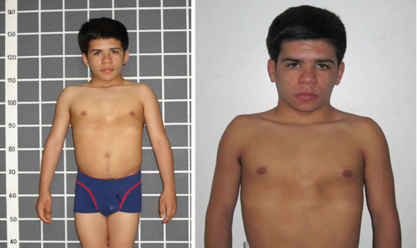
Uncategorized

Delayed puberty

delayed puberty

Delayed puberty

Delayed puberty is when the physical signs of puberty do not appear by age 13 for girls and age 14 for boys. Puberty is considered delayed when there are no signs of breast development by 13 years of age in girls or testicular enlargement by 14 years of age in boys 1). Puberty is the time when a child’s body starts to change to an adult’s. Normally, these changes begin in girls when they’re between 8 and 14 years old. In boys, they start between the ages of 9 and 15. This wide range in age is normal, and it’s why kids may develop several years earlier or later than many of their friends. Sometimes, though, kids pass this normal age range for puberty without showing any signs of body changes. This is called delayed puberty. Clinicians should suspect pubertal delay if there is halting or regression of pubertal development. In girls with initial pubertal changes, absence of menarche (a girl’s first period) by 15 years of age is also concerning 2).

Signs of delayed puberty in boys include:

  • the penis and testicles not starting to grow larger by age 14
  • genital growth that takes longer than 5 years
  • short stature compared with their peers, who now are growing faster

In girls, signs include:

  • no breast development by age 14
  • not starting to menstruate within 5 years of when breasts start to grow or by age 16

Delayed puberty, which is more common in boys, can happen for different reasons. Many children with delayed puberty will eventually go through an otherwise normal puberty, just at a late age. Delayed puberty can be hereditary with a condition called constitutional delay of puberty; meaning that the late onset of puberty may run in families. However, delayed puberty may also be due to chromosomal abnormalities, genetic disorders, chronic illnesses, or tumors that damage the pituitary gland or the hypothalamus, which affect maturation.

Delayed puberty can cause significant psychological distress and low self-esteem 3). Doctors usually can help teens with delayed puberty develop so they can catch up with their peers.

Delayed puberty treatment depends on the cause. Children with constitutional delay of growth and puberty don’t need treatment. They will eventually catch up to other children their age.

Medicine might be needed if puberty is very delayed or there is a hormone problem. If your child has a health problem or is underweight, treating the condition that is causing delayed puberty can help.

When should you take your child to see a doctor?

Girls should see the doctor if they have:

  • Signs of puberty before eight years of age
  • No signs of puberty by 13 years of age
  • No menstrual period by 15 years of age
  • Puberty that starts but then stops

Boys should see the doctor if they have:

  • Signs of puberty before nine years of age
  • No signs of puberty by 14 years of age
  • Puberty that starts but then stops

What is normal puberty?

Puberty is when a child starts to mature into an adult. Hormones (estrogen and testosterone) cause the body to change during puberty. Normal puberty starts in the brain. The brain tells the ovaries (in girls) and testicles (in boys) to make estrogen and testosterone. The adrenal glands sit on top of the kidneys. They make testosterone-like hormones.

  • For girls, breasts normally start to grow between eight and 13 years of age. Body odor, a growth spurt, pubic and underarm hair are other signs of puberty in girls. A girl’s first menstrual period usually happens about 2.5 years after the breasts start to grow. On average, girls have their first period at 12 or 13 years of age.
  • For boys, the testicles start to grow between nine and 14 years of age. Body odor, a growth spurt, pubic and underarm hair, voice changes, and facial hair follow.

The physical changes of puberty are a result of gonadal sex hormone production, the start of which (gonadarche) indicates pubertal onset. Gonadarche is triggered by the pulsatile release of gonadotropin-releasing hormone, which activates the hypothalamic-pituitary-gonadal axis 4). Adrenarche (i.e., adrenal androgen production leading to pubic and axillary hair, body odor, and mild acne) is a separate but usually concurrent process and does not in itself indicate true pubertal onset in boys or girls 5).

In girls, signs of puberty include:

  • breast development
  • pubic or underarm hair development
  • rapid height growth (a growth spurt)
  • wider hips and a curvier body shape
  • start of menstruation (periods)

In boys, signs include:

  • enlargement of the testicles and penis
  • pubic, underarm, or facial hair development
  • rapid height growth (a growth spurt)
  • broader shoulders and a more muscular build
  • voice deepening

These changes are caused by the sex hormones testosterone (in boys) and estrogen (in girls) that their bodies start making in much larger amounts than before.

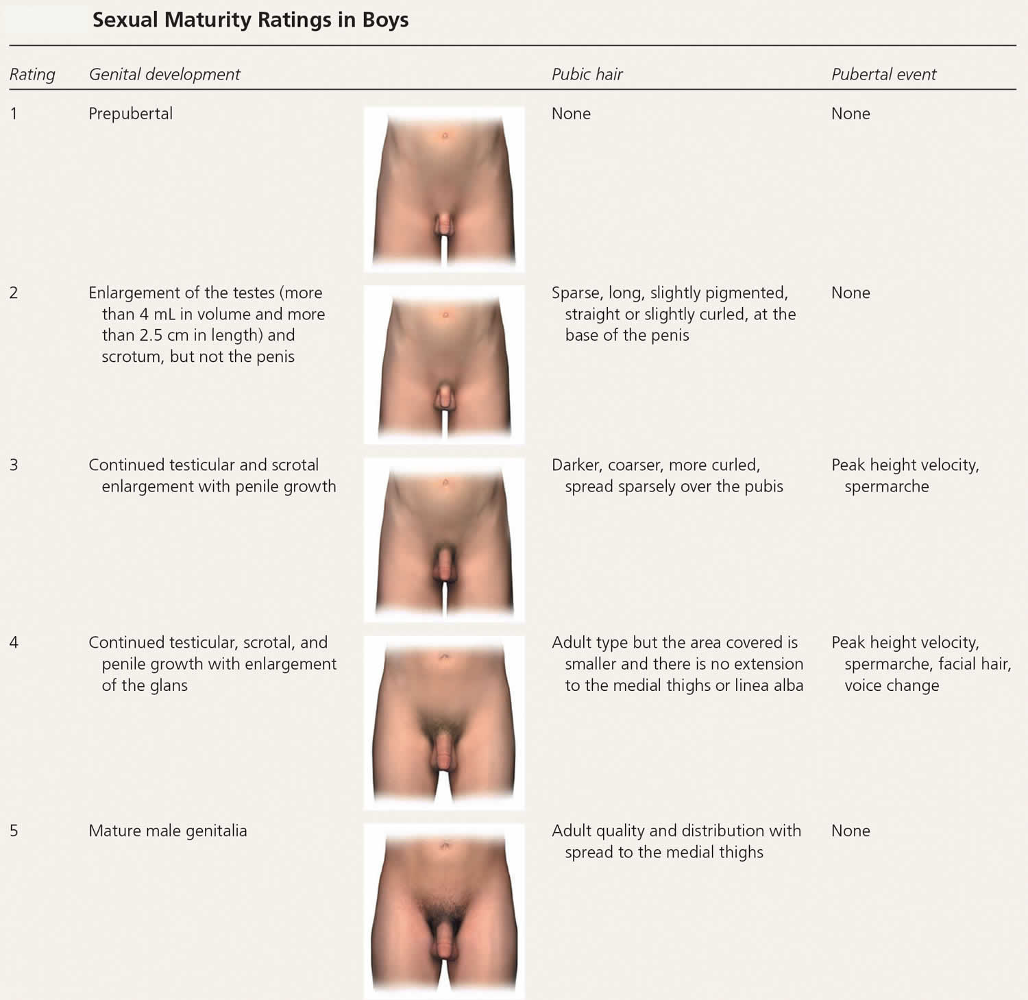
In girls, increased ovarian estradiol secretion causes breast development at a mean age of 10 years (range: eight to 12 years). Menarche typically follows 2.5 years after the onset of breast development, at an average age of 12.5 years (range: nine to 15 years) 6). In boys, testicular enlargement to at least 4 mL in volume or 2.5 cm in length is the first sign of true puberty and occurs at an average age of 11.5 years (range: 9.5 to 14 years) 7). Physical changes are described using sexual maturity ratings (Table 1 and Table 2), such as Tanner stages, and are affected by body habitus and demographic factors 8).

Linear growth velocity is about 5 cm per year from four years of age to puberty with a nadir before the pubertal growth spurt. Girls achieve peak height velocity during sexual maturity ratings 2 and 3 (mean: 8.3 cm per year, age 11 or 12 years) and boys during sexual maturity ratings 3 and 4 (mean: 9.5 cm per year, age 13 or 14 years). On average, girls complete linear growth at 15 years of age and boys at 17 years of age. After menarche, girls grow an average of 7 cm 9).

Figure 1. Normal puberty

Normal puberty

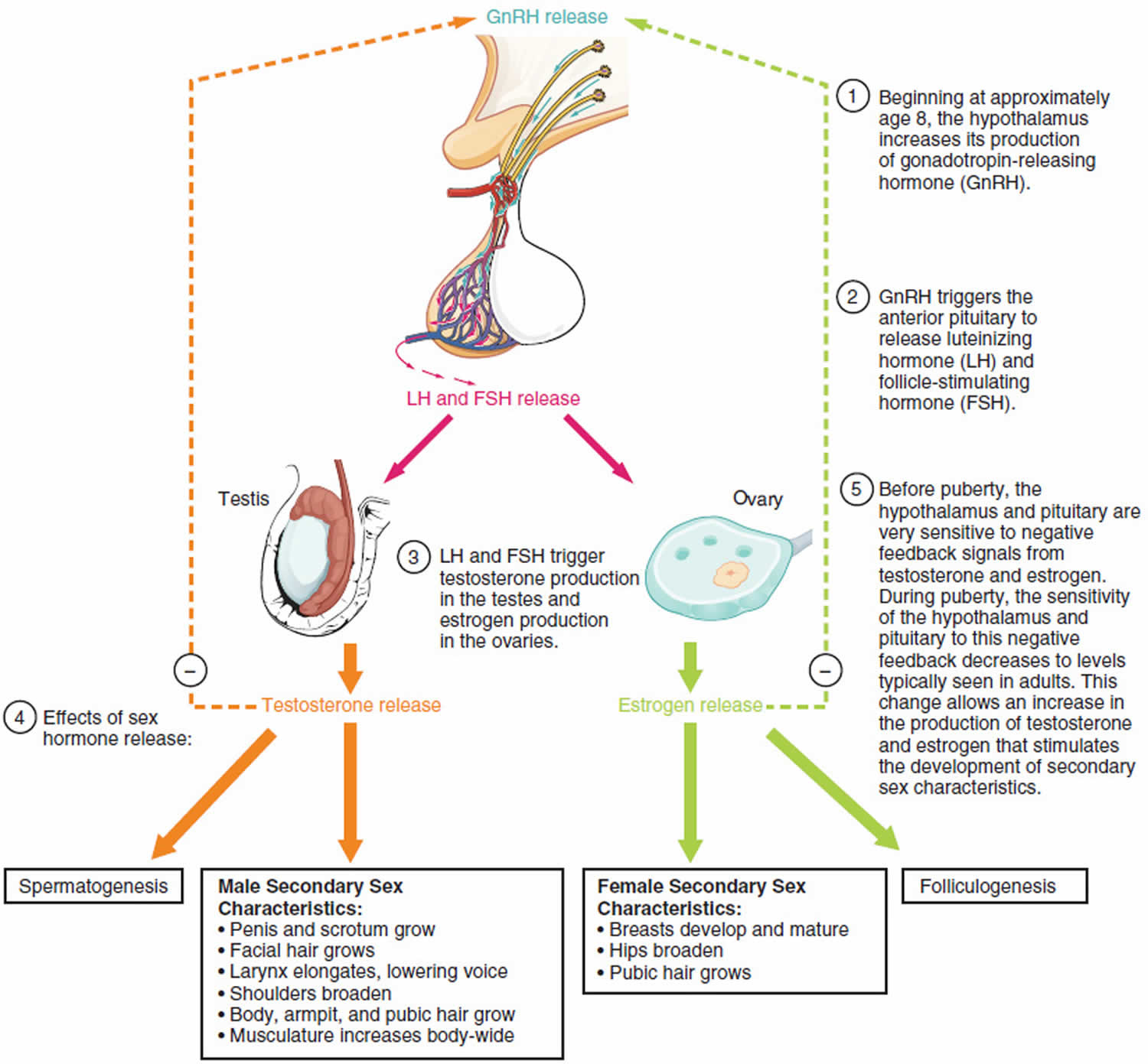
Table 1. Sexual Maturity Ratings in Girls

Sexual Maturity Ratings in Girls

[Source 10) ]

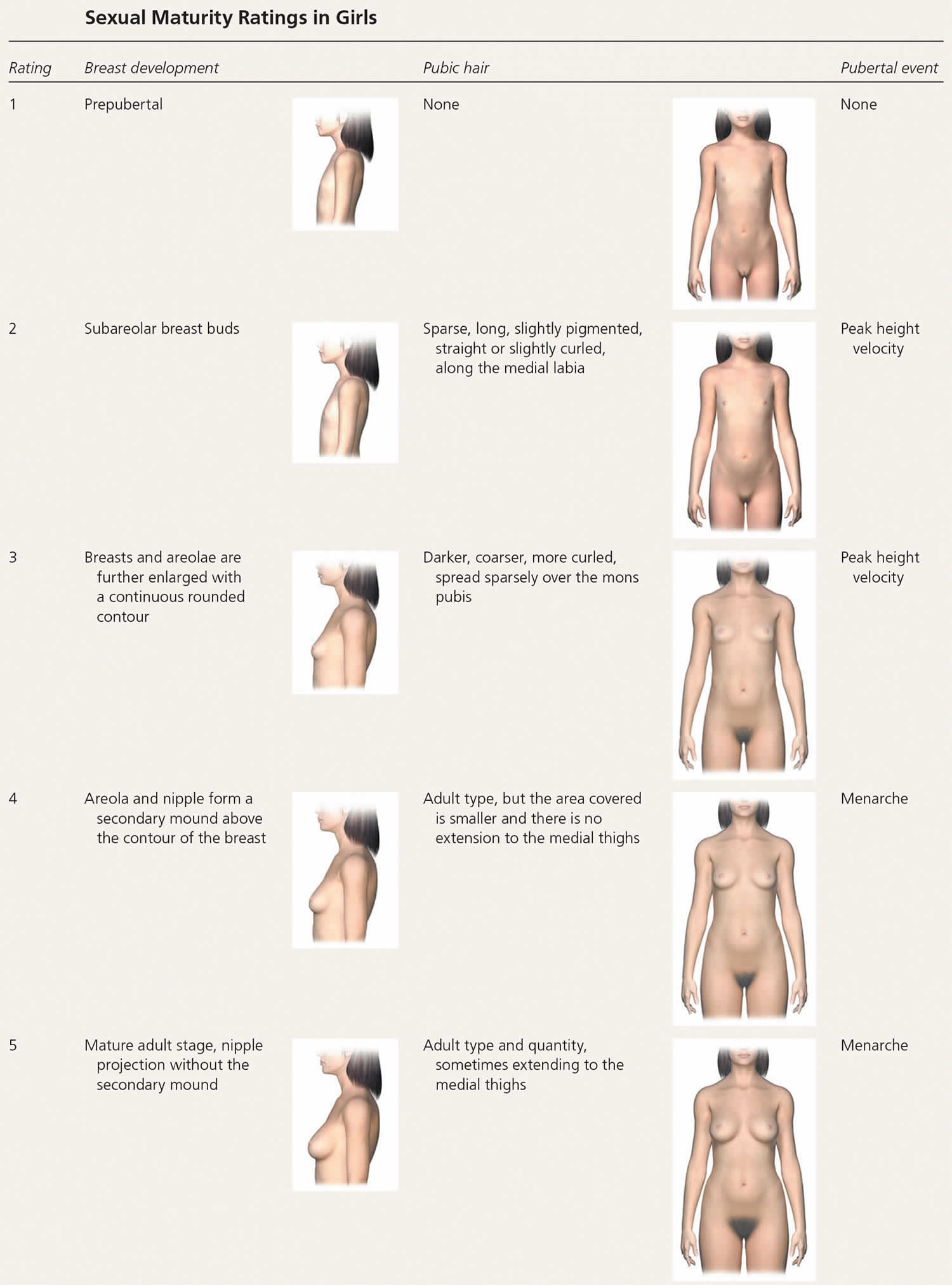
Table 2. Sexual Maturity Ratings in Boys

Sexual Maturity Ratings in Boys

[Source 11) ]

Delayed puberty causes

Most children with delayed puberty, especially boys, are “late bloomers” and otherwise healthy. This is called constitutional delay of growth and puberty. Usually, at least one parent was also a late bloomer. Some health problems and being underweight can cause delayed puberty. Less commonly, a problem with hormones causes it.

Puberty can be delayed in children who have not gotten proper nutrition due to long-term illnesses. Also, some young girls who undergo intense physical training for a sport, such as running or gymnastics, start puberty later than normal 12).

In other cases, the delay in puberty is not just due to slow maturation but occurs because the child has a long-term medical condition known as hypogonadism, in which the sex glands (the testes in men and the ovaries in women) produce few or no hormones.

Hypogonadism can be divided into two categories: secondary hypogonadism and primary hypogonadism.

  1. Primary hypogonadism also known as hypergonadotropic hypogonadism, the problem lies in the ovaries or testes, which fail to make sex hormones normally. In hypergonadotropic hypogonadism (primary hypogonadism), gonadal insufficiency delays puberty and results in elevated levels of follicle-stimulating hormone (FSH) and luteinizing hormone (LH). Conditions causing hypergonadotropic hypogonadism can be congenital or acquired and are collectively more common in girls (26%) than in boys (7%) with delayed puberty 13). Some causes include 14):
    • Genetic disorders, especially Turner syndrome (in women) and Klinefelter syndrome (in men)
    • Certain autoimmune disorders
    • Developmental disorders
    • Radiation or chemotherapy
    • Infection
    • Surgery
  2. Secondary hypogonadism also known as central hypogonadism or hypogonadotropic hypogonadism, is caused by a problem with the pituitary gland or hypothalamus (part of the brain). In secondary hypogonadism, the hypothalamus and the pituitary gland fail to signal the gonads to properly release sex hormones. Hypogonadotropic hypogonadism (secondary hypogonadism or central hypogonadism) is characterized by low levels of FSH and LH and further classified by the pathology. Causes of secondary hypogonadism include 15):
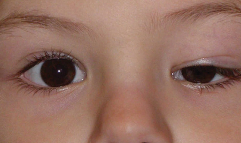
    • Kallmann syndrome, a genetic problem that also diminishes the sense of smell
    • Isolated hypogonadotropic hypogonadism, a genetic condition that only affects sexual development but not the sense of smell
    • Prior radiation, trauma, surgery, or other injury to the brain or pituitary
    • Tumors of the brain or pituitary.

Functional hypogonadotropic hypogonadism is caused by chronic disease, stress, or inadequate nutrition, and the condition may be transient or reversed. Persistent hypogonadotropic hypogonadism is caused by a congenital abnormality in the hypothalamic-pituitary-gonadal axis or an acquired etiology such as a central nervous system tumor, trauma, surgery, or radiation 16). Patients with persistent hypogonadotropic hypogonadism require treatment to induce puberty, maintain normal adult levels of sex steroids, and optimize fertility 17). Table 3 includes the differential diagnosis of delayed or absent puberty.

Table 3. Delayed or Absent Puberty differential diagnosis

Diagnosis Characteristics Treatment
Generally benign variant
Constitutional delay of growth and puberty Normal growth velocity, history of delayed puberty in parents, delayed bone age Surveillance every 6 months to evaluate for progression of pubertal development
Functional hypogonadotropic hypogonadism *
Celiac disease Abdominal pain, malabsorption, anemia, poor weight gain; short stature may be the only symptom; positive serology results, confirmed with endoscopic biopsy Gluten-free diet, surveillance
Diabetes mellitus Polyuria, polydipsia, polyphagia, weight loss, or known but poorly controlled disease; confirmed by serology Treat underlying disease
Hyperthyroidism Weight loss, heat intolerance, insomnia, tachycardia, hypertension; confirmed with serology Treat underlying disease
Hypothyroidism Weight gain, cold intolerance, fatigue, bradycardia; confirmed with serology Treat underlying disease
Inadequate nutrition for metabolic needs (e.g., eating disorder) Weight loss or poor weight gain, excessive exercise, food restriction, purging Weight restoration, treatment of underlying disorder
Inflammatory bowel disease Abdominal pain, constipation, diarrhea, hematochezia, poor weight gain, elevated serum erythrocyte sedimentation rate and C-reactive protein; confirmed with endoscopic biopsy Treat underlying disease
Persistent hypogonadotropic hypogonadism
Genetic†
Congenital hypogonadotropic hypogonadism Gonadotropin-releasing hormone deficiency, bilateral cryptorchidism, micropenis, unilateral renal agenesis, synkinesis (mirror movements), cleft lip or palate, hearing loss, dental agenesis, skeletal malformations Referral to a pediatric endocrinologist for hormone therapy
Kallmann syndrome ‡ Anosmia in addition to congenital hypogonadotropic hypogonadism presentation Referral to a pediatric endocrinologist for hormone therapy
Acquired
Central nervous system trauma, surgery, or radiation History of trauma, surgery, or central nervous system radiation for prior malignancy; may present similarly to central nervous system tumor if acute Referral to a pediatric endocrinologist for hormone therapy; other referrals as necessary for treatment of underlying disease
Central nervous system tumors Headaches, vision changes, seizures, suggestive magnetic resonance imaging findings of the brain and pituitary Referral for diagnosis and treatment of underlying disease (e.g., neurosurgeon, endocrinologist)
Hypergonadotropic hypogonadism §
Chemotherapy, radiation, or trauma to gonads History Referral to a pediatric endocrinologist for hormone therapy
Klinefelter syndrome (boys) Tall stature, learning disabilities, relatively small testes (3 to 6 mL) for degree of androgenization; 47,XXY karyotype Referral to a pediatric endocrinologist for hormone therapy
Oophoritis or orchitis History of mumps infection in boys Referral to a pediatric endocrinologist for hormone therapy
Turner syndrome (girls) Short stature, facial dysmorphism, webbed neck, brachydactyly, heart defects; in cases of mosaicism, short stature may be the only sign; 45,X or related karyotype Referral to a pediatric endocrinologist for hormone therapy and other comprehensive care

Footnotes:

* Other causes include cystic fibrosis, juvenile rheumatoid arthritis, systemic lupus erythematosus, sickle cell disease, thalassemia, chronic renal disease, and malnutrition.

Other syndromes include septo-optic dysplasia, Prader-Willi, Laurence-Moon, Bardet-Biedl, and CHARGE.

50% of congenital hypogonadotropic hypogonadism cases; five times more prevalent in boys. It is caused by disrupted migration of gonadotropin-releasing hormone–secreting neurons and the olfactory bulbs.

§ Other causes include gonadal dysgenesis, premature ovarian insufficiency (often autoimmune in nature), vanishing testes syndrome, testicular biosynthetic defects, and luteinizing hormone and follicle-stimulating hormone receptor defects.

[Source 18) ]

Constitutional delayed puberty

Most often, delayed puberty is a pattern of growth and development in a family. A child’s parents, uncle, aunt, brothers, sisters, or cousins might have developed later than usual too. This is called constitutional delay and usually doesn’t need any treatment. These “late bloomers” in time will develop normally, just later than most of their peers.

Constitutional delay of growth and puberty is the most common cause of delayed puberty in boys (60%) and girls (30%) 19). It represents an extreme of the normal spectrum of pubertal timing and is a diagnosis of exclusion 20). For more than 75% of patients with constitutional delay of growth and puberty, family history may reveal parental pubertal delay 21).

Medical problems

Some medical problems can cause delays in puberty:

  • Some kids and teens with chronic illnesses like diabetes, cystic fibrosis, kidney disease, or even asthma may go through puberty at an older age. That’s because their illnesses can make it harder for their bodies to grow and develop. Proper treatment and better control of these conditions can help make delayed puberty less likely.
  • Being malnourished — not eating enough food or not getting good nutrients — can make someone develop later than peers who eat a healthy, balanced diet. This can happen because of food insecurity, as well as disordered eating or excess physical activity. Teens with the eating disorder anorexia nervosa, for example, often lose so much weight that their bodies can’t develop properly. Girls who are extremely active in sports may be late developers because their level of exercise keeps them so lean. Girls’ bodies need enough fat before they can go through puberty or get their periods.
  • Problems in the pituitary gland or thyroid gland, which make hormones important for body growth and development, also can delay puberty.
  • Chromosome disorders can delay puberty in some people. Chromosomes are made up of DNA that contain our body’s construction plans. So when they have problems, it can affect normal growth processes. For example:
    • Turner syndrome is when one of a female’s two X chromosomes is abnormal or missing. This causes problems with how her body grows and makes sex hormones, and how her ovaries develop. Women who have Turner syndrome are shorter than normal, may not go through puberty in the usual way, and may have other medical problems. Sometimes, puberty starts at a normal time, and then stalls or stops after a few years.
    • Klinefelter syndrome is when males are born with an extra X chromosome (XXY instead of XY). This condition can affect testicular function and sexual development. These boys usually are tall for their age, might have learning problems, and may have other medical problems. Puberty usually starts at a normal time, but then stalls.

Delayed puberty signs and symptoms

Lacking signs of puberty is the primary indicator that a child may be experiencing delayed puberty. The following are the most common symptoms of delayed puberty. However, each child may experience symptoms differently. The symptoms of delayed puberty may resemble other problems or medical conditions. Always consult your child’s doctor for a diagnosis.

Symptoms of delayed puberty may include:

Delayed puberty girls

  • Lack of any breast development by age 13
  • More than four years between initial breast growth and first menstrual period
  • Failure to menstruate by age 14

Delayed puberty boys

  • Lack of testicular enlargement by age 14
  • Lack of pubic hair by age 15
  • More than four years to complete adult genital development

Delayed puberty diagnosis

If a boy or girl hasn’t shown signs of puberty as they move into the teen years, doctors will:

  • Do an exam.
  • Take a medical history, including whether others in the child’s family had a similar growth pattern.
  • Ask about any medicines the child takes.
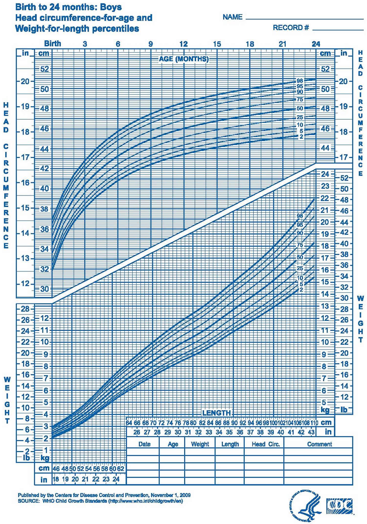
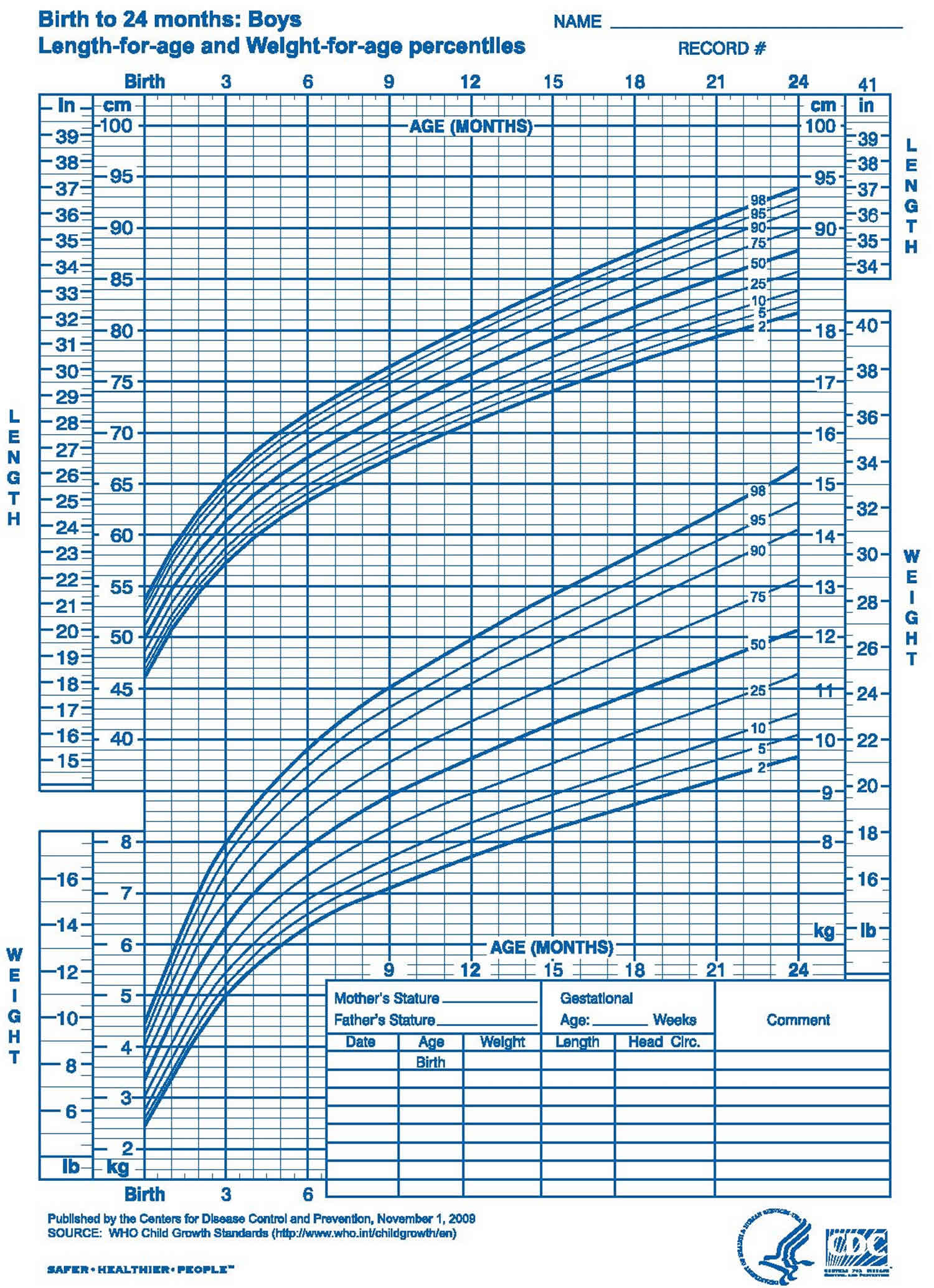
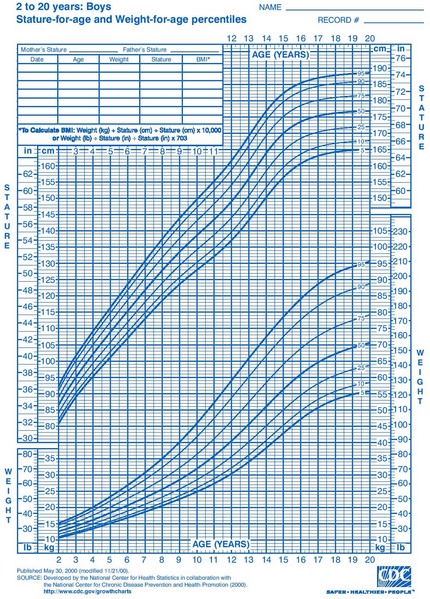
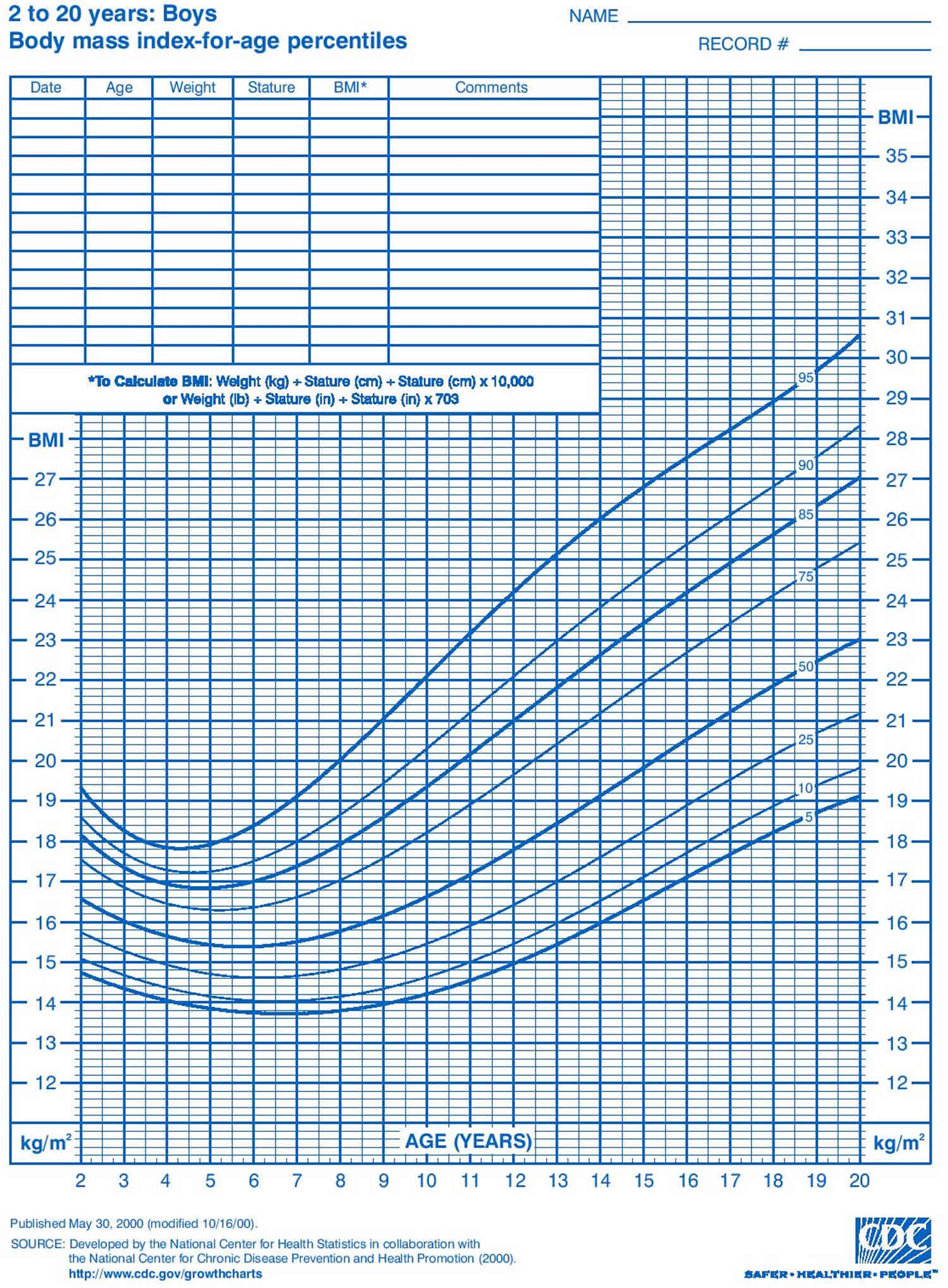
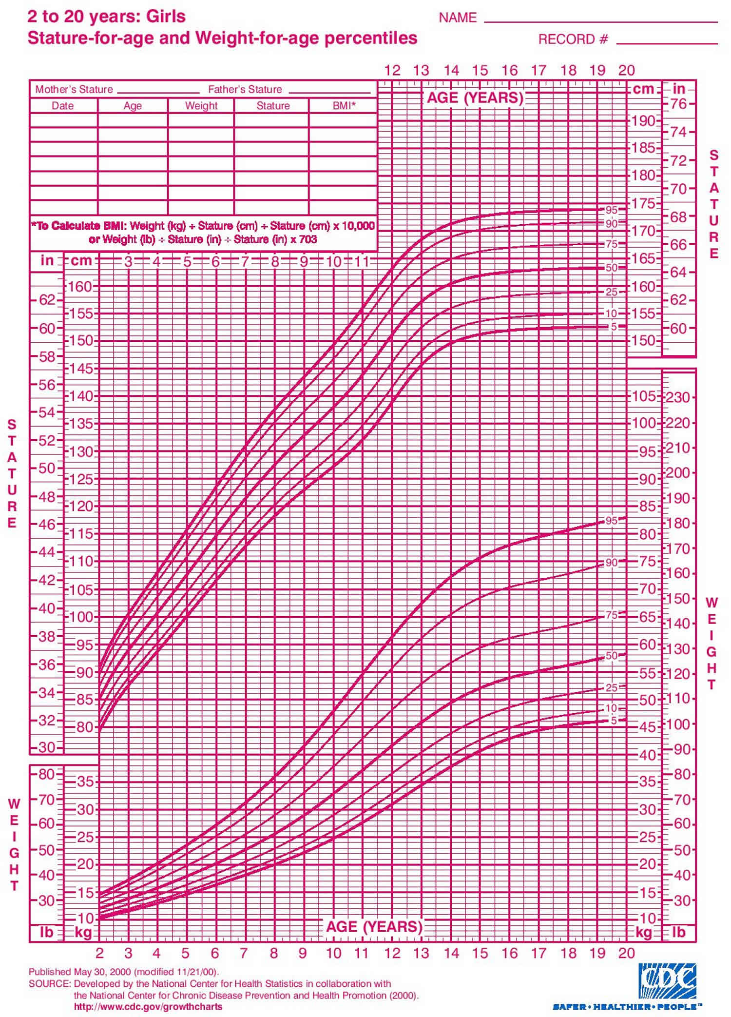
  • Check the growth chart to see if there’s a pattern that points to a problem.

Figure 2. Diagnostic approach to delayed puberty or absent pubertal development

Diagnostic approach to delayed puberty

[Source 22) ]

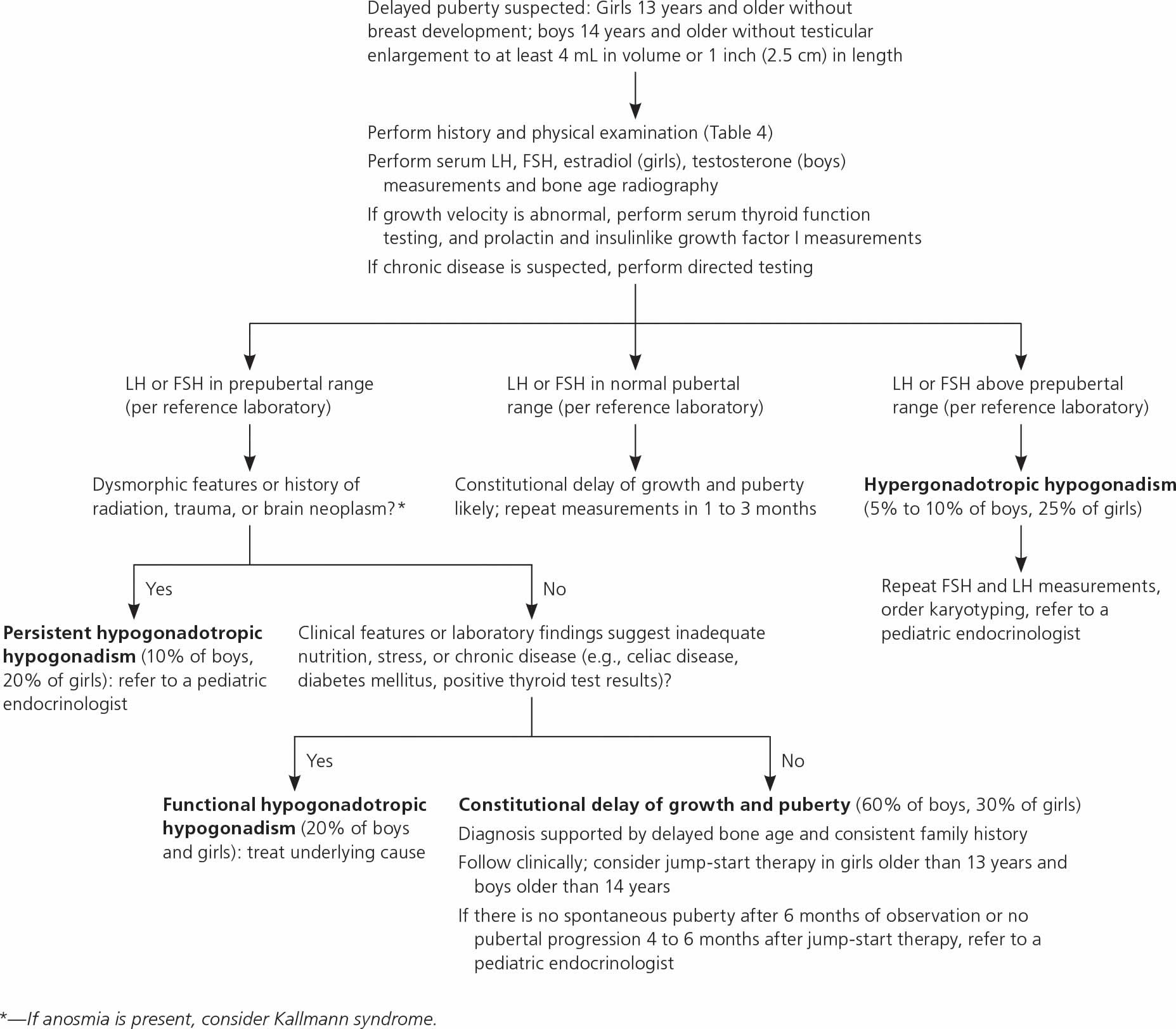
Table 4. Delayed puberty history and physical examination findings

Findings Possible diagnoses
Abdominal pain Gastrointestinal disease
Anosmia Kallmann syndrome
Asymmetric testes Oophoritis or orchitis
Body mass index and weight (on growth charts) Low: eating disorder, caloric insufficiency, gastrointestinal or other systemic disease
Chemotherapy, radiation treatment, brain tumor Hypogonadism
Cryptorchidism or orchidopexy Hypogonadism
Dysmorphic features (webbed neck, short stature, low hairline) Turner syndrome
Enlarged thyroid Hypothyroidism
Family history of late puberty Constitutional delay of growth and puberty
Galactorrhea Hyperprolactinemia
Growth velocity Peripubertal growth slowing, pathologic growth due to underlying condition
Height (growth chart) Short stature: Turner syndrome, constitutional delay of growth and puberty
Tall stature: Klinefelter syndrome
Joint pain Inflammatory disorder
Neurologic assessment (abnormal examination findings or symptoms such as headaches, vision changes) Intracranial pathology
Red (vs. dull pink) or thin vaginal mucosa Lack of estrogen exposure (hypogonadism)
Sexual maturity rating Delayed pubertal development (unspecified)
Small, firm testes Klinefelter syndrome
Temperature intolerance, gastrointestinal symptoms, tremor, depression, palpitations Thyroid disease
Trauma (head) Hypogonadism
Vasomotor symptoms in girls Ovarian insufficiency
Weight loss, stress, excessive exercise, inadequate nutrition, fatigue Eating disorder, caloric insufficiency
[Source 23) ]

In addition to a complete medical history and physical examination, diagnosis of delayed puberty may include:

  • Blood tests. Tests to check for chromosomal abnormalities, to measure hormone levels, and to test for diabetes, thyroid, pituitary, chromosomal, anemia, and other conditions that may delay puberty.
  • X-ray. A diagnostic test that uses invisible electromagnetic energy beams to produce images of internal tissues, bones, and organs onto film. Simple X-ray of the left hand and wrist bones is often performed to determine bone maturity.
  • Computed tomography scan (also called a CT or CAT scan). A diagnostic imaging procedure that uses a combination of X-rays and computer technology to produce horizontal, or axial, images (often called slices) of the body. A CT scan shows detailed images of any part of the body, including the bones, muscles, fat, and organs. CT scans are more detailed than general X-rays.
  • Magnetic resonance imaging (MRI). A diagnostic procedure that uses a combination of large magnets, radiofrequencies, and a computer to produce detailed images of organs and structures within the body.

If doctors find a problem, they usually refer families to a pediatric endocrinologist, a doctor who specializes in treating kids and teens who have growth problems, or to another specialist for more tests or treatment.

Initial workup should include measurements of serum FSH, LH, testosterone in boys or estradiol in girls, and bone age radiography (Table 5). If abnormal growth velocity is a concern, serum thyroid function, prolactin, and insulin like growth factor I should be assessed 24). Constitutional delay of growth and puberty can be difficult to distinguish from persistent hypogonadotropic hypogonadism; the latter may be diagnosed at 18 years of age if there is inadequate response to jump-start therapy (which is defined later in this section), and sex steroid replacement is still required 25).

Bone age indicates the degree of sex steroid effect on bone maturation and future growth potential 26). For example, patients with constitutional delay of growth and puberty generally have a delay of more than two years, but this finding is nonspecific 27).

Often, the doctor will find no underlying physical problem. Most kids with delayed puberty are just developing a bit later than average and will catch up.

Table 5. Diagnostic testing in the evaluation of pubertal disorders

Test Condition-specific findings


Precocious puberty Delayed puberty
Laboratory testing (refer to local reference values)
First-line
Serum estradiol Elevated (girls): estrogen exposure; if markedly elevated (> 100 pg per mL [367 pmol per L]), evaluate for ovarian tumor, especially if luteinizing hormone is suppressed Low (girls): prepubertal, may suggest poor ovarian function in response to gonadotropins
Serum testosterone Elevated: testicular (boys), adrenal, or exogenous source Low (boys): prepubertal, poor response of testes to gonadotropin stimulation
Serum LH and follicle-stimulating hormone Prepubertal levels: benign variant or peripheral precocious puberty High: gonadal insufficiency, Turner syndrome, Klinefelter syndrome
Postpubertal levels > 0.3 mIU per mL (0.3 IU per L): central precocious puberty Low: hypogonadotropic hypogonadism, constitutional delay of growth and puberty
If indicated
Directed testing (e.g., for celiac disease; diabetes mellitus; or hepatic, renal, or inflammatory conditions) Functional hypogonadotropic hypogonadism, seek underlying cause
Gonadotropin-releasing hormone analogue stimulation test Elevated LH: central precocious puberty (vs. benign variant) in complex clinical scenarios Used in complex clinical scenarios
Suppressed LH but elevated sex steroids: peripheral precocious puberty
Karyotyping Turner syndrome, Klinefelter syndrome
Serum 17-hydroxyprogesterone Elevated: nonclassic (late onset) congenital adrenal hyperplasia
Serum dehydroepiandrosterone sulfate Elevated: adrenal source, premature adrenarche (mild elevation) vs. peripheral precocious puberty Normal for age: may suggest persistent hypogonadotropic hypogonadism rather than constitutional delay of growth and puberty
Serum human chorionic gonadotropin (boys) Elevated: human chorionic gonadotropin–secreting germ cell tumor
Serum insulinlike growth factor I Low: growth hormone deficiency (if low for both bone and chronologic age)
Serum prolactin High: prolactin-secreting tumor, hypothyroidism, other neoplasm
Serum thyroid-stimulating hormone and free thyroxine Thyroid disease Thyroid disease
Imaging
First-line
Bone age radiography Advanced (> 2 standard deviations): more likely to be central or peripheral precocious puberty, less likely to be benign pubertal variant Delayed: constitutional delay of growth and puberty, underlying chronic disease
If indicated
Adrenal imaging Adrenal tumor
Magnetic resonance imaging (brain and pituitary) Central nervous system lesion Central nervous system lesion
Pelvic or testicular ultrasonography Ovarian or testicular tumor; greater ovarian volume may indicate central precocious puberty (vs. benign variant) Absence of the uterus (e.g., androgen insensitivity, Müllerian system abnormalities)
[Source 28) ]

Delayed puberty treatment

Specific treatment for delayed puberty will be determined by your child’s doctor based on:

  • Your child’s age, overall health, and medical history
  • Extent of the condition
  • Your child’s tolerance for specific medications, procedures, or therapies
  • Expectations for the course of the condition
  • Your opinion or preference

Treatment for delayed puberty depends on the cause of the problem. Often, when the underlying cause is treated, puberty proceeds normally.

Many children with delayed puberty will eventually go through an otherwise normal puberty, just at a late age. If the delayed puberty is due to heredity, no treatment is usually necessary. Some late bloomers struggle with waiting for the changes of puberty to start. So doctors may offer hormone treatment:

  • Boys might get a short course of treatment with testosterone (usually a monthly injection for 4–6 months) to get the changes of puberty started.
  • Girls might get low doses of estrogens for 4–6 months to start breast development.

Girls older than 13 years and boys older than 14 years with possible constitutional delay of growth and puberty or gonadotropin-releasing hormone deficiency may be offered jump-start therapy to induce puberty 29). For example, treating boys with testosterone cypionate or enanthate (e.g., 50 to 100 mg intramuscularly per month) and girls with overnight transdermal estradiol (e.g., 6.2 mcg, one-fourth of the 25-mcg 24-hour patch) for three to six months may accelerate attainment of final adult height and generally does not lead to premature epiphysis closure 30). After treatment ends, the teen’s own hormones usually take over to complete the process of puberty. If they don’t, the doctor will discuss long-term sex hormone replacement. If pubertal progression does not occur within four to six months after completing therapy, further evaluation for persistent hypogonadotropic hypogonadism and long-term hormone therapy should be initiated 31).

In some cases, surgery is necessary to correct an anatomical problem.

Other children have a long-lasting condition known as hypogonadism in which the sex glands (the testes in men and the ovaries in women) produce few or no hormones. For example, hypogonadism can occur in girls with Turner syndrome or in individuals with hypogonadotropic hypogonadism, which occurs when the hypothalamus produces little to no gonadotropin-releasing hormone (GnRH).

References   [ + ]

read more

Growth hormone deficiency

growth hormone deficiency

Growth hormone deficiency

Growth hormone deficiency is a rare condition in which the body does not make enough growth hormone. Growth hormone is made by the anterior pituitary gland, a small organ at the base of the brain. In children, growth hormone is essential for normal growth, muscle and bone strength, and distribution of body fat. Growth hormone also helps control glucose (sugar) and lipid (fat) levels in the body. Without enough growth hormone, a child is likely to grow slowly and be much shorter than other children of the same age and gender. The incidence of growth hormone deficiency is estimated to be 1 in 4,000 to 10,000 individuals worldwide. An estimated 6,000 adults are diagnosed with growth hormone deficiency every year in the United States 1). Adult growth hormone deficiency has been estimated to affect 1 in 100,000 people annually 2). In adults, growth hormone plays a role in regulating bone density, muscle mass, and glucose and lipid metabolism. It can also affect heart and kidney function. Deficiencies may have begun in childhood or develop in adulthood. A deficiency can develop, for example, because of damage to the pituitary gland caused by a head injury, brain tumor, or surgery or radiation treatment. This can result in a decrease in pituitary hormones (hypopituitarism). The deficiency in growth hormone can lead to decreased bone density, less muscle mass, and altered lipid levels. However, testing for growth hormone deficiency is not routine in adults who have decreased bone density and/or muscle strength or increased lipids. growth hormone deficiency is a very rare cause of these disorders.

It’s important for parents to know that there are many reasons for slow growth and below-average height in children. At times, slow growth is normal and temporary, such as right before puberty starts. A pediatric endocrinologist (children’s hormone specialist) or primary care doctor can help find out why a child is growing slowly. Most children with growth hormone deficiency grow less than two inches (5 centimeters) each year.

There are four types of isolated growth hormone deficiency differentiated by the severity of the condition, the gene involved, and the inheritance pattern.

Isolated growth hormone deficiency type 1A is caused by an absence of growth hormone and is the most severe of all the types. In people with type 1A, growth failure is evident in infancy as affected babies are shorter than normal at birth.

People with isolated growth hormone deficiency type 1B produce very low levels of growth hormone. As a result, type 1B is characterized by short stature, but this growth failure is typically not as severe as in type 1A. Growth failure in people with type 1B is usually apparent in early to mid-childhood.

Individuals with isolated growth hormone deficiency type 2 have very low levels of growth hormone and short stature that varies in severity. Growth failure in these individuals is usually evident in early to mid-childhood. It is estimated that nearly half of the individuals with type II have underdevelopment of the pituitary gland (pituitary hypoplasia). The pituitary gland is located at the base of the brain and produces many hormones, including growth hormone.

Isolated growth hormone deficiency type 3 is similar to type 2 in that affected individuals have very low levels of growth hormone and short stature that varies in severity. Growth failure in type 3 is usually evident in early to mid-childhood. People with type 3 may also have a weakened immune system and are prone to frequent infections. They produce very few B cells, which are specialized white blood cells that help protect the body against infection (agammaglobulinemia).

Growth hormone deficiency in children treatment involves growth hormone shots (injections) given at home. The shots are most often given once a day. Older children can often learn how to give themselves the shot.

Treatment with growth hormone is long-term, often lasting for several years. During this time, the child needs to be seen regularly by the pediatrician to ensure the treatment is working. If needed, your child’s endocrinologist will change the dosage of the medicine.

Serious side effects of growth hormone treatment are rare. Common side effects include:

  • Headache
  • Fluid retention
  • Muscle and joint aches
  • Slippage of the hip bones

Growth hormone deficiency causes

Growth hormone deficiency may result from disruption of the growth hormone axis in the higher brain, hypothalamus, or pituitary. Most instances of growth hormone deficiency are idiopathic (doctors can find no cause). Some children are born with growth hormone deficiency (congenital growth hormone deficiency). Others develop it after birth due to a brain injury, a tumor, or radiation treatment to the head (acquired growth hormone deficiency).

Isolated growth hormone deficiency is caused by mutations in one of at least three genes. Isolated growth hormone deficiency types 1A and 2 are caused by mutations in the GH1 gene. Type 1B is caused by mutations in either the GH1 or GHRHR gene. Type 3 is caused by mutations in the BTK gene.

The GH1 gene provides instructions for making the growth hormone protein. Growth hormone is produced in the pituitary gland and plays a major role in promoting the body’s growth. Growth hormone also plays a role in various chemical reactions (metabolic processes) in the body. Mutations in the GH1 gene prevent or impair the production of growth hormone. Without sufficient growth hormone, the body fails to grow at its normal rate, resulting in slow growth and short stature as seen in isolated growth hormone deficiency types 1A, 1B, and 2.

The GHRHR gene provides instructions for making a protein called the growth hormone releasing hormone receptor. This receptor attaches (binds) to a molecule called growth hormone releasing hormone. The binding of growth hormone releasing hormone to the receptor triggers the production of growth hormone and its release from the pituitary gland. Mutations in the GHRHR gene impair the production or release of growth hormone. The resulting shortage of growth hormone prevents the body from growing at the expected rate. Decreased growth hormone activity due to GHRHR gene mutations is responsible for many cases of isolated growth hormone deficiency type 1B.

The BTK gene provides instructions for making a protein called Bruton tyrosine kinase (BTK), which is essential for the development and maturation of immune system cells called B cells. The BTK protein transmits important chemical signals that instruct B cells to mature and produce special proteins called antibodies. Antibodies attach to specific foreign particles and germs, marking them for destruction. It is unknown how mutations in the BTK gene contribute to short stature in people with isolated growth hormone deficiency type 3.

Some people with isolated growth hormone deficiency do not have mutations in the GH1, GHRHR, or BTK genes. In these individuals, the cause of the condition is unknown. When this condition does not have an identified genetic cause, it is known as idiopathic isolated growth hormone deficiency.

A mutation in a transcription factor (POUF-1, also known as PIT-1) is known to result in familial growth hormone deficiency 3). As many as 14 different mutations have been described. In addition to growth hormone deficiency, affected individuals have had prolactin deficiencies and variable thyroid-stimulating hormone (TSH) deficiencies. Imaging of the pituitary gland usually reveals a hypoplastic or ectopic posterior pituitary.

Growth hormone deficiency with other hypopituitarism associated with inactivating mutations of the PROP1 (Prophet of PIT-1) transcription factor gene have been documented in reports. Patients with this mutation usually do not produce luteinizing hormone (LH) or follicle-stimulating hormone (FSH), and thus, do not spontaneously progress into puberty. They may also have TSH (thyroid-stimulating hormone) deficiency. Imaging of the pituitary gland of patients with PROP1 mutations may show either a small anterior pituitary or an intrapituitary mass.

Congenital growth hormone deficiency may be associated with an abnormal pituitary gland (seen on MRI) or may be part of a syndrome such as septooptic dysplasia (de Morsier syndrome), which may include other pituitary deficiencies, optic nerve hypoplasia, and absence of the septum pellucidum; it occurs with an incidence of about 1 in 50,000 births. Septooptic dysplasia (de Morsier syndrome) may be associated with a mutation in the gene for another transcription factor, HESX1.

Acquired growth hormone deficiency may result from trauma, infections (eg, encephalitis, meningitis), cranial irradiation (somatotrophs appear to be the most radiation-sensitive cells in the pituitary), and other systemic diseases (particularly histiocytosis). Although most instances of isolated growth hormone deficiency are idiopathic, specific etiologies cause most growth hormone deficiency associated with other pituitary deficiencies. A reported 12-86% of children with apparent isolated growth hormone deficiency have sellar developmental defects.

Of more than 20,000 children receiving growth hormone in the National Cooperative Growth Study (a database of patients receiving growth hormone therapy), approximately 25% of the patients with growth hormone deficiency had a acquired growth hormone deficiency. These causes included the following 4):

  • Central nervous system (CNS) tumor, including craniopharyngioma – 47%
  • Central nervous system (CNS) malformation – 15%
  • Septooptic dysplasia (de Morsier syndrome) – 14%
  • Leukemia – 9%
  • Central nervous system (CNS) radiation – 9%
  • Central nervous system (CNS) trauma – 3%
  • Histiocytosis – 2%
  • Central nervous system (CNS) infection – 1%

Multiple Pituitary Hormone Deficiency is a condition caused by a shortage of several hormones produced by the pituitary gland. It has similar characteristics to growth hormone deficiency but has additional complications caused by the absence of other hormones including:

  • Thyroid-simulating hormone (TSH) and cortisol, the hormone associated with the body’s fright and flight response.
  • The gonadotrophins – follicle stimulating hormone (FSH) and lutenising hormone (LH)
  • Adrenal-stimulating hormone (ACTH) is much less frequently involved, but the deficiency of this hormone is extremely important to detect.

Growth hormone deficiency symptoms

Slow growth may first be noticed in infancy and continue through childhood. The pediatrician will most often draw the child’s growth curve on a growth chart. Children with growth hormone deficiency have a slow or flat rate of growth. The slow growth may not show up until a child is 2 or 3 years old.

The child will be much shorter than most children of the same age and gender. The child will still have normal body proportions, but may be chubby. The child’s face often looks younger than other children of the same age. The child will have normal intelligence in most cases.

In older children, puberty may come late or may not come at all, depending on the cause.

Signs of growth hormone deficiency:

  • Slowed growth in height in infants, children, or adolescents (teenagers)
  • A young-looking face compared with other children of the same age
  • A chubby body, small hands and feet, and poorly developed muscles
  • Low blood glucose levels (in infants and toddlers with severe growth hormone deficiency)
  • A very small penis (in male newborns with severe growth hormone deficiency)
  • Delayed puberty

Growth hormone deficiency diagnosis

Your doctor will review your child’s medical history and growth charts, and look for signs of growth hormone deficiency and other conditions that affect growth. Your doctor may do tests to help find the cause of slow growth. These include:

  • An X-ray of the hand to check bone growth (bone age) and assess growth potential
  • Blood tests and other laboratory tests to rule out other conditions that affect growth
  • Specific tests for growth hormone deficiency include
  • Insulin-like growth factor (IGF-1). A blood test checks levels of IGF-1, a hormone that reflects growth hormone levels.
  • Growth hormone stimulation test. The child is given medicines that stimulate the pituitary to release growth hormone. Then, if growth hormone levels in the blood don’t rise to a certain level, it can mean the pituitary is not making enough growth hormone.
  • Magnetic resonance image (MRI). An MRI (imaging test) of the head will look for a problem with the pituitary or the brain, and can rule out a brain tumor.

Growth hormone deficiency test

Testing is most often done after the pediatrician has looked into other causes of poor growth. Tests that may be done include:

Growth hormone testing

Growth hormone testing is primarily used to identify growth hormone deficiency and to help evaluate pituitary gland function, usually as a follow-up to other abnormal pituitary hormone test results.

Because growth hormone is released in pulses, a single measurement of the blood level is not normally clinically useful. Therefore, testing for the suppression or stimulation of growth hormone release from the pituitary is usually done.

  • Growth hormone stimulation tests help to diagnose growth hormone deficiency and hypopituitarism. For a stimulation test, a sample of blood is drawn after 10-12 hours of fasting. Then, under close medical supervision, a person is given an intravenous solution of a substance that normally stimulates the release of growth hormone from the pituitary. Blood samples are then drawn at timed intervals and growth hormone levels are tested in each to see if the pituitary gland was stimulated to produce the expected levels of growth hormone. The most commonly used stimulant is arginine, but others include clonidine and glucagon. Since exercise normally causes an increase in growth hormone, vigorous exercise may also be used as the stimulant for growth hormone release.
  • Growth hormone stimulates the production of insulin-like growth factor-1 (IGF-1). IGF-1 is a hormone that mediates the effects of growth hormone and helps promote normal bone and tissue growth and development. However, unlike growth hormone, its level is stable in the blood throughout the day. This makes IGF-1 a useful indicator of average growth hormone levels and the IGF-1 test is often used to help evaluate growth hormone deficiency or growth hormone excess.
  • Other blood tests that may be used to evaluate pituitary gland function include prolactin, free T4, TSH, cortisol, FSH, LH, and testosterone. These tests are usually performed prior to growth hormone testing to make sure that they are normal and/or controlled with medication before growth hormone testing is done. For example, hypothryoidism must be treated prior to testing for growth hormone deficiency in children; otherwise, a falsely low growth hormone result may be seen.

If they pituitary gland is failing to produce sufficient qunatities of all the hormones it produces the condition is known as Panhypopituitarism.

  • MRI of the head can show the hypothalamus and pituitary glands.
  • Tests to measure other hormone levels may be done, because a lack of growth hormone may not be the only problem.

Growth hormone deficiency treatment

Children with growth hormone deficiency receive treatment with daily injections of synthetic (manufactured) human growth hormone, a prescription medicine. The growth hormone, given at home, is injected under the skin. Growth is usually monitored every 3 to 6 months by a pediatric endocrinologist, who will adjust the dose as needed.

The best results occur when growth hormone deficiency is diagnosed and treated early. In some children, growth hormone can lead to four inches (10 centimeters) of growth during the first year of treatment. Others grow less, but usually faster than without treatment. Some children need treatment until adolescence; others need it into adulthood.

Side effects of growth hormone therapy

Mild to moderate side effects are uncommon. They include:

  • Headaches
  • Muscle or joint pain
  • Mildly under active thyroid gland
  • Swelling of hands and feet
  • Curvature of the spine (scoliosis)
  • Development of breast tissue in boys

Rare but serious side effects include:

  • Severe headache with vision problems
  • A hip problem, when the top of the thigh bone slips out of place
  • Inflamed pancreas (pancreatitis)

For most children, the benefits of taking growth hormone outweigh the risks.

Overall, growth hormone has been shown to be a safe hormone when used at recommended doses. There are excellent large databases for evaluation of possible safety signals that occur during treatment with growth hormone. Growth hormone adverse events have been carefully documented in a review by Krysiak R et al. 5). Most adverse events have been local injection site reactions, which rarely lead to discontinuation. Headache, nausea, and fever have been generally self-limiting and are well tolerated. Adverse events such as edema or carpal tunnel syndrome are seen more often in adults than children, and they may be the result of fluid retention caused by growth hormone. Adverse events seen particularly in children have included transient idiopathic intracranial hypertension (also known as pseudotumor cerebri), gynecomastia, and slipped capital femoral epiphysis. The idiopathic intracranial hypertension resolved after discontinuation of growth hormone and restarting at a low dose.

There have been concerns about cancer associated with growth hormone administration, and these concerns have stemmed from several observations. First, acromegaly (a condition of growth hormone excess) is known to increase the risk of colorectal cancer. Second, epidemiological studies have shown a relationship between tall statue and cancer risk, between insulin like growth factor I (IGF-I) levels and the risk of prostate cancer, and an increase in breast cancer associated with levels of free IGF-I. One study has suggested that there may be reason for concern because of cases of Hodgkin disease and colorectal cancer found in long-term follow up of patients who had received human-derived growth hormone. Although the incidence of these diseases was greater than the population at large, it was not outside the confidence ranges.

Further, follow up of patients receiving human-derived growth hormone in the United States has not shown such a correlation. There has been recent concern from analysis of data in French children who were treated with growth hormone between 1985 and 1996, and then followed until 1996 (the SAGhE study) 6). A retrospective analysis of mortality in this population suggests the possibility of increased cardiovascular disease and bone tumors in adults who received growth hormone as children. The cardiovascular disease was primarily attributed to subarachnoid or intracerebral hemorrhages. Overall cancer mortality rates were not higher than the general population, but bone tumor–related deaths were 5 times higher than expected. There appeared to be a dose relationship (risk was highest in patients receiving doses >50 mcg/d).

The study is flawed by not having a control group (data from those who took growth hormone as children were compared to the population at large, which may not be an appropriate comparison). In addition, there was no apparent relationship with duration of growth hormone therapy, which one would expect if the increase in mortality was actually related growth hormone therapy, suggesting that the increase in mortality in this group could be more likely related to the reason they were short and taking growth hormone, rather than an effect of the growth hormone itself.

Similar data from Sweden, The Netherlands, and Belgium 7) have shown no increase in mortality rates, and all of the deaths were attributable to accidents or suicide, further suggesting that the French data could be misleading. Clearly, what is most needed is long-term adult follow up of those patients who received growth hormone as children

Growth hormone deficiency prognosis

The earlier growth hormone deficiency is treated, the better the chance that a child will grow to near-normal adult height. Many children gain 4 or more inches (about 10 centimeters) during the first year and 3 or more inches (about 7.6 centimeters) during the next 2 years. The rate of growth then slowly decreases.

Growth hormone therapy does not work for all children.

Left untreated, growth hormone deficiency may lead to short stature and delayed puberty. Average adult height for untreated patients with severe isolated growth hormone deficiency is 143 cm in men and 130 cm in women. Approximately 5% of children with growth hormone deficiency also have episodes of hypoglycemia, particularly in infancy, which resolve with growth hormone therapy.

Adults with untreated growth hormone deficiency have altered body composition (eg, excess body fat, lower lean body mass), decreased bone mineralization, cardiovascular risk factors (in particular, altered blood lipids), and decreased exercise tolerance. In addition, these patients may be socially isolated.

Growth hormone deficiency can occur with deficiencies of other hormones such as those that control:

  • Production of thyroid hormones
  • Water balance in the body
  • Production of male and female sex hormones
  • The adrenal glands and their production of cortisol, DHEA, and other hormones

Mortality in children with growth hormone deficiency is due almost entirely to other pituitary hormone deficiencies 8). These children have an increased relative risk of death in adulthood from cardiovascular causes resulting from altered body composition and dyslipidemia.

References   [ + ]

read more

Failure to thrive

failure to thrive

What is failure to thrive

Failure to thrive is defined as decelerated or arrested physical growth (height and weight measurements fall below the third or fifth percentile, or a downward change in growth across two major growth percentiles) and is associated with abnormal growth and development 1). Failure to thrive is defined as children or baby who don’t meet the expected standards of weight gain and have poor linear (height) growth within the first few years of life 2). Many things can cause failure to thrive, including illnesses and eating problems or inadequate nutrition. Once doctors find the cause of the problem, they can work with families to help get a child back into a healthy growth pattern.

Failure to thrive lacks a clear definition, in part because it’s not a disease or disorder itself. Previously, failure to thrive was categorized as either organic (underlying medical condition) or non-organic (no known medical condition). However, this categorization is considered outdated as the causes and effects of malnutrition are usually intertwined in most children. Rather, failure to thrive is a sign that a child is undernourished. In general, children who fail to thrive don’t receive or cannot take in, keep, or use the calories that would help them grow and gain enough weight.

Doctors usually diagnose failure to thrive in infants and toddlers — an important time of physical and mental development. After birth, a child’s brain grows as much in the first year as it will grow for the rest of a child’s life. Poor nutrition during this period may have lasting harmful effects on brain development.

Most babies double their birth weight by 4 months and triple it by age 1, but children who fail to thrive usually don’t meet those milestones. Sometimes, a child who starts out “plump” and seems to be growing well may gain less weight later. After a while, height growth may slow as well.

If failure to thrive continues, undernourished infants might:

  • lose interest in their surroundings
  • avoid eye contact
  • become fussy
  • not reach developmental milestones like sitting up, walking, and talking at the usual age
When to see a medical professional

If you notice a drop in weight gain or your baby doesn’t seem to have a normal appetite, see your child’s doctor. Also call about any major change in eating patterns. Toddlers and older children may have days and sometimes weeks when they show little interest in eating, but that shouldn’t happen in infants.

If you have trouble feeding your baby, your doctor can help. When a child doesn’t readily eat, parents can become frustrated and feel they’re doing something wrong. That can make the problem worse, causing stress for you and your baby. Instead, get help for both of you by talking to your doctor.

Who is affected by failure to thrive?

Infants born into families with inadequate support or understanding of infant needs may not provide the right kinds or amounts of food. For example, too much fruit juice, problems breastfeeding, or failure to introduce solids at an appropriate age may lead to too little calories being consumed. Babies and children with developmental delay or problems swallowing may consume too few calories. Conditions such as cystic fibrosis, celiac disease, or severe allergy or intolerance may consume enough food but not be able to absorb it properly. A child with a chronic medical condition, such as congenital heart disease or a genetic syndrome, may need more calories than expected. In severe cases, neglect or abuse may be associated with failure to thrive if food is purposely withheld from a baby.

Does my child have failure to thrive?

If you’re worried that your child is failing to thrive, remember that many things can cause slower weight gain. For instance, breastfed babies and bottle-fed babies often gain weight at different rates in the early newborn period.

Genetics also play a big role in weight gain. So if a baby’s parents are slim, the baby may not put on pounds quickly. However, infants should still gain weight steadily. As a general guideline, babies usually eat often in a 24-hour period and should gain about 1 ounce a day in the first month of life. It can be hard to judge this from home (even if you have a scale), so it’s important to see your child’s doctor regularly. Doctors can check for problems at regular well-child checkups, so it’s important to keep these appointments.

Failure to thrive causes

Failure to thrive has many different causes, and sometimes more than one cause may contribute to the condition at the same time. If an infant is not offered enough food or is not willing to eat enough food, or vomits repeatedly (such as with severe gastroesophageal reflux), there will not be enough calories to support growth. A child who is unable to absorb enough calories (such as with severe allergies or a medical condition like cystic fibrosis) will also not grow as expected. Any condition that causes a child to need more calories than normally expected may also lead to failure to thrive.

A number of things can cause failure to thrive, including:

  • Not enough food offered. In some cases, parents mistakenly cause the problem. Sometimes a parent measures formula incorrectly, causing an infant to get too few calories. Problems with breastfeeding or transitioning to solids also can cause failure to thrive. Parents who worry their child will get fat may restrict the amount of calories they give their infants. And sometimes parents don’t pay enough attention to their children’s hunger cues or can’t afford enough food for their children.
  • The child eats too little. Some children have trouble eating enough food because of prematurity, developmental delays, or conditions like autism in which they do not like eating foods of certain textures or tastes.
  • Health problems involving the digestive system. Several health conditions can prevent a child from gaining weight. These include gastroesophageal reflux (GER), chronic diarrhea, cystic fibrosis, chronic liver disease, and celiac disease. With reflux, the esophagus may become so irritated that a child refuses to eat because it hurts. Persistent diarrhea can make it hard for the body to hold on to the nutrients and calories from food. Cystic fibrosis, chronic liver disease, and celiac disease all cause problems with the body’s ability to absorb nutrients. The child may eat a lot, but the body doesn’t absorb and retain enough of that food. Kids with celiac disease are allergic to gluten, a protein found in wheat and other grains. The immune system’s abnormal response to this protein damages the lining of the intestine so it can’t absorb nutrients properly.
  • Food intolerance. A food intolerance, which is different from a food allergy, means the body is sensitive to certain foods. For example, milk protein intolerance means the body can’t absorb foods that have milk proteins, such as yogurt and cheese, which could lead to failure to thrive.
  • An ongoing illness or disorder. A child who has trouble eating — because of prematurity or a cleft lip or palate, for example — may not take in enough calories to support normal growth. Other conditions involving the heart, lungs, or endocrine system can increase the amount of calories a child needs and make it hard for the child to eat enough to keep up.
  • Infections. Parasites, urinary tract infections (UTIs), tuberculosis, and other infections can force the body to use nutrients rapidly and decrease appetite. This can lead to short- or long-term failure to thrive.
  • Metabolic disorders. Metabolic disorders are health conditions that make it hard for the body to break down, process, or take energy from food. They also can cause a child to eat poorly or vomit.

Factors in the child’s environment include:

  • Loss of emotional bond between parent and child
  • Poverty
  • Problems with child-caregiver relationship
  • Parents do not understand the appropriate diet needs for their child
  • Exposure to infections, parasites, or toxins
  • Poor eating habits, such as eating in front of the television and not having formal meal times

Sometimes a mix of medical problems and environmental factors leads to failure to thrive. For instance, if a baby has severe GER and is reluctant to eat, feeding times can be stressful. The baby may become upset and frustrated, and the caregiver might be unable to feed the child enough food.

Other times, doctors aren’t sure exactly what causes the problem.

Failure to thrive symptoms

Children who fail to thrive do not grow and develop normally as compared to children of the same age. They seem to be much smaller or shorter. Teenagers may not have the usual changes that occur at puberty.

The following are the most common symptoms of failure to thrive. However, each child may experience symptoms differently. Symptoms may include:

  • Lack of appropriate weight gain
  • Irritability
  • Easily fatigued
  • Excessive sleepiness
  • Lack of age-appropriate social response (i.e., smile)
  • Does not make vocal sounds
  • Delayed motor development
  • Learning and behavior difficulties later in childhood

Symptoms of failure to thrive include:

  • Height, weight, and head circumference do not match standard growth charts
  • Weight is lower than third percentile of standard growth charts or 20% below the ideal weight for their height
  • Growth may have slowed or stopped

The following may be delayed or slow to develop in children who fail to thrive:

  • Physical skills, such as rolling over, sitting, standing and walking
  • Mental and social skills
  • Secondary sexual characteristics (delayed in adolescents)

Babies who fail to gain weight or develop often lack interest in feeding or have a problem receiving the proper amount of nutrition. This is called poor feeding.

Other symptoms that may be seen in a child that fails to thrive include:

  • Constipation
  • Excessive crying
  • Excessive sleepiness (lethargy)
  • Irritability

The symptoms of failure to thrive may resemble other conditions or medical problems. Always consult your child’s physician for a diagnosis.

Failure to thrive possible complications

  • Permanent mental, emotional, or physical delays can occur.
  • When to Contact a Medical Professional

Failure to thrive diagnosis

Many babies go through brief periods when their weight gain levels out, or they even lose a little weight. This is not unusual. However, if a baby doesn’t gain weight for 3 months in a row during the first year of life, doctors usually become concerned.

Failure to thrive is usually discovered and diagnosed by the infant’s physician. Infants are always weighed and measured when seen by their physicians for well-baby check-ups. Doctors use standard growth charts to plot weight, length, and head circumference, which are measured at each well-child exam. Children may have failure to thrive if they fall below a certain weight range for their age or fail to gain weight at the expected rate.

Your child’s doctor will initiate a more complete evaluation when your infant’s growth, development, and functioning are found to be delayed. Doctors might order tests (such as a complete blood count or urine test) to check for underlying medical problems. If a particular disease or disorder is suspected, the doctor might order other tests to check for that condition.

A special test called the Denver Developmental Screening Test may be used to show any delays in development. A growth chart outlining all types of growth since birth is created.

The following tests may be done:

  • Complete blood count (CBC)
  • Electrolyte balance
  • Hemoglobin electrophoresis to check for conditions such as sickle cell disease
  • Hormone studies, including thyroid function tests
  • X-rays to determine bone age
  • Urinalysis

A few different growth chart patterns might signal a health problem, such as:

  • When a child’s weight or height percentile changes from a certain pattern it’s been following. For example: If height and weight consistently are on the 60th percentile line until a child is 5 years old, then the height has dropped to the 30th percentile at age 6, that might indicate that there’s a growth problem because the child is not following his or her previous growth pattern. Many kids may show changes in growth percentiles at certain points in development, when it’s normal for growth rates to vary more from child to child. This is particularly common during infancy and puberty.
  • When kids don’t get taller at the same rate at which they’re gaining weight. Let’s say a boy’s height is in the 40th percentile and his weight is in the 85th percentile. (So he’s taller than 40% of kids his age, but weighs more than 85% of kids his age.) That might be a problem. On the other hand, if he’s in the 85th percentile for height and weight and follows that pattern consistently over time, that usually means that he’s a normal child who’s just larger than average.

If you have any questions about your child’s growth — or growth charts — talk with your doctor.

To see if there’s a problem, doctors will ask for a child’s detailed health history, including a feeding history. This helps doctors see whether underfeeding, household stresses, or feeding problems might be to blame. A nutritionist or other health care professional also may track the calories in a child’s diet to make sure the child is getting enough.

Growth Charts

Growth charts are a standard part of any checkup, and they show health care providers how kids are growing compared with other kids of the same age and gender. They also allow doctors and nurses to see the pattern of kids’ height and weight gain over time, and whether they’re developing proportionately.

Let’s say a child was growing along the same pattern until he was 2 years old, then suddenly started growing at a much slower rate than other kids. That might indicate a health problem. Doctors could see that by looking at a growth chart.

Doctors interpret the growth charts in the context of the child’s overall well-being, environment, and genetic background. Is the child meeting other developmental milestones? Are there other signs that a child is not healthy? How tall or heavy are the child’s parents and siblings? Was the child born prematurely? Has the child started puberty earlier or later than average? These are all factors that the doctor will use to help understand the numbers on the growth chart.

Girls and boys are measured on different growth charts because they grow in different patterns and at different rates.

And one set of charts is used for babies, from birth to 24 months. Another set of charts is used for kids ages 2 to 20 years old.

Up until the time babies are 24 months old, doctors measure weight, length, and head circumference.

With older kids (2 to 20 years old), doctors measure weight, height, and body mass index (BMI). It’s important to look at and compare weight and height measurements to get a full picture of a child’s growth.

Commonly used standard growth charts include the following:

Figure 1. Boys Growth Charts from Birth to 24 Months (Head circumference-for-age and Weight-for-length percentiles)

Boys Growth Charts from Birth to 24 Months for Head circumference-for-age and Weight-for-length percentiles

Figure 2. Boys Growth Charts from Birth to 24 Months (Length-for-age and Weight-for-age percentiles)

Boys Growth Charts from Birth to 24 Months for Length-for-age and Weight-for-age percentiles

Figure 3. Boys Growth Charts from 2 to 20 Years (Stature-for-age and Weight-for-age percentiles)

Boys Growth Charts from 2 to 20 Years for Stature-for-age and Weight-for-age percentiles

Figure 4. Boys Growth Charts from 2 to 20 Years (Body mass index-for-age percentiles)

Boys Growth Charts from 2 to 20 Years for Body mass index-for-age percentiles

Figure 5. Girls Growth Charts from Birth to 24 Months (Head circumference-for-age and Weight-for-length percentiles)

Girls Growth Charts from Birth to 24 Months for Head circumference-for-age and Weight-for-length percentiles

Figure 6. Girls Growth Charts from Birth to 24 Months (Length-for-age and Weight-for-age percentiles)

Girls Growth Charts from Birth to 24 Months for Length-for-age and Weight-for-age percentiles

Figure 7. Girls Growth Charts from 2 to 20 Years (Stature-for-age and Weight-for-age percentiles)

Girls Growth Charts from 2 to 20 Years for Stature-for-age and Weight-for-age percentiles

Figure 8. Girls Growth Charts from 2 to 20 Years (Body mass index-for-age percentiles)

Girls Growth Charts from 2 to 20 Years for Body mass index-for-age percentiles

Why is Head Circumference Measured?

In babies, head circumference (the distance around the largest part of the head) can provide clues about brain development. If a baby’s head is bigger or smaller than most other kids’ or the head circumference stops increasing or increases quickly, it may indicate a problem.

For example, an unusually large head may be a sign of hydrocephalus, a buildup of fluid inside the brain. A head that’s smaller than average may be a sign that the brain is not developing properly or has stopped growing.

What are Percentiles?

Percentiles are measurements that show where a child is compared with others. On the growth charts, the percentiles are shown as lines drawn in curved patterns.

When doctors plot a child’s weight and height on the chart, they see which percentile line those measurements land on. The higher the percentile number, the bigger a child is compared with other kids of the same age and gender, whether it’s for height or weight; the lower the percentile number, the smaller the child is. For example, if a 4-year-old boy’s weight is in the 10th percentile, that means that 10% of boys that age weigh less than he does and 90% of 4-year-old boys weigh more.

How are Percentiles Determined?

The Centers for Disease Control and Prevention (CDC) created the growth charts that are most commonly used in the United States. They were last updated in 2000. After collecting growth measurements from thousands of U.S. children over a period of time, the CDC was able to show the range of these measurements on one chart, using percentile curves.

Being in a high or a low percentile does not necessarily mean that a child is healthier or has a growth or weight problem. Let’s say that 4-year-old boy, who is in the 10th percentile for weight, is also in the 10th percentile for height. So 10% of kids are shorter and weigh less than he does, and most kids — 90% — are taller and weigh more. That just means that he’s smaller than average, which usually doesn’t mean there is a problem. If his parents and siblings are also smaller than average, and there are other signs that he’s healthy and developing well, doctors would likely conclude that there’s no cause for concern.

What’s the Ideal Percentile for my child?

There is no one ideal number. Healthy children come in all shapes and sizes, and a baby who is in the 5th percentile can be just as healthy as a baby who is in the 95th percentile.

Ideally, each child will follow along the same growth pattern over time, growing in height and gaining weight at the same rate, with the height and weight in proportion to one another. This means that usually a child stays on a certain percentile line on the growth curve. So if our 4-year-old boy on the 10th percentile line has always been on that line, he is continuing to grow along his pattern, which is a good sign.

Failure to thrive treatment

Treatment for children who fail to thrive involves making sure that the child gets the calories needed to grow and addressing any underlying feeding issues. This often requires the help of a care team that may include:

  • the primary care doctor or pediatrician
  • a registered dietitian to evaluate the child’s dietary needs
  • occupational therapists to help caregivers and the child develop successful feeding behaviors
  • speech therapists to address any sucking or swallowing problems
  • a social worker if a family has trouble getting enough food for the child
  • psychologists and other mental health professionals if there are any behavioral issues
  • specialists (such as a cardiologist, neurologist, or gastroenterologist) to treat underlying health conditions

Specific treatment for failure to thrive will be determined by your child’s physician based on:

  • Your child’s age, overall health, and medical history
  • Extent of your child’s symptoms
  • Cause of the condition
  • Your child’s tolerance for specific medications, procedures, or therapies
  • Expectations for the course of the condition
  • Your opinion or preference

The individual issues involved in causing failure to thrive are almost always complex. Treatment depends on the cause of the condition and may involve a team of healthcare providers, including social workers, nutritionists, physical therapists, geneticists, and other specialists.

Treatment depends on the cause of the delayed growth and development. Delayed growth due to nutritional problems can be helped by showing the parents how to provide a well-balanced diet.

Do not give your child dietary supplements such as Boost or Ensure without talking to your provider first.

Other treatment depends on how severe the condition is. The following may be recommended:

  • Increase the number of calories and amount of fluid the infant receives
  • Correct any vitamin or mineral deficiencies
  • Identify and treat any other medical conditions

Usually, children who have failure to thrive can be treated at home along with regular doctor visits. The doctor will recommend high-calorie foods and may place an infant on a high-calorie formula. Depending on the child’s feeding habits, doctors may recommend offering foods of certain textures, spacing out meals to make sure children are hungry, avoiding “empty” calories like juices and candies, and other strategies depending on the child’s condition and family situation.

In cases of severe failure to thrive, a child who is not growing with initial treatment may need tube feedings. In tube feedings, a tube is put in that runs from the nose into the stomach. Liquid nutrition is provided at a steady rate through the tube and is usually given at nighttime only. The child can continue with daily activities and eat freely during the day. Once he or she starts getting more calories, the child will feel better and will probably start to eat more on his or her own. At that point, the tube can be removed.

Children with extreme failure to thrive might need to be treated in a hospital. There, they will be fed and monitored around the clock for 10 to 14 days (or longer), until they gain some weight. After that, it still can take months until the symptoms of severe malnutrition are gone.

How long treatment lasts can vary. Weight gain takes time, so it might be several months before a child is back in the normal range. When the condition is due to a chronic illness or disorder, children may have to be monitored regularly at their doctor’s office. In those cases, treatment may take even longer, perhaps for a lifetime.

Failure to thrive prognosis

Normal growth and development may be affected if a child fails to thrive for a long time.

Normal growth and development may continue if the child has failed to thrive for a short time, and the cause is determined and treated.

References   [ + ]

read more

Retinitis pigmentosa

retinitis pigmentosa

What is retinitis pigmentosa

Retinitis pigmentosa is a group of inherited eye diseases that affect the light-sensitive part of the eye (retina). Retinitis pigmentosa causes cells in the retina to die, causing progressive vision loss. The first sign of retinitis pigmentosa usually is night blindness (nyctalopia), which becomes apparent in childhood. Problems with night vision can make it difficult to navigate in low light. Later, as the condition progresses affected individuals develop blind spots in the side (peripheral) vision. Over time, these blind spots merge to produce tunnel vision. The disease progresses over years or decades to affect central vision, which is needed for detailed tasks such as reading, driving, and recognizing faces. In adulthood, many people with retinitis pigmentosa become legally blind.

The signs and symptoms of retinitis pigmentosa are most often limited to vision loss. When retinitis pigmentosa occurs by itself, it is described as nonsyndromic retinitis pigmentosa. Researchers have identified several major types of nonsyndromic retinitis pigmentosa, which are usually distinguished by their pattern of inheritance: autosomal dominant, autosomal recessive, or X-linked.

Less commonly, retinitis pigmentosa occurs as part of syndromes that affect other organs and tissues in the body. These forms of retinitis pigmentosa are described as syndromic retinitis pigmentosa. The most common form of syndromic retinitis pigmentosa is Usher syndrome 1), which is characterized by the combination of vision loss and hearing loss beginning early in life. Retinitis pigmentosa is also a feature of several other genetic syndromes, including Bardet-Biedl syndrome; Refsum disease; and neuropathy, ataxia, and retinitis pigmentosa.

Retinitis pigmentosa may be caused by mutations in any of at least 60 genes. Association with RPE65 is important as there is genetic therapy available which is effective for young patients 2). Inheritance can be autosomal dominant, autosomal recessive, or X-linked 3). In 10 to 40 percent of all cases of retinitis pigmentosa, only one person in a family is affected. In these families, the disorder is described as simplex. It can be difficult to determine the inheritance pattern of simplex cases because affected individuals may have no affected relatives or may be unaware of other family members with the disease. Simplex cases can also result from a new gene mutation that is not present in other family members 4). Treatment options to slow the progression of vision loss include light avoidance, use of low-vision aids, and vitamin A supplementation. Researchers are working to develop new treatment options for the future such as gene therapy, stem cell transplantation and prosthetic implants 5).

The prevalence of retinitis pigmentosa in the United States and Europe is estimated to be between 1 in 3,500 to 1 in 4,000 individuals 6).

Figure 1. Retinitis pigmentosa

retinitis pigmentosa

Figure 2. Normal human retina

Normal human retina

Can retinitis pigmentosa be cured?

There is currently no cure for retinitis pigmentosa 7). Available treatments aim to slow the progression of the disease and primarily include light avoidance and the use of low-vision aids. Some practitioners also consider vitamin A as a possible treatment option. However, taking too much vitamin A can be toxic and the effects of vitamin A on the disease appear to be relatively weak 8). Studies have explored potential treatment with docosahexaenoic acid (DHA), an omega-3 fatty acid naturally found in fish. While DHA (docosahexaenoic acid) is known to play a structural role in retinal cells, more research is needed to determine whether supplements should be recommended 9).

Current research is focused on the development of new treatments including gene therapy, retinal transplantation, and the use of a retinal prosthesis. Stem cell transplantation would involve the injection and integration of stem cells into the retina, in hopes these cells will replace dead cells and provide the missing enzymes and chemicals needed for sight. Gene therapy could potentially be used when the disease-causing mutation is known and would aim to restore production of the missing or abnormal protein. Studies with retinal prosthetics have tested devices that transform light into electrical signals that can be sent directly to the inner retina and brain, avoiding the diseased part of the outer retina. Though challenges remain, preliminary research into these technologies has been promising 10), 11).

Retinitis pigmentosa causes

Mutations in more than 60 genes are known to cause nonsyndromic retinitis pigmentosa 12). More than 20 of these genes are associated with the autosomal dominant form of the disorder. Mutations in the RHO gene are the most common cause of autosomal dominant retinitis pigmentosa, accounting for 20 to 30 percent of all cases. At least 35 genes have been associated with the autosomal recessive form of the disorder. The most common of these is USH2A; mutations in this gene are responsible for 10 to 15 percent of all cases of autosomal recessive retinitis pigmentosa. Changes in at least six genes are thought to cause the X-linked form of the disorder. Together, mutations in the RPGR and RP2 genes account for most cases of X-linked retinitis pigmentosa.

The genes associated with retinitis pigmentosa play essential roles in the structure and function of specialized light receptor cells (photoreceptors) in the retina. The retina contains two types of photoreceptors, rods and cones. Rods are responsible for vision in low light, while cones provide vision in bright light, including color vision.

Mutations in any of the genes responsible for retinitis pigmentosa lead to a gradual loss of rods and cones in the retina. The progressive degeneration of these cells causes the characteristic pattern of vision loss that occurs in people with retinitis pigmentosa. Rods typically break down before cones, which is why night vision impairment is usually the first sign of the disorder. Daytime vision is disrupted later, as both rods and cones are lost.

Some of the genes associated with retinitis pigmentosa are also associated with other eye diseases, including a condition called cone-rod dystrophy. Cone-rod dystrophy has signs and symptoms similar to those of retinitis pigmentosa. However, cone-rod dystrophy is characterized by deterioration of the cones first, followed by the rods, so daylight and color vision are affected before night vision.

General Pathology

Histopathologic studies suggest that retinitis pigmentosa results from a primary defect in the rod and cone photoreceptors 13). Pathologic findings of an enucleated eye in a patient with autosomal recessive retinitis pigmentosa showed that the rod and cone outer segments were shortened and disorganized in the patient’s best field of vision, while in the area of visual loss; there was total loss of outer segments and a decrease in photoreceptors number 14). Two types of pigmented cells were found invading the retina: typical RPE cells that were migrating away from the retinal pigment epithelial layer, and macrophage-like cells that contained melanin. These changes were thought to be a reactive response to photoreceptor damage, since the RPE appeared relatively normal morphologically in areas of early photoreceptor involvement. A recent review described histopathologic findings in 10 patients with autosomal dominant retinitis pigmentosa, including poorly organized, shortened or absent outer segments with shortened inner segments. Inclusion bodies and/or perinuclear cytoplasmic membranous swirls were found in three cases 15).

Pathophysiology

The pathophysiology of retinitis pigmentosa has been studied in several animal models 16). In the rat, retinal degeneration caused by failure of retinal pigment epithelium to phagocytose the shed rod outer segment discs, leading to accumulation of rod outer segement debris. In mice homozygous recessive for retinal degeneration mutation, rod photoreceptors stop to develop and undergo degeneration before cellular maturation completes. A defect in cGMP-phosphodiesterase, which leads to toxic level of cyclic guanosine monophosphate, has also been documented. This is also found to be true in some autosomal recessive models of the dog. It is unknown whether the defect in these animal retinal degenerations is the pathophysiologic mechanism of human retinitis pigmentosa.

Molecular genetics of retinitis pigmentosa

More than 100 gene loci that cause retinitis pigmentosa have been mapped or identified 17). Genes that cause retinitis pigmentosa can be categorized into those that affect the phototransduction cascade, the retinoid cycle, photoreceptor structure, or other biological function of photoreceptor and retinal pigment epithelium. The most frequent known causes are mutations in the rhodopsin (phototransduction cascade), USH2A (photoreceptor structure), or RPGR (maintenance of cilia or ciliated cells with possible role in trafficking) genes. Patients with the same gene defect can have variable severity of disease at a given age. Despite recent advances, about 50% of cases still have an unknown molecular genetic basis. There is genetic treatment for RPE 65 defects in children.

Retinitis pigmentosa prevention

Since retinitis pigmentosa is a genetic disorder, there is currently no intervention that would prevent manifestations of retinitis pigmentosa 18).

Retinitis pigmentosa genetics

Mutations in more than 60 genes are known to cause nonsyndromic retinitis pigmentosa.

Genetics counseling

Resources for locating a genetics professional in your community are available online:

Retinitis pigmentosa inheritance

Retinitis pigmentosa can be inherited in an autosomal dominant, autosomal recessive, or X-linked manner. The mode of inheritance in a particular family is determined by evaluating the family history and, in some instances, by molecular genetic testing. There are many potential complications in interpreting the family history, so in some cases, identifying the responsible gene with genetic testing is needed.

In 10 to 40 percent of all cases of retinitis pigmentosa, only one person in a family is affected. In these families, the disorder is described as simplex. It can be difficult to determine the inheritance pattern of simplex cases because affected individuals may have no affected relatives or may be unaware of other family members with the disease. Simplex cases can also result from a new gene mutation that is not present in other family members.

Retinitis pigmentosa autosomal dominant inheritance

Autosomal dominant inheritance means that having a change (mutation) in only one copy of the responsible gene in each cell is enough to cause features of the condition. In some cases, an affected person inherits the mutated gene from an affected parent. In other cases, the mutation occurs for the first time in a person with no family history of the condition. When a person with a mutation that causes an autosomal dominant condition has children, each child has a 50% chance to inherit that mutation.

Figure 3. Retinitis pigmentosa autosomal dominant inheritance pattern

Retinitis pigmentosa autosomal dominant inheritance pattern

Retinitis pigmentosa autosomal recessive inheritance

Autosomal recessive inheritance means that to be affected, a person must have a mutation in both copies of the responsible gene in each cell. Affected people inherit one mutated copy of the gene from each parent, who is referred to as a carrier. Carriers of an autosomal recessive condition typically are unaffected. When 2 carriers of an autosomal recessive condition have children, each child has a:

  • 25% chance to be affected
  • 50% chance to be an unaffected carrier like each parent
  • 25% chance to be unaffected and not a carrier.

Figure 4. Retinitis pigmentosa autosomal recessive inheritance pattern

Retinitis pigmentosa autosomal recessive inheritance pattern

Retinitis pigmentosa X-linked inheritance

X-linked inheritance means that the responsible gene is located on the X chromosome. Males have one X chromosome (and one Y chromosome), while females have two X chromosomes. Males who have a mutation on their X chromosome will be affected, while female carriers of the mutation may be affected or unaffected, because they have another X chromosome with a normal copy of the gene.

  • All the daughters of an affected male will inherit the mutation; none of his sons will inherit the mutation.
  • The sons of a female with a mutation have a 50% chance to inherit the mutation and be affected; the daughters have a 50% chance to inherit the mutation (and be affected or unaffected).

Figure 5. Retinitis pigmentosa X-linked inheritance pattern (mother is the carrier of retinitis pigmentosa)

Retinitis pigmentosa X-linked inheritance pattern

Figure 6. Retinitis pigmentosa X-linked inheritance (father has retinitis pigmentosa)

Retinitis pigmentosa X-linked inheritance

What are the chances for me and my children to develop retinitis pigmentosa if my father is affected?

The chances for children and grandchildren of an affected person to develop retinitis pigmentosa (retinitis pigmentosa) depend on the mode of inheritance in the family (autosomal dominant, autosomal recessive, or X-linked – see the description above).

Each child of a person with autosomal dominant retinitis pigmentosa has a 50% chance of inheriting the mutation 19). If a child does not inherit the mutation, it cannot be passed on to future generations (i.e. the grandchildren will be unaffected). If a child does inherit the mutation, the chance for each grandchild to be affected would likewise be 50%.

Each child of a person with autosomal recessive retinitis pigmentosa will be an unaffected carrier of autosomal recessive retinitis pigmentosa. A carrier of autosomal recessive retinitis pigmentosa is at risk to have affected children only if his/her partner is also a carrier.

The risk to children and grandchildren of a male with X-linked retinitis pigmentosa depends on the sex of the children. All the daughters of an affected male will inherit the mutation and be carriers; none of his sons will inherit the mutation (they will be unaffected) 20).

People with personal questions about the genetic cause and inheritance of this condition are encouraged to speak with a genetic counselor or other genetics professional. A genetics professional can help by:

  • thoroughly evaluating the family history
  • addressing questions and concerns
  • assessing recurrence risks
  • facilitating genetic testing if desired
  • discussing reproductive options

Resources for locating a genetics professional in your community are available online:

Is it possible to avoid having a child with retinitis pigmentosa?

With advanced planning and appropriate testing, it may be possible to find out if a fetus or child will be affected, and/or to avoid having a child with retinitis pigmentosa. The optimal time for determining genetic risk and discussing the availability of prenatal testing is before pregnancy. If the disease-causing mutation(s) in a family have been identified, prenatal testing or preimplantation genetic diagnosis for a pregnancy at increased risk may be an option 21).

Prenatal genetic testing is performed during a pregnancy to determine if a fetus has inherited the disease-causing mutation(s) in a family. Genetic testing may be performed on a sample obtained by chorionic villus sampling (at about 10 to 12 weeks gestation), or by amniocentesis (usually performed at about 15 to 18 weeks gestation). If the condition is confirmed in the fetus by either method, planning for an affected child and/or pregnancy management options may be discussed with a health care provider.

As an alternative to prenatal diagnosis during the pregnancy, preimplantation genetic diagnosis (PGD) before a pregnancy may be an option. Preimplantation genetic diagnosis is done after in vitro fertilization (IVF) to diagnose a genetic condition in an embryo before it is introduced into the uterus. When having preimplantation genetic diagnosis, only embryos known to be unaffected are introduced into the uterus for a possible pregnancy.

Additional options for couples with a substantial risk to have an affected child may include adoption, gamete (egg or sperm) donation, and embryo donation.

People interested in learning more about their reproductive options should speak with a genetic counselor or other genetics professional. Requests for prenatal testing for diseases such as retinitis pigmentosa are not common. Medical professionals and individuals within families may have different opinions about the use of prenatal testing, particularly if it is being considered for the purpose of pregnancy termination (rather than early diagnosis). While most centers offering prenatal testing consider decisions to belong to the parents, discussion of these issues with a qualified professional is appropriate 22).

How does one know what type of retinitis pigmentosa they have?

Frequently (around 40-50% of the time), a person with retinitis pigmentosa has no family history of the condition. This may be because the retinitis pigmentosa mutation was a new event in that person, other family members have retinitis pigmentosa but have not been diagnosed, or the retinitis pigmentosa mutation has been in the family for a long time, but by chance, no other family members have been affected. For autosomal recessive retinitis pigmentosa, carriers may have been present in the mother’s and father’s side of the family for several generations, but a child won’t develop retinitis pigmentosa unless both parents are carriers and both pass on a mutation to their child 23).

There are many different gene mutations that lead to a diagnosis of retinitis pigmentosa, retinitis pigmentosa mutations can be inherited in different ways, and people with even the same genetic mutation can present with very different symptoms 24). As a result discovering the underlying mechanism for specific cases of retinitis pigmentosa can be challenging. As you seek to learn more about the cause of your retinitis pigmentosa, we recommend that you work with a genetic professional. A genetic professional will review your personal and family health history and can discuss with you your genetic testing options.

Retinitis pigmentosa prognosis

Symptoms of retinitis pigmentosa vary greatly, even among affected family members. Retinitis pigmentosa disease severity and long term outlook can be difficult to predict, even when assessed by a healthcare provider experienced in treating these conditions. Results from a electroretinogram (a record of the electrical signals in the retina produced when you see things) can be a useful aid to doctors as they estimate the long-term visual outlook for individual patients 25).

People with retinitis pigmentosa usually do not go completely blind, but in the later stages of the disease, may be considered “legally blind” 26). Legal blindness is defined as having best corrected vision equal to or worse than 20/200 or less than 20 degrees on a visual field in both eyes 27). Many people with retinitis pigmentosa meet criteria for legal blindness by age 40 28). Studies in retinitis pigmentosa patients have estimated the visual field to diminish at a rate of around 5 to 12 percent each year on average 29). Visual acuity declines more slowly. Age of symptom onset (e.g., childhood vs adulthood) is not a reliable predictor of disease severity 30). Knowing the specific retinitis pigmentosa causing gene mutation and type of retinitis pigmentosa occasionally provides additional information regarding the condition’s severity.

Some studies suggest that the rate of progression, age of onset, and eventual visual loss are related to the mode of inheritance. Autosomal dominant retinitis pigmentosa has the best prognosis, with the majority of patients under 30 years having visual acuity of 20/30 or better 31). X-linked is the most severe form with appreciable impairment of central visual acuity to 20/200 or less by the fifth decade of life. Autosomal recessive and sporadic cases were intermediate in severity 32). In terms of visual field loss, a study of 104 patients with autosomal dominant retinitis pigmentosa shows 93% of patients under age 20, 89% of those from 20-40, and 60% over the age of 40 had a central visual field radius of 10 degrees or greater with the IV4e test object 33).

Retinitis pigmentosa symptoms

Patients typically presents with night vision problems (unable to see in the dark or slow to adjusting to dark), progressive peripheral vision restriction, and tunnel vision at later stage of retinitis pigmentosa 34). It is rare for patients to lose all vision in both eyes 35). In a large study involving close to 1,000 patients with retinitis pigmentosa and Usher Syndrome at age 45 or older, one fourth of the patients had a visual acuity of 20/200 or worse in both eyes, and more than half had a visual acuity of 20/40 or better in at least one eye 36). Only 0.5% of patients were completely blind in both eyes 37). In one study, about 50% of retinitis pigmentosa patients reported having headaches, and 35% of retinitis pigmentosa patients reported light flashes 38).

Retinitis pigmentosa symptoms:

  • Night blindness (nyctalopia) — Hallmark; most commonly the earliest symptom in retinitis pigmentosa 39)
  • Visual loss, usually peripheral; in advanced cases, central visual loss
  • Photopsia (seeing flashes of light)

Symptoms of retinitis pigmentosa are most often recognized in children, adolescents and young adults, with progression of the disease continuing throughout the individual’s life. The pattern and degree of visual loss are variable.

As retinitis pigmentosa progresses and more rod cells breakdown, patients lose their peripheral vision (tunnel vision). Individuals with retinitis pigmentosa often experience a ring of vision loss in their periphery, but retain clear central vision. Others report the sensation of tunnel vision, as though they see the world through a straw. Many patients with retinitis pigmentosa retain a small degree of central vision throughout their life.

Other forms of retinitis pigmentosa, sometimes called cone-rod dystrophy, first affect central vision. Patients first experience a loss of central vision that cannot be corrected with glasses or contact lenses. With the loss of cone cells also comes disturbances in color perception. As the disease progresses, rod cells degenerate causing night blindness and peripheral vision.

The symptoms described above may not necessarily mean that you have retinitis pigmentosa. However, if you experience one or more of these symptoms, contact your eye doctor for a complete exam.

Retinitis pigmentosa diagnosis

Because retinitis pigmentosa is a collection of many inherited diseases, significant variability exists in the physical findings. Ocular examination involves assessment of visual acuity and pupillary reaction, as well as anterior segment, retinal, and funduscopic evaluation 40).

Medical History

Patients with retinitis pigmentosa characteristically develop night blindness and difficulty with mid-peripheral visual field in adolescence. Timing of onset can vary among pedigrees. As their condition progresses, they lose mid-peripheral followed by far-peripheral visual field, but often maintain central vision until the very end stage of retinitis pigmentosa 41).

Physical examination and signs

The classic clinical triad of retinitis pigmentosa is arteriolar attenuation, retinal pigmentary changes (could be either hypopigmentation and/or hyperpigmentation in form of bone-spicule and pigment clumpings), and waxy disc pallor 42). The characteristic pigmentary changes occur in the mid-peripheral fundus, which is predominantly populated by rods. There is often a high degree of symmetry in the fundus findings between the two eyes. Other common signs include vitreous cells, depigmentation and atrophy of the RPE, posterior subcapsular cataracts, cystic macular lesions, and refractive errors including myopia and astigmatism 43).

Systemic examination for retinitis pigmentosa can be helpful to rule out syndromic retinitis pigmentosa, which are conditions that have pigmentary retinopathy and mimic retinitis pigmentosa, such as the following:

  • Syndromes associated with retinitis pigmentosa and hearing loss: Usher syndrome 44), Waardenburg syndrome, Alport syndrome, Refsum disease
  • Kearns-Sayre syndrome: External ophthalmoplegia, lid ptosis, heart block, and pigmentary retinopathy
  • Abetalipoproteinemia: Fat malabsorption, fat-soluble vitamin deficiencies, spinocerebellar degeneration, and pigmentary retinal degeneration
  • Mucopolysaccharidoses (e.g., Hurler syndrome, Scheie syndrome, Sanfilippo syndrome): Can be affected with pigmentary retinopathy
  • Bardet-Biedl syndrome: Polydactyly, truncal obesity, kidney dysfunction, short stature, and pigmentary retinopathy
  • Neuronal ceroid lipofuscinosis: Dementia, seizures, and pigmentary retinopathy; infantile form is known as Jansky-Bielschowsky disease, juvenile form is Vogt-Spielmeyer-Batten disease, and adult form is Kufs syndrome

In general, the diagnosis of retinitis pigmentosa is established when the following findings are present 45).

  • Bilateral involvement (can be asymmetric);
  • Impairment of night vision and loss of peripheral vision;
  • Rod dysfunction evidenced by elevated rod final threshold on dark adaptation and/or rod responses on ERG testing that are either reduced in b-wave amplitude and prolonged in implicit time or are essentially non-detectable(extinguished ERG);
  • Progressive loss in photoreceptor function.

Testing

The following laboratory tests are useful in excluding masquerading diseases or in detecting conditions that are associated with retinitis pigmentosa:

  • Infectious studies for syphilis (VDRL, FTA-ABS), toxoplasmosis (when suspected; serum IgG)
  • Inherited/syndromic disease studies for Refsum disease (serum phytanic acid in the presence of other neurologic abnormalities), gyrate atrophy (ornithine levels), Kearns-Sayre syndrome (ECG to help rule out heart block), and abetalipoproteinemia (lipid profile with possible protein electrophoresis)
  • Neoplasm related studies for antiretinal antibodies (particularly antirecoverin antibodies), especially in cancer-associated retinopathy (CAR) or in severe retinitis pigmentosa

Other studies that may be helpful include the following:

  • Full-Field Electroretinogram (ERG): Most critical diagnostic test for retinitis pigmentosa. ERG measures the electrical potential generated by rods and cones after a light stimulus and is essential in the diagnosis of retinitis pigmentosa. The most important parameters being measured include a- and b-wave amplitudes and implicit times. In early stages of the disease, there is reduction in a- and b-wave amplitudes but implicit time can be prolonged or normal. Patients with advanced stages have non-detectable ERG.
  • Electro-oculogram (EOG): Electrooculogram (EOG) is a measurement of standing potential between the cornea and the retina and is a measurement of function of the RPE and photoreceptors. It is usually abnormal in retinitis pigmentosa. However, ERG is considered a more sensitive test for detection of photoreceptor function and consequently EOG is not routinely done. Not helpful in diagnosing retinitis pigmentosa, but central macular changes, normal ERG findings, and abnormal EOG findings suggest Best vitelliform macular dystrophy (Best disease)
  • Formal visual field testing: Most useful measure for ongoing follow-up care of patients with retinitis pigmentosa; Goldmann (kinetic) perimetry is recommended. Kinetic perimetry with Goldmann perimeter characteristically shows a ring scotoma in the mid-periphery of the visual field. They usually start as a group of isolated scotomas around 20 degrees from fixation, and gradually coalesce to form a partial followed by a complete ring. The outer edge of the ring expands relatively quickly to the periphery, while the inner edge constricts slowly toward fixation. Patients often have good central vision from a small central island (“tunnel vision”) until their 50’s or 60’s 46). Visual field testing is useful in monitoring the progression of disease and document the status of legal blindness.
  • Color testing: Commonly, mild blue-yellow axis color defects, although most patients with retinitis pigmentosa do not clinically complain of major difficulty with color perception
  • Dark adaptation study: Visual threshold is the minimum intensity of light that will stimulate the rods or cones to elicit a subjective response. Dark adaptometry measures the absolute threshold of rods at given time intervals as the retina adapts to the dark. In retinitis pigmentosa, there is increased absolute rod threshold and dark adaptation is usually prolonged. This test maybe useful in detecting early cases of retinitis pigmentosa 47).
  • Genetic subtyping: Definitive test for diagnosis to identify the particular defect.
  • Optical coherence tomography (OCT): OCT is a quick, inexpensive, and widely available tool to detect cystic macular lesions, epiretinal membrane, and vitreomaular traction syndrome observed in some retinitis pigmentosa patients with decreased central vision. One study also showed mild inner retinal layer thinning and severe outer retinal layer thinning using spectral domain OCT 48).
  • Fluorescein angiography (FA): FA may have a role in documenting early deterioration of the retinal pigment epithelium and especially in female carriers of X-linked retinitis pigmentosa. It has a role in patients with cystic macular lesions and exudative vasculopathy.

Imaging tests

Fluorescein angiography is rarely useful in diagnosing retinitis pigmentosa; however, the presence of cystoid macular edema can be confirmed by this test. Likewise, although optical coherence tomography (OCT) is not useful in helping to establish a diagnosis of retinitis pigmentosa, this imaging study can be helpful to document the extent and/or presence of cystoid macular edema.

Procedures

Biopsy for histologic examination in patients with retinitis pigmentosa is not clinically helpful, owing to the general good health of these patients and the chronic nature of the disease. Generally, specimens are obtained only on chronically atrophic retinas.

Retinitis pigmentosa treatment

Available treatments aim to slow the progression of the disease and primarily include light avoidance and the use of low-vision aids. Some practitioners also consider vitamin A as a possible treatment option. However, taking too much vitamin A can be toxic and the effects of vitamin A on the disease appear to be relatively weak 49). Studies have explored potential treatment with docosahexaenoic acid (DHA), an omega-3 fatty acid naturally found in fish. While DHA (docosahexaenoic acid) is known to play a structural role in retinal cells, more research is needed to determine whether supplements should be recommended 50).

Current research is focused on the development of new treatments including gene therapy, retinal transplantation, and the use of a retinal prosthesis. Stem cell transplantation would involve the injection and integration of stem cells into the retina, in hopes these cells will replace dead cells and provide the missing enzymes and chemicals needed for sight. Gene therapy could potentially be used when the disease-causing mutation is known and would aim to restore production of the missing or abnormal protein. Studies with retinal prosthetics have tested devices that transform light into electrical signals that can be sent directly to the inner retina and brain, avoiding the diseased part of the outer retina. Though challenges remain, preliminary research into these technologies has been promising 51), 52).

FDA-Approved Treatments

The medication(s) listed below have been approved by the Food and Drug Administration (FDA) as orphan products for treatment of this condition.

  • Voretigene neparvovec-rzyl (Brand name: Luxturna) – Manufactured by Spark Therapeutics, Inc. FDA-approved indication: An adeno-associated virus vector-based gene therapy indicated for the treatment of patients with confirmed biallelic RPE65 mutation-associated retinal dystrophy. Patients must have viable retinal cells determined by a treating physician.

Medications

Medications used in the management of retinitis pigmentosa include the following 53):

  • Fat-soluble vitamins (e.g., vitamin A, vitamin E, ascorbic acid)
  • Calcium-channel blockers (e.g., diltiazem)
  • Carbonic anhydrase inhibitors (eg, acetazolamide, methazolamide)

The following are medications with potential adverse effects in retinitis pigmentosa 54):

  • Isotretinoin (Accutane)
  • Sildenafil (Viagra)
  • High-dose vitamin E

Many treatments have been explored without proven benefit for the isolated forms of retinitis pigmentosa 55). These include various vitamins and minerals, vasodilators, tissue therapy with placental extract, cortisone, cervical sympathectomy, injections of a hydrolysate of yeast RNA, ultrasound, transfer factor, dimethyl sulfoxide, ozone, muscle transplants, and subretinal injections of fetal retinal cells 56). None of the above treatments were conducted in randomized, controlled clinical trials. It is important to note that anecdotal treatment with subjective improvement of visual function should be interpreted with caution due to fluctuation in visual acuity and visual fields in this disease. Electroretinogram (ERG) is a better objective measure of remaining retinal function. Any potential therapy will likely require several years of follow-up to assess efficacy due to the nature of slow progression of this disease.

Controversies exist regarding the use of high dose vitamin A, docosahexaenoic acid (DHA), and lutein to slow the progression of retinitis pigmentosa. Berson et al. 57), 58), 59) conducted three large randomized, controlled, double-masked trials. In the first study, 601 adult patients were randomized to one of four treatment groups: vitamin A, 15,000 IU/day plus vitamin E 3 IU/day; vitamin A 75 IU/day plus vitamin E, 3 IU/day; vitamin A, 15,000 IU/day plus vitamin E, 400 IU/day; and vitamin A, 75 IU/day plus vitamin E, 400 IU/day. The main outcome variable was the 30-Hz cone flicker ERG. In summary, patients who are on the higher dose of vitamin A had the slowest annual rate of decline in remaining ERG amplitude (8.3% of decline per year) while those on high dose vitamin E had the fastest (11.8%). The results were more significant in the cohort with higher amplitudes to start with (i.e., > 0.68 μV).

In the second study, patients who were given vitamin A palmitate 15,000 IU/day were randomized to either DHA capsules (1200 mg/day) or control fatty acid capsules. The main outcome variable was the total point score of the 30-2 Humphrey visual field. Overall, DHA supplementation by capsules did not slow the course of retinitis pigmentosa over a 4-year interval. However, for those who are taking vitamin A for the first time, a subgroup analysis concluded DHA supplement slowed the rate of visual field loss and log ERG amplitude loss in years 1 and 2, but not in years 3 and 4 after the start of treatment.

In the third study, they evaluated the supplemental effects of lutein 12 mg/day combined with high dose vitamin A and high dietary intake of DHA on the rate of retinitis pigmentosa visual field loss. The investigators reported no difference between groups in the rate of decline in the total point score for the HFA 30-2 program (primary outcome measure, p=0.66), nor loss of HFA 30-2 plus 60-4 total point score, logERG amplitude, and logMAR visual acuity (secondary outcomes). However, they did report a significant effect of treatment on the rate of decline for the HFA 60-4 total point score.

Based on these studies, the authors concluded that patients with retinitis pigmentosa would benefit from taking 12 mg of lutein per day in addition to 15,000 IU/d of vitamin A and weekly meals of oily fish, of which DHA is a major component. However, there were some debates regarding these recommendations 60). For example, members of the Data and Safety Monitoring Committee from the first study reported that much of the originally reported significant difference was a consequence of pooling the data and could be attributed to early and consistently large differences between the vitamin E group and all of the other groups 61). In the 2nd and 3rd study, conclusions were drawn based on secondary outcomes and subgroup analyses, rather than primary outcome 62). Therefore, the use of high dose vitamin A and other supplements must be weighed against their potential side effects (see complications).

The precise mechanism by which vitamin A supplementation provides its benefit is not known. It has been speculated that vitamin A rescues remaining cones, thereby explaining how one supplement may help a group of patients with different rod-specific gene defects. Vitamin E may lead to an adverse effect on the course of retinitis pigmentosa by inhibiting the absorption or transport of vitamin A. DHA is thought to facilitate the release of vitamin A from its carrier protein (interphotoreceptor retinoid binding protein) in the subretinal space.

Other treatment considerations

Patients who develop cystic macular lesions (about 30%) may benefit from oral acetazolamide 63), topical dorzolamide drops 64), and intravitreal steroids in some cases. Anti-VEGF intravitreal injection has also been shown to be effective in a small case series 65). The long-term efficacy of topical dorzolamide in improving the macular cystic lesions in patients with retinitis pigmentosa and Usher syndrome has been been demonstrated in a retrospective series with a mean follow-up of 39 months 66).

Although light deprivation has not been shown to be of benefit in altering the course of retinal degeneration 67), it is generally advisable for patients to use ultraviolet and short-wavelength (blue) blocking sunglasses for outdoor activities. Audiology consults should be considered for patients with possible or known diagnosis of Usher syndrome. Low vision services are designed to benefit those whose ability to function is compromised by visual impairment. A low vision examination may be useful to help optimize the use of remaining visual function. Genetic counseling can provide patients and families with information on the inheritance and implications of their genetic disorders and can help them make informed medical and personal decisions.

Medical follow up

Annual ocular examinations usually are sufficient to measure visual acuity and Goldmann visual field. If medical treatment is initiated, more frequent visits and laboratory blood work may be indicated. For example, patients with red blood cell (RBC) docosahexaenoic acid (DHA) level of at least 4% of total RBC fatty acids has been reported to have, on average, a slower rate of decline of visual field sensitivity than those with lower levels 68). Vitamin A levels and liver function tests should also be done annually if treatment has been initiated (see Complications).

Complications

In general, toxicity from vitamin A treatment is rare. As a safety measure, patients should have a pretreatment assessment of fasting serum vitamin A levels and liver function and annually thereafter. Because of the potential for birth defects, women who are pregnant or planning to conceive are advised not to take high doses of vitamin A (15,000 IU/day). In older adults, long-term vitamin A supplementation has been associated with a decrease in bone density and up to a 1% increased risk of hip fractures 69). Therefore, postmenopausal women and men over the age of 49 who are taking vitamin A should consult with their primary care physician regarding their bone health. Patients with renal failure or renal transplant should not take vitamin A due to excessive renal re-absorption. Finally, vitamin A should not be given to patients on chronic doxycycline because the combination can lead to increased intracranial pressure.

The 5 year study of the ARGUS II Implant supports the long term safety and benefit of the implant for those blind from retinitis pigmentosa 70). A collaborative has also recently published their recommendations to optimize patient outcomes 71). Most common complications are conjunctival erosion and hypotony. It is rare that the implant would require removal.

Surgery

There is now an FDA approved Humanitarian Device, called the ARGUS II implant, which may help patients with end-stage retinitis pigmentosa. It is approved for use in patients with bare light to no light perception. It consists of 3 parts: a video recorder, a transmitter and the implant itself. The implant is an epiretinal electrode chip coated in silicone that stimulates the retina electrically. It is connected to a silicone strip that carries the electrodes from the receiver. This strip encircles the eyeball and is surgically sewn onto the sclera. The wireless receiver receives electrical signals from a video recorder which is mounted to glasses on the patient’s face. The video unit converts the video images into electrical impulses which are transmitted to the receiver. The retinal stimulation results in the patient seeing lines or dots of light that indicate edges or objects in the patient’s field of vision. The patient does not see in color and the resolution does not allow for “seeing faces or small details.” Prior research on the ARGUS II showed that patients are better able to find doors, walk along a path and identify the location and movement of objects with the device turned “on” than without the device 72).

In patients with another form of retinitis pigmentosa, Leber’s variant, gene therapy for RPE 65 is being performed 73). This technique requires the injection of the gene into the eye, specifically into the space under the center of the retina (macular subretinal space). Replacement of the gene in younger patients (versus adults) has allowed patients to gain vision. This treatment is produced by Spark Therapeutics.

If the patient develops a cataract, it is generally advisable to defer surgical removal until the patient can no longer read with the better eye. In one study of 30 patients with retinitis pigmentosa, 83% improved by 2 lines on the Snellen visual acuity chart with cataract surgery 74).

Investigational procedures with potential in managing retinitis pigmentosa include the following:

  • Surgical placement of growth factors
  • Transplantation of retinal or retinal pigment epithelial (retinitis pigmentosaE) tissue
  • Placement of retinal prosthesis or phototransducing chip
  • Subretinal gene therapy

Future directions

Retinitis pigmentosa gene therapy

Although there is currently no cure for retinitis pigmentosa, well-characterized animal models and a developed understanding of the genetic basis of the disease allow gene therapy to be a potentially viable therapeutic strategy 75). For example, in the Rds mouse model which carries a mutation in Prph2, the rhodopsin promoted delivery of Prph2 rescues both ultrastructure and function in differentiated photoreceptors 76). In the Rd mouse, which carries a cyclic GMP phosphodiesterase (PDE) mutation, expression of this gene prolongs photoreceptor survival and induces a twofold increase in light sensitivity 77). In general, better improvements are generally seen in younger mice compared to adult mice 78). In humans, retinal gene therapy mediated by adeno-associated virus (AAV) based gene transfer was shown to be safe and effective in improving photoreceptor function in some patients with an inherited retinal blinding disorder associated with mutations in the retinitis pigmentosaE65 gene known as Leber Congenital Amaurosis 79). While this is very exciting for autosomal recessive diseases with well defined genetic mutations, one big challenge for retinitis pigmentosa is the heterogeneous nature of genetic mutations 80). For example, in autosomal dominant forms of retinitis pigmentosa, where one mutant allele causes disease, researchers must develop strategies to knock down expression of the mutated allele, while adding the normal allele by gene transfer. For those patients suffering from retinitis pigmentosa with unknown mutations, an AAV based transfer of bacterial forms of rhodopsin in the central retina might be an option to reactivate residual cones in the future.

Ciliary neurotrophic factor

Ciliary neurotrophic factor has been shown to slow retinal degeneration in a number of animal models. In human, an encapsulated form of retinitis pigmentosaE cells producing ciliary neurotrophic factor (Neurotech) was implanted into the eye of three patients with Usher syndrome and retinitis pigmentosa 81). Sham surgery was performed in the opposite eye as control. After 24 months of follow-up, although there is significant difference in structure (thicker retina and higher cone density in treated group), there is no significant change in visual acuity, visual field sensitivity, or ERG response 82). Recently, the ciliary neurotrophic factor trial results showed no long term benefit (60-96 months), with regards to efficacy for visual acuity, visual field sensitivity, or OCT measures of retinal structure with the implant 83).

Retinal prothesis

A retinal prosthesis or phototransducing chip can be surgically placed on the retinal surface and the healthy ganglion cell layer of the retina can be stimulated 84). Subjective improvements were noted in a few patients. Post approval, a paper has studied the outcomes in the real word 85).

Are there clinical trials investigating the use of gene therapy for treatment of retinitis pigmentosa?

ClinicalTrials.gov (https://www.clinicaltrials.gov/) currently lists several studies that either use gene therapy or are aimed at the development of this technology for the treatment of retinitis pigmentosa. Since gene therapy is still relatively new, the studies are fairly small and are focused on very specific genes. To access the studies currently recruiting participants, click here (https://clinicaltrials.gov/ct2/results?term=retinitis+pigmentosa+AND+gene+therapy&recr=Open). After you click on a study, review its “eligibility” criteria to determine its appropriateness. Use the study’s contact information to learn more. Check this site often for regular updates.

You can contact the Patient Recruitment and Public Liaison Office at the National Institutes of Health (https://clinicalcenter.nih.gov/) if you have questions.

Are there clinical trials involving retinal cell transplant or retinal prosthesis that are enrolling people with retinitis pigmentosa?

Yes. You can learn more about ongoing trials through ClinicalTrials.gov (https://www.clinicaltrials.gov/). Currently there are several clinical trials identified as enrolling individuals with retinitis pigmentosa. One trial, titled “Safety Study in Retinal Transplantation for Retinitis Pigmentosa” (https://clinicaltrials.gov/ct2/show/NCT00345917) involves retinal cell transplant, another trial, titled “Safety and Efficacy of Subretinal Implants for Partial Restoration of Vision in Blind Patients” (https://clinicaltrials.gov/ct2/show/NCT01024803) involves inserting a retinal prosthesis. Click on the study titles above and review the ‘eligibility’ criteria to determine their appropriateness. Use the study’s contact information to learn more.

Click here (https://clinicaltrials.gov/ct2/results?term=retinitis+pigmentosa+AND+gene+therapy&recr=Open) to view other retinitis pigmentosa trials, including trials that are completed as well as ones that are ongoing but not accepting new participants at this time. We suggest checking ClinicalTrials.gov (https://www.clinicaltrials.gov/) often for regular updates

You can also contact the Patient Recruitment and Public Liaison Office at the National Institutes of Health (https://clinicalcenter.nih.gov/).

Is retinal cell transplant and gene therapy the same thing?

No. Gene therapy introduces a healthy copy of a defective gene into the patient’s cells, while the aim of retinal cell transplant is to transplant neural retinal tissue and retinal pigment epithelium into the eyes of people with retinitis pigmentosa.

The National Human Genome Research Institute provides the following description regarding current aims for retinitis pigmentosa research on their Web site at the following link: (https://www.genome.gov/13514348)

“Research is also being conducted in areas such as gene therapy research, transplant research, and retinal prosthesis. Since retinitis pigmentosa is usually the result of a defective gene, gene therapy has become a widely explored area for future research. The goal of such research would be to discover ways healthy genes can be inserted into the retina. Attempts at transplanting healthy retinal cells into sick retinas are being made experimentally and have not yet been considered as clinically safe and successful. Retinal prosthesis is also an important area of exploration because the prosthesis, a man-made device intended to replace a damaged body part, can be designed to take over the function of the lost photoreceptors by electrically stimulating the remaining healthy cells of the retina. Through electrical stimulation, the activated ganglion cells can provide a visual signal to the brain. The visual scene captured by a camera is transmitted via electromagnetic radiation to a small decoder chip located on the retinal surface. Data and power are then sent to a set of electrodes connected to the decoder. Electrical current passing from individual electrodes stimulate cells in the appropriate areas of the retina corresponding to the features in the visual scene.”

References   [ + ]

read more

Intrauterine growth restriction

Intrauterine growth restriction

Intrauterine growth restriction

Intrauterine growth restriction (IUGR) also called fetal growth restriction (FGR), is a medical term that describes a baby who is not growing at the normal rate inside the uterus during the pregnancy. In other words, at any point in the pregnancy, the baby is not as big as would be expected for how far along the mother is in her pregnancy, this timing is referred to as an unborn baby’s “gestational age”. These babies usually have a low weight at birth.

Babies who have IUGR or fetal growth restriction often have a low weight at birth. If the weight is below the 10th percentile for a baby’s gestational age (meaning that 90% of babies that age weigh more) the baby is also referred to as “small for gestational age” or SGA.

It’s important to note that not all babies who are small for gestational age had intrauterine growth restriction while in the womb. For example, some are healthy babies who are just born smaller than average because their parents are small in stature.

The two types of intrauterine growth restriction are:

  1. Symmetrical IUGR, in which a baby’s body is proportionally small (meaning all parts of the baby’s body are similarly small in size).
  2. Asymmetrical IUGR, which is when the baby has a normal-size head and brain but the rest of the body is small.

During your pregnancy, your doctor will do tests to find out if your baby is growing normally. The main test for checking a baby’s growth in the uterus is an ultrasound. The ultrasound exam lets your doctor see your baby in your uterus with an instrument that is moved across your abdomen.

While you are having an ultrasound exam, your doctor will measure the size of your baby’s head, abdomen, and legs. These measurements will tell you and your doctor if your baby is growing normally. Your doctor will also find out the amount of amniotic fluid in your uterus. In some babies who have IUGR, the amount of amniotic fluid is low. If your baby is small, you may need more frequent ultrasound exams to check your baby’s health.

Another test is fetal monitoring. It’s a way to check your baby’s health inside your uterus. Monitoring devices are strapped over your uterus as you lie down for about 30 minutes. You will hear your baby’s heartbeat as it is recorded. Your doctor can look at the recording and see if your baby’s heartbeat is normal.

More tests may be needed to screen for infection or genetic problems if IUGR is suspected.

You might also have an amniocentesis. During this test, a needle is put through your skin into your uterus. A few teaspoons of amniotic fluid are withdrawn in the needle. The fluid is tested to see if it shows the cause of the IUGR. The amniotic fluid can detect infection and some chromosomal abnormalities that can cause genetic problems.

When a woman learns that her baby has or might have IUGR, the best way to help your baby is to keep all of your prenatal visits with your doctor. You should also monitor how often your baby moves and kicks. A baby who moves around often is usually healthy. A baby who doesn’t move very often or who stops moving may be sick. If you notice your baby isn’t moving as much, see your doctor.

Another way you can help your baby is to take good care of your body. Eat plenty of healthy foods and take in the recommended amount of calories for a pregnant woman. Rest will help you feel better and it may even help your baby grow. Try to get 8 hours of sleep (or more) each night. An hour or 2 of rest in the afternoon is also good for you. Finally, if you smoke, drink alcohol, or use drugs, stop now. These things can hurt your baby. This may be all that is needed to improve your baby’s health, as well as your own.

Management depends on how serious the intrauterine growth restriction is. This is based on the ultrasound (estimated fetal weight) and Doppler ultrasound (blood flow to the baby), as well is risk factors and the number of weeks gestation.

Treatment may include:

  • Frequent monitoring. This means you will have prenatal visits more often, and ultrasound and Doppler ultrasound exams. You may have other tests.
  • Tracking fetal movements. Your healthcare provider may also ask you to keep track of fetal movements. If so, he or she will give you instructions.
  • Corticosteroid medicine
  • Hospital stay
  • Early delivery or emergency cesarean
When to contact a medical professional

Contact your doctor right away if you are pregnant and notice that the baby is moving less than usual.

After giving birth, see your doctor if your infant or child does not seem to be growing or developing normally.

If my baby has intrauterine growth restriction, will I have to give birth early?

Maybe not. The time of delivery depends on how well your baby is doing. Sometimes, babies with intrauterine growth restriction keep on growing in the uterus. If your baby keeps gaining some weight, an early delivery may not be needed. But if your baby is not growing at all or has other problems, your doctor may decide that an early delivery could help. In this case, your doctor may want to induce labor. Your baby’s heart rate and movements will be closely watched to help you and your doctor make this decision.

Will I need to have a cesarean section?

If there are no signs of problems with your baby during labor, a vaginal delivery is okay. Some babies with intrauterine growth restriction are weak. The stress of labor and delivery may be too much for a weak baby. If your baby has problems during labor, a cesarean section or a C-section may be safer.

Will my baby need to stay in the hospital longer than usual?

Probably, especially if your baby was born early. Babies who are small at birth need to stay in the hospital until they can breathe and feed normally. After your baby is born, the doctor will check your baby’s weight to make sure the baby is growing. Generally, babies stay in the hospital until they weigh about 5 pounds and can breathe and feed normally.

Do all small babies have intrauterine growth restriction?

No. About one-third of the babies who are small at birth have IUGR. The rest of them don’t have IUGR—they’re just smaller than normal. Just like there are different sizes of infants, children, and adults, there are also different sizes of babies in the uterus. Small babies tend to run in families. The parents or other children in the family may have been small when they were born, too.

If I have another baby, will that baby also have intrauterine growth restriction?

Generally, no. Intrauterine growth restriction usually doesn’t occur in another pregnancy. But in some women, it does happen again. Women who have another pregnancy affected by IUGR usually have an illness, such as hypertension, that causes IUGR. Good control of illnesses before and during pregnancy lowers the risk of having another baby with intrauterine growth restriction.

Intrauterine growth restriction causes

Intrauterine growth restriction or IUGR has various causes. The most common cause is a problem in the placenta (the tissue that carries oxygen, food, and blood to the baby), which prevents a baby from getting enough oxygen and nutrients. This lack of nourishment slows the baby’s growth. It can happen for a number of reasons. A common cause is placental insufficiency, in which the tissue that delivers oxygen and nutrients to the baby is not attached properly or isn’t working correctly.

Birth defects and genetic disorders can also cause intrauterine growth restriction.

If the mother is small, it may be normal for her baby to be small, but this is not due to intrauterine growth restriction.

Most of the causes of IUGR are beyond your control. Usually, nothing the mother did causes IUGR in her baby. But if you smoke cigarettes, drink alcohol, or abuse drugs, you can cause intrauterine growth restriction in your baby.

Depending on the cause of intrauterine growth restriction, the developing baby may be small all over. Or, the baby’s head may be normal size while the rest of the body is small.

An unborn baby may not get enough oxygen and nutrition from the placenta during pregnancy because of:

  • High altitudes
  • Multiple pregnancy, such as twins or triplets
  • Placenta problems
  • Preeclampsia or eclampsia

A baby may develop IUGR if the mother:

  • Has an infection.
  • Has high blood pressure.
  • Has kidney disease.
  • Has heart disease.
  • Has too few red blood cells (anemia) e.g., sickle cell anemia.
  • Has blood clotting disorders.
  • Has diabetes.
  • Has poor nutrition.
  • Has very low weight.
  • Has a large amount of excess weight (obese).
  • Is smoking.
  • Is drinking alcohol.
  • Is abusing drugs.
  • Has other chronic disease e.g., autoimmune conditions such as lupus.

Sometimes a prescribed medicine that the mother is taking causes IUGR.

Problems at birth (congenital abnormalities) or chromosome problems are often associated with below-normal weight.

Factors in the baby that can cause intrauterine growth restriction include:

  • Being one of a twin or triplets
  • Infections
  • Birth defects, such as heart defects
  • Problem with genes or chromosomes

Infections during pregnancy can also affect the weight of the developing baby. These include:

  • Cytomegalovirus
  • Rubella
  • Syphilis
  • Toxoplasmosis

Risk factors for intrauterine growth restriction

Intrauterine growth restriction is more likely to occur in women who are carrying more than one baby or who had a previous baby who was small for gestational age or had intrauterine growth restriction. Certain medical conditions, such as some types of heart, lung, blood, or autoimmune disease, and anemia also can increase a woman’s risk of developing intrauterine growth restriction. So can eating poorly or being underweight before or during pregnancy.

Intrauterine growth restriction prevention

Intrauterine growth restriction can happen in any pregnancy. But some factors, like cigarette smoking or alcohol or medicine use, increase the risk for intrauterine growth restriction. Regular and early prenatal care and a healthy diet and steady weight gain help to prevent intrauterine growth restriction and other problems.

Following these guidelines will help prevent intrauterine growth restriction:

  • Do not drink alcohol, smoke, or use recreational drugs.
  • Eat healthy foods.
  • Get regular prenatal care.
  • If you have a chronic medical condition or you take prescribed medicines regularly, see your doctor before you get pregnant. This can help reduce risks to your pregnancy and the baby.

Intrauterine growth restriction symptoms

A pregnant woman doesn’t have symptoms of intrauterine growth restriction. A pregnant woman may feel that her baby is not as big as it should be. The measurement from the mother’s pubic bone to the top of the uterus will be smaller than expected for the baby’s gestational age. This measurement is called the uterine fundal height.

One of the main reasons for regular prenatal exams is to make sure your baby is growing well. During pregnancy, the size of your baby is estimated in different ways, including:

  • Fundal height. To check fundal height, your healthcare provider measures from the top of your pubic bone to the top of your uterus (fundus). Fundal height, measured in centimeters (cm), is about the same as the number of weeks of pregnancy after the 20th week. For example, at 24 weeks gestation, your fundal height should be close to 24 cm. If the fundal height is less than expected, it may mean intrauterine growth restriction.

IUGR may be suspected if the size of the pregnant woman’s uterus is small. Intrauterine growth restriction is most often confirmed by ultrasound.

During an ultrasound, a technician coats the woman’s belly with a gel and then moves a probe (wand-like instrument) over it. High-frequency sound waves “echo” off the body and create pictures of the baby on a computer screen. These pictures can be used to estimate the baby’s size and weight.

Although these estimates might not be exact, they help health care providers track the baby’s growth and see if there’s a problem. Ultrasounds also can help identify other issues, such as problems with the placenta or low amniotic fluid levels.

But a baby with intrauterine growth restriction may have certain signs after birth, such as:

  • Low birth weight
  • Low blood sugar levels
  • Lower body temperature
  • High level of red blood cells
  • Trouble fighting infections.

Intrauterine growth restriction complications

Intrauterine growth restriction increases the risk of pregnancy and newborn complications, depending on the cause. Babies whose growth is restricted often become more stressed during labor and need C-section delivery.

Your baby may need to be delivered early and stay in the hospital. Your baby may have trouble breathing, infections, and other problems. Stillbirths and death may occur. As your child grows, he or she will be at higher risk for heart and blood vessel problems.

Intrauterine growth restriction diagnosis

Since not all babies who are small have intrauterine growth restriction, an accurate diagnosis is important. This starts with correctly determining the baby’s gestational age by accurately dating the pregnancy.

At first, gestational age is estimated using the first day of a woman’s last menstrual period. Later in the pregnancy (usually between weeks 8 and 13), it is confirmed through an ultrasound. Once a baby’s gestational age is known, doctors use it to watch the baby’s growth and compare it with the expected growth rate. If the baby is growing more slowly than expected (sometimes referred to as “small for dates”), doctors will continue to watch the baby’s growth and may do more tests to see whether the baby has intrauterine growth restriction.

Watching growth is done in several ways. A measurement called the uterine fundal height helps estimate a baby’s size by measuring a mother’s belly from the top of the pubic bone to the top of the uterus.

Another way is to use ultrasounds. In fact, intrauterine growth restriction is usually diagnosed through an ultrasound examination.

If your healthcare provider thinks you have intrauterine growth restriction, you will have ultrasound tests. These include:

  • Fetal ultrasound. Estimating fetal weight with ultrasound is the best way to find intrauterine growth restriction. Ultrasound uses sound waves to create images of the baby in the womb. Sound waves will not harm you or the baby. Your healthcare provider or a technician will use the images to measure the baby. A diagnosis of intrauterine growth restriction is based on the difference between actual and expected measurements at a certain gestational age.
  • Doppler ultrasound. You may also have this special type of ultrasound to diagnose intrauterine growth restriction. Doppler ultrasound checks the blood flow to the placenta and through the umbilical cord to the baby. Decreased blood flow may mean your baby has intrauterine growth restriction.

You may have repeat ultrasound exams, Doppler studies, and other tests.

Your doctors also might do other tests if they believe a baby has intrauterine growth restriction, such as:

  • Fetal monitoring to track the baby’s heart rate and movements
  • Screenings for infections
  • Amniocentesis to help find the cause of intrauterine growth restriction (and sometimes to help determine lung maturity and whether the baby is likely to be able to breathe on his or her own)

Intrauterine growth restriction treatment

When intrauterine growth restriction is diagnosed, treatment is decided based on your baby’s condition and the woman’s month of pregnancy.

Intrauterine growth restriction increases the risk that the baby will die inside the womb before birth. If your doctor thinks you might have intrauterine growth restriction, you will be monitored closely. This will include regular pregnancy ultrasounds to measure the baby’s growth, movements, blood flow, and fluid around the baby – to keep track of growth and watch for other potential problems.

Nonstress testing will also be done. This involves listening to the baby’s heart rate for a period of 20 to 30 minutes.

Treatment also might include managing any maternal illness and ensuring that the mother eats a healthy and nutritious diet and gains the appropriate amount of weight. Some women are placed on bed rest to try to improve blood flow to the baby.

Depending on the results of these tests, your baby may need to be delivered early.

In some cases, health care providers will recommend inducing labor and delivery early if monitoring shows that a baby has stopped growing or has other problems. Although early delivery might be necessary, the goal is to keep the baby safe in the womb for as long as possible.

A cesarean section (C-section) might be done if the stress of a vaginal delivery is considered too risky for the baby.

Intrauterine growth restriction prognosis

After delivery, the newborn’s growth and development depends on the severity and cause of intrauterine growth restriction. Unfortunately, babies with intrauterine growth restriction are more likely to have health problems both before and after birth. Discuss your baby’s outlook with your doctor.

In general, babies who are born prematurely or are very small at birth are more likely to have problems that result in longer hospital stays. They also might need special care after birth, such as help breathing or medicine to prevent infections.

Other problems that can be related to intrauterine growth restriction include:

  • increased likelihood of C-section delivery
  • problems with breathing and feeding
  • trouble maintaining body temperature
  • abnormal blood cell counts
  • low blood sugar level (hypoglycemia)
  • decreased ability to fight infection
  • neurological problems
  • increased likelihood of stillbirth (dying in the womb before birth)

Babies who have intrauterine growth restriction are more likely to have certain health problems both during pregnancy and after birth. Problems include:

  • A difficult time handling the stress of vaginal delivery.
  • Increased risk of being stillborn.
  • Low blood sugar level at birth.
  • Lower resistance to infection.
  • Trouble maintaining body temperature.
  • An abnormally high red blood cell count.

The long-term effects of intrauterine growth restriction may depend on the condition that caused the problem in the first place.

Will my baby grow up to be normal in height?

Your baby will probably catch up in size and have a normal height by about 2 years of age.

read more

Ptosis

ptosis

What is ptosis

Ptosis is the involuntary drooping of the upper eyelid that can be present at birth (congenital) or can occur later in life (acquired). The upper eyelid may droop just a little, or so much that it covers the pupil (the black dot at the center of your eye that lets light in) (see Figure 1). Normally, the upper eyelid covers 1.0-2.0mm of the superior part of the cornea. Ptosis can limit or even completely block normal vision. The levator palpebrae superioris muscle holds the upper eyelid in proper position and moves it up and down. Any condition that affects this muscle will also affect the eyelid position. Poor development of the levator palpebrae superioris muscle in the upper eyelid can lead to an inability to properly open the eye. It is the most common cause of congenital ptosis. Acquired ptosis has many possible causes. Acquired ptosis is usually because of a mechanical factor, such as the lid is too heavy for the muscle to lift or it may be associated with a neurological disease or paralytic disease. Less common causes include injury, previous eye surgery or diabetes. Most cases of ptosis in an adult come on gradually during the later years of life, as part of the normal aging process. The levator tendon stretches, thins, or loosens its attachment to the eyelid, causing it to sag. This age-related ptosis is called involutional ptosis, aponeurotic ptosis or senile ptosis. Ptosis can involve one or both upper eyelids, with or without symmetry (see Figure 3).

Acquired ptosis can be caused by neurologic conditions that affect the nerves and/or muscles of the eye. These include myasthenia gravis, progressive external ophthalmoplegia, Horner syndrome, and third cranial nerve palsy. The ptosis may be combined with an eye movement disorder with resultant double vision. An eyelid mass can also cause ptosis.

If a disease is found, it will be treated. Most cases of drooping eyelids are due to aging and there is no disease involved.

Eyelid lift surgery (blepharoplasty) is done to repair sagging or drooping upper eyelids.

  • In milder cases, it can be done to improve the appearance of the eyelids.
  • In more severe cases, surgery may be needed to correct interference with vision.
  • In children with ptosis, surgery may be needed to prevent amblyopia, also called “lazy eye.”

When amblyopia is present, appropriate treatment is initiated. When astigmatism is significant enough to potentially cause amblyopia, glasses are prescribed. Early eyelid surgery is usually indicated for a drooping eyelid that blocks vision (which may cause delayed vision development), or leads to a significant chin-up head position (which may cause neck problems and/or delay of developmental skills). Children are usually monitored regularly for vision abnormalities. Surgery may also be indicated during preschool years if the ptosis does not improve with normal growth and maturation of the face.

Figure 1. Ptosis (severe affecting both eye and the upper eyelids covering both pupils blocking vision)

Ptosis

Ptosis in children

Ptosis in children can affect one eye or both eyes. Ptosis may be present at birth, or may be acquired later in life. If a droopy eyelid is present at birth or within the first year of life, the condition is called congenital ptosis. In most cases of congenital ptosis, the problem is isolated and does not affect the vision. Ptosis in children can be caused by problems with the muscle that lifts the eyelid (called the levator muscle). Any ptosis that develops over a period of days or weeks can signal a serious medical problem and needs further neurologic and physical evaluation.

In most cases of congenital ptosis, a droopy eyelid results from a localized myogenic dysgenesis. Rather than normal muscle fibers, fibrous and adipose tissues are present in the muscle belly, diminishing the ability of the levator to contract and relax. Therefore, the condition is commonly called congenital myogenic ptosis.

Congenital ptosis can also occur when the innervation to the levator is interrupted through neurologic or neuromuscular junction dysfunction.

Congenital ptosis occurs equally among the different races.

Congenital ptosis occurs equally between males and females.

Congenital ptosis is usually present at birth but may manifest within the first year of life.

The most obvious sign of ptosis is a drooping eyelid. Another sign is when the upper eyelid creases do not line up evenly with each other. A child with ptosis may tip their head back, lift up their chin, or raise their eyebrows to try to see better. Over time, these movements can cause head and neck problems.

Sometimes, a child born with ptosis can also have other eye-related problems. They can include eye movement issues, eye muscle disease, tumors (on the eyelid or elsewhere) and other problems.

Having ptosis puts a child at risk for vision problems. If the child’s eyelid droops so much that it blocks vision, amblyopia (also called “lazy eye”) can develop. One eye will have better vision than the other. A child with ptosis can also have astigmatism, where they see blurry images. The child may also develop misaligned (crossed) eyes. Development of amblyopia is an indication for immediate surgical correction.

Figure 2. Ptosis in children

Ptosis in children

Ptosis in children causes

In most cases of congenital ptosis, the cause is idiopathic.

Histologically, the levator muscles of patients with congenital ptosis are dystrophic. The levator muscle and aponeurosis tissues appear to be infiltrated or replaced by fat and fibrous tissue. In severe cases, little or no striated muscle can be identified at the time of surgery. This suggests that congenital ptosis is secondary to local developmental defects in muscle structure.

Congenital ptosis may occur through autosomal dominant inheritance. Common familial occurrences suggest that genetic or chromosomal defects are likely.

Other potential causes of congenital blepharoptosis include 1):

  • Blepharophimosis syndrome: This condition consists of short palpebral fissures, congenital ptosis, epicanthus inversus, and telecanthus.
  • Third cranial nerve palsy: Signs of aberrant regeneration are usually present. The pupil may be paradoxically small and nonreactive.
  • Horner syndrome: Ipsilateral findings of mild ptosis, miosis, and anhidrosis characterize this syndrome. The ipsilateral lower eyelid may be elevated. Also, because of the lack of sympathetic innervation to the iris melanocyte development, a difference in the iris color between the eyes may result (called heterochromia).
  • Marcus Gunn jaw-winking syndrome: The motor nerve to the external pterygoid muscle is misdirected to the ipsilateral levator muscle. Lid elevation occurs with mastication or with movement of the jaw to the opposite side.
  • Birth trauma
  • Duane syndrome: In this condition, the sixth cranial nerve fails to innervate a lateral rectus muscle. Then, the muscle acquires an innervation of the third cranial nerve. Although the synkinesis produced does not involve lid innervation, enophthalmos with apparent ptosis may result. In Esotropic Duane syndrome, the upper eyelid droops further and the lower lid elevates when the eye is adducted because of a co-contraction of the horizontal rectus muscles.
  • Periorbital tumor: Neuroblastoma, plexiform neuromas, lymphomas, leukemias, rhabdomyosarcomas, neuromas, neurofibromas, or other deep orbital tumors may produce ptosis or proptosis.
  • Kearns-Sayre syndrome: This mitochondrial deletion disorder is characterized by progressive external ophthalmoplegia, heart block, retinitis pigmentosa, and central nervous system manifestations. This condition begins in childhood but is rarely present at birth. The conditions are most likely to become symptomatic in the first or second decade of life. Bilateral ptosis is a prominent feature of this syndrome.
  • Myotonic dystrophy: Patients with this condition may present with polychromatic cataracts, gonadal atrophy, or premature thinning and/or loss of hair. Myotonic dystrophy is an autosomal dominant disorder that is characterized clinically by myotonia and progressive muscular weakness.
  • Blepharochalasis: This condition is characterized by infiltrative processes that thicken the lids and produce ptosis.
  • Myasthenia gravis: A defect at the neuromuscular junction produces relative unresponsiveness to released acetylcholine, resulting in ptosis.
  • Pseudotumor of the orbit: Patients with this condition may present with ptosis due to inflammation and edema of the eyelid.
  • Pseudoptosis: Less tissue in the orbit (e.g., unilateral smaller eye, fat atrophy, blowout fracture) produces the appearance of ptosis secondary to the decreased volume of orbital contents.

Ptosis in children diagnosis

All pediatric patients presenting with either unilateral droopy eyelid or bilateral droopy eyelids need a thorough examination that includes a medical history, a family history, a history of drug or allergic reactions, and a review of systems.

  • Family photographs can help determine onset or variability of the ptosis. Providing photographs also gives the surgeon a chance to examine the other family members. A patient with a strong family history of congenital ptosis may not need an extensive workup.
  • In severe cases of congenital ptosis in which surgery is needed, historical emphasis should be placed on any anticoagulant use or bleeding disorder to avoid potential complications during surgery. The surgeon should also inquire about a family history of malignant hyperthermia and cardiac disorders. Patients with ptosis and Kearns-Sayre syndrome or chronic progressive external ophthalmoplegia may also have a cardiac conduction disorder.
  • A history of fluctuating ptosis with strabismus may indicate myasthenia gravis.
  • A careful medical history regarding cancer should be obtained. Metastatic or primary orbital tumors can result in malpositioning of the eyelid.
  • A history of trauma with orbital wall fractures can result in pseudoptosis with enophthalmos. Additionally, third cranial nerve palsy from trauma may result in ptosis.
  • A history of drug or allergic reactions may be helpful. Allergic reactions can result in eyelid edema and droopy eyelid.
  • A history of difference in the size of the pupil may be helpful in diagnosing Horner syndrome. Patients with Horner syndrome have ptosis and miosis on the same side. Cervical or apical thoracic tumors can cause damage to the sympathetic chain and result in this condition. Neuroblastoma, which is one of the most common childhood cancers, should be ruled out.
  • A history of dry eyes, intermittent epiphora, or chronic conjunctivitis can indicate a dry eye disorder or corneal surface disease.

Physical examination

All pediatric patients presenting with either unilateral droopy eyelid or bilateral droopy eyelids need a thorough physical evaluation.

  • Visual acuity, refractive error, and cycloplegic refraction should be recorded. In infants, the surgeon should make sure that the baby can fixate and follow objects with each eye individually.
  • The patient should be evaluated for strabismus (misalignment) and undergo a dilated fundus examination.
  • Serial external photographs of the eyes and the face may be included in the patient’s record for documentation.
  • Tear function should be evaluated if any doubt exists about the adequacy of tear production. This evaluation would include a slit-lamp examination with fluorescein stain to examine the cornea, tear meniscus, and tear break-up time. The Schirmer test can also be performed for dry eye syndrome; to do so, a filter paper is applied at the junction of the middle and lateral one third of the lower eyelid.
  • Corneal sensitivity should be tested if possible. This may be a difficult test in young pediatric patients.
  • An exophthalmometer can be used to assess relative proptosis or enophthalmos of each eye. In pseudoptosis, a proptosis of the contralateral eye gives the false impression that the normal upper eyelid is droopy.
  • The pupillary size and the iris color differences between the eyes should be examined for Horner syndrome.
  • The lid height (palpebral fissure distance) should be observed and measured in millimeters with each eye fixating on a distant target. The distance is the measurement of the greatest width of the palpebral fissure with the patient’s eyes in straight gaze. The lid position in downgaze should be noted. In congenital ptosis, the ptotic lid appears higher in downgaze.
  • After the palpebral fissure distance is measured, the levator function should be evaluated. The patient looks downward as a ruler is positioned with a mark adjacent to the upper lid margin. With the examiner’s hand eliminating any brow action by the patient, the patient looks upward as far as possible without a change in head position. Lid elevation is measured directly from the ruler and is recorded in millimeters of levator function.
  • The patient should be examined for Bell phenomenon. The patient closes both eyes tightly as the examiner holds the upper and lower lids apart. If the globe elevates during the forced lid closure, a normal Bell phenomenon is present. This evaluation can help the surgeon to determine the risk of exposure keratopathy following the eyelid surgery.
  • Careful external examination along with palpation of the eyelids and the orbital rim should be performed. A lid mass can cause extra weight in the lid, resulting in ptosis. Plexiform neuromas, lymphoma, or leukemia can result in an eyelid mass. Rhabdomyosarcoma may present with a mass that is palpable through the lid.

Laboratory test

If myasthenia gravis is suspected, check serum acetylcholine receptor antibody levels 2).

Imaging Studies

The following are indications to perform neuroimaging studies (e.g., MRI, CT) of the orbit and brain:

  • History not consistent and onset not clear
  • Other neurologic findings along with ptosis
  • Orbital wall fracture suspected with history of trauma
  • Visible or palpable lid mass
  • Orbital tumors (eg, lymphoma, leukemia, rhabdomyosarcoma) suspected
  • New onset of Horner syndrome with or without other neurologic findings
  • New onset of third cranial nerve palsy with or without other neurologic findings
  • Globe displacement with either enophthalmos or proptosis

Other Tests

If myasthenia gravis is suspected, the following tests are recommended:

  • Single fiber electromyography (EMG)
  • Tensilon or Ice test
  • Serum Acetylcholine receptor antibody

If a mitochondrial disorder is suspected, a muscle biopsy should be performed.

What problems can occur as a result of childhood ptosis?

One or more of the following abnormalities may accompany ptosis in childhood: astigmatism (refractive error), obstruction of the visual axis (the path that light takes into the eye), a chin-up head position, and amblyopia. The abnormal resting position of the eyelid on the cornea may result in astigmatism (a misshaping of the cornea) or other refractive errors, and is a risk factor for developing amblyopia (refractive amblyopia). Another risk factor for amblyopia is an eyelid drooping so low that it actually prevents light from entering the eye and creating an image on the retina at the back of the eye (deprivation amblyopia) Also, a chin-up head position may be present. This head position is adopted in order to be able to see beneath the edge of the drooping upper eyelid. Contraction of the frontalis muscle (in the forehead) to further elevate the upper eyelid is a very common compensatory mechanism.

When can my infant have ptosis surgery?

If the ptosis is mild we recommend waiting until your 4-month-old is preschool age. If the ptosis is moderate or severe or causing a refractive error eye surgeons recommend surgery as early as six months. Please consult your ophthalmologist to determine the correct treatment for your child.

Ptosis in adults

Adults get ptosis (called involutional ptosis or aponeurotic ptosis or senile ptosis) when the levator muscle stretches or separates away from their eyelid. This can be caused by aging or an eye injury. Sometimes ptosis happens as a side effect after certain eye surgery. Rarely, diseases or tumors can affect the eyelid muscle, causing ptosis.

Senile ptosis (involutional ptosis or aponeurotic ptosis) was fist described by Jones Quickert and Wobig in 1975 who demonstrated that the levator aponeurosis appeared dehisced or disinserted from the tarsus. This disinsertion may be congenital or acquired. Most patients with acquired ptosis (involutional ptosis or aponeurotic ptosis or senile ptosis) develop the condition secondary to involutional changes in the levator aponeurosis. Gradual stretching or dehiscence of this structure causes slowly progressive ptosis.

Most patients with acquired ptosis develop the condition secondary to involutional changes in the levator aponeurosis. Gradual stretching or dehiscence of this structure causes slowly progressive ptosis.

Some patients have revealed a normal levator aponeurosis but a myogenic degeneration of the muscle itself, characterized by a fatty degeneration in the area of the Whitnall’s ligament. This fatty infitration has been confimed by light microscopy and appears to be a degenerative change found in adults with acquired ptosis. Müller’s muscle appeared to be grossly intact, but microscopic fibrosis with plentiful collagen fibers was observed in Müller’s muscles of patients with acquired blepharoptosis induced by prolonged hard contact lens wear.

Your ophthalmologist will find the cause of your ptosis in order to recommend treatment. They will do a complete eye exam, and may also want you to have blood tests and imaging tests. The ophthalmologist will likely recommend surgery to help the eyelid muscle work better.

Risk factors for senile ptosis

Congenital aponeurotic ptosis is uncommon but this could be secondary to trauma by forceps delivery, vacuum extraction, fetal rotation, and shoulder distocia. There are multiple factors that can cause disinsertion of the levator aponeurosis, such as continuous rubbing of the eye, chronic use of contact lenses, inflammatory diseases, trauma or following eyelid or intraocular surgery. Approximately 6% of patients following cataract surgery develops ptosis.

Figure 3. Age-related ptosis (involutional ptosis)

Age-related ptosis

Ptosis in adults symptoms

Patients with aponeurotic ptosis may present with a spectrum of symptoms ranging from visually significant obstruction to minor, visually asymptomatic cosmetic eyelid asymmetry. Visual field obstruction results in functional blockage of the superior visual field. Symptoms are often worse when reading or downgaze. Patients tend to compensate with overaction of the frontalis muscle. Persistent brow elevation may lead to frontalis fatigue or even cephalgia.

Also it´s important to look for fluctuation in symptoms of fatigue throughout the day that may indicate myasthenia gravis. In these cases, patients should also be asked about their use of statin medications, cause there have been recent reports of myasthenialike syndromes that resulted in ptosis.

Ptosis in adults diagnosis

Physical examination

Here are some clinical key points that should be noticed, some measurements must be interpreted with regard and thinking in each patient as unique, cause besides exist a “normal” range all are variable depending of age, ethnicity, gender, genetic, etc.

Margin reflex distance. Defined as the distance from the upper eyelid margin to the corneal light reflex in primary position. Normal distance 4-5 mm.

Vertical palpebral fissure height. Defined as the widest point between the lower eyelid and the upper eyelid. This measurement is taken with the patient fixating on a distant object in primary gaze. Normal palpebral fissure measures between 12–15 mm.

Upper eyelid crease position. Defined as distance from upper eyelid crease to the eyelid margin Normal distance is 8-9 mm in males and 9-11 mm in females. Aponeurotic defects characteristically have a high or an absent upper eyelid crease, also any asymmetry, altered contour, lack of continuity or thinning of the eyelid superior to the upper tarsal plate may indicate the presence of levator aponeurosis disinsertion.

Levator function (upper eyelid excursion). It is estimated by measuring from downgaze to upgaze with frontalis muscle function negated. Crowell Beard reported normal eyelid excursion to be between 12-17 mm

The levator function is classified as:

  • Good >= 8 mm
  • Fair 5 -7 mm
  • Poor 4 mm

The levator muscles obey Hering’s law of equal innervation. wich means that are innervated symmetrically, resulting in equal central neural output, so in cases of bilateral asymmetrical ptosis, the less affected eyelid may maintain a normal level of elevation due to excessive innervational stimulation determined by the more ptotic eyelid. This condition can be detected prior to surgery by manually elevating the ptotic eyelid. An immediate fall of the contralateral eyelid confims the presence of bilateral, asymmetrical ptosis masked by levator “overaction.”

Myasthenia gravis should be considered in all patients with ptosis, and one may check for levator fatigability by asking the patient to alternate between upgaze and downgaze repeatedly, or by asking the patient to look in extreme upgaze for up to 1–2 min.

Visual acuity and refraction: This is important because it could change after ptosis surgery. This occurs because the weight of the upper eyelid on the cornea may affect the shape of the cornea, and hence refractive error may change after surgical repositioning of the eyelid. Corneal topography has usually demonstrated an increase in against the rule astigmatism. These changes tend to be temporary, with a decrease in refractive shift by 12 months after surgery. That´s why, one should avoid prescribing glasses to patients prior to and up to 3 months following ptosis surgery.

Elderly patients who have dermatochalasis must be assessed carefully to notice if the lower position of the eyelid is a mechanical effect of the redundant skin and/or due to an aponeurotic defect.

Pupil abnormalities, ocular, extraocular and facial movements should be normal.

Head position, chin elevation , brow position, and brow action in attempted upgaze must be appreciated as a sign of ptosis.

Bell phenomenon, corneal sensation, lagophthalmos and poor tear film quantity or quality may be explored cause a faillure in this ones may predispose a patient to complications of ptosis repair such as dryness and exposure keratitis,

Diagnostic procedures

Visual field testing with the eyelids untaped (in the natural, ptotic state) and taped (artificially elevated) helps determine the patient’s level of functional visual impairment.

External full-face photography is helpful to compare past and actual state of ptosis.

Pharmacologic testing may be helpful to exclude some clinical diagnosis like Horner síndrome (Phenyleprine 10%) or myasthenia gravis (edrophonium chloride, ice- pack, or acetylcholine receptor antibody tests)

Ptosis of eyelid prognosis

A drooping eyelid can stay constant, worsen over time (be progressive), or come and go (be intermittent).

The expected outcome depends on the cause of the ptosis. In most cases, surgery is very successful in restoring appearance and function.

In children, more severe drooping eyelids may lead to lazy eye or amblyopia. This may result in long-term vision loss.

Droopy eyelid symptoms

Drooping of one or both eyelids is present. The lid may cover only the upper eye, or the entire pupil will be covered.

Problems with vision will also be present.

  • At first, just a sense that the very upper field of vision is being blocked.
  • When the drooping eyelid covers the pupil of the eye, the upper part of vision becomes blocked.
  • Children may tip their head back to help them see under the eyelid.
  • Tiredness and achiness around the eyes may also be present.

Increased tearing despite a feeling of dry eyes may be noticed.

Ptosis causes

Eyelid drooping can develop at any time, regardless of age. It is often seen in older people due to the aging process, but can also signal an underlying medical condition, some of which may be neurologic, mechanical or muscular in nature.

A drooping eyelid is most often due to:

  • Weakness of the muscle that raises the eyelid
  • Damage to the nerves that control that muscle
  • Looseness of the skin of the upper eyelids

Drooping eyelid can be:

  • Caused by the normal aging process
  • Present before birth
  • The result of an injury or disease

Diseases or illnesses that may lead to eyelid drooping include:

  • Tumor around or behind the eye
  • Eyelid tumor
  • Diabetes
  • Horner syndrome
  • Myasthenia gravis
  • Stroke
  • Brain aneurysm
  • Brain tumor
  • Cancer
  • Horner’s syndrome
  • Migraines
  • Myotonic dystrophy
  • Nerve injury
  • Swelling in the eyelid, such as with a chalazion or stye

A physical examination and tests are necessary to determine if any of these conditions are the cause of the eyelid droop.

Droopy eyelid causes in infants and children

Ptosis in infants and children is often due to a problem with the muscle that raises the eyelid. A nerve problem in the eyelid can also cause it to droop.

Ptosis may also occur due to other conditions. Some of these include:

  • Trauma at birth (such as from the use of forceps)
  • Eye movement disorders
  • Brain and nervous system problems
  • Eyelid tumors or growths

Other conditions to consider when evaluating a drooping eyelid

Dermatochalasis

Dermatochalasis, often referred to as “baggy eyes”, is excess skin and/or fat on the upper or lower eyelid. If there is enough excess skin and/or fat on the upper eyelid, eyelid drooping can occur. Patients may experience varying degrees of this condition. A mild to moderate case of dermatochalasis can result in an appearance that is sad or tired looking. More severe cases can result in changes in vision and may require surgical correction.

Brow ptosis

Brow ptosis, or a drooping of the eyebrow, is also a consideration when evaluating drooping eyelids. When the brow descends to a lower position, a drooping eyelid can result. Brow ptosis condition can coexist with both dermatochalasis and ptosis.

Droopy eyelid diagnosis

After completing a medical history and physical exam, your doctor may want to run tests, such as the slit-lamp test, Tensilon test, and visual field test, to find the cause of the eyelid drooping.

Slit-lamp Exam

The slit-lamp uses a low-power microscope and a high-intensity light source that can be focused to provide a thin beam. This test allows your doctor to examine the eyes, especially the eyelids, cornea, conjunctiva, sclera, and iris. Drops may be placed in the eyes to dilate the pupils, which may cause some discomfort. The slit-lamp examination is then repeated using another small lens held close to the eye, so the back of the eye can be examined.

Tensilon Test

The drug Tensilon is injected into your veins. The doctor will ask the patient to perform eye movements to access if the drug improves muscle strength. This test will help determine whether the eyelid drooping is due to neuromuscular issues from an autoimmune process.

Visual Field Test

The visual field refers to the total area in which objects can be seen in the side (peripheral) vision while you focus your eyes on a central point. This eye exam uncovers any vision deficiencies in your visual field and helps determine the cause. Abnormal results may be due to diseases or central nervous system disorders, such as tumors that damage or press on (compress) the parts of the brain that deal with vision.

Droopy eyelid treatment

Ptosis treatment for children

Ophthalmologists consider the following factors when deciding the best way to treat ptosis in children:

  • The child’s age
  • Whether one or both eyelids are involved
  • The eyelid height
  • The strength of the eyelid’s muscle
  • The eye’s movements

Although not all patients with congenital ptosis need surgical intervention, patients need to be closely monitored for the possible development of deprivational amblyopia. Since amblyopia may not be reversed after age 7-10 years, appropriate and timely medical and surgical treatment of congenital ptosis is critical to preserve the child’s vision.

  • Uncorrected congenital ptosis can result in amblyopia secondary to deprivation or uncorrected astigmatism.
  • An abnormal eyelid position can have negative psychosocial effects.
  • Uncorrected acquired blepharoptosis results in decreased field of vision and frontal headaches.

General treatment

  • Early consultation to avoid amblyopia
  • Must be able to rule out and document other possible causes of ptosis (eg, Horner syndrome, third cranial nerve palsy)

Patients with congenital ptosis may have other conditions that need to be addressed. These conditions include amblyopia, strabismus, craniofacial abnormalities, and other neurologic findings. Appropriate consultation may be needed depending on the associated findings.

  • Pediatric ophthalmologist
  • Pediatric oculoplastic service
  • Pediatric neurologist
  • Cardiologist (if mitochondrial disorder suspected)

In most cases, ophthalmologists recommend surgery to treat ptosis in children. This is to either tighten the levator muscle or attach the eyelid to other muscles that can help lift the eyelid. The goal is to improve vision.

If the child also has amblyopia, that condition must be treated as well. Amblyopia may be treated by wearing an eye patch or special eyeglasses, or using certain eye drops, to strengthen the weaker eye.

All children with ptosis—whether or not they have surgery—should see their ophthalmologist regularly for eye exams. Ask your child’s ophthalmologist how often exams are needed. Because kids’ eyes grow and change shape, they need to be checked for amblyopia, refractive disorders, and other eye problems.

Medical therapy

Observation is only required in mild cases of congenital ptosis if no signs of amblyopia, strabismus, and abnormal head posture are present.

  • Depending on the severity of the congenital ptosis, patients should be monitored every 3-12 months for signs of amblyopia due to congenital ptosis.
  • External photographs can be helpful in monitoring patients.
  • Head posture should be carefully examined. If the patient acquires a chin-up posture due to the worsening of ptosis, surgery may be indicated.
  • The patient should be checked for astigmatism due to the compression of the droopy eyelid.

Medical follow up

  • Patients who underwent surgery for congenital ptosis are initially monitored every 2-4 weeks for signs of exposure keratopathy, infection, granuloma formation, and overcorrection and undercorrection. External photographic documentation can be helpful in monitoring patients.
  • Following the surgery, visual acuity, head posture, and refractive error should be carefully monitored. Any residual amblyopia should be treated aggressively.

Congenital ptosis surgery

Congenital ptosis has physical, functional, and psychological consequences. The method of repair depends on treatment goals, the underlying diagnosis, and the degree of levator function. Although the primary reason for the repair is functional, the surgeon has an opportunity through this procedure to produce symmetry in lid height, contour, and eyelid crease for better cosmesis 3).

Surgical correction of congenital ptosis can be undertaken at any age depending on the severity of the disease. Earlier intervention may be required if significant amblyopia or ocular torticollis is present. Severe cases of ocular torticollis may delay mobility in infants and toddlers because of the balance problems from extreme chin-up head posture. If intervention is not urgent, surgery is often delayed until age 3-4 years. Waiting until this age allows for more accurate measurements preoperatively 4).

Surgery for ptosis in patients with a history of dry eyes, seventh cranial nerve palsy, or significant extraocular muscle abnormalities, such as severe Graves ophthalmopathy, double elevator palsy, or progressive external ophthalmoplegia, should be approached with great caution to avoid exposure keratopathy following the surgery.

Levator muscle resection

This procedure is the shortening of the levator-aponeurosis complex through a lid-crease incision. The skin incision is hidden either in the existing lid fold or in a new lid fold created to match that of the contralateral eyelid.

  • Indications: Moderate levator function must be present to offer a chance for correction with a levator resection. If the levator function is greater than 4 mm but less than 6 mm, a levator resection of greater than or equal to 22 mm is recommended. If the levator function is 6-8 mm, a levator resection of 16-18 mm is indicated. If the levator function is greater than 8 mm, a levator resection of 10-13 mm is indicated.
  • Contraindications: An external levator resection is not indicated when the levator function is less than 4 mm. In such cases, a long-term surgical outcome may result in undercorrection. Poor Bell phenomenon (limited elevation of the eye), reduced corneal sensitivity, or poor tear production can produce exposure keratopathy.

Frontalis suspension procedure

This procedure is designed to augment the patient’s lid elevation through brow elevation. Frontalis suspension procedures produce lagophthalmos in most cases. Some surgeons prefer to perform a bilateral suspension procedure for severe unilateral congenital ptosis to obtain symmetry.

  • Indications: The procedure is indicated when the levator function is less than 4 mm.
  • Relative contraindications: Poor Bell phenomenon (limited elevation of the eye), reduced corneal sensitivity, or poor tear production can produce exposure keratopathy. If surgery is still indicated, these patients need close postoperative follow-up care to avoid corneal exposure, infection, corneal ulcer and amblyopia.
  • Surgical technique: Several materials are available to secure the lids to the frontalis muscles 5). These materials include:
    • Autogenous fascia lata: Autogenous fascia lata can be obtained from the leg of patients older than 3 years.
    • Preserved (tissue bank) fascia lata
    • Nonabsorbable suture material (eg, 2-0 Prolene, Nylon (Supramid) or Mersilene)
    • Silicone bands, silicone rods
    • ePTFE (expanded Poly Tetra Fluoro Ethylene), Gore-Tex
    • Autogenous materials used less frequently include palmaris longus tendon and temporalis fascia.
  • Surgical outcome: Patients may not be able to close their eyelids during sleep from a few weeks to several months following surgery. Families must be warned of this outcome before the operation. The problem of open lids during sleep improves with time; however, aggressive lubrication is needed to avoid exposure keratopathy.

Fasanella-Servat procedure

The upper lid is elevated by removing a block of tissue from the underside of the lid. This tissue includes the tarsus, conjunctiva, and Müller muscle. This procedure is not commonly performed for cases of congenital ptosis.

Müller muscle–conjunctival resection

This surgery is chosen if the eyelid has had a good response to phenylephrine.

  • The conjunctiva and the Müller muscle are marked off, clamped, and sutured. The tissues are resected. Then, the conjunctival layer is closed.
  • This procedure is not commonly performed for cases of congenital ptosis, although its use has been well-documented and its utility has increased in recent literature.

Congenital ptosis surgical follow up

Close follow up is necessary in the first few weeks following surgery to make sure that exposure keratoconjunctivitis doesn’t develop and is controlled if it does develop. This also allows for evaluation of the wound itself and for signs of infection or inflammation of any foreign implanted material.

Congenital ptosis surgery complications

Complications associated with the frontalis suspension procedure for congenital ptosis repair include the following:

  • Granuloma: If suspension materials are not placed well beneath the skin, granuloma formation may occur. Granulomas should be treated conservatively because they tend to eventually resolve.
  • Lid asymmetry
  • Overcorrection with exposure keratopathy and dry eyes
  • Undercorrection: Suspension materials may dissolve or break. Suture material may tear through soft tissue. Undercorrected congenital ptosis repair may require repeat surgery.
  • Infection

Congenital ptosis surgery prognosis

  • The repair of congenital ptosis can produce excellent functional and cosmetic results.
  • With careful observation and treatment, amblyopia can be treated successfully.
  • Of patients who require surgical intervention, 50% or more may require repeat surgery in 8-10 years following the initial surgery.

Adult ptosis surgery

Once the diagnosis of aponeurotic ptosis is made, different surgical options are available. The goal of surgery is to reattach a disinserted or dehisced aponeurosis to the superior anterior surface of the tarsus, or shorten and tighten a weak levator muscle, is usually done under local anesthesia, with or without IV sedation. Ptosis surgery is done as an outpatient procedure in your ophthalmologist’s office. A local anesthesia will be used to numb your eye and the area around it.

Sometimes, the surgeon may only need to make a small adjustment to the lid’s lifting muscle. For more severe ptosis, the levator muscle may need to be strengthened and reattached to the eyelid.

Anterior levator aponeurotic-muscle reattachment or resection is effective. Some surgeons perform a posterior resection of Müller’s muscle in patients who demonstrate adequate elevation of the eyelid following instillation of topical phenylephrine. Also a small anterior, minimal dissection procedure was compared with a traditional, anterior aponeurotic approach, and results of the less invasive surgery were as efficacious as the traditional approach. The small incision procedure uses a surgical opening of about 4 mm, in contrast to the traditional 10 mm. In the study, those in the small incision group also experienced better eyelid contour outcome. Moreover, with the minimal dissection technique, operating time was significantly less overcorrection or undercorrection.

As with any type of surgery, there are possible risks and complications with ptosis repair. Your ophthalmologist will discuss these with you.

Before eyelid surgery, be sure to tell your ophthalmologist about all the medicines you take. Include all prescription and over-the-counter medications, vitamins, and supplements. It is important for your eye surgeon to know if you take aspirin (or aspirin-containing drugs) or blood thinners, or if you have a bleeding problem.

Ptosis surgery complications

The most common complication of ptosis surgery is undercorrection which is seen after about 10–15% of cases. This patients should be observed until edema has resolved and the eyelid position has stabilized. Overcorrection should be observed until the lid position has become stable. Digital massage or “squeezing” exercises occasionally lower the eyelid, improving mild overcorrection. Surgical revision is considered in patients with persistent symptomatic ptosis or overcorrection.

Changes in corneal astigmatism can be seen in up to 72% of patients undergoing ptosis repair. It is generally with-the-rule, and in most cases regress back toward the preoperative level within 1 year.

Other potential complications are hemorrhage, infection, poor wound healing traumatize the superior oblique muscle or lacrimal gland ductules. unsatisfactory or asymmetric eyelid contour, scarring, wound dehiscence, eyelid crease asymmetry, conjunctival prolapse, tarsal eversion, and lagophthalmos with exposure keratitis.

Ptosis surgery prognosis

The majority of ptosis procedures are successful and restore a normally functioning eyelid.

References   [ + ]

read more

Tympanoplasty

tympanoplasty

Tympanoplasty

Tympanoplasty also called eardrum repair, is a surgery to repair a perforated eardrum (tympanic membrane) with the placement of a graft or the small bones of the middle ear. The eardrum is a thin layer of tissue that vibrates in response to sound (see Figures 1 and 2). Eardrum perforation may result from chronic infection or, less commonly, from trauma to the eardrum.

The goal of this surgical procedure is not only to close the perforation but also to improve hearing. The success of the operation depends on the ability to eradicate disease from the middle ear (eg, inflamed granulation tissue and cholesteatoma). Various techniques have been developed and refined, and a number of grafting materials are available. Tympanoplasty operation usually takes from 30 minutes to 2 hours.

Tympanoplasty is a safe and effective outpatient procedure used to both eradicate disease from the middle ear and restore hearing and middle ear function 1). Paramount to success are the preoperative assessment, good hemostasis intraoperatively, and thoughtful surgical planning with careful placement of the graft.

Most perforated eardrums heal without treatment within a few weeks. Your doctor may prescribe antibiotic drops if there’s evidence of infection. If the tear or hole in your eardrum doesn’t heal by itself, treatment will involve procedures to close the perforation. These may include:

  • Eardrum patch. If the tear or hole in your eardrum doesn’t close on its own, an ear, nose, and throat (ENT) specialist may seal it with a patch. With this office procedure, your ENT doctor may apply a chemical to the edges of the tear to stimulate growth and then apply a patch over the hole. The procedure may need to be repeated more than once before the hole closes.
  • Surgery. If a patch doesn’t result in proper healing or your ENT doctor determines that the tear isn’t likely to heal with a patch, he or she may recommend surgery. The most common surgical procedure is called tympanoplasty. Your surgeon grafts a tiny patch of your own tissue to close the hole in the eardrum. This procedure is done on an outpatient basis, meaning you can usually go home the same day unless medical anesthesia conditions require a longer hospital stay.

Home remedies for a ruptured or perforated eardrum

A ruptured eardrum usually heals on its own within weeks. In some cases, healing takes months. Until your doctor tells you that your ear is healed, protect it by doing the following:

  • Keep your ear dry. Place a waterproof silicone earplug or cotton ball coated with petroleum jelly in your ear when showering or bathing.
  • Refrain from cleaning your ears. Give your eardrum time to heal completely.
  • Avoid blowing your nose. The pressure created when blowing your nose can damage your healing eardrum.
When should you see your doctor?

See your doctor if:

  • You see fresh (bright red) blood on the bandage or packing, or oozing from the ear or incision.
  • The ear or incision is more tender, red, or swollen than it was when your child left the hospital.
  • Your child has a fever over 100.4°F (38°C).
  • Your child’s pain gets worse.

Human ear anatomy

Tympanic membrane

An understanding of the tympanic membrane anatomy is critical to successful repair. Tympanoplasty technique mandates an understanding of the layers. The tympanic membrane typically consists of the following 3 layers:

  1. Lateral epithelial layer
  2. Middle fibrous layer
  3. Medial mucosal layer

The outer epithelial layer is composed of stratified squamous epithelium, which is continuous with the skin of the external auditory canal. This is significant because in-growth of this outer epithelial portion through the perforation can result in an epithelial cyst called an acquired cholesteatoma. Untreated, this cyst then releases destructive enzymes that can enlarge the size of the perforation and ultimately cause ossicular erosion. The lateral grafting technique that is discussed later in this text requires that this entire epithelial layer be stripped from the drum remnant prior to placement of the graft so as to avoid iatrogenic cholesteatoma formation.

The middle fibrous layer is composed of connective tissue consisting of outer radial fibers and inner circular fibers. It provides strength to the drum. A healed perforation is also commonly deficient of this middle fibrous layer. The epithelial and endothelial layers regenerate creating a “dimeric” membrane. This miscalculation can be corrected when carefully examined under binocular microscopy. Because this middle layer is absent in the pars flaccida superiorly, the posterior-superior aspect of the drum can be drawn inward toward the middle ear as a retraction pocket.

The inner layer of the tympanic membrane consists of simple cuboidal and columnar epithelium cells. This layer is identical to the mucosal lining of the rest of the middle ear mucosal tissue and is considered to be critical to ensure healing of tympanic membrane perforations, and the surgeon often abrades or rasps the undersurface of the tympanic membrane remnant to stimulate regrowth.

Annulus

The peripheral edge of the tympanic membrane is rimmed by a dense fibrous layer called the annulus, which is essentially a thickening of the pars tensa. Successful elevation of the annulus is critical for medial grafting technique. The annulus is deficient superiorly at the “12 o’clock” location. This area is the notch of Rivinus and can guide the surgeon to a natural plane to elevate the annulus.

Ear canal

The ear canal has bone in the medial component (inner one-third). The lateral portion, which extends into the pinna, is composed of cartilage. The boney/cartilaginous interface is located at the medial two-thirds junction. Most incisions that are made to raise a tympanomeatal flap or perform either an endaural or transcanal approaches are made at this location as well. The superiorly placed vascular strip is another critical area within the ear canal. This region is demarcated by the tympanosquamous suture line superiorly and the tympanomastoid junction line inferiorly. Canal incisions are often made along these junctions.

Middle ear

The middle ear is an air-filled space bordered by the bony labyrinth of the inner ear medially, the tympanic membrane laterally, and the cranium superiorly. This space contains the ossicles, nerves (facial nerve, chorda tympani, Jacobsen nerve), small muscles (stapedius and tensor tympani), ligaments, and blood vessels. The petrous portion of the internal carotid artery and the internal jugular vein, which are both in proximity to the middle ear space, can be dehiscent and should be noted on any preoperative imaging. Rarely, middle ear pathology can involve these structures.

In order for successful grafting of the tympanic membrane to improve hearing, an intact ossicular chain must be present. The malleus transmits energy from the tympanic membrane to the incus, which itself is connected to the stapes superstructure resting on the oval window. Diarthrodial joints connect the 3 ossicles and allow the transmission of acoustic energy from the tympanic membrane to the inner ear. The incudostapedial joint is the most fragile and, hence, has the highest likelihood to require repair.

Mastoid

The middle ear communicates with the mastoid air cells via the mastoid antrum. The temporal bone air cells are usually pneumatized by 3 years of age. However, the air cells can remain underdeveloped and sclerotic in patients with persistent eustachian tube dysfunction. A poorly pneumatized or fluid-filled mastoid bone predisposes a patient to require a more extensive tympanomastoidectomy to improve the chances of successful graft placement.

Eustachian tube

The eustachian tube connects the middle ear with the nasopharynx and allows pressure equilibration in the middle ear. Enlarged adenoids or biofilms within this lymphoid tissue are hypothesized to predispose a patient to persistent middle ear disease. This bony-cartilaginous tube is approximately 45° from the horizontal in adults but only 10° from horizontal in infants. In addition, the infant eustachian tube is about 50% of the adult length.

Inner ear

The inner ear is composed of the cochlea, which is the end-organ for hearing, and the vestibular organs. The vestibular organs include the utricle, saccule, and the 3 semicircular canals and are involved in balance.

Figure 1. Human ear anatomy

Human ear anatomy

Figure 2. Normal ear drum (tympanic membrane) (otoscopic view)

tympanic membrane

Figure 3. Ear drum anatomy (tympanic membrane removed to reveal the middle ear bones or auditory ossicles)

Ear drum anatomy

Figure 4. Middle ear and auditory ossicles

Middle ear and auditory ossicles

Tympanoplasty indications

Although most eardrum or tympanic membrane perforations heal spontaneously, those that persist after dry ear precautions, ototopical drops, or myringoplasty should be considered for surgical repair.

Doctors do a tympanoplasty when the eardrum or tympanic membrane has a hole that doesn’t close on its own. Tympanoplasty is done to improve hearing and prevent water from getting into the middle ear.

Kids can get a hole in an eardrum from:

  • infections that cause the eardrum to burst
  • ventilation (ear) tubes that fall out or are removed
  • injury, such as puncturing the eardrum with a cotton swab
  • cholesteatoma, a growth within or behind the eardrum

Most of the time, the eardrum can repair itself. So at first, doctors closely watch a hole in a child’s eardrum rather than fix it right away. They might wait years to repair one in a very young child. This lets the ear develop enough to help prevent complications after the surgery. Surgery might also wait if a child has ongoing problems with ear infections.

Tympanoplasty contraindications

Operating on an actively infected ear is contraindicated 2).

Tympanoplasty types

A number of surgical approaches and grafting techniques are available for use by the surgeon.

Myringoplasty

Myringoplasty can be used for small perforations, such as nonhealing tympanic membranes after pressure-equalizing tube extrusion or traumatic perforations. The technique involves freshening the edges of the perforation to promote healing and placing a carefully trimmed graft lateral to the defect 3). Grafting materials for myringoplasty include fat, Gelfilm, Gelfoam, AlloDerm, and cigarette paper. Gelfoam can also be placed as packing in the middle ear to support the graft.

Tympanoplasty

When planning tympanoplasty, the surgeon must consider the location of the perforation (marginal versus central), and size (total versus subtotal). Areas of myringosclerosis and tympanosclerosis should be noted. Important comorbidities worth noting include craniofacial disorders and underlying environmental allergies or chronic allergic rhinitis. Critical factors that make tympanoplasty less successful include adhesive otitis media, severe eustachian tube dysfunction with either perforation of the contralateral ear or ongoing intermittent otorrhea, cholesteatoma, and previous surgical repair 4).

Various techniques and grafting materials can be used, and these are covered later. Which approach is used depends on the size and location of the perforation, the presence or absence of cholesteatoma or granulation tissue, the status of the ossicles and mastoid, other anatomical considerations (eg, narrow external auditory canals), as well as the surgeon’s preference and expertise 5).

Examining the middle ear and ossicles and removing any elements of adhesions or cholesteatoma is critical. The chosen approach should provide optimal visualization of the perforation and tympanic membrane. One should be careful not to disrupt an intact and mobile ossicular chain if the hearing loss is only low-frequency conductive, as is often the case with hearing loss secondary to a perforation 6).

Grafting materials

Various materials exist for use for tympanic membrane grafting. True temporalis fascia is the most common graft because of its ease of harvest and its abundant availability, even in revision cases. Some surgeons prefers loose areolar fascia also known as “fool’s fascia” and prefer to save the true fascia for revision cases. Also, the “fool’s fascia” is considered by some to be more pliable, have less donor site morbidity, and to be more transparent after healing. It is available via the same postauricular incision that can be used for tympanoplasty, or a separate incision can be made in or beyond the postauricular hairline if a transcanal or endaural technique is used. A mild amount of donor site morbidity occurs, with postoperative pain over the temporalis muscle being the most common symptom.

  • The postauricular incision is marked and injected with lidocaine with epinephrine.
  • Dissection is carried down onto the fascia (loose areolar /true temporalis).
  • The graft is harvested.
  • Muscle is removed from the fascia graft, and the graft is then set on the back table for later use.

Cartilage is available to be harvested easily from either the tragus or the conchal bowl, if a post-auricular approach is being used. Tragal cartilage is harvested with perichondrium attached via a small incision on the internal surface of the tragus 7). This graft is an appropriate size and carries very little donor site morbidity. In addition, the perichondrium can be reflected to stabilize the graft. Conchal cartilage also carries no additional significant morbidity. Other grafting materials include lobular fat, periosteum, perichondrium, vein, and AlloDerm.

Tympanoplasty pre-op

Pre-procedure planning

History

Evaluation of the patient for tympanoplasty involves a detailed history and physical examination. Important aspects of the history include the duration of the perforation, severity of otalgia, otorrhea, hearing loss, vertigo, previous attempts at repair, otitis media, and otitis externa. The number and frequency of infections (including time of most recent infection) provide insight into the severity of disease. Past otologic surgical history is critical and should include any history of ventilating tubes (tympanostomy tubes or pressure equalizing tubes) and details of any prior tympanoplasties (including approaches, grafts used, outcomes).

Physical examination

The physical examination should focus on pneumatic otoscopy and otomicroscopy. In the office, tuning forks can give an important assessment of hearing; in older patients, tuning forks can be used to confirm audiometric findings. Additional assessments include documentation of facial nerve functionality and inspection for previous incisions. Although a small amount of cerumen is tolerated in the routine otoscopic examination, obstructive cerumen should be removed when evaluating a patient for tympanoplasty in order to provide an unimpeded view of the entire tympanic membrane.

Monomeric (or more accurately, dimeric) areas may appear as perforations until inspected more closely under microscopy. Retraction pockets should be closely inspected for accumulation of squamous debris. Considering the status of the contralateral ear when considering repair of a tympanic membrane perforation is essential. The ear with more significant hearing loss is usually operated on first if bilateral perforations exist.

Patient preparation

Prior to considering surgery in any patient, acute and chronic infections should be controlled using ototopical, oral, and/or intravenous antibiotics or antifungals, if indicated. Ototopical drops with steroids may also be needed if granulation tissue or aural polyps are visualized to improve inflammation. Ideally, an ear should be “dry” for 3-4 months before surgery is performed to enhance the chance of success. Individuals who undergo surgery must keep the operated ear dry for a period of several weeks or until the graft has healed. Operating on an actively infected ear is contraindicated.

A child will have a hearing test before the surgery. This lets doctors compare the results with hearing tests done after the surgery.

Your health care provider will tell you what and when your child can eat and drink before the surgery. Your child’s stomach must be empty on the day of the procedure.

You can help prepare your child and ease any fears by talking about what to expect during and after the tympanoplasty.

Audiometric testing

Audiometry should be performed preoperatively in all surgical candidates. Tympanometry can add useful information in younger children who are difficult to properly examine. The primary reason for audiometric analysis is to establish the degree of conductive hearing loss. Perforations usually cause low-frequency conductive loss 8). If underlying ossicular discontinuity exists and is not addressed during surgery, then postoperative hearing can be worse despite an intact neo-tympanum. One should consider ossicular involvement if the conductive hearing loss is flat across all frequencies or greater than 35 db. Finally, the presence and degree of any sensorineural hearing loss should be documented preoperatively.

Radiographic testing

Computed tomography (CT) and magnetic resonance imaging (MRI) is not essential but may be indicated in patients in whom a concern for cochlear, labyrinthine, or intracranial pathology exists. Other patients who might be considered for preoperative imaging include patients with a history of facial palsy, children with craniofacial abnormalities, and revision cases in which the anatomy may be distorted.

Pre-operative imaging assists the surgeon in preoperatively identifying pathology and planning surgery. CT scan should be ordered when concern exists for cholesteatoma and in patients with previous mastoid surgery, otalgia, or vertigo. MRI is beneficial for delineating the integrity of the dura as well as detecting small retrocochlear lesions such as acoustic neuromas.

Tympanoplasty surgery

An ear, nose, and throat (ENT) surgeon will do the tympanoplasty. Your child will get general anesthesia to sleep through the procedure. The anesthesiologist will carefully watch your child and keep him or her safely and comfortably asleep.

During a tympanoplasty, the hole in the eardrum is patched. The patch, also called a graft, can be made of:

  • tissue taken from around your child’s ear (cartilage or fascia)
  • manmade material

The surgeon will put packing material behind and on top of the eardrum to keep the graft in place. This material dissolves over several weeks.

Transcanal approach

The transcanal approach is especially good for small posterior perforations, but can be used for medium-sized perforations if the anterior tympanic membrane is easily visualized. This technique can be challenging for significant anterior perforations, narrow/stenotic ear canals, or individuals with a significant anterior canal bulge. Inspecting the perforation prior to preparing the patient and determining that at least a 5-mm speculum can be placed is important. Canalplasty can be used to improve visualization if slightly limited.

Medial graft

When performing a transcanal medial tympanoplasty procedure, the following steps are followed 9):

  1. The ear canal is suctioned and surgical Betadine used during the surgical prep is removed.
  2. The external auditory canal is cleaned and injected with 1% lidocaine with 1:100,000 epinephrine or 0.5% lidocaine with 1:200,000 epinephrine, primarily for vasoconstriction to optimize visualization during the procedure.
  3. The edge of the perforation is dissected and removed using a sharp pick and cup forceps; this “postage-stamping” and “freshening” of the perforation is critical to ensure that the graft incorporates into the native tympanic membrane remnant.
  4. Next, a tympanomeatal flap is created. It is customized based on the location of the perforation and surgeon’s preference. The flap design should be such that it can be easily and atraumatically raised and the undersurface of tympanic membrane perforation can be readily accessed. A medially-based tympanomeatal flap is usually created with radial incisions at 12 o’clock and 6 o’clock (ie, superiorly and inferiorly) that either connect directly or via a semilunar incision in the posterior canal just medial to the bony-cartilaginous junction.
  5. The tympanomeatal flap is raised medially with a round knife. To avoid traumatic tearing, take great care not to suction on the flap.
  6. When the annulus is reached, the tympanotomy is made such that the instrument of choice (eg, round knife, gimmick, sickle knife, pick) lifts the annulus while hugging the bony groove from which the fibrous annulus can be dissected. The fibrous annulus is then dissected circumferentially with care not to injure the ossicles, the chorda tympani nerve, or residual drum. The flap is then positioned, usually anteriorly, such that the perforation is exposed.
  7. A canalplasty of the posterior or anterior external auditory canal can be performed to optimize visualization. Take care not to injure the facial nerve or temporomandibular joint.
  8. If indicated, the middle ear and ossicles are inspected and palpated to confirm ossicular continuity. Middle ear disease (granulation tissue, tympanosclerosis, adhesions, cholesteatoma) is completely removed. Removing hypertrophic middle ear mucosa with either a McCabe dissector or Duckbill elevator, particularly mucosa abutting the fibrous annulus in anterior tympanic membrane perforations, is important.
  9. Ossicular reconstruction can be performed if necessary, followed by grafting of the perforation. Elevating the tympanic membrane remnant off the long process of the malleus with a sickle knife may be necessary. This allows both closer inspection of the ossicles and better placement of the graft.
  10. The middle ear must be carefully packed with the surgeon’s preferred material – either Gelfilm, Gelfoam, or Surgicel. This is often soaked in either oxymetazoline, antibiotic ear drops, or diluted epinephrine (1:10,000). Packing the mesotympanum and hypotympanum is important, although excess packing should be avoided near the ossicles so as to prevent adhesions.
  11. The graft is trimmed on the back table. The graft should adequately cover the entire defect. Hemostasis is critical to intraoperative visualization and successful placement of the graft. The graft should be well supported so as to avoid shifting or displacement.
  12. Some surgeons advocate that nitrous oxide anesthetic be switched off at this point because this particular agent has a tendency to accumulate in spaces such as the middle ear and can potentially dislodge the graft.
  13. The tympanomeatal flap is laid back down over the graft, and the posterior canal skin edges are laid flat. Pieces of Gelfoam, Surgicel, or antibiotic ointment are placed along the tympanic membrane and graft and layered laterally to cover the canal incisions.
  14. Antibiotic ointment is placed in the lateral canal, and either Vaseline-coated or antibiotic-coated sculpted cotton ball is placed in the external auditory meatus.
  15. An optional Glasscock or mastoid pressure dressing is placed at the end of the case, particularly if a mastoidectomy has been performed.

Endaural approach

The endaural technique is useful with many perforations, especially when a small atticotomy is anticipated (when improved access to and visualization of the epitympanum is needed). Many of the steps involved in the transcanal technique are similarly performed in the endaural tympanoplasty as well. When performing an endaural medial tympanoplasty procedure, the following steps are followed:

  1. The canal is prepared as detailed above, but the injection may continue laterally to the lateral external auditory canal and tragus.
  2. An incision is made at 12 o’clock and extended superolaterally between the tragus and helical root.
  3. A medially-based tympanomeatal flap is raised, and the middle ear is entered with the same care as described previously. The tympanic membrane is freed superiorly and inferiorly.
  4. Middle ear work is carried out as indicated. Atticotomy can be performed using a small curette or drill if access into the epitympanum is needed.
  5. Grafting is performed and the tympanomeatal flap is laid back down. Gelfoam is placed along the tympanic membrane and fills the canal. Antibiotic ointment and a cotton ball are used laterally.

Postauricular approach

The postauricular technique is the most commonly performed approach for either revision tympanoplasties or those in which a mastoidectomy is anticipated. This technique offers the best visualization of the anterior tympanic membrane and is preferred for large anterior perforations. In addition, it can be combined with mastoidectomy if disease is found in the mastoid that requires the surgeon’s attention. A basic outline of the procedure follows:

  1. The canal is prepared in a similar fashion to the transcanal technique.
  2. Radial and horizontal canal incisions are made as described previously, and the canal is packed with cotton soaked in oxymetazoline or epinephrine.
  3. A postauricular incision is marked 5 mm posterior to the auricular crease in a curvilinear fashion, extending form the mastoid tip to the temporal line. The incision is injected with 1% lidocaine with either 1:100,000 epinephrine or 0.5% lidocaine with 1:200,000 epinephrine. The incision is carried down through the skin and subcutaneous tissue with care not to enter the ear canal. When the temporalis fascia is reached, a graft can be harvested using a Freer elevator and scissors. If multiple previous grafts have been harvested, either tissue from the contralateral ear or AlloDerm can be used as well.
  4. A periosteal incision is made in a “T” or “7” fashion, and the periosteum is raised into the lateral ear canal until the previously-made canal incisions are reached. The cotton in the ear canal is removed.
  5. A rubber Penrose drain can be inserted to retract the lateral canal and auricle anteriorly. Self-retaining retractors such as Weitlaner or Perkins retractors are used to provide further exposure. The perforation is visualized and prepared.
  6. The tympanomeatal flap is raised medially and the middle ear is entered as described previously. Do not suction on the flap.
  7. Canalplasty can be performed and middle ear work is carried out as indicated, including ossicular reconstruction. The perforation is grafted, and the tympanomeatal flap is laid back down with Gelfoam layered lateral to the tympanic membrane.
  8. The auricle and lateral ear canal are relaxed and the postauricular incision is closed in a layered fashion. The remainder of the ear canal is packed with Gelfoam and antibiotic ointment. A pressure dressing is applied to prevent a postauricular hematoma.

Grafting technique

Although variations exist, 2 primary grafting techniques exist: medial grafting (or underlay) and lateral grafting (or overlay). These terms refer to the position of the graft in relation to the fibrous annulus, not to the malleus or tympanic remnant.

The medial grafting technique is performed as described previously. The primary advantage of the medial graft technique is that it is quicker and easier to perform than lateral grafting. It also carries a high success rate (approximately 90% in experienced hands). The biggest disadvantage is its limited exposure and poor utility for larger perforations and its difficulty with repair of near-total perforations.

Advantages of the lateral graft technique include wide exposure and versatility for larger perforations and for any needed ossicular reconstruction. Disadvantages include the requirement of a higher technical skill level, a longer operative time, slower healing rate, and the risk of blunting and lateralization of the graft. The lateral graft technique is championed by the some doctors as a technique more suited for total drum replacement. The basic steps involved in lateral grafting are described as follows:

  1. Lateral (overlay) tympanoplasty is performed through the previously-described postauricular incision. Important differences exist in the canal incisions. In this procedure, a vascular strip is created by making radial incisions at about 2 o’clock and 5 o’clock. These incisions are connected medially just lateral to the annulus on the posterior canal wall and laterally just medial to the bony-cartilaginous junction along the anterior canal wall.
  2. The skin of the anterior external auditory canal is raised medially. When the annulus is reached, squamous epithelium is raised off of the tympanic remnant, and the canal skin is removed in continuity with the remnant skin and stored in saline solution. This maneuver is done with a cupped forceps.
  3. Bony canalplasty can be performed anteriorly to ensure visualization of the entire annulus. Protecting the flap with a portion of trimmed Silastic as a shield is helpful. Care must be taken not to enter the glenoid fossa, which risks injuring the temporomandibular joint (TMJ) and causing prolapse into the ear canal.
  4. Antibiotic-soaked Gelfoam is packed into the middle ear to support the tympanic remnant. The fascia graft is placed medial to the malleus and draped onto the posterior canal wall for stabilization. If possible, the graft should not extend onto the anterior canal wall in an effort to prevent blunting of the graft.
  5. The canal skin/tympanic remnant is returned and placed lateral to the graft and carefully positioned. Gelfoam is then packed tightly into the anterior aspect of the medial canal to prevent blunting, and the vascular strip is laid back down, covering the lateral extension of the fascia graft to improve its blood supply. Antibiotic-soaked Gelfoam is then packed into the rest of the external auditory canal.
  6. The postauricular incision is closed in layers, and antibiotic ointment is placed on the incision and in the lateral canal. A cotton ball is placed in the external auditory meatus, and a mastoid pressure dressing is applied.

What happens after tympanoplasty?

The surgery team will give you care instructions. For example, your child might need to:

  • avoid nose-blowing
  • sneeze with the mouth open
  • avoid getting water in the ear
  • use ear drops, if prescribed

For about a week after surgery, your child may have:

  • mild ear pain that gets better with pain medicine
  • a small amount of blood or fluid draining from the ear
  • a feeling of fullness in the ear

Usually, any packing placed in the ear will dissolve over time. At the first post-operative visit 2–3 weeks after surgery, the surgeon may try to remove any that is left. You can expect your child’s hearing to improve over 2 or 3 months after the surgery. The surgery team will schedule a repeat hearing test 8–12 weeks after surgery.

To help your child after a tympanoplasty:

  • Follow the surgeon’s care instructions.
  • Give your child pain medicine, when needed.

Make sure your child avoids:

  • getting water in the ear
  • heavy lifting
  • vigorous exercise and contact sports
  • any activities that may cause changes in pressure (swimming, diving, airplane travel)

Most children can return to regular activities a couple of days after surgery.

Tympanoplasty risks

There’s a very small risk of bleeding or infection from a tympanoplasty. Other risks include:

  • graft failure
  • hearing that doesn’t improve or that gets worse
  • ringing in the ear
  • funny taste in the mouth
  • dizziness

Tympanoplasty complications

Complications of tympanoplasty surgery include recurrence of the perforation, tympanic membrane retraction, otorrhea, cholesteatoma development, persistence or worsening of any conductive hearing loss, sensorineural hearing loss (rare), and taste disturbances 10). Post-auricular incisions are at risk for hematoma, and a mastoid pressure dressing is recommended for the first postoperative night. Outcomes can be optimized by a proper and detailed preoperative assessment and the careful construction of an effective surgical plan.

The graft can fail because of infection, failure to pack the graft securely in place, technical error, failure to clear mastoid and middle ear disease, and because of a concurrent undetected cholesteatoma. Excising all tympanosclerosis at the edge of the perforation so as to allow vascularized perimeters to incorporate the graft is critical.

Tympanoplasty recovery

Tympanoplasty is an outpatient procedure for adults and for most children. Postoperative hearing should be immediately assessed in the recovery room with a tuning fork. If a pressure dressing is applied, it should be removed on the first or second postoperative day depending on surgeon preference.

Although the ear must be kept otherwise dry, patients are allowed to wash their hair, while keeping a cotton ball with Vaseline in the canal for dry ear measures. Pain is usually managed with acetaminophen and/or ibuprofen. Narcotics such as hydrocodone (Vicodin or Norco) are usually prescribed when a post-auricular approach is used. Oral antibiotics are the surgeon’s preference and can be given for 5-7 days. A cotton ball is replaced in the external auditory meatus as needed for bleeding or drainage. Ototopical drops are typically administered postoperatively for 7-21 days after surgery and continued until the first postoperative visit.

At the first postoperative visit (3-4 weeks after surgery), the ear is examined under the microscope, and any canal packing or residual antibiotic is removed. At this time, a good assessment can be made as to the healing and neovascularization of the graft. Granulation tissue at the tympanomeatal flap is addressed. Ototopical drops are continued as the graft continues to heal. Postoperative audiometric testing is delayed until healing is complete (typically 6-12 weeks). Follow-up visits are scheduled to ensure complete proper healing and restoration of hearing.

Tympanoplasty success rate

The indications and outcomes vary depending on the specific clinical problem. Success rates of tympanic membrane closure vary greatly in the literature (35-98%) but are usually greater than 80% and depend largely on the size and location of the perforation, surgical technique, and overall health of the middle ear 11).

References   [ + ]

read more

Stuttering

stuttering

What is stuttering

Stuttering is a speech disorder characterized by disruptions in the production of speech sounds with repetition of sounds, syllables, or words; prolongation of sounds; and interruptions in speech known as blocks. These problems cause a break in the flow of speech (called disfluency). An individual who stutters exactly knows what he or she would like to say but has trouble producing a normal flow of speech. Stuttering affects the fluency of speech. Stuttering is sometimes referred to as stammering and by a broader term, disfluent speech. These speech disruptions may be accompanied by struggle behaviors, such as rapid eye blinks or tremors of the lips. Stuttering can make it difficult to communicate with other people, which often affects a person’s quality of life and interpersonal relationships. Stuttering can also negatively influence job performance and opportunities, and treatment can come at a high financial cost.

While the root cause of stuttering is not fully known, brain functioning (neurology), genetics, and environmental factors all play a role. Because speaking and communication are highly complex and depend on interaction with others, many factors can make stuttering worse in some situations. Stuttering most often begins in children between the ages of 2 and 6 as they are developing their language skills and in some cases, lasts throughout life. Most people produce brief disfluent speech from time to time. For instance, some words are repeated and others are preceded by “um” or “uh.” Disfluencies are not necessarily a problem; however, they can impede communication when a person produces too many of them. Approximately 5 to 10 percent of all children will stutter for some period in their life, lasting from a few weeks to several years. Boys are 2 to 3 times as likely to stutter as girls and as they get older this gender difference increases; the number of boys who continue to stutter is three to four times larger than the number of girls. Most children outgrow stuttering. Approximately 75 percent of children recover from stuttering. For the remaining 25 percent who continue to stutter, stuttering can persist as a lifelong communication disorder.

  • Many preschoolers who show early signs of stuttering will outgrow it. However, scientists cannot predict who will recover spontaneously. Speech therapy at an early age can increase the likelihood that the child will recover.

Because stuttering is a multi-dimensional disorder, it is often defined in three parts:

  • Affective: The way you feels about stuttering, such as: feeling ashamed, embarrassed, anxious, etc.
  • Behavioral: The observable characteristics, such as repetitions (c-c-car), prolongations (mmmine), blocks (—-book), physical struggle, avoidance behaviors, etc.
  • Cognitive: The way you think about stuttering and yourself, such as: thinking people don’t like you, thinking you are stupid, thinking you are less of a person, etc.

While people who stutter may experience embarrassment, anxiety, stress, and fear regarding speaking, stuttering is NOT an emotional or psychological disorder.

Symptoms of stuttering can vary significantly throughout a person’s day. In most cases, stuttering has an impact on at least some daily activities. The specific activities that a person finds challenging to perform vary across individuals. For some people, communication difficulties only happen during specific activities, for example, talking on the telephone or talking before large groups, while singing, reading, or speaking in unison may temporarily reduce stuttering. For most others, however, communication difficulties occur across a number of activities at home, school, or work. Some people may limit their participation in certain activities. Such “participation restrictions” often occur because the person is concerned about how others might react to stuttering. Other people may try to hide their stuttering from others by rearranging the words in their sentence (circumlocution), pretending to forget what they wanted to say, or declining to speak. Other people may find that they are excluded from participating in certain activities because of stuttering. Clearly, the impact of stuttering on daily life can be affected by how the person and others react to the disorder.

Stuttering can lead to:

  • Problems communicating with others
  • Being anxious about speaking
  • Not speaking or avoiding situations that require speaking
  • Loss of social, school, or work participation and success
  • Being bullied or teased
  • Low self-esteem

There are no cures for stuttering. Speech therapy can help a person communicate effectively and make long-term changes over time.

Children and adults who stutter may benefit from treatments such as speech therapy, using electronic devices to improve speech fluency or cognitive behavioral therapy.

There is no 1 best treatment for stuttering. Most early cases are short-term and resolve on their own.

Speech therapy may be helpful if:

  • Stuttering has lasted more than 3 to 6 months, or the “blocked” speech lasts several seconds
  • The child appears to be struggling when stuttering, or is embarrassed
  • There is a family history of stuttering

Speech therapy can help make the speech more fluent or smooth.

Parents are encouraged to:

  • Avoid expressing too much concern about the stuttering, which can actually make matters worse by making the child more self-conscious.
  • Avoid stressful social situations whenever possible.
  • Listen patiently to the child, make eye contact, don’t interrupt, and show love and acceptance. Avoid finishing sentences for them.
  • Set aside time for talking.
  • Talk openly about stuttering when the child brings it up to you. Let them know you understand their frustration.
  • Talk with the speech therapist about when to gently correct the stuttering.

Taking medicine has not been shown to be helpful for stuttering.

It is not clear whether electronic devices help with stuttering.

Self-help groups are often helpful for both the child and family.

I read about a new cure for stuttering. Is there such a thing?

There are no instant miracle cures for stuttering. Therapy, electronic devices, and even drugs are not an overnight process. However, a specialist in stuttering can help not only children but also teenagers, young adults and even older adults make significant progress toward fluency.

When to see a doctor or speech-language pathologist

It’s common for children between the ages of 2 and 5 years to go through periods when they may stutter. For most children, this is part of learning to speak, and it gets better on its own. However, stuttering that persists may require treatment to improve speech fluency.

See your doctor for a referral or contact a speech-language pathologist directly for an appointment if stuttering:

  • Lasts more than six months
  • Occurs with other speech or language problems
  • Becomes more frequent or continues as the child grows older
  • Occurs with muscle tightening or visibly struggling to speak
  • Affects the ability to effectively communicate at school, at work or in social interactions
  • Causes anxiety or emotional problems, such as fear or avoidance of situations where speaking is required
  • Begins as an adult

What are signs and symptoms of stuttering?

Stuttered speech often includes repetitions of words or parts of words, as well as prolongations of speech sounds. These disfluencies occur more often in persons who stutter than they do in the general population. Some people who stutter appear very tense or “out of breath” when talking. Speech may become completely stopped or blocked. Blocked is when the mouth is positioned to say a sound, sometimes for several seconds, with little or no sound forthcoming. After some effort, the person may complete the word. Interjections such as “um” or “like” can occur, as well, particularly when they contain repeated (“u- um- um”) or prolonged (“uuuum”) speech sounds or when they are used intentionally to delay the initiation of a word the speaker expects to “get stuck on.”

Symptoms of stuttering may include:

  • Feeling frustrated when trying to communicate
  • Pausing or hesitating when starting or during sentences, phrases, or words, often with the lips together
  • Putting in (interjecting) extra sounds or words (“We went to the…uh…store”)
  • Repeating sounds, words, parts of words, or phrases (“I want…I want my doll,” “I…I see you,” or “Ca-ca-ca-can”)
  • Tension in the voice
  • Very long sounds within words (“I am Booooobbbby Jones” or “Llllllllike”)
  • Difficulty starting a word, phrase or sentence
  • Prolonging a word or sounds within a word
  • Repetition of a sound, syllable or word
  • Brief silence for certain syllables or words, or pauses within a word (broken word)
  • Addition of extra words such as “um” if difficulty moving to the next word is anticipated
  • Excess tension, tightness, or movement of the face or upper body to produce a word
  • Anxiety about talking
  • Limited ability to effectively communicate

Other symptoms that might be seen with stuttering include:

  • Eye blinking
  • Jerking of the head or other body parts
  • Jaw jerking
  • Tremors of the lips or jaw
  • Facial tics
  • Clenching fists

Stuttering may be worse when the person is excited, tired or under stress, or when feeling self-conscious, hurried or pressured. Situations such as speaking in front of a group or talking on the phone can be particularly difficult for people who stutter.

However, most people who stutter can speak without stuttering when they talk to themselves and when they sing or speak in unison with someone else.

Some examples of stuttering include:

  • “W- W- W- Where are you going?” (Part-word repetition: The person is having difficulty moving from the “w” in “where” to the remaining sounds in the word. On the fourth attempt, he successfully completes the word.)
  • “SSSS ave me a seat.” (Sound prolongation: The person is having difficulty moving from the “s” in “save” to the remaining sounds in the word. He continues to say the “s” sound until he is able to complete the word.)
  • “I’ll meet you – um um you know like – around six o’clock.” (A series of interjections: The person expects to have difficulty smoothly joining the word “you” with the word “around.” In response to the anticipated difficulty, he produces several interjections until he is able to say the word “around” smoothly.)

Children with mild stuttering are often unaware of their stuttering. In severe cases, children may be more aware. Facial movements, anxiety, and increased stuttering may occur when they are asked to speak.

When does stuttering typically start?

Usually, the symptoms of developmental stuttering first appear between the ages of 2½ and 4 years. Although less common, stuttering may start during elementary school. Stuttering is more common among males than females. Among elementary school-age children, it is estimated that boys are three to four times more likely to stutter than girls. Preschoolers may show little or no awareness of their speech difficulties, particularly during the early stages of the problem. Throughout the school years and beyond, however, most people who stutter become increasingly aware of their speech difficulties and how others react when they do not speak fluently.

The development of stuttering varies considerably across individuals. Some children show significant difficulty with speech fluency within days or weeks of onset. Others show a gradual increase in fluency difficulties over months or years. Furthermore, the severity of children’s stuttering can vary greatly from day to day and week to week. With some children, the disfluencies may appear to go away for several weeks, only to start again for no apparent reason. For teens and adults who stutter, the symptoms of stuttering tend to be more stable than they are during early childhood. Still, teen and adult speakers may report that their speech fluency is significantly better or worse than usual during specific activities.

About 75% of preschoolers who begin to stutter will eventually stop. Many children who “recover” from stuttering do so within months of the time their stuttering started. Nonetheless, there are some people who have stuttered for many years and then improve. Why some people recover is unclear, and it is not possible to say with certainty whether the stuttering symptoms for any particular child will continue into adulthood. Children’s recovery from stuttering may happen when they receive speech therapy. The role of speech therapy in the recovery process needs to be studied further, however, because some preschoolers appear to recover without ever having seen an speech-language pathologist. It is hoped that, with continued research, speech-language pathologists will someday be able to precisely answer questions about why and how recovery takes place, both with and without speech therapy.

Basic Facts about Stuttering

  • Stress, anxiety, and nerves can increase a child’s stuttering, but they are not the cause of stuttering.
  • More than 76 million people worldwide stutter, which is about 1% of the population. In the United States, that’s over 3 million Americans who stutter.
  • Stuttering exists in all languages and all parts of the world.
  • Stuttering is more common among boys than girls; about 4 times more adult males stutter than females.
  • Stuttering usually begins in childhood, between the ages of 2 and 5 years.
  • Stuttering behaviors will develop and vary throughout the lifespan, although stuttering can begin suddenly in childhood.
  • Sometimes, in early childhood years, children will have periods in which the stuttering will appear to “go away”, only to return in a more severe pattern. As many as 80% of preschool children who begin to stutter appear to develop out of their stuttering. For those who continue to stutter into the school-age years and adolescence, however, there is a much greater likelihood that stuttering will be something that the individual will deal with throughout his or her life.
  • Many people who stutter report that they experience significant variability in their stuttering–sometimes they stutter a lot, and sometimes they may stutter just a little.
  • For most, when they are stuttering it feels like their speech is out of their control. The loss of control is intermittent and unpredictable. This can be disconcerting and commonly causes embarrassment, anxiety about speaking, and fear of stuttering again.
  • These feelings may result in the child who stutters trying to speak quickly or trying to force his way through dysfluent moments. These behaviors usually increase the likelihood that more stuttering will result.
  • From one perspective it can be said that stuttering and feeling out of control lead to anxieties about speaking and a series of behaviors that increase the frequency and severity of stuttering — a cycle which perpetuates the stuttering.
  • Children and adults who stutter are no more likely to have psychological or emotional problems than children and adults who do not. There is no reason to believe that emotional trauma causes stuttering.
  • Stuttering is generally not caused by psychological or physical trauma.
  • Stuttering is not related to intelligence

Common Myths About Stuttering

These are just a few of the common myths out there. Instead of perpetuating such myths, it is important to have the facts about stuttering!

Myths about stuttering persist today. Here are just a few of them:

  • People stutter because they are nervous. Because fluent speakers occasionally become more disfluent when they are nervous or under stress, some people assume that people who stutter do so for the same reason. While people who stutter may be nervous because they stutter, nervousness is not the cause.
  • People who stutter are shy and self-conscious. Children and adults who stutter often are hesitant to speak up, but they are not otherwise shy by nature. Once they come to terms with stuttering, people who stutter can be assertive and outspoken. Many have succeeded in leadership positions that require talking
  • Stuttering is a psychological disorder. Emotional factors often accompany stuttering but it is not primarily a psychological condition. Stuttering treatment often includes counseling to help people who stutter deal with attitudes and fears that may be the result of stuttering.
  • People who stutter are less intelligent or capable. People who stutter are disproving this every day. The stuttering community has its share of scientists, writers, and college professors. People who stutter have achieved success in every profession imaginable.
  • Stuttering is caused by emotional trauma. Some have suggested that a traumatic episode may trigger stuttering in a child who already is predisposed to it, but the general scientific consensus is that this is not usually the root cause of the disorder.
  • Stuttering is caused by bad parenting. When a child stutters, it is not the parents’ fault. Stress in a child’s environment child can exacerbate stuttering, but is not the cause.
  • Stuttering is just a habit that people can break if they want to. Although the manner in which people stutter may develop in certain patterns, the cause of stuttering itself is not due to a habit. Because stuttering is a neurological condition, many, if not most, people who stutter as older children or adults will continue to do so—in some fashion—even when they work very hard at changing their speech.
  • Children who stutter are imitating a stuttering parent or relative. Stuttering is not contagious. Since stuttering often runs in families, however, children who have a parent or close relative who stutters may be at risk for stuttering themselves. This is due to shared genes, not imitation.
  • Forcing a left-handed child to become right-handed causes stuttering. This was widely believed early in the 20th century but has been disproven in most studies since 1940. Although attempts to change handedness do not cause stuttering, the stress that resulted when a child was forced to switch hands may have exacerbated stuttering for some individuals.
  • Identifying or labeling a child as a stutterer results in chronic stuttering. This was the premise of a famous study in 1939. The study was discredited decades ago, but this outdated theory still crops up occasionally. Today, we know that talking about stuttering does not cause a child to stutter.

Types of stuttering

The precise mechanisms that cause stuttering are not understood. Stuttering is commonly grouped into two types termed developmental and neurogenic.

Developmental stuttering

Developmental stuttering occurs in young children while they are still learning speech and language skills. It is the most common form of stuttering. Some scientists and clinicians believe that developmental stuttering occurs when children’s speech and language abilities are unable to meet the child’s verbal demands. Most scientists and clinicians believe that developmental stuttering stems from complex interactions of multiple factors. Recent brain imaging studies have shown consistent differences in those who stutter compared to nonstuttering peers. Developmental stuttering may also run in families and research has shown that genetic factors contribute to this type of stuttering. Starting in 2010, researchers at the National Institute on Deafness and Other Communication Disorders have identified four different genes in which mutations are associated with stuttering. More information on the genetics of stuttering can be found in the research section of this fact sheet.

Neurogenic stuttering

Neurogenic stuttering may occur after a stroke, head trauma, or other type of brain injury. With neurogenic stuttering, the brain has difficulty coordinating the different brain regions involved in speaking, resulting in problems in production of clear, fluent speech.

At one time, all stuttering was believed to be psychogenic, caused by emotional trauma, but today scientists know that psychogenic stuttering is rare.

Stuttering in children

Approximately 5 percent of all children go through a period of stuttering that lasts six months or more. Three-quarters of those will recover by late childhood, leaving about 1% with a long-term problem. The best prevention tool is early intervention. If you think your child is stuttering, it is best to seek ways that you can help as soon as possible. If the stuttering persists beyond three to six months or is particularly severe, you may want to seek help from a speech-language pathologist who specializes in stuttering right away.

If your child has difficulty speaking and tends to hesitate on or repeat certain syllables, words, or phrases, he may have a stuttering problem. But he may simply be going through periods of normal disfluency that most children experience as they learn to speak.

The normally disfluent child

  • The normally disfluent child occasionally repeats syllables or words once or twice, li-li-like this. Disfluencies may also include hesitancies and the use of fillers such as “uh”, “er”, “um”.
  • Disfluencies occur most often between ages one and one-half and five years, and they tend to come and go. They are usually signs that a child is learning to use language in new ways. If disfluencies disappear for several weeks, then return, the child may just be going through another stage of learning.

The child with milder stuttering

  • A child with milder stuttering repeats sounds more than twice, li-li-li-li-like this. Tension and struggle may be evident in the facial muscles, especially around the mouth.
  • The pitch of the voice may rise with repetitions, and occasionally the child will experience a “block” — no airflow or voice for several seconds.
  • Disfluencies may come and go but are now present more often than absent.
  • Try to model slow and relaxed speech when talking with your child, and encourage other family members to do the same. Don’t speak so slowly that it sounds abnormal, but keep it unhurried, with many pauses. Television’s Mr. Rogers is a good example of this style of speech.
  • Slow and relaxed speech can be the most effective when combined with some time each day for the child to have one parent’s undivided attention. A few minutes can be set aside at a regular time when you are doing nothing else but listening to your child talk about whatever is on his mind.
  • When your child talks to you or asks you a question, try to pause a second or so before you answer. This will help make talking to your child less hurried, more relaxed.
  • Try not to be upset or annoyed when stuttering increases. Your child is doing his best as he copes with learning many new skills all at the same time. Your patient, accepting attitude will help him immensely.
  • Effortless repetitions or prolongations of sounds are the healthiest form of stuttering. Anything that helps your child stutter like this instead of stuttering tensely or avoiding words is helping.
  • If your child is frustrated or upset at times when his stuttering is worse, reassure him. Some children respond well to hearing, “I know it’s hard to talk at times…but lots of people get stuck on words…it’s okay.” Other children are most reassured by a touch or a hug when they seem frustrated.

The child with more severe stuttering

  • If your child stutters on more than 10% of his speech, stutters with considerable effort and tension, or avoids stuttering by changing words and using extra sounds to get started, he will profit from having therapy with a specialist in stuttering. Complete blocks of speech are more common than repetitions or prolongations. Disfluencies tend to be present in most speaking situations now.
  • Seek help from speech pathologists with a Certificate of Clinical Competence from the American Speech-Language-Hearing Association.
  • The suggestions for parents of a child with mild stuttering are also appropriate when the child has a severe problem. Try to remember that slowing and relaxing your own speaking style is far more helpful than telling the child to slow down.
  • Encourage your child to talk to you about his stuttering. Show patience and acceptance as you discuss it. Overcoming stuttering is often more a matter of losing fear of stuttering than a matter of trying harder.

7 Tips For Talking With Your Child

  1. Speak with your child in an unhurried way, pausing frequently. Wait a few seconds after your child finishes speaking before you begin to speak. Your own slow, relaxed speech will be far more effective than any criticism or advice such as “slow down” or “try it again slowly.”
  2. Reduce the number of questions you ask your child. Children speak more freely if they are expressing their own ideas rather than answering an adult’s questions. Instead of asking questions, simply comment on what your child has said, thereby letting him know you heard him.
  3. Use your facial expressions and other body language to convey to your child that you are listening to the content of her message and not to how she’s talking.
  4. Set aside a few minutes at a regular time each day when you can give your undivided attention to your child. During this time, let the child choose what he would like to do. Let him direct you in activities and decide himself whether to talk or not. When you talk during this special time, use slow, calm, and relaxed speech, with plenty of pauses. This quiet, calm time can be a confidence-builder for younger children, letting them know that a parent enjoys their company. As the child gets older, it can be a time when the child feels comfortable talking about his feelings and experiences with a parent.
  5. Help all members of the family learn to take turns talking and listening. Children, especially those who stutter, find it much easier to talk when there are few interruptions and they have the listeners’ attention.
  6. Observe the way you interact with your child. Try to increase those times that give your child the message that you are listening to her and she has plenty of time to talk. Try to decrease criticisms, rapid speech patterns, interruptions, and questions.
  7. Above all, convey that you accept your child as he is. The most powerful force will be your support of him, whether he stutters or not.

What should I do when my child stutters?

The most important thing to do when someone is stuttering is be a good communicator yourself.

  • Keep eye contact and give your child enough time to finish speaking.
  • Try not to fill in words or sentences.
  • Let your child know by your manner and actions that you are listening to what she says-not how she says it.
  • Model wait time-taking two seconds before you answer a child’s question-and insert more pauses into your own speech to help reduce speech pressure.

Try not to make remarks like “slow down,” “take a deep breath,” “relax,” or “think about what you’re going to say, then say it.” We often say these things to children because slowing down, relaxing, or thinking about what we are going to say helps us when we feel like we’re having a problem tripping over our words. Stuttering, though, is a different kind of speaking problem and this kind of advice is simply not helpful to someone who stutters.

How to Help Your Child Right Away

  1. Try to model slow and relaxed speech when talking with your child, and encourage other family members to do the same. Don’t speak so slowly that it sounds abnormal, but keep it unhurried, with many pauses. Television’s Mr. Rogers is a good example of this style of speech.
  2. Slow and relaxed speech can be the most effective when combined with some time each day for the child to have one parent’s undivided attention. Set aside a few minutes at a regular time when you are doing nothing else but listening to your child talk about whatever is on his mind.
  3. When your child talks to you or asks you a question, try to pause a second or so before you answer. This will help make talking less hurried, more relaxed.
  4. Try not to be upset or annoyed when stuttering increases. Your child is doing his best as he copes with learning many new skills all at the same time. Your patient, accepting attitude will help him.
  5. If your child is frustrated or upset at times when her stuttering is worse, reassure her. Some children respond well to hearing, “I know it’s hard to talk at times, but lots of people get stuck on words…it’s okay.” Other children are most reassured by a touch or a hug when they seem frustrated.

What should I do when my child is having a difficult speaking day?

It’s always best to check with your child about what he would like you to do on days when talking is more difficult.

Children and teens who stutter vary greatly in how they want their families, teachers, and peers to respond when they are having an especially difficult time talking. One child may prefer that his teacher treat him in the same way as she would any other day, by spontaneously calling on him or asking him to read aloud.

On the other hand, another child may want his teacher to temporarily reduce her expectations for his verbal participation, by calling on him only if his hand is raised or allowing him to take a pass during activities such as round-robin reading.

What should I do when my child interrupts someone else?

Handle interruptions the same way that you would if your child didn’t stutter. Children who stutter sometimes interrupt others because it’s easier to get speech going while others are talking. Scientists are not sure exactly why it’s easier to talk over others, but it may be because less attention is called to the child at the beginning of her turn when stuttering is most likely to occur.

Even though it may be easier to get her speech going by interrupting someone else, it’s important for your child to learn the rules for good communication.

What about oral reports and other classroom demands?

There are many things you can do to help make oral reports a positive experience for your child. Together, you and your child can develop a plan, considering factors such as:

  • Order ‘ whether he wants to be one of the first to present, in the middle, or one of the last to present;
  • Practice opportunities ‘ ways he can practice that will help him feel more comfortable, such as at home with you, with a friend, or at a speech therapy session;
  • Audience size ‘ whether to give the oral report in private, in a small group, or in front of the entire class;
  • Other issues ‘ whether he should be timed, or whether grading criteria should be modified because of his stuttering;
  • Being called on ‘ whether he feels comfortable being called on at any time in class, only when his hand is raised, or when signaled first by the teacher; and
  • How to talk with his teacher about his preferences for talking in class.

How should I handle teasing?

One of the most painful experiences we can have as a parent is knowing that our child is being teased. Teasing is an experience common to many children, not just those who stutter.

What types of things can I say to encourage my child to talk?

The best way to encourage a child who stutters to talk is to let him know through your words and actions that what he says is important, not the way he says it. Other ways you can encourage the child:

  • Praise him for sharing his ideas;
  • Tell him that stuttering does not bother you;
  • Give him opportunities to talk, such as starting a conversation or asking him for
    his opinion; and,
  • Let him know it’s ok with you to stutter.

What causes stuttering?

The exact cause of stuttering is unknown. There are four factors most likely to contribute to the development of stuttering:

  1. Genetics (approximately 60% of those who stutter have a family member who does also);
  2. Child development (children with other speech and language problems or developmental delays are more likely to stutter);
  3. Neurophysiology (recent neurological research has shown that people who stutter process speech and language slightly differently than those who do not stutter); and
  4. Family dynamics (high expectations and fast-paced lifestyles can contribute to stuttering).

Recent studies suggest that genetics plays a role in the disorder. It is thought that many, if not most, individuals who stutter inherit traits that put them at risk to develop stuttering. The exact nature of these traits is presently unclear. Whatever the traits are, they obviously impair the individual’s ability to string together the various muscle movements that are necessary to produce sentences fluently.

Stuttering may occur when a combination of factors comes together and may have different causes in different people. It is probable that what causes stuttering differs from what makes it continue or get worse.

Not everyone who is predisposed to stutter will develop the disorder. For many, certain life events are thought to “trigger” fluency difficulty. One of the triggers for developmental stuttering may be the development of grammar skills. Between the ages of 2 and 5 years, children learn many of the grammatical rules of language. These rules allow children to change immature messages (“Mommy candy”) into longer sentences that require coordination to produce fluently (“Mommy put the candy in my backpack”). A child who is predisposed to stutter may have no difficulty speaking fluently when sentences are only one or two words long. However, when the child starts trying to produce longer, more complex sentences, he or she may find himself or herself not quite up to the challenge-and disfluent speech results.

After stuttering has started, other factors may cause more disfluencies. For example, a child who is easily frustrated may be more likely to tighten or tense speech muscles when disfluencies occur. Such tension may increase how long a disfluency lasts. Listeners’ responses to stuttering (e.g., teasing) can aggravate fluency difficulties as well. People who stutter vary widely in how they react to the disfluencies in their speech. Some appear to be minimally concerned. Others-especially those who have encountered unfavorable reactions from listeners-may develop emotional responses to stuttering that hinder speech production further. Examples of these emotions include shame, embarrassment, and anxiety.

Stuttering resulting from other causes

Speech fluency can be disrupted from causes other than developmental stuttering. A stroke, traumatic brain injury, or other brain disorders can cause speech that is slow or has pauses or repeated sounds (neurogenic stuttering).

Speech fluency can also be disrupted in the context of emotional distress. Speakers who do not stutter may experience dysfluency when they are nervous or feeling pressured. These situations may also cause speakers who stutter to be less fluent.

Speech difficulties that appear after an emotional trauma (psychogenic stuttering) are uncommon and not the same as developmental stuttering.

Risk factors for stuttering

Certain factors may place children at risk for stuttering. Knowing these factors will help you decide whether or not your child needs to see a speech-language pathologist 1), 2), 3), 4). Certain factors may place children at risk for stuttering. Knowing these factors will help you decide whether or not your child needs to see a speech-language pathologist.

Figure 1 Stuttering risk factors chart below. If your child has one or more of these risk factors, you should be more concerned. You may want to schedule a speech screening with a speech therapist who specializes in stuttering. The therapist will decide whether your child is stuttering, and then determine whether to wait a bit longer or begin treatment right away.

Figure 1. Stuttering risk factors

Stuttering risk factors

[Source 5)]

Family history

There is now strong evidence that almost half of all children who stutter have a family member who stutters. The risk that your child is actually stuttering instead of just having normal disfluencies increases if that family member is still stuttering. There is less risk if the family member outgrew stuttering as a child.

Age at onset

Children who begin stuttering before age 3 1/2 are more likely to outgrow stuttering; if your child begins stuttering before age 3, there is a much better chance she will outgrow it within 6 months.

Time since onset

Between 75% and 80% of all children who begin stuttering will begin to show improvement within 12 to 24 months without speech therapy. If your child has been stuttering longer than 6 months, or if the stuttering has worsened, he may be less likely to outgrow it on his own.

Gender

Girls are more likely than boys to outgrow stuttering. In fact, three to four boys continue to stutter for every girl who stutters. Why this difference? First, it appears that during early childhood, there are innate differences between boys’ and girls’ speech and language abilities. Second, during this same period, parents, family members, and others often react to boys somewhat differently than girls. Therefore, it may be that more boys stutter than girls because of basic differences in boys’ speech and language abilities and differences in their interactions with others. That being said, many boys who begin stuttering will outgrow the problem. What is important for you to remember is that if your child is stuttering right now, it doesn’t necessarily mean he or she will stutter the rest of his or her life.

Other speech and language factors

A child who speaks clearly with few, if any, speech errors would be more likely to outgrow stuttering than a child whose speech errors make him difficult to understand. If your child makes frequent speech errors such as substituting one sound for another or leaving sounds out of words, or has trouble following directions, you should be more concerned. The most recent findings dispel previous reports that children who begin stuttering have, as a group, lower language skills. On the contrary, there are indications that they are well within the norms or above. Advanced language skills appear to be even more of a risk factor for children whose stuttering persists 6).

These risk factors place children at higher risk for developing stuttering. If your child has any of these risk factors and is showing some or all of the warning signs mentioned previously, you should be more concerned. You may want to schedule a speech screening with a speech therapist who specializes in stuttering. The therapist will decide whether your child is stuttering, and then determine whether to wait a bit longer or begin treatment right away.

Can stuttering be treated?

Yes, there are a variety of successful approaches for treating both children and adults. In general, the earlier, the better is good advice.

Why Go To Speech Therapy?

Many teens and adults who stutter have been to speech therapy for their stuttering at least once in their lives. Some people have been through years of therapy. Just because you may have had treatment for your stuttering in the past does not mean you shouldn’t consider it again. It is common for stuttering to change over time or for emotions and attitudes about your speech to change as you have new experiences.

It is important for you to have a clear idea about your motivation for going to therapy because your reasons for seeking treatment will help you decide:

  • The speech-language pathologist who is right for you;
  • The amount, length, and cost of treatment;
  • Possible goals for speech therapy; and,
  • The amount of success to be expected.

Choosing a Speech-Language Pathologist

The key to success with any kind of treatment is finding someone who is knowledgeable about that particular treatment. This is especially true of stuttering.

If none are located near you, contact a local university, hospital, or speech and hearing clinic. Universities that have training programs in speech pathology often have a speech clinic that will provide therapy for stuttering.

Once you’ve contacted a speech pathologist, interview them. There are many important questions you will want to ask, but a few in particular are very important.

  • How comfortable are you with treating stuttering? This is important because some speech pathologists are not comfortable working with stuttering.
  • How many teens and adults who stutter have you worked with? This will help you determine whether the speech pathologist has the kind of experience you need.
  • What do you think the primary goals of stuttering therapy should be for a teen/adult? This will help you decide whether the speech pathologist’s ideas about goals match your own.
  • What approaches do you use in speech therapy? How often is therapy scheduled? These questions are important because some types of therapy work best when you can go on an intensive schedule (i.e., every day for several hours each day across several weeks). Sometimes the therapy schedule the speech pathologist offers will not work for you because of your job or family commitments. It’s important to know this up front.

Therapy Amount, Length, and Cost

The amount of stuttering therapy needed and length of time involved are related to each other and are usually different for each person. The decision about how much therapy is needed and how often it should be scheduled is usually made following a stuttering evaluation.

A thorough evaluation usually ranges from two to four hours and may cost between $300 and $500, depending on your location and the speech pathologist’s charges. These charges can vary greatly, so be sure to ask about costs when making the initial call to the speech pathologist. Also, check to see if your health insurance covers the cost of the evaluation.

Once you’ve completed the evaluation process, the speech pathologist will explain your results to you and then the two of you will begin thinking about the length of time that you can expect to be in therapy and how often it should be scheduled. Therapy length and amount needed depend on your goals, the type of therapy itself, and the severity of the stuttering handicap.

Some therapy programs offer a standard amount of therapy in a set length of time, such as 40 hours across a three-week time period in an intensive program. For many people, however, it takes a longer period of time to overcome the negative feelings about stuttering that build up over the years. In this situation, intensive therapy may not be the right approach to treat the stuttering. Keep in mind also that some speech pathologists do not offer intensive therapy.

If any of these factors are true for your situation, you might want to go to therapy one or two times a week for an hour across several months or even a year. In general, many adults who are seeking long-term changes in stuttering will attend twice-weekly therapy anywhere from 6 to 18 months. Hourly therapy charges can range from fifty to eighty-five dollars. Again, these charges will depend on your location and the speech pathologist’s hourly charges. Local university speech and hearing clinics often charge less because of their training mission. At many university programs, it is possible to get an evaluation and therapy at lower rates than those listed here.

Contact your insurance company before you get an evaluation or go for therapy to find out whether they cover stuttering therapy. It’s important to ask about stuttering therapy in particular because many insurance companies will pay for speech therapy that is restorative (i.e., after a stroke or brain injury), but may not pay for stuttering therapy when it’s viewed as a chronic speech disability.

Stuttering diagnosis

Identifying stuttering in an individual’s speech would seem like an easy task. Disfluencies often “stand out” and disrupt a person’s communication. Listeners can usually detect when a person is stuttering. At the same time, however, stuttering can affect more than just a person’s observable speech. Some characteristics of stuttered speech are not as easy for listeners to detect. As a result, diagnosing stuttering requires the skills of a certified speech-language pathologist.

Stuttering is usually diagnosed by a speech-language pathologist, a health professional who is trained to test and treat individuals with voice, speech, and language disorders. The speech-language pathologist will consider a variety of factors, including the child’s case history (such as when the stuttering was first noticed and under what circumstances), an analysis of the child’s stuttering behaviors, and an evaluation of the child’s speech and language abilities and the impact of stuttering on his or her life.

During an evaluation, an speech-language pathologist will note the number and types of speech disfluencies a person produces in various situations. The speech-language pathologist will also assess the ways in which the person reacts to and copes with disfluencies. The speech-language pathologist may also gather information about factors such as teasing that may make the problem worse. A variety of other assessments (e.g., speech rate, language skills) may be completed as well, depending upon the person’s age and history. Information about the person is then analyzed to determine whether a fluency disorder exists. If so, the extent to which it affects the ability to perform and participate in daily activities is determined.

For young children, it is important to predict whether the stuttering is likely to continue. An evaluation consists of a series of tests, observations, and interviews designed to estimate the child’s risk for continuing to stutter. Although there is some disagreement among speech-language pathologists about which risk factors are most important to consider, factors that are noted by many specialists include the following:

  • a family history of stuttering
  • stuttering that has continued for 6 months or longer
  • presence of other speech or language disorders
  • strong fears or concerns about stuttering on the part of the child or the family

No single factor can be used to predict whether a child will continue to stutter. The combination of these factors can help speech-language pathologists determine whether treatment is indicated.

For older children and adults, the question of whether stuttering is likely to continue is somewhat less important, because the stuttering has continued at least long enough for it to become a problem in the person’s daily life. For these individuals, an evaluation consists of tests, observations, and interviews that are designed to assess the overall severity of the disorder. In addition, the impact the disorder has on the person’s ability to communicate and participate appropriately in daily activities is evaluated. Information from the evaluation is then used to develop a specific treatment program, one that is designed to:

  • help the individual speak more fluently,
  • communicate more effectively, and
  • participate more fully in life activities.

Stuttering treatment

Although there is currently no cure for stuttering, there are a variety of treatments available. The nature of the treatment will differ, based upon a person’s age, communication goals, and other factors. If you or your child stutters, it is important to work with a speech-language pathologist to determine the best treatment options.

Most treatment programs for people who stutter are “behavioral.” They are designed to teach the person specific skills or behaviors that lead to improved oral communication. For instance, many speech-language pathologists teach people who stutter to control and/or monitor the rate at which they speak. In addition, people may learn to start saying words in a slightly slower and less physically tense manner. They may also learn to control or monitor their breathing. When learning to control speech rate, people often begin by practicing smooth, fluent speech at rates that are much slower than typical speech, using short phrases and sentences. Over time, people learn to produce smooth speech at faster rates, in longer sentences, and in more challenging situations until speech sounds both fluent and natural. “Follow-up” or “maintenance” sessions are often necessary after completion of formal intervention to prevent relapse.

Goals for Speech Therapy

Stuttering therapy for teens and adults usually means changing long-standing speech behaviors, emotions, and attitudes about talking and communication in general. As a result, length and type of therapy can vary greatly depending on your goals. A list of sample therapy goals for teens and adults includes:

  • Reducing the frequency of stuttering;
  • Decreasing the tension and struggle of stuttering moments;
  • Working to decrease word or situation avoidances;
  • Learning more about stuttering;
  • Using effective communication skills such as eye contact or phrasing; and,
  • Determining whether goals relate to long-term change or to meet a specific short-term need, such as a job interview.

Working together with a speech pathologist who is knowledgeable about stuttering will help you identify your personal goals.

What do speech-language pathologists do when working with individuals who stutter

Speech-language pathologists work to help people who stutter lessen the impact or severity of disfluency when it occurs. The goal is not so much to eliminate disruptions in fluency-which many people find difficult to do-but to minimize their impact upon communication when they do occur. People may be taught to identify how they react to or cope with breaks in speech fluency. They learn other reactions that will lead to fluent speech and effective communication. For instance, a person who often produces long, physically tense disfluencies would learn to modify these disfluencies so that they become fleeting, relatively effortless breaks in speech. As people become better at managing fluency in therapy, they practice the newly learned skills in real-life situations.

People usually find that these behavioral strategies are relatively easy to implement during therapy activities. In contrast, people may find that day-to-day fluency management-at least in the early stages of treatment-is hard work! Use of the various fluency management techniques requires mental effort. It is one thing to manage or monitor speech rate in a quiet, controlled setting like a therapy room, but quite another in a noisy, fast-paced office or classroom. For this reason, speech-language pathologists often work with family members, teachers, and others on what to expect from therapy. Generally, it is not reasonable to expect that a person who stutters will be able to monitor or control his speech fluency at all times of the day in all situations.

Traditionally, there has been some reluctance to treat stuttering during the preschool years. This reluctance has stemmed from at least two sources: the observation that many children “outgrow” stuttering, and the belief that therapy heightens a child’s awareness of fluency difficulty which in turn increases the child’s risk for persistent stuttering. Current thinking is somewhat different from these traditional views, however. It is now generally agreed that early intervention for stuttering is desirable. That said, an speech-language pathologist still may recommend a “wait and see” approach for children who have been stuttering for only a few months and who otherwise appear to be unconcerned and at low risk for persistent stuttering. If treatment is recommended for preschoolers, the approaches taken usually are somewhat different from those used with older children and adults. For example, parents may be trained to provide youngsters with feedback about their speech fluency, praising the fluent speech (“That was very smooth!”), and occasionally highlighting instances of disfluent speech (“That sounded a little bumpy”). Parents and/or speech-language pathologists may model smooth speech. speech-language pathologists teach parents when, where, and how to implement these treatments. Recent research suggests that intervention programs like these are quite effective at reducing, if not eliminating, the symptoms of stuttering with preschoolers.

In addition to the approaches described above, a variety of assistive devices have been developed to help those who stutter speak more smoothly. Most of these assistive devices alter the way in which an individual hears his or her voice while speaking. The devices often are small, so that they fit in or behind a speaker’s ear. Laboratory research suggests that some individuals who stutter speak more fluently when they hear their voice played back to them at a slight delay or at a higher or lower pitch, or when “white noise” is played into their ear as they speak. How effective these devices are in real-life settings continues to be studied. Early findings suggest that some people find some auditory feedback devices very helpful, while others do not.

Research is ongoing to identify:

  • why some people benefit from the devices more than others
  • whether the devices can be made to be more effective
  • how much improvement one might expect in fluency when a device is used either alone or with speech therapy
  • whether the benefits last over time

In addition to treatment provided by speech-language pathologists, some people who stutter have found help dealing with their stuttering through stuttering self-help and support groups. In general, stuttering support groups are not therapy groups. Instead, they are groups of individuals who are facing similar problems. These individuals work together to help themselves cope with the everyday difficulties of stuttering.

Many such groups exist around the world. In the United States stuttering support groups have a long-standing and strong tradition of helping people overcome the burden of stuttering. Support groups often have local chapters that consist of anywhere from a few to a few dozen members who meet regularly (e.g., weekly or monthly) to discuss issues related to their stuttering. Some groups also have e-mail lists and chat rooms, newsletters and books, and annual conferences that bring together hundreds of people who stutter and their families.

Many support group members report that their experiences in the support group improve their ability to use techniques learned in therapy. Others report that the support group meets needs that their formal speech therapy did not meet. Thus, many people benefit from participating in treatment provided by an speech-language pathologist and a stuttering support group. Indeed, most support groups have developed strong partnerships with the speech-language pathology community to promote and expand treatment options for people who stutter.

Self-help groups

Many people find that they achieve their greatest success through a combination of self-study and therapy. Self-help groups provide a way for people who stutter to find resources and support as they face the challenges of stuttering. It can be helpful for children, parents and adults who stutter to connect with other people who stutter or who have children who stutter. Several organizations offer support groups. Along with providing encouragement, support group members may offer advice and coping tips that you might not have considered.

This list is not exhaustive:

Therapy for children

For very young children, early treatment may prevent developmental stuttering from becoming a lifelong problem. Certain strategies can help children learn to improve their speech fluency while developing positive attitudes toward communication. Health professionals generally recommend that a child be evaluated if he or she has stuttered for 3 to 6 months, exhibits struggle behaviors associated with stuttering, or has a family history of stuttering or related communication disorders. Some researchers recommend that a child be evaluated every 3 months to determine if the stuttering is increasing or decreasing. Treatment often involves teaching parents about ways to support their child’s production of fluent speech. Parents may be encouraged to:

  • Provide a relaxed home environment that allows many opportunities for the child to speak. This includes setting aside time to talk to one another, especially when the child is excited and has a lot to say.
  • Listen attentively when the child speaks and focus on the content of the message, rather than responding to how it is said or interruptng the child.
  • Speak in a slightly slowed and relaxed manner. This can help reduce time pressures the child may be experiencing.
  • Listen attentively when the child speaks and wait for him or her to say the intended word. Don’t try to complete the child’s sentences. Also, help the child learn that a person can communicate successfully even when stuttering occurs.
  • Talk openly and honestly to the child about stuttering if he or she brings up the subject. Let the child know that it is okay for some disruptions to occur.

Stuttering therapy

Many of the current therapies for teens and adults who stutter focus on helping them learn ways to minimize stuttering when they speak, such as by speaking more slowly, regulating their breathing, or gradually progressing from single-syllable responses to longer words and more complex sentences. Most of these therapies also help address the anxiety a person who stutters may feel in certain speaking situations.

Drug therapy

The U.S. Food and Drug Administration has not approved any drug for the treatment of stuttering. However, some drugs that are approved to treat other health problems—such as epilepsy, anxiety, or depression—have been used to treat stuttering. These drugs often have side effects that make them difficult to use over a long period of time.

Electronic devices

Some people who stutter use electronic devices to help control fluency. For example, one type of device fits into the ear canal, much like a hearing aid, and digitally replays a slightly altered version of the wearer’s voice into the ear so that it sounds as if he or she is speaking in unison with another person. In some people, electronic devices may help improve fluency in a relatively short period of time. Additional research is needed to determine how long such effects may last and whether people are able to easily use and benefit from these devices in real-world situations. For these reasons, researchers are continuing to study the long-term effectiveness of these devices.

Coping and support

If you’re the parent of a child who stutters, these tips may help:

  • Listen attentively to your child. Maintain natural eye contact when he or she speaks.
  • Wait for your child to say the word he or she is trying to say. Don’t jump in to complete the sentence or thought.
  • Set aside time when you can talk to your child without distractions. Mealtimes can provide a good opportunity for conversation.
  • Speak slowly, in an unhurried way. If you speak in this way, your child will often do the same, which may help decrease stuttering.
  • Take turns talking. Encourage everyone in your family to be a good listener and to take turns talking.
  • Strive for calm. Do your best to create a relaxed, calm atmosphere at home in which your child feels comfortable speaking freely.
  • Don’t focus on your child’s stuttering. Try not to draw attention to the stuttering during daily interactions. Don’t expose your child to situations that create a sense of urgency, pressure, or a need to rush or that require your child to speak in front of others.
  • Offer praise rather than criticism. It’s better to praise your child for speaking clearly than to draw attention to stuttering. If you do correct your child’s speech, do so in a gentle, positive way.
  • Accept your child just as he or she is. Don’t react negatively or criticize or punish your child for stuttering. This can add to feelings of insecurity and self-consciousness. Support and encouragement can make a big difference.

References   [ + ]

read more

Ototoxicity

ototoxicity

Ototoxicity

Ototoxicity is a medical term for ear poisoning (oto = ear, toxicity = poisoning), which results from exposure to drugs or chemicals that damage the inner ear or the vestibulo-cochlear nerve (the nerve sending balance and receiving/sending sounds from the inner ear to the brain). Because the inner ear is involved in both hearing and balance, ototoxicity can result in disturbances of either or both of these senses. The parts of the brain that receive hearing and balance information from the inner ear can also be affected by poison, but this is not technically considered ototoxicity. Poisoning of the brain is classified as neurotoxicity.

Two areas can be damaged or destroyed through ototoxicity: the hair cells within the inner ear, and the vestibulo-cochlear nerve that links the inner ear to the brain. When damage occurs, any degree and combination of hearing loss and balance disruption are possible depending upon the part(s) affected and on what type of drug a person is taking, how much, and for how long. Some people may have no problems or very minimal hearing loss and “ringing in the ears” (tinnitus). But other people might have major problems with balance and/or profound hearing loss (sensorineural deafness).

Hair cells are located in both the cochlea and the vestibular areas of the inner ear. They are composed of a cell body with a hair-like attachment. When these “hairs” are normally bent with sound vibrations or movement, they send electrical signals to the brain about hearing or balance function. In ototoxicity, these hairs can be damaged to the point that they no longer stand up, thus reducing the auditory and/or balance signals sent to the brain.

The occurrence and degree of ototoxicity (inner ear poisoning) depends upon the drug involved as well as other factors such as heredity. Ototoxicity can be temporary or permanent. The effect of certain drugs is often temporary, while other drugs typically produce permanent changes to the ear. Some drugs can cause either temporary or permanent problems. It is important to note here that the broad majority of people who experience ototoxicity have a temporary or reversible form that does not result in a major or long-term disruption in their lives.

With cochleotoxicity, hearing loss or the start or worsening of tinnitus (ringing in the ears) can occur through damage to the cochlea (the hearing apparatus) or the cochlear branch of the vestibulo-cochlear nerve. Vestibular ototoxicity or vestibulotoxicity are terms used to describe ototoxicity that affects the balance organs or the vestibular branch of the vestibulo-cochlear nerve.

When ototoxicity is caught early, doctors can work to prevent problems from becoming worse and get patients the rehabilitation they need to deal with any damage that’s been done.

It is important to note that no drug is known to cause Ménière’s disease, benign paroxysmal positional vertigo, or any other vestibular disorder causing fluctuating function.

Figure 1. Ear anatomy

Ear anatomy

Ear anatomy
Figure 2. Inner ear anatomy

Inner ear anatomy

Figure 3. The Cochlea (cross section view)

The Cochlea

Note: a) Cross section of the cochlea. (b) The spiral organ and the tectorial membrane.

Figure 4. Inner ear maculae respond to changes in head position

Inner ear maculae

Note: (a) Macula of the utricle with the head in an upright position. (b) Macula of the utricle with the head bent forward.

Figure 5. Dynamic inner ear balance organs (crista ampullaris) within the Semicricular ducts

Dynamic inner ear balance organs

Drugs that cause ototoxicity

Scientific studies are required to confirm whether a drug is ototoxic. Unfortunately, such research often involves years of study. When assessing the safety of a drug prior to releasing it on the market, the U.S. Food and Drug Administration (FDA) does not require testing of inner ear function or examination of the inner ear structures. This is one reason it is almost impossible to say with confidence how many and which drugs cause ototoxicity and how many or which people are affected by it.

Problems with a particular drug are usually only discovered after enough people have suffered the consequences and when physicians or other health care professionals can see a probable connection between the symptoms or problems and a drug. This was the case with aspirin and quinine centuries ago, with the antibiotic streptomycin in the 1940s, and more recently with some anti-cancer drugs. Since then, scientific studies have shown that these drugs cause ototoxicity in animals and people. Other, newer drugs have been implicated as ototoxic as well, but solid scientific proof is often lacking.

Many chemicals have ototoxic potential, including over-the-counter drugs, prescription medications, and environmental chemicals. The information below includes substances thought to cause ototoxicity. The discussion is incomplete because of the limited research thus far. If you are taking drugs on the advice of your physician, DO NOT STOP TAKING THEM just because you see them listed here. Speak with your doctor about your concerns to determine the best choice in your own unique situation.

Aspirin and quinine

Aspirin (acetylsalicylic acid) and quinine are well known to cause temporary ototoxicity resulting in tinnitus. They may also reduce hearing, particularly when given at high doses. Quinine products can also temporarily reduce balance ability. Once aspirin or quinine is stopped, the ototoxicity generally disappears. Some quinine products include:

  • chloroquine
  • quinidine
  • quinine (including Q-vel)
  • tonic water

Loop diuretics

Loop diuretics are a specific family of “water pills” that is known to occasionally cause temporary ototoxicity. These drugs cause ringing in the ears or decreased hearing that reverses when the drug is stopped.

An increased probability of ototoxicity is thought to occur with loop diuretics when they are administered during the same time period that an aminoglycoside antibiotic (see next section) is given. The loop diuretics include:

  • bumetanide (Bumex)
  • ethacrynic acid (Edecrin)
  • furosemide (Lasix)
  • torsemide (Demadex)

Hydrochlorothiazide and Maxide (triamterene and hydrochlorothiazide) diuretics commonly prescribed to people with Ménière’s disease or other forms of endolymphatic hydrops—are not loop diuretics.

Aminoglycoside antibiotics

All members of the aminoglycoside antibiotic family are well known for their potential to cause permanent ototoxicity if they enter the inner ear. Some of these drugs are more likely to cause hearing loss; others are more likely to cause vestibular loss. Others can cause either problem.

Members of the aminoglycoside family include:

  • amikacin
  • netilmicin
  • dihydrostreptomycin
  • ribostamycin
  • gentamicin
  • streptomycin
  • kanamycin
  • tobramycin
  • neomycin

A higher risk for aminoglycoside-antibiotic induced ototoxicity occurs when a person receives concurrent ototoxic drugs (such as a loop diuretic or another antibiotic—vancomycin), has insufficient kidney function or is receiving a drug that causes insufficient kidney function, or has a genetic vulnerability.

The risk of ototoxicity also increases with an increasing amount of the drug that enters the blood stream, the longer the drug is in the body, and the duration of time the drug is taken.

Aminoglycoside antibiotics can enter the inner ear through the blood system or via diffusion from the middle ear into the inner ear. They enter the blood stream in largest amounts when given intravenously (by IV) and in the least amounts by pill. Inhaled drugs also enter the blood stream; an example of this is the use of inhaled tobramycin for long-term treatment of cystic fibrosis.

Ear drops containing aminoglycosides can be problematic if they find their way into the middle ear in large enough quantities, such ear drops can diffuse into the inner ear and cause damage. Physicians do not agree about how often and under what circumstances this occurs. Many papers in medical journals address this argument.

Anti-neoplastics (anti-cancer drugs)

Anti-cancer drugs work by killing cancer cells. Unfortunately some can also damage or kill cells elsewhere in the body, including the ears. Cisplatin is well known to cause massive and permanent hearing loss. Carboplatin is also known to be ototoxic.

Environmental chemicals

Environmental chemicals have long been implicated in ototoxicity. Little research has been done to substantiate their precise effect on ears, but most are associated with hearing disturbances that may be permanent. In addition, mercury has also been linked to permanent balance problems. These include:

  • butyl nitrite
  • mercury
  • carbon disulfide
  • styrene
  • carbon monoxide
  • tin
  • hexane
  • toluene
  • lead
  • trichloroethylene
  • manganese
  • xylene

Ototoxicity prevention

Limit using drugs to those that are absolutely needed and follow the instructions carefully for those medications that are prescribed for you. If possible, avoid taking multiple types of ototoxic drugs (aspirin, quinine, loop diuretics, and aminoglycosides). When using airborne chemicals that are potentially ototoxic, good ventilation should be used. Open the windows, turn on a fan, and refrain from using the chemical for any longer than necessary. Stay well hydrated.

Ototoxicity signs and symptoms

The symptoms of ototoxicity can come on suddenly after a course of medicine or show up gradually over time.

Cochleotoxicity symptoms [hearing loss or the start or worsening of tinnitus (ringing in the ears) can occur through damage to the cochlea (the hearing apparatus) or the cochlear branch of the vestibulo-cochlear nerve] range from mild tinnitus to total hearing loss, depending upon each person and the form and level of exposure to the ototoxin. They can include one-sided or two-sided hearing loss and constant or fluctuating tinnitus. Some people may notice obvious hearing problems, usually in both ears (called bilateral hearing loss). They may have trouble hearing certain things, from high-pitched sounds to talking if there’s background noise. Or they may have tinnitus, which can cause annoying ringing in the ears as well as other strange sounds like hissing, buzzing, humming, and roaring. Sometimes, though, there’s only limited damage, and people might not even notice a problem. Or they might just have a hard time hearing high-frequency sounds while everything else sounds perfectly clear.

Signs of hearing problems include:

  • limited, poor, or no speech
  • being frequently inattentive
  • having problems at work, in school or difficulty learning
  • constantly turning up the volume on the TV or stereo
  • not responding to conversation-level speech or noises as expected (in babies and pre-verbal children, this would mean not startling or turning their heads when they hear a loud sound)
  • becoming tired more quickly or more often after hours of careful listening (such as after work or school)

Vestibulotoxicity symptoms [vestibular ototoxicity describe ototoxicity that affects the balance organs or the vestibular branch of the vestibulo-cochlear nerve] range from mild imbalance to total incapacitation. Symptoms of a vestibular or balance function loss depend upon the degree of damage, if the damage occurred rapidly or slowly, if it’s one-sided or two-sided, and how long ago the damage occurred. A slow one-sided loss might not produce any symptoms, while a rapid loss could produce enough vertigo, vomiting, and nystagmus (eye jerking), to keep a person in bed for days. Most of the time, the symptoms slowly pass, allowing a person to return to normal activities.

A two-sided loss in vestibulotoxicity typically causes headache, a feeling of ear fullness, imbalance to the point of being unable to walk, and a bouncing and blurring of vision (oscillopsia) rather than intense vertigo, vomiting, and nystagmus. It also tends to produce inability to tolerate head movement, a wide-based gait (walking with the legs farther apart than usual), difficulty walking in the dark, unsteadiness or the sensation of unsteadiness, lightheadedness, and significant fatigue. If the damage is severe, symptoms such as oscillopsia and problems with walking in the dark or with the eyes closed will not diminish with time.

Ototoxicity diagnosis

Ototoxicity diagnosis is based upon the patient’s history, symptoms, and test results. There is no specific test for ototoxicity; this makes a positive history for ototoxin exposure crucial to the diagnosis. Some of the tests that may be used to determine how much hearing or balance function have been lost involve the vestibular autorotation test, vestibulo-ocular reflex testing equipment (VORTEQ), electronystagmography (ENG), computerized dynamic posturography, rotary chair, head-shaking, electrocochleography, auditory brainstem response (ABR), otoacoustic emissions, pure tone audiometry, speech discrimination, and most other tests often used to identify and quantify inner ear problems.

Ototoxicity treatment

Research continues on ways to prevent or reverse ototoxicity, but so far there are no treatments that can reverse the damage. Currently available treatments focus on reducing the effects of the damage and rehabilitating function. Specifically, individuals with serious damage to the inner ear and with hearing loss may be helped with amplification device and hearing aids; those with profound bilateral (two-sided) hearing loss have been shown to benefit from cochlear implants. In fact, many early recipients of cochlear implants were victims of ototoxicity. An amplification device called an FM system can help reduce background noise. FM systems, sometimes called “auditory trainers,” may be provided to improve hearing in group or noisy environments and also can be fitted for personal or home use. Other assistive listening or alerting devices may help older people. Hearing aids come in various forms that fit inside or behind the ear and make sounds louder. They are adjusted by an audiologist so that the sound coming in is amplified enough to allow the person to hear it clearly. Sometimes, the hearing loss is so severe that the most powerful hearing aids can’t amplify the sound enough. In those cases, a cochlear implant may be recommended. Cochlear implants are surgically implanted devices that bypass the damaged inner ear and send signals directly to the auditory nerve. A small microphone behind the ear picks up sound waves and sends them to a receiver that has been placed under the scalp. This receiver then transmits impulses directly to the auditory nerve. These signals are perceived as sound and allow the person to hear.

When a loss of balance function has occurred, balance therapy also called vestibular rehabilitation can help the brain become accustomed to the altered balance signals coming from the inner ear. This may include training exercises that help strengthen balance skills and coordination. Exercises may involve bending down, standing or walking with eyes open and then with eyes closed, or having a therapist reposition the patient’s head at different angles to move fluid or debris out of certain parts of the ear. Balance therapy can also assist an individual in developing other ways to maintain balance such as emphasizing the use of vision and proprioception—the sensation felt by the soles of the feet, the ankles, knees, and hips— and structuring a program of general physical conditioning and exercises designed to strengthen and tone muscles.

The major long-term goals include continuing with conditioning activities to improve balance function, protecting the other systems involved with maintaining balance, and preventing further ototoxic damage.

Protection of other components of balance—vision and proprioception—is essential. Good vision is crucial in the face of a severe vestibular loss. Yearly ophthalmological examinations that include a glaucoma check should become routine. Use of ultraviolet (UV) eye protection in the sun and eye protection in the wind (such as goggles or sunglasses) should be considered.

Protecting proprioception involves taking precautions such as avoiding walking barefooted on any surface that could injure or damage the soles (such as on a macadam road surface), not wearing clothing that restricts circulation to the legs and feet (such as a tight girdle), and taking off excess body weight that can cause knee and hip difficulties.

Avoidance of ototoxic substances is also very important because individuals who have suffered from ototoxicity have a higher likelihood of experiencing it again, if exposed. A medic alert tag might be helpful for warning health care professionals about the need to avoid prescribing ototoxic medications unless needed to save your life. Such tags might also serve to flag an existing reduction in balance and/or hearing function.

read more

Necrotizing enterocolitis

necrotizing enterocolitis

What is necrotizing enterocolitis

Necrotizing enterocolitis (NEC) is the most frequent and lethal acquired disease of the gastrointestinal tract of premature infants, affecting newborn babies at a rate of 1–3 per 1000 births per year in North America 1). Necrotizing enterocolitis (NEC) is characterized by submucosal edema and hemorrhage, infiltration of the intestinal wall by neutrophils, disruption of the intestinal villus architecture, and in severe cases, full thickness necrosis or intestinal wall perforation 2). Bowel perforation occurs in one third of the affected infants 3). Although 5% to 25% of necrotizing enterocolitis cases occur in term infants, necrotizing enterocolitis is primarily a disease of preterm infants with the majority of cases occurring in very low birth weight infants (infants with birth weight < 1500 g) 4).

Necrotizing enterocolitis is categorized into three different stages, with clinical symptoms varying from feeding intolerance to severe cardiovascular compromise, coagulopathy, and peritonitis with or without pneumoperitoneum 5).

Classic early clinical signs of necrotizing enterocolitis include abdominal distension, feeding intolerance, and bloody stool in infants around 1 week old. Abdominal radiographs can demonstrate pneumatosis intestinalis and/or portal venous gas (see Figure 1) 6). Although the cause of NEC is not entirely known, milk feeding and bacterial growth play a role 7).

In North America, necrotizing enterocolitis occurs in about 7% of infants born between 500 and 1500 g 8) which translates into an incidence of around 1.1 per 1000 live births 9). Necrotizing enterocolitis has a mortality rate of approximately 30 %; lower birth weight infants and infants who require surgical treatment of necrotizing enterocolitis experience a higher mortality rate than larger babies or infants in whom necrotizing enterocolitis can be managed medically 10). Necrotizing enterocolitis costs the United States health care system over one billion dollars per year 11) with an average cost for surgical necrotizing enterocolitis between 300,000 and 600,000 dollars per patient 12). In addition to the immediate morbidity and economic costs associated with necrotizing enterocolitis, the disease results in long term sequel in around 25% of the time, such as neurodevelopmental delays or short gut syndrome 13).

Despite several decades of experience in treating patients with necrotizing enterocolitis 14), the overall mortality and approach to treatment have remained largely unchanged since the initial descriptions of the disease several decades ago 15).

Although the cause of necrotizing enterocolitis is not entirely known, milk feeding and bacterial growth play a role 16).

Necrotizing enterocolitis is a disease that occurs predominately in premature infants; the likelihood of developing necrotizing enterocolitis is inversely proportional to birth weight and gestational age 17). Interestingly, the onset of necrotizing enterocolitis appears to be most related to post gestational age (corrected postnatal age) as opposed to actual postnatal age. The peak incidence of necrotizing enterocolitis seems to occur at approximately 31 weeks post conceptual age. This highlights the relationship between host development and the development of necrotizing enterocolitis 18). There are several key differences between the preterm and the term neonate that contribute to the increased propensity of preterm neonates to develop necrotizing enterocolitis. The gastrointestinal tract of the preterm neonate demonstrates decreased intestinal barrier function 19), an impaired intestinal immune defense system 20), and an increased inflammatory propensity 21). Furthermore, the immune system of a preterm neonate is less developed than a baby born at term. In all neonates both the adaptive and the innate components of the immune system are immature owing to reduced physical barriers and impaired and delayed function of most cell types 22). Compared to term neonates, preterm infants have a stunted immune system possessing a smaller quantity of monocytes and neutrophils. The quality of these cells is also impaired with a reduced ability to kill pathogens. In addition, preterm neonates’ ability to produce cytokines is lowered translating into limited T cell activation (Table 1) 23).

Table 1. Comparison of the Term and Premature Neonatal Immune System

Term Preterm
↓ Physical barriers ↓↓↓ Physical barriers
↑ Effectiveness of immune cells to target pathogens ↓ Number of monocytes and neutrophils
↓ Overall ability to produce cytokines
↓ T cell activation
↓ Number of natural killer cells
↓ Bactericidal/permability-increasing protein ↓↓ Bactericidal/permability-increasing protein
↓ Passive Immunity (level of IgG depends on transplacental transfer and thus increases with gestation age)

Footnote: ↑ indicates increased; ↓ indicates decreased

[Source 24)]

Factors linked to increased necrotizing enterocolitis incidence 25)

Factors related to the infant

  • Prematurity (highest risk with lowest gestational age)
  • Very low birth weight (<1,500 g)
  • Low Apgar score at 5 min
  • Formula feeding
  • Mechanical ventilation
  • Congenital defects
    • Congenital heart disease
    • Patent ductus arteriosus
    • Gastroschisis
  • Pharmacological interventions
    • Indomethacin
    • Histamine H2 receptor antagonists
    • Prolonged empirical antibiotic use (≥5 days)
    • Concomitant use of indomethacin and glucocorticoids
    • Indomethacin tocolysis
  • Anemia

Factors related to the mother

  • HIV-positive status
  • Illicit drug abuse (including opiates, cannabinoids and cocaine)
  • Chorioamnionitis
  • Vaginal delivery

Necrotizing enterocolitis stages

Despite considerable research, preventive strategies have remained elusive for several decades, reflecting the lack of a clear delineation of what constitutes the diagnosis of classic necrotizing enterocolitis. Thus, the term “necrotizing enterocolitis” often reflects a spectrum of intestinal conditions that differ with respect to pathogenesis and the strategies required for prevention and treatment.

Three forms of neonatal intestinal injury occur most often: conditions primarily seen in term infants, spontaneous intestinal perforations, and classic necrotizing enterocolitis. Although necrotizing enterocolitis is considered to be a disease that primarily affects preterm infants, necrotizing enterocolitis–like symptoms also occur in term and late preterm infants. In these more mature neonates, the disease usually occurs in the first week after birth, but it differs from that seen in preterm infants in that it is more often associated with other problems, such as maternal illicit drug use, intestinal anomalies (e.g., aganglionosis or atresias), congenital heart disease, and perinatal stress that may affect mesenteric blood flow 26). Among preterm infants, spontaneous intestinal perforations have at times been categorized as necrotizing enterocolitis but probably represent a different disease entity with a different pathogenesis 27). Spontaneous intestinal perforation usually occurs in the first several days after birth and is not associated with enteral feeding. This disorder is characterized by only minimal intestinal inflammation and necrosis, as evidenced by low levels of serum inflammatory cytokines. It has been associated with the administration of indomethacin and with glucocorticoids such as dexamethasone or hydrocortisone 28).

The lack of universally reliable diagnostic criteria makes it difficult to establish the diagnosis. A systematic description of necrotizing enterocolitis, the staging system described by Bell et al. 29), was first published in 1978 and subsequently refined 30). This system includes three stages 31):

  • Bell stage 1 (Mild) criteria are highly nonspecific findings and may include feeding intolerance, mild abdominal distention, or both.
  • Bell stage 2 (Moderate) criteria are radiographic findings such as pneumatosis intestinalis, which may be hard to detect on radiographs.
  • One of the most important criteria for Bell stage 3 (Severe) is a perforated viscus, which may or may not be associated with intestinal necrosis and which could, in fact, be a spontaneous intestinal perforation or dissected air from the pleural cavity.

Furthermore, whether necrosis is actually present may not be clear in individual patients, since peritoneal drains may be placed without direct visualization and histopathological evaluation 32).

Table 2. Bell’s staging and suggested management for necrotizing enterocolitis

Bell’s stage Severity Clinical signs and symptoms Radiological Treatment
I Mild necrotizing enterocolitis, suspected necrotizing enterocolitis Mild systemic signs and intestinal signs Nonspecific
  • Close clinical observation
  • Discontinuation of enteral feeding
II Moderate necrotizing enterocolitis
  • Moderate systemic signs with prominent abdominal distension, abdominal tenderness and wall edema
  • Thrombocytopenia and metabolic acidosis
Pneumatosis intestinalis, portal venous gas
  • Medical management, such as nasograstric decompression, intravenous fluids and broad-spectrum antibiotics
  • Close clinical, laboratory and radiographic observation
III Advanced necrotizing enterocolitis
  • Worsening stage II signs and symptoms plus hypotension
  • Signs of peritonitis
  • Severe metabolic acidosis and shock.
Pneumoperitoneum
  • Exploratory laparotomy and resection of necrotic bowel
  • Peritoneal drainage in selected cases (abdominal compartment syndrome or weight <750 g)

Abbreviation: NEC = necrotizing enterocolitis

[Source 33)]

Another classification system used to define necrotizing enterocolitis more specifically is published in the Vermont Oxford Network Manual of Operations 34). This manual describes clinical and radiographic findings, with one or more of each type of finding (clinical or radiographic) required to establish a diagnosis of necrotizing enterocolitis. The clinical findings include bilious gastric aspirate or emesis, abdominal distention, and occult gross blood in the stool, with the absence of anal fissures. The imaging findings include pneumatosis intestinalis, hepatobiliary gas, and pneumoperitoneum. However, the Vermont–Oxford diagnostic approach has shortcomings similar to those of the criteria described by Bell et al. 35), since severe necrotizing enterocolitis requiring surgery can develop in patients even though pneumatosis intestinalis or portal gas has not been detected on imaging. These patients may only have abdominal distention, without intraluminal bowel gas, on presentation 36). Thus, the ominous progression of the disease may be missed, with a failure to intervene early enough. A more reliable staging approach that allows for aggressive preventive measures is needed, but it will probably require the development of biomarkers that accurately predict the full expression of necrotizing enterocolitis.

Necrotizing enterocolitis symptoms

Necrotizing enterocolitis is categorized into three different stages, with clinical symptoms varying from feeding intolerance to severe cardiovascular compromise, coagulopathy, and peritonitis with or without pneumoperitoneum 37).

The typical neonate with necrotizing enterocolitis is a premature infant who is thriving, yet suddenly presents with feeding intolerance, abdominal distension, bloody stools and signs of sepsis (that is, changes in heart rate, respiratory rate, temperature and blood pressure) 38). An important consideration in the diagnosis of necrotizing enterocolitis is the gestational age at which these symptoms present, owing to the existence of an inverse relationship between gestational age and the onset and severity of symptoms in patients with necrotizing enterocolitis 39). Specifically, an infant born at ~27 weeks of gestation will typically present with necrotizing enterocolitis at ~4–5 weeks of age and has a substantially higher risk of necrotizing enterocolitis development than an infant born at closer to 37 weeks of gestation, for whom onset typically occurs within the first 2 weeks after birth 40). A late onset of necrotizing enterocolitis in the most premature infants might be related to delayed microbial colonization of the gut and establishment of virulent microbial agents, in part owing to the use of broad-spectrum antibiotics and prolonged hospital stay 41). Signs of sepsis can be associated with high gastric residuals (defined as the volume that remains in the stomach before the next enteral feeding) of ≥2 ml/kg or >50% of the previous feeding volume, which could indicate the presence of feeding intolerance 42). Although feeding intolerance is the most common early gastrointestinal symptom associated with necrotizing enterocolitis 43), some controversy persists as to the use of gastric residuals as an objective measure and their predictive value in the context of the disease progression, owing to the inherent variability in sampling gastric contents through a small nasogastric or orogastric tube, as well as to the lack of standardization in the procedure of obtaining gastric aspirates 44).

The most typical initial signs and symptoms of “classic” necrotizing enterocolitis in a preterm infant include feeding intolerance, abdominal distention (Figure 1A), and bloody stools after 8 to 10 days of age 45). The pathognomonic findings on abdominal radiography are pneumatosis intestinalis, portal venous gas, or both (Figure 1B) 46). Early imaging signs that should raise the suspicion of necrotizing enterocolitis include dilated loops of bowel, a paucity of gas, and gas-filled loops of bowel that are unaltered on repeated examinations. Extraluminal air (“free air”) outside the bowel is a sign of advanced necrotizing enterocolitis. Symptoms may progress rapidly, often within hours, from subtle signs to abdominal discoloration, intestinal perforation, and peritonitis, leading to systemic hypotension that requires intensive medical support, surgical support, or both (Figure 1C) 47).

Although no specific laboratory markers have been validated in making the diagnosis of necrotizing enterocolitis, neutropenia and thrombocytopenia are often present 48). Consideration of alternative diagnoses is critical for infants who present with necrotizing enterocolitis and in whom overlapping signs and symptoms might be present, including those who have spontaneous intestinal perforation, ileus secondary to sepsis, sensitivity to cow milk, food protein intolerance, ischaemic bowel disease associated with heart disease or haematological disturbances (for example, polycythaemia).

Figure 1. Necrotizing Enterocolitis clinical and radiographic features

Necrotizing Enterocolitis clinical and radiographic features

Footnote:

Panel A shows an infant with a shiny, distended abdomen with periumbilical erythema (redness).

Panel B the radiograph shown in the upper arrow points to portal venous gas, and the lower arrow points to a ring of intramural gas, which is indicative of pneumatosis intestinalis.

In Panel C, the arrow points to an area of necrotic bowel in an infant with necrotizing enterocolitis found upon surgical exploration.

[Source 49)]

Necrotizing enterocolitis long term effects

The outcome of children with necrotizing enterocolitis is characterized by high overall morbidity ranging from 20–50%, as patients experience recurrence, intestinal strictures, short bowel syndrome, growth delay and neurodevelopmental impairment 50). Infants with necrotizing enterocolitis have longer hospitalization stays, increased risk of death before discharge and accrue higher financial costs compared with premature infants without necrotizing enterocolitis 51). In the long term, patients who survive necrotizing enterocolitis are frequently affected by neurodevelopmental impairment, demonstrated by their impaired performance in cognitive and developmental assessments such as the Bayley Scales of Infant Development, the Griffiths Quotient and the Stanford–Binet test 52), underscoring the far-reaching sequelae of this disease 53). A detailed list of complications and outcomes is presented in Table 3.

Table 3. Complications and outcomes in patients with necrotizing enterocolitis

Type of complication or outcome Incidence Associated factors
Recurrence 4–10% Nonoperative management, congenital heart disease
Mortality 15–63%
  • Main predictor is gestational age
  • Patients managed surgically have the highest mortality
Intestinal strictures 12–35%
  • Most frequent in patients managed medically
  • Affects colon in up to 80%
Stoma complications 50%
  • Most common include: prolapse, stricture and retraction
  • Proximal jejunostomies can cause substantial electrolyte and fluid losses, impaired weight gain and peristomal skin complications
Short Bowel Syndrome 20–35%
  • Relative risk up to 85.9
  • Increased risk associated with a residual intestinal length <25% of predicted for gestational age
Neurodevelopmental impairment 30–50% Necrotizing enterocolitis vs. no necrotizing enterocolitis (OR: 1.82). Surgical necrotizing enterocolitis versus medical necrotizing enterocolitis (OR: 2.34)
Growth delay 10%
  • Affected children fall below 50th percentile for weight and height
  • Problem more severe in patients with short bowel syndrome after necrotizing enterocolitis compared with age-matched controls without necrotizing enterocolitis

Footnotes:

NEC = necrotizing enterocolitis

OR = Odds ratio is a measure of association between an exposure and an outcome.

OR=1 Exposure does not affect odds of outcome
OR>1 Exposure associated with higher odds of outcome
OR<1 Exposure associated with lower odds of outcome

[Source 54)]

Necrotizing enterocolitis causes

Despite decades of investigation into the pathophysiology of necrotizing enterocolitis, it still not well defined. The importance of bacterial colonization in the development of necrotizing enterocolitis was recognized decades ago by Santulli et al. 55). Despite this no single causative agent has been identified. As such, most theories on the pathogenesis of necrotizing enterocolitis focus on not a specific pathogen but a generalized microbial imbalance of intestinal flora called dysbiosis 56). One evolving school of thought is that the disruption of normal neonatal intestinal bacterium, or microbiome, induces a proinflammatory state, allowing bacterial translocation across intestinal epithelia 57). In 2016 Nino et al. 58) eloquently proposed a “unifying hypothesis for the development of necrotizing enterocolitis: that the intestine of the premature neonate exists in a hyper-reactive state relative to the full-term intestine, which favors necrotizing enterocolitis development upon colonization with an appropriate microbial milieu in a patient with a permissive genetic background”. At present, necrotizing enterocolitis is thought to develop in the premature infant in the setting of bacterial colonization, often after administration of non-breast milk feeds, and disease onset is thought to be due in part to a baseline increased reactivity of the premature intestinal mucosa to microbial ligands as compared with the full-term intestinal mucosa 59). The increased reactivity leads to mucosal destruction and impaired mesenteric perfusion and partly reflects an increased expression of the bacterial receptor Toll-like receptor 4 (TLR4) in the premature gut, as well as other factors that predispose the intestine to a hyper-reactive state in response to colonizing microorganisms 60). The increased expression of TLR4 in the premature gut reflects a surprising role for this molecule in the regulation of normal intestinal development through its effects on the Notch signalling pathway 61).

Toll like receptor 4 (TLR4) plays a critical role in the development of necrotizing enterocolitis – its activation leads to mucosal injury and reduced epithelial repair 62). Furthermore, Toll-like receptor 4 (TLR4) is upregulated in the premature gut as compared to the gut of the full term neonate 63). Toll-like receptor 4 (TLR4) has an important role in the regulation of normal gut development in utero; levels of TLR4 expression typically fall throughout gestation 64). As a result, TLR4 levels are high in preterm neonate. When the gut is subsequently colonized with numerous gram negative bacteria, there are deleterious consequences of exaggerated TLR4 signaling including increased release of proinflammatory cytokines, increased enterocyte apoptosis, and impaired mucosal healing. In addition, bacterial translocation through the gut mucosa activate TLR4 on the endothelia of the intestinal vasculature, resulting in reduction of blood flow and development of intestinal ischemia and necrosis 65). A 2017 study by Hui et al. 66) demonstrated increased pro-inflammatory cytokines and enhanced expression of TLR4 in resected intestinal samples from 28 to 29 week old infants with necrotizing enterocolitis.

In addition to increased TLR4 signaling there are other factors that predispose the premature gut to the development of necrotizing enterocolitis (Figure 2). The premature gut displays decreased digestion, decreased nutrient absorption 67) and impaired intestinal motility 68). It also has a high baseline level of cellular endoplasmic reticulum stress. This increases the likelihood of apoptosis in the intestinal epithelium. Furthermore, there are decreased physical barriers in the premature gut, with a decreased number of mucus-producing goblet cells 69), immature tight junctions 70), and increased microvascular tone in the intestinal mesentery 71). Although outside of the scope of this review, in addition to the TLR4 pathway, other pathways and cell types are thought to be important in the development of necrotizing enterocolitis including platelet-activating factor and macrophages 72).

Figure 2. Factors that Predispose the Immature Gut to Necrotizing Enterocolitis

Factors that Predispose the Immature Gut to Necrotizing Enterocolitis

[Source 73)]

The microbiome in necrotizing enterocolitis

Several studies validate the notion that the microbiome of the neonate with necrotizing enterocolitis is fundamentally different from the microbiome of the neonate who is unaffected by necrotizing enterocolitis. However, there are a range of organisms implicated in these studies, further highlighting the lack of a single causative agent. Additionally, direct comparison of these studies are difficult due to limitations in 16S rRNA sequencing (speciation is dependent on the quality and length of the sequence, challenging primer design, and inability to distinguish between living and dead bacteria) and heterogeneous populations studied- including a wide range of post gestational ages at which necrotizing enterocolitis develops 74).

Despite these limitations studies investigating the microbiome of a neonate with necrotizing enterocolitis have been informative. Among those studies, Wang et al. 75) reported a study of 20 preterm infants from a single institution, 10 suffering from necrotizing enterocolitis and 10 without the disease. These patients included four twin pairs. Bacterial DNA from fecal samples were obtained and underwent sequencing of the 16S rRNA gene. All 20 infants had low levels of diversity in the intestinal bacterial colonization but patients with necrotizing enterocolitis had a significantly reduced level of diversity compared to unaffected neonates. They had an increase in the colonization of Gammaproteobacteria with a decrease in other bacterial species. Mai et al. 76) collected weekly stool samples from infants with a gestation age < 32 weeks or a birth weight ≤ 1250 g. They then used 16S rRNA sequencing to compare the diversity of the microbiota and the prevalence of specific bacteria in nine infants with necrotizing enterocolitis and nine matched controls. Patients with necrotizing enterocolitis has an increase in Proteobacteria and a decrease in Firmicutes between 1 week and < 72 hours prior to the detection of clinical necrotizing enterocolitis.

Investigators have also searched for a microbial pattern that appears prior to necrotizing enterocolitis onset. Morrow et al. 77) analyzed stool samples from infants < 29 weeks gestational age and compared infants who developed necrotizing enterocolitis to matched controls. Infants who developed necrotizing enterocolitis not only had lower diversity in their microbiome but distinct patterns. In postnatal days 4 to 9, infants who developed necrotizing enterocolitis were dominated by members of the Firmicutes phylum. During days 10 to 16, samples from the remaining necrotizing enterocolitis cases were dominated by Proteobacteria. Interestingly, infants with Firmicutes dysbiosis developed necrotizing enterocolitis earlier than infants with Proteobacteria dysbiosis. All infants with necrotizing enterocolitis lacked Propionibacterium and were preceded by either Firmicutes or Proteobacteria dysbiosis. However, it should be noted that 25% of controls had this phenotype as well. Multiple studies have shown that Proteobacteria can be associated with an increased incidence of necrotizing enterocolitis; a fact that has been validated in the 2017 meta-analysis of 14 previous studies of intestinal dysbiosis in preterm infants who subsequently developed necrotizing enterocolitis by Pammi and et al. 78).

Figure 3. Factors Impacting the Neonatal Gut Microbiome

Factors Impacting the Neonatal Gut Microbiome

Footnote: Factors contributing to the development of the neonatal microbiome include both prenatal factors such as the maternal microbiome, the microbiome of the amniotic fluid, the degree of prematurity and the mode of delivery, and postnatal exposures including antibiotics, diet, and acid suppressing medications

[Source 79)]

Prenatal development of the microbiome

PCR studies of amniotic fluid have estimated the prevalence of microbial invasion of the amniotic cavity to be more than 30–50% higher than previously detected by culture based methods 80). The placental basal plate was found to have a microbiome of its own with many commensal bacterial species including organisms from the phyla Firmicutes, Tenericutes, Proteobacteria, Bacteriodetes, and Fusobacteria 81). It is unclear whether this colonization has any impact on the neonatal GI tract but, given that the fetus swallows large volumes of amniotic fluid during gestation, it is logical that the fetal intestine would be exposed to amniotic fluid microbes 82). This notion is further supported by the findings of low levels of microbial DNA in first-pass meconium 83). Jimenez et al. 84) were able to isolate low numbers of Enterococcus, Staphylococcus, and Streptococcus in the umbilical blood from scheduled, elective cesarean sections. In a later study they tested the meconium from term infants prior to breast feeding and found similar organisms: Enterococcus, Staphylococcus, and Escherichia coli.

The impact of mode of delivery on the microbiome

In the United States the caesarean section rate continues to rise, reaching 33.1% in 2013 85). Several studies have demonstrated a difference in the microbiome of infants born via cesarean delivery compared to vaginally delivered neonates. Infants born via the vaginal canal are typically seeded with vaginal flora including Lactobacillus and Prevotella. In contrast, infants born via cesarean section are typically seeded with skin flora 86). Infants born via cesarean section display delayed onset of colonization of Bifidobacterium and Bacteroides with increased levels of colonization by the Enterobacteriaceae family 87). In 2011 Domingiuez-Bello et al. 88) used sequencing technology to demonstrate that the gastrointestinal microbiota of infants born vaginally were colonized with Lactobacillus, but infants born via cesarean delivery were colonized by bacteria typically found in skin and hospitals such as Staphylococcus and Acinetobacter. They later demonstrated that exposing neonates delivered via cesarean section to maternal vaginal fluids at birth could redirect the microbiome, making it similar to neonates delivered vaginally 89). Large numbers of epidemiologic studies have demonstrated compelling evidence suggesting a link between cesarean delivery and increased risk of obesity, asthma, allergies, immune deficiencies, and other atopic disease 90). However, to date, a direct link between delivery by cesarean and necrotizing enterocolitis has not been found. Prognostic studies indicate that cesarean section is a risk factor for necrotizing enterocolitis but this is likely correlated not causative 91).

Dietary impact on the microbiome

Multiple studies over several decades have demonstrated that enteral feeding with human milk as opposed to formula decreases the incidence of necrotizing enterocolitis 92). Breast milk contains immunoglobulins, cytokines, lactoferrin, and growth factors 93). Breast milk also contains glycoproteins that have been shown to decrease organ injury and inflammation in sepsis in mouse models 94)). In addition human milk contains human milk oligosaccharides that stimulates the growth of “healthy” bacteria- Bifidobacteria and Bacteroides species both possess the proper enzymes to digest human milk oligosaccharides and metabolize them for energy. Human milk oligosaccharides are the third most abundant ingredient in breast milk 95). Human milk oligosaccharides may help to select for beneficial microbes by providing them with substrates for growth, allowing them to thrive. This may decrease the ability of opportunistic pathogenic microbes to gain a foothold in the neonatal gut 96). Furthermore, one way in which breast milk is thought to be beneficial is downregulation of TLR4 signaling 97).

In addition to helping shape the intestinal microbiome by nutrient selection, breast milk has its own microbiome which evolves over time. Initially colostrum contains Staphylococcus, Streptococcus, Lactobacillus and Weissella but over time the microbes are more consistent with maternal oral flora (Veillonella, Leptotrichia, and Prevotella). Interestingly, while milk samples from mothers who underwent elective cesarean sections varied in bacterial composition from milk samples from mothers who experienced vaginal delivery, the microbiome in the breast milk of mothers who underwent nonelective cesarean sections was similar to the microbiome of milk among mothers with vaginal deliveries. This suggests that maternal stress and hormones influence breast milk microbiome more directly than mode of delivery 98).

After birth, breast fed infants are first colonized with aerobic or facultative anaerobic bacteria followed by a bloom of anaerobic bacteria. Formula fed infants’ gastrointestinal microbiomes differ by having fewer anaerobes and a plethora of gram negative bacteria 99) and have increased levels of Enterobacteriaceae, Bacteroides, and Clostridium in their stools compared to infants who receive breast milk. The effect on breast versus formula feeding on the levels of the Bifidobacterium species are less clear with some studies finding significantly reduced amounts in formula fed infants and other studies showing no difference at all 100).

Impact of antibiotics on the microbiome

Antibiotic exposure has a large impact on the neonatal microbiome delaying the colonization of beneficial bacteria and reducing the diversity of the intestinal microbiome, both factors which are thought to predispose the neonate to necrotizing enterocolitis 101). Years of research and numerous studies have demonstrated that use of antibiotics may be associated with development of necrotizing enterocolitis 102). Alexander et al. demonstrated there was a direct correlation between duration of antibiotics and risk of developing necrotizing enterocolitis among infants without culture-proven sepsis 103). For more detailed review of the topic, Esaiassen et al. published a meta-analysis in 2017 demonstrating the same: prolonged antibiotic exposure in uninfected preterm infants is associated with an increased risk of necrotizing enterocolitis and/or death 104).

Impact of acid suppression on the microbiome

Acid suppression therapy has a known impact on the preterm microbiome. Gupta et al. 105) demonstrated that the use of H2 blockers in premature infants shifts the microflora pattern towards Proteobacteria and limits the diversity of the fecal microbiome. These alterations may predispose an infant to necrotizing enterocolitis. Romaine et al. 106) performed a retrospective cohort study and found that the use of H2 blockers are associated with increased risk of the combined outcome of death, necrotizing enterocolitis, or sepsis in hospitalized very low birth weight infants.

Strategies for necrotizing enterocolitis prevention

Given that necrotizing enterocolitis occurs in a well-defined population of patients — that is, those who are premature — there might be benefit in identifying specific preventive strategies that, if administered successfully to the appropriate patients, could reduce the incidence of necrotizing enterocolitis. In this regard, there has been tremendous interest in developing specific nutritional and pharmacological strategies to reduce the incidence of necrotizing enterocolitis.

Nutritional approaches for necrotizing enterocolitis prevention: the use of breast milk

Multiple randomized clinical trials have now validated the empirical observation that breast milk statistically significantly reduces the incidence of necrotizing enterocolitis 107). Human milk contains a variety of beneficial bioactive factors, among which several have been shown to reduce necrotizing enterocolitis incidence and progression108). Below is a list of human milk components and the experimental evidence supporting their protective effects. Considerable research efforts have been deployed to identify these critical factors in the hope that new preventive strategies can be developed 109). Although the precise mechanisms by which breast milk protects against necrotizing enterocolitis are not yet fully understood, emerging experimental evidence suggests that breast milk inhibits TLR4 signalling by preventing glycogen synthase kinase 3β activity 110). Consequently, breast-milk-mediated downregulation of TLR4 signalling can reverse the inhibition in intestinal stem cell proliferation and mucosal healing, which are themselves inhibited by TLR4 111). Moreover, these effects were shown to be partially dependent upon activation of epidermal growth factor receptor signalling 112). Whether the development of necrotizing enterocolitis in association with formula feeding represents the presence of an injurious component in infant formula, or the deficiency of a protective agent only present in breast milk remains to be determined37,69,124. The lack of availability of human breast milk (which can arise for a number of reasons, such as insufficient production by the mother of an infant) remains a major challenge in neonatal care 113), and has led to the use of donor breast milk as a potential substitute or supplement to formula-feeding. Multiple reports support the use of donor human milk as a potentially effective strategy for reducing the incidence of necrotizing enterocolitis 114). For those instances in which no human breast milk is available, emphasis has been placed on determining the best evidence-based strategies for formula-feeding 115). Although no specific feeding regimen (that is, composition, volume and rate of feeding) has been validated to prevent necrotizing enterocolitis 116), the use of standardized feeding guidelines (for example, patient-specific orders with set thresholds to manage feeding intolerance) 117) have been implemented in multiple centres and have been proven to be effective to reduce the incidence and severity of the disease 118).

Necrotizing enterocolitis-protective factors in human milk

  • Nitrate and/or nitrite and antioxidant factors
  • L-arginine
  • Human milk oligosaccharides and prebiotics
  • Lactoferrin
  • Secretory IgA
  • Platelet-activating factor acetylhydrolase
  • Growth factors: –
    • Epidermal growth factor
    • Heparin-binding EGF-like growth factor
    • Transforming growth factor β2
    • Erythropoietin180

Necrotizing enterocolitis treatment

Despite considerable advances in neonatal care, necrotizing enterocolitis remains a devastating disease that lacks a cure. Current management is largely nonspecific and includes the administration of broad-spectrum antibiotics, initiation of bowel rest and the provision of fluid and inotropic support to maintain cardiorespiratory function 119). Surgical intervention is required in up to 50% of the necrotizing enterocolitis cases in large, population-based and hospital-based multicentre studies coordinated by neonatal research networks 120) and typically includes the removal of necrotic intestine. In rare cases, the placement of a peritoneal drain and abdominal irrigation might be sufficient.

Definitive necrotizing enterocolitis may require medical or surgical management based on the clinical presentation (Table 4). Medical intervention typically includes abdominal decompression, bowel rest, broad-spectrum intravenous antibiotics, and intravenous hyperalimentation. Surgical interventions are generally required in patients with intestinal perforation or deteriorating clinical or biochemical status (e.g., shock or a decreasing platelet count, neutrophil count, or both). Surgical procedures may involve drain placement, exploratory laparotomy with resection of diseased bowel, and enterostomy with creation of a stoma.

Two commonly used methods for treating advanced necrotizing enterocolitis with intestinal perforation are laparotomy and primary peritoneal drainage without laparotomy. The relative benefits of these methods have been controversial. Two large multicenter studies attempted to address this controversy 121), 122). The first concluded that the type of procedure does not influence survival or other clinically important early outcomes 123). The second study also showed no significant differences in outcomes between the groups, but it showed that infants treated with peritoneal drainage very often required a subsequent laparotomy 124). Further analysis of data from the latter study examined whether peritoneal drainage improved the patient’s immediate clinical status, and it showed no improvement when peritoneal drainage was used for this purpose 125). In addition, a systematic review of several studies suggested mortality was increased by more than 50% with peritoneal drainage as compared with laparotomy 126). Follow-up examinations at 18 to 22 months in infants who had undergone surgery for necrotizing enterocolitis in the neonatal period showed a significantly reduced risk of death or neurodevelopmental impairment among those who had undergone a laparotomy as compared with those who had undergone peritoneal drainage 127). These studies indicate that once surgery is required, the outcome may be poor, a finding that underscores the need for effective prevention.

Table 4. Diagnostic Criteria for and Treatment of Necrotizing Enterocolitis

Diagnosis and Signs and Symptoms Treatment Strategy
Suspected necrotizing enterocolitis
Abdominal distention without radiographic evidence of pneumatosis intestinalis, portal venous gas, or free intraperitoneal air Close clinical observation for increased abdominal distention and feeding intolerance
Unexpected onset of feeding intolerance Consideration of bowel decompression and brief discontinuation of feeding (e.g., 24 hr); abdominal radiograph (anteroposterior and left lateral decubitus); monitoring of white cell, differential, and platelet counts (sudden decreases suggest progression of disease); consideration of blood cultures and short course of intravenous antibiotics
Definitive medical necrotizing enterocolitis
Abdominal distention with pneumatosis intestinalis, portal venous gas, or both Bowel decompression and discontinuation of enteral feedings for approximately 7–10 days
Other radiographic signs such as fixed, dilated loops of intestine and ileus patterns are not pathognomonic but should be treated as such Close monitoring of white-cell, differential, and platelet counts (sudden decreases suggest progression of disease); blood culture and intravenous antibiotics for 7–10 days; close monitoring of abdominal radiographs (anteroposterior and left lateral decubitus); notification of surgical team
Surgical necrotizing enterocolitis
Free intraperitoneal air on abdominal radiograph after initial medical signs and symptoms Exploratory laparotomy with resection if necessary
Persistent ileus pattern, abdominal distention, and radiographs that show an absence of bowel gas, coupled with deteriorating clinical and laboratory values (e.g., decreasing neutrophil and platelet counts) Placement of drain
[Source 128)]

Therapeutic alteration of the neonatal microbiome

Research over the last decade has demonstrated the importance of the gut microbiome on human health and disease. Microbiome alterations have been associated with a vast array of diseases ranging from cardiovascular disease to colorectal cancer, obesity, diabetes, and rheumatoid arthritis 129). Furthermore, microbiome manipulation has already proven beneficial in the treatment of clostridium difficile infection 130) and has demonstrated promising results in the treatment of inflammatory bowel disease 131) and in experimental models of obesity 132).

Given the link between gut dysbiosis and necrotizing enterocolitis, it is logical then, that future prevention and treatment of the disease will also include a component of microbiome manipulation and altering the microbiome is a promising target for future therapies 133). A 2014 Cochrane review of randomized and quasi-randomized trials found that enteral supplementation of probiotics prevents severe necrotizing enterocolitis and all cause mortality in preterm infants 134). In 2016 Denkel et al. found that dual-strain probiotics reduced necrotizing enterocolitis and mortality in preterm infants in a German newborn intensive care unit 135). However, the evidence regarding probiotics is difficult to interpret. Although the meta-analyses of probiotics usage have shown a beneficial effect, not all individual randomized control trials have demonstrated the same. Trials are difficult to generalize as many use a different study design, differing probiotics, and differing infant diets and feeding times 136). The strain of probiotics used is likely to be important. The PiPs trial did not demonstrate any benefit with routine administration of Bifidobacterium breve 137). Furthermore, there are conflicting opinions regarding giving live bacteria to particular vulnerable preterm neonates.

There are three major options for an approach to microbiome-based therapies: additive, subtractive, or modulatory therapies. Additive therapy includes the manipulation of the microbiome by supplementing the microbiome of the host with either specific strains of organisms or groups of natural or engineered microorganisms. Subtractive therapy involves the removal of specific deleterious members of the microbiome to cure disease. Modulatory therapies involve administration of nonliving agents, called prebiotics, to modify the composition or activity of the host microbiome 138).

However, before probiotics can routinely be used in the prevention of necrotizing enterocolitis, dose, strain, and timing of administration need to be standardized. Probiotics might require regulatory approval for use in the neonate before they can become standard of care. In addition to commercially available probiotics the development of genetically engineered probiotics are underway, although this process is still in its infancy. Bacterial cells could be altered to allow recombinant expression of therapeutic biomolecules. This would overcome issues with bioavailability and drug inactivation with oral administration. Protein synthesis of the therapeutics could be tied to conditions associated with the disease 139).

Quantitative metagenomics can be used to directly map the human gut microbiome. In the future this could be used for risk detection 140). Current efforts are aimed at risk detection of chronic diseases, but given the association between gut dysbiosis and necrotizing enterocololitis, and the knowledge that certain bacterial strains appear more frequently in patients who develop necrotizing enterocolitis, this strategy could be applied to the disease in the future. At risk preterm infants would be good targets for microbiome analysis. Microbiome patterns thought to be associated with an increased risk for the development of necrotizing enterocolitis could then be ideal candidates for microbiome alteration.

References   [ + ]

read more