Uncategorized

Cystic fibrosis

cystic fibrosis

What is cystic fibrosis

Cystic fibrosis is an inherited genetic disease characterized by the buildup of thick, sticky mucus that can damage many of your body’s organs – severely affecting your lungs, your digestive system (pancreas and liver) and sometimes the reproductive system. Cystic fibrosis affects the exocrine glands, which produce body fluids such as saliva, sweat, mucus, tears and enzymes. Mucus is a slippery substance that lubricates and protects the linings of the airways, digestive system, reproductive system, and other organs and tissues. In people with cystic fibrosis, the body produces mucus that is abnormally thick and sticky. This abnormal mucus can clog the airways, leading to severe problems with breathing and bacterial infections in the lungs. These infections cause chronic coughing, wheezing, and inflammation. Over time, mucus buildup and infections result in permanent lung damage, including the formation of scar tissue (fibrosis) and cysts in the lungs.

Most people with cystic fibrosis also have digestive problems. Some affected babies have meconium ileus, a blockage of the intestine that occurs shortly after birth. Other digestive problems result from a buildup of thick, sticky mucus in the pancreas. The pancreas is an organ that produces insulin (a hormone that helps control blood sugar levels). The pancreas also makes enzymes that help digest food. In people with cystic fibrosis, mucus blocks the ducts of the pancreas, reducing the production of insulin and preventing digestive enzymes from reaching the intestines to aid digestion. Problems with digestion can lead to diarrhea, malnutrition, poor growth, and weight loss. In adolescence or adulthood, a shortage of insulin can cause a form of diabetes known as cystic fibrosis-related diabetes mellitus.

A person who has cystic fibrosis often need to eat foods high in calories, fats, sugars and salt. However, the features of cystic fibrosis and their severity varies among affected individuals.

Cystic fibrosis is the most common life threatening genetic disease affecting children. Cystic fibrosis used to be considered a fatal disease of childhood. Through newborn screening using the heel prick test, most babies are diagnosed within the first few weeks of life. Every state in the U.S. now routinely screens newborns for cystic fibrosis. Early diagnosis means treatment can begin immediately. Symptoms usually start in early childhood and vary from child to child, but the condition gets slowly worse over time, with the lungs and digestive system becoming increasingly damaged. Advances in treatment have seen improved quality of life and life expectancy for those with cystic fibrosis, but there is no cure and complications can increase with age.

Cystic fibrosis is a common genetic disease within the white population in the United States. The disease occurs in 1 in 2,500 to 3,500 white newborns. Cystic fibrosis is less common in other ethnic groups, affecting about 1 in 17,000 African Americans and 1 in 31,000 Asian Americans.

Cystic fibrosis is an autosomal recessive condition – that means that a person needs two defective genes (one from each parent) to develop the condition. Approximately 12 million Americans or 1 in every 20 people living in this country, is a cystic fibrosis carrier. The gene that has been thought to cause cystic fibrosis is called cystic fibrosis transmembrane conductance regulator or CFTR. This gene has been located on chromosome 7.

In general, the condition results in defective chloride ion transport across the epithelial cells and increased viscosity of bodily secretions, specifically secretions from the respiratory tract (i.e. lungs, throat) and pancreas. As a result, the patient is predisposed to pancreatic insufficiency and recurrent chest infections. Due to the abnormal transport of chloride ions, this abnormality can be detected in sweat. This abnormality lead to a diagnostic test for cystic fibrosis, named the sweat test.

Adults with cystic fibrosis experience health problems affecting the respiratory, digestive, and reproductive systems. Most men with cystic fibrosis have congenital bilateral absence of the vas deferens, a condition in which the tubes that carry sperm (the vas deferens) are blocked by mucus and do not develop properly. Men with congenital bilateral absence of the vas deferens are unable to father children (infertile) unless they undergo fertility treatment. Women with cystic fibrosis may experience complications in pregnancy.

Although cystic fibrosis requires daily care, people with the condition are usually able to attend school and work, and often have a better quality of life than people with cystic fibrosis had in previous decades.

With improved treatments and better ways to manage the disease, many people with cystic fibrosis now live well into adulthood.

Cystic fibrosis prognosis

The severity and extent of the disease varies greatly amongst cystic fibrosis patients. Cystic fibrosis condition may also lead to bowel problems such as malaborption, bowel obstructions and constipation.

Due to recent developments into the management of cystic fibrosis, the prognosis has greatly improved. Over 90% of children diagnosed with cystic fibrosis will live to their teens. The estimated median survival age of children born in after 1990 will be 40 years. This may be further improved upon in the future by gene therapy techniques.

Cystic fibrosis life expectancy

Improvements in screening and treatments mean people with cystic fibrosis now may live into their mid- to late 30s, on average, and some are living into their 40s and 50s.

Cystic fibrosis causes

What causes cystic fibrosis

In cystic fibrosis, a defect (mutation) in the cystic fibrosis transmembrane conductance regulator (CFTR) gene changes a protein that regulates the movement of salt in and out of cells. The result is thick, sticky mucus in the respiratory, digestive and reproductive systems, as well as increased salt in sweat.

Many different defects can occur in the gene. The type of gene mutation is associated with the severity of the condition.

Children need to inherit one copy of the gene from each parent in order to have the disease. If children inherit only one copy, they won’t develop cystic fibrosis. However, they will be carriers and possibly pass the gene to their own children.

Risk factors for cystic fibrosis

  • Family history. Because cystic fibrosis is an inherited disorder, it runs in families.
  • Race. Although cystic fibrosis occurs in all races, it is most common in white people of Northern European ancestry.

Cystic fibrosis genetics

The gene responsible for cystic fibrosis, the cystic fibrosis transmembrane conductance regulator (CFTR) gene was discovered in 1989. More than 2,000 mutations (different forms of the gene) have been discovered, but only a few are common. The cystic fibrosis transmembrane conductance regulator (CFTR) gene provides instructions for making a channel that transports negatively charged particles called chloride ions into and out of cells. Chloride is a component of sodium chloride, a common salt found in sweat. Chloride also has important functions in cells; for example, the flow of chloride ions helps control the movement of water in tissues, which is necessary for the production of thin, freely flowing mucus.

Mutations in the CFTR gene disrupt the function of the chloride channels, preventing them from regulating the flow of chloride ions and water across cell membranes. As a result, cells that line the passageways of the lungs, pancreas, and other organs produce mucus that is unusually thick and sticky. This mucus clogs the airways and various ducts, causing the characteristic signs and symptoms of cystic fibrosis.

Other genetic and environmental factors likely influence the severity of the condition. For example, mutations in genes other than CFTR might help explain why some people with cystic fibrosis are more severely affected than others. Most of these genetic changes have not been identified, however.

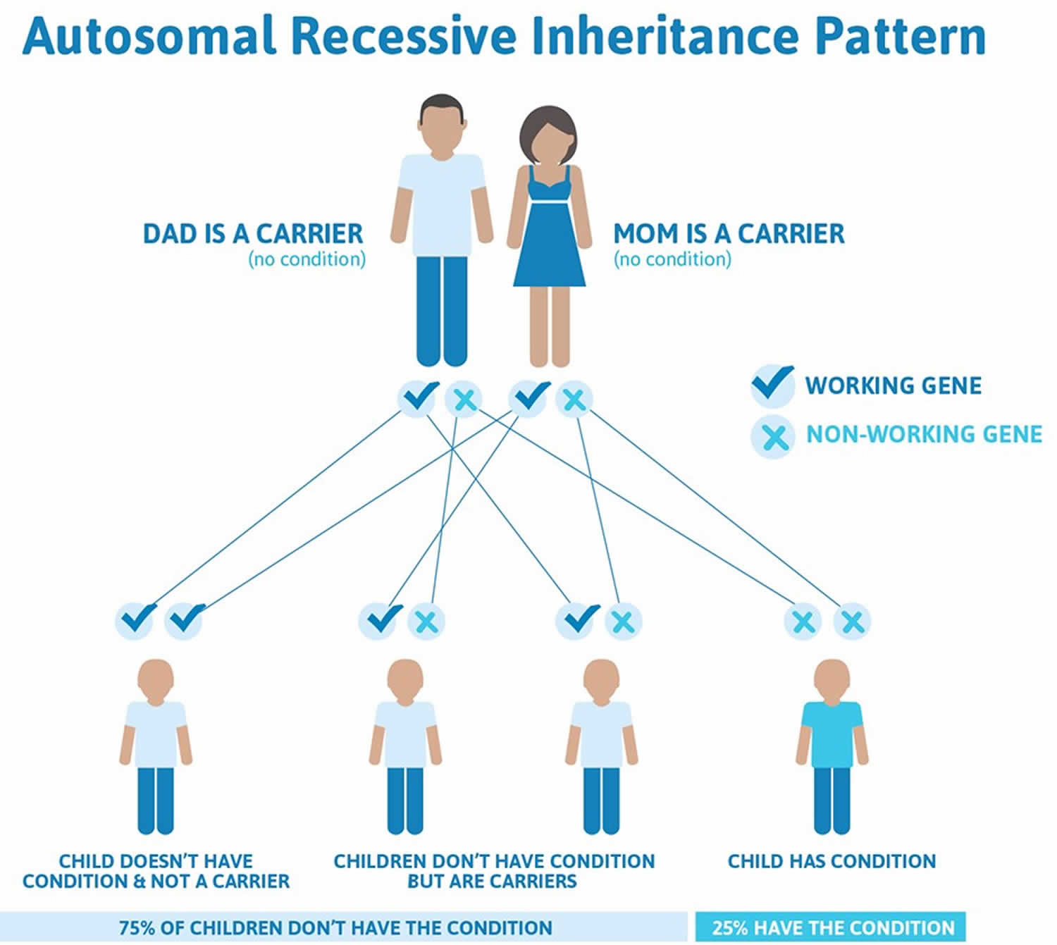
Cystic fibrosis is inherited in an autosomal recessive pattern, which means both copies of the gene in each cell have mutations. The parents of an individual with an autosomal recessive condition each carry one copy of the mutated gene, but they typically do not show signs and symptoms of the condition.

For a baby to be born with cystic fibrosis, both parents must be carriers of the faulty cystic fibrosis gene. The child will have inherited the faulty gene from each parent.

  • Approximately 12 million Americans or 1 in every 20 people living in this country, is a cystic fibrosis carrier.

The carrier parents, who are not themselves affected by cystic fibrosis, have one unaffected gene and one faulty cystic fibrosis gene. If both parents are carriers, a child has:

  • A one-in-four chance of being born with cystic fibrosis,ƒƒ
  • A two-in-four chance of being a carrier, like their parents, but not having the disease, andƒƒ
  • A one-in-four chance of being completely free of the condition – neither having cystic fibrosis nor being a carrier of the faulty cystic fibrosis gene

Note that the odds are the same for each successive pregnancy.

Figure 1. Cystic fibrosis autosomal recessive inheritance pattern

Cystic fibrosis autosomal recessive inheritance pattern

People with specific questions about genetic risks or genetic testing for themselves or family members should speak with a genetics professional.

Resources for locating a genetics professional in your community are available online:

Cystic fibrosis carrier

People with only one faulty copy of the cystic fibrosis transmembrane conductance regulator (CFTR) gene that causes cystic fibrosis will not have the condition and are not at risk of developing it. Carriers do not experience any detrimental symptoms as a result of their status, although any children they have with another carrier could be born with cystic fibrosis.

Can I find out if my partner and I are carriers?

It is now possible to test for the CFTR gene using a simple mouthwash or blood test. Check with your local laboratory if mouthwash testing is available. In the case of a mouthwash test, cells lining the cheek are collected by spitting saliva into a special tube; DNA can be extracted from these cells and then analyzed. In relatives of people diagnosed with cystic fibrosis, the exact gene alterations found in their affected family member are established wherever possible, no matter how rare the alteration. A test for the more common gene alterations is performed on the partner who does not have a family history of the condition.

Because the test for common mutations only detects about 90 – 95% of mutations, a negative result does not entirely rule out the possibility of the person being a cystic fibrosis carrier, but the statistical likelihood is reduced to less than 1 in 250. Depending on the number of genetic cystic fibrosis alterations tested for in your health region, this figure may differ slightly. A positive result shows that the person is definitely a carrier, even if there is no family history.

Very occasionally, neither cystic fibrosis gene alteration is identified in the person with cystic fibrosis, because the mutations may be very rare ones that are not detected in the routine test. In these cases, testing for cystic fibrosis carriers in a relative becomes a little more complicated, but is generally still possible by testing the entire cystic fibrosis gene for mutations. Once the cystic fibrosis-carrying gene from each parent has been identified, it is then possible to test for that pattern in the relative. Except in the case of a full brother or sister of someone with cystic fibrosis, a negative result does not confirm with 100% certainty that you are not a carrier, but it does mean that you have a much lower risk. The Table 1 below shows how the level of risk of having a child with cystic fibrosis can be estimated once testing has taken place and suggests possible courses of action.

Table 1. Cystic Fibrosis – Family Genetic Testing

[table “C436” not found /]
[Source 1)]

What are the options if my partner and I are both carriers of the cystic fibrosis gene?

All available options will be explained during your genetic counseling session, so that you can decide what is right for you. All decisions are for you to make, and there will be trained staff available in the genetics department to clarify the situation and to discuss the implications of all possible courses of action. Antenatal tests, with the options of ending or continuing the pregnancy, are discussed, including any risks that may be associated with the tests. Pre-implantation genetic diagnosis using IVF (in-vitro fertilization) techniques to ensure that only an embryo free of cystic fibrosis is implanted into the womb are also explained.

The future

Work is being carried out on techniques capable of detecting cystic fibrosis cells in the embryo using a sample of the mother’s blood. This would avoid the need for chorionic biopsy (where a piece of the developing placenta is taken at 10 – 12 weeks) or amniocentesis (where a sample of the fluid surrounding the fetus is taken), the current tests available for antenatal diagnosis.

Also as our ability to detect more cystic fibrosis genes improves, it will be possible to test for the rarer mutations that currently make the carrier status test less than 100% accurate due to missed, rare or previously unidentified mutations. This is called a false-negative result.

Home testing

The Cystic Fibrosis Foundation recommends that genetic testing is undertaken with professional clinical support and that the results of a genetic test are discussed with a genetic counselor, who can explain in full the results you may get and the options available to you as an individual.

Using a home genetic testing kit would mean that you do not have professional support available to you if and when you need it, as well as being less reliable. For these reasons, the Cystic Fibrosis Foundation advises against the use of home genetic testing kits.

Cystic fibrosis prevention

If you or your partner has close relatives with cystic fibrosis, you both may want to undergo genetic testing before having children. The test, which is performed in a lab on a sample of blood, can help determine your risk of having a child with cystic fibrosis.

If you’re already pregnant and the genetic test shows that your baby may be at risk of cystic fibrosis, your doctor can conduct additional tests on your developing child.

Genetic testing isn’t for everyone. Before you decide to be tested, you should talk to a genetic counselor about the psychological impact the test results might carry.

Cystic fibrosis symptoms

Cystic fibrosis signs and symptoms vary, depending on the severity of the disease. Even in the same person, symptoms may worsen or improve as time passes. Some people may not experience symptoms until adolescence or adulthood.

Children presenting with cystic fibrosis may experience recurrent chest infections and failure to thrive (i.e. not growing to normal weight and height compared to children of the same age). The child may also suffer from very smelly, light colored stools. 10% of children that had a history of meconium ileus will have cystic fibrosis.

The build-up of sticky mucus in the lungs can cause breathing problems and increases the risk of lung infections. Over time, the lungs may stop working properly.

Mucus also clogs the pancreas (the organ that helps with digestion), which stops enzymes reaching food in the gut and helping with digestion.

This means most people with cystic fibrosis don’t absorb nutrients from food properly and need to eat more calories to avoid malnutrition.

Symptoms of cystic fibrosis include:

  • recurring chest infections
  • wheezing, coughing, shortness of breath and damage to the airways (bronchiectasis)
  • difficulty putting on weight and growing
  • jaundice
  • diarrhea, constipation, or large, smelly poo
  • a bowel obstruction in newborn babies (meconium ileus) – surgery may be needed

People with the condition can also develop a number of related conditions, including diabetes, thin, weakened bones (osteoporosis), infertility in males, and liver problems.

Respiratory signs and symptoms

The thick and sticky mucus associated with cystic fibrosis clogs the tubes that carry air in and out of your lungs. This can cause signs and symptoms such as:

  • A persistent cough that produces thick mucus (sputum)
  • Wheezing
  • Breathlessness
  • Exercise intolerance
  • Repeated lung infections
  • Inflamed nasal passages or a stuffy nose

Digestive signs and symptoms

The thick mucus can also block tubes that carry digestive enzymes from your pancreas to your small intestine. Without these digestive enzymes, your intestines aren’t able to completely absorb the nutrients in the food you eat. The result is often:

  • Foul-smelling, greasy stools
  • Poor weight gain and growth
  • Intestinal blockage, particularly in newborns (meconium ileus)
  • Severe constipation

Frequent straining while passing stool can cause part of the rectum — the end of the large intestine — to protrude outside the anus (rectal prolapse). When this occurs in children, it may be a sign of cystic fibrosis. Parents should consult a physician knowledgeable about cystic fibrosis. Rectal prolapse in children may sometimes require surgery. Rectal prolapse in children with cystic fibrosis is less common than it was in the past, which may be due to earlier testing, diagnosis and treatment of cystic fibrosis.

Cystic fibrosis diagnosis

In United States, all newborn babies are screened for cystic fibrosis as part of the newborn blood spot test (heel prick test) carried out shortly after they’re born.

A heel prick blood test is performed to check for the enzyme IRT (immune reactive trypsin) is measured. If the levels are high, its suggestive of cystic fibrosis and the infants undergoes a sweat test.

Cystic fibrosis testing

If the screening test suggests a child may have cystic fibrosis, they’ll need these additional tests to confirm they have the condition:

  • The sweat test. This is the gold-standard diagnostic test. It is based on the pathological feature of cystic fibrosis, which is abnormal chloride ion transport. If the patient has two successive tests showing chloride levels above 60mmol/L (normal is under 15mmol/L), its conclusive that the patient has cystic fibrosis.
  • A genetic test (CF genotyping) may be used to confirm the diagnosis. A sample of blood or saliva is checked for the faulty gene that causes cystic fibrosis
  • A blood test is also performed on infants.

These tests can also be used to diagnose cystic fibrosis in older children and adults who didn’t have the newborn test.

The genetic test can also be used to see whether someone is a “carrier” of cystic fibrosis in cases where the condition runs in the family.

This test can be important for someone who thinks they may have the faulty gene and wishes to have children.

Cystic fibrosis complications

People with cystic fibrosis also have a higher risk of developing other conditions.

These include:

  • weak and brittle bones (osteoporosis). People with cystic fibrosis are at higher risk of developing a dangerous thinning of bones – medicines called bisphosphonates can sometimes help
  • electrolyte imbalances and dehydration. Because people with cystic fibrosis have saltier sweat, the balance of minerals in their blood may be upset. Signs and symptoms include increased heart rate, fatigue, weakness and low blood pressure.
  • cystic fibrosis-related diabetes – insulin and a special diet may be needed to control blood sugar levels
  • nasal polyps and sinus infections – steroids, antihistamines, antibiotics or sinus flushes can help
  • liver problems
  • fertility problems – it’s possible for women with cystic fibrosis to have children, but men won’t be able to father a child without help from fertility specialists (see a doctor or fertility specialist for more advice)

They’re more likely to pick up infections, and more vulnerable to complications if they do develop an infection, which is why people with cystic fibrosis shouldn’t meet face to face.

Respiratory system complications

  • Damaged airways (bronchiectasis). Cystic fibrosis is one of the leading causes of bronchiectasis, a condition that damages the airways. This makes it harder to move air in and out of the lungs and clear mucus from the airways (bronchial tubes).
  • Chronic infections. Thick mucus in the lungs and sinuses provides an ideal breeding ground for bacteria and fungi. People with cystic fibrosis may often have sinus infections, bronchitis or pneumonia.
  • Growths in the nose (nasal polyps). Because the lining inside the nose is inflamed and swollen, it can develop soft, fleshy growths (polyps).
  • Coughing up blood (hemoptysis). Over time, cystic fibrosis can cause thinning of the airway walls. As a result, teenagers and adults with cystic fibrosis may cough up blood.
  • Pneumothorax. This condition, in which air collects in the space that separates the lungs from the chest wall, also is more common in older people with cystic fibrosis. Pneumothorax can cause chest pain and breathlessness.
  • Respiratory failure. Over time, cystic fibrosis can damage lung tissue so badly that it no longer works. Lung function usually worsens gradually, and it eventually can become life-threatening.
  • Acute exacerbations. People with cystic fibrosis may experience worsening of their respiratory symptoms, such as coughing and shortness of breath, for several days to weeks. This is called an acute exacerbation and requires treatment in the hospital.

Digestive system complications

  • Nutritional deficiencies. Thick mucus can block the tubes that carry digestive enzymes from your pancreas to your intestines. Without these enzymes, your body can’t absorb protein, fats or fat-soluble vitamins.
  • Diabetes. The pancreas produces insulin, which your body needs to use sugar. Cystic fibrosis increases the risk of diabetes. Around 30 percent of people with cystic fibrosis develop diabetes by age 30.
  • Blocked bile duct. The tube that carries bile from your liver and gallbladder to your small intestine may become blocked and inflamed, leading to liver problems and sometimes gallstones.
  • Intestinal obstruction. Intestinal obstruction can happen to people with cystic fibrosis at all ages. Children and adults with cystic fibrosis are more likely than are infants to develop intussusception, a condition in which a section of the intestines folds in on itself like an accordion.
  • Distal intestinal obstruction syndrome (DIOS). DIOS is partial or complete obstruction where the small intestine meets the large intestine.

Reproductive system complications

Almost all men with cystic fibrosis are infertile because the tube that connects the testes and prostate gland (vas deferens) is either blocked with mucus or missing entirely. Certain fertility treatments and surgical procedures sometimes make it possible for men with cystic fibrosis to become biological fathers.

Although women with cystic fibrosis may be less fertile than other women, it’s possible for them to conceive and to have successful pregnancies. Still, pregnancy can worsen the signs and symptoms of cystic fibrosis, so be sure to discuss the possible risks with your doctor.

Cystic fibrosis treatment

There’s no cure for cystic fibrosis, but a range of treatments can help control the symptoms, prevent or reduce complications, and make the condition easier to live with.

Cystic fibrosis is a disease that affects many systems. Therefore, its essential that a multidisciplinary approach is taken in the management of this disease.

The main goals in the management of cystic fibrosis are:

  • Promote sufficient growth and nutrition
  • Preventing and controlling infections that occur in the lungs
  • Removing and loosening mucus from the lungs
  • Prevent the progression of further lung disease
  • Treating and preventing intestinal blockage

This is achieved with a team of health professionals including pediatricians, physiotherapists, dieticians, counselors and family doctors. Specific treatments for cystic fibrosis are:

  • Antibiotics to reduce risk of chest infections
  • Pain relievers such as Ibuprofen may reduce lung deterioration
  • Mucus-thinning drugs to reduce viscosity of mucus, making it easier to breathe
  • Bronchodilators to increase air entry
  • Physio exercises to remove mucus in the chest
  • Improved nutrition ( high calorie diet, vitamin and mineral rich) to maintain weight and ensure essential nutrients are consumed
  • Refer for counseling and support groups as chronic diseases can cause a great amount of stress for the patient and family
  • Oral enzymes should be taken to aid in digestion and absorption of food
  • In severe cases, a lung transplant may be suggested due to severity of disease and poor quality of life

Medicines for lung problems

People with cystic fibrosis may need to take different medicines to treat and prevent lung problems. These may be swallowed, inhaled or injected.

Medicines for lung problems include:

  • antibiotics to prevent and treat chest infections
  • medicines to make the mucus in the lungs thinner and easier to cough up – for example, dornase alfa, hypertonic saline and mannitol dry powder
  • medicine to help reduce the levels of mucus in the body – for example, ivacaftor taken on its own (Kalydeco) or in combination with lumacaftor (Orkambi, but this is only available on compassionate grounds if people fulfill several criteria set by the manufacturer)
  • bronchodilators to widen the airways and make breathing easier
  • steroid medicine to treat small growths inside the nose (nasal polyps)

It’s also important that people with cystic fibrosis are up-to-date with all routine vaccinations and have the flu jab each year once they’re old enough.
Exercise

Any kind of physical activity, like running, swimming or football, can help clear mucus from the lungs and improve physical strength and overall health.

A physiotherapist can advise on the right exercises and activities for each individual.

Airway clearance techniques

Airway clearance helps to stop thick mucus from building up and blocking airways in the lungs, reducing infections and preventing lung damage.

There are many different techniques, and your specialist cystic fibrosis physiotherapist will work closely with you to find the technique, or combination of techniques, that works best for you. Everyone’s lungs are different and the amount of airway clearance needed will vary from person to person. No one technique has been shown to be better than another, and this may be because each person’s chest can behave very differently, and people may prefer different techniques at different times in their life. It is important that you are happy with the technique you’re using as, despite it being essential, physiotherapy to clear the chest can be a treatment that people struggle to do regularly. Some techniques require no equipment, and others use a device.

These include:

  • Active Cycle of Breathing Techniques – a cycle of deep breathing, huffing, coughing and relaxed breathing to move mucus.
  • Autogenic Drainage – a series of gentle controlled breathing techniques that moves mucus from smaller to bigger airways using controlled breathing at different levels. Uses fast flow rates as you sigh out but ensures the airways are kept open.
  • Positive Expiratory Pressure – breathing out through a mask or mouthpiece against a resistance to build up pressure behind mucus to move it.
  • Oscillating Positive Expiratory Pressure e.g. Flutter®, Acapella® – breathing out against a resistance to create positive expiratory pressure and also vibrate your airways to move mucus.
  • High Frequency Chest Wall Oscillation (the vest) – an electric air pulse generator that connects to an inflatable jacket (vest) to vibrate the chest.

The evidence for airway clearance techniques in people with cystic fibrosis has been analyzed in five Cochrane reviews published between 2000 and 2011. There is currently insufficient evidence to suggest that one technique is better than another.

However two recent well-constructed studies in cystic fibrosis show that if using High Frequency Vest Oscillation, (the Vest), the patients may not breathe as well using it or may get more infections and get them more quickly. These studies were done in children. There are at least two studies in adults that show less or just the same amount of secretions cleared with the Vest compared to other techniques.

The Association of Chartered Physiotherapists in Cystic Fibrosis therefore recommends that the Vest is not used as the only airway clearance technique especially in children with cystic fibrosis. The Association of Chartered Physiotherapists in Cystic Fibrosis recognizes that some patients may prefer to use the Vest and feel benefit from using it. We advise people thinking about wanting to use the Vest to discuss the pros and cons with their physiotherapist. The Association of Chartered Physiotherapists in Cystic Fibrosis recommends that for people with cystic fibrosis choosing to use the Vest some other airway clearance technique such as at least huffing and coughing is done with the Vest on.

Physiotherapists should consider patient choice and circumstances, and clinical reasoning of the person with cystic fibrosis’s signs and symptoms when recommending techniques. Techniques may be combined depending on patient needs.

There are other devices not named above. Be sure to discuss any new airway clearance technique that you would like to try with your cystic fibrosis physiotherapist. They will discuss whether there is good research for the device and whether there are any positives and/or negatives to consider.

How much airway clearance do you need?

The length and number of airway clearance sessions will vary from person to person, and may need to increase if you have a chest infection.

For few or no lung secretions, treatment sessions may only need to last 10–15 minutes, but if there are many it could take 45–60 minutes. Most people do two treatments a day, but the number and length of your sessions may need to increase depending on the volume of secretions your lungs are producing. If no mucus is present, exercise may play a greater role in your airway clearance techniques. Your cystic fibrosis physiotherapist will be able to advise what is suitable for you.

When should airway clearance start?

A specialist cystic fibrosis physiotherapist should assess all newly diagnosed people with cystic fibrosis and advise when and how they should start doing airway clearance. This may not be immediately in screened infants with no symptoms, and exercise/activity may play a greater role. At all ages, the advice will be based on an individual assessment.

Who will do my airway clearance?

The families of babies with cystic fibrosis will be taught how to do airway clearance with their child. As you grow older with cystic fibrosis, your physiotherapist will aim to find a technique that you can carry out independently so that you do not have to rely on someone else to help. Breathing exercises can be introduced from the age of two to three in the form of a game. Older children can start doing airway clearance themselves with supervision. Most teenagers and adults become completely independent, and only need help if they have increased mucus.

What about exercise?

Exercise helps with general fitness, health and well-being, and bone health. Improved fitness has been shown to be important in the long term prognosis of cystic fibrosis, and people with cystic fibrosis should aim to maintain an active lifestyle. Physical exercise is also often used as part of an airway clearance regime. Exercise can help loosen mucus in the lungs and make airway clearance techniques quicker and easier. Your cystic fibrosis physiotherapist should be able to answer any questions about the type and intensity of exercise suitable for you.

What about your posture?

As you get older it is easy to adopt poor postures, which can lead to back pain and impact upon lung function. It is therefore important for people with cystic fibrosis to maintain good posture. Your physiotherapist will be able to offer advice and help to assess and treat any joint or back problems you may have.

What about incontinence?

Some people with cystic fibrosis may develop problems with urinary or fecal incontinence (leaking wee or poo) when coughing and/or exerting themselves. If this should happen to you it is important to tell your physiotherapist as soon as possible and not be embarrassed, as they can teach you pelvic floor exercises to help. They may also refer you on to a specialist. For more information see our fact sheet.

Inhalation therapy

Many people with cystic fibrosis will take inhaled medications as part of their treatment. This may be to loosen the mucus, hydrate the airways, open up the airways, reduce inflammation of the airways and/or to deliver antibiotics to treat infections. These medications may come in nebulized (inhaling a mist) or dry powder form, and often the timing of these alongside airway clearance and exercise is important to ensure your treatment is as effective as possible. Please ask your doctor for advice.

Cystic fibrosis diet

Eating well is important for people with cystic fibrosis because the mucus can make it difficult to digest food and absorb nutrients.

The pancreas often doesn’t work properly, making it even harder to digest food.

A dietitian will advise on how to take in extra calories and nutrients to avoid malnutrition.

They may recommend a high-calorie diet, vitamin and mineral supplements, and taking digestive enzyme capsules with food to help with digestion.

Only around 10% of people with cystic fibrosis retain any useful pancreatic function, meaning that digestive juices (enzymes) do not reach the stomach in order to break down food properly so that the body can use it to produce energy.

This means that most people with cystic fibrosis require enzyme capsules with all meals and snacks. The amount of capsules needed depends on the food being eaten, and varies from person to person, but it can be as many as 60 a day.

Being a healthy weight can improve the chances of fighting off chest infections, so a suitable diet is important. It is also necessary to build up reserves in case of weight loss during times of illness.

A healthy diet for someone with cystic fibrosis is:

  • High in energy (calories) – the body of someone with cystic fibrosis has to work harder, and has higher energy requirements.
  • Rich in fat and protein – to compensate for the amount wasted as food is not fully digested.

As with any diet, the exact amount of these elements varies by age and from person to person.

As a general principle, people with cystic fibrosis often require 20 to 50% more calories each day than people without cystic fibrosis, however some may need considerably more than this.

This high calorie intake is often hard to achieve as many people with cystic fibrosis experience reduced appetite, especially during episodes of infection – the very time when your body’s energy requirements are at their highest. Many people with cystic fibrosis need to take enzymes to digest and absorb fat-containing foods. Forgetting or taking too few enzymes will cause you to lose fat (energy) in your stools and this results in some of the fat you have eaten being wasted. This can also lead to weight loss or difficulty in gaining weight despite eating well. Even if you are taking your enzymes appropriately you will continue to lose some fat in your stools and this contributes to your increased energy requirements.

What is a high energy diet?

This is a diet that contains an increased amount of calories. Calories are a measure of how much energy a food contains. Dietary fat provides the most energy (calories) in the smallest volume and is the reason that your dietician encourage you to use high fat foods and snacks whenever possible, together with the appropriate number of enzymes. This should be in combination with the right balance of foods from other food groups – starchy foods (bread, pasta, rice and cereals), proteins (meat, fish, cheese and meat substitutes), dairy foods (milk, cheese, and yogurts) and fruit and vegetables.

Although a diet high in energy can be hard to achieve, there are simple guidelines that you can follow to help increase your calorie intake:

Fatty foods

  • Fat is the richest source of energy (calories). One gram of fat contains nine calories, which is more than twice the amount found in protein or carbohydrate. Fat is also a good source of essential fatty acids and fat soluble vitamins
  • Where possible you should choose unsaturated fats and oils as they give the same amount of calories but in a healthier form. Monounsaturated fats include olive oil and rapeseed oil. Polyunsaturated fats include sunflower, soya, sesame and corn oil. Soft spreads made from olive oil, rapeseed oil etc can be used to replace butter and lard
  • The essential omega 3 fats are also important and are found mainly in oily fish. You should try to include these in your diet
  • Try to eat plenty of fat, making sure you take enough enzymes with your food, as fatty foods require more enzymes
  • Fry your food in olive oil or rapeseed oil, add olive oil, butter or margarine to vegetables and potatoes and spread butter or margarine generously on bread
  • Olive oil can be drizzled on food to increase the flavor and energy content
  • Toss pasta in olive oil before serving or drizzle olive oil over pasta dishes
  • Add mayonnaise, olive oil, dips or dressings to sandwiches and salads
  • Choose higher fat foods such as pastries, crisps, nuts, seeds, chocolate, cakes, biscuits and chips
  • Single, double or whipped cream can be added to puddings, sauces and soups
  • Add extra olive oil/butter/margarine to vegetables, potatoes, pasta, bread and toast
  • Try to have more fried foods
  • When grilling or roasting foods, drizzle them with olive oil or vegetable oil and baste with oil regularly
  • Add olive oil/mayonnaise/salad cream to sandwiches and salads
  • Always add a dressing (oil or mayonnaise) to your salads
  • Avoid using low fat spread or eating reduced fat foods

Starchy (carbohydrate) foods

  • Starchy foods are another good energy provider and should be a part of every meal
  • Starchy foods include breakfast cereals, pasta, potatoes, rice and bread
  • Make them higher in calories by brushing with olive oil, garnishing with a drizzle of oil, frying them or adding lots of milk, butter, olive oil, mayonnaise etc
  • Mash potatoes with butter, olive oil spread or margarine and mix in some cream, crème fraiche or cheese
  • Toss pasta in olive oil before serving or drizzle olive oil over pasta dishes
  • Add chopped nuts or dried fruit to cereal, yogurt, milk pudding, fruit and ice cream

Sugary foods

  • Sugary foods such as jam, honey, marmalade, syrup, fizzy drinks, tinned fruit in syrup, cakes, biscuits, sweets and chocolates are also energy rich
  • Put plenty of sugar into hot drinks, on cereals and in desserts
  • Spread jam, honey, marmalade and chocolate spread thickly on bread and toast
  • Add sugar, honey, jam or syrup to cereal, yogurt, milk pudding, fruit and ice cream
  • Avoid foods which are labelled as being ‘low sugar’ or sweetened with artificial sweetener, such as diet yogurts, diet squashes and diet fizzy drinks

Your teeth are very important to your health. Remember to clean your teeth after eating sugary food or taking sweetened drinks and to visit your dentist regularly.

Milk and dairy products

  • Avoid low fat dairy products such as low fat yogurts, semi-skimmed and skimmed milk and low fat cheeses such as cottage cheese. If these are the only types of dairy products you like they are still a good source of calcium
  • Milk and dairy products are an important source of energy and calcium
  • Use full cream or Channel Island milk and try to have between one and two pints daily in drinks, desserts such as custard, instant whips or milk pudding, savoury sauces such as cheese or parsley sauce and on cereals
  • Add double cream to ordinary milk when making any milk-based savory and sweet dishes or milky drinks
  • Use milk instead of water when making up condensed or packet soup.
  • Stir in double cream or olive oil just before serving
  • Always add milk and olive oil/butter/margarine to mashed potato
  • Cheese is an ideal snack; serve as cheese on toast, cheese and crackers etc. In addition cheese can be used to add extra calories to your food, for example it
  • can be added to mashed potatoes, baked beans or pasta dishes
  • Sprinkle grated cheese on top of vegetables, potatoes, soups and sauces
  • Yogurts and ice cream make a quick dessert or snack. Try to choose whole milk, thick and creamy, Greek- or custard- style yoghurts and real dairy ice creams

Protein foods

  • Protein foods come in two varieties:
    • Animal proteins such as meat, fish, eggs, milk and dairy products
    • Vegetable proteins such as beans, peas, lentils and nuts
  • Try to keep your protein intake high by having a good helping at each meal, including breakfast. It will also help if your snacks include some protein
  • Fatty or oil-rich fish are good source of protein as well as a good source of the omega 3 fatty acids. Try eating oil-rich fish such as salmon, herring, sardines,
  • mackerel, pilchards, trout, kippers and fresh tuna regularly
  • If you are vegetarian make sure you replace meat or fish with a variety of protein sources such as soya mince or other meat substitutes, beans, seeds, lentils, tofu, nuts, cheese or eggs
  • Avoid eating raw eggs and do not add them to high energy drinks. They do not add many calories and may be a Salmonella risk

Fruit and vegetables

  • Fruit and vegetables are generally low in energy, but do provide vitamins and some minerals, so they are an important part of a balanced diet
  • Try to eat fruit and vegetables everyday – fresh, frozen or tinned – but if your appetite is poor do not fill yourself up with fruit and vegetables at the expense of higher calorie foods
  • Some fruit and vegetables e.g. bananas, beans, pulses and root vegetables such as potato, turnip and parsnip are higher in calories
  • Try to have a glass of fruit juice or squash with added vitamin C daily
  • Use fruit in pies and crumbles or serve with double cream or dairy ice cream
  • Add double cream, ice cream or custard to fruit
  • Add sauces, olive oil or butter to vegetables or fry them

Calcium

  • Calcium is essential to the body for maintaining strong and healthy bones. Therefore it is important that you have plenty of calcium rich foods each day
  • The best sources of calcium are dairy products such as milky drinks, cheese, yogurts and dairy ice cream
  • If you don’t like these types of foods, non-dairy calcium sources include baked beans, tinned fish with bones (salmon, sardines and pilchards) and white bread
  • Also look out for foods that may have calcium added to them such as soya milk, drinks and cereal bars
  • Generally non-dairy foods contain less calcium and therefore you will need to eat more of them to ensure an adequate calcium intake. If you only like low fat varieties of milk and milk products continue to have these as they contain similar amounts of calcium as full cream versions
  • If you are concerned about your calcium intake talk to your cystic fibrosis dietitian

Is this really a healthy diet?

For most people a high fat and high calorie diet would not be considered a healthy diet. However for many people with cystic fibrosis this is the most appropriate diet to help achieve and maintain a good healthy weight. This in turn helps to fight infections and stay healthy for longer. It is almost the opposite of the diet recommended for most other adults which is low in fat, sugar and salt with an increased intake of dietary fiber. This type of diet is not suitable for most people with cystic fibrosis because it is bulky and filling and is unlikely to provide enough energy.

Not everyone is the same and some people with cystic fibrosis may gain too much weight. These patients may need to reduce their fat and energy intake and should be assessed and advised on an individual basis by a specialist cystic fibrosis dietitian.

To give your high fat diet a more healthy emphasis where possible you should try to use monounsaturated cooking oils or spreads such as olive oil, rapeseed oil, olive oil spread instead of lard or butter and to eat fruit and vegetables daily. These can be incorporated as high fiber, high energy snacks including dried fruit, fruit pies and crumbles served with cream or ice cream. Additional fat e.g. olive oil, butter, cream, cheese can be added to dishes after the rest of the family has been served to ensure the whole family get the type of diet they need.

Pancreatic enzymes

Approximately 85% or more of adults with cystic fibrosis are pancreatic insufficient. This means the pancreas is unable to produce or release enough digestive enzymes into the small intestine and so food is not digested and can not be absorbed in the gut. To help to compensate for the lack of digestive enzymes most people with cystic fibrosis have to take replacement pancreatic enzymes to digest their food properly. The enzyme dose needed varies from one adult to another. Your cystic fibrosis team will advise on appropriate enzyme doses and it is important to discuss any changes to your enzyme intake with the team before changing our dose. There is a wide range of strengths of pancreatic enzymes available. When taking pancreatic enzymes it is also important to drink plenty of fluids.

Making your enzymes work for you

To get the most benefit from your enzymes, follow these guidelines:

  • Take enzymes with every meal, fat-containing snack or milky drink
  • Spread your enzymes throughout the meal
  • High fat meals will need more enzymes than low fat meals
  • Most snacks will require fewer enzymes than a meal but be cautious as some snacks are very fatty and can require as many or even more enzymes than a meal
  • Be flexible about the dosage and timing of enzymes
  • Enzymes do not need to be taken with fruit, jelly, sorbet, boiled, chewy or jelly sweets, squash, fizzy drinks or fruit juices

Remember there is no standard dose of enzyme. You should take the amount required to control your bowels. Sometimes people with cystic fibrosis produce too much stomach acid and this can make pancreatic enzymes less effective. If you are experiencing problems with heartburn, reflux or abdominal cramps then please discuss with your cystic fibrosis team and they can prescribe appropriate medications to help.

Distal intestinal obstruction syndrome, fluid and fiber

Distal intestinal obstruction syndrome (a blockage in the bowel) – usually referred to as DIOS – may be caused by not taking enough enzymes and if you are dehydrated this can make things worse. Sometimes it can occur for no known reason. You should make sure you drink enough fluid and aim to have 30 ml per kg body weight of fluid each day. For example if you weigh 50 kg this is 1500 ml per day, the equivalent of about six to eight glasses/mugs of liquid each day.

  • Some drinks such as sweet, fizzy caffeine-containing drinks or strong coffee may dehydrate you further. Water or diluted squash are best
  • When the weather is hot or you are in a hot room you will need more fluid
  • If you are doing a lot of exercise the fluid you lose by sweating will need to be replaced by drinking more
  • Your body will lose fluid if your blood sugars are too high. Make sure that if you have diabetes it is well controlled
  • Taking more fiber in your diet may help prevent distal intestinal obstruction syndrome. Try including a high fiber cereal at breakfast, some wholemeal bread, and increasing the amount of fruit and vegetables you eat. If you increase your fiber intake you will need to increase the amount of fluid you drink.

Vitamin supplements

Malabsorption of the fat-soluble vitamins A, D, E and K is likely in most people with cystic fibrosis, especially those who are pancreatic insufficient. Without supplements, blood levels of these vitamins may become low and occasionally deficiency symptoms can occur. Fat-soluble vitamins should be taken at a mealtime when enzymes will also be taken.

Recommended daily supplements (starting doses):

  • Vitamin A – 4000-8000 iu (International Units)
  • Vitamin D – 400-800 iu
  • Vitamin E – 50-200 iu

Cystic fibrosis-related diabetes

Diabetes is common in adults and adolescents with cystic fibrosis. It occurs in approximately 30% of people with cystic fibrosis by the age of 25 years. Cystic fibrosis-related diabetes is less common in children.

Cystic fibrosis-related diabetes is different from the two main types of diabetes (Type 1 and Type 2 diabetes) and has features of both.

In addition, people with cystic fibrosis may develop high blood sugar levels
during periods of lung infection or while taking oral steroids. This may be temporary and may resolve when the acute infection is treated or when the steroids are reduced or stopped.

Some people may be concerned that the sugary diet that they have eaten may contribute to them developing cystic fibrosis-related diabetes. There are many other risk factors that cause or contribute to people with cystic fibrosis developing cystic fibrosis-related diabetes. Any concerns should be discussed with your doctor or dietitian.

Cystic fibrosis-related diabetes treatment

While some people can control their blood sugar levels by taking tablets, in cystic fibrosis-related diabetes most people are best treated with injections of insulin. Insulin cannot be taken by mouth because it is destroyed in the stomach. It is usually given as an injection two to four times a day.

Maintaining a healthy body weight is one of the most important steps you can take to ensure good health. People with cystic fibrosis-related diabetes still need to eat their usual high calorie, high protein and high fat diet to help achieve and maintain a healthy body weight. This is the opposite of the usual advice for diabetics and it can become confusing.

In cystic fibrosis, more energy (calories) is needed in the diet and the dose of insulin can usually be tailored to individual requirements and people can be taught to adjust their insulin dose to their dietary intake. Keeping your blood sugars at a near normal level will also help to maintain/improve your weight.

Surgical and other procedures

  • Nasal polyp removal. Your doctor may recommend surgery to remove nasal polyps that obstruct breathing.
  • Oxygen therapy. If your blood oxygen level declines, your doctor may recommend that you breathe pure oxygen to prevent high blood pressure in the lungs (pulmonary hypertension).
  • Endoscopy and lavage. Mucus may be suctioned from obstructed airways through an endoscope.
  • Feeding tube. Cystic fibrosis interferes with digestion, so you can’t absorb nutrients from food very well. Your doctor may suggest temporarily using a feeding tube to deliver extra nutrition while you sleep. This tube may be inserted in your nose and guided to your stomach, or it may be surgically implanted into the abdomen.
  • Bowel surgery. If a blockage develops in your bowel, you may need surgery to remove it. Intussusception, where a section of bowel has folded in on itself, also may require surgical repair.

Lung transplants

In severe cases of cystic fibrosis, when the lungs stop working properly and all medical treatments have failed to help, a lung transplant may be recommended.

If you have severe breathing problems, life-threatening lung complications or increasing resistance to antibiotics used to treat lung infections, lung transplantation may be an option. Because bacteria line the airways in diseases that cause permanent widening of the large airways (bronchiectasis), such as cystic fibrosis, both lungs need to be replaced.

A lung transplant is a serious operation that carries risks, but it can greatly improve the length and quality of life for people with severe cystic fibrosis.

Cystic fibrosis does not recur in transplanted lungs. However, other complications associated with cystic fibrosis — such as sinus infections, diabetes, pancreas problems and osteoporosis — can still occur after a lung transplant.

Lifestyle and home remedies

You can manage your condition and minimize complications in several ways. Always talk to your doctor before starting home remedies.

Pay attention to nutrition and fluid intake

Cystic fibrosis can cause malnourishment because the enzymes needed for digestion can’t reach your small intestine, preventing food from being absorbed. People with cystic fibrosis may need a significantly higher number of calories daily than do people without the condition.

A healthy diet is important to maintain good lung function. It’s also important to drink lots of fluids, which can help thin the mucus in your lungs. You may work with a dietitian to develop a nutrition plan.

Most people with cystic fibrosis need to take pancreatic enzyme capsules with every meal and snack. In addition, your doctor may recommend:

  • Antacids
  • Supplemental high-calorie nutrition
  • Special fat-soluble vitamins
  • Extra fiber to prevent intestinal blockage
  • Extra salt, especially during hot weather or before exercising
  • Adequate water during hot weather

Keep immunizations up to date

In addition to other usual childhood vaccines, people with cystic fibrosis should have the annual flu vaccine and any other vaccines their doctor recommends. Cystic fibrosis doesn’t affect the immune system, but children with cystic fibrosis are more likely to develop complications when they become sick.

Exercise

Regular exercise helps loosen mucus in your airways, and strengthens your heart. For many people with cystic fibrosis, participating in sports can improve confidence and self-esteem. Anything that gets you moving, including walking and biking, can help.

Eliminate smoke

Don’t smoke in your home or car, and don’t allow other people to smoke around you or your child. Secondhand smoke is harmful for everyone, but especially for people with cystic fibrosis.

Encourage hand-washing

Teach all the members of your family to wash their hands thoroughly before eating, after using the bathroom, when coming home from work or school, and after being around a person who is sick. Hand-washing is the best way to protect against infection.

Attend medical appointments

You’ll have ongoing care from your doctor and other medical professionals. Make sure to attend your regular follow-up appointments. Take your medications as prescribed and follow therapies as instructed. Contact your doctor if you experience any signs or symptoms such as severe constipation, more mucus than usual, blood in your mucus or reduced energy.

Coping and support

If you or someone you love has cystic fibrosis, you may experience strong emotions such as anger or fear. These issues are especially common in teens. Talking openly about how you feel can help. It also may help to talk with others who are dealing with the same issues.

That might mean joining a support group for parents of children with cystic fibrosis. Older children with the disorder may want to join a cystic fibrosis group to meet and talk with others who have the disease.

If you or your child is depressed or anxious, it may help to meet with a psychologist. He or she may suggest medications or other treatments as well.

Spend time with friends and family. Having their support can help you manage stress and reduce anxiety. Ask your friends or family for help if you need it.

Take time to learn about your or your child’s condition. If your child has cystic fibrosis, encourage him or her to learn about the condition. Find out how medical care is managed for children with cystic fibrosis as they grow older into adulthood. Ask your child’s doctor if you have questions about your child’s care as he or she becomes older.

References   [ + ]

read more

Burners and stingers

Burners and stingers

Burners and stingers

Burners and stingers are intense pains that occur when the nerves that run from your neck to your arm are stretched or compressed after an impact. These injuries are common in contact or collision sports where the shoulder may be pushed backward or the head and neck is forcibly pushed to the side. Burners and stingers are named for the stinging or burning pain that spreads from your shoulder to your hand. A burner or stinger can feel like an electric shock or lightning bolt down your arm.

Burners and stingers are most common in football players but are also common in those who participate in rugby, hockey, wrestling, lacrosse, gymnastics and diving. Burners can also happen in a motor vehicle crash when the head is pushed to one side or something hits the neck and shoulder.

In most cases, burners and stingers are temporary and symptoms quickly go away.

Athletes with a burner should be evaluated by a physician and should not return to their sport until they have fully recovered. A single burner or the effects of recurrent burners can lead to permanent neurologic damage. Nerves that have been injured are more susceptible to injury. Testing to evaluate nerve injury and recovery should be done by a trained medical specialist. Furthermore, burners can easily be confused with other neck injuries. Athletes who have tenderness over the bones in their neck or symptoms in both arms or a leg should be stabilized on the playing field and transported to a facility that can evaluate the athlete for possible spinal cord injury.

When to see a doctor

Brachial plexus injuries can cause permanent weakness or disability. Even if yours seems minor, you may need medical care. See your doctor if you have:

  • Recurrent burners and stingers
  • Weakness in your hand or arm
  • Weakness in any part of the arm following trauma
  • Complete paralysis of the upper extremity following trauma
  • Neck pain
  • Symptoms in both arms
  • Symptoms in upper and lower limbs

It’s important to be evaluated and treated within six to seven months after the injury. Delays in treatment may compromise outcomes of nerve surgeries.

The brachial plexus

Nerves are like electrical cables that travel through the spinal canal carrying messages between your brain and muscles. The nerves that provide feeling and movement to the arm branch out of the spinal canal at the neck. They join together to form a larger bundle, or cord of nerves called the brachial plexus. All of the nerve supply to the arm runs through this plexus.

The roots (anterior rami) of spinal nerves C5–C8 and T1 form the brachial plexus, which extends inferiorly and laterally on either side of the last four cervical and first thoracic vertebrae (Figures 1 and 2). The brachial plexus passes above the first rib posterior to the clavicle and then enters the axilla (armpit).

Figure 1. Brachial plexus

brachial plexus

Figure 2. Brachial plexus origin and nerve branches

Brachial plexus origin and nerve branches

Figure 3. Brachial plexus motor and sensory innervation of the upper limb

Brachial plexus motor and sensory innervation of the upper limb

Burners and stingers cause

The brachial plexus nerve network begins with nerve roots at the spinal cord in the neck and reaches to the armpit. Nerves branch out from there and continue down the arm to the forearm, hand, and fingers.

An injury to the brachial plexus can cause a burner or stinger. When a strong force increases the angle between the neck and shoulders, the brachial plexus nerves might stretch or tear. This often happens when the head is forcefully pushed sideways and down. This bends the neck and pinches the surrounding nerves. The injury may also pull the nerve roots of the brachial plexus from the spinal cord. Damaged nerves conduct sensation poorly and weaken muscle movements.

Figure 4. Burners and stingers cause

Burners and stingers cause

Footnote: Many brachial plexus injuries occur when the arm is pulled downward and the head is pushed to the opposite side.

Risk factors for developing burners and stingers

Contact sports

Athletes who engage in contact sports are more likely to suffer a burner or stinger. These injuries often occur with a fall onto the head, such as in a wrestling takedown or a football tackle. In fact, tackling or blocking in American football is the athletic activity that most often causes burners or stingers. Football defensive players and linemen frequently suffer this injury.

Spinal stenosis

In addition to playing contact sports, a small spinal canal may put you at greater risk for a burner or stinger. Athletes with recurrent stingers or burners may have smaller spinal canals than players who do not suffer recurrent injury. This condition is called spinal stenosis.

Burners and stingers prevention

To make burners less likely if you play contact sports, be sure to:

  • Keep your neck and shoulder muscles as strong and flexible as possible.
  • Gently stretch the neck muscles before any athletic activity.
  • Use protective gear (like a football neck collar or specially designed shoulder pads).
  • Use proper sports technique (never lead with your head during a football game, etc.).

The best way to prevent a burner is for athletes to use proper tackling technique (“see what you hit”) and strengthen their neck muscles. This will help limit excess motion of the neck from contact or collisions and reduce either stretch or compression to the nerves. In football, various collars (neck rolls, cowboy collars) have been created that can be attached to the shoulder pads to limit neck motion. The efficacy of these devices in preventing burners is unclear. Athletes who use these collars must make sure they can still extend their necks and look up during a tackle. In football, being able to see what you hit generally reduces the risk of serious injury that can occur when the neck is bent forward at the time of impact. Burners can also be prevented by avoiding contact or collisions until the effects of a previous burner have completely resolved.

Burners and stingers symptoms

Burner and stinger symptoms typically occur in one arm only. They usually last seconds to minutes, but in some cases they can last hours, days, or even longer. The most common symptoms of a burner or stinger include:

  • A burning or electric shock sensation
  • Arm numbness and weakness immediately following the injury
  • A warm sensation.

Athletes who have just sustained a burner will typically hold their arm limply at their sides or be observed shaking their arm to get rid of the tingling or burning sensation. A burning or stinging pain runs from the neck and shoulder down the arm even into the hand. In addition to the burning pain, it may feel like the arm has fallen asleep or like “pins and needles.” There may also be weakness in the shoulder and arm. Neck pain and spasm typically follow an injury that leads to a burner, but pain over the bones in the neck, pain radiating to both arms, or pain radiating to the legs suggests a possible spinal cord injury.

Burners and stingers diagnosis

In order to determine whether your injury is a burner or stinger, your doctor will discuss your symptoms and how the injury occurred. A doctor will usually recognize a burner from your symptoms and a physical exam. The doctor may check arm strength, reflexes, and range of motion in the arm. Imaging tests, such as x-rays, magnetic resonance imaging (MRI) scans, and nerve studies are not usually needed.

A more extensive examination like X-rays or magnetic resonance imaging (MRI) is needed if you have any of the following symptoms:

  • Weakness lasting more than several days
  • Neck pain or decreased range of motion in the neck
  • Symptoms in both arms
  • History of recurrent stingers/burners
  • Problems with thinking, speech, or memory

The tests can help doctors see the extent of the injury and rule out a more serious condition, such as a spine fracture.

Burners and stingers treatment

Treatment begins by removing the athlete from further injury. The main treatment for a burner is rest until the symptoms completely go away and muscle strength is regained. Athletes are not allowed to return to sports activity until their symptoms are completely gone and muscle strength is regained. This can take a few minutes or several days. Most burners last seconds to minutes. Athletes should never be allowed to return to sports if they have weakness or neck pain.

Ice to the base of the neck for at least 20 minutes, 3 or 4 times a day, may be helpful for the first 48 to 72 hours after the injury. Use of nonsteroidal anti-inflammatory drugs (NSAIDs), such as ibuprofen or naproxen, may also be helpful. While it is unusual for a burner to cause permanent nerve damage in young athletes, any athlete with a burner should be examined by a physician. In addition, they should not be allowed to return to practice and play until a physician has determined that they have full sensation, strength, and neck motion.

Although the injury gets better with time, if your symptoms last for several days you may need to work with a trainer or therapist to regain strength and motion.

Some athletes are more prone to burners than others. Athletes with frequent burners (such as multiple burners in a season or multiple seasons with a burner) should see a doctor. If symptoms are lasting longer or are becoming more severe, a longer rest period would be a good idea. Once the nerves have been injured, generally they are reinjured more easily.

If you have had recurrent stingers, your doctor may recommend a special neck roll or elevated shoulder pads to wear during sports activities.

Examples of special shoulder pads include “spider pads” worn under the shoulder pads or a “cowboy collar” worn on top of shoulder pads.

read more

Benign rolandic epilepsy

benign rolandic epilepsy

Benign rolandic epilepsy

Benign rolandic epilepsy also called benign rolandic epilepsy with centrotemporal spikes or benign epilepsy of childhood with centrotemporal spikes, is the most common form of childhood epilepsy. The official modern name is “childhood epilepsy with centrotemporal spikes” or CECTS. Yet, many people still just use the term benign rolandic epilepsy to refer to this syndrome. Benign rolandic epilepsy affects 15% of children with epilepsy and can start at any time between the ages of 3 and 10. Benign rolandic epilepsy is referred to as “benign” because most children outgrow the condition by puberty. Benign rolandic epilepsy is characterized by seizures involving a part of the brain called the rolandic area (the lower portion of the central gyrus of Rolando) 1). These seizures typically begin between the ages of 3 and 12 years and occur during the nighttime 2).

The seizures that happen in benign rolandic epilepsy are usually focal seizures, which start in only one part of the brain. Seizures often start as the child is asleep or just about to wake up in the morning. They have a feeling of tingling (like pins and needles) on one side of their mouth, involving the tongue, lips, gum and inner side of their cheek. Children sometimes describe their tongue or lips as being “fizzy” or “buzzing”. The seizure may also involve the throat, which may cause the child’s speech to be unclear and therefore difficult to understand. The child may make strange throaty or gurgling noises, and it is often this which alerts parents that something may be wrong. The child often knows what they want to say but cannot speak properly.

The seizure may also cause twitching movements (called clonic movements) or a stiffness (called a tonic movement) of one side of the mouth or face. These movements may then spread to the arm and/or the leg, usually on the same side as the movements in the face.

Occasionally, the seizure activity may spread to affect both sides of the brain. When this happens, the child loses consciousness, becomes stiff and has regular, jerking movements of their arms and legs. This is called a focal to bilateral tonic-clonic seizure. The child may also be incontinent. After this seizure, the child will be sleepy and some children may sleep for a few hours.

Other features of benign rolandic epilepsy include headaches or migraines and behavioral and/or learning differences 3). Benign rolandic epilepsy is thought to be a genetic disorder because most affected individuals have a family history of epilepsy 4). Treatment for benign rolandic epilepsy may depend on the symptoms and severity in each person. Because benign rolandic epilepsy resolves on its own before adulthood, many children with benign rolandic epilepsy who have infrequent seizures that only occur at night do not take anti-epileptic drugs 5). However, there have been studies suggesting that benign rolandic epilepsy may cause lasting cognitive or behavioral problems in some people 6). Medication is more likely to be recommended in children with frequent or daytime seizures, cognitive impairment, or a learning disorder 7). Medicine can also be considered if there are very frequent discharges on the EEG in sleep. Each family must consult with their physician(s) and make their own decision about whether to treat benign rolandic epilepsy 8). The most commonly used medication is levetiracetam (Keppra). Other medicines including oxcarbazepine, carbamazepine, gabapentin, zonisamide or lacosamide (Vimpat) may also be used. In most cases, seizures respond well to antiseizure medications.

Benign rolandic epilepsy accounts for about 15% of all epilepsies in children. The average age when these seizures begin is about 6 to 8 years old, but they may be seen in children from age 3 to 13. They are a bit more likely to affect boys. Children with benign rolandic epilepsy generally have normal intelligence, which is not affected by the seizures. Much less commonly, children may have learning difficulties and behavioral problems during the period of time that they have seizures. The problems typically disappear once the seizures stop and the EEG (electroencephalogram) goes back to normal.

Benign rolandic epilepsy causes

The cause of benign rolandic epilepsy is unknown. Benign rolandic epilepsy is thought to be a genetic disorder. Benign rolandic epilepsy is more common in children who have close relatives with epilepsy. Studies suggest that certain regions on chromosome 11 (11p13) and chromosome 15 (15q14) may be involved in benign rolandic epilepsy, but a specific gene has not been identified 9). Although in most cases it is seen in those without any other affected family members.

Benign rolandic epilepsy symptoms

Seizures in benign rolandic epilepsy can occur when the child is awake or during sleep. Benign rolandic epilepsy typically begins between the ages of 3 and 13 years with nighttime seizures. The episodes usually begin with twitching and stiffness of one side of the face, but often progress to a generalized tonic-clonic seizure that often wakes up the individual 10). They typically are seen shortly after the child goes to sleep or just before they wake up. They can also be seen during daytime naps. There may also be a tingling feeling on one side of the mouth that involves the tongue, lips, gums and inside of the cheek 11). Seizures that begin during wakefulness involve twitching, numbness, or tingling of one side of the face or tongue. These symptoms can interfere with speech and may cause drooling. The child remains fully aware. These seizures are typically brief, lasting no more than 2 minutes in most cases, and are usually infrequent.

The seizure can also involve the throat, which may make speech unclear and difficult to understand 12). Occasionally, both sides of the body may be affected, which can lead to stiffness and jerking movements of the arms and legs, and loss of consciousness. Loss of bladder control (incontinence) may also occur 13).

Some individuals with benign rolandic epilepsy experience headaches or migraines, learning difficulties, and behavioral problems during the period of time that they have seizures. In many children, once seizures stop and brain activity returns to normal, these issues resolve 14). Children may have very few seizures and most become seizure-free by the age of 16. However, there have been studies suggesting that cognitive or behavioral problems may persist in some people 15). More studies regarding whether there is an increased chance of long-term impairments in those with benign rolandic epilepsy are needed 16).

Benign rolandic epilepsy diagnosis

The diagnosis of benign rolandic epilepsy involves a full and accurate history of the seizure(s) and an electroencephalogram (EEG) test. The EEG records the electrical activity in the brain. In benign epilepsy of childhood with centrotemporal spikes, the EEG will pick up epileptic activity in the centro-temporal – also known as the rolandic – area of the brain. Sometimes the EEG may be normal. This doesn’t mean that the child doesn’t have this epilepsy syndrome. Occasionally, if the first EEG is normal, the hospital doctor will arrange a sleep-deprived EEG recording. This is because abnormal brain activity on the EEG is more easily seen when someone is tired or falls asleep.

Brain scans, including magnetic resonance imaging (MRI) scans are usually normal. The majority of children with a very clear and obvious history and EEG of benign rolandic epilepsy with centrotemporal spikes do not need to have an MRI brain scan.

Benign rolandic epilepsy treatment

Treatment for benign rolandic epilepsy may depend on the symptoms and severity in each person. In general, benign rolandic epilepsy typically does not require intensive therapy 17). Because seizures may be infrequent and usually occur at night, and because of the potential side affects of anti-epileptic drugs, many children with benign rolandic epilepsy do not take medication 18). However, emerging data on neuropsychological problems in people with benign rolandic epilepsy suggests that the syndrome may not be entirely without long-term effects 19). A recently recognized concern in children with benign rolandic epilepsy is a higher incidence of neuropsychological deficits 20). Each family must consult with their physician(s) and make their own decision about whether they are more comfortable treating or not treating benign rolandic epilepsy 21). The need for medication is generally bigger if a child has frequent seizures, daytime seizures, cognitive problems, or a learning disorder 22).

When benign rolandic epilepsy is treated, medications may include anti-epileptic drugs such as carbamazepine (Tegretol), gabapentin, levetiracetam (Keppra), lamotrigine (Lamictal), or sodium valproate (Epilim). Most children with benign rolandic epilepsy respond to a low dose of a single drug, but some have seizures that are more drug-resistant, requiring higher doses or more than one drug 23).

Benign rolandic epilepsy prognosis

Most children stop having seizures within 2 to 4 years after they begin. Nearly all children with benign rolandic epilepsy will outgrow the tendency to have seizures when they reach puberty, and nearly always before their 16th birthday. Medicines can be withdrawn under the guidance of the treating physician without seizure recurrence.

Children who have benign rolandic epilepsy do not usually have learning difficulties. However, some children will have difficulties with reading and language, and might need some support in school. Most children cope well with school work.

References   [ + ]

read more

Tricuspid atresia

tricuspid atresia

What is tricuspid atresia

Tricuspid atresia is a congenital (present at birth) heart defect, that occurs when the tricuspid heart valve is missing or abnormally developed. The defect blocks blood flow from the right atrium (upper receiving chamber) to the right ventricle (lower pumping chamber) (see Figure 2). Ultimately blood cannot enter the lungs, where it must go to pick up oxygen. A baby born with tricuspid atresia often has serious symptoms soon after birth because blood flow to the lungs is much less than normal.

The symptoms of tricuspid atresia vary from one baby to another. Some children look remarkably well while some children become blue if there is too little blood flowing to the lungs. Others become breathless early in life if there is too much blood flowing to the lungs. Children who are breathless may not gain weight normally.

Normally, blood flows from the body into the right atrium, then through the tricuspid valve to the right ventricle and on to the lungs (see Figure 1). If the tricuspid valve does not open, the blood cannot flow from the right atrium to the right ventricle. Because of the problem with the tricuspid valve, blood ultimately cannot enter the lungs. This is where it must go to pick up oxygen (becomes oxygenated).

Instead, the blood passes through a hole between the right and left atrium. In the left atrium, it mixes with oxygen-rich blood returning from the lungs. This mix of oxygen-rich and oxygen-poor blood is then pumped out into the body from the left ventricle. This causes the oxygen level in the blood to be lower than normal.

In people with tricuspid atresia, the lungs receive blood either through a hole between the right and left ventricles (described above), or through maintenance of a fetal vessel called the ductus arteriosus. The ductus arteriosus connects the pulmonary artery (artery to the lungs) to the aorta (main artery to the body). It is present when a baby is born, but normally closes by itself shortly after birth.

When to contact a medical professional

Contact your health care provider right away if your infant has:

  • New changes in breathing patterns
  • Problems eating
  • Skin that is turning blue

Figure 1. Normal heart valves

Normal heart valves

Figure 2. Tricuspid valve atresia

Tricuspid valve atresia

What happens as my child grows up?

Tricuspid atresia is a complex condition, and even with surgical treatment it cannot be corrected. Children with this condition are often limited to some extent in their physical activities, but specific restrictions on exercise are not always necessary. Your child’s cardiologist will tell you if there are any specific forms of exercise or activities they should avoid.

Although surgery can give a better quality of life, it is not possible to correct the heart abnormality and it’s uncertain how long children with this condition will live for. The longest survivors at present are in their later 30s. Heart transplantation may be an option for some patients, although this is rarely considered before adulthood.

Adults with tricuspid atresia

If you’re an adult with tricuspid atresia, you need to be seen regularly throughout your life by a doctor trained in adult congenital heart conditions. Your doctor is likely to recommend regular tests to evaluate your condition at these appointments.

Your doctor might recommend that you take preventive antibiotics before certain dental or medical procedures to prevent infective endocarditis.

Ask your doctor about what activities are best for you, and if there are sports or activities that you should limit or avoid.

Tricuspid atresia and pregnancy

Women with tricuspid atresia who are considering pregnancy should talk to a doctor who specializes in adult congenital heart diseases as well as a maternal-fetal medicine specialist. If you do become pregnant, it’s best to see a doctor who specializes in pregnancies in women with congenital heart disease.

For women who have had a Fontan procedure, pregnancy will be considered high-risk. Some women, such as those with a history of heart failure, will be discouraged from becoming pregnant.

What is the risk of having another child with a congenital heart condition?

If you have one child with a congenital heart condition, there is around a 1 in 40 chance that if you have another child, they will have a heart condition too 1). However, this risk may be higher (or lower) depending on the type of congenital heart condition your child has. Because your risk of having another child with a congenital heart condition is higher than it is for other people, your doctor may offer you a special scan at an early stage in future pregnancies, to look at the baby’s heart.

Ask your doctor for more information on having a scan earlier than usual. Do be aware that if you have more than one child with a congenital heart condition, the specific condition may not always be the same.

Tricuspid atresia types

Tricuspid atresia is a very serious type of congenital heart disease. There are three main abnormalities:

  1. The tricuspid valve failed to develop, so there is no connection between the right atrium and the right ventricle.
  2. The right ventricle is very small
  3. There is a hole in the ventricular septum (the wall between the ventricles). This is called a ‘ventricular septal defect’ (VSD).

In the normal heart, blood flows from the right atrium through the tricuspid valve to the right ventricle, and from there it goes through the pulmonary artery to the lungs (see Figure 1). In children with tricuspid atresia, the blood cannot flow from the right atrium into the right ventricle (Figure 2). Instead, it flows from the right atrium to the left atrium through a hole in the atrial septum. From the left atrium, the blood flows to the left ventricle, which pumps blood to both the aorta and (through the ventricular septal defect) to the pulmonary artery.

Children with tricuspid atresia may also have other abnormalities of the heart. In some children, the main arteries are ‘transposed’ (transposition of the great arteries). This means that the aorta arises from the right ventricle instead of the left, and the pulmonary artery arises from the left ventricle instead of the right.

In some children there may be a narrowing of the aorta, known as coarctation of the aorta. Others may have pulmonary stenosis (narrowing of the pulmonary valve), or pulmonary atresia (complete blockage of the pulmonary valve). So, some children will have too much blood flowing to the lungs, and some will have too little. Your child’s cardiologist will discuss your child’s individual condition with you.

Tricuspid atresia causes

No one knows why the the tricuspid valve doesn’t grow normally. Tricuspid valve atresia is an uncommon form of congenital heart disease. It affects about 5 in every 100,000 live births. One in 5 people with tricuspid valve atresia will also have other heart problems.

A baby is more likely to have tricuspid atresia if:

  • the baby has Down syndrome (trisomy 21)
  • either parent has a congenital heart defect
  • the mother had a rubella (German measles) infection or other viral infection during pregnancy
  • the mother has poorly controlled diabetes or lupus (an autoimmune disease)
  • the mother uses certain anti-acne or anti-seizure medicines during pregnancy

But, having one or more risk factors doesn’t mean that a baby will have tricuspid atresia. Tricuspid atresia can happen without any risk factors.

Risk factors for developing tricuspid atresia

In most cases, the cause of a congenital heart defect, such as tricuspid atresia, is unknown. However, several factors might increase the risk of a baby being born with a congenital heart defect, including:

  • A mother who had German measles (rubella) or another viral illness during early pregnancy
  • A parent who has a congenital heart defect
  • Older parental age at conception
  • Mother’s obesity
  • Drinking alcohol during pregnancy
  • Smoking before or during pregnancy
  • A mother who has poorly controlled diabetes
  • Use of some types of medications during pregnancy, such as the acne drug isotretinoin (Claravis, Amnesteem, others), some anti-seizure medications and some bipolar disorder medications
  • The presence of Down syndrome, a genetic condition that results from an extra 21st chromosome

Tricuspid atresia prevention

Congenital heart defects such as tricuspid atresia usually aren’t preventable. If you have a family history of heart defects or a child with a congenital heart defect, a genetic counselor and a cardiologist experienced in congenital heart defects can help you look at risks associated with future pregnancies.

Some steps you can take that might reduce your baby’s risk of heart and other birth defects in pregnancy include:

  • Get adequate folic acid. Take 400 micrograms of folic acid daily. This amount, which is often in prenatal vitamins, has been shown to reduce brain and spinal cord defects, and folic acid may help prevent heart defects, too.
  • Talk with your doctor about medication use. Whether you’re taking prescription or over-the-counter drugs, an herbal product or a dietary supplement, check with your doctor before using them during pregnancy.
  • Avoid smoking or drinking alcohol during pregnancy. Either can increase the risk of congenital heart defects.
  • Avoid chemical exposure, whenever possible. While you’re pregnant, it’s best to stay away from chemicals, including cleaning products and paint, as much as you can.

Tricuspid atresia symptoms

Tricuspid atresia symptoms include:

  • Bluish color to the skin (cyanosis) due to low oxygen level in the blood
  • Fast breathing
  • Fatigue
  • Have problems feeding
  • Get tired quickly when feeding
  • Poor growth
  • Shortness of breath
  • Be less active than most babies

In tricuspid atresia, the right side of the heart can’t pump enough blood to the lungs because the tricuspid valve is missing. A sheet of tissue blocks the flow of blood from the right atrium to the right ventricle. As a result, the right ventricle is usually small and underdeveloped (hypoplastic).

Blood instead flows from the right atrium to the left atrium through a hole in the wall between them (septum). This hole is either a heart defect (atrial septal defect) or an enlarged natural opening that’s supposed to close soon after birth (patent foramen ovale or patent ductus arteriosus). A baby with tricuspid atresia might need medication to keep the natural opening from closing after birth or surgery to create an opening.

Many babies born with tricuspid atresia have a hole between the ventricles (ventricular septal defect). In these cases, some blood can flow through the hole between the left ventricle and the right ventricle, and then blood is pumped to the lungs through the pulmonary artery.

However, the valve between the right ventricle and the pulmonary artery (pulmonary valve) might be narrowed, which can reduce blood flow to the lungs. If the pulmonary valve isn’t narrowed and if the ventricular septal defect is large, too much blood can flow to the lungs, which can lead to heart failure.

Some babies may have other heart defects as well.

Tricuspid atresia possible complications

A life-threatening complication of tricuspid atresia is a lack of oxygen to your baby’s tissues (hypoxemia).

Complications later in life

Although treatment greatly improves the outcome for babies with tricuspid atresia, complications can develop later in life, including:

  • Formation of blood clots that can lead to a clot blocking an artery in the lungs (pulmonary embolism) or cause a stroke
  • Irregular, fast heart rhythms (arrhythmias)
  • Chronic diarrhea (from a disease called protein-losing enteropathy)
  • Easy tiring when participating in activity or exercise
  • Heart failure
  • Fluid in the abdomen (ascites) and in the lungs (pleural effusion)
  • Kidney or liver disease
  • Blockage of the artificial shunt
  • Strokes and other nervous system complications
  • Sudden death

Tricuspid atresia diagnosis

Tricuspid atresia may be discovered during routine prenatal ultrasound imaging or when the baby is examined after birth. Bluish skin is present at birth. A heart murmur is often present at birth and may increase in loudness over several months.

A fetal echocardiogram (a more detailed ultrasound study of the unborn baby’s heart) can give more information and help the delivery team plan treatment.

A screening pulse oximeter test usually is done on all newborns right after birth using a light on a fingertip or toe. If tricuspid atresia isn’t found before birth, this test will show that the baby’s blood is not carrying as much oxygen as expected. The delivery team will then do other tests to find the problem and help plan treatment.

Tests may include the following:

  • Pulse oximeter monitoring. This measures the oxygen in your or your baby’s blood using a sensor placed over the end of your or your baby’s finger.
  • ECG also called EKG, a recording of the heart’s electrical activity
  • Echocardiogram (ultrasound images and videos of the heart). This test uses sound waves that bounce off your baby’s heart to produce moving images the doctor can view on a video screen. In a baby with tricuspid atresia, the echocardiogram reveals the absence of a tricuspid valve, irregular blood flow and other heart defects.
  • Chest x-ray. This might show whether the heart and its chambers are enlarged. It can also show whether there is too much or too little blood flow to the lungs.
  • Cardiac catheterization. A thin, flexible tube (catheter) is inserted into a blood vessel at your child’s groin and guided into the heart. Rarely used to diagnose tricuspid atresia, this test might be used to examine the heart before surgery to treat tricuspid atresia.
  • MRI of the heart
  • CT scan of the heart

Tricuspid atresia treatment

Once tricuspid atresia diagnosis is made, the baby will often be admitted to the neonatal intensive care unit (NICU). A medicine called prostaglandin E1 may be used to keep the ductus arteriosis open so that blood can circulate to the lungs.

Generally, patients with tricuspid atresia require surgery. If the heart is unable to pump enough blood out to the lungs and rest of the body, the first surgery most often takes place within the first few days of life. In this procedure, an artificial shunt is inserted to keep blood flowing to the lungs. In some cases, this first surgery is not needed.

Afterward, the baby goes home in most cases. The child will need to take one or more daily medicines and be closely followed by a pediatric cardiologist. This doctor will decide when the second stage of surgery should be done.

The next stage of surgery is called the Glenn shunt or hemi-Fontan procedure. This procedure connects half of the veins carrying oxygen-poor blood from the upper half of the body directly to the pulmonary artery. The surgery is most often done when the child is between 4 to 6 months old.

During stage 1 and 2, the child may still look blue (cyanotic).

Stage 3, the final step, is called the Fontan procedure. The rest of the veins carrying oxygen-poor blood from the body are connected directly to the pulmonary artery leading to the lungs. The left ventricle now only has to pump to the body, not the lungs. This surgery is usually performed when the child is 18 months to 3 years old. After this final step, the baby’s skin is no longer blue.

Tricuspid atresia surgery

It’s not possible to correct tricuspid valve atresia with surgery, but there are operations that can help children to have a better quality of life. The type and timing of surgery recommended for a baby with tricuspid atresia will depend on which additional abnormalities they may have, and how severe they are.

These surgical steps (called the single ventricle pathway) can improve blood flow to the lungs in a baby with tricuspid atresia:

  • At age 2 weeks or less (Shunt operation): Babies with too little blood flowing to the lungs need surgery to correct it – called a shunt operation. A Blalock-Taussig shunt redirects some of the left ventricle’s output from the body to the lungs. –
    • OR – If the blood flow to the lungs is too high, as can happen with a large ventricular septal defect, a band around the pulmonary artery lessens the flow to prevent damage, this surgery is called pulmonary artery banding. The band reduces the high blood flow to the lungs, reducing breathlessness and lowering the blood pressure in the pulmonary artery, to try to prevent lung damage. The surgery usually leaves a scar at the side of the chest rather than in the middle.
    • Babies who also have coarctation of the aorta (narrowing of the aorta) usually need surgery to repair the narrowing of the aorta within the first few weeks of life. If your child needs surgery for coarctation of the aorta, the surgeon will place a clamp on the aorta to stop the blood flow and make it easier to operate. He or she will then cut out the narrowed part of the aorta and sew the ends back together. Or, the surgeon may use a patch made of a special material to enlarge the narrowing. After the operation, your child will have a scar either on the left side of the chest or under their arm, or on the middle of the chest.
  • At age 4‒6 months: The Glenn Shunt (cavopulmonary shunt) procedure allows blood returning from the upper part of the body to flow directly to the lungs. The Blalock-Taussig shunt is removed at the same time. The Glenn Shunt involves connecting the superior vena cava directly to the pulmonary arteries (the arteries that takes blood to the lungs). The Glenn Shunt (cavopulmonary shunt) procedure procedure is used to increase the blood flow to the lungs, and also to reduce the workload of the heart. A Glenn Shunt (cavopulmonary shunt) does not correct the underlying heart abnormality. Further surgery after this usually involves redirecting the blood flow from the inferior vena cava to the pulmonary artery. This is called a total cavopulmonary connection.
    • Most children survive the Glenn Shunt (cavopulmonary shunt) surgery, but they may become more blue and short of breath on exertion as they grow. The risk of death and other complications – such as narrowing where the superior vena cava has been joined to the pulmonary artery, brain damage, stroke or internal bleeding – varies based on the exact type of heart condition your child has. Other possible complications include pleural effusion (fluid around the lungs) and kidney damage. Your pediatric cardiologist or cardiac surgeon will discuss your child’s individual risk with you before surgery.
  • At age 1.5‒3 years: The Fontan procedure channels blood from the lower half of the body to the lungs so the heart pumps oxygen-rich blood to the body only. Blood returning from the body flows to the lungs before passing through the heart. The purpose of Fontan procedure is to improve the amount of oxygen in the blood and in most cases to improve exercise capacity. This is achieved by connecting both the inferior and superior vena cava to the pulmonary artery. Many modifications to the original Fontan operation technique have been developed, including: a modified Fontan, a fenestrated Fontan, and total cavopulmonary connection. Any type of Fontan operation is a major operation, and your child’s cardiac surgeon will explain exactly which operation your child needs. Your child will be given a general anaesthetic. The heart will be stopped and the heart’s function will be taken over by a heart-lung machine. The surgeon will redirect the flow of blood from the inferior vena cava to the pulmonary artery. In most cases, the superior vena cava has already been connected (see Figure 6). The illustration shows the total cavopulmonary connection type of Fontan operation. After surgery, your child will have a scar in the middle of the chest, along the breastbone. A Fontan-type operation will not make your child’s heart normal, but – if the operation is successful – it should allow an adequate blood supply to the lungs to allow your child to grow.
    • Most children survive the Fontan procedure. The risk of death and major complications – such as brain damage – varies depending on the exact type of heart condition your child has. Other possible complications include pleural effusion (fluid around the lungs), pericardial effusion (fluid around the heart), and kidney damage. Some children can develop heart rhythm disturbances which need to be treated with medicines, or less commonly with a pacemaker. The length of time your child will need to stay in hospital will vary, depending on how well he or she recovers after surgery. There is an increased risk of developing a blood clot after the surgery, so most children will need to take either warfarin or aspirin to help prevent this.

Doctors decide which steps to take based on what they learn from all the tests.

Figure 3. Shunt operation

Shunt operation

Figure 4. Pulmonary artery banding

Pulmonary artery banding

Figure 5. Cavopulmonary shunt (Glenn Shunt) (superior vena cava connected A to pulmonary artery)

Cavopulmonary shunt

Figure 6. Total cavopulmonary connection (blood flow from both inferior and superior vena cava has been redirected to the right pulmonary artery)

Total cavopulmonary connection

Cardiac Catheterization

Cardiac catheterization can make or enlarge openings in the wall between the two atria and between the two ventricles. It also can be used to place a stent (mesh tube) in the ductus to keep it open.

Lifestyle and home remedies

Here are some tips for caring for your child with tricuspid atresia:

  • Strive for good nutrition. Your baby might not be getting enough calories because of tiring during feeding and an increased need for calories. It’s often helpful to give your baby frequent, small feedings. Breast milk is an excellent source of nutrition, but formula works well, too. Your doctor might prescribe a special high-calorie formula.
  • Preventive antibiotics. Your or your child’s cardiologist will likely recommend preventive antibiotics be taken before certain dental and other procedures to prevent bacteria from infecting the inner lining of the heart (infective endocarditis).
  • Practicing good oral hygiene — brushing and flossing teeth, getting regular dental checkups — also helps prevent infection.
  • Stay active. Encourage as much normal play and activity as you or your child can tolerate or as your doctor recommends, with ample opportunity for rest. Staying active helps your or your child’s heart stay fit.
  • Keep up with routine medical and well-child care. Standard immunizations are encouraged for children with congenital heart defects, as well as vaccines against the flu, pneumonia and respiratory syncytial virus (RSV) infections. Your child should take all medications as prescribed.
  • Keep follow-up appointments with your or your child’s doctor. Your child will need at least annual appointments with a doctor trained in congenital heart conditions. Your child’s doctor is likely to recommend several tests to evaluate your or your child’s heart condition.

Tricuspid atresia prognosis

In most cases, surgery will improve the tricuspid valve atresia. Treatments for tricuspid atresia improve the baby’s condition, but can’t make the heart work like one without a defect. A child born with tricuspid atresia will regularly see a cardiologist who specializes in congenital heart disease. (a doctor who treats heart problems) throughout childhood and as an adult.

Your or your child’s cardiologist will tell you whether you or your child needs to take preventive antibiotics before dental and other procedures. In some cases, your child’s cardiologist might recommend limiting vigorous physical activity.

The short- and intermediate-term outlook for children who have a Fontan procedure is generally promising. A variety of complications can occur over time and require additional monitoring and procedures.

Failure of the circulation system created by the Fontan procedure might make a heart transplant necessary.

Tricuspid atresia life expectancy

Although surgery can give a better quality of life, it is not possible to correct the heart abnormality and it’s uncertain how long children with this condition will live for. The longest survivors at present are in their later 30s. Heart transplantation may be an option for some patients, although this is rarely considered before adulthood.

References   [ + ]

read more

Retinopathy of prematurity

retinopathy of prematurity

Retinopathy of prematurity

Retinopathy of prematurity (ROP) is an eye disorder caused by abnormal disorganized blood vessels growth in the light sensitive part of the eyes (retina) of premature infants, which may lead to scarring, retinal detachment and blindness 1). ROP generally affects infants born before week 31 of pregnancy and weighing 2.75 pounds (about 1,250 grams) or less at birth. In most cases, ROP resolves without treatment, causing no damage. Advanced retinopathy of prematurity, however, can cause permanent vision problems or blindness.

In retinopathy of prematurity, blood vessels swell and overgrow in the light-sensitive layer of nerves in the retina at the back of the eye. When the condition is advanced, the abnormal retinal vessels extend into the jellylike substance (vitreous) that fills the center of the eye. Bleeding from these vessels may scar the retina and stress its attachment to the back of the eye, causing partial or complete retinal detachment and potential blindness.

Despite advances in diagnosis and treatment, as medicine and technology advances and premature infants are surviving at earlier gestational ages, ROP continues to be a significant problem. The incidence of premature births is increasing throughout the world, and with it, retinopathy of prematurity is now appearing in countries with the technology to save preterm infants. Thus, retinopathy of prematurity has become a leading cause of childhood blindness worldwide 2).

Figure 1. Structure of the human eye

Structure of the human eye

Figure 2. The retina (inside human eye) – the retina is the section of the eye that processes light signals and converts them to nerve impulses for processing in the brain.

The retina

Figure 3. Retinopathy of prematurity

retinopathy of prematurity

Footnote: Stages of Retinopathy of Prematurity in Zone II in Preterm Infants. Panel A shows a line between the vascularized and avascularized retina (stage 1). Panel B shows a ridge between the vascularized and avascularized retina (stage 2). Panel C shows a thickened ridge with aberrant intravitreal angiogenesis (stage 3). Panel D shows partial retinal detachment (stage 4), which is most evident at the right side of the image where the underlying choroidal vascular detail is out of focus.

[Source 3) ]

Figure 4. Retinal detachment

Retinal detachment

Why do premature babies get ROP?

Scientists do not understand why some premature babies develop ROP and others do not. Much research continues to be done to solve this mystery. Serious ROP is very rarely seen in babies weighing more than 1,250 grams at birth. Many years ago ROP was more common and that was linked to excessive use of oxygen. Now the supply of oxygen to premature babies is very closely monitored and the amount of oxygen given to your baby is very carefully calculated and controlled. As a result ROP is less common but it still occurs. It is well known that the sicker and smaller a baby is the more likely it is that ROP will develop.

How will I know if my baby is getting ROP?

Doctors cannot predict which babies will develop ROP. All babies weighing less than 1250 grams at birth will need to have regular eye examinations after their gestational age reaches 30 to 31 weeks. These examinations are undertaken by a medical eye specialist known as an ophthalmologist. To look at the back of a baby’s eyes the pupil (black circle in the middle of the coloured part at the front of the eye) needs to be dilated. Eye drops are used to dilate the pupils. The examination only takes a couple of minutes. Your baby may be minimally upset by these examinations and will settle very quickly once the eyes have been checked.

What is the risk of my baby developing ROP that needs treatment?

The following table gives results for babies screened for ROP at the neonatal nursery of the Royal Women’s Hospital, Melbourne, Australia between July 1992 and June 2002. At the Royal Women’s, Melbourne only about 5 % of babies checked for ROP actually need treatment. These results will vary from nursery to nursery and from time to time and are only intended to be a guide to give some idea of the possibility of babies of certain birth weights needing treatment. It is clear form this table that most premature infants do not develop ROP severe enough to warrant treatment.

Low birth weight and prematurity are strongly associated with an increased risk of retinopathy of prematurity 4). In the Early Treatment for Retinopathy of Prematurity study, the disorder developed in 68% of premature infants born in the United States and weighing less than 1251 g; among infants with the disorder, severe retinopathy of prematurity developed in almost 37% 5).

Table 1. Risk of premature baby developing ROP

Birth weight Number of Babies Screened Number of babies with ROP

That needed treatment (%)

> 1250 g 245 0
1000 -1250 g 489 4 (0.8%)
750 – 999 g 425 15 (3.3%)
500 – 749 g 207 33 (16%)
< 500 g 20 5 (25%)

If my baby has ROP, what happens?

In most babies ROP is mild and over a period of weeks the ROP gradually disappears. The ophthalmologist will check your baby’s eyes every one or two weeks and you will be told the result of each examination.

Can ROP be treated?

Yes, but remember very few babies develop ROP that is bad enough to need treatment. Mild ROP does not need treatment as it almost always goes away by itself. If your baby needs treatment this is called threshold disease (severe stage three ROP). If your baby develops threshold ROP treatment options will be discussed with you. The treatment is generally done with laser and in 90% of babies needing treatment the ROP disappears and sight is preserved.

If your baby may need treatment more information will be given to you and you can discuss the situation with the paediatricians and ophthalmologist caring for your baby.

Will my baby need to have eye checks after leaving hospital?

It is recommended that all babies who are small enough to have their eyes checked while in hospital should be offered an eye examination at one year of age. A small number of these children are found to have turned eyes or need glasses for clearer vision, even if they did not have any ROP while in hospital. It is strongly recommended that you make sure your baby has an eye examination at about one year of age. Depending on the finding at this check-up further eye checks may be needed. See follow-up under treatment below.

Retinopathy of prematurity causes

Retinopathy of prematurity occurs in premature infants who are born before the retinal vessels complete their normal growth. The eye starts to develop at 16 weeks gestation, when the blood vessels of the retina begin to form at the optic nerve. The blood vessels gradually grow outwards towards the edge of the developing retina, supplying oxygen and nutrients. During the last 12 weeks of pregnancy the eye develops rapidly. When an infant is born full term, the retinal blood vessel growth is mostly complete. When an infant is born prematurely (those born < 31 weeks gestation or < 1250g are thought to be the most at risk), the blood vessels have not reached the edges of the retina. Normal vessel growth may stop and the periphery of the retina may not get enough oxygen and nutrients. It is believed that the periphery then sends out signals by various mediators, such as Vascular Endothelial Growth Factor (VEGF) to other areas of the retina for nourishment. As a result of this, new blood vessels begin to grow. These new blood vessels are fragile and weak and can bleed leading to retinal scarring. If the scarring shrinks, it can pull on the retina, causing it to detach. Retinal detachment is the main cause of visual impairment and blindness in premature neonates.

Local ischemia plays a role, just as in other proliferative retinopathies like diabetic retinopathy and sickle cell retinopathy 6). Its incidence varies inversely with birth weight and gestational age. Oxygen has long been known to have a role in the disease process. Excessive oxygen can cause vaso-obliteration in the immature retina. Studies have shown that keeping the oxygen saturation at a lower level from birth can reduce the rate of advanced retinopathy of prematurity 7).

In utero, the fetus is in a hypoxic state in contrast to after birth. When infants are born prematurely, the growth of retinal vessels is stimulated by vascular endothelial growth factor (VEGF). However if the immature retina is exposed to ongoing hyperoxia, the vessels will stop growing. Over time, the avascular retina becomes ischemic and stimulates VEGF in some cases leading to arterial venous shunts and neovascularization.

In histological studies of infants with ROP, the earliest lesions seen in acute phase were arteriovenous shunts. Other lesions include neovascularization that may penetrate the vitreous, microvascular changes including tufting, and obliteration of capillaries around arteries and veins 8).

Figure 5. Revised Two-Phase Hypothesis of Retinopathy of Prematurity

Two-Phase Hypothesis of Retinopathy of Prematurity

Footnotes: In retinopathy of prematurity, there is initially delayed physiologic retinal vascular development, resulting in a peripheral avascular area of the retina (phase 1). Later, vasoproliferation in the form of intravitreal angiogenesis can occur at the junction of avascularized and vascularized retina (phase 2). As shown in the lower panel, increased vascular endothelial growth factor (VEGF) induced by hypoxia delays physiologic retinal vascular development by interfering with ordered vascular development; decreased VEGF in high oxygen also delays physiologic retinal vascular development by reducing developmental angiogenesis.

Abbreviations: EPO = erythropoietin, ERK = extracellular signal-regulated kinase, HIF = hypoxia-inducible factor, IGF-1 = insulin-like growth factor 1, MEK = mitogen-activated protein–ERK, O2 = oxygen, pAKT = phosphorylated protein kinase B, PI3 = phosphatidylinositol 3, pJAK = phosphorylated Janus kinase, pSTAT3 = phosphorylated signal transducer and activator of transcription 3, ROS = reactive oxygen species, and VEGFR vascular endothelial growth factor receptor.

[Source 9) ]

Risk factors for developing retinopathy of prematurity

Key risk factors for retinopathy of prematurity include:

  • Low birthweight
  • Low gestational age
  • Extended supplemental oxygen, although the exact role is not fully understood.

High levels of oxygen given to preterm neonates used to be an important risk factor but with newer technologies and monitoring of oxygen levels, this risk has diminished.

Suggested risk factors

  • Intraventricular hemorrhage, resuscitation at birth, respiratory distress syndrome and prolonged ventilation, sepsis, white race, anemia and blood transfusions, multiple infections and multiple births 10).

Retinopathy of prematurity prevention

Screenings of infants at risk with appropriate timing of eye exams and follow up is essential in identifying infants in need of treatment 11). It is important to recognize that screening recommendations may vary by location. The text and table below summarizes the current recommendations for the United States 12).

The following infants should be screened for ROP:

  • All neonates born < 1250 grams, regardless of gestation.
  • All neonates born < 31 weeks gestation, regardless of weight.
  • Neonates will be screened at 4 weeks of age but no earlier than 31 weeks corrected gestational age.
  • Neonates greater than 31 weeks gestation, born > 1250g, with additional medical concerns, will be screened at the discretion of the consultant and require a special consultation request.
  • 1500 grams < birthweight < 2000g grams or gestational age > 30 weeks who are believed by their pediatrician or neonatologist to be at risk for ROP (e.g. history of hypotension requiring inotropic support, received supplemental oxygen for more than a few days or without saturation monitoring

Infants should be screened “by an ophthalmologist who is experienced in the examination of preterm infants for ROP using a binocular indirect ophthalmoscope” 13).

Table 2: Recommended timing of first eye exam based on gestational age in the United States

Gestational Age at Birth Postmenstrual age (PMA) (weeks) Chronologic (weeks)
22 weeks 31 9, consider earlier screening per clinical judgment
23 weeks 31 8, consider earlier screening per clinical judgment
24 weeks 31 7
25 weeks 31 6
26 weeks 31 5
27 weeks 31 4
28 weeks 32 4
29 weeks 33 4
30 weeks 34 4
>30 weeks with high risk factors 4
[Source 14) ]

Retinopathy of prematurity complications

The most feared complication in ROP is retinal detachment or macular folds. There are a number of other complications related to this disease that can effect visual development. Myopia is a common finding in premature infants with our without ROP. Infants with regressed ROP also have an increased incidence of strabismus, amblyopia, and anisometropia.

Retinopathy of prematurity diagnosis

Ophthalmologists with adequate knowledge of ROP should perform retinal exams in preterm infants. The initial exam should be based on the infant’s age (see Table 1).

Following pupillary dilation using eye drops, the retina is examined using an indirect ophthalmoscope. The peripheral portions of the retina are pushed into view using scleral depression. Either separate sterile equipment or appropriate cleaning protocols should be used to avoid possible cross-contamination by infectious agents between infants.

Caution: When using dilation drops, be aware of possible adverse effects to the cardiorespiratory and gastrointestinal system of the infant.

The International Committee for Classification of Retinopathy of Prematurity developed a diagnostic classification in 1984, and since has been further refined 15). ROP is defined by location (Zone), severity (stage) and vascular characteristics in the posterior pole (normal, pre-plus or plus disease) 16).

Figure 6. Retinopathy of prematurity diagnosis

Retinopathy of prematurity diagnosis

Footnote: Scheme of retina of the right and left eyes showing zone borders and clock hours used to describe the location and extent of retinopathy of prematurity. Diagrammatic representation of the potential total area of the premature retina, with zone I (the most posterior) symmetrically surrounding the optic nerve head (the earliest to develop). A larger retinal area is present temporally (laterally) than nasally (medially) (zone III). Only zones I and II are present nasally. The retinal changes are usually recorded on a diagram such as this.

[Source 17) ]

Retinopathy of prematurity location (Zone)

For the purpose of defining the location, three concentric zones were defined. Since retinal vascular development proceeds from the optic nerve to the ora serrata, the zones are centered on the optic disc rather than the macula. Zone is based on the most posterior zone (as the retina may be vascularized to different extents in different regions of the retina, i.e. nasal vs temporal vs superior vs inferior).

  • Zone I: The area defined by a circle centered on optic nerve, the radius of which extends from the center of the optic disc to twice the distance from the center of the optic disc to the center of the macula.
  • Zone II: The area extending centrifugally from the edge of zone I to a circle with a radius equal to the distance from the center of the optic disc to the nasal ora serrata.
  • Zone III: The residual temporal crescent of retina anterior to zone II. By convention, zones II and III are considered to be mutually exclusive.

Retinopathy of prematurity stages (Disease Severity)

Prior to the development of retinopathy of prematurity in the premature infant, vascularization of the retina is incomplete or “immature” (Stage 0).

  • Stage 1 Demarcation Line: This line is thin and flat (in the retina plane) but definite structure that separates the avascular retina anteriorly from the vascularized retina posteriorly.
  • Stage 2 Ridge: The ridge arises from the demarcation line and has height and width, which extends above the plane of the retina. The ridge may change from white to pink and vessels may leave the plane of the retina posterior to the ridge to enter it. Small isolated tufts of neovascular tissue lying on the surface of the retina, commonly called “popcorn” may be seen posterior to this ridge structure and do not constitute the degree of fibrovascular growth that is a necessary condition for stage 3.
  • Stage 3 Extraretinal Fibrovascular Proliferation: Neovascularization extends from the ridge into the vitreous. This extraretinal proliferating tissue is continuous with the posterior aspect of the ridge, causing a ragged appearance as the proliferation becomes more extensive.
  • Stage 4 Partial Retinal Detachment: Stage 4, in the initial classification was the final stage and initially known as the cicatricial phase 18). It was later divided into extrafoveal (stage 4A) and foveal (stage 4B) partial retinal detachments. Stage 4 retinal detachments are generally concave and most are circumferentially oriented. Retinal detachments usually begin at the point of fibrovascular attachment to the vascularized retina and the extent of detachment depends on the amount of neovascularization present 19).
    • Stage 4A – Partial retinal detachment that does not involve the fovea.
    • Stage 4B – Partial retinal detachment that involves the fovea.
  • Stage 5 Total Retinal Detachment: Retinal detachments are generally tractional and usually funnel shaped. The configuration of the funnel itself is used for subdivision of this stage depending if the anterior and posterior portions are open or narrowed 20).
  • Plus Disease: Relates to the increased venous dilatation and arteriolar tortuosity of the posterior retinal vessels. Plus disease may also include iris vascular engorgement, poor pupillary dilation (rigid pupil) and vitreous haze.
  • Pre-Plus Disease: Relates to vascular abnormalities of the posterior pole that are insufficient for the diagnosis of plus disease, but in general, it is considered to be less than 2 quadrants of plus disease.

More than one stage may be present in the same eye, staging for the eye as a whole is determined by the most severe stage present. Most infants who develop ROP will have stage 1 or 2, but a small number will worsen. Sometimes ROP can worsen very rapidly which can destroy vision.

Extent

The extent of disease is recorded as hours of the clock or as 30° sectors. As the observer looks at each eye, the 3-o’clock position is to the right and nasal in the right eye and temporal in the left eye, and the 9-o’clock position is to the left and temporal in the right eye and nasal in the left eye 21).

Vascular characteristics in the posterior pole (normal, pre-plus or plus disease)

Plus disease

Additional signs of increased venous dilatation and arteriolar tortuosity of the posterior retinal vessels which can increase in severity to include iris vascular engorgement, poor pupillary dilatation, and vitreous haze was referred to as plus disease in the original classification 22). Subsequent clinical trials have used a “standard” photograph to define the minimum amount of vascular dilatation AND tortuosity that must be present in at least 2 quadrants that are required to make the diagnosis of plus disease 23).

Pre-Plus disease

There is a spectrum of abnormal dilatation and tortuosity of which Plus disease is the severe form. Pre-plus disease was later described as vascular abnormalities of the posterior pole that are insufficient for the diagnosis of plus disease but that demonstrate more arterial tortuosity AND more venous dilatation than normal 24).

Aggressive Posterior ROP (AP-ROP)

An uncommon, rapidly progressing, severe form of ROP is designated AP-ROP was later added to the classification 25). Characteristic features of this type of ROP are a posterior location, plus disease, and the ill-defined nature of the retinopathy, which usually progresses to stage 5 if untreated. This rapidly progressing has also been referred to as “type II ROP” and “Rush disease”.

Retinopathy of prematurity differential diagnosis

  • Familal Exudative Vitreoretinopathy is a genetic disorder that appears similar to ROP but occurs in full-term infants.
  • Persistent Fetal Vascular can cause a traction retinal detachment difficult to differentiate but typically unilateral and does not have a correlation to prematurity.

Retinopathy of prematurity treatment

How ROP is treated depends on its severity. Some of the treatments have side effects of their own. Newer research has shown promise in treating advanced cases of retinopathy of prematurity with a combination of traditional therapy and drugs.

The treatment for ROP accepted to be safe and effective was cryotherapy to the avascular retina as designated by the CRYO- ROP study in 1986 26). This produced a reduction in unfavorable outcomes in eyes with threshold ROP 27). Subsequently, argon and diode lasers have been used similarly to treat the avascular retina to reduce unfavorable outcomes. The laser units are preferred because they are more portable and better tolerated by patients 28). Currently ROP treatment guidelines are based on the Early Treatment of Retinopathy of Prematurity Study 29).

Cryotherapy treatment is currently recommended for the following (defined as “type 1” ROP):

  • Zone I: any stage ROP with plus disease
  • Zone I: stage 3 ROP without plus disease
  • Zone II: stage 2 or 3 ROP with plus disease
  • Eyes meeting these criteria should be treated as soon as possible, at least within 72 hours.

The number of clock hours of disease is no longer the determining factor for treatment.

Anti-VEGF treatment has shown promise (compared to conventional laser therapy) for treatment of stage 3 ROP with plus disease in Zone I (not Zone II) 30). Anti-VEGF treatments are NOT currently approved by the US Food and Drug Administration (FDA) for the treatment of ROP and the optimal dose, safety profile, and follow up schedule following treatment are still under investigation.

Follow-up is recommended in 3-7 days following laser photocoagulation or anti-VEGF injection 31). Surgically treated eyes must be watched carefully for regression. Very late recurrences of proliferative ROP have been reported following anti-VEGF therapy. Despite treatment, some eyes will progress to retinal detachment. In the CRYO-ROP study, approximately 30% of eyes progressed to posterior pole macular fold or retinal detachment 32). These eyes may need vitreoretinal surgery. At the reported 15-year outcome from the CRYO-ROP study, “between 10 and 15 years of age, new retinal folds, detachments, or obscuring of the view of the posterior pole occurred in 4.5% of treated and 7.7% of control eyes” 33). Thus, they recommended that eyes that experience threshold should have long-term, regular follow up.

Table 3. Current management of retinopathy of prematurity

Current management of retinopathy of prematurity

[Source 34) ]

Laser therapy

The standard treatment for advanced retinopathy of prematurity, laser therapy burns away the area around the edge of the retina, which has no normal blood vessels. This procedure typically saves sight in the main part of the visual field, but at the cost of side (peripheral) vision. Laser surgery also requires general anesthesia, which may be risky for preterm infants.

Cryotherapy

This was the first treatment for retinopathy of prematurity. Cryotherapy uses an instrument to freeze a specific part of the eye that extends beyond the edges of the retina. It is used rarely now because outcomes from laser therapy are generally better. As with laser therapy, the treatment destroys some peripheral vision and must be done under general anesthesia.

Medications

Research on anti-vascular endothelial growth factor (anti-VEGF) drugs to treat retinopathy of prematurity is ongoing. Anti-VEGF drugs work by blocking the overgrowth of blood vessels in the retina. The medication is injected into the eye while the infant is under a brief general anesthesia. Although no drugs have received US Food and Drug Administration (FDA) approval to treat retinopathy of prematurity specifically, some medications approved for other uses are being explored as alternatives to laser therapy, or to be used in conjunction with it.

Bevacizumab (Avastin) has FDA approval for treating colon cancer, but is also widely used to curb the overgrowth of retinal blood vessels in two serious adult eye diseases, wet macular degeneration and advanced diabetic retinopathy. The drug has shown some promise in treating ROP in initial research and may be an option for preterm infants at highest risk of vision loss. Other FDA-approved drugs for eye injections, such as ranibizumab (Lucentis), aflibercept (Eylea) and pegaptanib (Macugen), also are being used and studied as retinopathy of prematurity treatments.

Combination treatment

Studies have shown that anti-VEGF drugs may improve outcomes when used in conjunction with laser therapy.

More research is needed into the timing of anti-VEGF drugs for a preterm infant, the optimal dose of the medication and how long its effects last. Doctors don’t yet know the long-term impact of using these drugs in preterm infants. Some concern exists that the drugs might slow down the formation of normal blood vessels in other parts of a baby’s body.

Follow up

Follow up recommendations were updated in 2019 by the American Academy of Pediatrics and depend on the location and stage 35).

The timing of follow up examinations 36) are based on retinal exam findings as classified by the International Classification of Retinopathy of Prematurity revisited 37).

  • Recommended follow up in 1 week or less:
    • Zone I: stage 0 (immature vascularization), 1, or 2 ROP
    • Posterior Zone II: immature vascularization
    • Suspected presence of Aggressive Posterior ROP (AP-ROP)
  • Recommended follow up in 1-2 weeks:
    • Zone I: unequivocally regressing ROP
    • Posterior Zone II: immature vascularization
    • Zone II: stage 2 ROP
  • Recommended follow up in 2 weeks:
    • Zone II: Stage 0 (immature vascularization) or 1, or unequivocally regressing ROP
  • Recommended follow up in 2-3 weeks:
    • Zone II: regressing ROP
    • Zone III: stage 1 or 2 ROP

Termination of acute retinal screening examinations based on age and retinal finding. Examinations can be stopped when:

  • Retina is fully vascularized
  • Zone III retinal vascularization without previous ROP in Zone I or II (may need a confirmatory exam if PMA <35 weeks)
  • Postmenstrual age (PMA) = 45 weeks and no type 2 ROP (“prethreshold disease”) (i.e. stage 3 ROP in zone II, any ROP in zone I) or worse ROP
  • If previously treated with anti-VEGF (vascular endothelial growth factor) injection, follow until at least postmenstrual age (PMA) =65 weeks (infant needs close follow up during time of highest risk for disease reactivation postmenstrual age (PMA): 45-55 weeks)
  • ROP has fully regressed (ensure there is no abnormal vascular tissue present that can reactivate and progress)

Long-term follow up:

After termination of acute retinal screening. Prematurely-born infants should be seen within 4-6 months after discharge from the neonatal intensive care unit (NICU) because they are at increased risk for developing strabismus, amblyopia, high refractive error, cataract, and glaucoma.

Retinopathy of prematurity prognosis

Infants with ROP are also at increased risk of developing other eye problems later in life such as retinal detachment, myopia, strabismus, and amblyopia. In many cases these problems can be treated or controlled. If ROP progresses leading to retinal detachment, the outcome is visually devastating. The CRYO-ROP study showed that at the 15-year follow-up treatment reduces the risk of unfavorable outcome from 52% to 30% 38). The same study showed improved outcomes in the treated group for visual acuity at the 3-year, 10-year, and 15-year follow up.

References   [ + ]

read more

Newborn screening tests

newborn screening tests

Newborn screening tests

Newborn screening tests check babies for serious, rare disorders that may not be visible at birth, before your baby leaves the hospital. The three newborn screening tests are blood spot test (heel stick), hearing screening test and pulse oximetry screening test. The conditions that newborn babies are screened for varies by state. Some disorders are more common in some states, making these individual tests more important in those states.

  1. Blood spot screening or heel-prick test checks for about 60 rare but treatable disorders. Early detection can help prevent serious health problems, disability, and even death. Three conditions were added for screening in January 2017. A heel-prick is used to sample the baby’s blood. The blood drops are collected in a small vial or on a special paper. The blood is then sent for testing. The baby’s heel may have some redness at the pricked site, and some babies may have bruising, but this usually disappears in a few days.
  2. Hearing screening checks for hearing loss. Identifying hearing loss early helps babies stay on track with speech, language, and communication skills.
  3. Pulse oximetry screening also called heart screening, checks for a set of serious heart defects known as critical congenital heart disease (CCHD). If detected early, babies with critical congenital heart disease can often be treated with surgery or other medical interventions.

Your baby can be born with a health condition but may not show any signs of the problem at first. If a health condition is found early with newborn screening, it often can be treated. This makes it possible to avoid more serious health problems for your baby.

Results of the hearing and pulse oximetry screen will be available on the same day of screening and will be discussed with families at that time. The blood spot screening process takes a few days. The newborn screen reports may include a potential diagnosis, recommend additional testing, and information about the needed referral to a specialist.

Approximately 1 in every 300 newborns in the U.S. has a condition that can be detected through screening. With the early detection afforded by newborn screening, affected infants receive prompt treatment, which can prevent permanent disability, developmental delay and even death. No child should suffer or die when there is a means to identify and treat the condition.

Newborn screening tests may include:

  • Phenylketonuria (PKU). PKU is an inherited disease in which the body cannot metabolize a protein called phenylalanine. It is estimated that 1 baby in 25,000 is born with PKU in the U.S. Without treatment, PKU can cause intellectual disability. Newborn screening for PKU is required in all 50 states.
  • Congenital hypothyroidism. This is a condition in which the baby is born with too little thyroid hormone. Hypothyroidism is also quite common and has almost doubled in the past 20 years to approximately 1 in 2,000 births in the U.S. Untreated low thyroid hormone levels can lead to mental developmental problems and poor growth. All 50 states screen for hypothyroidism.
  • Galactosemia. This is an inherited disorder in which the baby is unable to metabolize galactose, a milk sugar. It is estimated to occur in about 1 baby in every 53,000 births. Without treatment (avoidance of milk), galactosemia can be life threatening. Symptoms may begin in the first two weeks of life. All states screen for galactosemia.
  • Sickle cell disease. This inherited disorder occurs primarily in African Americans, but may also occur in Hispanics and Native Americans. The disease causes a severe form of anemia. There are different types of the disease. The disease occurs in about one out of every 500 African American births and 1 out of every 36,000 Hispanic American births. Early diagnosis of sickle cell disease can help lower some of the risks which include severe infections, blood clots, and stroke.
  • Maple syrup urine disease. This is an inherited disorder that is very common in the Mennonite population. The disorder is caused by an inability of the body to properly process certain parts of protein called amino acids. The name comes from the characteristic odor of maple syrup in the baby’s urine caused by the abnormal protein metabolism. If untreated, it is life threatening as early as the first two weeks of life. Even with treatment, severe disability and paralysis can occur.
  • Homocystinuria. This inherited disorder affects 1 in 100,000 babies and causes intellectual disability, bone disease, and blood clots. It is caused by a deficiency of an enzyme necessary to digest an amino acid called methionine.
  • Biotinidase deficiency. This inherited disorder is characterized by a deficiency of the biotinidase enzyme. This enzyme is important in metabolizing biotin, a B vitamin. It affects 1 in 60,000 to 75,000 babies in the U.S. and is most common in the people of European descent. Lack of the enzyme can lead to severe acid build up in the blood, organs, and body systems.
  • Congenital adrenal hyperplasia (CAH). Most states screen for this inherited disease of the adrenal glands. Babies born with congenital adrenal hyperplasia (CAH) cannot make enough of the hormone cortisol, which helps control energy, sugar levels, blood pressure, and how the body responds to the stress of injury or illness. The Endocrine Society estimates the incidence of CAH at 1 in about 15,000, depending on the severity of the disease. It is extremely common in a certain group of Eskimos in western Alaska. CAH may also affect the development of the genitals and the hormones of puberty.
  • Medium chain acyl-CoA dehydrogenase deficiency (MCAD). This disorder of fatty acid oxidation can cause sudden death in infancy and serious disabilities in survivors, such as intellectual disability. MCAD affects about 1 baby in 6,400 to 46,000, almost exclusively in people of northwestern European descent.
  • Hearing loss. Three in every 1,000 newborns have significant hearing loss, and nearly all states are currently testing newborns.

Other tests screen for disorders including congenital toxoplasmosis and cystic fibrosis. Some states are using a new testing technique called tandem mass spectrometry (MS/MS) which can detect more than 30 disorders using a simple blood sample, including those involving protein and fatty acid metabolism.

Most screenings cannot be performed until a baby has received at least 24 hours of breast milk or formula. Your baby may need follow-up testing if you are discharged before this time or the baby is unable to be tested before discharge. Most states mandate a second blood test to be done at 2 weeks of age.

Newborn screening tests key points:

  • All babies in the United States get newborn screening. The conditions newborns are screened for differ in each state. Each state decides which tests are required.
  • Most states screen for 29 of the 35 conditions recommended by the Advisory Committee on Heritable Disorders in Newborns and Children. Although these conditions are rare, each year over 5,000 babies are identified with a newborn screening condition.
  • Ask your baby’s health care provider which tests your baby will have.
  • There are three parts to newborn screening. A heel stick to collect a small blood sample, pulse oximetry to look at the amount of oxygen in the baby’s blood, and a hearing screen.
  • The blood test is generally performed when a baby is 24 to 48 hours old. This timing is important because certain conditions may go undetected if the blood sample is drawn before 24 hours of age.
  • If your baby has a newborn screening test result that’s not normal, he should have a different kind of test to make sure he’s healthy.
  • Newborn screening helps identify rare but serious health conditions. Many of these can be treated if found early.
  • Newborn screening does not confirm a baby has a condition. If a positive screen is detected, parents will be notified immediately and follow-up testing will be done.
  • Every baby born in the United States will be screened unless a parent decides to opt out for religious reasons.

Do parents have to ask for newborn screening?

No – it is normal hospital procedure to screen every baby regardless of whether the parent asks for it and whether the parents have health insurance. The screening test is normally included in the forms for standard medical procedures that the newborn may need after birth. Parents sign this form upon arrival at the hospital for the birth of their baby. All states require screening to be performed on newborns, but most will allow parents to refuse for religious purposes. Any decision to decline or refuse testing should first be discussed with a health professional, since newborn screening is designed to protect the health of the baby.

When is newborn screening test done?

Your baby gets newborn screening before he/she leaves the hospital after birth, when she’s/he’s 1 to 2 days old. This timing is important because certain conditions may go undetected if the blood sample is drawn before 24 hours of age. If the blood is drawn after 48 hours of age, there could be a life-threatening delay in providing care to an infant that has the condition. Some states require babies to undergo a second newborn screen when they are two weeks old. This precaution ensures that parents and health professionals have the most accurate results.

Ideally, the newborn hearing screen should be performed before the baby leaves the hospital.

If your baby isn’t born in a hospital, talk to her healthcare provider about getting newborn screening at 1 to 2 days of age. Some states require that babies have newborn screening again about 2 weeks later.

Newborn screening and home births

Even babies who are not born in a hospital are required to have newborn screening. If a home birth is planned, the licensed midwife may be qualified to complete the newborn screening blood test and hearing screen. If newborn screening cannot be completed in the home, parents should bring the infant to a hospital or clinic for the newborn screening blood test within a few days of birth. A hearing screen should also be scheduled with the baby’s health care provider at no later than one month of age.

Newborn Screening for Preterm, Low Birth Weight, NICU or Sick Newborns

Babies born preterm, sick or with a low birth weight often have certain medical problems that require special treatments. These treatments or procedures can affect the newborn screening results. These infants may require a special process for newborn screening. For example, many preterm, sick or low birth weight infants require more than one blood draw throughout their hospital stay to ensure accurate testing. To find out more about your hospital’s protocol, speak with your obstetrician or the baby’s doctor.

Newborn screening and Adoption

For international adoptions, some adoption agencies may be able to arrange overseas newborn screening during the pre-adoption period with the consent of the infant’s legal guardian. For children adopted from the United States, most states recommend that contact information for the adoptive parents, adoption agency, or lawyer be included on the newborn screening card, rather than that of the birth mother. This will allow timely follow-up with the child’s caregivers in the event of an abnormal test result.

Adopted children who are born at home, in independent clinics or in other countries may not have had newborn screening, or their results may be unavailable. If results cannot be confirmed during the initial medical assessment of an adopted infant, screening should be done promptly. Clinical testing may be more appropriate than newborn screening for adopted children older than one year of age and for children whose medical history suggests they have a health condition.

Newborn screening for Military Families

Babies born at Military Treatment Facilities (MTF’s) will have newborn screening provided through the state they were born in. This applies to all branches of the military (Army, Navy, Air Force). For babies born in MTFs outside of the continental United States, the Military Treatment Facility will send the sample to a preselected state program on the mainland for testing. Military babies born outside of Military Treatment Facility’s will get the screening used by the hospital they are born in. For information on state programs, please visit the state page (https://www.babysfirsttest.org/newborn-screening/states) of where the baby will be born to learn about that state’s panel.

What if newborn screening results aren’t normal?

Most newborn screening results are normal. In rare cases when your baby’s screening results aren’t normal, it may simply mean she needs more testing. Your baby’s doctor then recommends another kind of test, called a diagnostic test, to see if there is a health problem. If the diagnostic test results are normal, no more testing is needed. If the diagnostic test results are not normal, your doctor can guide you about next steps for your baby.

How will parents find out the results?

Parents will learn if their baby’s newborn screening result is out of the normal range from their baby’s health care provider and/or the state newborn screening program. An abnormal newborn screen result does not necessarily mean your baby is ill. It may occur because the blood sample was collected too soon after birth, not enough blood was obtained, or your infant did not have enough breast or bottle feedings prior to the testing. However, sometimes an out-of-range result indicates a serious, but treatable, health problem. It is important for parents to follow up with the baby’s primary healthcare provider immediately to learn the cause of the out-of-range result.

Why are all babies screened at birth?

Most babies are born healthy. However, some infants have a serious medical condition even though they look and act like all newborns. These babies generally come from families with no previous history of a condition. Newborn screening allows health professionals to identify and treat certain conditions before they make a baby sick. Most babies with these conditions who are identified at birth and treated early are able to grow up healthy with normal development.

How are newborn screening costs covered?

Newborn screening test costs vary by state because individual states finance their newborn screening programs in different ways. Most states collect a fee for screening, but health insurance or other programs often cover all or part of it. Babies will receive newborn screening regardless of health insurance status.

For more information regarding the cost of newborn screening in your state, contact your state’s newborn screening coordinator. Find the contact info for your state coordinator by searching for your state here (https://www.babysfirsttest.org/newborn-screening/states).

If one of my children has a health condition, will my baby have it, too?

Almost all of the health conditions found by newborn screening are inherited. This means they are passed from parents to children.

When one child in a family has an inherited health condition, the chance of a brother or sister having the same condition is higher than if no child in the family has the condition.

If you have a child with a health condition and you want to have another baby, talk to your health care provider or a genetic counselor. A genetic counselor is a person who is trained to know about genetics, birth defects and other medical problems that run in families.

Sometimes hearing loss is not inherited. For example, it can be caused by an infection during pregnancy. In this case, it usually doesn’t happen in another pregnancy.

People with specific questions about genetic risks or genetic testing for themselves or family members should speak with a genetics professional.

Resources for locating a genetics professional in your community are available online:

What if I want newborn screening test not provided by my state?

Because the conditions found on each state’s newborn screening panel is determined by the state, the number and type of conditions a baby will be screened vary depending on the state in which he or she is born. Most states will screen for all the conditions found on the Recommended Uniform Screening Panel. Some states screen for more conditions. Parents have the option of pursuing additional screening for their child if they are concerned about a specific condition not being screened for in their state.

What is additional screening?

Additional screening also known as supplemental screening, refers to additional testing that can be performed after participating in your state’s newborn screening program. While each state screens for many conditions, there are more conditions that can be detected at birth. It is recommended that all babies be screened for all conditions on the Recommended Uniform Screening Panel, which currently contains 35 conditions. These conditions are chosen because they are able to be detected by newborn screening and have effective treatments available if caught early. Some of the additional conditions that can be identified by supplemental screening do not necessarily have a good treatment plan available. If you have more questions about additional screening based on your family history or other health concerns, we recommend that you discuss them with a health care professional. Be sure to ask about what conditions are covered in your state and what additional information this screening may provide. You may also want to contact your insurance company to determine its policy regarding additional screening coverage, since the state program does not pay for additional screening or the follow-up treatment.

Private and nonprofit companies such as those below can provide information about their expanded newborn screening services. Please be aware that the resources listed below are for informational purposes only and do not indicate an endorsement or guarantee any outcomes.

2M Associates, Inc.

  • 2M Associates, Inc. is associated with The University of Colorado Health Sciences Center, Denver, (U.S.A.) and provides expanded newborn screening in the U.S., India, the United Arab Emirates, and a number of other countries. All of the samples are processed in U.S. laboratories.
  • Phone number: 440-498-7484

Baylor Medical Center Institute of Metabolic Disease

  • This site offers educational material and information on newborn screening disorders. It also includes information on ordering supplemental newborn screening tests offered at Baylor.
  • Toll free number: 1-800-422-9567
  • Website: https://www.baylorgenetics.com

Mayo Medical Laboratories

PerkinElmer Genetics, Inc

  • PerkinElmer Genetics Screening Laboratory provides comprehensive newborn screening through a simple, fast and accurate product, StepOneTM. StepOne Comprehensive Newborn Screening detects more than 50 disorders in newborns from just a few drops of blood. This screening allows parents the opportunity to protect their babies from the preventable complications of undiagnosed disorders.
  • Toll free number: 1-866-463-6436
  • Website: https://newbornscreening.perkinelmer.com

University of Colorado Expanded Newborn Screening Program

  • The Expanded Newborn Screening Program allows parents to have their babies screened for over 20 additional disorders. Tests are run at the Biochemical Genetics Laboratory University of Colorado Health Sciences Center At Fitzsimons – Aurora, Colorado.
  • Phone number: 303-724-3826

What are newborn screening pilot programs?

When a state believes that adding a health condition to the newborn screening panel could benefit both individuals and public health, it generally begins a study of the new test, called a pilot study. In most states, the parents will be asked whether they want their baby to be screened for the conditions in the pilot programs after the baby is born. If they say yes, no additional blood will be taken from the baby, but he or she will be screened for a number of conditions in addition to the routine newborn screening panel. Results of pilot studies are reported with routine screening results. As with routine newborn screening, if there is an out-of-range result, the parents will be contacted with follow-up testing instructions. Sometimes these pilot programs are offered to families who give birth at a particular hospital, care facility, or network.

Newborn screening test procedure

There are three parts to newborn screening:

  1. Blood test. Most newborn screening is done with a blood test. A health care provider pricks your baby’s heel to get a few drops of blood. He collects the blood on a special paper and sends it to a lab within 1 day (24 hours) for testing. Your provider gets results for serious health conditions within 5 days and results for all conditions by 7 days. Following these timeframes is critical in case your baby has a disorder that needs to be identified and treated as early as possible. You can check with the hospital staff to make sure your baby’s sample was sent to the lab on time.
  2. Hearing screening. For this test, your provider places a tiny, soft speaker in your baby’s ear to check how your baby responds to sound. Your baby gets this test before she leaves the hospital after birth.
  3. Heart screening is also called pulse oximetry. It checks the amount of oxygen in your baby’s blood by using a sensor attached to his finger or foot. This test is used to screen babies for a group of heart conditions called critical congenital heart disease. Your baby gets this test before he leaves the hospital after birth.

To find out more about the timeframes used for sending blood samples to lab and getting test results back, ask your baby’s doctor or the hospital staff. Some states have websites with information about how well your hospital uses the timeframes. Check your state’s health department website to see if this information is available about your hospital.

Blood test

First, a physician, nurse, midwife, or other trained member of the hospital staff will fill out a newborn screening card. One part of this card is the filter paper to collect the baby’s blood sample. The other part is for important information for the lab performing the screen, such as the baby’s name, sex, weight, date/time of birth, date/time of heel stick collection, and date/time of first feeding. It will also include the contact information of the parents and the baby’s primary care provider for the follow-up results.

During the blood test, which is sometimes called a heel stick, the baby’s heel will be pricked to collect a small sample of blood. Parents are welcome to be a part of this process by holding their baby while the heel stick is performed. Studies show that when mothers or health professionals comfort babies during this process, the babies are less likely to cry. The health professional will put drops of blood onto the filter paper card to create several “dried blood spots.” The newborn screening card is then sent to the state laboratory for analysis.

Families can make requests for additional screening, also known as supplemental screening. Additional screening refers to extra testing that can be performed after participating in your state’s newborn screening program. This is sometimes done if there is family history of certain conditions or other health concerns. While each state screens for many conditions, there are more conditions that can be detected at birth. We recommend discussing additional screening and any concerns you might have with a health care professional. Make sure to ask what conditions are covered in your state and what information additional screening could provide. It is also important to contact your insurance company to determine their policy regarding additional screening coverage, since state programs do not pay for additional screening or the follow-up treatment.

Will the newborn screening blood test hurt my baby?

Most babies experience some brief discomfort from the heel stick, but it heals quickly and leaves no scar. The following suggestions may help make the screening experience more comfortable for you and your baby:

  1. Nurse/feed the baby before and/or after the procedure.
  2. Hold the baby during the procedure.
  3. Make sure the baby is warm and comfortable during the procedure.

Studies show that when mothers or health professionals comfort babies during the heel stick, the babies are less likely to cry.

What uses do residual dried blood spots have for a family?

Dried blood spots can be used in the event that a baby requires retesting, providing a fast alternative to bringing the parents and infant back to the hospital for a new blood draw. This is critical, as many of the conditions screened for by newborn screening need to be diagnosed as quickly as possible. Many states try to leave one full spot on the card.

The dried blood spots can also be made available to the parents for further health-related tests for their newborn, and can be used for identification purposes in the case of a missing or deceased child. The dried blood spots can be used to provide a match to help identify the child at the parent’s request.

Where are residual dried blood spots stored?

Typically, the same laboratory that conducted the newborn screening testing also manages the storage of dried blood spots. Storage facilities are located either at state public health laboratories, university and medical center laboratories, or private laboratories contracted by the state.

How do states protect the privacy of personally identifiable information?

In all states, the primary concern of the dried blood spots storage program is security. In most states, once newborn screening is completed, the filter paper containing the residual dried blood spots is separated from the newborn screening card that contains the newborn’s identifying information. The residual dried blood spots sample is assigned a code and is stored in a locked facility accessible only by employees with extensive data privacy training. When the dried blood spots sample is used for research purposes, the sample is assigned yet another code or tracking number, which ensures that the research team is many steps removed from any identifying information. Additionally, all requests for the use of residual dried blood spots for research must be reviewed, at minimum, by an Institutional Review Board before the de-identified spots can be released for research. Exact procedures vary state-by-state, but the above practices are typical of those implemented in all states and territories practicing dried blood spots storage.

How do residual dried blood spots help state public health programs?

Residual dried blood spots are used in quality assurance and quality control procedures to ensure a laboratory’s equipment is working properly. The samples also aid in the development of new newborn screening tests that can be made available to improve the health outcomes of our nation’s newborns. Our current newborn screening system is built upon such practices.

How do residual dried blood spots help biomedical research?

Public health programs utilize dried blood spots for population-based research. Furthermore, dried blood spots provide states with an unbiased, complete sample that allows states to better understand factors that contribute to the health of their residents, and to better address public health issues.

How long do states retain dried blood spots?

Depending upon the state, dried blood spots can be retained anywhere from one month to indefinitely. Most states store residual dried blood spots for over one year.

Have residual dried blood spots ever been misused?

To date, there have been no published reports on the misuse of residual dried blood spots. Privacy protections and patient confidentiality rules ensure that blood spots cannot be accessed by a third party, including insurers and law enforcement. Protecting the interests of the infants from whom the dried blood spots are obtained is of the utmost importance to state public health programs. States continue to develop guidelines for the persistent and expanded use of residual samples.

Hearing screen

Two different tests can be used to screen for hearing loss in babies. Both tests are quick (5-10 minutes), safe and comfortable with no activity required from your child. In fact, these tests are often performed while a baby is asleep. One or both tests may be used.

  1. Otoacoustic Emissions (OAE) Test: This test is used to determine if certain parts of the baby’s ear respond to sound. During the test, a miniature earphone and microphone are placed in the ear and sounds are played. When a baby has normal hearing, an echo is reflected back into the ear canal, which can be measured by the microphone. If no echo is detected, it can indicate hearing loss.
  2. Auditory Brain Stem Response (ABR) Test: This test is used to evaluate the auditory brain stem (the part of the nerve that carries sound from the ear to the brain) and the brain’s response to sound. During this test, miniature earphones are placed in the ear and sounds are played. Band-Aid-like electrodes are placed along the baby’s head to detect the brain’s response to the sounds. If the baby’s brain does not respond consistently to the sounds, there may be a hearing problem.

To find contact information for your state’s Early Hearing Detection and Intervention program, click here (http://www.infanthearing.org/status/cnhs.php).

Pulse Oximetry testing

Pulse oximetry, or pulse ox, is a non-invasive test that measures how much oxygen is in the blood. Infants with heart problems may have low blood oxygen levels, and therefore, the pulse ox test can help identify babies that may have Critical Congenital Heart Disease (CCHD). The test is done using a machine called a pulse oximeter, using a painless sensor placed on the baby’s skin. The pulse ox test only takes a couple of minutes and is performed after the baby is 24 hours old and before he or she leaves the newborn nursery.

Newborn screening diseases list

Each state requires different tests, so ask your baby’s health care provider which tests your baby will have. To see which health conditions your state screens for, see the BabysFirstTest.org Find a Conditions Screened By your State page (https://www.babysfirsttest.org/newborn-screening/states) or view their interactive map (https://www.babysfirsttest.org/newborn-screening/rusp-conditions).

Although newborn screening programs differ state by state, there are national recommendations to guide and support states in the development of their program. The committee that works to set these national guidelines is called the Advisory Committee on Heritable Disorders in Newborns and Children. They meet regularly to discuss proposals from parent advocates, organizations and experts in order to keep newborn screening up to date. In addition, the Secretary of the U.S. Department of Health and Human Services reviews the Committee’s recommendations.

The Committee and the Secretary work together to create the Recommended Uniform Screening Panel or RUSP. The Recommended Uniform Screening Panel is a list of conditions, including 35 core conditions and 26 secondary conditions, which the Committee recommends every baby should be screened for. The Recommended Uniform Screening Panel recommendation is not a law, but it serves as a helpful guide for the states. After consulting the Recommended Uniform Screening Panel, each state chooses which health conditions it will include in their newborn screening program. You can view the Recommended Uniform Screening Panel on the Advisory Committee on Heritable Disorders in Newborns and Children website here (https://www.hrsa.gov/advisory-committees/heritable-disorders/rusp/index.html). Many of these health conditions can be treated if found early.

Health conditions divided into seven groups

Note: Conditions with an asterisk (*) are part of the federally recommended uniform screening panel. Conditions screened vary by state.

1. Organic acid metabolism disorders. Babies with these problems don’t metabolize food correctly. Metabolism is the way your body changes food into the energy it needs to breathe, digest and grow.

  • 2-Methyl-3-Hydroxybutyric Acidemia (2M3HBA)
  • 2-Methylbutyrylglycinuria (2MBG)
  • 3-Hydroxy-3-Methylglutaric Aciduria also called hydroxymethylglutaric aciduria (HMG) *
  • 3-Methylcrotonyl-CoA Carboxylase Deficiency (3-MCC) *
  • 3-Methylglutaconic Aciduria (3MGA)
  • Beta-Ketothiolase Deficiency (BKT) *
  • Ethylmalonic Encephalopathy (EME)
  • Glutaric Acidemia, Type I (GA-1) *
  • Holocarboxylase Synthetase Deficiency also called multiple carboxylase deficiency (MCD) *
  • Isobutyrylglycinuria (IBG)
  • Isovaleric Acidemia (IVA) *
  • Malonic Acidemia (MAL)
  • Methylmalonic Acidemia (Cobalamin Disorders) (Cbl A,B) *
  • Methylmalonic Acidemia (Methymalonyl-CoA Mutase Deficiency) (MUT) *
  • Methylmalonic Acidemia with Homocystinuria (Cbl C, D, F)
  • Propionic Acidemia (PROP) *

2. Fatty acid oxidation disorders. When your body runs out of sugar, it usually breaks down fat for energy. A baby with fatty acid oxidation problems can’t change fat into energy.

  • 2,4 Dienoyl-CoA Reductase Deficiency (DE RED)
  • Carnitine Acylcarnitine Translocase Deficiency (CACT)
  • Carnitine Palmitoyltransferase I Deficiency (CPT-IA)
  • Carnitine Palmitoyltransferase Type II Deficiency (CPT-II)
  • Carnitine Uptake Defect (CUD) *
  • Glutaric Acidemia, Type II (GA-2)
  • Long-Chain L-3 Hydroxyacyl-CoA Dehydrogenase Deficiency (LCHAD) *
  • Medium-Chain Acyl-CoA Dehydrogenase Deficiency (MCAD) *
  • Medium-Chain Ketoacyl-CoA Thiolase Deficiency (MCAT)
  • Medium/Short-Chain L-3 Hydroxyacyl-CoA Dehydrogenase Deficiency (M/SCHAD)
  • Short-Chain Acyl-CoA Dehydrogenase Deficiency (SCAD)
  • Trifunctional Protein Deficiency (TFP) *
  • Very Long-Chain Acyl-CoA Dehydrogenase Deficiency (VLCAD) *

3. Amino acid metabolism disorders. Babies with these problems can’t process amino acids in the body. Amino acids help the body make protein.

  • Argininemia (ARG)
  • Argininosuccinic Aciduria (ASA) *
  • Benign Hyperphenylalaninemia (H-PHE)
  • Biopterin Defect in Cofactor Biosynthesis (BIOPT-BS)
  • Biopterin Defect in Cofactor Regeneration (BIOPT-REG)
  • Carbamoyl Phosphate Synthetase I Deficiency (CPS)
  • Citrullinemia, Type I (CIT) *
  • Citrullinemia, Type II (CIT II)
  • Classic Phenylketonuria (PKU) *
  • Homocystinuria (HCY) *
  • Hypermethioninemia (MET)
  • Hyperornithine with Gyrate Deficiency (Hyper ORN)
  • Maple Syrup Urine Disease (MSUD) *
  • Nonketotic Hyperglycinemia (NKH)
  • Ornithine Transcarbamylase Deficiency (OTC)
  • Prolinemia (PRO)
  • Tyrosinemia, Type I (TYR I) *
  • Tyrosinemia, Type II (TYR II)
  • Tyrosinemia, Type III (TYR III)

4. Hemoglobin disorders. These problems affect red blood cells. Red blood cells carry oxygen to the rest of the body.

  • Glucose-6-Phosphate Dehydrogenase Deficiency (G6PD)
  • Hemoglobinopathies (Var Hb)
  • Hb S, Beta-Thalassemia (Hb S/ßTh) *
  • Hb S/C disease (Hb S/C) *
  • Sickle Cell Anemia (Hb SS) *

5. Lysosomal storage disorders. Babies with these problems can’t break down certain types of complex sugars. This causes harmful substances to build up in the body.

  • Fabry (FABRY)
  • Gaucher (GBA)
  • Krabbe disease
  • Mucopolysaccharidosis Type-I (MPS I) *
  • Mucopolysaccharidosis Type-II (MPS II)
  • Niemann-Pick Disease (NPD)
  • Pompe (POMPE) *

6. Adrenal gland disorders. These problems affect the adrenal glands, which sit on top of the kidneys and help the body make hormones.

  • Congenital Adrenal Hyperplasia (CAH) *
  • X-linked adrenoleukodystrophy (X-ALD)

7. Other disorders

  • Adrenoleukodystrophy (ALD) *
  • Biotinidase Deficiency (BIOT) *
  • Classic Galactosemia (GALT) *
  • Congenital Cytomegalovirus
  • Congenital Hypothyroidism (CH) *
  • Congenital Toxoplasmosis (TOXO)
  • Critical Congenital Heart Disease (CCHD) *
  • Cystic Fibrosis (CF) *
  • Formiminoglutamic Acidemia (FIGLU)
  • Galactoepimerase Deficiency (GALE)
  • Galactokinase Deficiency (GALK)
  • Guanidinoacetate Methyltransferase Deficiency (GAMT)
  • Hearing loss (HEAR) *
  • Human Immunodeficiency Virus (HIV)
  • Hyperornithinemia-Hyperammonemia-Homocitrullinuria Syndrome (HHH)
  • Pyroglutamic Acidemia (5-OXO)
  • Severe Combined Immunodeficiency (SCID) *
  • Spinal Muscular Atrophy (SMA) *
  • T-cell Related Lymphocyte Deficiencies

Health conditions listed alphabetically

Conditions with an asterisk (*) are part of the federally recommended uniform screening panel (https://www.babysfirsttest.org/newborn-screening/conditions). Conditions screened vary by state.

  • 2,4 Dienoyl-CoA Reductase Deficiency (DE RED)
  • 2-Methyl-3-Hydroxybutyric Acidemia (2M3HBA)
  • 2-Methylbutyrylglycinuria (2MBG)
  • 3-Hydroxy-3-Methylglutaric Aciduria (HMG) *
  • 3-Methylcrotonyl-CoA Carboxylase Deficiency (3-MCC) *
  • 3-Methylglutaconic Aciduria (3MGA)
  • Adrenoleukodystrophy (ALD) *
  • Argininemia (ARG)
  • Argininosuccinic Aciduria (ASA) *
  • Benign Hyperphenylalaninemia (H-PHE)
  • Beta-Ketothiolase Deficiency (BKT) *
  • Biopterin Defect in Cofactor Biosynthesis (BIOPT-BS)
  • Biopterin Defect in Cofactor Regeneration (BIOPT-REG)
  • Biotinidase Deficiency (BIOT) *
  • Carbamoyl Phosphate Synthetase I Deficiency (CPS)
  • Carnitine Acylcarnitine Translocase Deficiency (CACT)
  • Carnitine Palmitoyltransferase I Deficiency (CPT-IA)
  • Carnitine Palmitoyltransferase Type II Deficiency (CPT-II)
  • Carnitine Uptake Defect (CUD) *
  • Citrullinemia, Type I (CIT) *
  • Citrullinemia, Type II (CIT II)
  • Classic Galactosemia (GALT) *
  • Classic Phenylketonuria (PKU) *
  • Congenital Adrenal Hyperplasia (CAH) *
  • Congenital Cytomegalovirus
  • Congenital Toxoplasmosis (TOXO)
  • Critical Congenital Heart Disease (CCHD) *
  • Cystic Fibrosis (CF) *
  • Ethylmalonic Encephalopathy (EME)
  • Fabry (FABRY)
  • Formiminoglutamic Acidemia (FIGLU)
  • Galactoepimerase Deficiency (GALE)
  • Galactokinase Deficiency (GALK)
  • Gaucher (GBA)
  • Glucose-6-Phosphate Dehydrogenase Deficiency (G6PD)
  • Glutaric Acidemia, Type I (GA-1) *
  • Glutaric Acidemia, Type II (GA-2)
  • Guanidinoacetate Methyltransferase Deficiency (GAMT)
  • Hearing loss (HEAR) *
  • Hemoglobinopathies (Var Hb)
  • Holocarboxylase Synthetase Deficiency (MCD) *
  • Homocystinuria (HCY) *
  • Human Immunodeficiency Virus (HIV)
  • Hypermethioninemia (MET)
  • Hyperornithine with Gyrate Deficiency (Hyper ORN)
  • Hyperornithinemia-Hyperammonemia-Homocitrullinuria Syndrome (HHH)
  • Isobutyrylglycinuria (IBG)
  • Isovaleric Acidemia (IVA) *
  • Krabbe
  • Long-Chain L-3 Hydroxyacyl-CoA Dehydrogenase Deficiency (LCHAD) *
  • Malonic Acidemia (MAL)
  • Maple Syrup Urine Disease (MSUD) *
  • Medium-Chain Acyl-CoA Dehydrogenase Deficiency (MCAD) *
  • Medium-Chain Ketoacyl-CoA Thiolase Deficiency (MCAT)
  • Medium/Short-Chain L-3 Hydroxyacyl-CoA Dehydrogenase Deficiency (M/SCHAD)
  • Methylmalonic Acidemia (Cobalamin Disorders) (Cbl A,B) *
  • Methylmalonic Acidemia (Methymalonyl-CoA Mutase Deficiency) (MUT) *
  • Methylmalonic Acidemia with Homocystinuria (Cbl C, D, F)
  • Mucopolysaccharidosis Type-I (MPS I) *
  • Mucopolysaccharidosis Type-II (MPS II)
  • Niemann-Pick Disease (NPD)
  • Nonketotic Hyperglycinemia (NKH)
  • Ornithine Transcarbamylase Deficiency (OTC)
  • Pompe (POMPE) *
  • Primary Congenital Hypothyroidism (CH) *
  • Prolinemia (PRO)
  • Propionic Acidemia (PROP) *
  • Pyroglutamic Acidemia (5-OXO)
  • S, Beta-Thalassemia (Hb S/ßTh) *
  • S, C Disease (Hb S/C) *
  • Severe Combined Immunodeficiency (SCID) *
  • Short-Chain Acyl-CoA Dehydrogenase Deficiency (SCAD)
  • Sickle Cell Anemia (Hb SS) *
  • Spinal Muscular Atrophy (SMA) *
  • T-cell Related Lymphocyte Deficiencies
  • Trifunctional Protein Deficiency (TFP) *
  • Tyrosinemia, Type I (TYR I) *
  • Tyrosinemia, Type II (TYR II)
  • Tyrosinemia, Type III (TYR III)
  • Very Long-Chain Acyl-CoA Dehydrogenase Deficiency (VLCAD) *

The Recommended Uniform Screening Panel

Table 1. Recommended Uniform Screening Panel – Core Conditions (as of July 2018)

Core Condition Metabolic Disorder Endocrine Disorder Hemoglobin Disorder Other Disorder
Organic acid condition Fatty acid oxidation disorders Amino acid disorders
Propionic acidemia X
Methylmalonic acidemia (methylmalonyl-CoA mutase) X
Methylmalonic acidemia (cobalamin disorders) X
Isovaleric acidemia X
3-Methylcrotonyl-CoA carboxylase deficiency X
3-Hydroxy-3-methyglutaric aciduria X
Holocarboxylase synthase deficiency X
ß-Ketothiolase deficiency X
Glutaric acidemia type I X
Carnitine uptake defect/carnitine transport defect X
Medium-chain acyl-CoA dehydrogenase deficiency X
Very long-chain acyl-CoA dehydrogenase deficiency X
Long-chain L-3 hydroxyacyl-CoA dehydrogenase deficiency X
Trifunctional protein deficiency X
Argininosuccinic aciduria X
Citrullinemia, type I X
Maple syrup urine disease X
Homocystinuria X
Classic phenylketonuria X
Tyrosinemia, type I X
Primary congenital hypothyroidism X
Congenital adrenal hyperplasia X
S,S disease (Sickle cell anemia) X
S, βeta-thalassemia X
S,C disease (Sickle cell disease) X
Biotinidase deficiency X
Critical congenital heart disease X
Cystic fibrosis X
Classic galactosemia X
Glycogen Storage Disease Type II (Pompe) X
Hearing loss X
Severe combined Immunodeficiencies X
Mucopolysaccharidosis Type 1 X
X-linked Adrenoleukodystrophy X
Spinal Muscular Atrophy due to homozygous deletion of exon 7 in SMN1 X

Table 2. Advisory Committee on Heritable Disorders in Newborns and Children Recommended Uniform Screening Panel – Secondary Conditions (as of July 2018)

Secondary Condition Metabolic Disorder Hemoglobin Disorder Other Disorder
Organic acid condition Fatty acid oxidation disorders Amino acid disorders
Methylmalonic acidemia  with homocystinuria X
Malonic acidemia X
Isobutyrylglycinuria X
2-Methylbutyrylglycinuria X
3-Methylglutaconic aciduria X
2-Methyl-3-hydroxybutyric aciduria X
Short-chain acyl-CoA dehydrogenase deficiency X
Medium/short-chain L-3-hydroxyacyl-CoA dehydrogenase deficiency X
Glutaric acidemia type II X
Medium-chain ketoacyl-CoA thiolase deficiency X
2,4 Dienoyl-CoA reductase deficiency X
Carnitine palmitoyltransferase type I deficiency X
Carnitine palmitoyltransferase type II deficiency X
Carnitine acylcarnitine translocase deficiency X
Argininemia X
Citrullinemia, type II X
Hypermethioninemia X
Benign hyperphenylalaninemia X
Biopterin defect in cofactor biosynthesis X
Biopterin defect in cofactor regeneration X
Tyrosinemia, type II X
Tyrosinemia, type III X
Various other hemoglobinopathies X
Galactoepimerase deficiency X
Galactokinase deficiency X
T-cell related lymphocyte deficiencies X

What is the difference between core and secondary conditions in the Recommended Uniform Screening Panel?

Core conditions are the conditions that newborn screening is specifically designed to identify. A condition on the newborn screening panel is classified as a “core condition” if:

  • There is a specific and sensitive test available to detect it
  • The health outcomes of the condition are well understood
  • There is an available and effective treatment
  • Identification of the condition could affect the future reproductive decisions of the family.

Secondary conditions are the genetic conditions that can be identified when looking for a core condition. A condition on the newborn screening panel is classified as a “secondary condition” if it is identified unintentionally when screening for one of the core conditions, or as a consequence of confirmatory testing for an out-of-range result of a core condition.

Newborn screening test results

After the birth of a baby, most parents do not hear back from their baby’s health care provider about the newborn screening results, as families are not contacted if the screening result was within the normal range. Sometimes, families will receive a phone call from the state newborn screening program or their baby’s health care provider about 2-3 weeks after the screen was performed. In these instances, there may have been an out-of-range result that could indicate the child has one of the conditions included in the state’s screening panel.

If a baby’s newborn screening result is positive or “out-of-range,” the baby’s health care provider or the state newborn screening program will notify parents and provide instructions for the follow-up testing process.

It is important to remember that an out-of-range screening result does not mean that a child has the condition. In fact, most babies with out-of-range newborn screens are healthy and have follow-up test results within the normal range.

Follow up with the baby’s healthcare provider immediately.

Some infants with out-of-range results do end up having a treatable condition. Finding and treating these conditions at an early age can often prevent serious problems, such as brain damage, organ damage, and even death. It is very important to follow the healthcare provider or state public health program’s instructions quickly and carefully.

What does a negative (in-range) result mean?

A “negative” or “in-range” result means that the baby’s blood test did not show any signs of the conditions included on the newborn screening panel.

In terms of the newborn hearing screening, often an in-range or negative result will be referred to as “passing” the newborn screening. This means that when your baby was tested, his or her hearing was well that day.

In most states, parents are only contacted in the event of a positive result. If you do not receive information about your baby’s newborn screening results and would like to confirm that the results were negative, contact your baby’s primary healthcare provider.

What does a positive (out-of-range) result mean?

A “positive” or “out-of-range” result means that the baby’s screening exam did show signs that the baby may be at higher risk of having one or more of the conditions included on the newborn screening panel. This does not mean that the baby definitely has a medical condition. In fact, most babies who receive positive results do not have the condition of concern. However, follow-up testing must be performed immediately to determine if a condition is actually present.

A “positive” or “out-of-range” newborn hearing screen is often referred to as “not passing” the newborn hearing screen. This means that the newborn hearing screen did not provide a clear result as to whether the baby could hear or not. Following a hearing screen in which the baby does not pass, the family will be referred to an audiologist who will perform further testing to determine if the baby actually has hearing loss.

In the event of a positive result, the parents will be notified by the baby’s primary healthcare provider or the state newborn screening program and will be given directions about what to do next. Because of the serious nature of the conditions on the newborn screening panel, it is important to follow the directions quickly and carefully. The sooner follow-up testing can be completed, the sooner the treatment can begin if a baby is found to have the condition. This will ensure the best possible outcome for the baby.

The results of your baby’s newborn hearing screen will be reported to the Early Hearing Detection and Intervention (EHDI) program of the state that your baby was tested in. Most states have laws in place that mandate how the results must be reported and what steps should be taken following a positive diagnosis for hearing loss. To see if your state has a law regarding the Early Hearing Detection and Intervention program go here (https://www.asha.org/advocacy/federal/ehdi).

Each condition has a unique confirmatory test. Go to the “Find a Condition” section (https://www.babysfirsttest.org/newborn-screening/conditions) and either type in a condition or select a condition from the list to find condition-specific information about the confirmatory testing process.

Confirmatory testing must be performed to determine whether or not the child has the condition

Newborn screening identifies babies who may have a condition so that confirmatory testing can be offered to find out if the condition is truly present. The baby’s health care provider may perform the confirmatory diagnostic testing or they will refer to a specialist clinic.

What are False Positives?

Newborn screening is not the same as diagnostic testing. A diagnostic test can tell with more certainty whether or not a child has a genetic condition. On the other hand, a screening test simply indicates that a child may have a condition. The purpose of a screening test is to catch all babies that may have a condition. This means that many children with an out-of-range screening result are healthy. When a child with an out-of-range newborn screening result has a follow-up test result within the normal range, it is sometimes called a “false positive”.

Is my child really healthy?

If your health care provider tells you that confirmation testing shows a false positive result, your child does not have the condition that was suggested by newborn screening. Since genetic conditions are present from birth, the child will not develop this condition in the future. No further follow up testing or treatments are needed, but if you continue to have concerns, tell a health care provider.

Why did this happen?

A false positive result can occur for many reasons. Newborn screening evaluates the levels of different substances in a baby’s blood. Anything that can cause the levels to be higher or lower than expected can lead to a false positive result. For example, a healthy baby may have an out-of-range newborn screening result if he or she has not eaten enough before the screen, the specimen has been exposed to heat, the initial blood sample was too small, or the test was performed too early. Sometimes a screen comes back positive for a specific condition that the baby ends up not having, but allows the baby’s doctor to see if there is another medical issue. Again, screening is meant to find babies that may be sick early in their lives.

What is Carrier Identification?

Occasionally, when a baby’s newborn screen is out-of-range for conditions like cystic fibrosis or sickle cell anemia, follow-up testing reveals that he or she is a carrier of the condition.

Most of the conditions identified through newborn screening are autosomal recessive genetic conditions. Babies with autosomal recessive conditions inherited two non-working copies of a particular gene – one from their mother and one from their father. If a child only inherits one non-working copy of the gene, he or she is considered a “carrier” of the condition.

Is my child really healthy?

In recessive conditions, a single non-working gene is not enough to cause the serious and life-threatening symptoms that are typically associated with a condition. In fact, most carriers are healthy and do not even know they have a non-working copy of the gene.

What does this mean for my family?

Children inherit pairs of genes from their parents – one from their father and one from their mother. Therefore, if a child has a non-working copy of a gene, they most likely inherited it from one of their parents. This can have implications for the entire family.

When a child is identified as a carrier of a genetic condition, it is possible that one or both parents are carriers of the condition, as well. Therefore, the parents may want to undergo genetic testing and counseling to determine their own carrier status, especially if they plan on having more children in the future. When one parent is a carrier of the condition and the other parent has two working copies of the gene:

  • There is a 1 in 2 chance (50%) of having a child with 2 working genes.
  • There is a 1 in 2 chance (50%) of having a child who is a carrier (1 working copy and 1 non-working copy of the gene).

Recessive genetic conditions

When both parents are carriers of a recessive genetic condition:

  • There is a 1 in 4 chance (25%) of having a child affected by a recessive condition.
  • There is a 1 in 4 chance (25%) of having a child with 2 working genes.
  • There is a 1 in 2 chance (50%) of having a child who is a carrier (1 working copy and 1 non-working copy of the gene).

Depending on the carrier status of the parents, siblings may have inherited a non-working copy of the gene, as well. They may wish to pursue carrier testing when they reach reproductive age to determine their own chance of having a child with a genetic condition.

Getting a diagnosis

A small percentage of babies with out-of-range results do have the condition. When these babies undergo confirmatory testing, the result will be out-of-range, as well. In these cases, the newborn screening result is considered a “true positive” since follow-up testing confirms that the child does have the condition. The next step is to get the baby treatment.

Do I need to see a specialist?

If possible, a child with a genetic condition should be treated by a team of doctors and other health care professionals who have special knowledge of the condition. Many of the conditions identified through newborn screening are classified as metabolic conditions because they make it difficult for the body to break down and utilize certain substances found in food. Individuals with these conditions generally benefit from meeting with a metabolic specialist and a dietician since they have unique nutritional needs. Other health issues can be associated with these conditions including developmental delay, which may require special screening, management and therapy. Your baby’s health care provider can help coordinate care with specialists or other medical resources in the community.

Because most conditions found on the newborn screening panel are genetic disorders, families may want to meet with a genetic counselor. The goal of genetic counseling is to help families understand the results of a screen, the causes of genetic conditions, and the impact this diagnosis could have on other family members and future pregnancies. For a referral, speak with your child’s health care provider.

Resources for locating a genetics professional in your community are available online:

How can I keep my baby healthy?

A primary health care provider will work with a team of specialists to determine a treatment plan that meets a child’s individual needs. The plan will depend on many things such as the child’s age, weight, general health, and test results. Each condition found on the newborn screening panel is different. Visit the find a condition page (https://www.babysfirsttest.org/newborn-screening/conditions) to learn more about the newborn screening panels and how conditions are selected. Many of these conditions are treated with special diets, while others require medication or other medical interventions. When a treatment plan is started early and followed carefully, a baby has the best chance for normal growth and development. In fact, many babies identified through newborn screening grow up to lead a normal, healthy life.

What does this mean for my family?

Most of the conditions identified through newborn screening are genetic conditions that follow an autosomal recessive pattern of inheritance. This means that they occur when a baby inherits two non-working copies of a particular gene. Children receive pairs of genes from their parents – one set of genes from the father and one set from the mother. Therefore, babies with recessive genetic conditions inherit one non-working gene from the mother and the other from the father.

When a recessive diagnosis is confirmed in a child, it usually means that the parents are “carriers” of the genetic condition. This means that each parent has one copy of the gene that is working correctly and one that is not working correctly. Because recessive conditions result from two non-working copies of the gene, carriers rarely have symptoms of the condition. However, they can pass the non-working copy of the gene on to their children. Therefore, the parents may want to undergo genetic testing to determine their own carrier status, especially if they plan on having more children in the future.

Depending on the carrier status of the parents, siblings may have inherited a non-working copy of the gene, as well. They may wish to pursue carrier testing when they reach reproductive age to determine their own chance of having a child with a genetic condition.

read more

Ebstein anomaly

ebstein-anomaly

What is Ebstein’s anomaly

Ebstein’s anomaly is a rare heart defect where the valve on the right side of the heart (the tricuspid valve), which separates the right atrium from the right ventricle, doesn’t develop properly 1). In this condition the tricuspid valve is elongated and displaced downward towards the right ventricle. The abnormality causes the tricuspid valve to leak blood backwards into the right atrium. This means blood can flow the wrong way within the heart, and the right ventricle may be smaller and less effective than normal. The backup of blood flow can lead to heart swelling and fluid buildup in the lungs or liver. Sometimes, not enough blood gets out of the heart into the lungs and the person may appear blue. The condition is congenital, which means it is present at birth. Symptoms range from mild to very severe. Treatment depends on the severity of the defect and may include medications, oxygen therapy, or surgery.

Ebstein’s anomaly can occur on its own, but it often occurs with an atrial septal defect, pulmonic stenosis, and Wolff-Parkinson-White syndrome. It’s estimated that Ebstein’s anomaly accounts for less than 1% of congenital heart disease cases. The estimated risk of Ebstein anomaly in the general population is 1 in 20,000 live births with no predilection for either gender 2).

  • Atrial septal defect. About half the people with Ebstein’s anomaly have a hole between the two upper chambers of the heart called an atrial septal defect (ASD). This hole may allow deoxygenated blood in the right atrium to mix with oxygenated blood in the left atrium, decreasing the amount of oxygen available in your blood. This causes a bluish discoloration of the lips and skin (cyanosis). The atrial septal defect associated with Ebstein’s anomaly can increase your risk of a blood clot passing from the veins in your heart into the blood vessels leading to your brain and causing a stroke. If you have surgery to repair your tricuspid valve, your surgeon will also close the atrial septal defect at the same time.
  • Abnormal heartbeats (arrhythmias). Some people with Ebstein’s anomaly have an abnormal heart rhythm (arrhythmia) characterized by rapid heartbeats (tachycardia). These types of arrhythmias (tachyarrhythmias) can make your heart work less effectively, especially when the tricuspid valve is leaking severely. In some cases, a very fast heart rhythm may cause fainting spells (syncope).
  • Wolff-Parkinson-White (WPW) syndrome. Some people with Ebstein’s anomaly may also have a condition known as Wolff-Parkinson-White syndrome — an abnormal electrical pathway in the heart. The presence of Wolff-Parkinson-White syndrome can lead to very fast heart rates and fainting spells.

The tricuspid valve is normally made of three parts, called leaflets or flaps. The leaflets open to allow blood to move from the right atrium (top chamber) to the right ventricle (bottom chamber) while the heart relaxes. They close to prevent blood from moving from the right ventricle to the right atrium while the heart pumps.

In persons with Ebstein anomaly, the leaflets are placed deep in the right ventricle instead of the normal position. The leaflets are often larger than normal. The defect most often causes the valve to work poorly, and blood may go the wrong way. Instead of flowing out to the lungs, the blood flows back into the right atrium. The backup of blood flow can lead to heart enlargement and fluid buildup in the body. There may be narrowing of the valve that leads to the lungs (pulmonary valve). Newborns who have a severe leakage across the tricuspid valve will have a very low level of oxygen in their blood and significant heart enlargement. The health care provider may hear abnormal heart sounds, such as a murmur, when listening to the chest with a stethoscope.

In many cases, patients also have a hole in the wall separating the heart’s two upper chambers (atrial septal defect) and blood flow across this hole may cause oxygen-poor blood to go to the body. This can cause cyanosis, a blue tint to the skin caused by oxygen-poor blood.

Ebstein anomaly occurs as a baby develops in the womb. The exact cause is unknown. The use of certain drugs (such as lithium or benzodiazepines) during pregnancy may play a role. The condition is rare. It is more common in white people.

There is a remarkably wide spectrum of presentation, ranging from severely cyanotic newborns to cardiomegaly with mild cyanosis in childhood to a previously asymptomatic adult presenting with atrial arrhythmias or reentry supraventricular tachycardia. The onset of symptoms depends on the degree of tricuspid valve anatomic and functional derangement and presence of accessory pathways (e.g, Wolff-Parkinson-White syndrome). When symptoms result from a severely dysfunctional tricuspid valve, surgical repair should be considered.

If you have no signs or symptoms associated with Ebstein’s anomaly, careful monitoring of your heart may be all that’s necessary. If signs and symptoms bother you, or if the heart is enlarging or becoming weaker, treatment for Ebstein’s anomaly may be necessary. Treatment options include medications and surgery.

What activities will my child be able to do?

If valve leakage is mild and tests show no abnormal heart rhythms, your child can usually participate in most sports. Your cardiologist may recommend avoiding certain intense competitive sports. Ask your child’s cardiologist which activities are appropriate.

What problems might my child have?

Children with Ebstein’s anomaly may have a rapid heart rhythm called supraventricular tachycardia (SVT) often as a result of a condition called Wolf-Parkinson-White syndrome (WPW). An episode of supraventricular tachycardia (SVT) may cause palpitations (older children may feel your heart racing). Sometimes this is associated with fainting, dizziness, lightheadedness or chest discomfort. Infants may be unusually fussy or have other symptoms that can’t easily be connected with rapid heart rhythm. If you child has had these symptoms, contact your doctor. If your symptoms persist, seek immediate attention. Recurrent supraventricular tachycardia (SVT) may be prevented with medicines. In many cases, the source of the abnormal heart rhythm may be removed by a catheter procedure called radiofrequency ablation.

If the valve abnormality is especially severe, you may have decreased stamina, fatigue, cyanosis, and sometimes fluid retention. Infants may not feed or grow normally. The symptoms may respond to medicines such as diuretics. In some instances surgery (described above) may be recommended.

Figure 1. Ebstein’s anomaly of the tricuspid valve

ebstein anomaly

Heart valves

Your heart is a strong muscle about the size of the palm of your hand. Your body depends on the heart’s pumping action to deliver oxygen- and nutrient-rich blood to the body’s cells. When the cells are nourished properly, the body can function normally. Just like an engine makes a car go, the heart keeps your body running. The heart has two pumps separated by an inner wall called the septum. The right side of the heart pumps blood to the lungs to pick up oxygen. The left side of the heart receives the oxygen-rich blood from the lungs and pumps it to the body.

The heart has four chambers 3), two on the right and two on the left:

  • Two upper chambers are called atrium (two is called an atria). The atria collect blood as it flows into the heart.
  • Two lower chambers are called ventricles. The ventricles pump blood out of the heart to the lungs or other parts of the body.

The heart also has four valves that open and close to let blood flow from the atria to the ventricles and from the ventricles into the two large arteries connected to the heart in only one direction when the heart contracts (beats). The four heart valves are:

  • Tricuspid valve, located between the right atrium and right ventricle
  • Pulmonary or pulmonic valve, between the right ventricle and the pulmonary artery. This artery carries blood from the heart to the lungs.
  • Mitral valve, between the left atrium and left ventricle
  • Aortic valve, between the left ventricle and the aorta. This aorta carries blood from the heart to the body.

Each valve has a set of flaps (also called leaflets or cusps). The mitral valve has two flaps; the others have three. Valves are like doors that open and close. They open to allow blood to flow through to the next chamber or to one of the arteries. Then they shut to keep blood from flowing backward. Blood flow occurs only when there’s a difference in pressure across the valves, which causes them to open. Under normal conditions, the valves permit blood to flow in only one direction.

The heart four chambers and four valves and is connected to various blood vessels. Veins are blood vessels that carry blood from the body to the heart. Arteries are blood vessels that carry blood away from the heart to the body.

The heart pumps blood to the lungs and to all the body’s tissues by a sequence of highly organized contractions of the four chambers. For the heart to function properly, the four chambers must beat in an organized way.

When the heart’s valves open and close, they make a “lub-DUB” sound that a doctor can hear using a stethoscope 4).

  • The first sound—the “lub”—is made by the mitral and tricuspid valves closing at the beginning of systole. Systole is when the ventricles contract, or squeeze, and pump blood out of the heart.
  • The second sound—the “DUB”—is made by the aortic and pulmonary valves closing at the beginning of diastole. Diastole is when the ventricles relax and fill with blood pumped into them by the atria.

Figure 2. The anatomy of the heart valves

anatomy of the heart valves

Figure 3. Top view of the 4 heart valves

the four heart valves

Figure 4. Normal heart blood flow

Normal heart blood flow

Figure 5. Heart valves function

Heart valves function

Ebstein’s anomaly in adults

Because the tricuspid valve is malformed in Ebstein’s anomaly, it often doesn’t work properly and may leak. If the valve leaks, some of the blood pumped by the right ventricle goes backwards through the valve with each heartbeat. This may result in significant enlargement of the right atrium. In more extreme cases the size of the right ventricle is too small to allow for enough blood to go to the lungs.

If the leakage of the tricuspid valve is moderate or severe, symptoms including exercise intolerance and swelling of the abdomen and legs may develop. Heart rhythm problems may also occur. In extreme cases when the right ventricle is underformed, babies may be very blue. In these cases, patients may have required surgeries similar to patients with single ventricles.

Ebstein’s anomaly is mild in most adults who have it, so they don’t need surgery. But sometimes the tricuspid valve leaks severely enough to result in heart failure or cyanosis. Then surgery may be required. Several different operations have been used in patients with Ebstein’s anomaly. The most common involves a repair of the tricuspid valve. The valve can’t be made normal, but often surgery significantly reduces the amount of leaking. In some cases the tricuspid valve can’t be adequately repaired. Then it’s replaced with an artificial valve. If there’s an atrial septal defect, it’s usually closed at the same time. In some patients, the atrial septal defect is the main problem and can be closed either with a device or with surgery.

People with Ebstein’s anomaly may have a rapid heart rhythm called supraventricular tachycardia (SVT). An episode of SVT may cause palpitations. (You feel your heart racing.) Sometimes this is associated with fainting, dizziness, lightheadedness or chest discomfort. If you have these symptoms, contact your doctor. If your symptoms persist, seek immediate attention. Recurrent SVT may be prevented with medicines. In many cases, the source of the abnormal heart rhythm may be treated by a catheter procedure called radiofrequency ablation.

If the valve abnormality is especially severe, you may have decreased stamina, fatigue, cyanosis and sometimes fluid retention. These problems usually develop because the valve has become leakier. If you have these symptoms, contact your cardiologist. The symptoms may respond to medicines such as diuretics, which cause you to lose excess fluid. In some instances surgery may be recommended.

Ebstein’s anomaly life expectancy

In general, the earlier symptoms develop, the more severe the disease. The earlier that heart failure or abnormal rhythms begin, the more serious is the condition. Some patients may have either no symptoms or very mild symptoms. Others may worsen over time, developing blue coloring (cyanosis), heart failure, heart block, or dangerous heart rhythms.

For those children diagnosed after one year of life, the outcome is usually very good with people living normal lives. Of course they will have to see a cardiologist for routine exams, echocardiograms and medication management. Depending on how leaky the valve is, your child may be restricted from certain activities or intense competitive sports. Your child will also have to take antibiotics prior to and just after any dental procedures or other surgeries.

The average life expectancy at birth of individuals with Ebstein’s anomaly determined from 219 cases was 37 years 5). It was 33 years for males and nearly 39 for females. The differences in survival rates between male and female patients at ages 10 and 15 was significant and favored the male.

A severe leakage can lead to swelling of the heart and liver, and congestive heart failure.

Other complications may include:

  • Abnormal heart rhythms (arrhythmias), including abnormally fast rhythms (tachyarrhythmias) and abnormally slow rhythms (bradyarrhythmias and heart block)
  • Blood clots from the heart to other parts of the body
  • Brain abscess

Ebstein’s anomaly complications

Many people with mild Ebstein’s anomaly have few complications. However, you may need to take some precautions in certain situations:

  • Being active. If you have mild Ebstein’s anomaly with a nearly normal heart size and no heart rhythm disturbances, you can probably participate in most physical activities. Depending on your signs and symptoms, your doctor may recommend that you avoid certain competitive sports, such as football or basketball. Your doctor can help you decide which activities are right for you.
  • During pregnancy. In many cases, women with mild Ebstein’s anomaly can safely have children. But pregnancy does have risks. If you plan on becoming pregnant, be sure to talk to your doctor ahead of time. He or she can tell you if it’s safe for you to become pregnant and help decide how much extra monitoring you may need throughout pregnancy and childbirth. He or she may also suggest other treatments for your condition or symptoms before you become pregnant. Being pregnant puts additional strain on your heart and circulatory system not only during pregnancy, but also during labor and delivery. However, vaginal delivery may be possible. Rarely, severe complications can develop that can cause death to the mother or baby.

Other complications that may result from Ebstein’s anomaly include heart failure, heart rhythm problems and, less commonly, sudden cardiac arrest or stroke.

Ebstein’s anomaly causes

Ebstein’s anomaly is a heart defect that you have at birth (congenital). Why it occurs is still unknown.

In Ebstein’s anomaly, the tricuspid valve sits lower than normal in the right ventricle. This makes it so that a portion of the right ventricle becomes part of the right atrium (becomes atrialized), causing the right atrium to be larger than usual. Because of this, the right ventricle can’t work properly.

Also, the tricuspid valve’s leaflets are abnormally formed. This can lead to blood leaking backward into the right atrium (tricuspid valve regurgitation).

The placement of the valve and how poorly it’s formed may vary among people. Some people may have a mildly abnormal valve. Others may have a valve that is extremely displaced, and it may leak severely.

The more the tricuspid valve leaks, the more the right atrium enlarges as it receives more blood. At the same time, the right ventricle enlarges (dilates) as it tries to cope with the leaky valve and still deliver blood to the lungs. Thus, the right-sided chambers of the heart enlarge, and as they do, they weaken, which may lead to heart failure.

Other heart conditions associated with Ebstein’s anomaly

Several other heart conditions may be associated with Ebstein’s anomaly. A few common conditions include:

  • Holes in the heart. Many people with Ebstein’s anomaly have a hole between the two upper chambers of the heart called an atrial septal defect or a small flap-like opening called a patent foramen ovale. A patent foramen ovale is a hole between the upper heart chambers that is present in all babies before birth but usually closes after birth, although it may remain open in some people without causing issues.These holes may allow oxygen-poor blood in the right atrium to mix with oxygen-rich blood in the left atrium, decreasing the amount of oxygen available in your blood. This causes a bluish discoloration of the lips and skin (cyanosis).
  • Abnormal heartbeats (arrhythmias). Some people with Ebstein’s anomaly have an abnormal heart rhythm (arrhythmia) characterized by rapid heartbeats (tachycardia). These types of arrhythmias can make your heart work less effectively, especially when the tricuspid valve is leaking severely. In some cases, a very fast heart rhythm may cause fainting spells (syncope).
  • Wolff-Parkinson-White (WPW) syndrome. Some people with Ebstein’s anomaly may also have a condition known as Wolff-Parkinson-White syndrome — an abnormal electrical pathway in the heart. The presence of Wolff-Parkinson-White syndrome can lead to very fast heart rates and fainting spells.

Risk factors for Ebstein’s anomaly

Congenital heart defects, such as Ebstein’s anomaly, happen early in the development of a baby’s heart.

It’s uncertain what risk factors might cause the defect. Genetic and environmental factors are both thought to play a role. People with a family history of heart defects may be more likely to have Ebstein’s anomaly. A mother’s exposure to certain medications, such as lithium, may be associated with Ebstein’s anomaly in the child.

Ebstein’s anomaly prevention

There is no known prevention, other than talking with your provider before a pregnancy if you are taking medicines that are thought to be related to developing this disease. You may be able to prevent some of the complications of the disease. For example, taking antibiotics before dental surgery may help prevent endocarditis.

Ebstein’s anomaly symptoms

Symptoms range from mild to very severe. Symptoms develop soon after birth, and include bluish-colored lips and nails due to low blood oxygen levels (cyanosis). In severe cases, the baby appears very sick and has trouble breathing. In mild cases, the affected person may be asymptomatic for many years.

Symptoms in older children may include:

  • Cough
  • Failure to grow
  • Fatigue
  • Rapid breathing
  • Shortness of breath
  • Very fast heartbeat

Ebstein’s anomaly diagnosis

To diagnose Ebstein’s anomaly, your doctor may review your signs and symptoms and conduct a physical examination. If your doctor suspects an underlying problem, such as congenital heart disease, or if you have other signs and symptoms that may suggest Ebstein’s anomaly, your doctor may recommend several tests, including:

  • Echocardiogram. This test is often used to diagnose Ebstein’s anomaly and other congenital heart defects. In this test, sound waves produce detailed images of your heart. This test assesses the structure of your heart, the tricuspid valve and the blood flow through your heart. Your doctor may also order a transesophageal echocardiogram. In this test, your doctor inserts a tube with a tiny sound device (transducer) into the part of your digestive tract that runs from your throat to your stomach (esophagus). Because the esophagus lies close to your heart, the transducer provides a detailed image of your heart.
  • Electrocardiogram (ECG). An ECG uses sensors (electrodes) attached to your chest and limbs to measure the timing and duration of your heartbeat. An ECG can help your doctor detect irregularities in your heart’s rhythm and structure, and offer clues as to the presence of an extra pathway.
  • Chest X-ray. A chest X-ray shows a picture of your heart, lungs and blood vessels. It can reveal if your heart is enlarged, which may be due to Ebstein’s anomaly.
  • Cardiac MRI. A cardiac MRI uses magnetic fields and radio waves to create detailed images of your heart. This test may be used to determine the severity of your condition, get a detailed view of the tricuspid valve, and assess the size and function of your lower right heart chamber (right ventricle).
  • Holter monitor. This is a portable version of an ECG. It’s especially useful in diagnosing rhythm disturbances that occur at unpredictable times. You wear the monitor under your clothing. It records information about the electrical activity of your heart as you go about your normal activities for a day or two.
  • Pulse oximetry. In this test, a sensor attached to your finger or toe measures the amount of oxygen in your blood.
  • Exercise stress test. During this test, you walk on a treadmill or ride a stationary bicycle while your blood pressure, heart rate, heart rhythm and breathing are monitored. A stress test may be used to get an idea of how your heart responds to exercise. It can help your doctor decide what level of physical activity is safe for you.
  • Electrophysiology study. This test may be used to diagnose irregular heart rhythms (arrhythmias). In this test, doctors thread thin, flexible tubes (catheters) tipped with electrodes through your blood vessels to a variety of spots within your heart. Once in place, the electrodes can map the spread of electrical impulses through your heart. In addition, your doctor can use the electrodes to stimulate your heart to beat at rates that may trigger — or halt — an arrhythmia. This may help your doctor to determine if medications may help treat the arrhythmia.
  • Cardiac catheterization. Doctors rarely use this test to diagnose Ebstein’s anomaly. However, in a few cases doctors may order it to obtain additional information, to confirm findings from other tests, or to check heart arteries. In this procedure, doctors insert a long, thin tube (catheter) into a blood vessel in your groin, arm or neck and guide it to your heart using X-ray imaging. A special dye injected through the catheter helps your doctor see the blood flow through your heart, blood vessels and valves; measure pressures and oxygen levels in your heart; and look for abnormalities inside the heart and lungs.

Ebstein’s anomaly treatment

Treatment depends on the severity of the defect and the specific symptoms. The goal of treatment is to reduce your symptoms and avoid future complications, such as heart failure and arrhythmias.

Medical care may include:

  • Medications to help with heart failure, such as diuretics
  • Oxygen and other breathing support
  • Surgery to correct the valve
  • Replacement of the tricuspid valve. This may be needed for children who continue to worsen or who have more serious complications.

Regular monitoring

If you have no signs or symptoms or abnormal heart rhythms, your doctor may recommend only careful monitoring of your heart condition with regular checkups.

Follow-up appointments generally include a physical examination and tests. Tests may include an electrocardiogram, echocardiogram, a Holter monitor test and an exercise stress test.

Medications

If you have heart rhythm disturbances, medications may help control your heart rate and maintain normal heart rhythm.

Your doctor may also prescribe medications for signs and symptoms of heart failure, if needed, such as drugs to prevent water retention (diuretics) and other medications. You also may be given medications to prevent blood clots if you have certain heart rhythm problems or a hole (atrial septal defect) between the upper heart chambers.

Some babies may be given a medication to keep a connection (ductus arteriosus) open between two major blood vessels leading from the heart — the aorta and pulmonary artery. This can help increase blood flow to the lungs. Some babies also may be given an inhaled substance called nitric oxide to help improve blood flow to the lungs.

Surgery

Your doctor may recommend surgery when your symptoms are affecting your quality of life. Surgery may also be recommended if you have mild symptoms but your heart is beginning to enlarge and your overall heart function is beginning to decrease. Because Ebstein’s anomaly is rare, choose a surgeon who’s familiar with the defect and who has training and experience performing procedures to correct it.

Several different types of procedures can be used to surgically treat Ebstein’s anomaly and associated defects, including:

  • Tricuspid valve repair. In this procedure, surgeons reduce the size of the valve opening and allow the existing valve leaflets to come together to work properly. A band may be placed around the valve to stabilize the repair. This procedure is usually done when there’s enough valve tissue to allow for repair. Some surgeons perform a newer form of tricuspid valve repair called cone reconstruction. In cone reconstruction, surgeons separate the leaflets of the tricuspid valve from the heart muscle. The leaflets are then rotated and reattached, creating a “leaflet cone.” In some cases, your valve may need to be repaired again or your valve may need to be replaced in the future.
  • Tricuspid valve replacement. If the existing valve can’t be repaired, your surgeon may replace the valve by removing the deformed valve and inserting either a biological tissue (bioprosthetic) or mechanical valve. Mechanical valves generally aren’t used often in tricuspid valve replacement. If a mechanical valve is used, you’ll need to take a blood-thinning medication for the rest of your life.
  • Closure of the atrial septal defect. If a hole is present between the two upper chambers of the heart (atrial septal defect), your surgeon can close the hole during surgery to repair or replace the defective valve. Your surgeon can also repair other associated heart defects that may be present during surgery to repair or replace the tricuspid valve.
  • Maze procedure. If you have fast heart rhythms, your surgeon may perform the maze procedure to correct the fast heart rhythms during surgery to repair or replace the tricuspid valve. In this procedure, your surgeon makes small incisions in the upper chambers of your heart to create a pattern or maze of scar tissue. Because scar tissue doesn’t conduct electricity, it interferes with stray electrical impulses that cause some types of fast heart rhythms. Extreme cold (cryotherapy) or radiofrequency energy may also be used to create the scars.

Radiofrequency catheter ablation

If you have fast or abnormal heart rhythms, your doctor may perform radiofrequency catheter ablation. In this procedure, your doctor threads one or more catheters through your blood vessels to your heart. Electrodes at the catheter tips can use radiofrequency energy to damage (ablate) a small spot of heart tissue and create an electrical block along the pathway that’s causing your arrhythmia. In some cases, repeat procedures may be necessary.

Heart transplantation

If you have severe Ebstein’s anomaly and poor heart function, a heart transplant may be necessary.

Coping and support

If you or your child has mild Ebstein’s anomaly, here are some steps that may help you cope:

  • Follow up on medical care. Be sure to follow up with your cardiologist trained in congenital heart disease for regular evaluations. Be an active participant in monitoring the condition and report any new or worsening signs or symptoms to your doctor. Timely treatment can keep the condition from becoming worse.
  • Take medications as prescribed. Taking medications at the right dose and the right time can help improve symptoms such as racing heartbeats, fatigue and shortness of breath.
  • Stay active. Be as physically active as your or your child’s doctor allows. Exercise can help strengthen the heart and improve blood circulation. If you’re a parent of a child with Ebstein’s anomaly, it’s natural to want to protect your child from harm. But remember that your child needs to live life as normally as possible. Encourage playtime with breaks as needed. Ask your doctor for a note you can give to your child’s teachers or caregivers describing any restrictions on his or her physical activity.
  • Develop a support network. Although many people with congenital heart defects lead normal, healthy lives, living with a heart defect isn’t always easy, particularly when you or your child needs continued specialized care. The physical, emotional and financial stress of coping with a serious health condition can be overwhelming. Having family and friends you can rely on is critical to successful coping. In addition, you may wish to ask your doctor about local support groups that may be helpful. Support groups can be a great source of practical information, comfort and friendship.

References   [ + ]

read more

Spherocytosis

spherocytosis

What is spherocytosis

Spherocytosis is the presence in the blood of spherocytes, i.e erythrocytes (red blood cells) that are sphere-shaped rather than bi-concave disk shaped as normal. Spherocytes are found in all hemolytic anemias to some degree. Hereditary spherocytosis and autoimmune hemolytic anemia are characterized by having only spherocytes 1). Spherocytosis most often refers to hereditary spherocytosis. This is caused by a molecular defect in one or more of the proteins of the red blood cell cytoskeleton, including spectrin, ankyrin, Band 3, or Protein 4.2 2). Because the cell skeleton has a defect, the blood cell contracts to a sphere, which is its most surface tension efficient and least flexible configuration. Though the spherocytes have a smaller surface area through which oxygen and carbon dioxide can be exchanged, they in themselves perform adequately to maintain healthy oxygen supplies. However, they have a high osmotic fragility—when placed into water, they are more likely to burst than normal red blood cells. These cells are more prone to physical degradation. Families with a history of spherocytosis should have their children screened for hereditary spherocytosis.

Spherocytosis can be diagnosed in Peripheral blood film by seeing spherical red blood cells rather than biconcave. Because Red blood cells are more prone to lysis in water (because the lack some proteins in their cytoskeleton) Osmotic Fragility test can also be helpful in the diagnosis.

Hereditary spherocytosis

Hereditary spherocytosis is an inherited a disorder of the red cell membrane (cytoskeleton protein deficiency) which results in red blood cells that are fragile causing premature breakdown of red blood cells and anemia (hemolytic anemia) 3). The red blood cells have a normal shape at first – flat discs, like a doughnut without the hole. Over time, small bits of their membranes come off when the cells pass through the spleen. This makes the cells become rounder, like spheres. These rounder red blood cells (spherocytes) are easily destroyed. They have a shorter life than normal red blood cells – as short as 10 to 30 days instead of 100 to 120 days for normal red blood cells.

Red blood cells contain a protein called hemoglobin that carries oxygen around your body, bringing it to cells that need it. Because so many red blood cells are destroyed in spherocytosis, you will have low level of red blood cells. This is called hemolytic anemia. If the anemia is severe, your tissues will get less oxygen than normal.

People with hereditary spherocytosis typically experience a shortage of red blood cells (anemia), yellowing of the eyes and skin (jaundice), and an enlarged spleen (splenomegaly) 4). Most newborns with hereditary spherocytosis have severe anemia, although it improves after the first year of life. Splenomegaly (enlarged spleen) can occur anytime from early childhood to adulthood. About half of affected individuals develop hard deposits in the gallbladder called gallstones, which typically occur from late childhood to mid-adulthood.

The morphologic hallmark of hereditary spherocytosis is the microspherocyte, which is caused by loss of red blood cells membrane surface area and has abnormal osmotic fragility in vitro.

Hereditary spherocytosis occurs in 1 in 2,000 individuals of Northern European ancestry. Hereditary spherocytosis is the most common cause of inherited anemia in that population. The prevalence of hereditary spherocytosis in people of other ethnic backgrounds is unknown, but it is much less common.

There are four forms of hereditary spherocytosis, which are distinguished by the severity of signs and symptoms. They are known as the mild form, the moderate form, the moderate/severe form, and the severe form. It is estimated that 20 to 30 percent of people with hereditary spherocytosis have the mild form, 60 to 70 percent have the moderate form, 10 percent have the moderate/severe form, and 3 to 5 percent have the severe form.

People with the mild form may have very mild anemia or sometimes have no symptoms. People with the moderate form typically have anemia, jaundice, and splenomegaly. Many also develop gallstones. The signs and symptoms of moderate hereditary spherocytosis usually appear in childhood. Individuals with the moderate/severe form have all the features of the moderate form but also have severe anemia. Those with the severe form have life-threatening anemia that requires frequent blood transfusions to replenish their red blood cell supply. They also have severe splenomegaly, jaundice, and a high risk for developing gallstones. Some individuals with the severe form have short stature, delayed sexual development, and skeletal abnormalities.

Surgery to remove the spleen (splenectomy) cures the anemia but does not correct the abnormal cell shape. Splenectomy is the standard treatment for patients with clinically severe hereditary spherocytosis, but can be deferred safely in patients with mild uncomplicated hereditary spherocytosis (hemoglobin level >11 g/dL). After the spleen is removed, the life span of the red blood cell returns to normal. Splenectomy usually results in full control of hereditary spherocytosis, except in the unusual autosomal recessive variant of the disorder 5).

Children should wait until age 5 to have splenectomy because of the infection risk. In mild hereditary spherocytosis cases discovered in adults, it may not be necessary to remove the spleen.

Children and adults should be given a pneumococcal vaccine before spleen removal surgery. They also should receive folic acid supplements. Additional vaccines may be needed based on the person’s history.

Figure 1. Hereditary spherocytosis

Hereditary spherocytosis

[Source 6)]

How might hereditary spherocytosis affect pregnancy?

There is limited information about the effect of hereditary spherocytosis on pregnancy. Hemolytic crisis and persistent anemia have been reported during pregnancy, especially in women who have not undergone splenectomy 7).

One article reported on 8 patients with hereditary spherocytosis who had a total of 19 pregnancies 8):

  • 10 pregnancies occurred in patients before splenectomy, and 9 occurred after splenectomy.
  • There were 13 term births, 4 spontaneous abortions (miscarriages), and 2 therapeutic abortions (pregnancy termination for medical indications).
  • Of the 19 pregnancies, 8 were complicated by anemia and all were in patients without splenectomy. A hemolytic crisis occurred in 6 pregnancies, and persistent anemia occurred in 2 pregnancies. Transfusion was required in 4 pregnancies 9).

It would appear that pregnancy may cause hemolytic anemia, but maternal morbidity and fetal outcome seem more favorable after splenectomy than before splenectomy 10).

In terms of pregnancy management, folic acid supplementation is necessary. Monitoring for worsening of anemia with complete blood counts and reticulocyte counts is recommended 11).

How might splenectomy affect pregnancy?

Few studies have investigated the effect of splenectomy on pregnancy. Most of the studies performed looked at neonatal outcome among women with immune thrombocytopenia (ITP) who have had a splenectomy, and very few have focused on obstetric outcome.

A study that investigated pregnancy in patients with hereditary spherocytosis (hereditary spherocytosis) found that only about one third of pregnancies in non-splenectomized women developed anemia, or anemia deteriorated. The authors noted that in splenectomized patients, the incidence of complaints was minimal 12).

There has been one retrospective study comparing pregnancies of women who have and have not undergone splenectomy (not specific to women with hereditary spherocytosis). The major finding of this study was that splenectomy is a significant risk factor for adverse obstetric outcomes, and specifically, is an independent risk factor for preterm delivery. However, while there were higher rates of preterm delivery, these deliveries were near term and had no impact on birth outcome 13).

Obstetric complications associated with splenectomy included C-section, maternal blood transfusion, pneumonia during pregnancy, and unspecified complications of anesthesia and sedation during labor. Higher rates of fertility treatments were also found among post-splenectomy women 14).

Importantly, pregnancies following splenectomy were not associated with adverse perinatal outcome – no significant differences were found between the groups regarding low Apgar scores, congenital malformations (birth defects), intrauterine growth restriction (IUGR), or perinatal death 15).

What are the long term effects of removal of spleen and gallbladder in children with hereditary spherocytosis?

Overall, individuals with hereditary spherocytosis who have had their spleen removed showed an improvement in quality of life 16).

Complete removal of the spleen (splenectomy) cures almost all patients with hereditary spherocytosis 17). The spleen, however, is important in fighting infection. Individuals, particularly children, who have had a splenectomy are more likely to contract a serious and possibly life-threatening infection (sepsis). Most septic infections have been observed in children whose spleens were removed in the first years of life, although older children and adults also are susceptible Hereditary Spherocytosis. https://emedicine.medscape.com/article/206107-overview. Subtotal (partial) splenectomy is an effective alternative to total splenectomy; decreasing (but not eliminating) hemolysis (breakdown of red blood cells) and reducing the need for blood transfusions, while maintaining spleen function 18). Subtotal splenectomy, however, is not effective in preventing gallstone formation 19).

Gallbladder removal (cholecystectomy) is a procedure that has been shown to help prevent biliary tract disease and, in some patients with mild hereditary spherocytosis, helps avoid the need for splenectomy. Removal of the gallbladder has not been known to cause any long-term adverse effects, aside from occasional diarrhea.

What are the current recommendations regarding post-splenectomy antibiotic prophylaxis in children?

The ideal duration of antibiotic prophylaxis for children is not clear. Recommendations for daily prophylaxis differ among different authorities 20). Guidelines in the United States suggest relatively limited courses of post-splenectomy prophylaxis (up to five years of age and for at least one year after splenectomy), whereas British guidelines recommend lifelong penicillin prophylaxis in high-risk individuals (defined as those less than 16 or more than 50 years of age, and those with an inadequate response to pneumococcal vaccination) 21).

The American Academy of Pediatrics Committee on Infectious Diseases published a policy statement in 2000 which included the following information 22):

  • Antibiotic prophylaxis is recommended for all children with sickle cell disease and functional or anatomic asplenia, regardless of whether they have received pneumococcal immunizations.
  • Although the efficacy of penicillin prophylaxis in children with functional or anatomic asplenia other than sickle cell disease has not been studied, it is reasonable to use prophylaxis in the same regimen.
  • Antibiotic prophylaxis should be begun before 2 months of age or as soon as sickle cell disease or asplenia occurs or is otherwise recognized or suggested by screening procedures.
  • Oral administration of penicillin V potassium is recommended at a dosage of 125 mg twice a day until 3 years of age and at a dosage of 250 mg twice a day after 3 years of age.
  • Children who have not experienced invasive pneumococcal infection and have received recommended pneumococcal immunizations may discontinue penicillin prophylaxis after 5 years of age 23).

It has also been stated that individuals in whom prophylaxis is being discontinued should have well-established, regular medical care, and understand the warning symptoms and signs, as well as the management, of possible post-splenectomy sepsis. Individuals with highly compromised immune systems, and survivors of pneumococcal post-splenectomy sepsis, are reasonable candidates for prophylaxis until age 18, or even for life 24).

Individuals looking for specific medical advice for themselves or family members should speak with their health care provider.

Does hereditary spherocytosis increase the risk of stroke or heart attack?

Very rarely, hereditary spherocytosis (hereditary spherocytosis) in people that have not undergone splenectomy has been associated with Moyamoya disease, which can increase the risk of blood clots, strokes, and transient ischemic attacks 25). However, because people who are anemic have lower cholesterol and whole blood viscosity than those who are not anemic, it has been suggested that people with hereditary spherocytosis who have not had their spleen removed should have fewer arteriosclerotic events (such as heart attack or stroke) than unaffected family members. Chronic anemia may slow down the development of arteriosclerosis 26).

People with hereditary spherocytosis who have undergone splenectomy may be at increased risk. Both venous and arterial vascular events have been associated with hereditary spherocytosis in patients who have undergone splenectomy compared to non-splenectomized patients 27). Long-term potential complications of splenectomy in adults may include an increased risk of atherosclerotic heart disease, and many adults who have had a splenectomy are on a low-dose aspirin therapy regimen. More recently, some evidence has pointed to a connection between splenectomy for hereditary spherocytosis and the development of venous thrombosis (blood clots). In some cases, a high platelet count has been suggested as a contributing factor. In others, the connection is less clear. In some patients, pulmonary hypertension has developed related to blood clots in the lung 28).

Hereditary spherocytosis causes

Hereditary spherocytosis is caused by changes (mutations) in at least five genes such as the ANK1, EPB42, SLC4A1, SPTA1, and SPTB genes 29). These genes provide instructions for producing proteins that are found on the membranes of red blood cells. These proteins transport molecules into and out of cells, attach to other proteins, and maintain cell structure. Some of these proteins allow for cell flexibility; red blood cells have to be flexible to travel from the large blood vessels (arteries) to the smaller blood vessels (capillaries). The proteins allow the cell to change shape without breaking when passing through narrow capillaries.

Mutations in red blood cell membrane proteins result in an overly rigid, misshapen cell. Instead of a flattened disc shape, these cells are spherical. Dysfunctional membrane proteins interfere with the cell’s ability to change shape when traveling through the blood vessels. The misshapen red blood cells, called spherocytes, are removed from circulation and taken to the spleen for destruction. Within the spleen, the red blood cells break down (undergo hemolysis). The shortage of red blood cells in circulation and the abundance of cells in the spleen are responsible for the signs and symptoms of hereditary spherocytosis.

Mutations in the ANK1 gene are responsible for approximately half of all cases of hereditary spherocytosis. The other genes associated with hereditary spherocytosis each account for a smaller percentage of cases of this condition.

Inheritance pattern

In about 75 percent of cases, hereditary spherocytosis is inherited in an autosomal dominant pattern, which means one copy of the altered gene in each cell is sufficient to cause the disorder 30). In some cases, an affected person inherits the mutation from one affected parent. Other cases result from new mutations in the gene and occur in people with no history of the disorder in their family.

Less commonly, hereditary spherocytosis is inherited in an autosomal recessive manner 31). This means that to be affected, a person must have a mutation in both copies of the responsible gene in each cell. Affected people inherit one mutated copy of the gene from each parent, who is referred to as a carrier. Carriers of an autosomal recessive condition typically do not have any signs or symptoms (they are unaffected). When 2 carriers of an autosomal recessive condition have children, each child has a:

  • 25% (1 in 4) chance to be affected
  • 50% (1 in 2) chance to be an unaffected carrier like each parent
  • 25% chance to be unaffected and not be a carrier

In some of the cases that result from new mutations in people with no family history of the condition, the inheritance pattern may be unclear 32).

Can my unaffected children pass hereditary spherocytosis on to their children?

The offspring of an individual affected with an autosomal dominant form of hereditary spherocytosis has a 50% (1 in 2) risk to inherit the same mutation in the disease-causing gene. If a child of an affected parent with autosomal dominant hereditary spherocytosis does not inherit the mutation, that child will not pass the mutation on to his/her children because it is not present in his/her genes.

The offspring of an individual with an autosomal recessive form of hereditary spherocytosis will definitely be a carrier of the condition. A carrier of an autosomal recessive condition is generally only at risk to have an affected child if his/her partner is also a carrier for the condition, having a mutation in the same disease-causing gene. When 2 carriers of an autosomal recessive condition have children, each child has a 25% (1 in 4) risk to be affected. If a carrier has a child with an individual who is not a carrier, that child will not be affected.

Hereditary spherocytosis pathophysiology

The following four abnormalities in red blood cell membrane proteins have been identified in hereditary spherocytosis:

  • Spectrin deficiency alone
  • Combined spectrin and ankyrin deficiency
  • Band 3 deficiency
  • Protein 4.2 defects

Spectrin deficiency

Spectrin deficiency is the most common defect in hereditary spherocytosis. The biochemical nature and the degree of spectrin deficiency are reported to correlate with the extent of spherocytosis, the degree of abnormality on osmotic fragility test results, and the severity of hemolysis.

Spectrin deficiency can result from impaired synthesis of spectrin or from quantitative or qualitative deficiencies of other proteins that integrate spectrin into the red cell membrane. In the absence of those binding proteins, free spectrin is degraded, leading to spectrin deficiency.

The spectrin protein is a tetramer made up of alpha-beta dimers. Mutations of alpha-spectrin are associated with recessive forms of hereditary spherocytosis, whereas mutations of beta-spectrin occur in autosomal dominant forms of hereditary spherocytosis 33).

Synthesis of alpha-spectrin is threefold greater than that of beta-spectrin. The excess alpha chains normally are degraded. Heterozygotes for alpha-spectrin defects produce sufficient normal alpha-spectrin to balance normal beta-spectrin production. Defects of beta-spectrin are more likely to be expressed in the heterozygous state because synthesis of beta-spectrin is the rate-limiting factor.

Red cell membranes isolated from individuals with autosomal recessive hereditary spherocytosis have only 40-50% of the normal amount of spectrin (relative to band protein 3). In the autosomal dominant form of hereditary spherocytosis, red cell spectrin levels range from 60-80% of normal.

Approximately 50% of patients with severe recessive hereditary spherocytosis have a point mutation at codon (969) that results in an amino acid substitution (alanine [Ala]/aspartic acid [Asp]) at the corresponding site in the alpha-spectrin protein. This leads to a defective binding of spectrin to protein 4.1. Mutations involving the alpha-spectrin beta-spectrin gene also occur, each resulting in spectrin deficiency.

Several other beta-spectrin mutations have been identified. Some of these mutations result in impaired beta-spectrin synthesis. Others produce unstable beta-spectrins or abnormal beta-spectrins that do not bind to ankyrin and undergo proteolytic degradation.

Ankyrin defects

hereditary spherocytosis is described in patients with translocation of chromosome 8 or deletion of the short arm of chromosome 8, where the ankyrin gene is located. Patients with hereditary spherocytosis and deletion of chromosome 8 have a decrease in red cell ankyrin content.

Ankyrin is the principal binding site for spectrin on the red cell membrane. Studies of cytoskeletal protein assembly in reticulocytes indicate that ankyrin deficiency leads to decreased incorporation of spectrin. In hereditary spherocytosis caused by ankyrin deficiency, a proportional decrease in spectrin content occurs, although spectrin synthesis is normal. Of particular interest, 75-80% of patients with autosomal dominant hereditary spherocytosis have combined spectrin and ankyrin deficiency and the two proteins are diminished equally.

Band 3 deficiency

Band 3 deficiency has been recognized in 10-20% of patients with mild-to-moderate autosomal dominant hereditary spherocytosis. These patients also have a proportionate decrease in protein 4.2 content on the erythrocyte membrane. In some individuals with hereditary spherocytosis who are deficient in band 3, the deficiency is considerably greater in older red blood cells. This suggests that band 3 protein is unstable.

Protein 4.2 (pallidin) deficiency

Hereditary hemolytic anemia has been described in patients with a complete deficiency of protein 4.2. Red blood cell morphology in these cases is characterized by spherocytes, elliptocytes, or sphero-ovalocytes.

Deficiency of protein 4.2 in hereditary spherocytosis is relatively common in Japan. One mutation that appears to be common in the Japanese population (resulting in protein 4.2 Nippon) is associated in the homozygous state with a red cell morphology described as spherocytic, ovalocytic, and elliptocytic. Another mutant protein 4.2 (protein 4.2 Lisboa) is caused by a deletion that results in a complete absence of protein 4.2. This is associated with a typical hereditary spherocytosis phenotype.

Aquaporin-1

In addition to abnormal levels of proteins affected by mutations, patients with hereditary spherocytosis may demonstrate aberrant distribution of other proteins in erythrocytes. Crisp et al 34) found reduced expression of the water channel protein aquaporin-1 (AQP1) in the membranes of erythrocytes from patients with hereditary spherocytosis, compared with normal controls. The AQP1 content in erythrocyte membranes correlated with the clinical severity of hereditary spherocytosis.

Red blood cell antibodies

Using a mitogen-stimulated direct antiglobulin test, Zaninoni and colleagues found red blood cell antibodies in 61% of patients with hereditary spherocytosis. Patients with red blood cell-bound IgG of more than 250 ng/mL (the positive threshold of autoimmune hemolytic anemia) had increased numbers of spherocytes and mainly had spectrin deficiency. These researchers concluded that the more evident hemolytic pattern in patients with red blood cell autoantibodies suggests that these antibodies have a pathogenic role in red blood cell opsonization and removal by the spleen 35).

Hereditary spherocytosis symptoms

Clinically, hereditary spherocytosis shows marked heterogeneity, ranging from an asymptomatic condition to fulminant hemolytic anemia. Patients with severe cases may present as neonates, while those with mild hereditary spherocytosis may not come to medical attention until adulthood, when an environmental stressor uncovers their spherocytosis.

The symptoms of hereditary spherocytosis are minor in some children, but for many children the condition is more serious. Your child may get these common symptoms of anemia:

  • Pale skin, lips or nail beds compared to their normal color
  • Feeling tired or irritable
  • Feeling dizzy or lightheaded
  • Rapid heartbeat

Your child may also have jaundice (yellow color in the whites of the eyes; maybe yellow tint in the skin for some skin colors). This happens when red blood cells break down and their pigment, called bilirubin, builds up in the body.

The extra bilirubin increases the chance that a newborn may need to be under blue lights (light therapy or phototherapy).

Extra bilirubin also increases the chance of having gallstones.

Hereditary spherocytosis complications

Some individuals with the severe form of hereditary spherocytosis have short stature, delayed sexual development, and skeletal abnormalities.

Many patients with hereditary spherocytosis have bone marrow that is able to compensate enough so that the child is only mildly anemic and does not have major symptoms. Whether your child has mild, moderate or severe hereditary spherocytosis, the major complications of hereditary spherocytosis are aplastic or megaloblastic crisis, hemolytic crisis, and cholecystitis and cholelithiasis 36).

  • Aplastic crises. This type of crisis often is associated with viral infections. The bone marrow is suppressed by the viral infection and the number of new red blood cells produced is decreased. The patient’s red blood cells are destroyed at their usual rate and this results in a worsening of the anemia. In this type of crisis the patient with hereditary spherocytosis may quickly develop severe anemia and may require a blood transfusion. Symptoms of an aplastic crisis may include increasing pallor (paleness), decreased energy and decreased appetite.
  • Hemolytic crises. This is the most frequent type of crisis that occurs in patients with hereditary spherocytosis. It is caused most often by a viral infection and results when there is a sudden increase in red blood cell destruction. It is rarely severe but will result in worsening anemia, increasing jaundice, enlargement of the spleen and an increased reticulocyte count. Occasionally this type of crisis requires a blood transfusion.
  • Gallstones. The excessive production of bilirubin from the destroyed red blood cells can lead to the formation of bilirubin gallstones. These may collect in the bile ducts or gall bladder and cause irritation or obstruction of bile flow. This is called a “gall bladder attack” or cholecystitis. These gallstones may occur in infancy but typically appear in adolescence and young adult life. Five percent of children under 10 with hereditary spherocytosis have gallstones. Studies have shown that approximately 50% of patients with hereditary spherocytosis who are between the ages of 10 and 30 years have gallstones. Over the age of 30 years the incidence continues to rise so that by age 50 almost all patients with hereditary spherocytosis have had gallstones.
  • Other health issues. Severe hereditary spherocytosis has been associated with short stature, delayed sexual maturation, changes in the growth of facial bones, gout, leg ulcers and extramedullary hematopoieses. Extramedullary hematopoiesis is the growth of bone marrow tissue in organs of the body outside of the bone marrow. All of these conditions are rare but can be treated by a splenectomy (removal of the spleen.)

Hereditary spherocytosis prognosis

Overall, the long-term outlook (prognosis) for people with hereditary spherocytosis is usually good with treatment 37). However, it may depend on the severity of the condition in each person. Hereditary spherocytosis is often classified as being mild, moderate or severe 38). People with very mild hereditary spherocytosis may not have any signs or symptoms unless an environmental “trigger” causes symptom onset 39). In many cases, no specific therapy is needed other than monitoring for anemia and watching for signs and symptoms 40). Moderately and severely affected people are likely to benefit from splenectomy 41). Most people who undergo splenectomy are able to maintain a normal hemoglobin level 42). However, people with severe hereditary spherocytosis may remain anemic post-splenectomy, and may need blood transfusions during an infection 43).

Information about life expectancy in the medical literature appears to be limited. However, we are not aware of reports that state that life expectancy is known to be significantly shortened in people without other medical problems who are managed appropriately. In all people who undergo splenectomy, there is a lifelong, increased risk of developing a life-threatening infection (sepsis) 44). Although most septic episodes have been observed in children whose spleens were removed in the first years of life, older children and adults also are susceptible. Fortunately, taking certain precautions can reduce this risk and can prevent minor infections from becoming life-threatening 45).

Hereditary spherocytosis diagnosis

To check for spherocytosis, the doctor will:

  • Ask about the health of your child and family members
  • Examine your child and feel their abdomen to see if their spleen is larger than normal
  • Do blood tests to learn more

Here are some of the things the doctor may look for in your child’s blood:

  • The level of red blood cells. This shows whether the child has anemia. The test is called a complete blood count, or CBC.
  • The percent of immature red blood cells in the blood. These are called reticulocytes. The level is higher in people with spherocytosis.
  • The shape of the red blood cells as seen under a microscope. Red blood cells that look round instead of flat are a sign of spherocytosis.
  • How much a special chemical binds to the red blood cell membrane. The test is called a hereditary spherocytosis screen.
  • Whether the blood contains antibodies that can destroy red blood cells.
  • The level of bilirubin.

The classic laboratory features of hereditary spherocytosis include the following 46):

  • Mild to moderate anemia
  • Reticulocytosis
  • Increased mean corpuscular hemoglobin concentration (MCHC)
  • Spherocytes on the peripheral blood smear
  • Hyperbilirubinemia
  • Abnormal results on the incubated osmotic fragility test. Osmotic fragility is the test performed to make the diagnosis of hereditary spherocytosis. The patient’s red blood cells are suspended in a salt solution and their destruction or fragility is measured. If this test is abnormal, a genetic test for the specific mutation associated with hereditary spherocytosis can be performed.

Hereditary spherocytosis treatment

In most patients with hereditary spherocytosis, no specific therapy is needed other than monitoring for anemia and watching for signs and symptoms of an aplastic crisis, a hemolytic crisis, and/or gallstones.

Your doctor will check your child regularly so they receive the right treatment at the right time. Unless your child’s hereditary spherocytosis case is very mild, your doctor will see them at least once a year to check:

  • Any symptoms
  • Their level of red blood cells
  • The size of their spleen
  • The risk for gallstones

In some patients who have severe anemia or other complications, a splenectomy (surgical removal of the spleen) is recommended. This can end the destruction of red blood cells, i.e., following a splenectomy, most patients with hereditary spherocytosis will have a normal levels of hemoglobin and bilirubin. Splenectomy also prevents aplastic and hemolytic crises and significantly decreases the risk of gallstones.

However, there are many potential complications to a splenectomy. Patients who have had a splenectomy are at greater risk for very serious bacterial infections. The exact incidence of infection is unknown but more recent studies have shown it to be 1 percent to 2 percent. These infections are more common in children less than five years of age. For this reason, splenectomy for hereditary spherocytosis is usually delayed until after age five. Vaccinations and use of preventive antibiotics decrease this risk but will not absolutely prevent infections.

It is recommended that all patients who are going to have a splenectomy have a hemophilus influenza B, pneumococcus and meningococcus vaccinations prior to the splenectomy. The pneumococcus vaccination should be repeated every five years. Prophylactic antibiotics are recommended for at least three years following splenectomy and may be recommended for a longer period of time. The most important point to remember is that any fever or illness in a child who’s had a splenectomy should be promptly evaluated by a physician.

Other potential complications of a splenectomy include bleeding (during or immediately following the surgery), pancreatitis and/or intestinal obstruction. Long-term potential complications include infection, portal vein thrombosis and intestinal obstruction. Some patients who have had a splenectomy may have a high platelet count. In adult life, this may increase the risk of atherosclerotic heart disease; many adults who have had a splenectomy are on a low-dose aspirin therapy regimen.

More recently, some evidence has pointed to a connection between splenectomy for hereditary spherocytosis and the development of venous thrombosis (blood clots.) In some cases, the high platelet count has been suggested as contributing to the problem of venous thrombosis but, in other patients, that connection is less clear. In some patients, pulmonary hypertension has developed related to blood clots in the lung.

Pulmonary hypertension is an apparently uncommon but potentially fatal complication that has been described anywhere from eight to 50+ years following a splenectomy. It is unknown if there are other factors that may contribute to the development of this complication and at this time there are no specific recommendations for prevention or routine testing for this problem.

Folic acid

A type of vitamin B (folic acid) may help your child’s body produce more red blood cells.

Folic acid used to be standard treatment, but now diets in the United States tend to be rich in folic acid, so most children don’t need to take extra.

Usually it’s only needed by children whose red blood cells break down very quickly. Ask your child’s doctor whether to use a supplement and, if so, how much to give each day.

Therapy for jaundice

Your newborn will need treatment if they have severe jaundice (yellowing of the whites of the eyes or skin). This is caused by a buildup of bilirubin, the pigment from red blood cells. High levels of bilirubin can cause brain damage if untreated.

The usual treatment is to place your baby under blue lights. This is called light therapy or phototherapy.

Blood transfusions

If your child has very low levels of red blood cells, they may need red blood cells from a healthy donor (a blood transfusion). Your child receives the blood through a vein in their arm. This is most likely to be needed when they are 3 to 8 weeks old.

But many children never need a transfusion, or need one only if they get a certain virus (parvovirus) that temporarily stops their body from making red blood cells.

If your child needs a blood transfusion, they can often get care without having to spend a night in the hospital.

Surgery on the spleen

Because red blood cells get destroyed in the spleen, your child’s condition may be helped by removing all or part of their spleen. This surgery is called a splenectomy or partial splenectomy.

Removing part or all of the spleen slows down how fast red blood cells break down. This improves red blood cell levels and reduces the risk of gallstones.

Some children with hereditary spherocytosis never need their spleen removed. It depends on their red blood cell level and other symptoms.

Because the spleen helps fight certain bacterial infections, doctors try to delay this surgery until your child is at least 5 years old. The goal of removing part – rather than all – of the spleen is to leave enough of the spleen tissue to help fight these certain bacterial infections.

In most cases, our surgeons can do laparoscopic surgery. They remove part of the spleen through small incisions with the aid of a tiny camera, rather than through a large cut.

In some children, the part of the spleen that remains grows larger. If that happens, they need another surgery to remove the whole spleen. Some families choose to have the whole spleen removed at the first surgery. Our team can discuss the pros and cons of these approaches and can arrange for you to have a clinic visit with a surgeon to learn more details.

Without a spleen, the risk of infection is higher. After surgery, your doctor will explain how to help avoid infections and what symptoms to watch for. Fever can be an emergency. It is important to keep immunizations up to date after a splenectomy.

Gallstone treatment

Children with spherocytosis have a greater chance of forming gallstones. These are small, stone like objects that form when the liquid in the gallbladder hardens. This liquid is called bile.

Gallstones can cause pain, infection or other problems if they get stuck in the tubes that lead out of the gallbladder.

If your child gets gallstones, they may need surgery to remove their gallbladder. In many patients, doctors do ultrasounds of the abdomen every few years to look for gallstones.

Spherocytosis causes

Spherocytes are most commonly found in immunologically-mediated hemolytic anemias and in hereditary spherocytosis, but the former would have a positive direct Coombs test and the latter would not. The misshapen but otherwise healthy red blood cells are mistaken by the spleen for old or damaged red blood cells and it thus constantly breaks them down, causing a cycle whereby the body destroys its own blood supply (auto-hemolysis). A complete blood count (CBC) may show increased reticulocytes, a sign of increased red blood cell production, and decreased hemoglobin and hematocrit. The term “non-hereditary spherocytosis” is occasionally used, albeit rarely 47).

Causes of spherocytosis can include 48):

  • Warm autoimmune hemolytic anemia
  • Cold autoimmune hemolytic anemia/paroxysmal cold hemoglobinuria
  • Acute and delayed hemolytic transfusion reactions
  • ABO hemolytic diseases of newborn/Rh hemolytic disease of newborn
  • Hereditary spherocytosis
  • Intravenous water infusion or drowning (fresh water)
  • Hypophosphatemia
  • Bartonellosis
  • Snake bite
  • Hyposplenism
  • Rh-null phenotype

References   [ + ]

read more

Glomerulonephritis

glomerulonephritis

What is glomerulonephritis

Glomerulonephritis is a general term for a group of disorders in which there is bilateral, symmetrical inflammation of the tiny filters in your kidneys (glomeruli). Most often, glomerulonephritis is caused by an autoimmune disease (your immune system attacking healthy kidney tissue), but it can also result from infection.

A renal glomerulus consists of a capillary plexus invaginating the blind end of the proximal renal tubule. There are about one million glomeruli in each kidney. The glomerular capillaries are lined by a glomerular basement membrane. Glomeruli remove excess fluid, electrolytes and waste from your bloodstream and pass them into your urine.

Glomerulonephritis doesn’t usually cause any noticeable symptoms. It’s more likely to be diagnosed when blood or urine tests are carried out for another reason.

Glomerulonephritis can come on suddenly (acute) or gradually (chronic). Glomerulonephritis occurs on its own or as part of another disease, such as lupus or diabetes. Severe or prolonged inflammation associated with glomerulonephritis can damage your kidneys. Although mild cases of glomerulonephritis can be treated effectively, for some people the condition can lead to long-term kidney problems.

Treatment for glomerulonephritis depends on the cause and severity of your condition. Mild cases may not need any treatment.

Treatment can be as simple as making changes to your diet, such as eating less salt to reduce the strain on your kidneys.

Medication to lower blood pressure, such as angiotensin-converting enzyme (ACE) inhibitors, is commonly prescribed because they help protect the kidneys.

If the condition is caused by a problem with your immune system, medication called immunosuppressants may be used.

Table 1. Classification of Primary Glomerular Disease Based on Clinical Syndrome

Nephrotic Syndrome
Minimal change disease
Membranous glomerular nephropathy
Focal segmental glomerulosclerosis
Membranoproliferative glomerulonephritis*
C1q nephropathy†
Fibrillary glomerulonephritis†
Acute Glomerulonephritis
Membranoproliferative glomerulonephritis
IgA nephropathy
Rapidly Progressive Glomerulonephritis
Antiglomerular basement membrane disease
Immune complex crescentic glomerulonephritis
Pauci-immune crescentic glomerulonephritis
Membranoproliferative glomerulonephritis
IgA nephropathy
Membranous glomerular nephropathy (rare)
Asymptomatic Hematuria and/or Proteinuria
IgA nephropathy
Membranoproliferative glomerulonephritis

Note:

* Usually with active sediment; e.g., red blood cell casts, dysmorphic red blood cells), unlike other causes of nephrotic syndrome.

† Extremely rare disorders.

[Source 1)]

Kidney Anatomy

A frontal section through the kidney reveals two distinct regions: a superficial, light red region called the renal cortex and a deep, darker reddish-brown inner region called the renal medulla (medulla = inner portion) (Figures 2 and 3). The renal medulla consists of several cone-shaped renal pyramids. The base (wider end) of each pyramid faces the renal cortex, and its apex (narrower end), called a renal papilla, points toward the renal hilum. The renal cortex is the smooth-textured area extending from the renal capsule to the bases of the renal pyramids and into the spaces between them. It is divided into an outer cortical zone and an inner juxtamedullary zone. Those portions of the renal cortex that extend between renal pyramids are called renal columns.

Together, the renal cortex and renal pyramids of the renal medulla constitute the parenchyma or functional portion of the kidney. Within the parenchyma are the functional units of the kidney—about 1 million microscopic structures called nephrons. Filtrate (filtered fluid) formed by the nephrons drains into large papillary ducts, which extend through the renal papillae of the pyramids. The papillary ducts drain into cuplike structures called minor and major calyces. Each kidney has 8 to 18 minor calyces and 2 or 3 major calyces. A minor calyx receives filtrate from the papillary ducts of one renal papilla and delivers it to a major calyx. Once the filtrate enters the calyces it becomes urine because no further reabsorption can occur. The reason for this is that the simple epithelium of the nephron and ducts becomes transitional epithelium in the calyces. From the major calyces, urine drains into a single large cavity called the renal pelvis and then out through the ureter to the urinary bladder.

Figure 1. Kidney location

Kidney location

Figure 2. Kidney anatomy

Kidney anatomy

Figure 3. Kidney structure

Kidney structure

Figure 4. Microcirculation of the kidney

Microcirculation of the kidney

Note: DCT = distal convoluted tubule; PCT = proximal convoluted tubule

Glomerulonephritis types

There are two types of glomerulonephritis—acute and chronic glomerulonephritis.

Acute glomerulonephritis

The acute glomerulonephritis develops suddenly. You may get it after an infection in your throat or on your skin. Sometimes, you may get better on your own. Other times, your kidneys may stop working unless the right treatment is started quickly. The early symptoms of the acute glomerulonephritis are:

  • puffiness of your face in the morning
  • blood in your urine (or brown urine)
  • urinating less than usual.

You may be short of breath and cough because of extra fluid in your lungs. You may also have high blood pressure. If you have one or all of these symptoms, be sure to see your doctor right away.

Acute glomerulonephritis causes

The acute disease may be caused by infections such as strep throat. It may also be caused by other illnesses, including lupus, Goodpasture’s syndrome, Wegener’s disease, and polyarteritis nodosa. Early diagnosis and prompt treatment are important to prevent kidney failure.

Acute glomerulonephritis treatment

The acute glomerulonephritis may go away by itself. Sometimes you may need medication or even temporary treatment with an artificial kidney machine to remove extra fluid and control high blood pressure and kidney failure. Antibiotics are not used for acute glomerulonephritis, but they are important in treating other forms of disease related to bacterial infection. If your illness is getting worse rapidly, you may be put on high doses of medicine that affect your immune system. Sometimes, your doctor may order plasmapheresis, a special blood filtering process to remove harmful proteins from your blood.

Chronic glomerulonephritis

The chronic glomerulonephritis may develop silently (without symptoms) over several years. Chronic glomerulonephritis often leads to complete kidney failure. Early signs and symptoms of the chronic glomerulonephritis may include:

  • Blood or protein in the urine (hematuria, proteinuria)
  • High blood pressure
  • Swelling of your ankles or face (edema)
  • Frequent nighttime urination
  • Very bubbly or foamy urine

Symptoms of kidney failure include:

  • Lack of appetite
  • Nausea and vomiting
  • Tiredness
  • Difficulty sleeping
  • Dry and itchy skin
  • Nighttime muscle cramps

Chronic glomerulonephritis causes

Sometimes, chronic glomerulonephritis runs in the family. This kind often shows up in young men who may also have hearing loss and vision loss. Some chronic glomerulonephritis are caused by changes in the immune system. However, in many cases, the cause is not known. Sometimes, you will have one acute attack of glomerulonephritis and develop the chronic glomerulonephritis years later.

Chronic glomerulonephritis treatment

There is no specific treatment for the chronic form of the illness. You doctor may tell you to:

  • Eat less protein, salt and potassium
  • Control your blood pressure
  • Take diuretics (water pills) to treat puffiness and swelling
  • Take calcium supplements

Membranoproliferative glomerulonephritis

Membranoproliferative glomerulonephritis is a form of glomerulonephritis caused by an abnormal immune response. Deposits of antibodies build up in a part of the kidneys called the glomerular basement membrane. This membrane helps filter wastes and extra fluids from the blood.

Biopsy specimens from patients with membranoproliferative glomerulonephritis are characterized by global capillary wall thickening and glomerular hypercellularity. The increased cellular content occurs because of proliferation of resident glomerular cells, as well as infiltrating mononuclear cells and neutrophils. Often seen, but not necessarily specific to membranoproliferative glomerulonephritis, is double contouring or splitting of the glomerular capillary basement membranes. A subset of patients with membranoproliferative glomerulonephritis may exhibit cellular crescents within Bowman’s space. Immunofluorescence studies demonstrate diffuse granular or bandlike intense staining of capillary loops and mesangium with C3, and to a lesser extent IgG and IgM. Membranoproliferative glomerulonephritis pathognomonic changes on electron microscopy are subendothelial and mesangial electron-dense deposits; the former are found in an expanded subendothelial region of the glomerular basement membrane formed by projections of mesangial cytoplasm. Some subepithelial deposits may also been seen, but they are not as prominent as in cases of membranous glomerulonephritis (discussed later).

The 2 forms of this disease are membranoproliferative glomerulonephritis I and membranoproliferative glomerulonephritis II.

Most people with the disease have type I. Membranoproliferative glomerulonephritis II is much less common. It also tends to get worse faster than membranoproliferative glomerulonephritis I.

A subset of membranoproliferative glomerulonephritis cases (type II membranoproliferative glomerulonephritis) have a different hallmark electron microscopic finding than the discrete subendothelial deposits (type I membranoproliferative glomerulonephritis). In the former, a bandlike, almost continuous, ribbon of the electron-dense material is found in the subendothelial space.

Damage to this membrane affects the kidney’s ability to create urine normally. It may allow blood and protein to leak into the urine. If enough protein leaks into the urine, fluid may leak out of the blood vessels into body tissues, leading to swelling (edema). Nitrogen waste products may also build up in the blood (azotemia).

Primary membranoproliferative glomerulonephritis is a disease mostly found in children, with more than 75% of cases diagnosed between ages 8 and 16 years 2). It accounts for approximately 10% of biopsy specimens of primary glomerular disorders.

Patients with membranoproliferative glomerulonephritis follow the rule of thirds. That is, approximately one third will have a spontaneous remission, one third will have persistent manifestations that intermittently wax and wane, and one third will have a progressive decline to end stage kidney disease. Factors that may predict the latter include heavier degrees of proteinuria or the nephrotic syndrome, or both, hypertension, advanced azotemia at baseline, and a nephritic presentation, especially with crescents on biopsy. After excluding important secondary causes of membranoproliferative glomerulonephritis, most notably hepatitis C infection, immunosuppressive therapy should be tried for these patients, in addition to conservative management. Most data on treatment have come from pediatric studies 3), but a potential adult regimen includes prednisone, 2 mg/kg every other day for 3 to 12 months, depending on the rate of response. If a significant response is seen, with a decline in proteinuria, stabilization of serum creatinine level, and improvement in activity of the urine sediment, the steroids may be tapered to 20 mg every other day and maintained for another few years. Other therapies, in addition to steroids, such as antiplatelet agents (e.g., aspirin, dipyridamole), with or without cytotoxic agents, have not convincingly proven to be of benefit.

Membranoproliferative glomerulonephritis causes

In membranoproliferative glomerulonephritis, your immune system starts to attack the healthy cells in your kidneys which damage the glomeruli. Your immune system makes proteins called antibodies to attack substances in your body that they see as harmful. The harmful substances are called antigens. Antigens combine with antibodies to make immune complexes. These immune complexes get stuck in your kidneys and cause damage.

Membranoproliferative glomerulonephritis is classified as an immune complex disease and the presumptive pathophysiologic mechanism is the inappropriate production of antibodies recognizing a nephritogenic antigen. Membranoproliferative glomerulonephritis is believed to occur as a result of deposition of circulating antigen-antibody complexes. These traverse the large pores found between glomerular endothelial cells and deposit between them and the glomerular basement membrane. Complement activation results from the deposition of these antigen-antibody complexes and results in a cascade of proinflammatory signals that stimulate local cell proliferation and recruitment of circulating immune cells, which augments the inflammatory reaction. This ongoing inflammation is believed to be the major reason for the ensuing renal damage.

Causes of membranoproliferative glomerulonephritis may include:

  • Autoimmune diseases (systemic lupus erythematosus, scleroderma, Sjögren syndrome, sarcoidosis)
  • Cancer (leukemia, lymphoma)
  • Infections (hepatitis B, hepatitis C, endocarditis, malaria)

Membranoproliferative glomerulonephritis prognosis

The disorder often slowly gets worse and eventually results in chronic kidney failure.

Half of people with this condition develop chronic kidney failure within 10 years. This is more likely in those who have higher levels of protein in their urine.

Membranoproliferative glomerulonephritis possible complications

Complications that may result from this disease include:

  • Acute nephritic syndrome
  • Acute renal failure
  • Chronic kidney disease

Membranoproliferative glomerulonephritis symptoms

Symptoms may include any of the following:

  • Blood in the urine (hematuria): Glomerular disease can cause your glomeruli to leak blood into your urine. Your urine may look pink or light brown from blood.
  • Protein in the urine (proteinuria): Glomerular disease can cause your glomeruli to leak protein into your urine. Your urine may be foamy because of the protein.
  • Changes in mental status such as decreased alertness or decreased concentration
  • Cloudy urine
  • Dark urine (smoke, cola, or tea colored)
  • Decrease in urine volume
  • Swelling of any part of the body (edema). Glomerular disease can cause fluid to build in your body. The extra fluid can cause swelling in body parts like your hands, ankles, or around your eyes.
  • Nephrotic Syndrome: A set of symptoms that happen together and affect your kidneys. These include:
    • Swelling in body parts like your legs, ankles, or around your eyes (edema)
    • Large amounts of protein in your urine (proteinuria)
    • Loss of protein in your blood
    • High levels of fat lipids in your blood (high cholesterol)
    • High blood pressure

Membranoproliferative glomerulonephritis diagnosis

The health care provider will examine you and ask about your symptoms. The provider may find that you have signs of too much fluid in the body, such as:

  • Swelling, often in the legs
  • Abnormal sounds when listening to your heart and lungs with a stethoscope
  • You may have high blood pressure

The following tests help confirm the diagnosis:

  • Urine test: A urine test will help find protein and blood in your urine.
  • Blood test: A blood test will help find levels of protein, cholesterol, and wastes in your blood.
  • Glomerular filtration rate (GFR): A blood test will be done to know how well your kidneys are filtering the wastes from your body.
  • Kidney biopsy (to confirm membranoproliferative glomerulonephritis type I or II): In this test, a tiny piece of your kidney is removed with a special needle, and looked at under a microscope. Because membranoproliferative glomerulonephritis is so rare, and because knowing the class of your disease can help your healthcare provider decide on the best treatment, it is very important that the person looking at your biopsy is an expert in glomerular diseases. You or your doctor may need to contact a large research center to find such an expert.
  • Genetic Testing: A test may be done to see if the cause of your membranoproliferative glomerulonephritis comes from genes you inherited from your family.

Membranoproliferative glomerulonephritis treatment

Treatment depends on the symptoms. The goals of treatment are to reduce symptoms, prevent complications, and slow the progression of the disorder.

Before a treatment plan is made, the doctor will try to find the cause of your membranoproliferative glomerulonephritis. If membranoproliferative glomerulonephritis is not caused by another disease, such as hepatitis C, your treatment plan will be different. Treatment with many medications can slow the progress of the disease and help you manage your symptoms like high blood pressure, proteinuria, and edema.

You may need a change in diet. This may include limiting sodium, fluids, or protein to help control high blood pressure, swelling, and the buildup of waste products in the blood.

Medicines that may be prescribed include:

  • Blood pressure medicines such as Angiotensin-converting enzyme (ACE) inhibitors and Angiotensin 2 receptor blockers (ARBs)
  • Dipyridamole, with or without aspirin
  • Diuretics
  • Medicines to suppress the immune system (immunosuppressive drugs), such as cyclophosphamide
  • Corticosteroids (often called “steroids”)
  • Diet change

Corticosteroids and immunosuppressive drugs: These medications are used to calm your immune system (your body’s defense system) and stop it from attacking your glomeruli.

Angiotensin-converting enzyme (ACE) inhibitors and angiotensin 2 receptor blockers (ARBs): These are blood pressure medications used to reduce protein loss and control blood pressure.

Diet change: Some diet changes may be needed, such as reducing salt (sodium) and protein in your food choices to lighten the load of wastes on the kidneys.

For both adults and children, the general treatment plan will be the same. The goal of your treatment is to stop your immune system from causing harm to your kidneys by giving you certain types of medications. There’s no cure for the disease. Treatment focuses on controlling your symptoms and slowing the progression of the disease.

Treatment is more effective in children than in adults. Dialysis or kidney transplant may eventually be needed to manage kidney failure.

Crescentic glomerulonephritis

Crescentic glomerulonephritis is defined as any glomerular disease characterized by extensive crescents involving more than 50% of the glomeruli with a rapid loss of renal function (at least 50% decline in the glomerular filtration rate {GFR} within 3 months) 4).

Crescentic glomerulonephritis is classified into 4 types according to immunofluorescence findings:

  • Type I was defined as linear IgG staining/deposition of immunoglobulins along the glomerular basement membrane [anti-GBM crescentic glomerulonephritis]. Approximately one-half of patients with anti-GBM glomerulonephritis have pulmonary capillaritis (Goodpasture’s syndrome). Patients with anti-GBM disease can have anti-GBM glomerulonephritis alone or in combination with anti-GBM, antibody-mediated pulmonary hemorrhage (Goodpasture’s syndrome). Approximately 40% to 60% of patients with anti-GBM disease have pulmonary hemorrhage 5). Other factors such as pulmonary alveolar capillary damage by cigarette smoke or other hydrocarbon exposure might predispose patients to lung involvement 6). Cigarette smoke likely predisposes to lung involvement via oxidant-induced neutralization of alveolar alpha 1-antiproteinase and resultant greater susceptibility to capillary injury by unopposed proteinases released by activated leukocytes 7).
  • Type II, as glomerular deposition of immune complex or immune complex mediated crescentic glomerulonephritis (lupus nephritis and immunoglobulin A [IgA] nephropathy) [granular glomerular staining on immunofluorescence].
  • Type III, as pauci-immune deposition (little or no staining for immunoglobulins or complement). Pauci-immune crescentic glomerulonephritis is one of the most common causes of rapidly progressive glomerulonephritis. The characteristic feature of pauci-immune crescentic glomerulonephritis is focal necrotizing and crescentic glomerulonephritis with little or no glomerular staining for Ig by immunofluorescence microscopy examination. The majority of patients with pauci-immune crescentic glomerulonephritis have glomerular diseases as a part of a systemic small vessel vasculitis, including Wegener’s granulomatosis, microscopic polyangiitis, and Churg-Strauss syndrome, or as a part of renal-limited vasculitis 8). In approximately 80% of patients, pauci-immune crescentic glomerulonephritis is associated with ANCA and thus can be called ANCA-associated crescentic glomerulonephritis 9). Approximately three-fourths of patients with pauci-immune or ANCA-associated crescentic glomerulonephritis have systemic small-vessel vasculitis 10). The pauci-immune glomerulonephritis in patients with no evidence for systemic vasculitis sometimes is called “renal-limited vasculitis” because it is pathologically identical to the glomerulonephritis in patients with concurrent vasculitis elsewhere. The three clinicopathologic categories of ANCA-associated, systemic, small-vessel vasculitis are microscopic polyangiitis, Wegener’s granulomatosis, and Churg-Strauss syndrome 11). Drugs known to be associated with the development of pauci-immune crescentic glomerulonephritis include propylthiouracil, benzylthiouracil, methimazole, d-penicillamine, minocycline, ciprofloxacin, and hydralazine 12). While most of the reported cases of drug-induced pauci-immune crescentic glomerulonephritis have a circulating ANCA, antibody positivity has not been observed for minocycline and d-penicillamine associated crescentic glomerulonephritis 13).
  • Type IV combination of anti-GBM (type 1) & pauci-immune (type III) 14).

Crescentic glomerulonephritis is one of the leading causes for acute or rapidly progressive renal failure. The incidence of crescentic glomerulonephritis varies with geographic location and policies of kidney biopsies from 2-10% in different studies 15). There is regional and temporal variation in aetiology, prevalence and prognosis of diffuse crescentic glomerulonephritis across the world 16).

The prognosis in crescentic glomerulonephritis is dependent on the age, aetiology, extent of the renal failure and the histological subtype 17). A strong predictor of outcome for all types of crescentic glomerulonephritis is the severity of renal insufficiency at the time of presentation 18). The other unfavorable predictors are elderly patients, presence of oliguria, requirement of haemodialysis, very late presentation, >75% circumferential crescents, Fibrous crescents and interstitial fibrosis tubular atrophy in histopathology 19).

Crescentic glomerulonephritis must be diagnosed promptly and precisely so that appropriate treatment can be initiated as quickly as possible. The best predictor of outcome for all types of crescentic glomerulonephritis is the severity of renal failure at the time therapy begins 20). Even several days’ delay in diagnosis and treatment can have a major negative impact on outcome because of the rapidly progressing loss of renal function that typically accompanies crescentic glomerulonephritis.

In a patient with rapidly progressive glomerulonephritis, statistically the most likely diagnosis is ANCA-associated pauci-immune crescentic glomerulonephritis unless the patient is a child 21). In children, immune-complex crescentic glomerulonephritis is most common because of the combined effect of less-frequent ANCA disease and a higher frequency of most types of immune-complex glomerulonephritis, including acute post-streptococcal glomerulonephritis, Henoch-Schönlein purpura nephritis, IgA nephropathy, membranoproliferative glomerulonephritis, and lupus nephritis. ANCA glomerulonephritis is by far the most common cause of rapidly progressive glomerulonephritis in adults, especially older adults. Approximately 80% of crescentic glomerulonephritis in patients over 60 years of age is pauci-immune disease, which is associated with ANCA approximately 80% of the time. Anti-GBM disease is uncommon at any age.

Anti-GBM glomerulonephritis is the most aggressive form of glomerulonephritis, with the highest frequency of renal insufficiency and the highest frequency of crescent formation at the time of diagnosis. More than 95% of patients with anti-GBM glomerulonephritis have crescents at the time of biopsy, and approximately 85% have 50% or more of glomeruli with crescents. ANCA glomerulonephritis is a close second; approximately 90% of patients have crescents, and approximately 50% have 50% or more of glomeruli with crescents. In contrast, all types of immune-complex glomerulonephritis have a much lower frequency of crescent formation and, when crescents are present, they rarely affect 50% or more of glomeruli.

The best laboratory predictors of pauci-immune crescentic glomerulonephritis and anti-GBM crescentic glomerulonephritis are serologic detection of ANCA or anti-GBM antibodies, respectively 22). The best laboratory predictors of immune-complex crescentic glomerulonephritis are various serologic markers for different types of immune-complex disease, for example, hypocomplementemia, antinuclear antibodies, cryoglobulins, or antibodies indicative of a potentially nephritogenic infection.

The standard treatment for anti-GBM glomerulonephritis, ANCA glomerulonephritis, and severe crescentic immune complex glomerulonephritis (for example, crescentic lupus nephritis) is high-dose corticosteroids and cytotoxic immunosuppressive drugs 23). Plasmapheresis is added for anti-GBM glomerulonephritis and for ANCA glomerulonephritis that is accompanied by pulmonary hemorrhage. Levy et al 24) found that the 1-year patient and renal survival rates for anti-GBM glomerulonephritis are 100% and 95%, respectively, if immunosuppression and plasma exchange are begun when the serum creatinine is less than 5.7 mg/dL. If the serum creatinine is 5.7 mg/dL or higher, the 1-year patient and renal survival rates are 83% and 82%, respectively, if dialysis is not necessary initially, but only 65% and 8%, respectively, if dialysis is required.

Optimal treatment for pauci-immune ANCA-associated crescentic glomerulonephritis is different than that for anti-GBM disease. In this case, plasmapheresis has not shown to add any advantage to a combination of cytotoxic and steroid therapies unless the patient also has hemoptysis or anti-GBM antibody present. Initial steroid dosing should be more aggressive in the form of pulse intravenous high doses, such as methylprednisolone, 1000 mg daily for 3 consecutive days, followed by oral prednisone at a dosage of 1 mg/kg daily. Cytotoxic therapy should accompany this steroid therapy, typically either 2 mg/kg of oral cyclophosphamide daily, or monthly intravenous pulse cyclophosphamide. Typical duration of therapy is 6 to 12 months, depending on how quickly the patient has entered remission. Patients who need dialysis should still be treated unless contraindicated, because the chance for renal recovery is higher than with anti-GBM disease, although adjusting down the dose of cyclophosphamide is necessary.

Post streptococcal glomerulonephritis

The epidemiology of acute post-streptococcal glomerulonephritis has changed substantially over the past 50 years. Before the 1980s acute post-streptococcal glomerulonephritis was relatively common worldwide with multiple large and recurrent epidemics reported, especially in the Native Americans in the United States and in Central and South America 25). Many of these epidemics were thought to be related to streptococcal skin rather than throat infection, often associated with preceding scabies 26). Over the past 20 years there has been a substantial decline in the reported incidence of acute post-streptococcal glomerulonephritis in many industrialized countries 27).

Despite this declining incidence of acute post-streptococcal glomerulonephritis in many developed countries, there is still a significant global burden of disease. It has been estimated that there are more than 470,000 cases of acute post-streptococcal glomerulonephritis worldwide annually with ~5,000 deaths, with 97% occurring in less developed countries 28). It is likely that acute post-streptococcal glomerulonephritis is underreported in many developing countries and these figures are likely to be an underestimate of the true burden of this condition.

The majority of these cases are related to pyoderma caused by infection with Streptococcus pyogenes (Group A Streptococcus [GAS]), often with underlying scabies 29). It has long been recognized that certain Group A Streptococcus M protein types, as now determined by the emm genotype (emm sequence type), are associated with nephritis and these are mostly different from the emm types that cause acute rheumatic fever 30). Although acute post-streptococcal glomerulonephritis has classically been associated with infection with Group A Streptococcus, it has occasionally been reported to occur after infection with other streptococcal species 31). Lancefield group C and G streptococci (GCS/GGS) have been associated with acute post-streptococcal glomerulonephritis, in particular Streptococcus zooepidemicus, which has been responsible for a number of acute post-streptococcal glomerulonephritis outbreaks associated with unpasteurized milk 32).

The short term prognosis for children with acute post-streptococcal glomerulonephritis is generally good with a mortality of < 0.5% and fewer than 2% progressing to end-stage renal failure 33). The long-term implications of acute post-streptococcal glomerulonephritis are less clear with studies reporting mixed outcomes 34). Although many studies have reported favorable outcomes, some are less reassuring, especially in the Indigenous Australian population. A recent study has found significantly higher rates of albuminuria in Indigenous Australians with previous acute post-streptococcal glomerulonephritis compared with controls 35). Given albuminuria is a marker of early chronic kidney disease, this suggests that acute post-streptococcal glomerulonephritis may be contributing to the extremely high rates of chronic renal failure seen in Indigenous Australian adults 36).

Post streptococcal glomerulonephritis causes

Poststreptococcal glomerulonephritis is a form of glomerulonephritis. It is caused by an infection with a type of streptococcus bacteria. The infection does not occur in the kidneys, but in a different part of the body, such as the skin or throat.

The strep bacterial infection causes the tiny blood vessels in the filtering units of the kidneys (glomeruli) to become inflamed. This makes the kidneys less able to filter the urine.

Poststreptococcal glomerulonephritis is uncommon today because infections that can lead to the disorder are commonly treated with antibiotics. The disorder may develop 1 to 2 weeks after an untreated throat infection, or 3 to 4 weeks after a skin infection.

It may occur in people of any age, but it most often occurs in children ages 6 through 10. Although skin and throat infections are common in children, poststreptococcal glomerulonephritis is a rare complication of these infections.

Risk factors include:

  • Strep throat
  • Streptococcal skin infections (such as impetigo)

Post streptococcal glomerulonephritis possible complications

Health problems that may result from this disorder include:

  • Acute renal failure (rapid loss of kidneys’ ability to remove waste and help balance fluids and electrolytes in the body)
  • Chronic glomerulonephritis
  • Chronic kidney disease
  • Heart failure or pulmonary edema (fluid buildup in the lungs)
  • End-stage renal disease
  • Hyperkalemia (abnormally high potassium level in the blood)
  • High blood pressure (hypertension)
  • Nephrotic syndrome (group of symptoms that include protein in the urine, low blood protein levels in the blood, high cholesterol levels, high triglyceride levels, and swelling)

Post streptococcal glomerulonephritis symptoms

Symptoms may include any of the following:

  • Decreased urine output
  • Rust-colored urine
  • Swelling (edema), general swelling, swelling of the abdomen, swelling of the face or eyes, swelling of the feet, ankles, hands
  • Visible blood in the urine
  • Joint pain
  • Joint stiffness or swelling

Post streptococcal glomerulonephritis diagnosis

A physical examination shows swelling (edema), especially in the face. Abnormal sounds may be heard when listening to the heart and lungs with a stethoscope. Blood pressure is often high.

Other tests that may be done include:

  • Anti-DNase B
  • Serum ASO (and streptolysin O)
  • Serum complement levels
  • Urinalysis
  • Kidney biopsy (usually not needed)

Post streptococcal glomerulonephritis treatment

There is no specific treatment for this disorder. Treatment is focused on relieving symptoms.

  • Antibiotics, such as penicillin, will likely be used to destroy any streptococcal bacteria that remain in the body.
  • Blood pressure medicines and diuretic drugs may be needed to control swelling and high blood pressure.
  • Corticosteroids and other anti-inflammatory medicines are generally not effective.

You may need to limit salt in the diet to control swelling and high blood pressure.

Patients with acute post-streptococcal glomerulonephritis should be treated as though they have active streptococcal infection. The reason for this recommendation is that positive cultures may sometimes be obtained from patients in whom upper respiratory or skin infections are not clinically evident. Treatment of a carrier state may prevent spread to other household members; in addition, at least one report suggested that patients who receive post-streptococcal glomerulonephritis antibiotic treatment have a milder clinical course 37).

Prophylactic penicillin treatment should be used in epidemics in closed communities and prescribed to household contacts of index cases in areas where post-streptococcal glomerulonephritis is very common or when clusters of cases are reported. This recommendation is based on the finding that cross-infection among family members of index cases in communities where post-streptococcal glomerulonephritis is prevalent is very high.

Membranous glomerulonephritis

Membranous nephropathy occurs when the small blood vessels in the kidney (glomeruli), which filter wastes from the blood, become damaged and thickened. As a result, proteins leak from the damaged blood vessels into the urine (proteinuria). For many, loss of these proteins eventually causes signs and symptoms known as nephrotic syndrome. Membranous glomerulonephritis is a slowly progressive disease of the kidney affecting mostly patients between ages of 30 and 50 years, usually Caucasian men in older than 40. It is the second most common cause of nephrotic syndrome in adults (33% of cases), with focal segmental glomerulosclerosis being the most common 38).

Membranous glomerulonephritis is a kidney disorder defined by characteristic microscopic and immunofluorescence findings. On light microscopy, there is diffuse thickening of glomerular capillary walls without associated hypercellularity. On immunofluorescence, there is diffuse granular staining of the glomerular capillary loops: typically, for IgG more than IgA or IgM, as well as C3. On electron microscopy, the pathologic hallmark confirming membranous glomerulonephritis is electron-dense deposits in the subepithelial region of the glomerular basement membrane. These deposits correspond to the immunoglobulins seen on immunofluorescence. Occasionally, these deposits are large enough that they can be seen with special stains on light microscopy directly, or may induce adjacent changes of glomerular basement membrane material, leading to a spike appearance on either side of the deposit.

In mild cases, membranous nephropathy may get better on its own, without any treatment. As protein leakage increases, so does the risk of long-term kidney damage. In many, the disease ultimately leads to kidney failure. There’s no absolute cure for membranous nephropathy, but successful treatment can lead to remission of proteinuria and a good long-term outlook.

Membranous nephropathy causes

Often, membranous nephropathy results from some type of autoimmune activity. Your body’s immune system mistakes healthy tissue as foreign and attacks it with substances called autoantibodies. These autoantibodies target certain proteins located in the kidney’s filtering systems (glomeruli). This is known as primary membranous nephropathy.

The cause of immunoglobulin deposition in the subepithelial location of the glomerular basement membranes in membranous glomerulonephritis and its subsequent damage and altered structure, function, or both, are not completely known.

Sometimes membranous nephropathy is brought on by other causes. When this happens, it’s called secondary membranous nephropathy. Causes may include:

  • Autoimmune disease, such as lupus erythematosus (SLE)
  • Infection with hepatitis B, hepatitis C or syphilis
  • Certain medications, such as gold salts and nonsteroidal anti-inflammatory drugs
  • Solid cancerous tumors or blood cancers

Membranous nephropathy may also occur along with other kidney diseases, such as diabetic nephropathy and rapidly progressive (crescentic) glomerulonephritis.

Risk factors for membranous nephropathy

Factors that can increase your risk of membranous nephropathy include:

  • Having a medical condition that can damage your kidneys. Certain diseases and conditions increase your risk of developing membranous nephropathy, such as lupus and other autoimmune diseases.
  • Use of certain medications. Examples of medications that can cause membranous nephropathy include nonsteroidal anti-inflammatory drugs and gold salts.
  • Exposure to certain infections. Examples of infections that increase the risk of membranous nephropathy include hepatitis B, hepatitis C and syphilis.
  • Genetic background. Certain genetic factors make it more likely that you’ll develop membranous nephropathy.

Membranous nephropathy complications

Complications associated with membranous nephropathy include:

  • High cholesterol. Levels of cholesterol and triglycerides are often high in people with membranous nephropathy, which greatly increases the risk of heart disease.
  • Blood clots. With proteinuria, you may lose proteins that help prevent clotting from your blood into your urine. This makes you more prone to having blood clots develop in deep veins or blood clots that travel to your lungs.
  • High blood pressure. Waste buildup in your blood (uremia) and salt retention can raise blood pressure.
  • Infections. You’re more susceptible to infections when proteinuria causes you to lose immune system proteins (antibodies) that protect you from infection.
  • Nephrotic syndrome. High protein levels in the urine, low protein levels in the blood, high blood cholesterol, and swelling (edema) of the eyelids, feet and abdomen occur with this syndrome.
  • Acute kidney failure. In cases of severe damage to the kidneys’ filtering units (glomeruli), waste products may build up quickly in your blood. You may need emergency dialysis to remove extra fluids and waste from your blood.
  • Chronic kidney disease. Your kidneys may gradually lose function over time to the point where you need dialysis or a kidney transplant.

Membranous nephropathy prognosis

The prognosis of membranous glomerulonephritis is diverse and varied, based on pertinent clinical factors at presentation and the occurrence of remission, whether it is spontaneous or induced by specific immunotherapy. In general, those patients free of remission typically have a slow progressive loss of renal function, leading to end stage kidney disease. A pertinent caveat of membranous glomerulonephritis is the higher degree of hypercoagulability with the nephrotic syndrome compared with other primary glomerular disorders. Although any venous thrombosis event appears to be more likely in patients with membranous glomerulonephritis compared with other primary glomerular diseases, a significant consideration that may significantly affect renal and patient survival is the development of renal vein thrombosis, which may occur in approximately 15% of cases.

Membranous nephropathy symptoms

Membranous nephropathy may develop gradually, so you may not suspect that anything is wrong. As you lose protein from your blood, swelling in your legs and ankles and weight gain from excess fluid can occur. Many people have lots of swelling from the very beginning of the disease, but others may not have any severe symptoms until they have advanced kidney disease.

Signs and symptoms of membranous nephropathy include:

  • Swelling in the legs and ankles
  • Weight gain
  • Fatigue
  • Poor appetite
  • Urine that looks foamy
  • High cholesterol
  • Increased protein in the urine (proteinuria)
  • Decreased protein in the blood, particularly albumin

Membranous nephropathy diagnosis

Membranous nephropathy may not cause any signs or symptoms. Sometimes, it’s diagnosed when a routine urine test — performed for another health reason — shows that you have high levels of protein in your urine (proteinuria).

If you do have signs or symptoms of protein in the urine, your doctor will ask questions about your medical history and perform a complete physical exam. Your blood pressure will be checked.

Blood, urine and imaging tests can tell your doctor how well your kidneys are working and diagnose membranous nephropathy. They can also help rule out other possible causes of your symptoms.

Tests that may be done include:

  • A urine test (urinalysis). You may be asked to provide a urine sample so your doctor can measure how much protein is in your urine.
  • Blood tests. A blood sample allows your doctor to check for high cholesterol, high triglycerides, high blood sugar and other factors that can affect the kidneys. A creatinine blood test gives information about your kidney function. Other blood tests can be done to check for autoimmune diseases or viral infections that can cause kidney damage, such as hepatitis B or C.
  • Glomerular filtration rate (GFR) test. The GFR test estimates your level of kidney function and can help your doctor determine your stage of kidney disease.
  • Antinuclear antibody (ANA) test. This blood tests looks for antinuclear antibodies, substances that attack your body’s own tissues. High levels of antinuclear antibodies are a sign of an autoimmune disease.
  • Kidney ultrasound or computed tomography (CT). Imaging scans allow your doctor to see the structure of your kidneys and urinary tract.
  • Kidney biopsy. A doctor removes a small piece of your kidney to be examined under a microscope. A kidney biopsy is usually needed to confirm the diagnosis. It can tell your doctor the type of kidney disease you have, the amount of kidney damage and what treatments may work best.
  • Anti-PLA2R antibody test. This new blood test looks for certain immune substances related to membranous nephropathy. It may help confirm or rule out the disease when a biopsy cannot be done. High levels of these antibodies are a sign of active disease. They’ve been linked to an increased risk of worsening kidney function.

Membranous nephropathy treatment

Treatment of membranous nephropathy focuses on addressing the cause of your disease and relieving your symptoms. There is no certain cure.

However, up to three out of 10 people with membranous nephropathy have their symptoms completely disappear (remission) after five years without any treatment. About 25 to 40 percent have a partial remission.

In cases where membranous nephropathy is caused by a medication or another disease — such as cancer — stopping the medication or controlling the other disease usually improves the condition.

Low risk of advanced kidney disease

With membranous nephropathy, you’re considered at low risk of developing advanced kidney disease in the next five years if:

  • Your urine protein level remains less than 4 grams a day for six months
  • Your blood creatinine level remains in the normal range for six months

If you’re at low risk of advanced kidney disease, treatment of membranous nephropathy usually begins by taking the following steps:

  • Take blood pressure medication. Doctors typically prescribe an angiotensin-converting enzyme (ACE) inhibitor or an angiotensin II receptor blocker (ARB) to keep your blood pressure under control.
  • Decrease swelling (edema). Water pills (diuretics) help remove sodium and water from your blood.
  • Control cholesterol. Medications called statins are used to keep your cholesterol in check.
  • Lower your risk for blood clots. People with membranous nephropathy are more likely to have deep vein thrombosis or pulmonary embolism. Doctors may prescribe blood-thinners, or anticoagulants, to prevent these dangerous events.
  • Cut back on salt. Salt can increase urine protein levels. It also makes your body retain fluid. Check the salt content in foods, drinks and condiments.

Doctors generally prefer to avoid using strong drugs (which can cause significant side effects) early in the course of the disease, when there’s a chance that the disease will improve on its own.

Moderate to high risk of advanced kidney disease

Your doctor may recommend more-intensive treatment as the amount of protein in your urine increases. The more protein you have in your urine (proteinuria), the greater the risk to your kidneys and well-being.

In the past, doctors have assessed risk based on the amount of protein in the urine over time:

  • Moderate risk. Urine protein level stays between 4 and 8 grams a day and blood creatinine level is at normal or near normal for six months of observation. About half the people with these signs develop serious kidney disease over five years.
  • High risk. Urine protein level is persistently greater than 8 grams a day for three months or kidney function is below normal or falls below normal during the observation period. About 3 out of 4 people with these signs are highly likely to develop serious kidney disease over 10 years.

A new approach for assessing risk allows doctors to evaluate antibody levels in the blood along with how much protein is in the urine. This approach also helps doctors determine how you’ll respond to therapy.

If you have a moderate to high risk of advanced kidney disease, your doctor may talk to you about these treatments for membranous nephropathy:

  • Steroids plus a chemotherapy drug. If your urine protein level keeps rising, your doctor may prescribe a corticosteroid medication with a chemotherapy drug to suppress your immune system. This can lower your urine protein levels and stop the progress toward kidney failure. However, immune suppressing medications don’t help everyone. They can also have significant side effects. Some of the side effects of chemotherapy drugs — such as risk of cancer or infertility — may occur many years after taking the drug.
  • Cyclosporine. If you don’t want to take a chemotherapy drug or cannot tolerate it, cyclosporine (a calcineurin inhibitor drug) is an option.
  • Rituximab (Rituxan). Rituximab has helped some people who have not improved with immunosuppressive therapy. Studies suggest it works at least as well as steroid therapy. The medication kills B cells in the immune system — the cells that produce substances, called antibodies, that damage the glomeruli. However, it’s expensive and not generally covered by insurance.

Sometimes, the disease comes back after treatment ends. This has happened to people taking any kind of immune suppressants. In some cases, if the first round of treatment doesn’t work or you have a relapse, you may benefit from a second round. Talk to your doctor about the best treatment plan for you.

Home remedies

Talk to your doctor about how to reduce your chances of developing kidney disease. Your doctor may suggest that you:

  • Have regular checkups
  • Follow your prescribed treatment for diabetes or high blood pressure
  • Lose excess weight by following a healthy diet and regular exercise program
  • Stop smoking, if you are a smoker
  • Limit use of over-the-counter pain medications
  • Make changes in your diet, such as eating less salt and less protein
  • Limit your intake of alcohol.

Glomerulonephritis complications

Although treatment for glomerulonephritis is effective in many cases, further problems can sometimes develop.

These include:

  • High blood pressure. Damage to your kidneys and the resulting buildup of wastes in the bloodstream can raise your blood pressure.
  • High cholesterol
  • Blood clots – including deep vein thrombosis (DVT) or a pulmonary embolism
  • Damage to other organs
  • Acute kidney failure. Loss of function in the filtering part of the nephron can result in rapid accumulation of waste products. You might need emergency dialysis — an artificial means of removing extra fluids and waste from your blood — typically by an artificial kidney machine.
  • Chronic kidney disease. Your kidneys gradually lose their filtering ability. Kidney function that deteriorates to less than 10 percent of normal capacity results in end-stage kidney disease, which requires dialysis or a kidney transplant to sustain life.
  • Nephrotic syndrome. With this syndrome, too much protein in your urine results in too little protein in your blood. Nephrotic syndrome can be associated with high blood cholesterol and swelling (edema) of the eyelids, feet and abdomen.

If you’re diagnosed with glomerulonephritis, your doctor may prescribe medication to help lower your blood pressure, lower your cholesterol or protect against blood clots.

Glomerulonephritis causes

Many conditions can cause glomerulonephritis. Sometimes the disease runs in families and sometimes the cause is unknown. Conditions that can lead to inflammation of the kidneys’ glomeruli include:

Infections

  • Post-streptococcal glomerulonephritis. Glomerulonephritis may develop a week or two after recovery from a strep throat infection or, rarely, a skin infection (impetigo). To fight the infection, your body produces extra antibodies that can eventually settle in the glomeruli, causing inflammation. Children are more likely to develop post-streptococcal glomerulonephritis than are adults, and they’re also more likely to recover quickly.
  • Bacterial endocarditis. Bacteria occasionally can spread through your bloodstream and lodge in your heart, causing an infection of one or more of your heart valves. You’re at greater risk of this condition if you have a heart defect, such as a damaged or artificial heart valve. Bacterial endocarditis is associated with glomerular disease, but the connection between the two is unclear.
  • Viral infections. Viral infections, such as the human immunodeficiency virus (HIV), hepatitis B and hepatitis C, can trigger glomerulonephritis.

Immune diseases

  • Systemic lupus erythematosus (SLE). A chronic inflammatory disease, lupus can affect many parts of your body, including your skin, joints, kidneys, blood cells, heart and lungs.
  • Goodpasture’s syndrome. A rare immunological lung disorder that can mimic pneumonia, Goodpasture’s syndrome causes bleeding in your lungs as well as glomerulonephritis.
  • IgA nephropathy. Characterized by recurrent episodes of blood in the urine, this primary glomerular disease results from deposits of immunoglobulin A (IgA) in the glomeruli. IgA nephropathy can progress for years with no noticeable symptoms.
  • Minimal change glomerulopathy

In some cases, the immune system abnormalities are triggered by an infection, such as:

  • HIV
  • Hepatitis B and hepatitis C – viral infections of the liver
  • Infection of the heart valves (endocarditis)

Vasculitis

  • Polyarteritis. This form of vasculitis affects small and medium blood vessels in many parts of your body, such as your heart, kidneys and intestines.
  • Granulomatosis with polyangiitis. This form of vasculitis, formerly known as Wegener’s granulomatosis, affects small and medium blood vessels in your lungs, upper airways and kidneys.

In most cases, glomerulonephritis doesn’t run in families. If you’re diagnosed with an inherited type of glomerulonephritis, your doctor can advise you about the chances of someone else in your family being affected. They may recommend screening, which can identify people who may be at increased risk of developing the condition.

Conditions likely to cause scarring of the glomeruli

  • High blood pressure. This can damage your kidneys and impair their ability to function normally. Glomerulonephritis can also lead to high blood pressure because it reduces kidney function and can influence how your kidneys handle sodium.
  • Diabetic kidney disease (diabetic nephropathy). This can affect anyone with diabetes, usually taking years to develop. Good control of blood sugar levels and blood pressure might prevent or slow kidney damage.
  • Focal segmental glomerulosclerosis. Characterized by scattered scarring of some of the glomeruli, this condition can result from another disease or occur for no known reason.

Infrequently, chronic glomerulonephritis runs in families. One inherited form, Alport syndrome, also might impair hearing or vision.

In addition to the causes listed above, glomerulonephritis is associated with certain cancers, such as multiple myeloma, lung cancer and chronic lymphocytic leukemia.

Glomerulonephritis prevention

There may be no way to prevent most forms of glomerulonephritis. However, here are some steps that might be beneficial:

  • Seek prompt treatment of a strep infection with a sore throat or impetigo.
  • To prevent infections that can lead to some forms of glomerulonephritis, such as HIV and hepatitis, follow safe-sex guidelines and avoid intravenous drug use.
  • Control high blood pressure, which lessens the likelihood of damage to your kidneys from hypertension.
  • Control your blood sugar to help prevent diabetic nephropathy.

If you have the chronic type of glomerulonephritis, it is very important to control your blood pressure since this may slow down kidney damage. Your doctor may tell you to eat less protein. A dietitian trained to work with kidney patients (a renal dietitian) can be very helpful in planning your diet.

Glomerulonephritis symptoms

Signs and symptoms of glomerulonephritis depend on whether you have the acute or chronic form and the cause. Your first indication that something is wrong might come from symptoms or from the results of a routine urinalysis.

In severe cases of glomerulonephritis, you may see blood in your urine. However, this is usually noticed when a urine sample is tested.

Your urine may be frothy if it contains a large amount of protein.

If a lot of protein leaks into your urine, swelling of the legs or other parts of the body (edema) can also develop. This is known as nephrotic syndrome.

Depending on your type of glomerulonephritis, other parts of your body can be affected and cause symptoms such as:

  • rashes
  • joint pain
  • breathing problems
  • tiredness

Many people with glomerulonephritis also have high blood pressure.

Glomerulonephritis diagnosis

Glomerulonephritis often comes to light when a routine urinalysis is abnormal.

If your doctor suspects glomerulonephritis, they’ll usually arrange:

  • Blood test – This test can provide information about kidney damage and impairment of the glomeruli by measuring levels of waste products, such as creatinine and blood urea nitrogen. Blood test to measure your creatinine level; if your kidneys aren’t working normally, the creatinine level in your blood rises and estimated glomerular filtration rate (eGDR) falls
  • Urine test – to check for blood or protein in your urine, either by dipping special strips into a sample of your urine or sending the sample to a laboratory for further testing. A urinalysis might show red blood cells and red cell casts in your urine, an indicator of possible damage to the glomeruli. Urinalysis results might also show white blood cells, a common indicator of infection or inflammation, and increased protein, which can indicate nephron damage. Other indicators, such as increased blood levels of creatinine or urea, are red flags.

If glomerulonephritis is confirmed, further blood tests may be needed to help determine the cause.

If your kidney problem needs to be investigated further, it may be recommended that you have:

  • an ultrasound scan – this is to check the size of your kidneys, make sure there are no blockages, and look for any other problems
  • a biopsy – this is to remove a small sample of kidney tissue, carried out using local anesthetic to numb the area; an ultrasound machine locates your kidneys and a small needle is used to take a sample. A kidney biopsy is almost always necessary to confirm a diagnosis of glomerulonephritis.

Glomerulonephritis treatment

Treatment of glomerulonephritis and your outcome depend on:

  • Whether you have an acute or chronic form of the disease
  • The underlying cause
  • The type and severity of your signs and symptoms

In mild cases, treatment isn’t always necessary. If treatment is needed, it’s usually carried out by a kidney specialist.

Some cases of acute glomerulonephritis, especially those that follow a Strep infection, might improve on their own and require no treatment. If there’s an underlying cause, such as high blood pressure, an infection or an autoimmune disease, treatment will be directed to the underlying cause.

In general, the goal of treatment is to protect your kidneys from further damage.

Dietary and lifestyle changes

Simple lifestyle changes can often help reduce high blood pressure (hypertension), although some people may need to take medication as well.

In mild cases, your doctor or dietitian will give you relevant advice about diet.

You may be advised to REDUCE your intake of:

  • foods that contain a high amount of salt
  • cutting your salt intake to less than 1.5 g (1500 mg) a day (that’s about half a teaspoon of salt)
  • foods or drinks that contain a high amount of potassium
  • eating a low-fat, balanced diet – including plenty of fresh fruit and vegetables
  • being active – getting more exercise
  • fluid
  • cutting down on alcohol
  • losing weight – if you’re overweight
  • drinking less caffeine – found in coffee, tea and cola
  • stopping smoking
  • getting at least six hours of sleep a night if you can

This should help control your blood pressure and ensure the amount of fluid in your body is regulated.

You should have a regular review to ensure your blood contains the right levels of potassium, sodium chloride and other salts.

Coping and support

Living with a chronic illness can tax your emotional resources. If you have chronic glomerulonephritis or chronic kidney failure, you might benefit from joining a support group. A support group can provide both sympathetic listening and useful information.

To find a support group, ask your doctor for a recommendation or contact the National Kidney Foundation (https://www.kidney.org/) to find the chapter nearest you.

Stopping smoking

Smoking may make kidney disease caused by glomerulonephritis worse more quickly.

It also increases the risk of complications like heart disease and stroke, which are already more common in people with glomerulonephritis.

Immunosuppressants

Severe cases of glomerulonephritis, caused by problems with the immune system, are sometimes treated with types of medicine known as immunosuppressants. These medicines suppress your immune system.

Suppressing your immune system can be effective, but it also increases your risk of infections and can cause other side effects.

If you’re offered treatment with immunosuppressant medicines, they’ll be adjusted to the level needed to treat your condition and will be carefully monitored.

Corticosteroids

You may be put on a course of medicines containing steroids (corticosteroids), such as Prednisolone.

Corticosteroids are used to reduce swelling and suppress your immune system.

Once your kidneys have started to recover, your dose of corticosteroid medicine will usually be lowered. You may continue to take a small dose, or this treatment may be stopped altogether.

Cyclophosphamide

Cyclophosphamide is an immunosuppressant used in very high doses to treat some cancers. It’s also an established treatment, in much lower doses, for glomerulonephritis.

Other immunosuppressants

Other medicines to help control your immune system include:

  • mycophenolate mofetil
  • azathioprine
  • rituximab
  • ciclosporin
  • tacrolimus

Other medicines

If your condition is thought to be linked to a viral infection, it may be treated with antiviral medication.

Individual symptoms can sometimes be treated. For example, swelling caused by a build-up of fluid may be treated with a type of medication called a diuretic.

Treating high blood pressure

Glomerulonephritis often leads to high blood pressure, which can cause further kidney damage and other health problems.

Your blood pressure will be carefully monitored by the healthcare professionals treating you.

You may need to take medicines that lower blood pressure and help reduce the amount of protein that leaks into your urine, such as:

  • Angiotensin-converting enzyme (ACE) inhibitors. Angiotensin-converting enzyme (ACE) inhibitors reduce blood pressure by relaxing your blood vessels. Common examples are enalapril, lisinopril, perindopril and ramipril. The most common side effect is a persistent dry cough. Other possible side effects include headaches, dizziness and a rash.
  • Angiotensin 2 receptor blockers (ARBs). Angiotensin 2 receptor blockers (ARBs) work in a similar way to ACE inhibitors. They’re often recommended if ACE inhibitors cause troublesome side effects. Common examples are candesartan, irbesartan, losartan, valsartan and olmesartan. Possible side effects include dizziness, headaches, and cold or flu-like symptoms.
  • Calcium channel blockers. Calcium channel blockers reduce blood pressure by widening your blood vessels. Common examples are amlodipine, felodipine and nifedipine. Other medicines such as diltiazem and verapamil are also available. Possible side effects include headaches, swollen ankles and constipation. Drinking grapefruit juice while taking some calcium channel blockers can increase your risk of side effects.
  • Diuretics. Sometimes known as water pills, diuretics work by flushing excess water and salt from the body through urine. They’re often used if calcium channel blockers cause troublesome side effects. Common examples are indapamide and bendroflumethiazide. Possible side effects include dizziness when standing up, increased thirst, needing to go to the toilet frequently, and a rash. Low potassium level (hypokalaemia) and low sodium level (hyponatraemia) may also be seen after long-term use.
  • Beta-blockers. Beta-blockers can reduce blood pressure by making your heart beat more slowly and with less force. They used to be a popular treatment for high blood pressure, but now only tend to be used when other treatments haven’t worked. This is because beta-blockers are considered less effective than other blood pressure medications. Common examples are atenolol and bisoprolol. Possible side effects include dizziness, headaches, tiredness, and cold hands and feet.

The medication recommended for you at first will depend on your age and ethnicity:

  • if you’re under 55 years of age – you’ll usually be offered an ACE inhibitor or an angiotensin-2 receptor blocker (ARB)
  • if you’re aged 55 or older, or you’re any age and of African or Caribbean origin – you’ll usually be offered a calcium channel blocker

Often, people who have high blood pressure and kidney disease need to take several medicines to control their blood pressure.

These medications are commonly prescribed, even if your blood pressure is not particularly high, as they can help protect the kidneys.

Treating high cholesterol

High cholesterol levels are common in people with glomerulonephritis.

Your doctor may recommend treatment with medication to reduce cholesterol and help protect you against complications such as heart and vascular disease.

There are several different types of cholesterol-lowering medication that work in different ways.

The most commonly prescribed medications are:

Statins

Statins block the enzyme (a type of chemical) in your liver that helps to make cholesterol. This leads to a reduction in your blood cholesterol level.

You’ll usually be started on a medication called atorvastatin. Other statins include simvastatin and rosuvastatin.

When someone has side effects from using a statin, it’s described as having an “intolerance” to it. Side effects of statins include headaches, muscle pain and stomach problems, such as indigestion, diarrhoea or constipation.

Statins will only be prescribed to people who continue to be at high risk of heart disease, because they need to be taken for life. Cholesterol levels start to rise again once you stop taking them.

Aspirin

In some cases, a low daily dose of aspirin may be prescribed, depending on your age (usually over 40 years old) and other risk factors.

Low-dose aspirin can help to prevent blood clots forming, particularly for someone who’s had a heart attack, has established vascular disease, or a high risk of developing cardiovascular disease (CVD).

You may also be advised to have periodic blood tests to ensure your liver is functioning well.

Ezetimibe

Ezetimibe is a medication that blocks the absorption of cholesterol from food and bile juices in your intestines into your blood. It’s generally not as effective as statins, but is less likely to cause side effects.

You can take ezetimibe at the same time as your usual statin if your cholesterol levels aren’t low enough with the statin alone. The side effects of this combination are generally the same as those of the statin on its own (muscle pain and stomach problems).

You can take ezetimibe by itself if you’re unable to take a statin. This may be because you have another medical condition, you take medication that interferes with how the statin works, or because you experience side effects from statins. Ezetimibe taken on its own rarely causes side effects.

Plasma exchange

Plasma is a fluid that is part of the blood. It contains proteins, such as antibodies that can cause your kidneys to become inflamed.

Plasma exchange involves removing some of the plasma from your blood.

During the procedure, you’re connected to a machine that gradually removes some of your blood.

The plasma is separated from the blood cells and removed. A plasma substitute is then added to the blood before it’s put back into your body.

Plasma exchange may be used in certain circumstances if your condition is particularly severe – usually if you have a type of glomerulonephritis called ANCA vasculitis or anti-glomerular basement membrane disease.

Therapies for associated kidney failure

For acute glomerulonephritis and acute kidney failure, dialysis can help remove excess fluid and control high blood pressure. The only long-term therapies for end-stage kidney disease are kidney dialysis and kidney transplant. When a transplant isn’t possible, often because of poor general health, dialysis is the only option.

Vaccinations

People with glomerulonephritis can be more prone to infections, particularly if:

  • you have nephrotic syndrome
  • you develop chronic kidney disease

It’s usually a good idea to help protect yourself against infection by having a seasonal flu jab and a pneumonia immunization.

References   [ + ]

read more

Port wine stains

port wine stain

Port wine stains

Port-wine stain also called nevus flammeus, is a type of birthmark that got its name because it looks like maroon (light pink to a dark red color) wine was spilled or splashed on the skin. A port-wine stain is caused by a malformation of tiny blood vessels called capillaries. Other small birthmarks that are related to port-wine stains are sometimes called salmon patches, which may also be called angel kisses (when they are on the baby’s face) and stork bites (when they are on the back of the baby’s neck). Like port-wine stains, salmon patches start as flat, pink or red patches; the difference between these birthmarks is that salmon patches tend to fade in the first year of life while port-wine stains become darker and grow along with the baby.

In the past, port-wine stains and salmon patches were considered to be variations of the same kind of birthmark, but now it is now known that port-wine stains are truly malformations of capillaries and will never improve on their own, while salmon patches are temporary dilatations (expansions) of capillaries that do typically improve on their own.

Port wine stains usually occur on the face and neck and less often on the trunk and limbs. Port wine stains do not usually cause any symptoms but they can produce long-term psychological trauma and problems with self-image and self-esteem if they are present on visible areas such as the face. A lot can be done to reduce their psychological impact, including the treatments discussed below. Cosmetic camouflage can also be helpful as can be advice from patient support groups.

Later in life port wine stains can become deeper red or purple in color and become raised or lumpy and more difficult to cover with makeup. The raised areas can bleed easily if they are scratched.

Port wine stains of the eyelid area and upper jaw sometimes lead to increased pressure within the eye (glaucoma). Rarely patients with facial port wine stains are prone to seizures and require further assessment and investigation. An extensive port wine stain of a limb may be associated with an increase in growth of that limb (Klippel-Trenaunay syndrome).

Port-wine stains are always present at birth, though they may change in appearance as the baby gets older and grows. Approximately 1 in 1,000 babies is born with a port-wine stain. There is no known association within families (genetic tendency) at this time. Although port-wine stains have no sex predilection, they are much less common in Asians and African Americans. Due to their similarity, it is important to note that salmon patches are much more common, and about 7 in 10 babies will have one of these.

These birthmarks are noncancerous, but port-wine stains are sometimes associated with other syndromes involving the brain and development. Klippel-Trenaunay syndrome involves malformations of the veins (venous malformations), port-wine stains (capillary malformations), and excessive growth of the soft tissues. Additionally, some individuals with Klippel-Trenaunay syndrome have one limb that is longer and larger than the other limb. The syndrome is most frequently diagnosed in infancy or early childhood.

Though port-wine stain birthmark often starts out looking pink at birth, port-wine stains tend to become darker (usually reddish-purple or dark red) as kids grow.

Uncomplicated port-wine stains do not usually cause any physical symptoms. However, they can upset the quality of life of the affected individual and their family.

Port-wine stains won’t go away on their own, but they can be treated. Laser therapies can make many port-wine stains much less noticeable.

Figure 1. Port wine stain face

Port wine stain face

Figure 2. Port wine stain birthmark

port wine stain birthmark

When to seek medical care

The skin of the port-wine stain should behave just like the skin on the rest of the baby, although the port-wine stain may darken and expand as the baby grows; if the skin bleeds or develops cracks or other problems, contact your child’s doctor for further advice.

Can a Port Wine stain be cured?

Unfortunately, a Port Wine stain does not go away on its own and usually cannot be cured, although it may become far less noticeable after treatment with Pulsed Dye Laser.

Port-wine stain causes

The cause of Port Wine stains is not known. However, Port Wine stains are thought to develop in areas of skin lacking the small nerves that control the constriction of small blood vessels. The blood vessels stay open, causing a permanent blush in the affected area of skin. Port wine stains occur in approximately 3 per 1000 births, affecting males and females and all racial groups equally.

It is proposed that port-wine stains develop due to irregularities in neural development (ie, axonal degeneration) and genetic mutations that may be familial or sporadic 1). Studies have shown that biopsies of port-wine stains specimen have a greater vessel-to-nerve ratio with overall decreased nerve density relative to normal skin 2). It has been proposed that irregular blood flow in port-wine stains leads to chronic ischemia and results in further axonal injury and degeneration. Furthermore, the lack of neural modulation of blood flow results in worsening of ectatic vessels 3). Port-wine stains typically occur in regions that are normally innervated by certain nerve branches, such as the trigeminal nerve, and show significantly decreased nerve fiber density compared to unaffected skin 4). They present as well-demarcated, solid, pink, red or purple patches and can affect any part of the body; however, they frequently effect the head and neck area, particularly in a V1 and V2 dermatomal distribution. Dilated capillaries and post-capillary venules in associated port-wine stains lesions results in increased hemoglobin content in the overlying skin, which causes the deeply red to purple pigmented skin. Capillary malformations do not proliferate and thus do not grow, but instead they demonstrate chronic vascular dilation with possible gradual darkening and thickening over many years with or without treatment 5). The deep reddish purple to violaceous color created by the hemoglobin pigment appears like Port-Wine, which is where they inherited their name. It is important to differentiate port-wine stains from other vascular tumors that may present during infancy or early childhood since the diagnosis may change treatment options and overall patient outcome.

Acquired Port Wine stains may appear without known cause, as part of an illness or in previously inflamed or injured skin.

Is a Port Wine stain hereditary?

Port Wine stains do not often run in families; however, they are relatively common, affecting about 1 in 300 babies, equally in both sexes. Port Wine stains are not contagious or cancerous.

Port-wine stain signs and symptoms

A port-wine stain is almost always present at birth. Port-wine stains are never painful or itchy. It is sometimes difficult to tell the difference between a port-wine stain and other birthmarks, such as a salmon patch or a hemangioma, but it can be diagnosed by your child’s doctor, based on its appearance.

Most port-wine stains affect the face, but they may involve any area of the skin.

The appearance of a port-wine stain tends to change during life. A flat faint red, purple or pink mark is usually seen at birth, which may become temporarily darker when the baby cries, has a temperature or is teething. Although the port-wine stain usually does not get any larger, it does grow in proportion with the child.

Port Wine stains often turn darker red or purple in adults and the skin becomes thicker. Lumps can form (a cobblestone-like appearance), which may bleed readily. The lip or nose, if involved, may in some people become slightly swollen.

Eczema (dermatitis) can develop over a port-wine stain, which may then be itchy or sore.

A port-wine stain may rarely be part of more widespread abnormalities, some of which are listed below:

  • Port-wine stain is occasionally associated with congenital glaucoma. A port-wine stain in the skin around the eye, may be associated with increased pressure in the eye (glaucoma). A referral to an eye-specialist may be required.
  • Sturge-Weber syndrome (intracranial angiomas). Rarely a port-wine stain on the upper face can be linked to abnormalities within the brain called Sturge-Weber syndrome. This can be investigated by the neurology specialists with scans.
  • Klippel-Trenaunay syndrome (limb hypertrophy). Klippel-Trenaunay syndrome occurs when there is enlargement of the limb affected by the port wine stain, which may also develop enlarged deeper varicose-type veins.
  • A port-wine stain on the central back overlying the spine can be linked to an underlying spine defect called spina bifida (Latin for ‘split spine’).

Port-wine stain distribution:

  • The face is the most commonly affected site
  • The nape of the neck and upper trunk are other common sites, although any part of the body can be affected

Port-wine stain morphology:

  • Pink to deep red / purple patches
  • Often unilateral with a distinct cut off
  • Lesions tend to persist, darken and thicken with age

Sturge-Weber syndrome

  • The usual cutaneous finding is a unilateral port-wine stain, involving roughly the areas served by the ophthalmic and maxillary divisions of the trigeminal nerve
  • Epilepsy occurs in up to 90% of cases – once seizures start neurological deterioration with mental retardation can be rapid
  • Patients without seizures fare much better
  • Glaucoma and other eye complications arise in 50-60% of all cases
  • Early referral is indicated

Figure 3. Port-wine stain with Sturge-Weber syndrome

Port-wine stain with Sturge-Weber syndrome

Klippel-Trenaunay syndrome

Klippel-Trenaunay syndrome is characterized by three features:

  1. a capillary malformation (port‐wine stain) of the skin associated with
  2. a soft‐tissue and bone overgrowth and hypertrophy in combination with
  3. varicose veins, with or without deep venous and lymphatic abnormalities.

Klippel-Trenaunay syndrome can be diagnosed if only two of the three features are present

The capillary/venular malformation (port‐wine stain) affects 98% of Klippel-Trenaunay syndrome and is usually present at birth. The abnormal veins affect 72% of cases, and as with this case may not affect typical sites; some of the varicosities are only visible once the child is walking. Limb hypertrophy affects 67% of affected individuals

The skin changes can be seen as follows:

  • Most commonly involve the lower limbs, followed by the arms, the trunk and rarely the head and neck
  • The capillary/venular malformation (port‐wine stain) is pink-reddish with linear borders, may darken with age to purple. 10% are nodular
  • The changes seldom cross the midline.

Figure 4. Port-wine stain with Klippel-Trenaunay syndrome

Port-wine stain with Klippel-Trenaunay syndrome

Port-wine stain diagnosis

A dermatologist can diagnose a Port Wine stain by taking a history about the skin changes and looking at the appearance of the skin.

Port-wine stain treatment

Your child’s doctor will likely advise you to wait and see how the patch develops; if a port-wine stain is very large or on the baby’s face, however, he/she will likely recommend that your baby have further testing to make sure that there are no associated syndromes that might involve the brain or the baby’s development and will also recommend that you see a dermatologist in order to begin planning treatment. There is no treatment necessary for a port-wine stain, but some people are bothered by the appearance – particularly if it occurs on the face – and will choose to begin treatment earlier rather than later. The treatment usually involves laser therapy of the skin.

Laser treatment

Various lasers are in use for treatment of Port Wine stains. Early treatment is possibly more effective, as the baby’s skin is thinner and the Port Wine Stain relatively small. However, even after successful early treatment, the remaining areas of port wine stain may darken again later in life.

The Pulsed Dye Laser is most commonly used. It emits a beam of special light, which reacts with the red color in blood. Adults do not usually require an anesthetic, apart from cooling the skin. Children may need a general anesthetic. The treatment causes immediate dark bruising and the skin is more sensitive to rubbing. Other possible temporary side effects include blistering and crusting. Scarring is rare. A course of laser treatment is usually required, with a few months between repeated treatments. The aim of treatment is to make the port wine stain paler, as it is often not possible to make it fade completely. If the port wine stain has developed bleeding areas, these can be successfully treated by laser.

Depending on the size and site of the birthmark, up to 10 treatment sessions may be required at intervals of approximately 8 weeks. Port wine stains on the limbs respond less well than those on the face. Treatments given early in life, before the birthmark becomes thickened, are more successful than those used later on.

In addition to the Pulsed Dye Laser, several other lasers and light-based treatments are in use, and research is on-going for treatment-resistant port wine stains.

Cosmetic camouflage

Skin camouflage can be very useful. Camouflage is a type of special, water resistant make-up matched to the color of the normal skin. You can get a prescription for a special type of camouflage make-up that covers up the birthmark.

Port-wine stains with the Sturge-Weber syndrome

  • Facial lesions need early referral for the following reasons:
    • To consider the possibility of the Sturge-Weber syndrome
    • The cosmetic impact of such lesions can be substantial – referral to a dermatology department with laser facilities is appropriate
    • Lesions on or near the eyelid can be associated with glaucoma – where the face is affected both above and below the eye the risk of glaucoma is particularly high. 40% of the cases of glaucoma arise in infancy, but glaucoma can also arise in adults and those at risk need early assessment and long-term follow up with an ophthalmologist

Port-wine stains with Klippel-Trenaunay syndrome

  • Effective treatment can be given to the capillary malformation using pulsed dye laser
  • Given the possible limb changes, difficulties with varicose veins and other potential complications (including genitourinary hemorrhage, gastrointestinal hemorrhage, hemothorax) a multidisciplinary approach is required

References   [ + ]

read more