Uncategorized

Stomach flu in kids

stomach flu in kids

What is stomach flu

Stomach flu is commonly known as viral gastroenteritis or ‘gastro’ – is not a type of flu (influenza) at all (flu viruses do not cause gastroenteritis), but a common illness that affects your gut (stomach and intestines) that can cause vomiting and diarrhea. Gastroenteritis is an inflammation of the lining of your stomach and intestines caused by a virus, bacteria or parasites. Viral gastroenteritis is the second most common illness in the U.S. The cause is often a norovirus infection. Gastroenteritis is easily spread through contaminated food or water, and contact with an infected person (or their vomit or poop). The best prevention is frequent hand washing. Good hand washing with soap and water before food preparation and eating; and after going to the toilet, changing nappies, and handling any ill person is important in helping to stop the spread of infection.

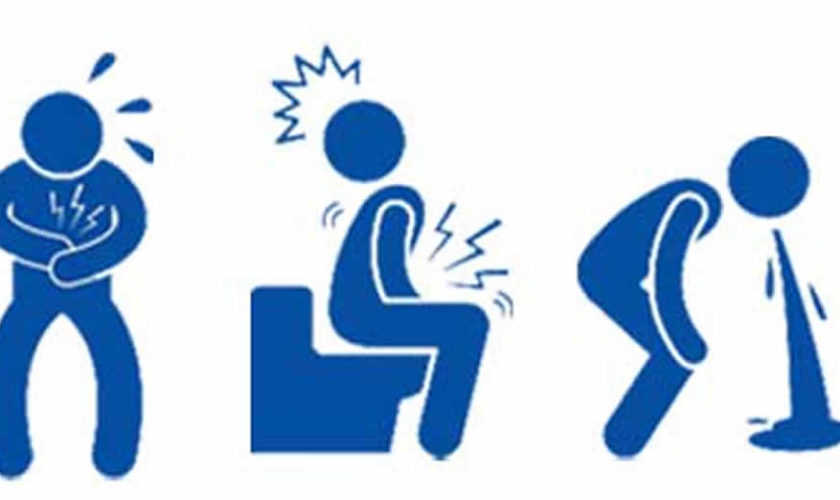
Symptoms of gastroenteritis include diarrhea, abdominal pain, vomiting, headache, fever and chills. Gastroenteritis is not usually serious but it can make you very dehydrated. Milder forms can be managed at home by drinking fluids. Most people recover with no treatment.

Stomach flu (gastroenteritis) may be caused by:

  • viruses (such as rotavirus or norovirus infections)
  • bacteria (including salmonella)
  • toxins produced by bacteria
  • parasites (such as giardia)
  • chemicals (such as toxins in poisonous mushrooms)

Gastroenteritis should only last for a few days. Stomach flu doesn’t usually require medication.

The most common problem with gastroenteritis is dehydration. This happens if you do not drink enough fluids to replace what you lose through vomiting and diarrhea. Dehydration is most common in babies, young children, the elderly and people with weak immune systems. Older people, young children and those with a weakened immune system are at risk of developing more serious illnesses.

It is very important to drink plenty of fluids. See a doctor immediately if your child cannot keep down a sip of liquid or has dehydration (dry mouth, no urine for 6 hours or more, or lethargy). If you are unwell with diarrhea or vomiting, you could have gastroenteritis. A doctor can diagnose gastroenteritis after talking to and examining you. If you’re not getting better, the doctor may want to do stool (poop) tests to find out what’s making you ill.

Stomach flu or viral gastroenteritis key points:

  • Viral gastroenteritis is an infection of the stomach and intestines, caused by a virus.
  • The main symptoms include diarrhea and vomiting.
  • Diarrhea and vomiting can cause a loss of fluids, also called dehydration.
  • If dehydration is severe, patients may have to be given fluid intravenously (IV) at the hospital.
  • Viral gastroenteritis can spread by sharing food, water and utensils. Frequent hand washing can help prevent the spread of infection to others.
  • Viral gastroenteritis is usually not a serious illness. However, people who have weak immune systems are at risk for more serious infection.

Viral gastroenteritis is NOT caused by any of the following, although the symptoms may be similar:

  • bacteria such as salmonella or E. coli
  • parasites such as Giardia
  • medications
  • other medical conditions
When to see a doctor

You should see your doctor if:

  • your child is very young or small (aged below 6 months or weighs less than 8 kg)
  • your child is born preterm, or has other chronic conditions
  • your child is passing less than 4 wet nappies/day
  • you or your child is passing any blood in the stool
  • you or your child is having dark green (bile) vomits
  • you or your child vomits blood
  • you or your child is having severe abdominal pain
  • you or your child less than 3 years old and has a fever more than 101.3 °F (38.5° C)
  • you or your child is showing signs of dehydration (very thirsty, cold hands and feet, dry lips and tongue, sunken eyes, sunken fontanelle, sleepy or drowsy)
  • you or your child is unable to tolerate any oral intake because of severe vomiting
  • you or your child becomes unusually drowsy
  • vomiting persists more than two days
  • diarrhea persists more than several days
  • diarrhea turns bloody
  • lightheadedness or fainting occurs with standing
  • confusion develops
  • worrisome abdominal pain develops

Flu shot, will it prevent the stomach flu?

The flu shot protects against influenza, which isn’t the same thing as the stomach flu (gastroenteritis). Gastroenteritis is an infection caused by a variety of viruses, including rotaviruses and noroviruses. Although it is often called the stomach flu, gastroenteritis is not caused by influenza viruses.

Influenza attacks your respiratory system — your nose, throat and lungs. Signs and symptoms of influenza may include:

  • Coughing
  • Congestion
  • Fever
  • Muscle aches

Gastroenteritis, on the other hand, attacks your intestines, causing signs and symptoms such as:

  • Diarrhea
  • Vomiting
  • Fever
  • Chills
  • Headache
  • Body aches

You can reduce your risk of influenza and gastroenteritis by washing your hands often with soap and water, as well as disinfecting contaminated and frequently touched surfaces.

The annual flu vaccine is the most effective way to reduce your risk of getting influenza or reducing its severity, if you do. Two oral rotavirus vaccines are available for young infants — RotaTeq and Rotarix, to protect against rotavirus gastroenteritis.

Food poisoning vs stomach flu

Food poisoning comes from eating foods that contain germs like bad bacteria or toxins. Bacteria are all around us, so mild cases of food poisoning are common. These can cause diarrhea and an upset stomach. When this happens, you might hear people calling it a stomach bug or stomach virus.

Bacteria are all around us, including in food, and sometimes they can be good for you (e.g., probiotics). You can learn how to avoid bad germs in food.

What are the signs of food poisoning?

Someone who has food poisoning might have:

  • an upset stomach (called nausea)
  • vomiting
  • stomach cramps
  • diarrhea, which may contain blood
  • a fever of 100.4° F (38 °C) or above
  • feeling generally unwell – such as feeling tired or having aches and chills

Sometimes feeling sick from food poisoning shows up within hours of eating the bad food. At other times, someone may not feel sick until several days later. With mild cases of food poisoning, you will not feel sick for very long and will soon be feeling fine again.

It can be hard to tell if you have food poisoning or something else. You might do a little detective work and see who else gets the same sickness. Did they eat the same thing you did? If only people who ate that food got sick, food poisoning could be the problem.

Which germs are to blame?

Foods from animals, raw foods, and unwashed vegetables all can contain germs that cause food poisoning. The most likely source is food from animals, like meat, poultry (such as chicken), eggs, milk, and shellfish (such as shrimp).

Some of the most common bacteria are:

  • Salmonella
  • Listeria
  • Campylobacter
  • E. coli
  • Norovirus

To avoid food poisoning, people need to prepare, cook, and store foods properly.

How you get food poisoning?

You can catch food poisoning if you eat something that has been contaminated with germs.

This can happen if food:

  • isn’t cooked or reheated thoroughly
  • isn’t stored correctly – for example, it’s not been frozen or chilled
  • is left out for too long
  • is handled by someone who’s ill or hasn’t washed their hands
  • is eaten after its “use by” date

How to treat food poisoning

You can usually treat yourself or your child at home. Read about how to treat diarrhea and vomiting yourself below.

The symptoms usually pass within a week.

The treatment you’ll get for food poisoning will depend on the germ that is making you sick. The doctor might give you medicine, but most of the time someone who has food poisoning doesn’t need to take medicine.

It’s also rare that a person with food poisoning would need to go to the hospital. Usually, only people who get really dehydrated have to go to the hospital. Being dehydrated means your body has lost too much fluid due to diarrhea and vomiting. A dehydrated person can get fluids and medicine through an IV at the hospital. To keep from getting dehydrated, try to keep drinking liquids when you’re sick.

You may also need to go to the hospital if you have blood in your poop. If you do see blood in your poop, you should definitely tell your doctor about it.

How can I prevent food poisoning?

Many things can be done to prevent food poisoning. These precautions should be taken at every stage a food takes — from preparation to cooking to storing leftovers. One of the best ways is to wash your hands if you’re preparing and making foods.

Before you start making foods you should wash your hands before — so germs from your hands don’t get on the food — and after so you don’t pass along germs from the food to yourself or anyone else.

Other ways to keep your food safe include:

  • Wash fruits and vegetables well before eating them.
  • Only eat foods that are properly cooked. If you cut into chicken and it looks pink and raw inside, tell a grown-up.
  • Look at what you’re eating and smell it too. If something looks or smells different from normal, don’t eat or drink it. Milk is a good example. If you’ve ever had a sip of sour milk, you know you never want to taste that again! Mold (which can be green, pink, white, or brown) is also often a sign that food has spoiled.
  • If you’re going to eat leftovers, make sure you heat them up. By heating them, you can kill bacteria that grew while it was in the fridge.
  • Check the date. Lots of packaged foods have expiration dates or “sell by” dates (which means that the food should leave store shelves by that time). Don’t eat a food if today’s date is after the expiration date. Use it before it expires.
  • Cover and refrigerate food right away. Bacteria get a good chance to grow in foods that sit at room temperature. By putting food in the fridge, you’re putting the chill on those bad germs!

Stomach flu contagious

Stomach flu (gastroenteritis) spreads easily from having contact with an infected person (or their vomit or stools). Stomach flu (gastroenteritis) can also spread via contaminated food or water.

How long am I contagious if I have the stomach flu?

You can be contagious from a few days up to two weeks or more, depending on which virus is causing your stomach flu (gastroenteritis).

A number of viruses can cause gastroenteritis, including noroviruses and rotaviruses. The contagious period — the time during which a sick person can give the illness to others — differs slightly for each virus.

  • Norovirus. With norovirus — the most common cause of viral gastroenteritis in adults — you’re contagious when you begin to feel ill. Symptoms usually appear within one to two days of exposure. Although you typically feel better after a day or two, you’re contagious for a few days after you recover. The virus can remain in your stool for up to two weeks or more after recovery. Children should stay home from school or child care for at least 48 hours after the last time they vomit or have diarrhea.
  • Rotavirus. Symptoms of rotavirus — the leading cause of viral gastroenteritis in infants and young children — usually appear one to three days after exposure. But you’re contagious even before you develop symptoms, and up to two weeks after you’ve recovered.

The viruses that cause gastroenteritis are spread through close contact with infected people, such as by sharing food or eating utensils, and by touching contaminated surfaces and objects. Eating contaminated food also can cause norovirus.

Washing your hands often with soap and water is the most effective way to stop the spread of these viruses to others. If you can’t wash your hands, use an alcohol-based hand sanitizer, which can reduce germs.

To help keep others from getting sick, disinfect contaminated surfaces immediately after someone vomits or has diarrhea. Wear disposable gloves, and use a bleach-based household cleanser or 2 cups (0.5 liters) of bleach in a gallon (3.8 liters) of water. Norovirus can survive for months on surfaces not adequately disinfected with bleach solution.

Also wear disposable gloves to immediately wash clothes or linens that might be contaminated.

How long does stomach flu last?

Depending on the cause of the gastroenteritis, symptoms may last from one day to more than a week.

Common causes of gastroenteritis are:

  • Viruses (rotaviruses and noroviruses).
  • Food or water contaminated by bacteria or parasites.
  • Reaction to a new food. Young children may develop signs and symptoms for this reason. Infants who are breast-fed may even react to a change in their mothers’ diets.
  • Side effect from medications.

How to treat diarrhea and vomiting yourself

You can usually treat yourself or your child at home.

The most important thing is to have plenty of fluids to avoid dehydration.

DO

  • stay at home and get plenty of rest
  • drink lots of fluids, such as water and squash – take small sips if you feel sick
  • carry on giving breast or bottle feeds to your baby – if they’re being sick, try giving small feeds more often than usual
  • for babies on formula or solid foods, give small sips of water between feeds
  • eat when you feel able to – you don’t need to have or avoid any specific foods
  • take paracetamol if you’re in discomfort – check the leaflet before giving them to your child

DON’T

  • have fruit juice or fizzy drinks – they can make diarrhea worse
  • make baby formula weaker – use it at its usual strength
  • give young children medicine to stop diarrhea
  • give aspirin to children under 16

How to treat diarrhea and vomiting in children

The main treatment is to give enough fluids to prevent your child becoming dehydrated. Babies and children below 3 years old are most at risk and may need to be checked by a doctor. Give small amounts of fluids frequently as they can usually tolerate this better than large volumes at a time. You should continue to give fluids even if they are vomiting. Many common medicines to reduce vomiting or diarrhea are often not helpful and may instead be harmful in children. Antibiotic treatment is also unnecessary and unhelpful in most cases because the infection is usually caused by viruses which do not respond to this treatment.

If you’re breast-feeding, let your baby nurse. If your baby is bottle-fed, offer a small amount of an Oral Rehydration Solutions (ORS) or regular formula.

Consider acetaminophen (Tylenol, others) for relief of discomfort, unless your child has liver disease. Don’t give your child aspirin.

What fluids to use

The best fluids to use are Oral Rehydration Solutions (ORS), e.g., CeraLyte, Enfalyte, Pedialyte, Hydralyte, Gastrolyte, Repalyte etc., which are available from your local chemist. They contain glucose and different salts which tend to be lost from the body during vomiting or diarrhea. Make them up EXACTLY as it says on the packet. Breast fed babies should continue to be breastfed but may need to be fed more frequently. Oral Rehydration Solutions (ORS) or water (boiled if the baby is less than 6 months old) may be offered to babies in addition to breast feeds. Bottle fed babies may need to have both Oral Rehydration Solutions (ORS) and their formula at normal strength.

What can I do if my child refuse to take the oral rehydration solution?

Chilling the fluids or making them into iceblocks may help your child to take them. Some children may still refuse to drink. In this situation water or other fluids such as diluted juice or soft drinks may be given, although they are not as good as Oral Rehydration Solutions (ORS) because they don’t have all the extra salts in the right amounts and have sugars which are not as well absorbed.

DO NOT GIVE UNDILUTED juice, sodas, sports drinks or other soft drinks as they have too much sugar and may make the diarrhea worse. Chicken broth is also not recommended as it has too much salt and no sugar.

How much fluid does my child need?

This depends on the age and size of the child and also how dehydrated they are.

The minimum daily requirements in children are:

  • 3-10kg (1-12months): 100ml/kg
  • 10-20kg (1-5yrs): 1000ml + 50ml/kg for each kg over 10kg
  • >20kg: 1500ml + 20ml/kg for each kg over 20kg

You may also need to give an extra 2ml/kg for every vomit and 10ml/kg for each diarrheal stool in addition to the maintenance amount of fluids required.
Give small volumes frequently, e.g. 5ml (1tsp) every 5 minutes, is better tolerated than 60ml all at once every hour.

What about eating food?

Doctors no longer recommend restricting food intake during gastroenteritis. Your child may not feel like eating initially but should be allowed to eat once they feel hungry. Gradually introduce bland, easy-to-digest foods, such as toast, rice, bananas and potatoes. Avoid giving your child full-fat dairy products, such as whole milk and ice cream, and sugary foods, such as sodas and candy. These can make diarrhea worse.

Bottle fed babies on infant formula should be given their formula at normal strength and not diluted down. The only foods to avoid are those with high sugar content such as undiluted juice, cordials, soft drinks, jelly, jam, sweets, chocolate etc. as they may make the diarrhea worse.

Lactose intolerance is uncommon in young American children but may occur temporarily after a bout of gastroenteritis. This may be suspected if their diarrhea worsens and is watery, frothy and explosive after drinking milk or formula. If this occurs, then a lactose free or soy formula may be used for a few weeks until the gut recovers.

Viral gastroenteritis

Viral gastroenteritis is an intestinal infection marked by watery diarrhea, abdominal cramps, nausea or vomiting, and sometimes fever.

The most common way to develop viral gastroenteritis — often called stomach flu — is through contact with an infected person or by ingesting contaminated food or water. If you’re otherwise healthy, you’ll likely recover without complications. But for infants, older adults and people with compromised immune systems, viral gastroenteritis can be deadly.

There’s no effective treatment for viral gastroenteritis, so prevention is key. In addition to avoiding food and water that may be contaminated, thorough and frequent hand-washings are your best defense.

Although viral gastroenteritis is commonly called stomach flu, gastroenteritis isn’t the same as influenza. Real flu (influenza) affects only your respiratory system — your nose, throat and lungs. Gastroenteritis, on the other hand, attacks your intestines, causing signs and symptoms, such as:

  • Watery, usually nonbloody diarrhea — bloody diarrhea usually means you have a different, more severe infection
  • Abdominal cramps and pain
  • Nausea, vomiting or both
  • Occasional muscle aches or headache
  • Low-grade fever

Depending on the cause, viral gastroenteritis symptoms may appear within one to three days after you’re infected and can range from mild to severe. Symptoms usually last just a day or two, but occasionally they may persist as long as 10 days.

Because the symptoms are similar, it’s easy to confuse viral diarrhea with diarrhea caused by bacteria, such as Clostridium difficile, salmonella and E. coli, or parasites, such as giardia.

Viral gastroenteritis causes

You’re most likely to contract viral gastroenteritis when you eat or drink contaminated food or water, or if you share utensils, towels or food with someone who’s infected.

A number of viruses can cause gastroenteritis, including:

  • Noroviruses. Both children and adults are affected by noroviruses, the most common cause of foodborne illness worldwide. Norovirus infection can sweep through families and communities. It’s especially likely to spread among people in confined spaces. In most cases, you pick up the virus from contaminated food or water, although person-to-person transmission also is possible.
  • Rotavirus. Worldwide, this is the most common cause of viral gastroenteritis in children, who are usually infected when they put their fingers or other objects contaminated with the virus into their mouths. The infection is most severe in infants and young children. Adults infected with rotavirus may not have symptoms, but can still spread the illness — of particular concern in institutional settings because infected adults unknowingly can pass the virus to others. A vaccine against viral gastroenteritis is available in some countries, including the United States, and appears to be effective in preventing the infection.

Some shellfish, especially raw or undercooked oysters, also can make you sick. Although contaminated drinking water is a cause of viral diarrhea, in many cases the virus is passed through the fecal-oral route — that is, someone with a virus handles food you eat without washing his or her hands after using the toilet.

Risk factors for getting gastroenteritis

Gastroenteritis occurs all over the world, affecting people of every age, race and background.

People who may be more susceptible to gastroenteritis include:

  • Young children. Children in child care centers or elementary schools may be especially vulnerable because it takes time for a child’s immune system to mature.
  • Older adults. Adult immune systems tend to become less efficient later in life. Older adults in nursing homes, in particular, are vulnerable because their immune systems weaken and they live in close contact with others who may pass along germs.
  • Schoolchildren, churchgoers or dormitory residents. Anywhere that groups of people come together in close quarters can be an environment for an intestinal infection to get passed.
  • Anyone with a weakened immune system. If your resistance to infection is low — for instance, if your immune system is compromised by HIV/AIDS, chemotherapy or another medical condition — you may be especially at risk.

Each gastrointestinal virus has a season when it’s most active. If you live in the Northern Hemisphere, for instance, you’re more likely to have rotavirus or norovirus infections between October and April.

Viral gastroenteritis complications

The main complication of viral gastroenteritis is dehydration — a severe loss of water and essential salts and minerals. If you’re healthy and drink enough to replace fluids you lose from vomiting and diarrhea, dehydration shouldn’t be a problem.

Infants, older adults and people with suppressed immune systems may become severely dehydrated when they lose more fluids than they can replace. Hospitalization might be needed so that lost fluids can be replaced intravenously. Dehydration can be fatal, but rarely.

Viral gastroenteritis prevention

The best way to prevent the spread of intestinal infections is to follow these precautions:

  • Get your child vaccinated. A vaccine against gastroenteritis caused by the rotavirus is available in some countries, including the United States. Given to children in the first year of life, the vaccine appears to be effective in preventing severe symptoms of this illness.
  • Wash your hands thoroughly. And make sure your children do, too. If your children are older, teach them to wash their hands, especially after using the toilet. It’s best to use warm water and soap and to rub hands vigorously for at least 20 seconds, remembering to wash around cuticles, beneath fingernails and in the creases of the hands. Then rinse thoroughly. Carry towelettes and hand sanitizer for times when soap and water aren’t available.
  • Use separate personal items around your home. Avoid sharing eating utensils, glasses and plates. Use separate towels in the bathroom.
  • Keep your distance. Avoid close contact with anyone who has the virus, if possible.
  • Disinfect hard surfaces. If someone in your home has viral gastroenteritis, disinfect hard surfaces, such as counters, faucets and doorknobs, with a mixture of two cups of bleach to one gallon of water.
  • Check out your child care center. Make sure the center has separate rooms for changing diapers and preparing or serving food. The room with the diaper-changing table should have a sink as well as a sanitary way to dispose of diapers.

Take precautions when traveling

When you’re traveling in other countries, you can become sick from contaminated food or water. You may be able to reduce your risk by following these tips:

  • Drink only well-sealed bottled or carbonated water.
  • Avoid ice cubes, because they may be made from contaminated water.
  • Use bottled water to brush your teeth.
  • Avoid raw food — including peeled fruits, raw vegetables and salads — that has been touched by human hands.
  • Avoid undercooked meat and fish.

Gastroenteritis prevention

To reduce your risk of catching or spreading gastroenteritis, wash your hands well after using the bathroom or changing nappies, and before preparing or eating food.

If you have gastroenteritis, it’s important to stay home (away from work, school or childcare) until the symptoms have been gone for at least 24 hours. If your work involves handling food or looking after children, the elderly, or patients, do not return to work until 48 hours after the symptoms have stopped.

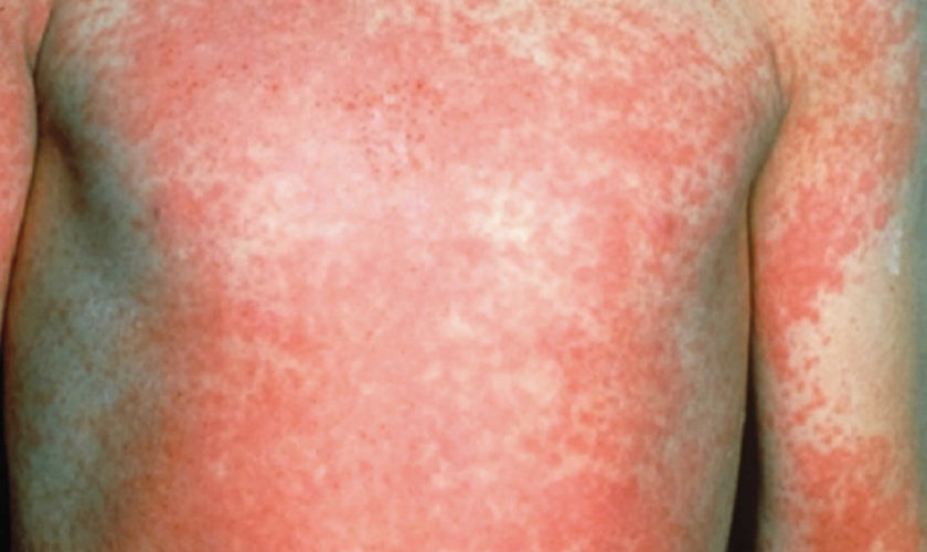
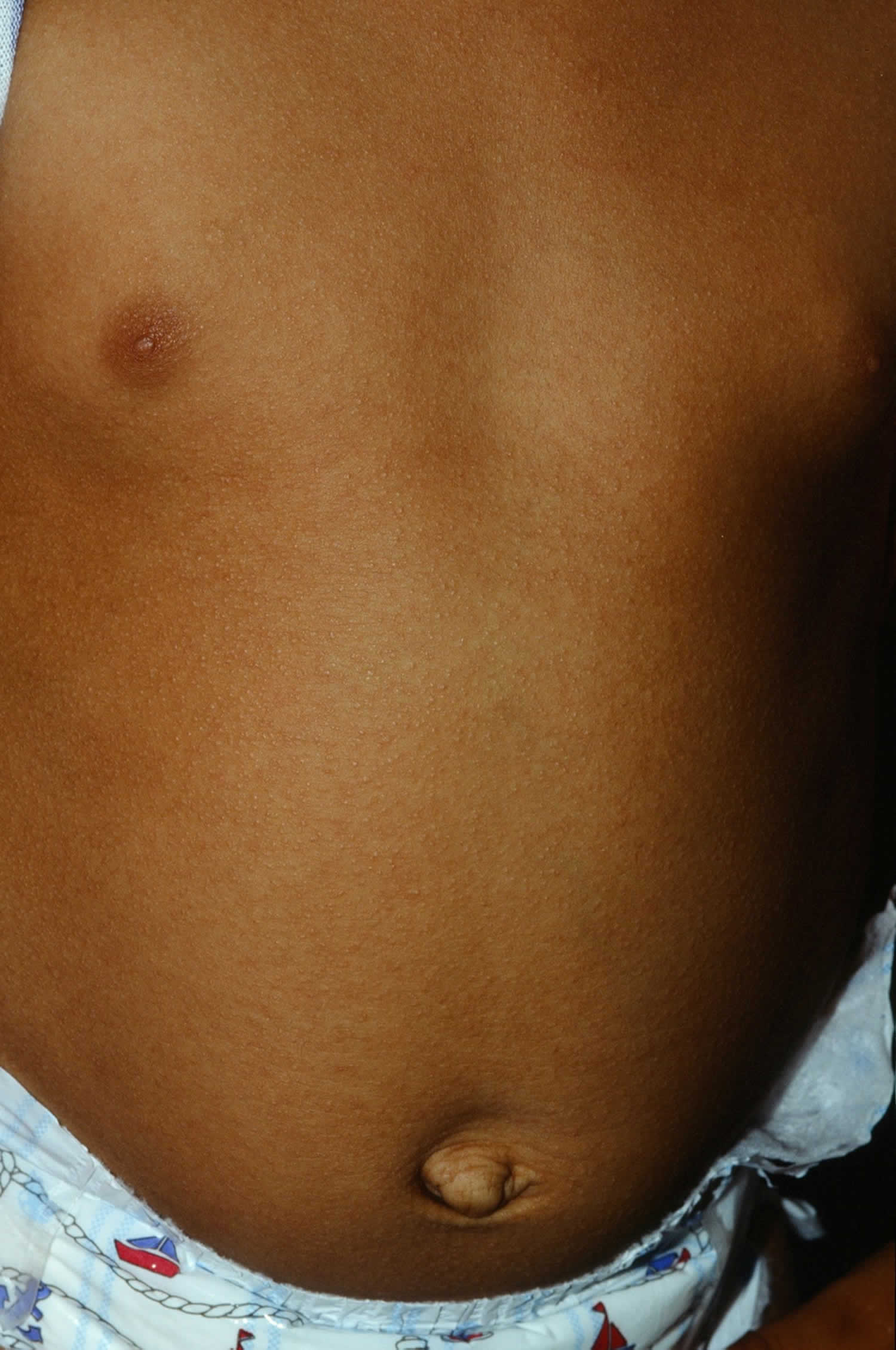
Rotavirus spreads easily among infants and young children. The virus can cause severe watery diarrhea, vomiting, fever, and abdominal pain. Children who get rotavirus disease can become dehydrated and may need to be hospitalized.

Two oral rotavirus vaccines are available for young infants — RotaTeq® (RV5) and Rotarix® (RV1). Vaccines for norovirus are in clinical trials.

Rotavirus vaccines

The best way to prevent rotavirus in children is to have them vaccinated. Rotavirus vaccine is the best way to protect your child against rotavirus disease. Most children (about 9 out of 10) who get the vaccine will be protected from severe rotavirus disease. About 7 out of 10 children will be protected from rotavirus disease of any severity.

Two rotavirus vaccines are currently licensed for infants in the United States:

  • RotaTeq® (RV5) is given in 3 doses at ages 2 months, 4 months, and 6 months
  • Rotarix® (RV1) is given in 2 doses at ages 2 months and 4 months

Both vaccines are given by putting drops in the child’s mouth. Each requires multiple doses.

The first dose of either vaccine should be given before a child is 15 weeks of age. Children should receive all doses of rotavirus vaccine before they turn 8 months old.

There is a very small risk that babies may develop a bowel problem called intussusception after receiving the vaccine.

Possible side effects of Rotavirus vaccine

Most babies who get rotavirus vaccine do not have any side effects. However, some babies can have side effects that are usually mild and go away on their own. Serious side effects are possible but rare.

Side effects or problems that have been associated with rotavirus vaccine include:

  • Mild problems: Being irritable, or having mild, temporary diarrhea or vomiting after getting a dose of rotavirus vaccine.
  • Serious problems: There is a small risk of intussusception, a type of bowel blockage that is treated in a hospital, and could require surgery. Intussusception happens in some babies every year in the United States, and usually there is no known reason for it. Intussusception from rotavirus vaccination usually occurs within a week of receiving a dose of vaccine. The risk of intussusception from rotavirus vaccination is estimated to range from about 1 in 20,000 to 1 in 100,000 US infants who get rotavirus vaccine. Your doctor can give you more information.
  • Problems that could happen after any vaccine: Any medication can cause a severe allergic reaction. Such reactions from a vaccine are very rare, estimated at less than 1 in a million doses, and usually happen within a few minutes to a few hours after the vaccination. As with any medicine, there is a very remote chance of a vaccine causing a serious injury or death.

Who should NOT get Rotavirus vaccine?

Your healthcare provider is the best source of information on the benefits and risks of vaccines. Before your child receives any vaccine, discuss with your healthcare provider:

  • health problems that your child may have
  • medications that your child is currently taking
  • concerns you might have about vaccination

Infants should NOT get rotavirus vaccine if they have any of the following:

  • a severe (life-threatening) allergic reaction to an earlier dose of rotavirus vaccine,
  • a severe (life threatening) allergy to any component of rotavirus vaccine. Tell your doctor if your baby has any severe allergies that you know of, including a severe allergy to latex,
  • severe combined immunodeficiency (SCID), a condition in which a child’s immune system cannot fight infections, or
  • a previous episode of a type of bowel blockage called intussusception.

Infants who are moderately or severely ill should wait to get the vaccine until they recover. This includes infants with moderate or severe diarrhea or vomiting. Babies who are mildly ill can get the vaccine.

Check with your doctor before vaccinating if your baby’s immune system is weakened because of:

  • HIV/AIDS, or any other disease that affects the immune system
  • Treatment with drugs such as steroids
  • Cancer, or cancer treatment with x-rays or drugs

Can Rotavirus vaccine be given with other vaccines?

Rotavirus vaccine can be safely given during the same doctor’s visit with DTaP vaccine, Hib vaccine, polio vaccine, hepatitis B vaccine, and pneumococcal conjugate vaccine.

Stomach flu symptoms

Someone with gastroenteritis may have:

  • vomiting
  • diarrhea
  • nausea (feeling sick in the stomach)
  • stomach pains
  • low grade fever
  • headaches
  • no appetite

Vomiting often occurs at the start of the illness and may last 2-3 days. Diarrhea, which is often runny may last up to 10 days. Your child may also have a fever and abdominal (tummy) pains with the illness.

Gastroenteritis diagnosis

Your doctor will likely diagnose gastroenteritis based on symptoms, a physical exam and sometimes on the presence of similar cases in your community. A rapid stool test can detect rotavirus or norovirus, but there are no quick tests for other viruses that cause gastroenteritis. In some cases, your doctor may have you submit a stool sample to rule out a possible bacterial or parasitic infection.

How to treat stomach flu

There’s no specific medicine for stomach flu (gastroenteritis). Antibiotics aren’t effective against viruses, and overusing them can contribute to the development of antibiotic-resistant strains of bacteria. The most important treatment for gastroenteritis is to drink fluids. Frequent sips are easier for young children than a large amount all at once. Keep drinking regularly even if you are vomiting.

If you have a baby or young child with gastroenteritis, it’s a good idea to have them checked by a doctor for dehydration. You can get rehydration fluids from a pharmacy. These are the best fluids to use in cases of gastroenteritis, especially for children.

If you can’t get any, or your child refuses to drink it, giving diluted fruit juice (one part juice to four parts of water) is reasonable. You could try a cube of ice or an iceblock if your child won’t drink. Avoid milk and other dairy products and do not give juice, sodas, sports drinks or other soft drinks as the sugar may make the diarrhea worse. It is fine to eat once you feel like it.

Babies can continue milk feeds throughout the illness, with rehydration fluid between feeds. Medication for nausea or diarrhea can be useful for adults, but may not be safe for kids. Antibiotics are rarely helpful.

If you are very sick with gastroenteritis, you may need to go to hospital where you may be put on a drip.

Home remedies

To help keep yourself more comfortable and prevent dehydration while you recover, try the following:

  • Let your stomach settle. Stop eating solid foods for a few hours.
  • Try sucking on ice chips or taking small sips of water. You might also try drinking clear soda, clear broths or noncaffeinated sports drinks. Drink plenty of liquid every day, taking small, frequent sips.
  • Ease back into eating. Gradually begin to eat bland, easy-to-digest foods, such as soda crackers, toast, gelatin, bananas, rice and chicken. Stop eating if your nausea returns.
  • Avoid certain foods and substances until you feel better. These include dairy products, caffeine, alcohol, nicotine, and fatty or highly seasoned foods.
  • Get plenty of rest. The illness and dehydration may have made you weak and tired.
  • Be cautious with medications. Use many medications, such as ibuprofen (Advil, Motrin IB, others), sparingly if at all. They can make your stomach more upset. Use acetaminophen (Tylenol, others) cautiously; it sometimes can cause liver toxicity, especially in children. Don’t give aspirin to children or teens because of the risk of Reye’s syndrome, a rare, but potentially fatal disease. Before choosing a pain reliever or fever reducer discuss with your child’s pediatrician.

For infants and children

When your child has an intestinal infection, the most important goal is to replace lost fluids and salts. These suggestions may help:

  • Help your child rehydrate. Give your child an oral rehydration solution, available at pharmacies without a prescription. Talk to your doctor if you have questions about how to use it. Don’t give your child plain water — in children with gastroenteritis, water isn’t absorbed well and won’t adequately replace lost electrolytes. Avoid giving your child apple juice for rehydration — it can make diarrhea worse.
  • Get your child back to a normal diet slowly. Gradually introduce bland, easy-to-digest foods, such as toast, rice, bananas and potatoes.
  • Avoid certain foods. Don’t give your child dairy products or sugary foods, such as ice cream, sodas and candy. These can make diarrhea worse.
  • Make sure your child gets plenty of rest. The illness and dehydration may have made your child weak and tired.
  • Avoid giving your child over-the-counter anti-diarrheal medications, unless advised by your doctor. They can make it harder for your child’s body to eliminate the virus.

If you have a sick infant, let your baby’s stomach rest for 15 to 20 minutes after vomiting or a bout of diarrhea, then offer small amounts of liquid. If you’re breast-feeding, let your baby nurse. If your baby is bottle-fed, offer a small amount of an oral rehydration solution or regular formula. Don’t dilute your baby’s already-prepared formula.

read more

Temper tantrums

temper-tantrums

What is temper tantrums

Tantrums also called temper tantrums, are extremely common in toddlers and preschoolers. Tantrums or temper tantrums are how young children deal with difficult feelings. It helps to tune in to your child’s emotions, and to avoid situations that trigger your child’s tantrums.

Temper tantrums usually start at around 18 months and are very common in toddlers. Temper tantrums are a normal part of child development and most often occur in kids between the ages of 2 and 3.

Tantrums come in all shapes and sizes. Temper tantrums can involve spectacular explosions of anger, frustration and disorganized behavior – when your child ‘loses it’.

You might see crying, screaming, stiffening limbs, an arched back, kicking, falling down, flailing about or running away. In some cases, children hold their breath, clench their teeth, vomit, break things, pound their fists or get aggressive as part of a tantrum. Hitting and biting are common, too.

Boys and girls both have tantrums and your child’s personality will also play a part.

Some children are naturally easygoing and positive, whereas others who are very active, intense and persistent may have more intense tantrums.

Tantrums tend to occur more often if a child is anxious, ill, moody, tired or lives in a stressful home.

One reason for this is toddlers want to express themselves, but find it difficult. They feel frustrated, and the frustration comes out as a tantrum.

Once a child can talk more, they’re less likely to have tantrums. By the age of four, tantrums are far less common.

Children’s brains develop as they grow. The section of the brain that is ‘firing’ at the age children commonly have a lot of tantrums (big feelings) is the limbic system (the emotional center of the brain). Young children see their world and react to many everyday situations through this emotional lens.

Until their cerebral cortex (the reasoning and thinking part of the brain) is developed, a young child’s only way of telling you about what is bothering them is through expressing their feelings – such as frustration about being misunderstood or not being able to make their needs clear, or being upset.

Some triggers that can spark a young child’s big feelings (tantrums) include being stressed, hungry, tired, frustrated or overstimulated.

Triggers for tantrums (big feelings)

It is important to remember that tantrums (big feelings) are a normal part of child development. However, certain factors make episodes of tantrums (big feelings) more likely.

Triggers that may spark tantrums (big feelings) include being:

  • stressed
  • hungry
  • tired
  • overstimulated – for example, by loud or noisy environments where there is a lot going on
  • frustrated – especially about not being understood or not having enough language skills to communicate needs
  • physically ill
  • upset – for example, because a parent has reacted angrily or laughed at the child
  • confused – for example, by inconsistent parenting or caregivers reacting differently to the big feelings in different circumstances.

If your child is expressing tantrums (big feelings), remember that:

  • these feelings are frightening to your child – they want to avoid them as much as you do, but lack the necessary skills to cope
  • they are not ‘doing this on purpose’
  • they need your calming influence to help them through it
  • they cannot calm themselves on their own
  • when the big feelings (tantrums) have passed, they need to know they are still loved.

While children are still too young to regulate their own emotions and behavior, adults can help them deal with big feelings (tantrums) by ‘co-regulating’ their mood – for example by being calm, soothing, caring, close and rational until the big feeling (tantrums) passes. This helps young children regain their dignity and know they are still loved.

Young children need reassurance, nurturing and understanding from adults, as they do not understand their big feelings and are not able to manage them on their own.

Self-regulation is the ability to understand and manage behavior and reactions. Children start developing it from around 12 months. As your child gets older, he/she will be more able to regulate his/her reactions and calm down when something upsetting happens. You’ll see fewer tantrums as a result.

Big feelings (tantrums) and child development

Young children are emotional beings and act out their feelings through their behavior. They do not have the cognitive thinking abilities to express themselves in a rational way. When the cortex is not developed, young children are unable to calm themselves on their own.

Young children need (calm and rational) adults to support and help them through their big feeling episodes and to regain a sense of calm and dignity. When a young child is being emotional, it is important that the adults are responding calmly and rationally.

Expressions of big feelings (tantrums) tend to occur at the age when young children do not have the ability to handle their feelings without adult help (co-regulation). This often occurs between the ages of 18 months and four years, but each child is different. Some children don’t need to express themselves through big feelings at all.

Older children learn how to reduce their own heightened emotional state without adult help, when the cognitive part of their brain is more developed.

Tips for preventing outbursts of tantrums (big feelings)

General suggestions include:

  • Spend time together doing enjoyable things when your child is calm.
  • Make sure your child gets enough rest and sleep.
  • Offer regular meals, and healthy snacks and drinks.
  • Find ways to help your child communicate – for example, encourage them to point at things. Two-year-olds speak only about 50 words. Lack of communication skills is thought to be a trigger for big feelings in younger children.
  • Talk and read to your child as much as possible to help with their speech development and communication skills.
  • Allow your child control over safe things – for example, let them choose which fruit to have at snack time or which toy to take to bed. This gives them a feeling of independence and control over one aspect of their world.
  • Think about your child’s request before you refuse it – for example, is it really that unreasonable for your child to have a small treat after they have grocery shopped with you, without complaining, for over an hour? It may be that your child’s request can be accommodated.
  • Keep your parenting consistent – for example, don’t change what you expect as reasonable behavior just because you feel tired and it seems easier to give in. It’s not easier in the long run.
  • Give your child plenty of attention and compliment them when they are behaving well.
  • Take note of which factors and events trigger these expressions of feelings and think up ways to cope. For example, if your child ‘loses it’ when they are in the car, plan for car travel to be more enjoyable by playing your child’s favorite music and stocking the car with special toys.
  • Take notice of your child’s behavior before a big feeling so that you can step in and avoid them altogether in the future – for example, an afternoon nap or distraction with a favorite storybook may work if tiredness is a trigger.
  • Encourage your child to use words to express their wants and needs as much as they are able to. Praise them for any attempts to use words.
  • Reward your child for coping with frustration – for example, using words to communicate wants and needs instead of kicking or screaming.

Tips for coping with outbursts of tantrums

Dealing with tantrums can be very draining and stressful. You might feel you need to step in to end a tantrum straight away. But if it’s safe, it can help to take a breather while you decide how to respond.

  • Don’t judge yourself as a parent based on how many tantrums your child has. Remember that all children have tantrums. Instead, focus on how you respond to the tantrums. And remember that you’re only human and part of parenting is learning from mistakes.

Top tantrum tips coping when your child is expressing big feelings (tantrums):

  • Keep calm – model the behavior that you want your child to display.
  • Try to not get angry and don’t resort to smacking or hitting your child.
  • Use distraction whenever possible – with a book or song, or anything else going on nearby, this is particularly effective for younger children who have short attention spans.
  • If you know that it helps, and you are somewhere that it is safe to do so, allow your child some space and time on their own until they calm down. Perhaps they may need to stay in their room.
  • Some children become more traumatized when left alone. If this is the case, keep them close by and make sure they are safe. Console them as soon as the big feeling is over.
  • Recognize when the big feeling has subsided and console the child immediately to reassure them they are okay, and that you love them.
  • Avoid giving in to their demands. If your child is having a big feeling because they don’t want to do something – for example, have a bath – wait until they are calm. Then tell them that it’s good that they’ve calmed down, but they still need a bath.
  • Try not to lose your temper. If you feel that you are becoming angry, distract yourself. If it is safe to do so, leave the room, play music, read a magazine or do anything else that works for you.
  • If you find you can’t calm down and your child is safe, move away for a while until you feel better.
  • It’s important to make sure you don’t accidentally reward tantrums. For example, if your child has a tantrum because you say no to buying her a lolly but then you buy the lolly, this rewards the tantrum. Shouting or pleading with your child when she has tantrums can also be a reward, because it gives your child attention.

Staying calm

It’s really important that you stay calm when your child is having a tantrum. This can be very hard to do but if you become stressed too your child will pick up on it. Keeping calm and in control shows your toddler that you are not overwhelmed by his or her emotions and while he or she feels out of control, you are in control. During her tantrum he or she may not be able to hear you but sometimes by speaking slowly and quietly, you can help calm the situation. Try to avoid worrying about what other people will think if you’re out in public – if you stay calm even if your child is screaming the place down they will more likely carry on by as you are dealing with the situation. Many of them will have had children themselves and know what you are going through.

Here are some more ideas for staying calm and keeping things in perspective:

  • Develop a strategy for tantrums. Have a clear plan for how you’ll handle a tantrum in whatever situation you’re in. Concentrate on putting your plan into action when the tantrum happens.
  • Accept that you can’t control your child’s emotions or behavior directly. You can only keep your child safe and guide your child’s behavior so tantrums are less likely to happen in the future.
  • Accept that it takes time for change to happen. Your child has a lot of growing up to do before tantrums are gone forever. Developing and practising self-regulation skills is a life-long task.
  • Beware of thinking that your child is doing it on purpose or is trying to get you. Children don’t have tantrums deliberately – they’re stuck in a bad habit or just don’t have the skills right now to cope with the situation.
  • Keep your sense of humor. But don’t laugh at the tantrum – if you do, it might reward your child with attention. It might also upset him even more if he thinks you’re laughing at him.
  • If other people give you dirty looks, ignore them. They’ve either never had children or it’s been so long since they had a young child that they’ve forgotten what it’s like.

If you can’t stay calm

Sometimes it can be really hard to stay calm when your toddler is having a tantrum and if you really feel like you can’t and if your child is in a safe place, just move away for a moment until you feel calmer.

Distract your child

Help your child calm down by distracting them with something else, such as reading a book, or something else to look at where they are like a bus going past. If you do something like giving them treats in the hope of calming them down, this may be quick fix but in can end up with your child thinking that a tantrum will be rewarded. Have a drink or snack with you in case a child is genuinely hungry or thirsty. If you want to try distracting him or her before the full blown tantrum, you may want to have or toy or something handy.

Give them a hug

Sometimes a child having a tantrum may just want your attention and giving the child a hug might help. However, this will not work if the child has already too far gone in the tantrum. Sometimes this can make the situation worse. A hug may not stop a tantrum, but holding a child firmly and gently while talking to him or her in a clear voice may help the child understand that you are not giving in to the tantrum. You and your child may enjoy a loving cuddle after the tantrum has subsided. You may need to explain that you know she was angry but still the behavior was not acceptable. Let him or her know what they can do next time when they are feeling frightened or angry. Give your child the words to let you know how he or she feels.

After the tantrum

Do not reward your child after a tantrum by giving in to their demands. This will only prove to your child that the tantrum was effective. Instead, praise your child for regaining control.

Children may feel vulnerable after a tantrum when they may know their behavior was not very desirable. This is a time for a hug and reassurance that your child is loved, no matter what.

Asking for help

If you feel like you just can’t cope, wherever possible, it is better to ask for help than to keep everything bottled up and suffer alone. This will help to alleviate your own distress and will help you feel more able to deal with your toddler. It’s also good to talk to someone such as a friend, or you can call your family doctor.

Try give yourself a break sometimes:

  • go for a walk or swim
  • sit down with a cuppa
  • read the paper
  • watch your favorite TV program

These suggestions will give you a chance to recharge batteries. If it feels like you just can’t think straight, try making an appointment with your doctor to see if there is any help you can get locally.

How to manage tantrums in public

Sometimes, your child will express big feelings (tantrums) in a public place such as a supermarket. The humiliation of having strangers judge your parenting performance can ruin your strategy, but try not to give in.

Suggestions for managing expressions of big feelings in public include:

  • Remember that everyone who is a parent will be feeling for you. Reassure yourself that most onlookers understand what you are going through.
  • Stick to your tantrum strategy no matter what. Stay calm.
  • Try not to lose your temper. Screaming at your child or hitting them may provoke outrage from onlookers, which will only make you feel worse and probably make the big feeling last even longer. We are trying to teach our children to deal with their emotional distress – this does not happen if adults cannot control theirs.
  • In public places or when the child is in danger of hurting themselves, pick your child up and take them to a quiet, safe place to calm down.
  • Leave the shop and go home if the tantrum is severe or prolonged. Both of you may need ‘time out’ at home. (You could finish your shopping online.)
  • Don’t put yourself down or lose hope if you do give in to your child’s big feelings. Just try to stick to your plans next time.

Tips for coping with strong expressions of tantrums in young children

Some children have the temperament and strength to express their big feelings (tantrums) often, or to extend their big feelings (tantrums) for a very long time, or both. This can fray a parent’s patience and turn family life upside down.

Suggestions include:

  • Keep calm – model the behavior that you want your child to display.
  • Plan your strategy in advance. Use the same strategy every time a big feeling starts. Discuss your strategy with other caregivers to ensure consistency.
  • If you know that it helps, and you are somewhere that it is safe to do so, allow your child some space and time on their own until they calm down. Perhaps they may need to stay in their room.
  • If your child becomes more traumatized when left alone, keep them close by and make sure they are safe. Console them as soon as the big feeling is over.
  • The child may be quite distressed at their own behavior.
  • Control your temper by distracting yourself. Make sure your child is safe, then leave the room, play music, read a magazine or do anything else that works for you.
  • Avoid changing the family routine because of your child’s big feelings. Remind yourself that your child will soon be able to manage their feelings in a more appropriate way as they grow older.
  • Seek professional help if your attempts don’t get results. Your doctor is a good starting point for information and referral.

Why tantrums happen?

Tantrums are very common in children aged 1-3 years. Tantrums mean your child is overwhelmed by their feelings. Tantrums means they need your help.

To some extent, tantrums are attention-seeking behavior. They often happen when children are tired, hungry or uncomfortable and want attention from parents or caregivers.

This is because children’s social and emotional skills are only just starting to develop at this age. Children often don’t have the words to express big emotions. They want more independence but fear being separated from you. And they’re discovering that they can change the way the world works.

So tantrums are one of the ways that young children express and manage feelings, and try to understand or change what’s going on around them.

Older children can have tantrums too. This can be because they haven’t learned more appropriate ways to express or manage feelings. Or some older children might be slower than others to develop self-regulation.

For both toddlers and older children, there are things that can make tantrums more likely to happen:

  • Temperament – this influences how quickly and strongly children react to things like frustrating events. Children who get upset easily might be more likely to have tantrums.
  • Stress, hunger, tiredness and overstimulation – these can make it harder for children to express and manage feelings and behavior.
  • Situations that children just can’t cope with – for example, a toddler might have trouble coping if an older child takes a toy away.
  • Strong emotions – worry, fear, shame and anger can be overwhelming for children.

Avoiding tantrums

Tantrums can’t always be avoided. But you can make them less likely by avoiding stress, identifying and anticipating what triggers them, and talking about emotions with your child. Here are some ideas to encourage positive behavior in your toddler.

  • Reward and praise specific good behavior – make sure your toddler gets enough attention when they are behaving well. When your child is behaving well praise them for that particular behavior.
  • Choices – try to give your child some control and choices over little things. This may fulfill the need for independence and can ward off tantrums.
  • Encourage kids to use words – encourage your child to use words rather than screaming.
  • Reduce temptations – keep things you do not want your child to touch out of sight and out of reach to reduce the likelihood of struggles developing over them. This is not always possible, especially outside of the home where the environment cannot be controlled.
  • Distraction – take advantage of your child’s short attention span by moving to a different environment, changing activities or offering them a different object.
  • Nurturing success – set your child up to succeed when your child is playing or trying to master a new task. Offer age-appropriate toys and games. Also start with something simple before moving on to more challenging tasks.
  • Know your child’s limits – if you know your child is tired, or feeling unwell, it’s not the best time to go to the supermarket or visit friends.

What is self-regulation?

Self-regulation is the ability to understand and manage your behavior and your reactions to feelings and the things happening around you.

Self-regulation includes being able to:

  • regulate reactions to emotions like frustration or excitement
  • calm down after something exciting or upsetting
  • focus on a task
  • refocus attention on a new task
  • control impulses
  • learn behavior that helps you get along with other people.

You will notice when your child begins to self-regulate. They will learn to stop themselves from doing something they really want to do (like eating the whole bowl of cake mixture) and motivate themselves to do something they don’t like (like sharing a toy with a sibling). Children who have learned to self-regulate will show more control over their impulses, be able to sustain their attention for longer periods of time and be better able to deal with day-to-day frustrations such as distractions, noise and conflict.

However, every child is different and some children find self-regulation easier than others. Even older children and teenagers sometimes struggle with self-regulation. Your child’s ability to self-regulate will depend on the strength and intensity of his/her emotions. Children who typically feel things strongly and intensely find it harder to self-regulate. It isn’t as hard for children who are more easygoing. But if you feel that something isn’t quite right, see your child health care provider.

Parents and carers can play an important part in helping children to self-regulate. You can:

  • Provide particular support at times when kids are upset, tired or angry. For example, your children might be less likely to cooperate with their siblings before bedtime.
  • Break down complicated tasks into smaller parts so children can practice self-regulation without becoming overwhelmed. Help your child get ready for school or their early childhood service by breaking down the morning’s jobs into more manageable things like breakfast, getting dressed and packing a bag instead of simply talking about “getting ready”, which can be overwhelming for many kids.
  • Lead by example and demonstrate appropriate self-regulation. Think about how you negotiate decisions at home, manage conflict or a change of plans, and communicate with your child’s teacher.

Why self-regulation is important

As your child grows, self-regulation will help him/her:

  • learn at school – for example, because self-regulation gives her the ability to sit still and listen in the classroom
  • behave in socially acceptable ways – for example, because self-regulation gives her the ability to control impulses and not make loud comments around people who look different from her
  • make friendships – for example, because self-regulation gives her the ability to take turns in games, share toys and express emotions like joy and anger in appropriate ways
  • become more independent – because self-regulation gives her the ability to make good decisions about her behaviour and learn how to behave in new situations with less guidance from you
  • manage stress – because self-regulation helps her learn that she can cope with strong feelings and gives her the ability to calm herself down after getting angry.

How self-regulation develops

Babies aren’t born with the ability to control their own reactions and behavior. Self-regulation develops most in the toddler and preschool years, but it also keeps developing right into adulthood.

Babies

Your baby is too young to learn self-regulation, but with your help he’ll start developing ways of handling his emotions.

When you respond quickly to your baby when she’s upset, and cuddle and comfort her, she calms down. This experience helps your baby learn about how to soothe herself – for example, she might suck her thumb to comfort herself. Being able to self-soothe is the first step towards learning self-regulation.

Toddlers

As your baby becomes a toddler he’ll start to develop some basic self-regulation skills. For example, he’ll learn how long he usually needs to wait for things like food or his turn to play.

From around two years your child will probably be able to follow simple instructions or rules like ‘Please put your hat on’ and ‘Don’t hit’.

And as she develops, your child will start to follow simple rules even when you aren’t there. But at this age you can still expect that she might break rules in tricky situations. For example, if another child has a toy your child really wants, she might snatch rather than wait for her turn.

Preschoolers

From around 3-4 years, your child will start to know what you expect of his behaviour. He’ll probably be able to control his behaviour with some supervision and help from you. For example, he might try to speak in a soft voice if you’re at the movies.

School-age children

By school age your child is likely to be better at planning – that is, imagining the consequences of her behaviour and deciding how to respond. For example, your child might start being able to disagree with other people without having an argument.

At this age, your child is learning to see ‘both sides’ of a situation. When he can imagine how somebody else sees and feels about a situation, he’s more likely to control how he expresses his own wants and needs.

Helping your child learn self-regulation

Here are some tips for helping your child learn self-regulation:

  • Try to model self-regulation for your child – for example, show your child how you can do a frustrating task without getting upset. You could say something like, ‘Wow that was hard. I’m glad I didn’t get angry because I mightn’t have been able to do it’.
  • Talk about emotions with your child – for example, ‘Did you throw your toy because you were frustrated that it wasn’t working? What else could you have done?’. When your child struggles with a difficult feeling, encourage him to name the feeling and what caused it. Wait until the emotion has passed if that’s easier.
  • Help your child find appropriate ways to react to difficult emotions – for example, teach her to put her hands in her pockets when she wants to touch, snatch or strike out. Say things like ‘Let’s relax’ and ‘I can help you if you like’.
  • Have clear rules that help your child understand what behavior you expect – for example, ‘Use your words to show your feelings’.
  • Talk with your child about the behavior you expect – for example, ‘The shop we’re going to has lots of things that can break. It’s OK to look, but please don’t touch’. Give your child a gentle reminder as you enter the shop. For example, ‘Remember – just looking, OK?’
  • Praise your child when he shows self-control and follows the rules. Descriptive praise will tell him what he has done well. For example, ‘You were great at waiting for your turn’, or ‘I liked the way that you shared with Sam when he asked’.

Be patient with your child – it can be very hard for young children to follow rules when they have strong feelings. Matching your expectations to your child’s age and stage of development can also help.

How parents and carers can help children manage feelings

Did you know children aren’t born with innate skills to regulate their emotions? In fact, they need to learn to manage their emotions, attention and behaviours. During the first few years of life, children learn how to concentrate, share and take turns, which helps them move away from depending on parents or carers to beginning to manage by themselves. This process is called self-regulation.

Children’s feelings are often intense. They can be quickly taken over by feelings of excitement, frustration, fear or joy.

When feelings take over children’s behavior, they can find it difficult to manage without adult support. This is why learning how to recognise and manage feelings is a very important part of children’s social and emotional development.

Understanding that all sorts of feelings are normal, that they can be named, and that there are ways of handling them are the first things children need to learn about feelings. Understanding that feelings affect behavior, and being able to recognize how this happens are important steps for learning to manage feelings.

1. Notice feelings

Before you can learn how to control feelings, you first have to notice them. You can help your children notice feelings by noticing them yourself and giving them labels: happy, sad, excited, frustrated, angry, embarrassed, surprised, etc. Giving feelings names helps to make them more manageable for children.

Learning to pay attention to how they are feeling helps children understand that they can have emotions without being controlled by them.

2. Talk about everyday feelings

Talking with children about what it’s like when you’re angry, sad, nervous or excited helps them find ways to express feelings without having to act them out through negative behaviors. Children learn these skills best when they hear adults and peers using words to express feelings and when they are encouraged to use words like this too.

Learning to name feelings helps children find ways to express them without having to act them out.

3. Create space for talking about difficult feelings

Help children to separate a feeling from a difficult reaction by helping them name it. Being able to say or think, “I am feeling angry,” means that children don’t have to act really angry before anyone takes notice. It allows them to choose how they will respond. The same idea works with other difficult feelings like nervousness or fear.

Learning to cope with feelings helps children manage their behavior at school and at home. It helps them learn better, relate to others better and feel better about themselves.

Things to remember

  • learning skills for managing feelings takes practice
  • noticing and naming feelings comes first
  • talking about everyday feelings in normal conversations makes it easier when the difficult feelings come up
  • talking about difficult feelings is usually best tried after the feelings have calmed down a bit, and when children, parents and carers are feeling relaxed.

Things to try at home

  • Use feeling words when you talk with children about everyday situations: “You scored a goal! How exciting was that!”; or: “It’s pretty disappointing that Sam can’t play with you today.”
  • Invite children to describe their own feelings: “I’m feeling pretty nervous about going to the dentist. How about you?”; or “How did you feel when…?”

Toddler temper tantrums when to worry

From time to time, different things can affect your child’s ability to self-regulate. For example – tiredness, illness and changes to your child’s routine can all affect her ability to regulate her reactions and behavior. Also, some children have great self-regulation at child care or school but find it hard at home. Other children struggle in busy, noisy places like shopping centers.

Although these problems with self-regulation are pretty normal, it’s a good idea to speak with a professional if you’re worried about your child’s behavior or you’re having trouble managing his behavior as he gets older. For example, you could talk to your doctor, your child and family health nurse, or your child’s child care educator or teacher.

Consider seeking professional help if:

  • your child seems to have more tantrums or difficult behavior than other children of the same age
  • your child is behaving in difficult or out-of-control ways more often as she gets older
  • your child’s behavior is a danger to herself or others
  • your child is difficult to discipline and your strategies for managing her behavior don’t seem to be working
  • your child is very withdrawn and has a lot of trouble interacting with others
  • your child doesn’t seem to have as many communication and social skills compared with other children of the same age.

If your child has challenging behavior and autism spectrum disorder or a disability, talk with the professionals who work with him. They’ll be able to suggest ways to manage his behavior and to help him learn self-regulation skills.

The following signs may indicate your child has a difficulty that needs professional attention:

  • Frequent, unexplained temper tantrums
  • Unusual fears
  • Difficulty in going to sleep or staying asleep
  • Sadness and feelings of hopelessness that don’t go away
  • Avoiding friends or family and wanting to be alone most of the time
  • Refusing to go to school on a regular basis
  • Inability to get along with other children
  • Hyperactive behavior or constant movement beyond regular playing
  • Noticeable disinterest or decline in school performance
  • Frequent aggressive reaction (more than typically expected in the situation)
  • Severe difficulties with concentration, attention and organization
  • Significant changes in behavior over a short period of time

Things to take into account when deciding on the need for treatment

  1. How severe the symptoms are in terms of:
    • how much distress they cause
    • how often they occur.
  2. How much impact the symptoms have on the child:
    • at home
    • at school
    • elsewhere
  3. How the child’s behavior and feelings compare with that of other children the same age.
  4. Any particular experiences within the child’s family, school, community or culture that may be influencing the behaviors of concern.
  5. How the difficulties are affecting the child’s:
    • behavior
    • emotions
    • thoughts
    • learning
    • social relationships.

Your doctor or school psychologist/counselor can provide further advice.

How to deal with and handle temper tantrums

These ideas may help you cope with tantrums when they happen.

Toddler tantrum tips

Find out why the tantrum is happening

  • Your child may be tired or hungry, in which case the solution is simple. They could be feeling frustrated or jealous, maybe of another child. They may need time, attention and love, even though they’re not being very loveable.

Understand and accept your child’s anger

  • You probably feel the same way yourself at times, but you can express it in other ways.

Find a distraction

  • If you think your child is starting a tantrum, find something to distract them with straight away. This could be something you can see out of the window. For example, you could say, “Look! A cat”. Make yourself sound as surprised and interested as you can.

Wait for it to stop

  • Losing your temper or shouting back won’t end the tantrum. Ignore the looks you get from people around you and concentrate on staying calm.

Don’t change your mind

Giving in won’t help in the long term. If you’ve said no, don’t change your mind and say yes just to end the tantrum.

Otherwise, your child will start to think tantrums can get them what they want. For the same reason, it doesn’t help to bribe them with sweets or treats.

If you’re at home, try going into another room for a while. Make sure your child can’t hurt themself first.

Be prepared when you’re out shopping

  • Tantrums often happen in shops. This can be embarrassing, and embarrassment makes it harder to stay calm. Keep shopping trips as short as possible. Involve your child in the shopping by talking about what you need and letting them help you.

Try holding your child firmly until the tantrum passes

Some parents find this helpful, but it can be hard to hold a struggling child. It usually works when your child is more upset than angry, and when you’re feeling calm enough to talk to them gently and reassure them.

Hitting, biting, kicking and fighting

Most young children occasionally bite, hit or push another child. Toddlers are curious and may not understand that biting or pulling hair hurts.

This doesn’t mean your child will grow up to be aggressive. Here are ways to teach your child that this behavior is unacceptable:

Don’t hit, bite or kick back

  • This could make your child think it’s acceptable to do this. Instead, make it clear that what they’re doing hurts and you won’t allow it.

Put your child in another room

  • If you’re at home, try this for a short period. Check they’re safe before you leave them.

Talk to them

  • Children often go through phases of being upset or insecure and express their feelings by being aggressive. Finding out what’s worrying them is the first step to being able to help.

Show them you love them, but not their behavior

  • Children may be behaving badly because they need more attention. Show them you love them by praising good behavior and giving them plenty of cuddles when they’re not behaving badly.

Help them let their feelings out in another way

Find a big space, such as a park, and encourage your child to run and shout. Letting your child know that you recognize their feelings will make it easier for them to express themselves without hurting anyone else.

You could try saying things like: “I know you’re feeling angry about … “. As well as showing you recognize their frustration, it will help them be able to name their own feelings and think about them.

read more

SIDS

sudden-infant-death-syndrome

Sudden infant death syndrome

Sudden infant death syndrome or SIDS, is the unexpected and unexplained death, usually during sleep, of a seemingly healthy baby less than a year old that doesn’t have a known cause even after a complete investigation. This investigation includes performing a complete autopsy, examining the death scene, and reviewing the clinical history. When a baby dies, health care providers, law enforcement personnel, and communities try to find out why. They ask questions, examine the baby, gather information, and run tests. If they can’t find a cause for the death, and if the baby was younger than 1 year old, the medical examiner or coroner may call the death SIDS.

Most unexpected deaths occur while the child is asleep in their cot at night. SIDS is sometimes known as “crib death” because the infants often die in their cribs. SIDS is the leading cause of death in children between one month and one year old. Most SIDS deaths occur when babies are between one month and four months old and the majority (90%) of SIDS deaths happen before a baby reaches 6 months of age. However, SIDS deaths can happen anytime during a baby’s first year 1). Infants born prematurely or with a low birth weight are at greater risk, and SIDS is also more common in baby boys, African Americans, and American Indian/Alaska Native infants 2). In the past, the number of SIDS deaths seemed to increase during the colder months of the year. But today, the numbers are more evenly spread throughout the year 3). About 1,360 babies died of SIDS in 2017, the last year for which such statistics are available 4).

Although the cause is unknown, it appears that SIDS might be associated with defects in the portion of an infant’s brain that controls breathing and arousal from sleep.

Researchers have discovered some factors that might put babies at extra risk. They’ve also identified measures you can take to help protect your child from SIDS. Perhaps the most important is placing your baby on his or her back to sleep.

Although the cause of SIDS is unknown, there are steps you can take to reduce the risk. These include:

  • Placing your baby on his or her back to sleep, even for short naps. “Tummy time” is for when babies are awake and someone is watching
  • Having your baby sleep in your room for at least the first six months. Your baby should sleep close to you, but on a separate surface designed for infants, such as a crib or bassinet.
  • Using a firm sleep surface, such as a crib mattress covered with a fitted sheet
  • Keeping soft objects and loose bedding away from your baby’s sleep area
  • Breastfeeding your baby
  • Making sure that your baby doesn’t get too hot. Keep the room at a comfortable temperature for an adult.
  • Not smoking during pregnancy or allowing anyone to smoke near your baby.

SIDS is NOT

SIDS is not the cause of every sudden infant death. Each year in the United States, thousands of babies die suddenly and unexpectedly. These deaths are called Sudden Unexpected Infant Death (SUID) or Sudden Unexpected Death in Infancy (SUDI), which is a term used to describe the sudden and unexpected death of a baby. Sudden Unexpected Infant Death (SUID) may be the result of a serious illness or a problem that baby may have been born with, but most SUID deaths occur as a result of either SIDS or a fatal sleep accident.

SUID includes all unexpected deaths: those without a clear cause, such as SIDS, and those from a known cause, such as suffocation. One-half of all SUID cases are SIDS. Many unexpected infant deaths are accidents, but a disease or something done on purpose can also cause a baby to die suddenly and unexpectedly.

“Sleep-related causes of infant death” are those linked to how or where a baby sleeps or slept. These deaths are due to accidental causes, such as suffocation, entrapment, or strangulation. Entrapment is when the baby gets trapped between two objects, such as a mattress and a wall, and can’t breathe. Strangulation is when something presses on or wraps around the baby’s neck, blocking the baby’s airway. These deaths are not SIDS.

Other things that SIDS is NOT:

  • SIDS is not the same as suffocation and is not caused by suffocation.
  • SIDS is not caused by vaccines, immunizations, or shots.
  • SIDS is not contagious.
  • SIDS is not the result of neglect or child abuse.
  • SIDS is not caused by cribs.
  • SIDS is not caused by vomiting or choking.
  • SIDS is not completely preventable, but there are ways to reduce the risk.

What is most common age for SIDS?

The majority (90%) of SIDS deaths occur before a baby reaches 6 months of age, and the number of SIDS deaths peaks between 1 month and 4 months of age. However SIDS deaths can occur anytime during a baby’s first year, so parents should still follow safe sleep recommendations to reduce the risk of SIDS until their baby’s first birthday.

What are other sleep-related causes of infant death?

Other sleep-related causes of infant death are those that occur in the sleep environment or during sleep time. They include accidental suffocation by bedding, entrapment (when a baby gets trapped between two objects, such as a mattress and wall, and can’t breathe), or strangulation (when something presses on or wraps around a baby’s neck, blocking the baby’s airway). These deaths are not SIDS, but they are Sudden Unexpected Infant Death (SUID) or Sudden Unexpected Death in Infancy (SUDI).

Sudden infant death syndrome causes

Scientists don’t know exactly what causes SIDS at this time. Even though the exact cause of SIDS is unknown, there are ways to reduce the risk of SIDS and other sleep-related causes of infant death.

Scientists and health care providers are working very hard to find the cause or causes of SIDS. More and more research evidence suggests that infants who die from SIDS are born with brain abnormalities or defects 5). These defects are typically found within a network of nerve cells that send signals to other nerve cells. The cells are located in the part of the brain that probably controls breathing, heart rate, blood pressure, temperature, and waking from sleep. At the present time, there is no way to identify babies who have these abnormalities, but researchers are working to develop specific screening tests.

But scientists believe that brain defects alone may not be enough to cause a SIDS death. Evidence suggests that other events (e.g., lack of oxygen, excessive carbon dioxide intake, overheating, an infection) must also occur for an infant to die from SIDS. Researchers use the Triple-Risk Model to explain this concept (see Figure 1). The Triple-Risk Model describes the convergence of three conditions at the same time for an infant to die from SIDS. Having only one of these factors may not be enough to cause death from SIDS, but when all three combine, the chances of SIDS are high 6).

  1. Vulnerable infant. An underlying defect or brain abnormality makes the baby vulnerable. In the Triple-Risk Model, certain factors—such as defects in the parts of the brain that control respiration or heart rate or genetic mutations—confer vulnerability.
  2. Critical developmental period. During the infant’s first 6 months of life, rapid growth and changes in homeostatic controls occur. These changes may be evident (e.g., sleeping and waking patterns), or they may be subtle (e.g., variations in breathing, heart rate, blood pressure, and body temperature). Some of these changes may destabilize the infant’s internal systems temporarily or periodically.
  3. Outside stressor(s). Most babies encounter and can survive environmental stressors, such as a stomach sleep position, overheating, secondhand tobacco smoke, or an upper respiratory tract infection. However, an already vulnerable infant may not be able to overcome them. Although these stressors are not believed to single-handedly cause infant death, they may tip the balance against a vulnerable infant’s chances of survival.

According to the Triple-Risk Model 7), all three elements must be present for a sudden infant death to occur:

  1. The baby’s vulnerability is undetected.
  2. The infant is in a critical developmental period that can temporarily destabilize his or her systems.
  3. The infant is exposed to one or more outside stressors that he or she cannot overcome because of the first two factors.

For example, many babies experience a lack of oxygen and excessive carbon -dioxide levels when they have respiratory infections or when they re-breathe exhaled air that has become trapped in bedding as they sleep on their stomachs. Normally, infants sense this inadequate air intake, and their brains trigger them to wake up or trigger their heartbeats or breathing patterns to change or compensate. In a baby with an abnormality in the brain stem, however, these protective mechanisms may be less effective, so the child may succumb to SIDS.

Such a scenario might explain why babies who sleep on their stomachs are more susceptible to SIDS.

If caregivers can remove one or more outside stressors, such as placing an infant to sleep on his or her back instead of on the stomach to sleep, they can reduce the risk of SIDS 8).

Figure 1. Triple-Risk Model for SIDS

Triple-Risk Model for SIDS

Genetic polymorphisms

Even though it is unlikely that one defective gene predisposes a baby to SIDS, genes may act in combination with environmental risk factors to result in SIDS 9). Predisposing factors could include polymorphisms in genes involved in metabolism and the immune system, as well as conditions that affect the brain stem and cause neurochemical imbalances in the brain. Polymorphisms may predispose infants to death in critical situations.

One example of a genetic polymorphism that may be associated with SIDS involves the immune system. Studies in Norway and Germany have revealed an association between partial deletions of the highly polymorphic C4 gene and mild respiratory infections in infants who have died of SIDS 10). Differences in C4 expression may contribute to differences in the strength of the immune system by regulating the predisposition to infectious and autoimmune diseases that put infants at higher risk of SIDS. Partial deletions of the C4 gene are fairly common and are found in up to 20 percent of the white population. Other examples of polymorphisms and genetic mutations are more common in SIDS infants, but how they are related to SIDS is unknown.

Also, many SIDS infants have an activated immune system, which may indicate that they are vulnerable to simple infections. In one study, approximately 50 percent of infants who died of SIDS had a mild upper airway infection before death 11).

Genetic mutations

Genetic mutations can give rise to genetic disorders that can cause sudden unexpected death. If there is a family history of a metabolic disorder, genetic screening can determine if one or both of the parents are carriers of the mutation, and, if so, the baby can be tested soon after birth. If the condition is not identified, however, the resulting death may be mistaken for SIDS.

When a sudden infant death does occur, it is important to search for genetic mutations; infants with such mutations should be excluded from those diagnosed as dying of SIDS 12). One example of a genetic mutation that may be misdiagnosed as SIDS is a deficiency in fatty acid metabolism. Some babies who die suddenly may be born with a metabolic disorder that prevents them from properly processing fatty acids. A buildup of fatty acid metabolites can lead to a rapid disruption in breathing and heart function—a disruption that can be fatal.

Some infants who have been diagnosed as dying from SIDS have rare mutations that affect the function of the cardiac conduction system. These mutations can lead to a deadly arrhythmia.

Physical and sleep environmental factors

A combination of physical and sleep environmental factors can make an infant more vulnerable to SIDS. These factors vary from child to child.

Physical factors

Physical factors associated with SIDS include:

  • Brain defects. Some infants are born with problems that make them more likely to die of SIDS. In many of these babies, the portion of the brain that controls breathing and arousal from sleep hasn’t matured enough to work properly.
  • Low birth weight. Premature birth or being part of a multiple birth increases the likelihood that a baby’s brain hasn’t matured completely, so he or she has less control over such automatic processes as breathing and heart rate.
  • Respiratory infection. Many infants who died of SIDS had recently had a cold, which might contribute to breathing problems.

Sleep environmental factors

The items in a baby’s crib and his or her sleeping position can combine with a baby’s physical problems to increase the risk of SIDS. Examples include:

  • Sleeping on the stomach or side. Babies placed in these positions to sleep might have more difficulty breathing than those placed on their backs.
  • Sleeping on a soft surface. Lying face down on a fluffy comforter, a soft mattress or a waterbed can block an infant’s airway.
  • Sharing a bed. While the risk of SIDS is lowered if an infant sleeps in the same room as his or her parents, the risk increases if the baby sleeps in the same bed with parents, siblings or pets.
  • Overheating. Being too warm while sleeping can increase a baby’s risk of SIDS.

Risk factors for SIDS

Although sudden infant death syndrome can strike any infant, researchers have identified several factors that might increase a baby’s risk.

Research shows that several factors put babies at higher risk for SIDS and other sleep-related causes of infant death. Babies who usually sleep on their backs but who are then placed to sleep on their stomachs, such as for a nap, are at very high risk for SIDS.

Babies are at higher risk for SIDS if:

  • Sex. Boys are slightly more likely to die of SIDS.
  • Age. Infants are most vulnerable between the second and fourth months of life.
  • Race. For reasons that aren’t well-understood, nonwhite infants are more likely to develop SIDS.
  • Family history. Babies who’ve had siblings or cousins die of SIDS are at higher risk of SIDS.
  • Secondhand smoke. Babies who live with smokers have a higher risk of SIDS.
  • Are exposed to cigarette smoke in the womb or in their environment, such as at home, in the car, in the bedroom, or other areas.
  • Being premature. Both being born early and having a low birth weight increase your baby’s chances of SIDS.
  • Sleep on their stomachs
  • Sleep on soft surfaces, such as an adult mattress, couch, or chair or under soft coverings
  • Sleep on or under soft or loose bedding
  • Get too hot during sleep
  • Sleep in an adult bed with parents, other children, or pets; this situation is especially dangerous if:
    • The adult smokes, has recently had alcohol, or is tired.
    • The baby is covered by a blanket or quilt.
    • The baby sleeps with more than one bed-sharer.
    • The baby is younger than 11 to 14 weeks of age.
  • Maternal risk factors. During pregnancy, the mother also affects her baby’s risk of SIDS, especially if she:
    • Is younger than 20
    • Smokes cigarettes
    • Uses drugs or alcohol
    • Has inadequate prenatal care

Sudden infant death syndrome prevention

There’s no guaranteed way to prevent SIDS, but research shows that there are several ways to reduce the risk of SIDS and other sleep-related causes of infant death.

You can help your baby sleep more safely by following these tips:

  • Back to sleep. Place your baby to sleep on his or her back, rather than on the stomach or side, every time you or anyone else put the baby to sleep for the first year of life. This isn’t necessary when your baby’s awake or able to roll over both ways without help. Don’t assume that others will place your baby to sleep in the correct position — insist on it. Advise baby sitters and child care providers not to use the stomach position to calm an upset baby.
    • The back sleep position is the safest position for all babies, until they are 1 year old. Babies who are used to sleeping on their backs, but who are then placed to sleep on their stomachs, like for a nap, are at very high risk for SIDS. If baby rolls over on his or her own from back to stomach or stomach to back, there is no need to reposition the baby. Starting sleep on the back is most important for reducing SIDS risk. Preemies (infants born preterm) should be placed on their backs to sleep as soon as possible after birth.
  • Keep the crib as bare as possible. Use a firm mattress and avoid placing your baby on thick, fluffy padding, such as lambskin or a thick quilt. Don’t leave pillows, fluffy toys or stuffed animals in the crib. These can interfere with breathing if your baby’s face presses against them.
  • Use a firm and flat sleep surface such as a mattress in a safety-approved crib, covered by a fitted sheet with no other bedding or soft items in the sleep area. Never place baby to sleep on soft surfaces, such as on a couch, sofa, waterbed, pillow, quilt, sheepskin, or blanket. These surfaces can be very dangerous for babies. Do not use a car seat, stroller, swing, infant carrier, infant sling or similar products as baby’s regular sleep area. Following these recommendations reduces the risk of SIDS and death or injury from suffocation, entrapment, and strangulation.
  • Don’t overheat your baby. To keep your baby warm, try a sleep sack or other sleep clothing that doesn’t require additional covers. Don’t cover your baby’s head.
  • Have your baby sleep in in your room. Ideally, your baby should sleep in your room with you, but alone in a crib, bassinet or other structure designed for infants, for at least six months, and, if possible, up to a year.
  • Adult beds aren’t safe for infants. A baby can become trapped and suffocate between the headboard slats, the space between the mattress and the bed frame, or the space between the mattress and the wall. A baby can also suffocate if a sleeping parent accidentally rolls over and covers the baby’s nose and mouth.
  • Breast-feed your baby, if possible. Breast-feeding for at least six months lowers the risk of SIDS.
    • What if I fall asleep while feeding my baby? Research shows that it is less dangerous to fall asleep with an infant in an adult bed than on a sofa or armchair. Before you start feeding your baby, think about how tired you are. If there’s even a slight chance you might fall asleep while feeding, avoid couches and armchairs. These surfaces can be very dangerous places for babies, especially when adults fall asleep with infants while on them. If you think you might fall asleep while feeding your baby in an adult bed, remove all soft items and bedding from the bed before you start feeding to reduce the risk of SIDS, suffocation, and other sleep-related causes of death.
  • Don’t use baby monitors and other commercial devices that claim to reduce the risk of SIDS. The American Academy of Pediatrics discourages the use of monitors and other devices because of ineffectiveness and safety issues.
  • Offer a pacifier. Sucking on a pacifier without a strap or string at naptime and bedtime might reduce the risk of SIDS. One caveat — if you’re breast-feeding, wait to offer a pacifier until your baby is 3 to 4 weeks old and you’ve settled into a nursing routine. If your baby’s not interested in the pacifier, don’t force it. Try again another day. If the pacifier falls out of your baby’s mouth while he or she is sleeping, don’t pop it back in.
  • Immunize your baby. There’s no evidence that routine immunizations increase SIDS risk. Some evidence indicates immunizations can help prevent SIDS.

Can my baby choke if placed on the back to sleep?

The short answer is no. Research shows that the back sleep position carries the lowest risk of SIDS. Research also shows that babies who sleep on their backs are less likely to get fevers, stuffy noses, and ear infections. The back sleep position makes it easier for babies to look around the room and to move their arms and legs.

Healthy babies naturally swallow or cough up fluids—it’s a reflex all people have. Babies may actually clear such fluids better when sleeping on their backs because of the location of the opening to the lungs in relation to the opening to the stomach. There has been no increase in choking or similar problems for babies who sleep on their backs.

When the baby is in the back sleep position, the trachea (tube to the lungs) lies on top of the esophagus (tube to the stomach). Anything regurgitated or refluxed from the stomach through the esophagus has to work against gravity to enter the trachea and cause choking. When the baby is sleeping on its stomach, such fluids will exit the esophagus and pool at the opening for the trachea, making choking much more likely.

Cases of fatal choking are very rare except when related to a medical condition. The number of fatal choking deaths has not increased since back sleeping recommendations began. In most of the few reported cases of fatal choking, an infant was sleeping on his or her stomach.

What if my baby can’t get used to sleeping on his or her back?

The baby’s comfort is important, but safety is more important. Parents and caregivers should place babies on their backs to sleep even if they seem less comfortable or sleep more lightly than when on their stomachs.

A baby who wakes frequently during the night is actually normal and should not be viewed as a “poor sleeper.”

Some babies don’t like sleeping on their backs at first, but most get used to it quickly. The earlier you start placing your baby on his or her back to sleep, the more quickly your baby will adjust to the position.

Is it okay if my baby sleeps on his or her side?

No. Babies placed to sleep on their sides are at increased risk for SIDS. For this reason, babies should sleep fully on their backs for naps and at night to reduce the risk of SIDS.

If my baby rolls onto his or her stomach during sleep, do I need to put my baby in the back sleep position again?

No. Rolling over is an important and natural part of your baby’s growth. Most babies start rolling over on their own around 4 to 6 months of age. If your baby rolls over on his or her own during sleep, you do not need to turn the baby back over onto his or her back. The important thing is that your baby start every sleep time on his or her back to reduce the risk of SIDS, and that there is no soft objects, toys, crib bumpers, or loose bedding under baby, over baby, or anywhere in baby’s sleep area.

Can another caregiver or grandparents place my baby to sleep on his or her stomach for naptime?

No. Babies who usually sleep on their backs, but who are then placed to sleep on their stomachs, like for a nap, are at very high risk for SIDS. So it is important for everyone who cares for babies to always place them on their backs to sleep, for naps and at night, to reduce the risk of SIDS.

Are there times when my baby should be on his or her stomach?

Yes, your baby should have plenty of Tummy Time when he or she is awake and when someone is watching. Supervised Tummy Time helps strengthen your baby’s neck and shoulder muscles, build motor skills, and prevent flat spots on the back of the head.

What is Tummy Time?

Tummy time describes the times when you place your baby on his or her stomach while your baby is awake and someone is watching 13).

Tummy time is important because it 14):

  • Helps prevent flat spots on the back of your baby’s head
  • Makes neck and shoulder muscles stronger so your baby can start to sit up, crawl, and walk
  • Improves your baby’s motor skills (using muscles to move and complete an action)

From the day they come home, babies benefit from 2 to 3 tummy time sessions each day for a short period of time (3 to 5 minutes). As the baby grows and shows enjoyment of tummy time, you can lengthen the sessions. Tummy time prepares babies for the time when they will be able to slide on their bellies and crawl. As babies grow older and stronger they will need more time on their tummies to build their own strength for sitting up, rolling over, crawling, and walking.

Tummy Time Tips

These suggestions1 can help you and your baby enjoy tummy time 15):

  • Spread out a blanket in a clear area of the floor for tummy time.
  • Try short tummy time sessions after a diaper change or after your baby wakes from a nap.
  • Put a toy or toys within your baby’s reach during tummy time to help your baby learn to play and interact with his or her surroundings.
  • Ask someone you trust to sit in front of your baby during tummy time to encourage interaction and bonding.
  • As your baby gets older, your tummy time sessions can last longer, and you can have them more often throughout the day.

Will my baby get flat spots on the back of the head from sleeping on his or her back?

Pressure on the same part of the baby’s head can cause flat spots if babies are laid down in the same position too often or for too long a time. Such flat spots are usually not dangerous and typically go away on their own once the baby starts sitting up. The flat spots also are not linked to long-term problems with head shape. Making sure your baby gets enough Tummy Time is one way to help prevent these flat spots. Limiting the time spent in car seats, once the baby is out of the car, and changing the direction the infant lays in the sleep area from week to week also can help to prevent these flat spots.

In addition to tummy time, parents and caregivers can try these other ways to help prevent flat spots from forming on the back of baby’s head:

  • Hold your baby upright when he or she is not sleeping. This is sometimes called “cuddle time.”
  • Limit the amount of time your baby spends in car seats, bouncers, swings, and carriers.
  • Change the direction your baby lies in the crib from one week to the next—for example, have your baby’s feet point toward one end of the crib one week, and then have the feet point toward the other end of the crib the next week.

Is a baby-sized cardboard box a safe alternative infant sleep surface?

Currently, the American Academy of Pediatrics Task Force on SIDS indicates that there is not yet enough evidence to say anything about the potential benefit or dangers of using cardboard boxes, wahakuras, or pepi-pods.

A firm and flat sleep area that is made for infants, like a safety-approved* crib or bassinet, and is covered by a fitted sheet with no other bedding or soft items in the sleep area is recommended by the American Academy of Pediatrics to reduce the risk of SIDS and other sleep-related causes of infant death. Keeping baby in your room and close to your bed, ideally for baby’s first year, but at least for the first 6 months is also recommended by the AAP. Room sharing reduces the risk of SIDS. Having a separate safe sleep surface for baby reduces the likelihood of suffocation, entrapment, and strangulation.

*A crib, bassinet, portable crib, or play yard that meets the safety standards of the Consumer Product Safety Commission is recommended by the American Academy of Pediatrics Task Force on SIDS. For information on crib safety, go to https://www.cpsc.gov

Why shouldn’t I use crib bumpers in my baby’s sleep area?

Bumper pads and similar products that attach to crib slats or sides are often used with the intent of protecting infants from injury. However, evidence does not support using crib bumpers to prevent injury. In fact, crib bumpers can cause serious injuries or death. Keeping them out of your baby’s sleep area is the best way to avoid these dangers.

Before crib safety was regulated, the spacing between the slats of the crib sides could be any width, which posed a danger to infants if they were too wide. Parents and caregivers used padded crib bumpers to protect infants. Now that cribs must meet safety standards, the slats don’t pose the same dangers. As a result, the bumpers are no longer needed.

Can I swaddle my baby to reduce the risk of SIDS?

There is no evidence that swaddling reduces SIDS risk. In fact, swaddling can increase the risk of SIDS and other sleep-related causes of infant death if babies are placed on their stomachs for sleep or roll onto their stomachs during sleep.

If you decide to swaddle your baby, always place baby fully on his or her back to sleep. Stop swaddling baby once he or she starts trying to roll over.

I saw a product being promoted to prevent SIDS and keep my baby in the right position during sleep. Can I use it to prevent SIDS?

There is currently no known way to prevent SIDS, nor are there any products that can prevent SIDS. Evidence does not support the safety or effectiveness of wedges, positioners, or other products that claim to keep infants in a specific position or to reduce the risk of SIDS, suffocation, or reflux. In fact, many of these products are associated with injury and death, especially when used in baby’s sleep area.

The U.S. Food and Drug Administration, the Consumer Product Safety Commission, the American Academy of Pediatrics, and other organizations warn against using these products because of the dangers they pose to babies. Avoid products that go against safe sleep recommendations, especially those that claim to prevent or reduce the risk of SIDS. For more info go here: https://www.cpsc.gov/Newsroom/News-Releases/2010/Deaths-prompt-CPSC-FDA-warning-on-infant-sleep-positioners

Sudden infant death syndrome treatment

There’s no treatment for sudden infant death syndrome, or SIDS. But there are ways to help your baby sleep safely. For the first year, always place your baby on his or her back to sleep. Use a firm mattress and avoid fluffy pads and blankets. Remove all toys and stuffed animals from the crib, and try using a pacifier. Don’t cover a baby’s head, and make sure your baby doesn’t get too hot. Your baby can sleep in your room, but not in your bed. Breast feeding for at least six months lowers the risk of SIDS. Vaccine shots to protect your baby from diseases may also help prevent SIDS.

References   [ + ]

read more

Scarlet fever

Scarlet fever rash

What is scarlet fever

Scarlet fever also called scarlatina is a contagious infection of the upper respiratory tract (mostly the throat) with a type of bacteria called group A Streptococcus or group A strep that characteristically produces red, sandpaper-like rash (see Figure 1 below). Scarlet fever is also known as ‘scarlatina’. The infection is caused by a poison (toxin) produced by a bacterium called group A beta hemolytic Streptococcus. Some people are more sensitive to the toxin than others, so not everyone in a family who is infected will have the scarlet fever rash, even if they have the Strep throat infection. The tonsil or pharynx is the usual site of infection (“strep throat”), but surgical wounds or other foci are possible. Sometimes the area of infection is the skin rather than the throat, a condition called impetigo.

The scarlet fever rash appears 1–2 days after onset of symptoms (i.e, sore throat and fever) and starts on the neck and spreads downward on the body. The rash generally covers all of the skin on your body with the exception of the face. Applying pressure to the rash will cause the skin to turn white. After 3–4 days, the rash begins to fade, and sometimes the skin peels when the rash disappears, similar to sunburn.

Scarlet fever usually affects school-aged children aged 5 to 15, but can affect people of any age.

Scarlet fever is contagious to people who come into close contact with an infected child.

Scarlet fever isn’t usually serious and can be treated with antibiotics from your doctor. Once you’ve had it, you’re unlikely to get it again.

Treatment with antibiotics means most people recover in about a week, but left untreated it can spread to other parts of the body and cause serious health problems. Complications are rare but can include deeper tissue infections, rheumatic fever, and kidney disease (post-streptococcal glomerulonephritis).

Scarlet fever is encountered much less frequently today than it was in the past, and it is very rare in infants, as they are protected by their mother’s immune system components that prevent infection (antibodies) given to them at birth. Scarlet fever occurs in cycles in the population, depending on the strength of the bacterium. It is spread by fluids from the airways (i.e, cough, saliva, mucus).

Children and certain adults are at increased risk

  • Scarlet fever is rare in children under the age of 2, because substances from the mother’s immune system (antibodies) protect the child up to that age.
  • The peak ages for infection are 4–8 years. By age 10, most children have developed their own immunity to the toxin.
  • Because infection is spread by fluids from the airways (respiratory secretions), infection rates are higher in crowded situations.

It is difficult to avoid infection of others who are not immune in the household. However, you might try to:

  • Keep eating and clothing items used by an ill child away from other people, and wash them in hot soapy water.
  • The child’s caregivers should wash their hands frequently.
  • Keep the child comfortable with acetaminophen (Tylenol®) or ibuprofen for fever relief.
  • Have your child eat soft foods, drink plenty of liquids, and apply lotions such as calamine for itching, if needed.

Pregnancy advice

There’s no evidence to suggest that getting scarlet fever during pregnancy will harm your baby. But it can make you feel unwell, so it’s best to avoid close contact with anyone who has it.

If you do get symptoms of scarlet fever, see your doctor for treatment.

The antibiotics used for scarlet fever are usually safe to take during pregnancy.

When to see your doctor

You should see a doctor if your child has a very sore throat and red rash.

If your child has scarlet fever, the doctor may prescribe antibiotics. If left untreated, the bacteria might spread to the tonsils, lungs, skin, kidneys, blood or middle ear. Antibiotics will prevent serious health problems including rheumatic fever, kidney disease, pneumonia and arthritis.

The doctor may also recommend rest, pain relief and that your child drinks a lot of water.

Your child will stop being infectious 24 hours after they start antibiotics. If they don’t have antibiotics, they can still be infectious for two to three weeks.

Who’s at risk of scarlet fever infection?

Anyone can get scarlet fever, but there are some factors that can increase the risk of getting this infection.

Scarlet fever, like strep throat, is more common in children than adults. It is most common in children 5 through 15 years old. It is rare in children younger than 3 years old. Adults who are at increased risk for scarlet fever include:

  • Parents of school-aged children
  • Adults who are often in contact with children

Close contact with another person with scarlet fever is the most common risk factor for illness. For example, if someone has scarlet fever, it often spreads to other people in their household.

Infectious illnesses tend to spread wherever large groups of people gather together. Crowded conditions can increase the risk of getting a group A strep infection. These settings include :

  • Schools
  • Daycare centers
  • Military training facilities

How do you get scarlet fever?

Scarlet fever is caused by a group A streptococcal (bacterial) infection. The bacteria live in the nose and throat. It is important to know that all infected people do not have symptoms or seem sick. When someone who is infected coughs or sneezes, the bacteria travel in small droplets of water called respiratory droplets. One of the ways you can get sick is if you breathe in those droplets or if you touch something that has the droplets on it and then touch your mouth, nose, or eyes.

Scarlet fever is spread by:

  • coughing and sneezing
  • contact with a contaminated surface, such as a plate or glass
  • touching or kissing an infected person.

Children can also catch it by touching the sores on the skin caused by group A strep (impetigo) of someone who has a scarlet fever skin infection.

Someone with scarlet fever is usually not able to spread the bacteria to others after they have taken the correct antibiotic for 24 hours or longer. If you are diagnosed with scarlet fever, you should stay home from work, school, or daycare until you no longer have a fever and have taken antibiotics for at least 24 hours so you don’t spread the infection to others.

Is scarlet fever contagious?

Scarlet fever is very contagious. It’s spread in the tiny droplets found in an infected person’s breath, coughs and sneezes.

You can be infected if the droplets get into your mouth, nose or eyes – either by being in close contact with an infected person, or by touching something that has droplets on it.

To help stop the infection spreading:

  • keep your child away from nursery or school for at least 24 hours after starting antibiotic treatment – adults should stay off work for at least 24 hours after starting treatment
  • cover your mouth and nose with a tissue when you cough or sneeze – throw away used tissues immediately
  • wash your hands with soap and water often, especially after using or disposing of tissues
  • avoid sharing utensils, cups and glasses, clothes, baths, bed linen, towels or toys.

Antibiotics help prevent spreading the infection to others

People with scarlet fever should stay home from work, school, or daycare until they:

  • No longer have a fever
  • AND
  • Have taken antibiotics for at least 24 hours

Take the prescription exactly as the doctor says to. Don’t stop taking the medicine, even if you or your child feel better, unless the doctor says to stop.

How long is scarlet fever contagious?

The infection is contagious from before the symptoms appear, until:

  • 24 hours after starting antibiotic treatment
  • up to two or three weeks later if you don’t take antibiotics

Scarlet fever usually clears up within a week, although the skin may peel for a few weeks after the other symptoms have passed.

Further problems due to scarlet fever are rare, but there’s a small risk of the infection spreading to other parts of the body and causing problems such as an ear infection or lung infection (pneumonia).

Contact your doctor if you or your child gets any new symptoms that you’re worried about in the weeks after a scarlet fever infection.

Scarlet fever rash

The scarlet fever rash:

  • usually starts on the chest or tummy, before spreading to other areas
  • is made up of pink-red blotches that may join up
  • feels like sandpaper (this may be the most obvious sign in someone with dark skin)
  • may be brightest red in body folds, such as the armpits or elbows
  • turns white if you press a glass on it

Figure 1. Scarlet fever rash (note the faint pink “sandpaper-like” rash of scarlet fever on the chest and abdomen. Numerous tiny red bumps of scarlet fever are sometimes more easily felt than seen)

Scarlet fever rash

Figure 2. Scarlet fever rash (the red rash typical of scarlet fever)

Scarlet fever rash

Figure 3. Scarlet fever rash (note the body folds, such as the elbow creases, often have a rash that appears like red lines)

Scarlet fever rash

Figure 4. Scarlet fever tongue (“white strawberry tongue” of scarlet fever, with a white coating with red dots on the surface)

Scarlet fever tongue

Scarlet fever tongue

Figure 5. Red face

scarlet-fever-rash-red-face

Figure 6. Scarlet fever rash – during the resolution phase of scarlet fever, the widespread rash begins to peel.

Scarlet fever rash

Scarlet fever rash

Scarlet fever long term effects

Complications are rare but can occur after having scarlet fever. This can happen if the bacteria spread to other parts of the body.

If scarlet fever goes untreated, the bacteria may spread to the:

  • Tonsils
  • Lungs
  • Skin
  • Kidneys
  • Blood
  • Middle ear

Complications can include:

  • Abscesses (pockets of pus) around the tonsils
  • Swollen lymph nodes in the neck
  • Ear, sinus, and skin infections
  • Pneumonia (lung infection)
  • Rheumatic fever (a heart disease)
  • Post-streptococcal glomerulonephritis (a kidney disease)
  • Arthritis (joint inflammation)

Post-streptococcal glomerulonephritis is an immunologically-mediated sequela of pharyngitis or skin infections caused by nephritogenic strains of group A Streptococcus or group A strep.

Rarely, scarlet fever can lead to rheumatic fever, a serious condition that can affect the:

  • Heart
  • Joints
  • Nervous system
  • Skin

Treatment with antibiotics can prevent most of these health problems.

Scarlet fever causes

What causes scarlet fever

Scarlet fever is caused by the same type of bacteria that cause strep throat, a strain of Streptococcus bacteria called “Streptococcus pyogenes” or “Group A Streptococcus” bacteria. In scarlet fever, the bacteria release a toxin that produces the rash and red tongue.

The infection spreads from person to person via droplets expelled when an infected person coughs or sneezes. The incubation period — the time between exposure and illness — is usually two to four days.

Streptococcal infections typically only lead to a suppurative (pus-producing) inflammation of the tonsils and throat, commonly known as “Strep throat.” The particular strains that cause scarlet fever also produce a toxin that leads to the typical rash. These bacteria are typically spread through droplets, for example in saliva or when people cough or sneeze.

Risk factors for getting scarlet fever

Anyone can get scarlet fever, but there are some factors that can increase your risk of getting this infection.

Scarlet fever, like strep throat, is more common in children than adults. It is most common in children 5 through 15 years old. It is rare in children younger than 3 years old. Parents of school-aged children and adults who are often in contact with children will have a higher risk for scarlet fever than adults who are not around children very often.

Close contact with another person with scarlet fever is the most common risk factor for illness. For example, if someone has scarlet fever, it often spreads to other people in their household.

Infectious illnesses tend to spread wherever large groups of people gather together. Crowded conditions — such as those in schools, daycare centers, or military training facilities — can increase the risk of getting a group A strep infection.

Scarlet fever prevention

The best way for your child to avoid scarlet fever is by washing their hands often, and to not share plates or utensils with other people.

To avoid giving it to other people, tell your child to cover his or her mouth and nose when coughing and sneezing to prevent the potential spread of germs.

Children with scarlet fever should stay home while they are unwell, and for at least 24 hours after starting antibiotics.

If your child has scarlet fever, wash his or her drinking glasses, utensils and, if possible, toys in hot soapy water or in a dishwasher.

Scarlet fever symptoms

Symptoms of scarlet fever usually appear between one and three days after infection and include a very red sore throat, fever and a red rash.

Red blotches are the first sign of the rash, which then changes to look like sunburn and feel like sandpaper. Scarlet fever sandpaper-like rash is characterized by 1–2 mm red bumps, which merge together, starting on the neck, then moving to the trunk, and finally to the arms and legs (extremities). It is sometimes a bit itchy. If scarlet fever develops on body creases (armpits, elbow folds), red streaks may appear.

The rash will probably appear about two days after the child starts feeling ill, but can appear before they feel sick or up to a week later. You may see it first on their neck, underarm or groin.

Fever, chills, body aches, nausea, vomiting, and loss of appetite may occur.

When the throat is the main area of infection, the tonsils may become enlarged, red, and tender. Other areas (lymph nodes) in the neck may become swollen. At first, the tongue has a white coating, giving a “white strawberry” tongue appearance, which then falls off (sheds) to reveal a bright red strawberry tongue. The rash does not affect the palms and soles at first, but later on, these areas may peel. The rash usually lasts for 4–5 days, and as it fades (subsides), skin on the neck and face start to peel, and eventually the hands and feet start to peel as well.

Scarlet fever signs and symptoms:

  • A whitish coating on their tongue or throat
  • Red bumps on their tongue – it looks like a strawberry
  • Headaches
  • Abdominal pain, nausea and vomiting
  • Fever of 101 °F (38.3 °C) or higher
  • Chills
  • Very sore throat and tonsils sometimes with white or yellowish patches with difficulty in swallowing
  • Enlarged glands in the neck (lymph nodes) that are tender to the touch
  • Swollen glands
  • Aches all over their body
  • Red lines. The folds of skin around the groin, armpits, elbows, knees and neck usually become a deeper red than the surrounding rash.
  • Flushed face. The face may appear flushed with a pale ring around the mouth.

Your child may also have flushed cheeks (Figure 5), a pale area around the mouth, or bright red skin creases under the arms, elbows and groin area.

The red face rash (Figure 5):

  • The rash doesn’t usually spread to the face, but the cheeks may turn very red.
  • This may look a bit like sunburn.
  • The area around the mouth usually stays pale.

The rash and the redness in the face and tongue usually last about a week. Once the rash fades, you may notice peeling skin around the finger tips, toes and groin (see Figures 6).

Scarlet fever diagnosis

An illness that includes a red rash and sore throat can be caused by many viruses and bacteria. It is very important to determine if group A strep is the cause. A rapid strep test or a throat culture is needed.

During the physical exam, your doctor will:

  • Look at the condition of your child’s throat, tonsils and tongue
  • Feel your child’s neck to determine if lymph nodes are enlarged
  • Assess the appearance and texture of the rash

Throat swab

If your doctor suspects strep is the cause of your child’s illness, he or she will also swab the tonsils and back of your child’s throat to collect material that may harbor the strep bacteria.

A rapid strep test involves swabbing the throat and testing the swab to quickly see if group A strep is causing the illness. Tests for the strep bacteria are important because a number of conditions can cause the signs and symptoms of scarlet fever, and these illnesses may require different treatments. If the test is positive, doctors can prescribe antibiotics.

If the rapid strep test is negative, but a doctor still strongly suspects scarlet fever, then they can take a throat culture swab to see if bacteria grow from the sample. This culture test requires more time to get the results, but can be important to use in children and teens because they are at risk of getting rheumatic fever if their scarlet fever infection is not treated. For adults, it is usually not necessary to do a throat culture following a negative rapid strep test since there is little risk of adults getting rheumatic fever following scarlet fever.

If there are no strep bacteria, then some other factor is causing the illness.

Scarlet fever treatment

If Streptococcus infection is confirmed, prescription antibiotics will be prescribed, to be taken for about 10 days. Either penicillin or amoxicillin are recommended as a first choice for people who are not allergic to penicillin. Doctors can use other antibiotics to treat scarlet fever in people who are allergic to penicillin.

Benefits of antibiotics include:

  • Decreasing how long someone is sick
  • Decreasing symptoms (feeling better)
  • Preventing the bacteria from spreading to others
  • Preventing serious complications like rheumatic fever

You or your child should start feeling better after a day or two, but make sure you finish the whole course of treatment. Failure to follow the treatment guidelines may not completely eliminate the infection and will increase your child’s risk of developing complications.

While taking antibiotics:

  • rest and drink plenty of fluids
  • take acetaminophen (paracetamol) or ibuprofen if you’re uncomfortable or have high temperature (don’t give aspirin to children under 16)
  • try to avoid spreading the infection

Your child can return to school when he or she has taken antibiotics for at least 24 hours and no longer has a fever.

Scarlet fever home remedies

You can take a number of steps to reduce your child’s discomfort and pain.

  • Treat fever and pain. Use ibuprofen (Advil, Children’s Motrin, others) or acetaminophen (Tylenol, others) to control the fever and minimize throat pain.
  • Provide adequate fluids. Give your child plenty of water to keep the throat moist and prevent dehydration.
  • Prepare a saltwater gargle. If your child is able to gargle water, give him or her salty water to gargle and then spit out. This may ease the throat pain.
  • Humidify the air. Use a cool mist humidifier to eliminate dry air that may further irritate a sore throat.
  • Offer lozenges. Children older than age 4 can suck on lozenges to relieve a sore throat.
  • Provide comforting foods. Warm liquids such as soup and cold treats like ice pops can soothe a sore throat.
  • Avoid irritants. Keep your home free from cigarette smoke and cleaning products that can irritate the throat.

Scarlet fever prognosis

When compared to the prognosis of scarlet fever in the early 20th century, the prognosis of scarlet fever is excellent. This mainly due to the introduction of antibiotics and hygiene. After the diagnosis is made and treatment is initiated, the patient can return to normal activity 24 hours after the fever has resolved. Left untreated, scarlet fever prognosis decreases and the likelihood of complications stemming from group A strep infection increases.

Scarlet fever is often a mild illness, even without any treatment – but there is a small chance that the infection could spread to other parts of the body and cause more severe illnesses. The middle ear or tonsils can become infected and produce pus. Suppurative and non-suppurative complications abscess near the local area of infection to kidney injury. Another possible complication of scarlet fever is rheumatic fever, where various joints or organs (e.g. the heart or kidneys) become inflamed. But nowadays these more severe complications are much less common in Germany and other countries than they used to be.

In very rare cases, germs get into wounds. This can be dangerous because the bacteria can then enter the bloodstream and cause sepsis (blood poisoning, also known as septicemia). Although these kinds of serious complications are rare, they can be life-threatening.

Taking antibiotics will usually make the symptoms of scarlet fever go away within a few days. People who take antibiotics are also no longer contagious after 24 hours. Those who don’t take antibiotics are contagious for up to three weeks.

read more

Premature baby

premature baby

Prematurity

Prematurity also called preterm or “preemie”, is a term for the broad category of babies born at less than 37 weeks’ gestation 1). Preterm birth is when a baby is born too early, before 37 weeks of pregnancy have been completed 2). So a baby born at 36 weeks and 6 days is officially premature. A baby born before the 37th week is known as a premature or pre-term baby. Preterm birth is the leading cause of neonatal mortality and the most common reason for antenatal hospitalization 3). Many premature babies also weigh less than 2,500 grams (5 lbs. 8 oz.) and may be referred to as low birthweight (LBW). For premature infants born with a weight below 1000 g, the three primary causes of mortality are respiratory failure, infection, and congenital malformation.

Normal pregnancy usually lasts an average of 40 weeks (normally between 38 and 42 weeks).

The degree of prematurity is often described by gestational age as:

  • Extremely premature: from 23-28 weeks
  • Very premature: 28-32 weeks
  • Moderately premature: 32-34 weeks
  • Late preterm (near-term): 34-37 weeks.
    • Late preterm infants are often much larger than very premature infants but may only be slightly smaller than full-term infants. Late preterm babies usually appear healthy at birth but may have more difficulties adapting than full-term babies. Because of their smaller size, they may have trouble maintaining their body temperature. They often have difficulty with breastfeeding and bottle feeding, and may need to eat more frequently. They usually require more sleep and may even sleep through a feeding, which means they miss much-needed calories. Late preterm infants may also have breathing difficulties, although these are often identified before the infants go home from the hospital. These infants are also at higher risk for infections and jaundice, and should be watched for signs of these conditions. Late preterm infants should be seen by a care provider within the first one or two days after going home from the hospital.

Premature infants are also categorized by birthweight:

  • < 1000 g: Extremely low birthweight (ELBW)
  • 1000 to 1499 g: Very low birthweight (VLBW)
  • 1500 to 2500 g: Low birthweight (LBW)

Babies born too early (especially before 32 weeks) have higher rates of death and disability. In 2017, preterm birth and low birth weight accounted for about 17% of infant deaths 4). Babies who survive may have:

  • Breathing problems
  • Feeding difficulties
  • Cerebral palsy
  • Developmental delay
  • Vision problems
  • Hearing problems

Preterm births may also take an emotional toll and be a financial burden for families.

Slightly fewer than 12 percent of all babies are premature. Overall, the rate of premature births is rising, mainly due to the large numbers of multiple births in recent years. Twins and other multiples are about six times more likely to be premature than single birth babies. The rate of premature single births is also slightly increasing each year.

In 2018, preterm birth affected 1 of every 10 infants born in the United States and the rate of preterm birth among African-American women (14%) was about 50 percent higher than the rate of preterm birth among white women (9%) 5).

According to the National Center for Health Statistics for 2011, 12 percent of babies born in the U.S. are born preterm, or before 37 completed weeks of pregnancy. Of all babies:

  • About 8 percent are born between 34 and 36 weeks of gestation (the time from conception to birth)
  • About 1.5 percent are born between 32 and 33 weeks of gestation
  • About 2 percent are born under 32 weeks of gestation

More than 90% of these premature babies survive. And survival rates keep getting better as medical knowledge gets better.

Survival is affected by how premature a baby is. For example, moderately preterm babies are more likely to survive than extremely preterm babies. Babies born after only 23 weeks have a reasonable chance of survival – more than 50%.

The majority of preterm children develop normally. The longer your baby’s gestation, the less chance there is of any health or developmental concerns.

Babies who are born late preterm generally have no serious long-term problems.

Extremely premature babies (born at 28 weeks or less) have an increased risk of developmental problems. But even in extremely premature babies, severe developmental problems are still quite uncommon.

Key points about prematurity:

  • Babies born before 37 weeks of pregnancy are considered premature or born too early.
  • Many premature babies also weigh less than 5 pounds, 8 ounces (2,500 grams). They may be called low birth weight.
  • Premature babies can have long-term health problems. In general, the more premature the baby, the more serious and long-lasting the health problems may be.
  • Prenatal care is a key factor in preventing preterm births and low-birth-weight babies.
  • Premature babies are at increased risk for sudden infant death syndrome (SIDS).
  • Even though they are otherwise ready for discharge, some premature babies still need special care when they go home.

What are the characteristics of prematurity?

The following are the most common characteristics of a premature baby. However, each baby may show different characteristics of the condition. Characteristics may include:

  • Small baby, often weighing less than 2,500 grams (5 lbs. 8 oz.)
  • Thin, shiny, pink or red skin, able to see veins
  • Little body fat
  • Little scalp hair, but may have lots of lanugo (soft body hair)
  • Weak cry and body tone
  • Genitals may be small and underdeveloped

The characteristics of prematurity may resemble other conditions or medical problems. Always consult your baby’s doctor for a diagnosis.

Why is prematurity a concern?

Premature babies are born before their bodies and organ systems have completely matured. These babies are often small, with low birthweight (less than 2,500 grams or 5 lbs. 8 oz.), and they may need help breathing, eating, fighting infection, and staying warm. Very premature babies, those born before 28 weeks, are especially vulnerable. Many of their organs may not be ready for life outside the mother’s uterus and may be too immature to function well.

Some of the problems premature babies may experience include:

  • Temperature instability–inability to stay warm due to low body fat.
  • Respiratory problems:
    • Infant respiratory distress syndrome (previously called hyaline membrane disease). A condition in which the air sacs cannot stay open due to lack of surfactant in the lungs.
    • Chronic lung disease/bronchopulmonary dysplasia. These are long-term respiratory problems caused by injury to the lung tissue.
    • Air leaking out of the normal lung spaces into other tissues
    • Incomplete lung development
    • Apnea (stopping breathing). This occurs in about half of babies born at or before 30 weeks.
  • Cardiovascular:
    • Patent ductus arteriosus (PDA). A heart condition that causes blood to divert away from the lungs.
    • Too low or too high blood pressure
    • Low heart rate. This often occurs with apnea.
  • Blood and metabolic:
    • Anemia. This may require blood transfusion.
    • Jaundice. This is due to immaturity of liver and gastrointestinal function.
    • Too low or too high levels of minerals and other substances in the blood, such as calcium and glucose (sugar)
    • Immature kidney function
  • Gastrointestinal:
    • Difficulty feeding. Many premature babies are unable to coordinate suck and swallow before 35 weeks gestation.
    • Poor digestion
    • Necrotizing enterocolitis (NEC). A serious disease of the intestine common in premature babies.
  • Neurologic:
    • Intraventricular hemorrhage. This is bleeding in the brain.
    • Periventricular leukomalacia. A softening of tissues of the brain around the ventricles (the spaces in the brain containing cerebrospinal fluid).
    • Poor muscle tone
    • Seizures. These may be due to bleeding in the brain.
    • Retinopathy of prematurity. This is abnormal growth of the blood vessels in a baby’s eye.
  • Infections. Premature infants are more susceptible to infection and may require antibiotics.

Premature babies can have long-term health problems as well. Generally, the more premature the baby, the more serious and long-lasting are the health problems.

What causes prematurity?

There are many factors linked to premature birth. Some directly cause early labor and birth, while others can make the mother or baby sick and require early delivery. The following factors may contribute to a premature birth:

  • Maternal factors
    • Preeclampsia (high blood pressure of pregnancy, also known as toxemia or gestational hypertension)
    • Chronic medical illness (such as heart or kidney disease)
    • Infection (such as group B streptococcus, urinary tract infections, vaginal infections, infections of the fetal/placental tissues)
    • Drug use (such as cocaine)
    • Abnormal structure of the uterus
    • Cervical incompetence (inability of the cervix to stay closed during pregnancy)
    • Previous preterm birth
  • Factors involving the pregnancy
    • Abnormal or decreased function of the placenta
    • Placenta previa (low lying position of the placenta)
    • Placental abruption (early detachment from the uterus)
    • Premature rupture of membranes (amniotic sac)
    • Polyhydramnios (too much amniotic fluid)
  • Factors involving the fetus
    • When fetal behavior indicates the intrauterine environment is not healthy
    • Multiple gestation (twins, triplets or more).

Who is at risk for prematurity?

Many women have no known risk factors for premature birth. But several things can make premature birth more likely.

Women with these risk factors are more likely to deliver early:

  • Having had a previous preterm labor or birth
  • Getting pregnant within a short time (less than a year) after having had a baby
  • Carrying twins, triplets, or more babies at one time
  • Having an abnormal cervix or uterus
  • Being younger than 16 or older than 35
  • Being African American
  • Having long-term health problems such as heart disease or kidney disease
  • Smoking
  • Using illegal drugs such as cocaine

In addition, women who develop any of the following problems during pregnancy are more likely to deliver early:

  • Infections
  • High blood pressure
  • Diabetes
  • Blood-clotting problems
  • Problems with the placenta
  • Vaginal bleeding

Certain developmental problems can put unborn babies at higher risk for prematurity.

What are the risk factors for a preterm birth?

Many times doctors do not know what causes a woman to deliver early, but several known factors may increase the likelihood that a woman could deliver early.

Social, personal, and economic characteristics:

  • Teens and women over age 35
  • Black race
  • Women with low income

Pregnancy and medical conditions:

  • Prior preterm birth
  • Infection
  • Carrying more than 1 baby (twins, triplets, or more)

Behavioral factors:

  • Tobacco use
  • Substance use
  • Stress.

Can anything be done to prevent a preterm birth?

Preventing preterm birth remains a challenge because there are many causes of preterm birth and because causes may be complex and not always well understood. However, pregnant women can take important steps to help reduce their risk of preterm birth and improve their general health.

Here are some steps to reduce your risk of having a preterm birth:

  • Quit smoking.
  • Avoid alcohol and drugs.
  • Get prenatal care as soon as you think you may be pregnant and throughout the pregnancy.
  • Seek medical attention for any warning signs or symptoms of preterm labor.
  • Talk with your doctor or other healthcare provider about the use of progesterone treatment if you had a previous preterm birth. Your healthcare provider may give you the hormone progesterone if you are at high risk for preterm birth. Progesterone can help if you have had a previous preterm birth.

Another step women and their partners can take to reduce the risk of preterm birth is waiting at least 18 months between pregnancies.

Prenatal care is a key factor in preventing preterm births and low birthweight babies. At prenatal visits, the health of both mother and fetus can be checked. Because maternal nutrition and weight gain are linked with fetal weight gain and birthweight, eating a healthy diet and gaining weight in pregnancy are essential. Prenatal care is also important in identifying problems and lifestyles that can increase the risks for preterm labor and birth. Some ways to help prevent prematurity and to provide the best care for premature babies may include the following:

  • Identifying mothers at risk for preterm labor
  • Prenatal education of the symptoms of preterm labor
  • Avoiding heavy or repetitive work or standing for long periods of time that can increase the risk of preterm labor
  • Early identification and treatment of preterm labor

Does using in-vitro fertilization (IVF) or assisted reproductive treatment (ART) increase my risk of having a preterm birth?

Women who conceive through ART are at higher risk for preterm birth, primarily because they are more likely to be pregnant with more than one baby at a time 6).

What are the warning signs and symptoms of preterm labor?

In most cases, preterm labor (labor that happens too soon, before 37 weeks of pregnancy) begins unexpectedly and the cause is unknown.

If you have any of the following symptoms, you should contact your health professional – that is, your midwife, doctor or hospital. These symptoms might or might not mean you’re in labor, but you should always have them checked out.

It might be that you just don’t feel right, even though you don’t have any particular symptoms. If this happens, trust your own instincts. See your doctor or go to the hospital.

Like regular labor, signs of early labor (preterm labor) are:

  • Contractions (the abdomen tightens like a fist) every 10 minutes or more often
  • Change in vaginal discharge (a significant increase in the amount of discharge or leaking fluid or bleeding from the vagina)
  • Pelvic pressure—a feeling that your baby is pushing down or a feeling of pressure in your pelvis
  • Low, dull backache
  • Cramps that feel like a menstrual period
  • Abdominal cramps with or without diarrhea
  • Swelling in your hands, feet or face
  • Nausea, vomiting or diarrhea
  • Blurriness, double vision or other eye disturbances
  • Abdominal cramps, much like period pain
  • Your baby’s movements slowing down or stopping

If you think you are experiencing premature labor, it is important that you see a healthcare provider right away. If you are less than 37 weeks pregnant and you experience any of the signs of premature labor (preterm labor), such as contractions, your waters breaking, bleeding, a ‘show’ of mucus from your vagina or a sudden decrease in your baby’s movements, contact your doctor or nearest delivery suite immediately. It may be possible to slow down or stop the labor and your doctor may be able to give you medicine so that the baby will be healthier at birth. But each day the baby stays inside your womb, the greater their chance of survival.

What are the symptoms of prematurity?

Each baby may show slightly different symptoms. The following are the most common symptoms of a premature baby:

  • Small size. Premature babies often weigh less than 5 pounds, 8 ounces.
  • Thin, shiny, pink, or red skin. You may be able to see veins through the skin.
  • Little body fat
  • Little scalp hair. But the baby may have lots of soft body hair (lanugo)
  • Weak cry
  • Low muscle tone
  • Male and female genitals are small and not yet fully developed

The symptoms of prematurity may look like other health conditions. Make sure your child sees his or her healthcare provider for a diagnosis.

What are the complications of prematurity?

Premature babies are cared for by a neonatologist. This is a doctor with special training to care for newborns. Other specialists may also care for babies, depending on their health problems.

Premature babies are born before their bodies and organ systems have completely matured. These babies are smaller than they would have been if they were born at full term. They may need help breathing, eating, fighting infection, and staying warm. Extremely premature babies, those born before 28 weeks, are at the greatest risk for problems. Their organs and body systems may not be ready for life outside the mother’s uterus. And they may be too immature to function well.

Some of the problems premature babies may have include:

  • Keeping their body temperature steady or staying warm
  • Breathing problems, including serious short- and long-term problems
  • Blood problems. These include low red blood cell counts (anemia), yellow-color to the skin from breaking down old red blood cells (jaundice), or low blood sugar levels (hypoglycemia).
  • Kidney problems
  • Digestive problems, including trouble feeding and poor digestion. In some cases there may be inflammation and death of parts of the intestine (necrotizing enterocolititis).
  • Nervous system problems, including bleeding in the brain or seizures
  • Infections

Premature babies can have long-term health problems as well. Generally, the more premature the baby, the more serious and long-lasting the health problems may be.

Cardiac

The overall incidence of structural congenital heart defects among premature infants is low. The most common cardiac complication is:

  • Patent ductus arteriosus (PDA): The ductus arteriosus is more likely to fail to close after birth in premature infants. The incidence of patent ductus arteriosus increases with increasing prematurity; patent ductus arteriosus occurs in almost half of infants whose birthweight is < 1750 g and in about 80% of those < 1000 g. About one third to one half of infants with patent ductus arteriosus have some degree of heart failure. Premature infants ≤ 29 weeks gestation at birth who have respiratory distress syndrome have a 65 to 88% risk of a symptomatic PDA. If infants are ≥ 30 weeks gestation at birth, the ductus closes spontaneously in 98% by the time of hospital discharge.

Central nervous system (CNS)

Central nervous system complications include:

  • Poor sucking and swallowing reflexes: Infants born before 34 weeks gestation have inadequate coordination of sucking and swallowing reflexes and need to be fed intravenously or by gavage.
  • Apneic episodes: Immaturity of the respiratory center in the brain stem results in apneic spells (central apnea). Apnea may also result from hypopharyngeal obstruction alone (obstructive apnea). Both may be present (mixed apnea).
  • Intraventricular hemorrhage: The periventricular germinal matrix (a highly cellular mass of embryonic cells that lies over the caudate nucleus on the lateral wall of the lateral ventricles of a fetus) is prone to hemorrhage, which may extend into the cerebral ventricles (intraventricular hemorrhage). Infarction of the periventricular white matter (periventricular leukomalacia) may also occur for reasons that are incompletely understood. Hypotension, inadequate or unstable brain perfusion, and blood pressure peaks (as when fluid or colloid is given rapidly IV) may contribute to cerebral infarction or hemorrhage. Periventricular white matter injury is a major risk factor for cerebral palsy and neurodevelopmental delays.
  • Developmental and/or cognitive delays: Premature infants, particularly those with a history of sepsis, necrotizing enterocolitis, hypoxia, and intraventricular and/or periventricular hemorrhages, are at risk of developmental and cognitive delays (see also Childhood Development). These infants require careful follow-up during the first year of life to identify auditory, visual, and neurodevelopmental delays. Careful attention must be paid to developmental milestones, muscle tone, language skills, and growth (weight, length, and head circumference). Infants with identified delays in visual skills should be referred to a pediatric ophthalmologist. Infants with auditory and neurodevelopmental delays (including increased muscle tone and abnormal protective reflexes) should be referred to early intervention programs that provide physical, occupational, and speech therapy. Infants with severe neurodevelopmental problems may need to be referred to a pediatric neurologist.

Eyes

Ocular complications include:

  • Retinopathy of prematurity (ROP): Retinal vascularization is not complete until near term. Preterm delivery may interfere with the normal vascularization process, resulting in abnormal vessel development and sometimes defects in vision including blindness (ROP). Incidence of ROP is inversely proportional to gestational age. Disease usually manifests between 32 weeks and 34 weeks gestational age.
  • Myopia and/or strabismus:Incidence of myopia and strabismus increases independently of ROP.

Gastrointestinal tract

Gastrointestinal complications include:

  • Feeding intolerance, with increased risk of aspiration: Feeding intolerance is extremely common because premature infants have a small stomach, immature sucking and swallowing reflexes, and inadequate gastric and intestinal motility. These factors hinder the ability to tolerate both oral and nasogastric feedings and create a risk of aspiration. Feeding tolerance increases over time, particularly when infants are able to be given some enteral feedings.
  • Necrotizing enterocolitis: Necrotizing enterocolitis usually manifests with bloody stool, feeding intolerance, and a distended, tender abdomen. Necrotizing enterocolitis is the most common surgical emergency in the premature infant. Complications of neonatal necrotizing enterocolitis include bowel perforation with pneumoperitoneum, intra-abdominal abscess formation, stricture formation, short bowel syndrome, septicemia, and death.

Infection

Infectious complications include:

  • Sepsis
  • Meningitis

Sepsis or meningitis is about 4 times more likely in the premature infant, occurring in almost 25% of very low-birthweight infants. The increased likelihood results from indwelling intravascular catheters and endotracheal tubes, areas of skin breakdown, and markedly reduced serum immunoglobulin levels.

Kidneys

Renal complications include:

  • Metabolic acidosis
  • Growth failure

Renal function is limited, so the concentrating and diluting limits of urine are decreased. Late metabolic acidosis and growth failure may result from the immature kidneys’ inability to excrete fixed acids, which accumulate with high-protein formula feedings and as a result of bone growth. Sodium and bicarbonate are lost in the urine.

Lungs

Pulmonary complications include:

  • Respiratory distress syndrome (RDS)
  • Respiratory insufficiency of prematurity
  • Chronic lung disease (bronchopulmonary dysplasia)

Surfactant production is often inadequate to prevent alveolar collapse and atelectasis, which result in respiratory distress syndrome (hyaline membrane disease). Many other factors can contribute to respiratory distress in the first week of life. Regardless of the cause, many extremely premature and very premature infants have persistent respiratory distress and an ongoing need for respiratory support (termed Wilson-Mikity disease, chronic pulmonary insufficiency of prematurity, or respiratory insufficiency of prematurity). Some infants are successfully weaned off support over a few weeks; others develop chronic lung disease (bronchopulmonary dysplasia) with need for prolonged respiratory support using a high-flow nasal cannula, continuous positive airway pressure (CPAP) or other noninvasive ventilatory assistance, or mechanical ventilation. Respiratory support may be given with room air or with supplemental oxygen. If supplemental oxygen is required, the lowest oxygen concentration that can maintain target oxygen saturation levels of 90 to 95% should be used.

Palivizumab prophylaxis for respiratory syncytial virus is important for infants with chronic lung disease.

Metabolic problems

Metabolic complications include:

  • Neonatal hypoglycemia and neonatal hyperglycemia
  • Hyperbilirubinemia: Hyperbilirubinemia occurs more commonly in the premature as compared to the term infant, and kernicterus (brain damage caused by hyperbilirubinemia) may occur at serum bilirubin levels as low as 10 mg/dL (170 micromol/L) in small, sick, premature infants. The higher bilirubin levels may be partially due to inadequately developed hepatic excretion mechanisms, including deficiencies in the uptake of bilirubin from the serum, its hepatic conjugation to bilirubin diglucuronide, and its excretion into the biliary tree. Decreased intestinal motility enables more bilirubin diglucuronide to be deconjugated within the intestinal lumen by the luminal enzyme beta-glucuronidase, thus permitting increased reabsorption of unconjugated bilirubin (enterohepatic circulation of bilirubin). Conversely, early feedings increase intestinal motility and reduce bilirubin reabsorption and can thereby significantly decrease the incidence and severity of physiologic jaundice. Uncommonly, delayed clamping of the umbilical cord (which has several benefits and is generally recommended) may increase the risk of hyperbilirubinemia by allowing the transfusion of red blood cells (RBCs) thus increasing red blood cell breakdown and bilirubin production.
  • Metabolic bone disease (osteopenia of prematurity): Metabolic bone disease with osteopenia is common, particularly in extremely premature infants. It is caused by inadequate intake of calcium, phosphorus, and vitamin D and is exacerbated by administration of diuretics and corticosteroids. Breast milk also has insufficient calcium and phosphorus and must be fortified. Supplemental vitamin D is necessary to optimize intestinal absorption of calcium and control urinary excretion.
  • Congenital hypothyroidism: Congenital hypothyroidism, characterized by low thyroxine (T4) and elevated thyroid-stimulating hormone (TSH) levels, is much more common among premature infants than full-term infants. In infants with a birthweight of < 1500 g, the rise in TSH may be delayed for several weeks, necessitating repeated screening for detection. Transient hypothyroxinemia, characterized by low T4 and normal TSH levels, is very common among extremely premature infants; treatment with L-thyroxine is not beneficial 7).

Temperature regulation

The most common temperature regulation complication is:

  • Hypothermia: Premature infants have an exceptionally large body surface area to volume ratio. Therefore, when exposed to temperatures below the neutral thermal environment, they rapidly lose heat and have difficulty maintaining body temperature. The neutral thermal environment is the environmental temperature at which metabolic demands (and thus calorie expenditure) to maintain normal body temperature (36.5 to 37.5° C rectal) are lowest.

How is prematurity diagnosed?

A baby born before 37 weeks of pregnancy is considered premature or born too early. Prematurity is defined as:

  • Early term infants. Babies born between 37 weeks and 38 weeks, 6 days.
  • Late preterm infants. Babies born between 34 weeks and 36 weeks, 6 days.
  • Very preterm. Babies born at or below 32 weeks.
  • Extremely preterm. Babies born at or below 28 weeks.

What is the treatment for premature birth?

Most preterm babies arrive early without warning. If a pregnancy is found to be at high risk of premature birth, the mother is treated to extend the pregnancy as far as possible. There are various ways this is done. Preventing preterm birth is important because it gives your baby more time to develop in the womb so that they are fully ready for life outside the womb.

Specific treatment for prematurity will be determined by your baby’s doctor based on:

  • Your baby’s gestational age, overall health, and medical history
  • Extent of the disease
  • Tolerance for specific medications, procedures, or therapies
  • Expectations for the course of the disease
  • Your opinion or preference

Treatment may include:

  • Prenatal corticosteroid therapy. One of the most important parts of care for premature babies is a medication called corticosteroids. Research has found that giving the mother a steroid medication at least 48 hours prior to preterm delivery greatly reduces the incidence and severity of respiratory disease in the baby. Another major benefit of steroid treatment is lessening of intraventricular hemorrhage (bleeding in the baby’s brain). Although studies are not clear, prenatal steroids may also help reduce the incidence of NEC and PDA. Mothers may be given steroids when preterm birth is likely between 24 and 34 weeks of pregnancy. Before that time, or after, the medication usually is not effective.
  • Premature babies usually need care in a special nursery called the Neonatal Intensive Care Unit (NICU). The NICU combines advanced technology and trained health professionals to provide specialized care for the tiniest patients. The NICU team is led by a neonatologist, who is a pediatrician with additional training in the care of sick and premature babies.

Care of premature babies may also include:

  • Temperature-controlled beds
  • Monitoring of temperature, blood pressure, heart and breathing rates, and oxygen levels
  • Giving extra oxygen by a mask or with a breathing machine
  • Mechanical ventilators (breathing machines) to do the work of breathing for the baby
  • Intravenous (IV) fluids, when feedings cannot be given, or for medications
  • Placement of catheters (small tube) into the umbilical cord to give fluids and medications and to draw blood
  • X-rays (for diagnosing problems and checking tube placement)
  • Special feedings of breast milk or formula, sometimes with a tube into the stomach if a baby cannot suck. Breast milk has many advantages for premature babies as it contains immunities from the mother and many important nutrients.
  • Medications and other treatments for complications, such as antibiotics
  • Kangaroo Care. A method of caring for premature babies using skin-to-skin contact with the parent to provide contact and aid parent-infant attachment. Studies have found that babies who “kangaroo” may have shorter stays in the NICU.

What is a premature baby?

A premature baby is one who is born too early, before 37 weeks of pregnancy. Each year, about 1 in 10 babies in the United States is born prematurely. Premature babies may not be fully developed at birth. They may have more health problems and may need to stay in the hospital longer than babies born later. Thanks to advances in medical care, even babies born very prematurely are more likely to survive today than ever before.

Your baby’s health care provider may use these terms to describe your baby’s birth:

  • Late preterm: Your baby is born between 34 and 36 completed weeks of pregnancy.
  • Moderately preterm: Your baby is born between 32 and 34 weeks of pregnancy.
  • Very preterm: Your baby is born at less than 32 weeks of pregnancy.
  • Extremely preterm: Your baby is born at or before 25 weeks of pregnancy.

Some health problems related to premature birth can last a lifetime. Other problems, like intellectual or developmental disabilities, can show up as your baby grows and later in childhood. These are problems with how the brain works that can cause a person to have trouble or delays in physical development, learning, communicating, taking care of himself or getting along with others.

The earlier in pregnancy a baby is born, the more likely he/she is to have health problems. Babies born before 34 weeks of pregnancy are mostly likely to have health problems, but babies born between 34 and 37 weeks of pregnancy are also at increased risk of having health problems related to premature birth. Some premature babies need to spend time in a hospital’s newborn intensive care unit also called NICU. This is the nursery in a hospital where sick newborns get medical care. Premature babies stay in the neonatal intensive care unit (NICU) until their organs develop enough to stay alive without medical support. Some babies need NICU care for weeks or months until they can breathe on their own, eat by mouth and maintain their body temperature and body weight.

Premature baby key points:

  • Premature babies may have more health problems than babies born later. These include problems with their brain, lungs, heart, eyes and other organs.
  • Some premature babies have to spend time in a hospital’s newborn intensive care unit (NICU) to get special medical care.
  • Premature birth can lead to long-term challenges for some babies, including intellectual and developmental disabilities.
  • After they leave the hospital, premature babies get regular checkups to monitor their health and development.
  • If you’re worried about your baby’s health or development at any time, tell your baby’s doctor right away.

What is my baby’s gestational age?

Gestational age is the length of time your baby has been developing in your uterus. It’s calculated from the first day of your last menstrual period (LMP). However, gestational age dating using the mother’s history can be unreliable because of uncertainty of the dates. About 20% of women have an uncertain last menstrual period.

Exact gestational age is important because the more premature babies are, the less developed they will be. And this means that they’ll probably need more medical support for their lungs, hearts, tummy and bowels, temperature control and feeding.

For example, most babies who are born at less than 32 weeks of pregnancy will need help with breathing. This means they’ll be cared for in a neonatal intensive care unit (NICU). If they’re more developed, they might be cared for in a special care nursery (SCN).

Confirming your baby’s gestational age

Gestational age assessment begins prenatally with obstetric ultrasonography in the first trimester. Discovery of many fetal anomalies, unsuspected multiple gestation, location of the placenta, and an accurate dating of the pregnancy are additional major benefits of early ultrasonography.

The Ballard Scoring System remains the main tool clinicians use after delivery to confirm gestational age by means of physical examination 8). The major parts of the anatomy used in determining gestational age include the following:

  • Ear cartilage (eg, a preterm infant at 28 weeks’ gestation has a small amount of ear cartilage and/or a flattened pinna)
  • Sole (eg, a preterm infant at 33 weeks’ gestation has only an anterior crease on the sole of the foot)
  • Breast tissue (eg, a preterm infant at 28 weeks’ gestation has no breast tissue, and the areolae are barely visible)
  • Genitalia

Neurologic criteria include muscle tone of the trunk and extremities and joint mobility. Reassessing the neurologic criteria 18-24 hours after birth is best to allow for recovery from maternal medication (eg, magnesium sulfate, analgesics), which may decrease tone and responsiveness.

Hittner et al 9) reported that regression of the vascularity of the lens capsule is an excellent tool to confirm a gestational age of 28-34 weeks.

How to calculate my baby’s corrected age?

When you’re judging whether your premature baby is developing normally, it is important to understand their ‘corrected age’.

The corrected age is your baby’s chronological age minus the number of weeks or months they were born early. For example, a 6-month-old baby who was born 2 months early would have a corrected age of 4 months. That means they may only be doing the things that other 4-month-olds do. Most pediatricians recommend correcting age when assessing growth and development until your child is 2 years old.

What is low birth weight?

Babies can be both premature and low birth weight.

Low birth weight is when babies weigh less than 2.5 kg (5 lbs. 8 oz.).

Low birth weight can happen because premature babies are born before they get the chance to put on weight in the last weeks or months of pregnancy. These babies have low birth weight but are the appropriate size for their gestational age.

What is extremely low birth weight?

An extremely low birth weight (ELBW) infant is defined as one with a birth weight of less than 1000 g (2 lb, 3 oz). Most extremely low birth weight infants are also the youngest of premature newborns, usually born at 27 weeks’ gestational age or younger. Infants born with a birth weight less than 1500 g are defined as very low birth weight (VLBW) infants.

Why are babies born prematurely?

The cause of premature birth is unknown in about half of all cases. However, some of the reasons babies are born prematurely include:

  • multiple pregnancy (twins or more)
  • the mother has a problem with her uterus or cervix, like uterine fibroids or a weakened cervix
  • the mother gets an infection
  • the mother has a medical condition that means the baby must be delivered early, such as pre-eclampsia
  • the mother has a health condition like diabetes and high blood pressure
  • a history of premature birth

There are also some other factors that are associated with a premature birth. These include poor or not enough nutrition, too much physical activity, smoking, alcohol and other drug use, too much stress, anxiety, depression, obesity, underweight and lack of prenatal care. Being under 17 years or over 35 years can also be a factor in premature birth.

The best way to make sure your pregnancy goes well is to follow your doctor’s advice about:

  • eating well
  • not smoking, not drinking alcohol and not taking other drugs
  • doing the right amount of physical activity (some, but not too much)
  • managing stress, depression and anxiety.

Even if you follow all the pregnancy advice, you might still have a premature baby. But if you look after yourself, you’ll have done the very best you can for your baby. If you think you might be at risk of premature birth, talk to your doctor or other health professional.

What will happen at the birth?

It is best for very premature babies to be born at a hospital that has a neonatal intensive care unit (NICU). If the hospital where the baby is born does not have an NICU, you and your baby may be transferred to another hospital.

When you are in labor, you may be given medicines to stop the contractions for a while. This allows you to be transferred to another hospital if necessary. You may also receive injections of corticosteroids 12 to 24 hours before the birth to help your baby’s lungs function more efficiently.

Premature babies can be born very quickly. They will usually be born through the vagina. However, in some cases the doctor may decide it is safest to deliver the baby via cesarean (C-section). Your doctor will discuss this decision with you.

A medical team from the neonatal (newborn) unit will be there for the birth. As soon as your baby is born, they will care for the baby in your room, possibly using a neonatal (baby) resuscitation bed. The team will keep your baby warm and help them to breathe with an oxygen mask or breathing tube, and possibly medicine. Some babies need help to keep their heart beating with cardio-pulmonary resuscitation (CPR) or an injection of adrenaline (epinephrine).

Once your baby is stable, they may be transferred to the NICU or special care nursery (SCN).

Respiratory management

  • Recruitment and maintenance of optimal lung volume in infants with respiratory distress: This step can be accomplished with early use of continuous positive airway pressure (CPAP) given nasally, by nasal mask, or by using an endotracheal tube when mechanical ventilation and/or surfactant is administered
  • Avoidance of hyperoxia or hypoxia with the aid of pulse oximetry: Always use blended oxygen with an oxygen saturation target range (SaO 2) of 90-95%. The lower limits of the pulse oximeter alarm should be set closer to the lower saturation limit, and the upper alarm limit should be no more than 95% 10).
  • Prevention or minimization of barotrauma or volutrauma should always be the goal when using a mechanical ventilator. Normal tidal volume is 4-7 mL/kg
  • Early administration (age <2 hours) of surfactant is recommended when indicated. Routine use of prophylactic surfactant solely for prematurity is not advisable

Thermoregulation

It is very important to maintain normal temperature in any newborn, but this is particularly important for premature infants. Use radiant warmers with skin probes to regulate the desired temperature (in general, a normal body temperature of 36.5º-37.5ºC [97.7º-99.5ºF] 11). A heated and humidified isolette is ideal for extremely low birth weight (ELBW) infants. Food-grade plastic wrap/sheets can also be very helpful immediately after birth to control humidity and prevent heat loss in ELBW infants. The environmental temperature should be maintained to at least 25ºC (77º F) 12).

Fluid and electrolyte management

Preterm infants require close monitoring of their fluid and electrolyte levels for several reasons (eg, immature skin increases transepidermal water loss; immature kidney function; the use of radiant warming, phototherapy, mechanical ventilation) 13). The degree of prematurity dictates the initial fluid management. The following are general principles of fluid and electrolyte management when caring for premature infants:

  • The initial fluid should be a solution of glucose and water
  • Calcium may be added in the initial fluid
  • Total parenteral nutrition should be started as early as possible, especially for ELBW infants

Close monitoring of glucose and electrolyte levels as well as acid-base balance is the key when managing ELBW infants. Strict monitoring of input and output is crucial. Thus, urine output, serum electrolyte levels, and daily weight are critical in handling fluids and electrolytes in premature infants.

What will my premature baby look like?

Premature babies look different from full-term babies. Their appearance depends on how early they were born.

When a baby is born at 34 to 37 weeks of gestation (late preterm), he’ll probably look like a small full-term baby.

As a baby’s gestational age decreases, her weight and size also decrease.

Extremely premature babies – for example, those born at 24 weeks of gestation – will be quite small and might fit snugly into your hand and look very fragile. They might look exhausted and have fragile, translucent skin. Their eyes might still be fused shut.

  • Skin: it might not be fully developed, and may appear shiny, translucent, dry or flaky. The baby may not have any fat under the skin to keep them warm.
  • Eyes: the eyelids of very premature babies may be fused shut at first. By 30 weeks they should be able to respond to different sights.
  • Immature development: your baby might not be able to regulate its body temperature, breathing or heart rate. They may twitch, become stiff or limp or be unable to stay alert.
  • Hair: your baby may have little hair on its head, but lots of soft body hair (called ‘lanugo’).
  • Genitals: the baby’s genitals may be small and underdeveloped.

Without much body fat or muscle, premature babies tend not to move very much. Some of their first movements can be jerky. But as their muscles develop and their nerves start connecting to the brain, their movements become more smooth and controlled.

As these tiny babies grow, parents can watch the developmental changes in their baby’s appearance, movement and ability to interact with their world.

Premature baby diagnosis

Initial laboratory testing in cases of prematurity is performed to identify issues that, if corrected, improve the patient’s outcome.

Such tests include the following:

  • Frequent blood glucose measurement: This is essential because premature infants are prone to hypoglycemia and hyperglycemia
  • Complete blood count (CBC): Anemia or polycythemia may be revealed that is not clinically apparent
  • White blood cell (WBC) count: A high or low WBC count and numerous immature neutrophil types may be found; an abnormal WBC count may suggest subtle infection
  • Blood type and antibody testing (Coombs test): These studies are performed to detect blood-group incompatibilities between the mother and infant and to identify antibodies against fetal red blood cells (RBCs); such incompatibilities increase the risk for jaundice and kernicterus
  • Serum electrolyte levels: Frequent determination of serum sodium, potassium, calcium, and glucose levels, in conjunction with monitoring of daily weight and urine output in extremely low birth weight (ELBW) infants, assist the clinician in managing fluid and electrolytes

Serum electrolytes analysis

At birth, most serum electrolyte levels reflect those of the mother. For example, if the mother received magnesium sulfate to inhibit labor, the baby’s respiratory effort may be compromised, and the serum magnesium value in the infant may be elevated.

The serum calcium may be low shortly after birth in small preterm babies.

Immature renal function, as well as limited bone and tissue reserves, result in the need for intravenous replacement of calcium, sodium, potassium, phosphate, and trace minerals in those infants who are taking nothing by mouth. Infants who can tolerate enteric nutrition receive adequate electrolytes and minerals from appropriate preterm formulas and fortified human milk.

Frequent laboratory determinations of serum sodium, potassium, calcium, and glucose levels in conjunction with monitoring of daily weight and urine output in extremely low birth weight (ELBW) infants assists the clinician in managing fluid and electrolytes.

Metabolic screening

Every state has a metabolic screening program. All programs include testing of newborn blood spots for a minimum of phenylketonuria, hypothyroidism, and galactosemia. The timing of obtaining the sample varies and a few samples may be required at different intervals. Referring to state guidelines can be very helpful.

In general, false-positive results are most common in preterm babies. Early detection and intervention minimizes the long-term neurologic risk.

Imaging studies

Imaging studies are specific to the organ system affected. Chest radiography is performed to assess the lung parenchyma and heart size in newborns with respiratory distress. Cranial ultrasonography is performed to detect occult intracranial hemorrhage in premature infants.

Lumbar puncture

Lumbar puncture is performed in premature infants with positive blood cultures and in those who have clinical signs of central nervous system infection. The decision to perform lumbar puncture in extremely low birth weight (ELBW) premature infants can sometimes be a difficult one because of their size and the surrounding clinical circumstances. However, when feasible, lumbar puncture should be performed; this will help in determining the duration of antibiotic therapy.

When will my premature baby be able to go home?

Premature babies often need time to “catch up” in both development and growth. In the hospital, this catch-up time may involve learning to eat and sleep, as well as steadily gaining weight. Depending on their condition, premature babies often stay in the hospital until they reach the pregnancy due date.

If a baby was transferred to another hospital for specialized NICU care, he or she may be transferred back to the “home” hospital once the condition is stable.

Your hospital will not send your baby home until they are confident both the baby and you are ready. Staff will make sure you understand how to care for your baby at home. They will also show you how to use any equipment you may need.

You will need appointments to see a neonatologist (newborn baby doctor) or pediatrician. Your local child and family health nurse will also see you regularly.

Consult your baby’s doctor for information about the specific criteria for discharge of premature babies at your hospital. General goals for discharge may include the following:

  • Serious illnesses are resolved
  • Stable temperature. The baby is able to stay warm in an open crib.
  • Taking all feedings by breast or bottle
  • No recent apnea or low heart rate
  • Parents are able to provide care including medications and feedings

Before discharge, premature babies also need an eye examination and hearing test to check for problems related to prematurity. Parents need information about follow-up visits with the pediatrician for baby care and immunizations. Many hospitals have special follow-up healthcare programs for premature and low birthweight babies.

Even though they are otherwise ready for discharge, some babies continue to have special needs, such as extra oxygen or tube feedings. With instruction and the right equipment, these babies are often able to be cared for at home by parents. A hospital social worker can often help coordinate discharge plans when special care is needed.

Ask your baby’s doctor about a “trial run” overnight stay in a parenting room at the hospital before your baby is discharged. This can help you adjust to caring for your baby while healthcare providers are nearby for help and reassurance. Parents may also feel more confident taking their baby home when they have been given instructions in infant CPR (cardiopulmonary resuscitation) and infant safety.

Premature infants are at increased risk for sudden infant death syndrome (SIDS) and should be sleeping on their back before being sent home from the hospital. Please talk with your infant’s healthcare providers about these recommendations from the American Academy of Pediatrics to reduce the risk for SIDS and other sleep-related infant deaths in infants from birth to age 1:

  • Make sure your baby is immunized. An infant who is fully immunized can reduce his or her risk for SIDS by 50 percent.
  • Breastfeed your infant. The American Academy of Pediatrics recommends breastfeeding for at least six months.
  • Place your infant on his or her back for sleep or naps. This can decrease the risk for SIDS, aspiration, and choking. Never place your baby on his or her side or stomach for sleep or naps. If your baby is awake, allow your child time on his or her tummy as long as you are supervising, to decrease the chances that your child will develop a flat head and strengthen the baby’s stomach muscles.
  • Always talk with your baby’s doctor before raising the head of their crib if he or she has been diagnosed with gastroesophageal reflux.
  • Offer your baby a pacifier for sleeping or naps, if he or she isn’t breastfed. If breastfeeding, the AAP recommends delaying the introduction of introducing a pacifier until breastfeeding has been firmly established. If your baby has already been taking a pacifier before he or she was mature enough to feed directly from the breast, don’t panic. Ask for help from a lactation consultant for the transition to feeding from the breast if the baby is strong enough to do so.
  • Use a firm mattress (covered by a tightly fitted sheet) to prevent gaps between the mattress and the sides of a crib, a play yard, or a bassinet. This can decrease the risk for entrapment, suffocation, and SIDS.
  • Share your room instead of your bed with your baby. Putting your baby in bed with you raises the risk for strangulation, suffocation, entrapment, and SIDS. Bed sharing is not recommended for twins or other higher multiples.
  • Avoid using infant seats, car seats, strollers, infant carriers, and infant swings for routine sleep and daily naps. These may lead to obstruction of an infant’s airway or suffocation.
  • Avoid using illicit drugs and alcohol, and don’t smoke during pregnancy or after birth.
  • Avoid overbundling, overdressing, or covering an infant’s face or head. This will prevent him or her from getting overheated, reducing the risks for SIDS.
  • Avoid using loose bedding or soft objects?bumper pads, pillows, comforters, blankets?in an infant’s crib or bassinet to help prevent suffocation, strangulation, entrapment, or SIDS.
  • Avoid using cardiorespiratory monitors and commercial devices?wedges, positioners, and special mattresses?to help decrease the risk for SIDS and sleep-related infant deaths.
  • Always place cribs, bassinets, and play yards in hazard-free areas?those with no dangling cords or wires?to reduce the risk for strangulation.

It is normal to feel a little worried when you are looking after your baby yourself after so long in hospital. Take it slowly in a calm and quiet environment until you both get used to being at home.

Taking care of your premature baby

Your premature baby’s bones

A premature baby’s bones aren’t always fully developed. In the last months of pregnancy, lots of minerals – including calcium and phosphorus – are transferred from mother to baby. In a full-term baby, this helps bones grow and get strong. But premature babies don’t get these important minerals in the womb.

Premature babies also often lose more minerals through their urine than full-term babies.

And full-term babies spend their last few months in the womb stretching and flexing their muscles, which also helps their bones to develop. Premature babies miss out on this too.

It can take quite a while for your premature baby’s bones to grow and get strong.

Helping your premature baby’s bones to grow

Staff in the neonatal intensive care unit (NICU) might recommend a powder containing supplementary calcium and phosphorus that can be added to expressed breastmilk for premature babies. This helps their bones grow and strengthen. Sometimes a specially formulated and fortified formula milk can be used.

Gentle exercises specially designed for premature babies – for example, bending and straightening their arms and legs – can help your baby gain weight and build stronger bones and larger muscles. These exercises can also make it less likely that your baby’s motor development will be delayed.

These exercises need to fit into your premature baby’s overall medical plan, so speak to your doctor before you try them. Your baby’s doctor will know when your baby is ready to start exercises. A hospital physiotherapist will probably do these special exercises with your baby to start with, while you learn how to do them.

When you take your premature baby home, you can play games that encourage her to move her arms and legs. For example, let your baby kick while lying on the floor, or play ‘Row, row, row your boat’ while gently moving your baby’s arms.

Your premature baby’s skin

When a premature baby is born, his skin might not be fully developed. It develops quickly, though.

Skin has two very important functions. It lets your baby sense the world through touch and temperature. It also protects all the vulnerable tissues and organs inside your baby’s body.

Sensation

Touch is the first of the senses to mature. Your premature baby learns about the world mainly through touch, and touch is a key way for you to bond with your premature baby.

It can be soothing for your premature baby if you warm your hands and place them gently on her back or head. Just keep them still. Too much pressure or the wrong kind of touch can be stressful for your baby. Your baby’s nurse will show you how to touch your baby to soothe and comfort her.

Protection

In a full-term baby, the skin acts as a barrier against bacteria and viruses that can infect the body. The fat under the skin also insulates the baby by keeping in heat and fluid. It prevents dehydration too.

Sometimes, the skin of very small premature babies – those born at around 26 weeks – hasn’t fully developed. It might look smooth and shiny or translucent, and it’s very fragile. It doesn’t yet act as an efficient barrier. So if anything gets on the baby’s skin, the hair follicles and glands might let it through – for example, any lotions or creams put onto the skin.

Later – at 30-32 weeks – your baby’s skin might look wrinkly and loose, because the skin surface has increased, but your baby doesn’t have much fat underneath the skin.

Because premature babies sometimes have very little fat, they can’t keep a steady body temperature. Your baby’s incubator will be warmed and might be humidified until his skin strengthens 2-3 weeks after birth.

It’s also normal for premature babies’ skin to get dry and flaky.

Taking care of your premature baby’s skin

Each NICU will have its own procedures for looking after premature babies’ fragile skin. For example, your NICU might use oil or cream for premature babies with very dry skin, and staff will take care handling premature babies with very fragile skin.

You can also help to care for your premature baby’s skin by:

  • using soft cotton baby clothes rather than synthetics that don’t breathe or wool that can be scratchy
  • not putting anything onto your baby’s skin without first checking with the nurse or doctor
  • learning how best to touch your baby – your baby’s nurse or doctor will be able to help you.

Premature baby’s development

Some common issues for premature babies include:

  • breathing problems
  • heart problems
  • problems in their digestive tract
  • jaundice
  • anemia
  • infections

Most premature babies will develop normally, but they are at higher risk of developmental problems so will need regular health and development checks at the hospital or with a pediatrician. If you are worried about your child’s development, talk to your doctor.

Problems that may occur later in children who were born prematurely include:

  • language delays
  • growth and movement problems
  • problems with teeth
  • problems with vision or hearing
  • thinking and learning difficulties
  • social and emotional problems

Do premature babies need special medical care?

Talk to your baby’s health care providers about any health conditions your baby has. He may be healthy enough to go home soon after birth, or he may need to stay in the NICU for special care. Your baby can probably go home from the hospital when he:

  • Weighs at least 4 pounds
  • Can keep warm on his own, without the help of an incubator. An incubator is a clear plastic bed that helps keep your baby warm.
  • Can breastfeed or bottle-feed
  • Gains weight steadily
  • Can breathe on his own

Your baby may need special medical equipment, medicine or other treatment after he leaves the hospital. Your baby’s provider and the staff at the hospital can help you with these things and teach you how to take care of your baby at home. They may recommend that you bring your baby to a neonatologist for checkups after your baby leaves the hospital. A neonatologist is a doctor who specializes in caring for premature babies and children. Talk to your baby’s provider if you have any questions about your baby’s health or long-term effects of premature birth. Hospital staff also can help you find parent support groups and other resources in your area that may be able to help you care for your baby.

What kinds of health problems can premature babies have?

Health problems that may affect premature babies include:

  • Anemia. This is when a baby doesn’t have enough healthy red blood cells to carry oxygen to the rest of the body. Anemia can cause low levels of oxygen and glucose (sugar) in a baby’s blood and make it hard for a baby’s organs to work properly. Premature babies in the NICU may have anemia because they get regular blood tests to check their health. They often can’t make new blood cells quickly enough to replace the blood cells they lose during blood tests. This can lead to anemia.
  • Breathing problems. These include:
    • Apnea of prematurity also called AOP. This is a pause in breathing for 15 to 20 seconds or more. It may happen together with a slow heart rate called bradycardia.
    • Bronchopulmonary dysplasia also called BPD. This is a lung disease that can develop in premature babies as well as babies who have treatment with a breathing machine. Babies with bronchopulmonary dysplasia have a higher risk of lung infections than other babies and bronchopulmonary dysplasia sometimes leads to lung damage.
    • Respiratory distress syndrome also called RDS. If a baby has respiratory distress syndrome, her lungs can’t make enough of a substance called surfactant. Surfactant is a slippery substance that keeps small air sacs in a baby’s lungs from collapsing.
  • Infections or neonatal sepsis. Premature babies can get infections more easily than other babies because their immune systems aren’t fully developed. The immune system protects your body from infection. Infection in premature babies can lead to sepsis, when the body has an extreme response to infection. Sepsis can be life-threatening.
  • Intraventricular hemorrhage also called IVH. This is bleeding in the fluid-filled spaces also called ventricles in the brain. The more premature a baby is, the more likely he is to have intraventricular hemorrhage.
  • Newborn jaundice. This is when your baby’s skin and the white parts of his eye look yellow. It’s caused by the build-up of a substance called bilirubin in your baby’s blood. Jaundice happens when a baby’s liver isn’t fully developed or isn’t working well.
  • Necrotizing enterocolitis also called NEC. This is a common, but very serious problem that can affect a newborn baby’s intestines. Intestines are long tubes that are part of your digestive system. Your baby’s digestive system helps his body break down food, take in nutrients and remove waste. Necrotizing enterocolitis happens when the tissue of the intestine is injured (damaged) or begins to die.
  • Patent ductus arteriosus also called PDA. This is a heart condition that happens when a blood vessel called the ductus arteriosus doesn’t close properly. The ductus arteriosus helps blood go around a baby’s lungs before birth. Once a baby’s born and her lungs fill with air, the ductus arteriosus isn’t needed anymore and usually closes on its own a few days after birth. If it doesn’t close properly, too much blood may flow into the lungs. This can cause heart and breathing problems.
  • Retinopathy of prematurity also called ROP. This is an eye disease that happens when a baby’s retina’s don’t fully develop in the weeks after birth. The retina is the nerve tissue that lines the back of the eye. Retinopathy of prematurity usually affects both eyes. Most babies with retinopathy of prematurity have a mild case and don’t need treatment. But babies with severe retinopathy of prematurity can have vision problems or blindness.

Prematurity prognosis

Mortality and morbidity are inversely proportional to gestational age and birth weight. Infants with extremely low birth weight (ELBW) who are born at tertiary care centers have outcomes more favorable than those who are born at level 1 or 2 centers and then transferred.

Roberts et al 14) found that children born at 22-27 weeks’ gestation have high rates of adverse neuro-developmental outcome at age 8 years. Assessment of a regional cohort of 144 survivors of preterm birth showed that, relative to matched term controls, the preterm cohort had substantially higher rates of blindness, deafness, cerebral palsy, and intellectual impairment and disabilities caused by these impairments. Comparison of preterm children born in 1997 with those born in 1991-1992 showed that the rates of moderate or severe disability were similar in the two cohorts (19%), but the rate of mild impairment was greater in 1997 (40% vs 24%); disability rates in control groups showed virtually no change over time 15).

Infants born at born at 23-25 weeks of gestation who receive antenatal exposure to corticosteroids appear to have a lower rate of mortality and complications compared with those who do not have such exposure 16). Infants born at at 34-36 weeks’ gestation with antenatal exposure to corticosteroids between 24 and 34 weeks of gestation also appear to have a lower incidence of respiratory disorders 17).

Morbidity and mortality

Preterm births account for approximately 70% of neonatal deaths and 36% of infant deaths, as well as 25-50% of cases of long-term neurologic impairment in children 18).

The mortality rate is high in developing countries, especially those of Sub-Saharan Africa. The perinatal mortality rate is 70 deaths per 1000 births; the neonatal mortality rate is 45 deaths per 1000 live births. Preterm birth is the strongest independent predictor of mortality in the United States. Preterm delivery accounts for 75-80% of all neonatal morbidity and mortality.

Since the early 1960s, survival rates of premature infants substantially increased because of technologic advances. From 1989-1990, infants with birth weights less than 751 g had a survival rate of 39% (range among centers, 23-48%). In 1992, the US Food and Drug Administration (FDA) approved exogenous surfactant therapy for respiratory distress syndrome (RDS), leading to a considerable improvement in survival rates. Since the FDA approved the use of surfactant and since the subsequent introduction of numerous natural surfactants, the mortality rate attributed to surfactant deficiency has been markedly reduced.

Data from the Vermont Oxford Network in 1994-1996 indicated that the survival rate of infants born weighing less than 1000 g was 74.9% 19). Survival of infants born weighing less than 1000 g and requiring cardiopulmonary resuscitation in the delivery room was substantially decreased (53.8%). The changes in obstetric and neonatal care in the first half of the decade of 1990s decreased mortality and morbidity for ELBW infants. No additional improvements in mortality and morbidity were observed at the end of the decade.

Obstetric and pediatric personnel must be familiar with their own institutional data in addition to national benchmarks related to gestational age and mortality rates. These data are essential for proper prenatal counseling of parents and/or caregivers regarding survival and resuscitation plans.

The three primary causes of mortality in infants born with a weight of less than 1000 g are respiratory failure, infection, and congenital malformation. Infection of the amniotic fluid leading to pneumonia is the major cause of mortality 20). In infants who weigh less than 500 g at birth, immaturity is listed as the only cause of mortality.

Women who have an intrauterine infection do not respond to tocolytics. Preterm premature rupture of membranes (PPROM) is associated with 30-40% of premature deliveries. Mortality of the premature infant increases with coexisting PPROM but depends on gestational age and the expertise of the maternal-fetal monitoring team. Postnatal findings of periventricular leukomalacia (PVL) on cranial ultrasonography are highly correlated with chorioamnionitis.

In premature infants with a congenital heart disease (CHD), excluding isolated patent ductus arteriosus, the actuarial survival rate is 51% at 10 years, whereas infants with both congenital heart disease and prematurity have substantially worsened outcomes than infants who only have one of these conditions 21). The survival rate improved as the study period (1976-1999) progressed. Congenital anomalies are an independent risk factor for mortality and morbidity in preterm birth.

In a longitudinal study of 1279 extremely premature children, (gestational age ≤28 week; birth weight 22). Among affected children, hearing loss was delayed in onset in 10% and progressive in 28%. Prolonged supplemental oxygen use was the most important marker for predicting hearing loss.

In a retrospective analysis of data from 20,231 live births recorded between 1995 and 2003, Werner et al found that very premature infants who are delivered vaginally have fewer breathing problems than do those delivered by cesarean section 23). All of the study’s infants were born after 24-34 weeks’ gestation, with 69.3% of them delivered vaginally. In comparison with the vaginally delivered infants, those delivered by cesarean section were more likely to be born in respiratory distress (39.2% compared with 25.6% for vaginal delivery). Infants in the study who underwent cesarean delivery were also more likely than vaginally delivered infants to have a 5-minute Apgar score of less than 7 (10.7% vs 5.8%, respectively) 24).

References   [ + ]

read more

Molluscum contagiosum

molluscum-contagiosum

What is molluscum contagiosum

Molluscum contagiosum is an infection caused by a poxvirus (molluscum contagiosum virus) 1). The result of the infection is usually a benign, mild skin disease characterized by lesions (growths) that may appear anywhere on the body. Within 6-12 months, Molluscum contagiosum typically resolves without scarring but may take as long as 4 years.

Children aged 2–5 years are most commonly affected with molluscum contagiosum, although it can occur in adolescents and adults. It is rare in children aged younger than one year. Molluscum contagiosum is more prevalent in warm climates than cool ones, and in overcrowded environments.

Children with atopic eczema (atopic dermatitis) may be more severely affected by molluscum contagiosum due to the skin barrier breaks and immune cell dysfunction in atopic skin. In addition, these patients may be more likely to autoinoculate (excoriation of primary lesions and spread to areas of normal skin) new areas of skin because of the underlying pruritus from their atopy.

The lesions, known as Mollusca, are small, raised, and usually white, pink, or flesh-colored with a dimple or pit in the center. They often have a pearly appearance. They’re usually smooth and firm. In most people, the lesions range from about the size of a pinhead to as large as a pencil eraser (2 to 5 millimeters in diameter). They may become itchy, sore, red, and/or swollen.

  • Whenever you can see the bumps on the skin, molluscum contagiosum is contagious.

Most people get about 10 to 20 bumps on their skin. If a person has a weakened immune system, many bumps often appear.

Mollusca may occur anywhere on the body including the face, neck, arms, legs, abdomen, and genital area, alone or in groups. The lesions are rarely found on the palms of the hands or the soles of the feet.

Molluscum contagiosum can be very extensive and troublesome in patients with human immunodeficiency virus (HIV) infection or that have other reasons for poor immune function.

Patients with HIV/AIDS and other immunocompromising conditions (e.g., solid organ transplant recipients) can develop “giant” lesions (≥15 mm in diameter), larger numbers of lesions (100 or more molluscum contagiosum bumps) and lesions that are more resistant to standard therapy. The following diseases should be considered in the differential diagnosis of molluscum contagiosum: cryptococcosis, basal cell carcinoma, keratoacanthoma, histoplasmosis, coccidioidomycosis, and verruca vulgaris. For genital lesions, condyloma acuminata and vaginal syringomas should be considered.

Molluscum contagiosum lesions have recently come to be classified in one of three ways:

  • The commonly seen skin lesions found largely on the faces, trunks, and limbs of children;
  • The sexually transmitted lesions found on the abdomen, inner thighs, and genitals of sexually active adults; and
  • The diffuse and recalcitrant eruptions of patients with AIDS or other immunosuppressive disorders.

Treatment of mild molluscum contagiosum infections is often not required because lesions will eventually resolve on their own. However, you can decrease the chance of spreading the infection to other parts of your body or to other people with the following guidelines:

  • Do not scratch or shave the affected areas.
  • Avoid sharing clothing, towels, and bedding with others.
  • If the affected area is small, keep it covered.

Figure 1. Molluscum contagiosum in children

molluscum contagiosum in children

Footnote: Each molluscum contagiosum starts out as a very small spot, about the size of a pinhead, and grows over several weeks into a larger bump that might become as large as a pea or pencil eraser. A tiny dimple (indentation) often develops on the top of each molluscum contagiosum.

What causes molluscum contagiosum?

Molluscum contagiosum is caused by a poxvirus, the molluscum contagiosum virus. There are at least 4 viral subtypes.

There are several ways the molluscum contagiosum virus can spread:

  • Direct skin-to-skin contact
  • Indirect contact via shared towels or other items
  • Auto-inoculation into another site by scratching or shaving
  • Sexual transmission in adults.

Transmission of molluscum contagiosum appears to be more likely in wet conditions, such as when children bathe or swim together. The incubation period is usually about 2 weeks but can be as long as 6 months.

How do people get molluscum contagiosum ?

The virus that causes molluscum spreads from direct person-to-person physical contact and through contaminated fomites. Fomites are inanimate objects that can become contaminated with virus; in the instance of molluscum contagiosum this can include linens such as clothing and towels, bathing sponges, pool equipment, and toys. Although the virus might be spread by sharing swimming pools, baths, saunas, or other wet and warm environments, this has not been proven. Researchers who have investigated this idea think it is more likely the virus is spread by sharing towels and other items around a pool or sauna than through water.

Someone with molluscum contagiosum can spread it to other parts of their body by touching or scratching a lesion and then touching their body somewhere else. This is called autoinoculation. Shaving and electrolysis can also spread mollusca to other parts of the body.

Molluscum contagiosum can spread from one person to another by sexual contact 2). Many, but not all, cases of molluscum in adults are caused by sexual contact 3).

Conflicting reports make it unclear whether the disease may be spread by simple contact with seemingly intact lesions or if the breaking of a lesion and the subsequent transferring of core material is necessary to spread the virus.

The molluscum contagiosum virus remains in the top layer of skin (epidermis) and does not circulate throughout the body; therefore, it cannot spread through coughing or sneezing 4).

Since the virus lives only in the top layer of skin, once the lesions are gone the virus is gone and you cannot spread it to others 5). Molluscum contagiosum is not like herpes viruses, which can remain dormant (“sleeping”) in your body for long periods and then reappear.

Who is at risk for molluscum contagiosum infection ?

Molluscum contagiosum is common enough that you should not be surprised if you see someone with it or if someone in your family becomes infected. Although not limited to children, it is most common in children 1 to 10 years of age.

People at increased risk for getting molluscum contagiosum include:

  • People with weakened immune systems (i.e., HIV-infected persons or persons being treated for cancer) are at higher risk for getting molluscum contagiosum. Their growths may look different, be larger, and be more difficult to treat.
  • Atopic dermatitis may also be a risk factor for getting molluscum contagiosum due to frequent breaks in the skin. People with this condition also may be more likely to spread molluscum contagiousm to other parts of their body for the same reason.
  • People who live in warm, humid climates where living conditions are crowded.

In addition, there is evidence that molluscum contagiosum infections have been on the rise in the United States since 1966, but these infections are not routinely monitored because they are seldom serious and routinely disappear without treatment.

How can you prevent molluscum contagiosum from spreading ?

The best way to avoid getting molluscum contagiosum is by following good hygiene habits. Remember that the virus lives only in the skin and once the lesions are gone, the virus is gone and you cannot spread the virus to others.

Wash your hands

There are ways to prevent the spread of molluscum contagiosum. The best way is to follow good hygiene (cleanliness) habits. Keeping your hands clean is the best way to avoid molluscum infection, as well as many other infections. Hand washing removes germs that may have been picked up from other people or from surfaces that have germs on them.

Don’t scratch or pick at molluscum contagiosum lesions

It is important not to touch, pick, or scratch skin that has lesions, that includes not only your own skin but anyone else’s. Picking and scratching can spread the virus to other parts of the body and makes it easier to spread the disease to other people too.

Keep molluscum contagiosum lesions covered

It is important to keep the area with molluscum lesions clean and covered with clothing or a bandage so that others do not touch the lesions and become infected. Do remember to keep the affected skin clean and dry.

Any time there is no risk of others coming into contact with your skin, such as at night when you sleep, uncover the lesions to help keep your skin healthy.

Be careful during sports activities

Do not share towels, clothing, or other personal items.

People with molluscum contagiosum should not take part in contact sports like wrestling, basketball, and football unless all lesions can be covered by clothing or bandages.

Activities that use shared gear like helmets, baseball gloves and balls should also be avoided unless all lesions can be covered.

Swimming should also be avoided unless all lesions can be covered by watertight bandages. Personal items such as towels, goggles, and swim suits should not be shared. Other items and equipment such as kick boards and water toys should be used only when all lesions are covered by clothing or watertight bandages.

Swimming Pools

Parents and others often ask if molluscum virus can spread in swimming pools. There is also concern that it can spread by sharing swimming equipment, pool toys, or towels.

Some investigations report that spread of molluscum contagiosum is increased in swimming pools. However, it has not been proved how or under what circumstances swimming pools might increase spread of the virus. Activities related to swimming might be the cause. For example, the virus might spread from one person to another if they share a towel or toys. More research is needed to understand if and for how long the molluscum virus can live in swimming pool water and if such water can infect swimmers.

Open sores and breaks in the skin can become infected by many different germs. Therefore, people with open sores or breaks from any cause should not go into swimming pools.

If a person with molluscum contagiosum is going swimming, they should:

  • Cover all visible lesions with watertight bandages
  • Dispose of all used bandages at home
  • Not share towels, kick boards, other equipment, or toys.

Day Care Centers and Schools

There is no reason to keep a child with molluscum infection home from day care or school.

If you notice bumps on a child’s skin, it is reasonable to inform the child’s parents and to request a doctor’s note. Only a healthcare professional can diagnose molluscum contagiosum because there are many other causes of growths on the skin, both infectious and non-infectious.

Lesions not covered by clothing should be covered with a watertight bandage. Change the bandage daily or when obviously soiled.

If a child with lesions in the underwear/diaper area needs assistance going to the bathroom or needs diaper changes, then lesions in this area should be bandaged too if possible.

Covering the lesions will protect other children and adults from getting molluscum contagiosum and will also keep the child from touching and scratching the lesions, which could spread the infection to other parts of his/her body or cause secondary (bacterial) infections.

Children should be reminded to wash their hands frequently.

If day care or school employees with regular physical contact with others are normally required to have skin examinations during pre-employment physicals, then infection molluscum contagiosum should be noted.

Work

Although there has been only one reported case of healthcare provider-to-patient transmission of molluscum contagiosum, if an employee who comes in physical contact with clients regularly, such as an aesthetician or health care provider, is diagnosed with molluscum contagiosum by a health care professional, it would be reasonable to require that he/she cover visible lesions with a watertight dressing while at work. Otherwise, no special precautions are needed.

Other precautions can prevent or reduce spread to uninfected people:

  • Frequent and correct hand hygiene practices. For handwashing tips and information, see the Clean Hands Saves Lives site.
  • Requiring notation of molluscum contagiosum on examinations requested for—
    • Employment
      • Recommended only for employees with regular physical contact with clients (e.g., aestheticians, health care professionals, day care employees) who are normally required to have skin examinations during pre-employment physicals.
    • Camp
    • Sports physicals
  • Recommending that all visible lesions be covered by a watertight dressing, if molluscum contagiosum is diagnosed by a healthcare professional during a physical examination.
  • Routinely disinfecting shared equipment (e.g., kick boards, wrestling mats). The molluscum contagiosum virus is not particularly difficult to kill and usual sanitation procedures should be sufficient.

Other ways to avoid sharing your molluscum contagiosum infection

Do not shave or have electrolysis on areas with lesions.

Don’t share personal items such as unwashed clothes, hair brushes, wrist watches, and bar soap with others.

If you have lesions on or near the penis, vulva, vagina, or anus, avoid sexual activities until you see a health care provider.

Molluscum contagiosum signs and symptoms

Molluscum contagiosum presents as clusters of small round dome-shaped papules (bumps). The papules range in size from 1 to 10 mm and may be white, skin-colored, pink, pearly or brown and occur in clusters and sometimes in a straight line. They often have a waxy, shiny look with a small central pit (this appearance is sometimes described as umbilicated). Each papule contains white cheesy material.

There may be few or hundreds of papules on one individual. They mostly arise in warm moist places, such as the armpit, behind the knees, groin or genital areas. They can arise on the lips or rarely inside the mouth. They do not occur on palms or soles.

The bumps are usually painless and may occasionally itch. When molluscum contagiosum is autoinoculated by scratching, the papules often form a row. Individual bumps may get bigger over the course of 6–12 weeks. Usually the bumps do not grow larger than 10 mm, but in patients with weak immune systems, they can be larger than a nickel.

Molluscum contagiosum frequently induces dermatitis around them and affected skin becomes pink, dry and itchy. As the papules resolve, they may become inflamed, crusted or scabby for a week or two.

Molluscum contagiosum infections may be:

  • Mild – under 10 lesions
  • Moderate – about 10–50 lesions
  • Severe – over 50 lesions

Common areas for molluscum contagiosum lesions are the chest, abdomen, back, armpits, groin, or backs of the knees. Occasionally, they can be seen on the face and genital region. Because the incubation period for molluscum contagiosum is 2 weeks to 6 months, lesions may not immediately be seen after contracting the virus. Molluscum contagiosum infection is self-limited, and lesions will go away on their own in 6–9 months, although they rarely can persist for a few years. As the bumps begin to resolve, they may initially appear more inflamed, with pus and crusting of the lesions, before they eventually fade. They usually do not leave a scar.

Molluscum contagiosum complications

Complications of Molluscum contagiosum can include:

  • Secondary bacterial infection from scratching (impetigo)
  • Conjunctivitis when the eyelid is infected
  • Disseminated secondary eczema; this represents an immunological reaction or ‘id’ to the virus
  • Numerous and widespread molluscum contagiosum that are larger than usual may occur in immune-deficient patients (such as uncontrolled HIV infection or in patients on immune
  • suppressing drugs), and often affect the face
  • Spontaneous, pitted scarring
  • Scarring due to surgical treatment.

Molluscum contagiosum diagnosis

Your doctor will most likely be able to diagnose molluscum contagiosum by its appearance. Molluscum is usually recognized by its characteristic clinical appearance or on dermatoscopy. White molluscum bodies can often be expressed from the center of the papules.

Very rarely, a biopsy is required. Histopathology shows characteristic intracytoplasmic inclusion bodies.

Molluscum contagiosum treatment

Molluscum contagiosum viral skin infection will resolve on its own within a few months. Talk to your child’s doctor about whether he or she recommends treatment or watchful waiting. No treatment is 100% effective since scientists are currently unable to kill the virus, and most can have side effects such as pain or irritation of the skin.

Treatment for molluscum is usually recommended if lesions are in the genital area (on or near the penis, vulva, vagina, or anus) 6). If lesions are found in this area it is a good idea to visit your healthcare provider as there is a possibility that you may have another disease spread by sexual contact.

  • Be aware that some treatments available through the internet may not be effective and may even be harmful.

What are the treatment options for molluscum contagiosum ?

Because molluscum contagiosum is self-limited in healthy individuals, treatment may be unnecessary. Nonetheless, issues such as lesion visibility, underlying atopic disease, and the desire to prevent transmission may prompt therapy.

Possible treatments include the following:

  • Cantharidin 0.7% or 0.9% liquid – This is an extract from the blister beetle. It is applied to the lesions and then washed off in 2–6 hours. It is not for use on the face or genitals.
  • Removal with freezing (cryosurgery), scraping (curettage), or burning (electrocautery) – All of these options may be painful.
  • Salicylic acid
  • Podofilox (Condylox®)
  • Tretinoin (Retin-A®)
  • Trichloroacetic acid
  • Silver nitrate paste
  • Imiquimod cream (Aldara®) – This may be useful for widespread, difficult-to-treat lesions.

Physical removal

Physical removal of lesions may include cryotherapy (freezing the lesion with liquid nitrogen), curettage (the piercing of the core and scraping of caseous or cheesy material), and laser therapy. These options are rapid and require a trained health care provider, may require local anesthesia, and can result in post-procedural pain, irritation, and scarring.

  • It is not a good idea to try and remove lesions or the fluid inside of lesions yourself. By removing lesions or lesion fluid by yourself you may unintentionally autoinoculate other parts of your body or risk spreading it to others. By scratching or scraping your skin you could cause a bacterial infection.

Oral therapy

Gradual removal of lesions may be achieved by oral therapy. This technique is often desirable for pediatric patients because it is generally less painful and may be performed by parents at home in a less threatening environment. Oral cimetidine has been used as an alternative treatment for small children who are either afraid of the pain associated with cryotherapy, curettage, and laser therapy or because the possibility of scarring is to be avoided. While cimetidine is safe, painless, and well tolerated, facial molluscum contagiosum do not respond as well as lesions elsewhere on the body.

Topical therapy

Podophyllotoxin cream (0.5%) is reliable as a home therapy for men but is not recommended for pregnant women because of presumed toxicity to the fetus. Each lesion must be treated individually as the therapeutic effect is localized. Other options for topical therapy include iodine and salicylic acid, potassium hydroxide, tretinoin, cantharidin (a blistering agent usually applied in an office setting), and imiquimod (T cell modifier). Imiquimod has not been proven effective for the treatment of molluscum contagiosum in children and is not recommended for children due to possible adverse events. These treatments must be prescribed by a health care professional.

Therapy for immunocompromised persons

Most therapies are effective in immunocompetent patients; however, patients with HIV/AIDS or other immunosuppressing conditions often do not respond to traditional treatments. In addition, these treatments are largely ineffective in achieving long-term control in HIV patients.

Low CD4 cell counts have been linked to widespread facial mollusca and therefore have become a marker for severe HIV disease. Thus far, therapies targeted at boosting the immune system have proven the most effective therapy for molluscum contagiosum in immunocompromised persons. In extreme cases, intralesional interferon has been used to treat facial lesions in these patients. However, the severe and unpleasant side effects of interferon, such as influenza-like symptoms, site tenderness, depression, and lethargy, make it a less-than-desirable treatment. Furthermore, interferon therapy proved most effective in otherwise healthy persons. Radiation therapy is also of little benefit.

What are the long-term effects of molluscum contagiosum ?

In immune competent hosts, molluscum contagiosum is a relatively harmless. The papules may persist for up to 2 years or longer. In children, about half of cases have cleared by 12 months, and two-thirds by 18 months, with or without treatment. Contact with another infected individual later on can lead to a new crop of mollusca.

Infection can be very persistent in the presence of significant immune deficiency.

Molluscum contagiosum doesn’t usually cause any long-term problems. The growths typically don’t leave any marks. Treatments might scar the skin, though, and some people develop an infection that needs to be treated with antibiotics.

Recovery from one molluscum infection does not prevent future infections. Molluscum contagiosum is not like herpes viruses which can remain dormant (“sleeping”) in your body for long periods of time and then reappear. If you get new molluscum contagiosum lesions after you are cured, it means you have come in contact with an infected person or object again.

Complications of molluscum contagiosum

The lesions caused by molluscum contagiosum are usually benign and resolve without scarring. However scratching at the lesion, or using scraping and scooping to remove the lesion, can cause scarring. For this reason, physically removing the lesion is not often recommended in otherwise healthy individuals.

The most common complication is a secondary infection caused by bacteria. Secondary infections may be a significant problem in immunocompromised patients, such as those with HIV/AIDS or those taking immunosuppressing drug therapies. In these cases, treatment to prevent further spread of the infection is recommended.

References   [ + ]

read more

Diaper rash

Baby diaper rash

What is diaper rash

Diaper rash also called nappy rash or napkin dermatitis are used to describe various skin conditions that affect the skin under a baby’s diaper. Diaper rash is most often a form of contact dermatitis.

Diaper rash is common and can happen no matter how careful you are about looking after your baby’s bottom. Almost all children who wear nappies get diaper rash at some stage. Up to a third of babies and toddlers in nappies have diaper rash at any one time. It doesn’t usually develop in newborns, but all babies can get diaper rash.

Diaper rash can be caused by:

  • your baby’s skin being in contact with urine or poo (feces) for a long time. Urine and feces contain irritating substances that can penetrate broken skin and cause irritation and inflammation (redness)
  • the diaper rubbing against your baby’s skin
  • not cleaning the diaper area or changing the nappy often enough
  • soap, detergent, bubble bath or synthetic cleaning agent, as is used in commercial ‘bubble bath’ products, dry out the skin and leave it open to cracks in the skin – which can then become a passage for infection.
  • alcohol-based baby wipes
  • your baby recently taking antibiotics

Irritated skin can quickly become infected with Candida, a fungal infection; this gives rise to small pustules (pimples) around the edge of the red area and requires a special cream from your chemist to clear it.

There may be red patches on your baby’s bottom, or the whole area may be red (see Figures 1 and 2). Their skin may look sore and feel hot to touch, and there may be spots, pimples or blisters.

Most babies with mild nappy rash don’t feel sore, but if the rash is severe your baby may feel uncomfortable and be distressed.

Diaper rash prevention

  • Change diapers as soon as possible after they become wet or soiled
  • Cleanse your baby with baby wipes or warm water and a cloth
  • Give your baby some ‘diaper free’ time each day
  • Avoid using plastic pants
  • Wash and rinse all cloth diapers thoroughly
When to see a doctor about diaper rash

You should take your child to the doctor if:

  • the rash hasn’t improved after three days, even when you use the treatment steps below
  • blisters, crusts or pimples appear
  • your child is upset and isn’t sleeping
  • your child has an unexplained fever
  • the rash is spreading
  • the redness is severe
  • the rash is in other places as well, such as your baby’s mouth, scalp, or the mother’s nipples
  • the end of your son’s penis is red and swollen, or has a scab on it.

Figure 1. Baby diaper rash

Baby diaper rash

diaper rash

Baby diaper rash

Figure 2. Severe diaper rash

Severe diaper rash

Diaper rash complications

It takes weeks for your baby’s skin to repair itself, so his/her skin will be more vulnerable to things that irritate it.

Secondary infections such as thrush (fungus or yeast) can happens. The warm, damp skin under your baby’s nappy provides an ideal place for Candida albicans fungi to grow. Diaper rash due to a Candida albicans fungal infection tends to be brighter red than simple diaper rash. These red areas have clearly defined borders. There may be small red or white pustules (satellite lesions) around the edge of the rash. The rash often spreads into the skin folds or onto the skin not covered by a nappy.

Candida albicans is a type of fungus normally found in the digestive (gastrointestinal) tract. If your baby is wearing a moist or blocked up (occluded) diaper, particularly one that is dirty (has fecal contamination), an inflammatory skin rash (dermatitis) may develop on the baby’s skin. Diarrhea increases the risk for developing candidal diaper dermatitis, and candida infection in the mouth (oral thrush) may also occur.

  • Candidal diaper dermatitis most commonly appears in the genitals and diaper area, particularly the deep folds, and it consists of red elevated areas (papules) and flat, solid areas of skin (plaques) with sharp edges and skin flakes (scale) as well as surrounding “satellite” skin elevations containing pus (pustules).
  • Redness (erythema) may appear to be joined into one area (confluent).
  • Breakdown (erosions) or loss of the upper layer of skin may be present.
  • Alternatively, the lesions may be merging together (coalescing) small pink bumps with overlying scale, without any redness.

Your baby may also have a fungal infection in their mouth, called oral thrush. Their mouth will be sore and look red, and there may be white patches on their tongue or inside their cheeks. If your baby is breastfeeding, your nipples may also be affected by thrush (see Figures 3 and 4 below).

Treat thrush (diaper Candidiasis) with an antifungal cream prescribed by your doctor. Thrush can take longer to clear than regular diaper rash and often comes back. Treatment can take a long time, which can be frustrating.

Treatments your doctor may prescribe:

Topical antifungal therapies, applied twice a day:

  • Nystatin cream
  • Econazole cream
  • Miconazole cream
  • Clotrimazole cream

The doctor may also reassure you that once your baby is toilet trained, the diaper dermatitis will no longer develop.

Figure 3. Yeast infection diaper rash (Candida infection)

Candida infection diaper rash

Figure 4. Yeast diaper rash

Yeast infection diaper rash

Figure 5. Adult diaper rash (Candida of the groin or thrush)

Adult diaper rash

Diaper rash due to bacterial infection

Sometimes if a baby’s skin has been irritated and damaged by simple diaper rash it can become infected by bacteria. The skin can be red, warm and swollen and the baby may develop a fever. Parts of the skin may be broken and weeping, and a yellow crusting may be noticeable. This type of diaper rash needs to be treated by a doctor.

Diaper rash causes

Many things can combine to cause diaper rash in your child.

  • The main cause is wearing a wet or dirty nappy for too long. Prolonged dampness, friction and ammonia substances released from baby’s urine can irritate your baby’s skin.
  • A baby’s urine is sterile (there are no germs in urine), but there are germs on the baby’s skin, in feces and on clothing (including diapers). These germs make ammonia when wet with urine or feces. Ammonia can burn the skin.
  • The nappy can rub on the skin (especially stiff cloth nappies).
  • Plastic pants over the nappy prevent air flow around the nappy often make diaper rash worse, and they also hide a diaper which may be very wet or dirty, so the skin stays wet for a long time.
  • Soaps and detergents left on cloth diapers after washing can also contribute to diaper rash.

Sometimes children also have other conditions such as eczema, psoriasis, thrush or impetigo, which might make diaper rash worse.

Diaper rash can be traced to a number of sources as well, including:

  • Chafing or rubbing. Tightfitting diapers or clothing that rubs against the skin can lead to a rash.
  • Irritation from a new product. Your baby’s skin may react to baby wipes, a new brand of disposable diapers, or a detergent, bleach or fabric softener used to launder cloth diapers. Other substances that can add to the problem include ingredients found in some baby lotions, powders and oils.
  • Bacterial or yeast (fungal) infection. What begins as a simple skin infection may spread to the surrounding region. The area covered by a diaper — buttocks, thighs and genitals — is especially vulnerable because it’s warm and moist, making a perfect breeding ground for bacteria and yeast. These rashes can be found within the creases of the skin, and there may be red dots scattered around the creases.
  • Introduction of new foods. As babies start to eat solid foods, the content of their stool changes. This increases the likelihood of diaper rash. Changes in your baby’s diet can also increase the frequency of stools, which can lead to diaper rash. If your baby is breast-fed, he or she may develop diaper rash in response to something the mother has eaten.
  • Sensitive skin. Babies with skin conditions, such as atopic dermatitis or seborrheic dermatitis (eczema), may be more likely to develop diaper rash. However, the irritated skin of atopic dermatitis and eczema primarily affects areas other than the diaper area.
  • Use of antibiotics. Antibiotics kill bacteria — the good kinds as well as the bad. When a baby takes antibiotics, bacteria that keep yeast growth in check may be depleted, resulting in diaper rash due to yeast infection. Antibiotic use also increases the risk of diarrhea. Breast-fed babies whose mothers take antibiotics are also at increased risk of diaper rash.

What triggers diaper rash?

  • Many babies get diaper rash when they are unwell, particularly if they have diarrhea. Their skin seems to become more sensitive.
  • Starting a new food seems to trigger the diaper rash for some babies.
  • Having a wet or dirty diaper on for too long – change diapers regularly. ‘Too long’ is very variable – it depends, for example, on:
    • the baby – some babies get diaper rash more easily than others
    • the type of diaper – babies were more likely to get diaper rash when they wore cloth diapers with pilchers (waterproof nappy covers)
    • whether or not there is feces there
  • Chemicals in disposable diapers were a problem for some babies, but this seems to be much less of a problem now.
  • An infection on the skin, such as thrush (Candida infection), can make the rash worse. Thrush normally lives in the gut and on skin without causing any problem, but when the skin is damaged thrush can make the damage worse.

How to change diapers or nappies

Choosing which diaper you will use for your baby will depend on what is important to you and your family. In the first few years of your child’s life, you’re going to be changing a lot of diapers – probably around 6000! There are a number of things to consider, such as cost, convenience and the environment.

There are several types of nappies that are available, some reusable, others disposable.

It isn’t clear whether cloth or disposable nappies are better at preventing diaper rash. The most important thing is to change a wet or dirty diaper straight away. Many parents prefer to use disposable diapers for a bad case of diaper rash, then go back to cloth diapers once baby’s skin has healed.

Reusable diapers

  • Cloth squares – these are folded and fastened with pins or clips, and work best with a good-quality waterproof cover or pilcher. They fit snugly and are made of fabric that absorbs liquid, usually cotton (terry, flannelette), and also hemp, bamboo or a blend. Detergent makes some cloth nappies go hard over time, so using a wool mix is better.
  • All-in-ones – these have a waterproof layer on the outside or near the outside layer. They’re as easy to use as disposables, but a lot cheaper. They don’t need extensive soaking or bleaching and can be fastened with velcro, clips or press studs.
  • Pocket diapers – these have a water-resistant outer fitted shell, with a layer sewn to the shell along three sides and open at one end. Absorbent inserts are placed between the shell and the layer to absorb the liquid. The absorbency level can be adjusted with inserts made of different materials.

If you think you want to use cloth diapers for your baby, you’ll need around 20-24 diapers to start with, depending on your washing and drying routine, climate and season.

Washing cloth diapers

If you use cloth diapers, careful washing can help prevent diaper rash. Washing methods vary and many routines work well. They key is to clean, disinfect and remove soap residue. Here’s one effective method:

  • Pre-soak heavily soiled cloth diapers in cold water.
  • Wash diapers in hot water with a mild detergent and bleach. Bleach kills germs. You could also add vinegar to the wash cycle to eliminate odors and rinse out soap residue.
  • Double rinse the diapers in cold water to remove traces of chemicals and soap.
  • Skip fabric softener and dryer sheets because they can contain fragrances that may irritate your baby’s skin.

Disposable diapers

  • Disposables – generally consist of a plastic outer layer, a layer of super-absorbent chemicals, and an inner liner. They come in different packet sizes and are made for a range of ages.
  • Biodegradable disposables – these use a non-chemical absorption method. When you throw them away, they break down completely in landfill over time. They’re made from a variety of materials, such as bamboo, fabrics and paper pulp. These diapers are better for the environment, but are often more expensive than non-biodegradable disposables.

If you think you want to use disposable diapers, it’s a good idea to try a few different brands to see which one best suits your baby and budget.

You’ll need up to 12 diapers a day for a newborn and 6-8 a day for a toddler.

Cost and convenience

When weighing up the pros and cons of disposable vs reusable diapers, you might want to think about some of the following questions:

  • What about financial costs? Cloth diapers are generally cheaper, but you might switch between types over the time your baby is in diapers, so it’s worth taking a look at the costs. You can do your own breakdown of the cost differences between reusable and disposable diapers – work out how many disposable diapers are in the packet and how many diapers you use every day. This will show you how much you’re spending on disposable diapers.
  • Will you want to wash diapers rather than throw them away? For example, you might consider the time spent washing versus the smell of soiled diapers in your bin.
  • What about when you’re out and about? Will you find reusables or disposable diapers more convenient? Does this matter to you?
  • What type of diaper will perform the best? Is one type likely to result in less leakage or fewer daily changes? For example, reusable diapers will need to be changed more frequently than highly absorbent disposable diapers.
  • What are the environmental costs? Are environmentally friendly options important to you?

Nappy hygiene

Put as much of the contents as you can down the toilet. If you’re using nappies with disposable liners the liner can be flushed away but it’s best to only flush liners that are soiled with poo. Don’t flush the nappy as it can block the toilet.

Disposable nappies can be rolled up and resealed, using the tabs. Put them in a plastic bag kept only for nappies, then tie it up and put it in an outside bin. Washable cloth nappies can be machine washed at 60°C (140 °F) or you could use a local nappy laundry service.

To avoid infection, wash your hands after changing a nappy and before doing anything else.

Safety tips

  • If you are soaking nappies in a bucket, remember that even small amounts of water can be a drowning hazard for babies and young children.
  • Never leave hot or boiling liquids unattended or within reach of children.
  • Keep chemicals and chemical solutions out of reach of children.

Genital hygiene for babies

It’s important to keep your baby’s genitals clean. This will help prevent infections and keep your baby healthy.

To clean your baby’s genitals, you can use warm water and a cotton ball. You don’t need to use soap. If you do use soap, choose a mild one with a built-in moisturizer and use only a small amount. Always make sure to wash all the soap off your baby’s genitals.

You can clean your baby’s genitals when changing his/her nappy and at bath times.

Avoid using talcum powder anywhere on your baby, including around baby’s genitals. Talcum powder has fine particles that your baby can breathe in.

Cleaning your son’s penis

  • If your baby boy is circumcised

If your son is circumcised, gently wash your baby’s penis and scrotum with warm water and a cotton ball. Lightly pat your baby’s penis and scrotum dry with a soft towel.

To prevent your baby’s penis from sticking to the nappy, it’s a good idea to moisten the front of the diaper with petroleum jelly, pawpaw cream or something similar.

  • If your baby boy is uncircumcised

If your son is uncircumcised, you need to clean only the outside of the foreskin. You can clean inside the foreskin when it easily pulls back on its own. This usually happens when your boy is 2-3 years old, but sometimes it might not happen until puberty.

It’s normal for a milky white substance (called smegma) to gather under the foreskin. This is just made of dead skin cells and natural secretions.

Cleaning your daughter’s labia

Diaper creams, sweat and other substances can collect in and around the labia.

To clean your baby girl’s labia, wet a cotton ball, hold your baby’s legs apart and wipe between the labia with the cotton ball. Start at the front and gently wipe backwards. Lightly pat your baby’s genital area dry with a soft towel.

Don’t use vaginal deodorants or douches. They can upset the natural chemical balance of your baby’s vagina and increase the risk of infection.

You might notice a discharge that looks a bit like egg white. This is perfectly normal, and you don’t need to clean it away. If you’re unsure about any other discharge, see your doctor or child health nurse.

It’s common for newborn baby girls to have bloody vaginal discharge in the first few weeks. This is a normal response to mum’s hormones, so there’s no need to be alarmed. But if this discharge doesn’t go away, you should see your doctor or child and family health nurse.

Diaper rash prevention

The best way to deal with diaper rash is to try to prevent your baby getting it in the first place.

The simple steps below will help prevent diaper rash as well as help you to clear it up.

  • Always keep your child’s diaper area clean and dry. Change your child’s diaper frequently and give his/her bottom air as often as you can.
  • Protective barrier creams like Vaseline, Dermeze or zinc and castor oil or or zinc and cod liver oil can help keep your child’s skin in good condition. Note: these creams stain clothes and diapers. Many people use disposable diapers to avoid stains.
  • If you are using cloth diapers, change them often and wash using soap powders which are labelled for sensitive skin. Drying them in a tumble dryer, if you have one, may make them softer than drying in the sun.
  • Avoid using plastic pants whenever possible.
  • Give your baby’s bottom more time without a diaper. When possible, let your baby go without a diaper. Exposing skin to air is a natural and gentle way to let it dry. To avoid messy accidents, try laying your baby on a large towel and engage in some playtime while he or she is bare-bottomed.
  • Consider using ointment regularly. If your baby gets rashes often, apply a barrier ointment during each diaper change to prevent skin irritation. Petroleum jelly and zinc oxide are the time-proven ingredients in many diaper ointments.
  • After changing diapers, wash your hands well. Hand-washing can prevent the spread of bacteria or yeast to other parts of your baby’s body, to you or to other children.

If your baby’s diaper is dirty, use the diaper to clean off most of the feces from your baby’s bottom. Then use the cotton wool and warm water (or baby lotion or sorbolene or baby wipes – alcohol free and made for baby’s sensitive skin) to remove the rest and get your baby really clean.

Girls should be cleaned from front to back to avoid getting germs into the vagina. Boys should be cleaned around the testicles (balls) and penis, but there’s no need to pull back the foreskin. It’s just as important to clean carefully when you’re changing a wet nappy.

  • Powders are NOT necessary. Talc powder and other powders may be breathed in by a baby, and this may cause some health problems.
  • The yeast that causes thrush feeds on cornstarch powder (corn flour), so this powder should NOT be used either.

If you’re using cloth diapers, put in a diaper liner then fasten the diaper around your baby. Adjust it to fit snugly round the waist and legs. If you’re using disposable nappies, take care not to get water or cream on the sticky tabs as they won’t stick if you do.

Diaper rash symptoms

The skin on your child’s bottom will look red and sore. Sometimes diaper rash might go up onto your child’s tummy or spread up towards your baby’s back. Some areas of skin might be raised or swollen, and there might be breaks in the skin. These breaks are called ulcers.

The skin folds aren’t usually affected because urine doesn’t get into them.

The diaper rash can cause discomfort and pain, which can make your baby irritable.

Diaper rash is characterized by the following:

  • Skin signs. Diaper rash is marked by red, tender-looking skin in the diaper region — buttocks, thighs and genitals.
  • Changes in your baby’s disposition. You may notice your baby seems more uncomfortable than usual, especially during diaper changes. A baby with a diaper rash often fusses or cries when the diaper area is washed or touched.

Diaper rash treatment

Simple measures are often the best. diaper rash will usually get better or go away within a few days of taking the following steps.

Change your baby’s diapers frequently

Frequent diaper changes keep the diaper area dry and give your child’s skin a chance to heal. Check your child every hour or so to see if his/her diaper is wet or soiled. Change wet or soiled diapers straight away.

Let your baby’s bottom ‘air’

Give your baby’s bottom some air for as long as possible every day. You can leave baby lying on an open nappy or a towel (naked from the waist down) even while she’s sleeping. You can also try fastening her diaper loosely, to allow air to circulate freely.

Clean your baby’s skin

Use lukewarm water and a mild soap to wash your baby’s skin. Rinse the skin thoroughly and gently pat him dry with a towel. Use running water to clean your baby where possible.

If you’re using disposable wipes, make sure they don’t contain alcohol or other irritants, which will sting and irritate the raw areas on your baby’s skin. Also, some babies might be allergic to the preservatives in disposable wipes.

Use an appropriate protective cream after each diaper change

Apply a simple cheap barrier cream – for example, zinc and castor oil, Vaseline or Dermeze – thickly with every change. You can get these creams from a supermarket or your chemist without a prescription.

Talcum-based powders aren’t recommended for diaper rash, because accidentally breathing in a puff of talcum powder can cause breathing difficulties in babies.

Rinse cloth diapers thoroughly after they’re washed

This gets rid of soap residue in your baby’s nappy. Bleach is most effective for killing bacteria, but make sure to rinse the nappies well in fresh water after using bleach-based detergents. Ensure the nappies are quite dry before using them again.

Avoid plastic overpants if you’re using cloth nappies.

Diaper rash cream

For severe diaper rash or nappy rash that won’t go away with simple measures, your doctor might recommend using cortisone creams or ointments (hydrocortisone 1%). Creams containing steroid medication should be used only after you’ve talked to your doctor. Always follow the instructions on the packet when putting these creams on your baby.

Your doctor might also prescribe particular creams or ointments if your baby has a secondary infection with bacteria or thrush.

Diaper rashes usually require several days to improve, and the rash may come back repeatedly. If the rash persists despite prescription treatment, your doctor may recommend that your baby see a specialist in skin conditions (dermatologist).

Home remedies for diaper rash

If your baby gets diaper rash, you can usually treat their skin yourself:

If the rash isn’t upsetting your baby, at each nappy change apply a thin layer of a barrier cream to protect their skin. Ask your health visitor or pharmacist to recommend one.

Follow this advice to help look after your baby’s skin.

  • Change wet or dirty diapers as soon as possible.
  • Clean the whole diaper area gently but thoroughly, wiping from front to back. Use water or fragrance-free and alcohol-free baby wipes.
  • Bath your baby daily – but avoid bathing them more than twice a day as that may dry out their skin.
  • Dry your baby gently after washing them – avoid vigorous rubbing.
  • Lie your baby on a towel and leave their nappy off for as long and as often as you can to let fresh air get to their skin.
  • DON’T use soap, bubble bath, or lotions.
  • DON’T use talcum powder as it contains ingredients that could irritate your baby’s skin.
  • Keeping diaper area clean and dry. The best way to keep your baby’s diaper area clean and dry is by changing diapers immediately after they are wet or soiled. Until the rash is better, this may mean getting up during the night to change the diaper.After you’ve gently cleaned and dried the skin, apply a cream, paste or ointment. Certain products, such as zinc oxide and petroleum jelly, work well to protect the skin from moisture. Don’t try to scrub off this protective layer completely at the next diaper change, as that could hurt the skin more. If you do want to remove it, try using mineral oil on a cotton ball.
  • Increasing airflow. To aid the healing of diaper rash, do what you can to increase air exposure to the diaper region. These tips may help:
    • Air out your baby’s skin by letting him or her go without a diaper and ointment for short periods of time, perhaps three times a day for 10 minutes each time, such as during naps.
    • Avoid airtight plastic pants and diaper covers.
    • Use diapers that are larger than usual until the rash goes away.

Diaper rash usually clears up after about three days if you follow these hygiene tips.

If the rash is causing your baby discomfort, your doctor may prescribe a diaper rash cream to treat it. You should apply the prescribed cream first and wait a few minutes before you apply the barrier cream.

  • Applying ointment, paste, cream or lotion. Various diaper rash medications are available without a prescription. Talk to your doctor or pharmacist for specific recommendations. Some popular over-the-counter products include A + D, Balmex, Desitin, Triple Paste and Lotrimin (for yeast infections). Zinc oxide is the active ingredient in many diaper rash products. They are usually applied to the rash throughout the day to soothe and protect your baby’s skin. It doesn’t take much – a thin covering will do. The product can be applied over medicated creams, such as an antifungal or a steroid, when necessary. You could also apply petroleum jelly on top, which helps keep the diaper from sticking to the cream.Ointments, pastes or creams may be less irritating than lotions. But ointments and pastes create a barrier over the skin and don’t allow it to receive air. Creams dry on the skin and allow air through. Talk with your doctor about what type of product would be better for your child’s rash.As a general rule, stick with products designed for babies. Avoid items containing baking soda, boric acid, camphor, phenol, benzocaine, diphenhydramine, or salicylates. These ingredients can be toxic for babies.

Other rashes in the nappy area

If the rash doesn’t go away or your baby develops a persistent bright red, moist rash with white or red pimples that spreads into the folds of their skin, they may have an infection.

Ask your pharmacist or doctor for advice. The pharmacist may advise a cream to use.

If the rash is severe, take your baby to the doctor who may prescribe cream or medicine. Follow your doctor’s instructions on whether and when to apply barrier cream as well as the prescribed cream.

It’s normal for babies to develop skin rashes, but it’s important to know the difference between a minor irritation and a condition that requires attention.

Pain relief

Nappy rash is painful.

  • Most babies with diaper rash will be more irritable than usual and may sleep poorly. They often need more attention, holding and comforting.
  • Pain relief using paracetamol or acetaminophen can help. Make sure the dose is the right one for your baby (check the label on the bottle).
  • Covering damaged skin using a barrier cream gently and often can be soothing. You could talk with your local pharmacist (chemist) to help choose one.

Diaper rash medications

More severe nappy rash may need short-term treatment with a mild steroid cream to settle the irritation. This medicine should be used on babies only when recommended by a doctor. Steroid creams and fungal treatments are often used together.

Medicines recommended or prescribed by doctors and dermatologists (skin doctors) for short-term treatment of nappy rash include combinations of:

  • Hydrocortisone 1% ointment (e.g. DermAid cream/DermAid Soft Cream)+
  • Methylprednisolone aceponate 0.1% ointment (e.g. Advantan Cream, Advantan Fatty Ointment)+ – for more severe nappy rash, not for very young babies
  • Triamcinolone acetonide 0.02% ointment (e.g. Aristocort, Tricortone)+
  • Nystatin cream (e.g. Mycostatin Topical)*
  • Clotrimazole cream (e.g. Canesten Clotrimazole)*
  • Clotrimazole plus hydrocortisone 1% cream (e.g. Hydrozole)*+
  • Miconazole plus zinc oxide (e.g. Resolve Nappy Rash)*.

Note: + = corticosteroid; * = antifungal; *+= corticosteroid plus antifungal

Your doctor will explain when and how long to use each medicine.

If your baby has a bacterial skin infection, your doctor may prescribe an antibiotic cream such as mupirocin (e.g. Bactroban), or oral antibiotics (tablets).

Antifungal preparations

e.g. clotrimazole (Canesten range) econazole (Pevaryl range), miconazole (Daktarin, Resolve Balm), miconazole + zinc oxide (Daktozin, Resolve Nappy Rash Ointment)

  • Antifungal creams should be used for one to two weeks after the rash has cleared to make sure the infection has been fully treated; check product information
  • Antifungal creams can be used with barrier creams to protect the skin; apply the antifungal cream first, then the barrier cream
  • Daktozin contains an antifungal agent and zinc oxide, which provides a barrier on the skin and helps soothe and heal nappy rash

Antifungal + hydrocortisone combinations

e.g. Canesten Plus Clotrimazole and Hydrocortisone Cream, Resolve Plus 0.5, Resolve Plus 1.0

  • these products are useful if the skin is particularly inflamed and red, or if the baby also has eczema or seborrheic dermatitis; they should not be used in babies except on medical advice.

Alternative medicine

The following alternative treatments have worked for some people:

  • Witch hazel (winter bloom), a flowering plant. A study showed that applying an ointment made with witch hazel to diaper rash helped. The study included 309 children.
  • Human breast milk. Results are mixed on whether human breast milk applied to diaper rash is better than other treatments. One study showed that applying breast milk to diaper rash is an effective and safe treatment. Infants with diaper rash were treated with either 1 percent hydrocortisone ointment or breast milk. The study included 141 infants. Treatment with breast milk was as effective as the ointment alone.Another study compared human breast milk with a cream made from zinc oxide and cod liver oil. Newborns with diaper rash were treated with the cream or the breast milk. The study included 63 newborns. Treatment with the cream was more effective.
  • Calendula and aloe vera. A study comparing aloe vera and calendula in the treatment of diaper rash in children found each to be an effective treatment of diaper rash.
  • Shampoo clay (bentonite). A study showed that shampoo clay was effective in healing diaper rash and that it worked faster than calendula. The study included 60 infants.
  • Other substances. Other natural remedies have been tried, including evening primrose and a mixture of honey, olive oil, and beeswax. Further study is needed to prove their effectiveness for treating diaper rash. Some of these substances may promote bacterial growth.
read more

RSV

rsv

RSV

RSV short for respiratory syncytial virus, is a common respiratory virus that usually causes mild, cold-like symptoms. Most people recover in a week or two, but RSV can be serious, especially for infants and older adults. In fact, RSV is the most common cause of bronchiolitis (inflammation of the small airways in the lung) and pneumonia (infection of the lungs) in children younger than 1 year of age in the United States. RSV is also a significant cause of respiratory illness in older adults.

Respiratory syncytial virus (RSV) was discovered in 1956 and has since been recognized as one of the most common causes of childhood illness 1). RSV causes annual outbreaks of respiratory illnesses in all age groups. In most regions of the United States, RSV usually circulates during fall, winter, and spring, but the timing and severity of RSV season in a given community can vary from year to year. Healthcare professionals should consider RSV in patients with severe respiratory illness, particularly during the RSV season.

Almost all children will have had an RSV infection by their second birthday. People infected with RSV usually show symptoms within 4 to 6 days after getting infected.

There is no specific treatment for RSV infection, though researchers are working to develop vaccines and antivirals (medicines that fight viruses).

Most RSV infections go away on their own in a week or two. You can manage fever and pain with over-the-counter fever reducers and pain relievers, such as acetaminophen or ibuprofen. Talk to your healthcare provider before giving your child nonprescription cold medicines, since some medicines contain ingredients that are not recommended for children. It is important for people with RSV infection to drink enough fluids to prevent dehydration (loss of body fluids).

Healthy infants and adults infected with RSV do not usually need to be hospitalized. But some people with RSV infection, especially infants younger than 6 months of age and older adults, may need to be hospitalized if they are having trouble breathing or are dehydrated. In most of these cases, hospitalization only lasts a few days.

Visits to a healthcare provider for an RSV infection are very common. During such visits, the healthcare provider will evaluate how severe the person’s RSV infection is to determine if the patient should be hospitalized. In the most severe cases, a person may require additional oxygen or intubation (have a breathing tube inserted through the mouth and down to the airway) with mechanical ventilation (a machine to help a person breathe).

RSV is a widespread pathogen of humans, due in part to the lack of long-term immunity after infection, making reinfection frequent. It infects 90% of children within the first 2 years of life and frequently reinfects older children and adults. The majority of patients with RSV will have an upper respiratory illness, but a significant minority will develop lower respiratory tract illness, predominantly in the form of bronchiolitis. Children under the age of one year are especially likely to develop lower respiratory involvement, with up to 40% of primary infections resulting in bronchiolitis. Worldwide, it is estimated that RSV is responsible for approximately 33 million lower respiratory tract illnesses, three million hospitalizations, and up to 199,000 childhood deaths; the majority of deaths are in resource-limited countries. There is seasonal variation in RSV incidence, but seasonal effects vary with worldwide geography; temperate climates have a marked winter-spring predominance, and tropical and equatorial climates may have less pronounced spikes with the more interseasonal disease. Morbidity and mortality are significantly higher in a subset of patients, including premature infants, patients with preexisting cardiac, pulmonary, neurologic, and immunosuppressive disorders, and the elderly 2).

Figure 1. Respiratory syncytial virus (RSV)

RSV

Footnote: The structure of respiratory syncytial virus (RSV). The RSV genome is 15.2 kb of nonsegmented negative-sense RNA encoding 11 viral proteins. Viral envelope of RSV contains three transmembrane glycoproteins: attachment glycoprotein (G), fusion protein (F), and small hydrophobic protein (SH). Matrix proteins (M) are present on the inner side of the viral envelope. Viral RNA is tightly encapsidated by nucleoproteins (N) and the large proteins (L), phosphoproteins (P), and M2-1 proteins that mediate viral RNA transcription. M2-2 protein regulates viral RNA synthesis.

[Source 3) ]

Is there vaccine for RSV?

There is no vaccine yet to prevent RSV infection, but scientists are working hard to develop one. While most acute respiratory viral infections, such as influenza (flu), elicit long-term durable immune responses, RSV infection only leads to relatively short-lived protective immunity, which is why frequent RSV reinfection can occur throughout a patient’s life 4). Several attempts have been made to develop an effective RSV vaccine. However, no vaccine exists today because candidates failed to induce persistent immune responses against RSV antigen without causing vaccine-associated disease enhancement 5).

However, there is a medicine that can help protect some babies at high risk for severe RSV disease. Healthcare providers usually give this medicine called palivizumab to premature infants and young children with certain heart and lung conditions as a series of monthly shots during RSV season. If you are concerned about your child’s risk for severe RSV infection, talk to your child’s doctor.

What is RSV?

RSV or respiratory syncytial virus, an enveloped, single-stranded, negative-strand, RNA virus belonging to the Paramyxoviridae family, and is in the genus Pneumovirus 6). RSV was discovered in chimpanzees in 1956 and subsequently confirmed to be a human pathogen shortly after that. There are several animal respiratory syncytial viruses in the same genus as human RSV, which do not infect humans. The structure of RSV is that of a bilipid-layer-envelope surrounding a ribonucleoprotein core, with several membrane proteins, one of which functions in attachment to host cells, and one of which functions in fusion to host cells. There is only one serotype of RSV, but it is classified into two strains, “A” and “B,” with differences consisting of variation in the structure of several structural membrane proteins, most especially the attachment protein 7).

RSV is classified into subgroups A and B based on reactivity against monoclonal antibodies, with most differences occurring in the G protein 8). A study demonstrated that subtype A is more virulent than subtype B 9). The 15.2 kb RSV genome is a non-segmented negative-sense RNA encoding 11 viral proteins, namely nonstructural proteins NS1 and NS2, nucleoprotein (N), phosphoprotein (P), matrix protein (M), small hydrophobic protein (SH), attachment glycoprotein (G), fusion protein (F), M2-1, M2-2, and large protein (L) 10). The RSV envelope contains three surface transmembrane glycoproteins, specifically G, F, and SH (see Figure 1). Airway epithelial cells have been considered a primary target of RSV, with binding and entry of RSV into host cells mediated by the G and F proteins 11). The G protein, which is expressed as soluble (Gs) and membrane-bound (Gm) forms, is responsible for viral attachment to host cells and immune modulation by RSV 12). RSV entry is mediated by the F protein, which undergoes a conformational change and fuses the viral envelope with the host cell membrane 13). Several candidate molecules have been proposed as an RSV receptor, including CX3 chemokine receptor 1 (CX3CR1) 14), DC-SIGN 15), heparan sulfate proteoglycans (HGPGs) 16), and annexin II 17). The G protein contains a CX3C motif that can bind the CX3CR1 receptor on host cells; mutation of this motif or inhibition of the G-CX3CR1 interaction with a blocking anti-CX3CR1 antibody is reported to reduce RSV infection 18). Recently, nucleolin was identified as a functional fusion receptor for RSV 19). Silencing lung nucleolin using specific siRNA resulted in diminished RSV titers in infected mice, suggesting nucleolin as a functional cellular receptor for RSV 20). The SH protein forms a pentameric ion channel that enhances membrane permeability in the host 21). Studies demonstrated that deletion of SH in RSV leads to viral attenuation 22). Although all three RSV surface proteins (F, G, and SH) are major targets of humoral immune responses, vaccine development for RSV has been focused primarily on the F protein, which is generally conserved across all known RSV strains 23). Published reports have shown that F protein-specific antibodies induce the most neutralizing activity, suggesting a critical role for this protein 24). M proteins, which are present on the interior side of the viral envelope, consist of a structural component and play an essential role in viral assembly and filament formation 25). Viral RNA is tightly encapsidated by N proteins and the L, P, and M2-1 proteins that carry out viral RNA transcription 26). The RSV M2-2 protein is involved in maintaining the balance between viral genome replication and transcription by negatively regulating viral transcription 27). Although the non-structural proteins NS1 and NS2 do not directly participate in RNA replication, NS proteins facilitate RSV replication by disrupting type I IFN signaling in the host 28).

The Th1 and cytotoxic CD8+ T cell responses are both crucial for viral clearance and pathogenesis following RSV infection 29). Moreover, RSV-specific neutralizing antibody responses confer protection against RSV infection 30). It was reported that RSV-specific serum-neutralizing antibody levels were positively related to the resistance against RSV infection in adults 31) and the elderly 32), and the severity of RSV reinfection was inversely related to the titers of serum-neutralizing antibodies in children 33). Further, passive transfer with Palivizumab, a humanized murine monoclonal neutralizing antibody to RSV F protein, achieved protection against infection with RSV in young children 34) indicating a protective role of antibodies during RSV infection. Interestingly, RSV-specific nasal IgA seems to be more effective than serum IgA to prevent RSV infection, but IgA+ memory B cells were undetectable at convalescence 35). As nasal IgA is responsible for protection against RSV, inducing durable nasal IgA responses is considered an effective approach for RSV vaccine development. In addition, recent studies showed that passive administration of antibodies to RSV G protein also efficiently prevents RSV infection in mice, while treatment with the neutralizing antibody Palivizumab, which targets the F protein of RSV, is the only FDA-approved method for prevention of RSV infection 36). Animal models 37) and human studies 38) of RSV infection demonstrated that Th2 cytokines (e.g., interleukin (IL)-4, IL-5, and IL-13) contribute to airway pathogenesis following RSV infection, suggesting that inappropriate activation of Th2 responses is harmful for RSV-infected hosts. Although there were several attempts to develop a safe and effective RSV vaccine, the potential candidates repeatedly failed to confer effective protection. Furthermore, some vaccine candidates caused enhanced respiratory disease, rather than protection, upon exposure to RSV. Studies conducted in 1966–1967 demonstrated that administration of formalin-inactivated RSV vaccines (FI-RSV) to infants and children resulted in severe respiratory disease upon subsequent natural RSV infection 39). Hospitalization was required for 80% of the participants, and two vaccinated infants died upon infection, implying that primary immunization with FI-RSV induced aberrant pathologic responses. Indeed, subsequent studies on animal models revealed that FI-RSV boosted Th2-mediated immune responses 40).

Although an imbalance between Th1/Th2 immune responses accounts for the immunopathology during RSV infection, regulatory T cells (Tregs) are also essential for regulating a robust inflammatory response. Treg depletion leads to enhanced RSV disease accompanied by severe weight loss and delayed recovery 41). Selective chemoattraction of Tregs to the airway by chemokine CCL17/22 administration ameliorated RSV vaccine-induced lung disease 42). Consistent with these findings, injection of an IL-2/anti-IL-2 immune complex resulted in Treg accumulation and reduced lung inflammation following RSV infection 43), indicating that Tregs are responsible for controlling disease severity during RSV infection.

RSV Transmission

RSV can spread when an infected person coughs or sneezes. You can get infected if you get droplets from the cough or sneeze in your eyes, nose, or mouth, or if you touch a surface that has the virus on it, like a doorknob, and then touch your face before washing your hands. Additionally, it can spread through direct contact with the virus, like kissing the face of a child with RSV.

People infected with RSV are usually contagious for 3 to 8 days. However, some infants, and people with weakened immune systems, can continue to spread the virus even after they stop showing symptoms, for as long as 4 weeks. Children are often exposed to and infected with RSV outside the home, such as in school or child-care centers. They can then transmit the virus to other members of the family.

RSV can survive for many hours on hard surfaces such as tables and crib rails. It typically lives on soft surfaces such as tissues and hands for shorter amounts of time.

People of any age can get another RSV infection, but infections later in life are generally less severe. People at highest risk for severe disease include:

  • premature infants
  • young children with congenital (from birth) heart or chronic lung disease
  • young children with compromised (weakened) immune systems due to a medical condition or medical treatment
  • adults with compromised immune systems
  • older adults, especially those with underlying heart or lung disease

In the United States and other areas with similar climates, RSV infections generally occur during fall, winter, and spring. The timing and severity of RSV circulation in a given community can vary from year to year.

RSV pathophysiology

RSV is spread from person to person via respiratory droplet, and the incubation period after inoculation with RSV ranges from 2 to 8 days, with a mean incubation of 4 to 6 days, depending on host factors such as the age of the patient and whether it is the patient’s primary infection with RSV. After inoculation into the nasopharyngeal or conjunctival mucosa, the virus rapidly spreads into the respiratory tract, where it targets its preferred growth medium: apical ciliated epithelial cells. There it binds to cellular receptors using the RSV-G glycoprotein, then uses the RSV-F fusion glycoprotein to fuse with host cell membranes and insert its nucleocapsid into the host cell to begin its intracellular replication. Host inflammatory immune response is triggered, including both humoral and cytotoxic T-cell activation, and a combination of viral cytotoxicity and the host’s cytotoxic response cause necrosis of respiratory epithelial cells, leading to downstream consequences of small airway obstruction and plugging by mucus, cellular debris, and DNA. More severe cases may also include alveolar obstruction. Other downstream effects include ciliary dysfunction with impaired mucus clearance, airway edema, and decreased lung compliance 44).

People at high risk for severe RSV infection

Most people who get an RSV infection will have mild illness and will recover in a week or two. Some people, however, are more likely to develop severe RSV infection and may need to be hospitalized. Examples of severe infections include bronchiolitis (an inflammation of the small airways in the lung) and pneumonia. RSV can also make chronic health problems worse. For example, people with asthma may experience asthma attacks as a result of RSV infection, and people with congestive heart failure may experience more severe symptoms triggered by RSV. The following groups of people are more likely to get serious complications if they get sick with RSV:

RSV in Infants and Young Children

RSV can be dangerous for some infants and young children. RSV infection can cause a variety of respiratory illnesses in infants and young children. It most commonly causes a cold-like illness but can also cause lower respiratory infections like bronchiolitis and pneumonia. One to two percent of children younger than 6 months of age with RSV infection may need to be hospitalized. Each year in the United States, an estimated 57,000 children younger than 5 years old are hospitalized due to RSV infection. Severe disease most commonly occurs in very young infants.

Infants and Young Children at greatest risk for severe illness from RSV include:

  • Premature infants
  • Very young infants, especially those 6 months and younger
  • Children younger than 2 years old with chronic lung disease
  • Children younger than 2 years old with chronic heart disease
  • Children with weakened immune systems
  • Children who have neuromuscular disorders, including those who have difficulty swallowing or clearing mucus secretions.

Infants and young children with RSV infection may have rhinorrhea and a decrease in appetite before any other symptoms appear. Cough usually develops one to three days later. Soon after the cough develops, sneezing, fever, and wheezing may occur. In very young infants, irritability, decreased activity, and apnea may be the only symptoms of infection.

Most otherwise healthy infants and young children who are infected with RSV do not need hospitalization. Those who are hospitalized may require oxygen, intubation, and/or mechanical ventilation. Most improve with supportive care and are discharged in a few days.

Severe RSV infection

Virtually all children get an RSV infection by the time they are 2 years old. Most of the time RSV will cause a mild, cold-like illness, but it can also cause severe illness such as:

  • Bronchiolitis (inflammation of the small airways in the lung)
  • Pneumonia (infection of the lungs)

One to two out of every 100 children younger than 6 months of age with RSV infection may need to be hospitalized. Those who are hospitalized may require oxygen, intubation, and/or mechanical ventilation (help with breathing). Most improve with this type of supportive care and are discharged in a few days.

What you should do if your child is at high risk for severe RSV infection

RSV season occurs each year in most regions of the U.S. during fall, winter, and spring. If you have contact with an infant or young child, especially those who were born prematurely, are very young, have chronic lung or heart disease or a weakened immune system, you should take extra care to keep the infant healthy by doing the following:

  • Wash your hands often
  • Wash your hands often with soap and water for 20 seconds, and help young children do the same. If soap and water are not available, use an alcohol-based hand sanitizer. Washing your hands will help protect you from germs.
  • Keep your hands off your face
  • Avoid touching your eyes, nose, and mouth with unwashed hands. Germs spread this way.
  • Avoid close contact with sick people
  • Avoid close contact, such as kissing, and sharing cups or eating utensils with people who have cold-like symptoms.
  • Cover your coughs and sneezes
  • Cover your mouth and nose with a tissue or your upper shirt sleeve when coughing or sneezing. Throw the tissue in the trash afterward.
  • Clean and disinfect surfaces
  • Clean and disinfect surfaces and objects that people frequently touch, such as toys and doorknobs. When people infected with RSV touch surfaces and objects, they can leave behind germs.
  • Also, when they cough or sneeze, droplets containing germs can land on surfaces and objects.
  • Stay home when you are sick
  • If possible, stay home from work, school, and public areas when you are sick. This will help protect others from catching your illness.

RSV in older Adults and Adults with Chronic Medical Conditions

RSV infections can be dangerous for certain adults. Each year, it is estimated that more than 177,000 older adults are hospitalized and 14,000 of them die in the United States due to RSV infection. Adults at highest risk for severe RSV infection include:

  • Older adults, especially those 65 years and older
  • Adults with chronic heart or lung disease
  • Adults with weakened immune systems

Severe RSV infection

When an adult gets RSV infection, they typically have mild cold-like symptoms. But RSV can sometimes lead to serious conditions such as:

  • Pneumonia (infection of the lungs)
  • More severe symptoms for people with asthma
  • More severe symptoms for people with chronic obstructive pulmonary disease (COPD) (a chronic disease of the lungs that makes it hard to breathe)
  • Congestive heart failure (when the heart can’t pump blood and oxygen to the body’s tissues)

Older adults who get very sick from RSV may need to be hospitalized. Some may even die. Older adults are at greater risk than young adults for serious complications from RSV because our immune systems weaken when we are older.

What you should do if you or a loved one is at high risk for severe RSV disease

RSV season occurs each year in most regions of the U.S. during fall, winter, and spring. If you are at high risk for severe RSV infection, or if you interact with an older adult, you should take extra care to keep them healthy:

  • Wash your hands often
  • Wash your hands often with soap and water for 20 seconds. If soap and water are not available, use an alcohol-based hand sanitizer. Washing your hands will help protect you from germs.
  • Keep your hands off your face
  • Avoid touching your eyes, nose, and mouth with unwashed hands. Germs spread this way.
  • Avoid close contact with sick people
  • Avoid close contact, such as kissing, and sharing cups or eating utensils with people who have cold-like symptoms.
  • Cover your coughs and sneezes
  • Cover your mouth and nose with a tissue or your upper shirt sleeve when coughing or sneezing. Throw the tissue in the trash afterward.
  • Clean and disinfect surfaces
  • Clean and disinfect surfaces and objects that people frequently touch, such as toys and doorknobs. When people infected with RSV touch surfaces and objects, they can leave behind germs.
  • Also, when they cough or sneeze, droplets containing germs can land on surfaces and objects.
  • Stay home when you are sick
  • If possible, stay home from work, school, and public areas when you are sick. This will help protect others from catching your illness.

RSV Prevention

There are steps you can take to help prevent the spread of RSV. Specifically, if you have cold-like symptoms you should:

  • Cover your coughs and sneezes with a tissue or your upper shirt sleeve, not your hands
  • Wash your hands often with soap and water for 20 seconds
  • Avoid close contact, such as kissing, shaking hands, and sharing cups and eating utensils, with others

In addition, cleaning contaminated surfaces (such as doorknobs) may help stop the spread of RSV.

Ideally, people with cold-like symptoms should not interact with children at high risk for severe RSV disease, including premature infants, children younger than 2 years of age with chronic lung or heart conditions, and children with weakened immune systems. If this is not possible, they should carefully follow the prevention steps mentioned above and wash their hands before interacting with such children. They should also refrain from kissing high-risk children while they have cold-like symptoms.

Parents of children at high risk for developing severe RSV disease should help their child, when possible, do the following:

  • Avoid close contact with sick people
  • Wash their hands often with soap and water
  • Avoid touching their face with unwashed hands
  • Limit the time they spend in child-care centers or other potentially contagious settings, especially during fall, winter, and spring. This may help prevent infection and spread of the virus during the RSV season.

Researchers are working to develop RSV vaccines, but none are available yet. A drug called palivizumab is available to prevent severe RSV illness in certain infants and children who are at high risk for severe disease. For example, infants born prematurely or with congenital (from birth) heart disease or chronic lung disease. The drug can help prevent serious RSV disease, but it cannot help cure or treat children already suffering from serious RSV disease, and it cannot prevent infection with RSV. If your child is at high risk for severe RSV disease, talk to your healthcare provider to see if palivizumab can be used as a preventive measure.

RSV Prophylaxis for High-Risk Infants and Young Children

Palivizumab is a monoclonal antibody recommended by the American Academy of Pediatrics 45) to be administered to high-risk infants and young children likely to benefit from immunoprophylaxis based on gestational age and certain underlying medical conditions. It is given in monthly intramuscular injections during the RSV season, which generally occurs during fall, winter, and spring in most locations in the United States.

Palivizumab prophylaxis is not recommended for primary asthma prevention or to reduce subsequent episodes of wheezing.

Because 5 monthly doses of palivizumab at 15 mg/kg per dose will provide more than 6 months (>24 weeks) of serum palivizumab concentrations above the desired level for most children, administration of more than 5 monthly doses is not recommended within the continental United States 46). For qualifying infants who require 5 doses, a dose beginning in November and continuation for a total of 5 monthly doses will provide protection for most infants through April and is recommended for most areas of the United States. If prophylaxis is initiated in October, the fifth and final dose should be administered in February, which will provide protection for most infants through March. If prophylaxis is initiated in December, the fifth and final dose should be administered in April, which will provide protection for most infants through May.

Variation in the onset and offset of the RSV season in different regions of Florida may affect the timing of palivizumab administration. Data from the Florida Department of Health may be used to determine the appropriate timing for administration of the first dose of palivizumab for qualifying infants. Despite varying onset and offset dates of the RSV season in different regions of Florida, a maximum of 5 monthly doses of palivizumab should be adequate for qualifying infants for most RSV seasons in Florida.

Sporadic RSV infections occur throughout the year in most geographic locations. During times of low RSV prevalence (regardless of proportion of positive results), prophylaxis with palivizumab provides the least benefit because of the large number of children who must receive prophylaxis to prevent 1 RSV hospitalization.

Summary of American Academy of Pediatrics Guidance for Palivizumab 47):

  • In the first year of life, palivizumab prophylaxis is recommended for infants born before 29 weeks, 0 days’ gestation.
  • Palivizumab prophylaxis is not recommended for otherwise healthy infants born at or after 29 weeks, 0 days’ gestation.
  • In the first year of life, palivizumab prophylaxis is recommended for preterm infants with chronic lung disease of prematurity, defined as birth at <32 weeks, 0 days’ gestation and a requirement for >21% oxygen for at least 28 days after birth.
  • Clinicians may administer palivizumab prophylaxis in the first year of life to certain infants with hemodynamically significant heart disease.
  • Clinicians may administer up to a maximum of 5 monthly doses of palivizumab (15 mg/kg per dose) during the RSV season to infants who qualify for prophylaxis in the first year of life. Qualifying infants born during the RSV season may require fewer doses. For example, infants born in January would receive their last dose in March.
  • Palivizumab prophylaxis is not recommended in the second year of life except for children who required at least 28 days of supplemental oxygen after birth and who continue to require medical intervention (supplemental oxygen, chronic corticosteroid, or diuretic therapy).
  • Monthly prophylaxis should be discontinued in any child who experiences a breakthrough RSV hospitalization.
  • Children with pulmonary abnormality or neuromuscular disease that impairs the ability to clear secretions from the upper airways may be considered for prophylaxis in the first year of life.
  • Children younger than 24 months who will be profoundly immunocompromised during the RSV season may be considered for prophylaxis.
  • Insufficient data are available to recommend palivizumab prophylaxis for children with cystic fibrosis or Down syndrome.
  • The burden of RSV disease and costs associated with transport from remote locations may result in a broader use of palivizumab for RSV prevention in Alaska Native populations and possibly in selected other American Indian populations.
  • Palivizumab prophylaxis is not recommended for prevention of health care-associated RSV disease.

Preterm infants without chronic lung disease of prematurity or congenital heart disease

Palivizumab prophylaxis may be administered to infants born before 29 weeks, 0 days’ gestation who are younger than 12 months at the start of the RSV season. For infants born during the RSV season, fewer than 5 monthly doses will be needed.

Available data for infants born at 29 weeks, 0 days’ gestation or later do not identify a clear gestational age cutoff for which the benefits of prophylaxis are clear. For this reason, infants born at 29 weeks, 0 days’ gestation or later are not universally recommended to receive palivizumab prophylaxis. Infants 29 weeks, 0 days’ gestation or later may qualify to receive prophylaxis on the basis of congenital heart disease (congenital heart disease), chronic lung disease (chronic lung disease), or another condition.

Palivizumab prophylaxis is not recommended in the second year of life on the basis of a history of prematurity alone.

Some experts believe that on the basis of the data quantifying a small increase in risk of hospitalization, even for infants born earlier than 29 weeks, 0 days’ gestation, palivizumab prophylaxis is not justified.

Preterm infants with chronic lung disease

Prophylaxis may be considered during the RSV season during the first year of life for preterm infants who develop chronic lung disease of prematurity defined as gestational age <32 weeks, 0 days and a requirement for >21% oxygen for at least the first 28 days after birth.

During the second year of life, consideration of palivizumab prophylaxis is recommended only for infants who satisfy this definition of chronic lung disease of prematurity and continue to require medical support (chronic corticosteroid therapy, diuretic therapy, or supplemental oxygen) during the 6-month period before the start of the second RSV season. For infants with chronic lung disease who do not continue to require medical support in the second year of life prophylaxis is not recommended.

Infants with hemodynamically significant congenital heart disease

Certain children who are 12 months or younger with hemodynamically significant congenital heart disease may benefit from palivizumab prophylaxis. Children with hemodynamically significant congenital heart disease who are most likely to benefit from immunoprophylaxis include infants with acyanotic heart disease who are receiving medication to control congestive heart failure and will require cardiac surgical procedures and infants with moderate to severe pulmonary hypertension.

Decisions regarding palivizumab prophylaxis for infants with cyanotic heart defects in the first year of life may be made in consultation with a pediatric cardiologist.

These recommendations apply to qualifying infants in the first year of life who are born within 12 months of onset of the RSV season.

The following groups of infants with congenital heart disease are not at increased risk of RSV infection and generally should not receive immunoprophylaxis:

  • Infants and children with hemodynamically insignificant heart disease (eg, secundum atrial septal defect, small ventricular septal defect, pulmonic stenosis, uncomplicated aortic stenosis, mild coarctation of the aorta, and patent ductus arteriosus)
  • Infants with lesions adequately corrected by surgery, unless they continue to require medication for congestive heart failure
  • Infants with mild cardiomyopathy who are not receiving medical therapy for the condition
  • Children in the second year of life

Because a mean decrease in palivizumab serum concentration of 58% was observed after surgical procedures that involve cardiopulmonary bypass, for children who are receiving prophylaxis and who continue to require prophylaxis after a surgical procedure, a postoperative dose of palivizumab (15 mg/kg) should be considered after cardiac bypass or at the conclusion of extracorporeal membrane oxygenation for infants and children younger than 24 months.

Children younger than 2 years who undergo cardiac transplantation during the RSV season may be considered for palivizumab prophylaxis.

Children with anatomic pulmonary abnormalities or neuromuscular disorder

No prospective studies or population-based data are available to define the risk of RSV hospitalization in children with pulmonary abnormalities or neuromuscular disease. Infants with neuromuscular disease or congenital anomaly that impairs the ability to clear secretions from the upper airway because of ineffective cough are known to be at risk for a prolonged hospitalization related to lower respiratory tract infection and, therefore, may be considered for prophylaxis during the first year of life.

Immunocompromised children

No population based data are available on the incidence of RSV hospitalization in children who undergo solid organ or hematopoietic stem cell transplantation. Severe and even fatal disease attributable to RSV is recognized in children receiving chemotherapy or who are immunocompromised because of other conditions, but the efficacy of prophylaxis in this cohort is not known. Prophylaxis may be considered for children younger than 24 months of age who are profoundly immunocompromised during the RSV season.

Children with Down syndrome

Limited data suggest a slight increase in RSV hospitalization rates among children with Down syndrome. However, data are insufficient to justify a recommendation for routine use of prophylaxis in children with Down syndrome unless qualifying heart disease, chronic lung disease, airway clearance issues, or prematurity (<29 weeks, 0 days’ gestation) is present.

Children with Cystic Fibrosis

Routine use of palivizumab prophylaxis in patients with cystic fibrosis, including neonates diagnosed with cystic fibrosis by newborn screening, is not recommended unless other indications are present. An infant with cystic fibrosis with clinical evidence of chronic lung disease and/or nutritional compromise in the first year of life may be considered for prophylaxis. Continued use of palivizumab prophylaxis in the second year may be considered for infants with manifestations of severe lung disease (previous hospitalization for pulmonary exacerbation in the first year of life or abnormalities on chest radiography or chest computed tomography that persist when stable) or weight for length less than the 10th percentile.

Recommendations for timing of prophylaxis for Alaska Native and American Indian Infants

On the basis of the epidemiology of RSV in Alaska, particularly in remote regions where the burden of RSV disease is significantly greater than the general US population, the selection of Alaska Native infants eligible for prophylaxis may differ from the remainder of the United States. Clinicians may wish to use RSV surveillance data generated by the state of Alaska to assist in determining onset and end of the RSV season for qualifying infants.

Limited information is available concerning the burden of RSV disease among American Indian populations. However, special consideration may be prudent for Navajo and White Mountain Apache infants in the first year of life.

Discontinuation of palivizumab prophylaxis among children who experience breakthrough RSV hospitalization

If any infant or young child receiving monthly palivizumab prophylaxis experiences a breakthrough RSV hospitalization, monthly prophylaxis should be discontinued because of the extremely low likelihood of a second RSV hospitalization in the same season (<0.5%).

Use of palivizumab in the second year of life

Hospitalization rates attributable to RSV decrease during the second RSV season for all children. A second season of palivizumab prophylaxis is recommended only for preterm infants born at <32 weeks, 0 days’ gestation who required at least 28 days of oxygen after birth and who continue to require supplemental oxygen, chronic systemic corticosteroid therapy, or bronchodilator therapy within 6 months of the start of the second RSV season.

Prevention of health care-associated RSV disease

No rigorous data exist to support palivizumab use in controlling outbreaks of health care-associated disease, and palivizumab use is not recommended for this purpose. Infants in a neonatal unit who qualify for prophylaxis because of chronic lung disease, prematurity, or congenital heart disease may receive the first dose 48 to 72 hours before discharge to home or promptly after discharge.

Strict adherence to infection-control practices is the basis for reducing health care-associated RSV disease.

RSV signs and symptoms

Early symptoms of RSV

RSV may not be severe when it first starts. However, it can become more severe a few days into the illness. Early symptoms of RSV may include:

  • runny nose
  • decrease in appetite
  • cough, which may progress to wheezing

Signs and symptoms of RSV infection usually include:

  • Runny nose
  • Decrease in appetite
  • Coughing
  • Sneezing
  • Fever
  • Wheezing

These symptoms usually appear in stages and not all at once. In very young infants with RSV, the only symptoms may be irritability, decreased activity, and breathing difficulties.

RSV can also cause more severe infections such as bronchiolitis, an inflammation of the small airways in the lung, and pneumonia, an infection of the lungs. It is the most common cause of bronchiolitis and pneumonia in children younger than 1 year of age.

When an adult gets RSV infection, they typically have mild cold-like symptoms. But RSV can sometimes lead to serious conditions such as:

  • Pneumonia (infection of the lungs)
  • More severe symptoms for people with asthma
  • More severe symptoms for people with chronic obstructive pulmonary disease (COPD) (a chronic disease of the lungs that makes it hard to breathe)
  • Congestive heart failure (when the heart can’t pump blood and oxygen to the body’s tissues)

Older adults who get very sick from RSV may need to be hospitalized. Some may even die. Older adults are at greater risk than young adults for serious complications from RSV because our immune systems weaken when we are older.

RSV in very young infants

Infants who get an RSV infection almost always show symptoms. This is different from adults who can sometimes get RSV infections and not have symptoms. In very young infants (less than 6 months old), the only symptoms of RSV infection may be:

  • irritability
  • decreased activity
  • decreased appetite
  • apnea (pauses while breathing)

Fever may not always occur with RSV infections.

RSV in older adults and adults with chronic medical conditions

Older children and adults who get infected with RSV usually have mild or no symptoms. Symptoms are usually consistent with an upper respiratory tract infection which can include rhinorrhea, pharyngitis, cough, headache, fatigue, and fever. RSV disease usually lasts less than five days.

Some adults, however, may have more severe symptoms consistent with a lower respiratory tract infection, such as pneumonia. Those at high risk for severe illness from RSV include:

  • older adults, especially those 65 years and older
  • adults with chronic lung or heart disease
  • adults with weakened immune systems

RSV can sometimes also lead to exacerbation of serious conditions such as:

  • asthma
  • chronic obstructive pulmonary disease (COPD)
  • congestive heart failure

RSV diagnosis

Clinical symptoms of RSV are nonspecific and can overlap with other viral respiratory infections, as well as some bacterial infections. Several types of laboratory tests are available for confirming RSV infection. These tests may be performed on upper and lower respiratory specimens.

The most commonly used types of RSV clinical laboratory tests are:

  • real-time reverse transcriptase-polymerase chain reaction (rRT-PCR), which is more sensitive than culture and antigen testing
  • antigen testing, which is highly sensitive in children but not sensitive in adults

Less commonly used tests include:

  • viral culture
  • serology, which is usually only used for research and surveillance studies

Some tests can differentiate between RSV subtypes (A and B), but the clinical significance of these subtypes is unclear. Consult your laboratorian for information on what type of respiratory specimen is most appropriate to use.

For infants and young children

Both real-time reverse transcriptase-polymerase chain reaction (rRT-PCR) and antigen detection tests are effective methods for diagnosing RSV infection in infants and young children. The RSV sensitivity of antigen detection tests generally ranges from 80% to 90% in this age group. Healthcare professionals should consult experienced laboratorians for more information on interpretation of results.

For older children, adolescents, and adults

Healthcare professionals should use highly sensitive rRT-PCR assays when testing older children and adults for RSV. rRT-PCR assays are now commercially available for RSV. The sensitivity of these assays often exceeds the sensitivity of virus isolation and antigen detection methods. Antigen tests are not sensitive for older children and adults because they may have lower viral loads in their respiratory specimens. Healthcare professionals should consult experienced laboratorians for more information on interpretation of results.

Treatment for RSV

Treatment for RSV falls into three categories: supportive care, immune prophylaxis, and antiviral medication 48). The majority of RSV and bronchiolitis cases require no specific medical intervention, and many attempted treatments throughout history are ineffective. Vaccines for RSV and therapeutic interventions in RSV remain a target of intense scientific interest.

The mainstay of treatment for patients with RSV is supportive care. The spectrum of supportive care includes nasal suction and lubrication to provide relief from nasal congestion, antipyretics for fever, assisted hydration in the event of dehydration (assistance may be by mouth, by nasogastric tube, or intravenously), and oxygen for patients experiencing hypoxia. Patients with severe presentation and respiratory compromise/failure may require ventilatory support in the form of a high-flow nasal cannula, continuous positive airway pressure (CPAP), or intubation, and mechanical ventilation. Hospitalization is recommended for patients who are experiencing or are at risk for moderate to severe disease, patients requiring supplemental fluids, and patients requiring respiratory support.

Effective passive immune prophylaxis for RSV exists in the form of palivizumab, a humanized murine monoclonal antibody with activity against the RSV membrane fusion protein required for fusion with host cell membranes. Palivizumab must be administered monthly for the duration of the RSV season. Palivizumab is relatively expensive and is the subject of some debate regarding cost-effectiveness. The American Academy of Pediatrics publishes guidelines regarding which patients are candidates for palivizumab and its discontinuation in breakthrough infection, and we refer readers to those guidelines for specific recommendations regarding palivizumab eligibility (see above under prophylaxis). Broadly, these recommendations include prophylaxis for children in the first year of life with: prematurity less than or equal to 29 weeks gestational age, chronic lung disease of prematurity, congenital heart disease, or neuromuscular disorders.

There is a single antiviral medication approved for use against RSV in the United States, ribavirin. It is a nucleoside analog with application in several RNA viruses, and it shows in vitro activity against RSV and may be administered in aerosolized form. However, its use in RSV remains controversial due to expense, questions of danger to exposed health care providers, and questions of efficacy, specifically regarding mortality, length of mechanical ventilation, and length of hospital stay. Ribavirin’s routine use is discouraged, but it may be considered on a case-by-case basis.

Many other treatment modalities for bronchiolitis have been tried in the past, and all others have failed to show broad, reproducible efficacy on clinically significant outcomes in RSV and bronchiolitis. These include albuterol, racemic epinephrine, steroids, hypertonic saline, antibiotics, and chest physical therapy, and routine use of these interventions is not recommended 49).

RSV prognosis

The majority of children with RSV have an excellent outcome. Even those who need admission are usually discharged in several days. However, high-risk infants with other co-morbidities may require longer admission and some may even require mechanical ventilation. The overall mortality for RSV is less than 1%, and in the United States, there are less than 400 deaths attributed to RSV each year. Infants with congenital heart disease, prematurity or chronic lung disease tend to have the highest mortality. Further infants who are immunocompromised also tend to have longer admissions compared to normal infants. In the long run, some infants with RSV may develop wheezing, but this is debatable. Recent studies do not show an increased risk of asthma 50).

References   [ + ]

read more

Baby with fever

fever in a newborn

Baby with fever

Fever or a temperature above 100.4 °F (38°C) (taken rectally) in infants and toddlers may indicate a serious infection. If your baby is under three months and has a fever above 100.4 °F (38°C), then you should bring your baby to see a doctor, even if your baby has no other symptoms. 100.4 °F (38°C) fever guideline is based on taking rectal temperature reading. Fever is not an illness in itself, but is the sign of an illness. While fevers can be concerning for parents, doctors will usually be more concerned about what is causing the fever, and not what the child’s temperature is. It is more important for you to monitor any symptoms of the underlying illness, rather than the fever itself.

Young febrile infants (ages 0– 3 months) often present with nonspecific symptoms and it is difficult to distinguish between infants with a viral syndrome and those with early serious bacterial illness (e.g., meningitis, bacteremia (presence of bacteria in the bloodstream), urinary tract infection (UTI), and pneumonia).

Fever is a normal response to many illnesses, the most common being an infection in the body. Fever itself is usually not harmful, in fact, fever is an important part of the body’s defense against infection and helps the body’s immune system fight off infection.

Most fevers are harmless and are caused by mild infections. Many older infants develop high fevers with even minor illnesses.

Overdressing a child may even cause a rise in temperature.

A febrile seizure is a convulsion in a child triggered by a fever. A temperature of 100.4°F (38°C) or above may cause febrile seizures in children. Febrile seizures occur in some children and can be scary to parents. However, most febrile seizures are over quickly and most of the time does not cause any harm. Febrile seizure do not mean your child has epilepsy, and do not cause any lasting harm. The child usually does not have a more serious long-term health problem.

When to see a doctor

If your baby is under three months and has a fever above 100.4 °F (38°C) (taken rectally) or if your child is immunocompromised (has a weakened immune system) due to a medical condition or medical treatment and has a fever above 100.4 °F (38°C), then you should see a doctor, even if they have no other symptoms.

For all other children, take them to see a doctor if their temperature is above 100.4 °F (38°C) and they have any of the following symptoms:

  • a stiff neck or light is hurting their eyes
  • vomiting and refusing to drink much
  • a rash
  • more sleepy than usual
  • problems with breathing
  • pain that doesn’t get better with pain relief medication.

Also take your child to a doctor if they:

  • have a fever above 1004 °F (40°C), but show no other symptoms
  • have had any fever for more than two days
  • seem be getting more unwell
  • have had a febrile convulsion.

How to take a child’s temperature

Always use a digital thermometer to check your child’s temperature. Mercury thermometers should not be used. The American Academy of Pediatrics encourages parents to remove mercury thermometers from their homes to prevent accidental exposure and poisoning.

There are a number of ways you can take a child’s temperature. Each method measures your child’s temperature in a different way, and the results can vary depending on the type of thermometer you use. Different methods include:

  • Digital multiuse thermometer: Reads body temperature when the sensor located on the tip of the thermometer touches that part of the body. Can be used rectally, orally, or axillary.
    • Where to take the temperature:
      • Rectal (in the bottom): Birth to 3 years
      • Oral (in the mouth): 4 to 5 years and older
      • Axillary (under the arm): Least reliable, technique, but useful for screening at any age. Taking an axillary temperature is less reliable. However, this methoid may be used in schools and child care centers to check (screen) a child’s temperature when a child has other signs of illness. the temperature is used as a general guide.
    • Note: Label thermometer “oral” or “rectal”. Don’t use the same thermometer in both places.
  • Infrared forehead thermometer: Reads the infared heat waves released by the temporal artery, which runs across the forehead just below the skin.
    • Where to take the temperature: On the side of the forehead over the temporal artery.
    • Age group: 3 months and older. Before 3 months, better as a screening device than armpit temperatures. May be reliable in newborns and infants younger than 3 months according to new research.
  • Ear (tympanic) thermometer: Reads the infrared heat waves released by the eardrum.
    • Where to take the temperature: In the ear. When used in older children it needs to be placed correctly in your child’s ear canal to be accurate.
    • Age group: 6 months and older. Not reliable for babies younger than 6 months.
    • Note: Too much earwax can cause the reading to be incorrect.
  • Plastic tape thermometers used on the forehead (these are not recommended as they are not reliable).

Some thermometers are more suitable for particular age groups so you should always read and follow the manufacturer’s directions to get an accurate reading.

Figure 1. Digital multi-use thermometer

digital multi use thermometer

Figure 2. Infrared forehead thermometer

Infrared forehead thermometer

Figure 3. Ear thermometer

Ear thermometer

How to use a digital multiuse thermometer

Rectal temperature

If your child is younger than 3 years, taking a rectal temperature gives the best reading. The following is how to take a rectal temperature:

  1. Clean the end of the thermometer with rubbing alcohol or soap and water. Rinse it with cool water. Do not rinse it with hot water.
  2. Put a small amount of lubricant, such as petroleum jelly, on the end.
  3. Place your child belly down across your lap or on a firm surface. Hold him by placing your palm against his lower back, just above his bottom. Or place your child face up and bend his legs to his chest. Rest your free hand against the back of the thighs.
  4. With the other hand, turn the thermometer on and insert it 1/2 inch to 1 inch into the anal opening. Do not insert it too far. Hold the thermometer in place loosely with 2 fingers, keeping your hand cupped around your child’s bottom. Keep it there for about 1 minute, until you hear the “beep.” Then remove and check the digital reading.
  5. Be sure to label the rectal thermometer so it’s not accidentally used in the mouth.

Oral temperature

Once your child is 4 or 5 years of age, you can take his temperature by mouth. The following is how to take an oral temperature:

  1. Clean the thermometer with lukewarm soapy water or rubbing alcohol. Rinse with cool water.
  2. Turn the thermometer on and place the tip under your child’s tongue toward the back of his mouth. Hold in place for about 1 minute, until you hear the “beep.” Check the digital reading.
  3. For a correct reading, wait at least 15 minutes after your child has had a hot or cold drink before putting the thermometer in his mouth.

Normal temperature for babies

Ordinarily, the following are considered normal, while higher readings indicate fever.

  • Rectal reading of 100.4 degrees Fahrenheit (38 degrees Celsius) or less
  • Oral reading of 99 degrees Fahrenheit (37.2 degrees Celsius) or less

A normal temperature range for children is 97.7 to 100.4 °F (36.5°C to 38°C). A fever is when your child’s rectal thermometer temperature is higher than 100.4 °F (38°C).

Baby with fever causes

Fever in newborns may be due to one of the following:

  • Infection: Fevers are normal responses to infection in adults, but only about half of newborns with an infection have fevers. Some, especially premature babies, may have a lowered body temperature with infection or other signs such as a change in behavior, feeding or color.
  • Overheating: While it is important to keep a baby from becoming chilled, a baby can also become overheated with many layers of clothing and blankets.
    • This can occur at home, near heaters or near heat vents. It can also occur when a baby is over bundled in a heated car.
    • Avoid placing a baby in direct sunlight, even through a window.
    • Never leave a baby in a hot car even for a minute, since her temperature can rise quickly and cause heat stroke and death.
    • An overheated baby may have a hot, red or flushed face, and may be restless.
    • To prevent overheating, keep rooms at a normal temperature, about 72 to 75 °F, and dress your baby just like you and others in the room.
  • Low fluid intake or dehydration: Some babies may not take in enough fluids which causes a rise in body temperature. This may happen around the second or third day after birth. If fluids are not replaced with increased feedings, dehydration (excessive loss of body water) can develop and cause serious complications. Intravenous (IV) fluids may be needed to treat your baby’s dehydration.

The World Health Organization (WHO) categorizes neonatal sepsis into early and late based on the age of onset. Early neonatal sepsis affects infants less than 72 hours of age; whereas, late-onset infections in infants older than 72 hours to 28 days of age.

The cause of early onset neonatal sepsis is predominantly group Beta Streptococcal infection followed by Escherichia coli 1). Risk factors are maternal group B streptococcal colonization, chorioamnionitis, premature or prolonged (greater than 18 hours) rupture of membranes, preterm birth (less than 37 weeks) and multiple gestations.

The cause of late-onset neonatal sepsis includes pathogens such as group B strep, Escherichia coli (E. coli), Coagulase-negative Staphylococci, Staphylococcus aureus, Klebsiella pneumonia, Enterococci (more common in preterm infants), Pseudomonas, and Candida albicans. Risk factors for late-onset neonatal sepsis are prematurity, low birth weight, prolonged indwelling catheter use, invasive procedures, ventilator-associated pneumonia, and prolonged antibiotic usage 2).

The World Health Organization (WHO) statistics cite over one million neonatal deaths around the world each year result from the neonatal sepsis/pneumonia making it the leading cause of infant mortality, whereas preterm infants are more at risk for neonatal sepsis in the United States. According to the Centers for Disease Control and Prevention (CDC), the estimated incidence of early onset neonatal sepsis in the United States is 0.77 to 1 per 1000 live births. With the establishment of guidelines for universal screening and treatment of maternal group B Streptococcal colonization, the incidence of early onset sepsis in full-term infants has decreased to 0.3 to 0.4/1000 live births 3).

Fever in a newborn

Newborns (≤28 days) with fever may have few clues on history and physical examination to guide therapy; however, 3% have a serious bacterial infection 4). Obtaining the pertinent medical history from the mother regarding the pregnancy, delivery, and early neonatal life of the febrile neonate is essential. Typically, infections that occur in the first 7 days of life are secondary to vertical transmission from mother to baby, and those infections occurring after the first 7 days are usually community acquired or hospital acquired.

The definitions of serious bacterial infection (serious bacterial illness) vary across published literature. Serious bacterial infection typically includes the diagnoses of meningitis, bacteremia, and urinary tract infection (UTI). Some studies have also included pneumonia, bone and joint infections, skin and soft tissue infections, and bacterial enteritis in the definition. Invasive herpes simplex virus (HSV) infections are grouped into meningoencephalitis; disseminated; or skin, eyes, and mouth. There is some overlap in these presentations.

The most common bacterial pathogen for serious bacterial illness in the young infant is Escherichia coli, with group B Streptococcus, Staphylococcus aureus, Listeria monocytogenes, and other gram-negative enteric bacteria being the other likely pathogens in this age group. Although uncommon, herpes simplex virus (HSV) infections are a major cause of morbidity and mortality among neonates (ages 0–28 days) with a case fatality rate of 15.5 percent 5). The prevalence of neonatal HSV infection has been reported to be between 25 and 50 per 100,000 live births in the United States 6). The prevalence of HSV infection in a febrile neonate is 0.3 percent which is similar
to the prevalence of bacterial meningitis in this age group 7).

Definitive identification of a serious bacterial infection requires laboratory investigation; a full sepsis evaluation; and a positive result in blood culture, cerebrospinal fluid (CSF), and/or urine. Bacterial meningitis is more common in the first month of life than at any other time. An estimated 5-10% of neonates with early onset group B streptococcal sepsis have concurrent meningitis 8).

Young infants

The general approach to fever in a febrile infant aged 28-60 days includes maintaining a high index of suspicion, because these patients often lack clues on physical examination. The prevalence of a serious bacterial infection in an infant younger than 3 months is approximately 6-10%, most often urinary tract infections (UTIs). Interestingly, infants aged 3 months or younger with a confirmed viral infection are at lower risk for a serious bacterial infection when compared with those in whom a viral infection is not identified 9); although a urinary tract infection is still a significant concurrent infection in infants with bronchiolitis.

Infants aged 3 months to children aged 3 years

According to guidelines from the Agency of Health Care Policy and Research published in 2012, in infants younger than 3 months with rectal temperatures of 100.4 °F (38°C) or higher, the prevalence of serious bacterial infection reported in studies conducted in North American emergency departments or primary care practices ranged from 4.1-25.1% 10).

Historically, children aged 3 months to 3 years with rectal temperatures of 101.3 °F (38.5 °C) or higher had a risk of 2-4% for occult bacteremia 11). The leading cause of bloodstream infection was Streptococcus pneumoniae, followed by Haemophilus influenzae type B. With the introduction of effective vaccines for these pathogens, the incidence and epidemiology of childhood bacteremia in the immunologically normal host has changed considerably; only 1 in 200 (0.5%) febrile children are now found to be bacteremic 12).

The incidence of occult bacteremia in this population now ranges from 0.25-0.7%; moreover, 2 of every 3 blood isolates from these children represent an artifact (contamination) and not a true pathogen 13). Streptococcus pneumoniae and Escherichia coli are the most common pathogens, accounting for two thirds of cases. In infants with Streptococcus pneumoniae, many isolates are strains not covered by the currently available heptavalent conjugate vaccine.

Children with pneumococcal bacteremia may present with acute otitis media, pneumonia, symptoms of sinusitis, meningitis, febrile seizures, cellulitis (including orbital or facial cellulitis), or nonspecific febrile illnesses. Escherichia coli (E. coli) bacteremia is most common in children younger than 1 year and is usually associated with urinary tract infection (UTI). Staphylococcus aureus accounts for 15% of bloodstream infections and may be associated with skin, soft tissues, or musculoskeletal infections. Salmonella species, Neisseria meningitides, and Streptococcus pyogenes (group A Streptococcus) account for most of the remaining infections.

As with most patients, the approach to the febrile child aged 3 months to 3 years consists of a targeted medical history, a complete physical examination, and the judicious use of the laboratory tests.

Baby with fever signs and symptoms

Your baby has a fever when his/her rectal temperature reads above 100.4 °F (38°C) on a digital multi-use thermometer. Neonatal sepsis has a varied presentation, the infants may have symptoms that are nonspecific (eg, poor feeding, irritability, lethargy) or specific symptoms (eg, diarrhea, cough).. The neonate could have hypo or hyperthermia, irritability or lethargy, apnea or tachypnea, bradycardia or tachycardia, poor feeding, excessive sleepiness or being fussy. Associated symptoms may be system specific (eg, diarrhea, cough) or nonspecific (eg, poor feeding, irritability, lethargy). Seizures have been reported in 20-50% of neonates with meningitis. Necrotizing enterocolitis is common in premature infants. The physical examination could be noncontributory or could show an ill-looking infant with abnormal or unstable vital signs.

A thorough history is very important for all neonates with fever. Exposures to sick contacts in the household or daycare should be ascertained, as well as a recent history of a previous illness, immunization, or antibiotic use while in the birth hospital or since discharge. History should also include both maternal and infant risk factors. Maternal risk factors are lack of or delayed prenatal care, maternal group B strep colonization, intrapartum antibiotic use, maternal medical history including diabetes, hypertension, thyroid disease and maternal drug abuse. Infant risk factors are prematurity, low birth weight, neonatal course, a detailed history of neonatal intensive care unit (NICU) stay, prolonged rupture of membranes, sick contacts and detailed feeding history 14).

Your child may also be:

  • unwell and hot to touch
  • irritable or crying
  • more sleepy than usual
  • vomiting or refusing to drink
  • shivering
  • in pain.

If your baby is under three months and has a fever above 38°C, then you should see a doctor, even if they have no other symptoms.

Prenatal history

A review of the prenatal history, including maternal history of sexually transmitted infections (human immunodeficiency virus [HIV], hepatitis B and hepatitis C, syphilis, gonorrhea, chlamydia, herpes simplex), maternal group B Streptococcus status and prophylaxis, mode of delivery, prolonged rupture of membranes, and history of maternal fever should be noted.

A birth weight of less than 2500 g, rupture of membranes before the onset of labor, septic or traumatic delivery, fetal hypoxia, maternal peripartum infection, and galactosemia are all risk factors for a serious bacterial infection in the neonate. Gestational age should be determined, because premature infants are at increased risk for serious bacterial infections.

A family history of a previous death in a young infant from an infectious disease increases the suspicion of congenital anomalies and primary immunodeficiencies.

Nursery course

The neonate’s nursery course should be noted, including the age at which the patient went home from the nursery, whether or not a male neonate has been circumcised, and the use of peripartum or antepartum antibiotics. Any underlying diseases or conditions, as well as the use of medications that may increase the risk of infection, should be ascertained. Diet (ie, quantity and description of milk consumed; breast milk vs formula; and, if pertinent, the method the caregiver uses for preparing and storing the formula) and sleep histories should be obtained, because decreased oral intake or an acute change in sleep patterns may be clues to an invasive infection.

Household contacts

Any ill contacts in the household should also be noted. Exposure to any animals inside the home of the caregiver or outside the home (eg, in daycare facility) should be determined. The vaccination status of household members should be determined. A history of maternal fetal loss or death due to an infectious disease in a previous infant increases the suspicion of congenital anomalies and primary immunodeficiencies.

Identifying who is in the neonate’s household, who is the primary caregiver, contact with recent immigrants, and exposure to homelessness and poverty all impact the care the neonate receives.

Baby with fever diagnosis

Diagnostic studies in infants and toddlers with fever are based on their age groups. Febrile neonates (< 28days) and young infants (28-60 days) may require a full sepsis workup. Young infants are generally assessed for urinary tract and respiratory infections as well as their risk for serious bacterial infections. Febrile children aged 3 months to 3 years are evaluated based on epidemiologic and focal findings revealed during the history taking and physical examination as well as whether or not these children are at low risk for serious bacterial infections.

Historically, febrile infants less than 3 months of age would undergo a complete evaluation for sepsis, including a lumbar puncture and would be admitted to a hospital for intravenous antibiotics for at least 48 hours pending culture results 15). The rationale for this approach is based on the high prevalence of serious bacterial illness in this group and the difficulty with the clinical assessment for sepsis in the young infant where clinical signs of sepsis are often subtle 16). Although this approach minimizes the risk of infectious complications, it leads to unnecessary hospitalization and treatment,
resulting in potential iatrogenic harms to infants.

The history and physical examination is the first step in the evaluation of the febrile infant. The initial clinical assessment of the infant involves deciding if the child appears unwell or “toxic.” The clinical features that define toxicity include irritability, lethargy, and decreased social interaction. There may be signs of compromised circulation with poor perfusion and cyanosis or respiratory distress.

The clinical diagnosis of serious bacterial illness in young infants is difficult; infants at this age may have serious bacterial illness in the absence of signs of toxicity. There is a limited range of behavior in the young infant and signs of serious bacterial infection may be subtle. In addition, in the young infant with meningitis, there are often nonspecific symptoms with no meningeal signs.

Several studies have used observation scales to help predict serious bacterial illness. In young infants, clinical observation scales have low sensitivity for the diagnosis of serious bacterial illness. Although clinical assessment cannot adequately predict serious bacterial illness, it may help define a group of infants who are at low risk for serious bacterial illness due to their high sensitivity in identifying serious bacterial illness 17).

There are several published protocols which combine clinical and laboratory criteria in an attempt to identify young infants at low risk of serious bacterial illness who can be safely managed as outpatients. Laboratory testing includes blood testing with white blood cell count, absolute band count or band to neutrophil ratio and blood culture. Urine testing is performed by catheterization or suprapubic aspiration with urinalysis and urine culture obtained. If the infant has diarrhea, stool microscopic testing and cultures are added. Some of the protocols include cerebrospinal fluid testing. Although this is the only test that will diagnose meningitis, lumbar puncture is the most invasive test.

The most commonly used criteria in practice are the Rochester criteria. Two modified versions of the Rochester criteria have been subsequently developed with the addition of either stool white blood cell (in presence of diarrhea) or normal inflammatory markers (erythrocyte sedimentation rate [ESR] or C-reactive protein levels [CRP]) 18). The Rochester criteria aims to identify a low-risk group of infants who are well appearing, previously healthy, with no evidence of bacterial illness on examination, and with normal laboratory testing. In the Rochester criteria, if the infant is considered low risk, no lumbar puncture is performed and antibiotics are not routinely used.

Other commonly used low risk criteria are the Boston criteria 19) and the Philadelphia protocol (original and modified versions) 20). For these criteria, all infants require to have an analysis of cerebrospinal fluid as part of the laboratory criteria. Low risk infants identified with these criteria receive intramuscular ceftriaxone and are treated as outpatients. Other criteria- the Milwaukee 21) also include lumbar puncture as part of the assessment but no antibiotics are given.

The use of above-mentioned criteria are recommended for different age groups of infants (Philadelphia: 29–60 days; Rochester: 60 days or younger; Boston: 28–89 days) 22).

Infants who present with a recognizable viral illness or who have a confirmed viral infection by laboratory testing may have a different rate of serious bacterial illness than those with no viral symptoms. The various low risk protocols do not include viral testing in the assessment of the febrile infant.

Large studies have not been performed on the diagnostic accuracy of clinical assessment for invasive HSV infection in an infant who presents with fever. The literature has been focused on patients with confirmed infections, thereby not allowing better understanding of the diagnostic accuracy of clinical and/or laboratory assessments.

Review of systems and physical examination

A thorough review of systems must be obtained to identify any other symptoms associated with the fever. A complete physical examination including vital signs (temperature 38°C = 100.4°F), pulse oximetry, and growth parameters with percentiles is necessary. General appearance should be noted for activity level, color, tone, and irritability. Signs of localized infection should be identified via a thorough examination of the skin, mucous membrane, ear, and extremities.

The presence of an umbilical stump after age 4 weeks should be noted, because it is a potential clue to leukocyte adhesion deficiency, and the lack of a circumcision in males should be noted, because it increases the risk for a urinary tract infection (UTI). In addition to fever, the most common clinical features of a UTI in a neonate include failure to thrive, jaundice (typically secondary to conjugated hyperbilirubinemia from cholestasis), and vomiting. Irritability, inconsolability, poor perfusion, poor tone, decreased activity, and lethargy can be signs of a serious infection in this age group.

Most neonates with bacterial meningitis have a full fontanelle with normal neck flexion at the time of presentation. Remember that neonates younger than 28 days with significant bacterial infections can appear to be at low risk when analyzing history, physical examination findings, and laboratory values; thus, a high index of suspicion must be maintained.

Diagnostic tests

The management of well looking previously healthy febrile infants from age 7-90 days is changing from the traditional teaching. Infants from 7 to 28 days of age received full septic work up in 58% of the cases. That number dropped to 25% in infants aged 29 days-60 days, further dropped to 5% in infants aged 61 days to 90 days 23).

Initial workup of neonates with suspected sepsis should include complete blood count (CBC), chemistry panel, and cultures of the blood, urine and cerebrospinal fluid (CSF). Some centers use a microscopic analysis of urine, C-reactive protein (CRP), and pro-calcitonin (PCT) in the risk stratification process to identify infants at low risk for serious bacterial infection. Based on the presentation, one could also include a respiratory pathogen panel or tests for respiratory syncytial virus (RSV), influenza, gastrointestinal pathogens, and possibly a chest x-ray. Rochester criteria, Philadelphia criteria, and Boston criteria all recommend a full septic work up in infants less than 28 days of age presenting with fever regardless of other risk factors 24). After the wide use of the streptococcal vaccination, the prevalence of bacteremia in febrile infants has decreased. Gomez et al. 25) validated the “Step by Step” approach, which showed that risk stratification is a workable strategy to identify low-risk infants with fever. Infants with fever at high risk for serious bacterial infection may demonstrate evidence of leukocytes in the urine, and elevated pro-calcitonin (greater than 0.5 Ng/ml), CRP (greater than 20 mg/L) and absolute neutrophil count (ANC) (greater than 10,000/mm³). Wallace and Brown et al. 26) showed that the frequency of bacterial meningitis with urinary tract infection (UTI) is minimal. Greenhow et al. 27) concluded that 24% of well-appearing neonates with fever despite having no laboratory studies done and none of them had delayed bacteremia or meningitis. Recently some centers are using the cerebrospinal fluid (CSF) molecular testing which has a turn around time of two hours to aid in the evaluation and management of febrile neonates, especially during enteroviral season. This test is also useful if the CSF is contaminated with blood, to differentiate between bacterial and viral pathogens.

Rochester criteria

The Rochester criteria are used to assess febrile (temperature > 38°C) infants aged 28-60 days 28). The risk for occult bacteremia in well-appearing febrile infants is 7-9%; if all Rochester criteria are present, the risk is less than 1%. Infants at high risk were hospitalized with empiric antibiotics, and infants at low risk were discharged with follow-up in 24 hours 29).

The Rochester low-risk criteria for occult bacteremia include the following 30):

  • Infant must appear generally well
  • Infant has been previously healthy: Born at term (≥ 37 weeks’ gestation), no perinatal antimicrobial therapy, no treatment for unexplained hyperbilirubinemia, no previous antimicrobial therapy, no previous hospitalization, no chronic or underlying illness, not hospitalized longer than the mother
  • Infant has no evidence of skin, soft-tissue, bone, joint, or ear infection
  • Infant has the following laboratory values: white blood cell (WBC) count of 5,000-15,000 cells/μL, absolute band count of 1,500 bands/μL or less, less than 10 white blood cells (WBCs) per high-power field (HPF) on microscopic examination of the urine, less than 5 WBCs/HPF on microscopic examination of stool in an infant with diarrhea

Boston criteria

The Boston criteria are used to assess febrile (temperature > 38°C) infants aged 28-89 days 31). Infants who met these criteria were managed as outpatients with 50 mg/kg ceftriaxone intramuscularly at the time of discharge. The scheduled follow-up visit was in 24 hours; 5.4% of patients had a serious bacterial infection at follow-up.

The Boston low-risk criteria for occult bacteremia are as follows 32):

  • No immunizations or antimicrobials within the preceding 48 hours
  • No evidence of dehydration or ear, soft-tissue, or bone infection
  • Well appearing
  • Caretaker available by telephone
  • Infant has the following laboratory values: WBC count less than 20,000 cells/μL, CSF with WBC count less than 10 cells/μL 33), urinalysis with less than 10 white blood cells per high-power field (HPF) on microscopic examination, no infiltrate on chest radiograph (if one was obtained)

Philadelphia criteria

The Philadelphia criteria were used to assess febrile infants aged 29-60 days with fever (> 38.2°C). All high-risk patients were hospitalized and treated with empiric antibiotics. Low-risk patients were not treated with antibiotics, with follow-up in 24 hours. Sensitivity for identifying patients with a serious bacterial infection was 98%, specificity was 42%, positive predictive value was 14%, and the negative predictive value was 99.7%.

The Philadelphia low-risk criteria for occult bacteremia included the following:

  • Well appearing
  • White blood cell (WBC) count of less than 15,000 cells/μL
  • Band-neutrophil ratio of less than 0.2
  • Urinalysis reveals less than 10 white blood cells per high-power field (HPF) on microscopic examination and a negative urine Gram stain result
  • CSF has less than 8 white blood cells (WBCs)/μL and a negative CSF Gram stain finding
  • Chest radiograph does not have an infiltrate (if a radiograph was obtained)
  • Stool has no blood and few or no white blood cells (WBCs) on the smear

Diagnostic studies in neonates

A full sepsis evaluation is often recommended in febrile neonates and young infants. This includes a complete blood cell (CBC) count, blood culture, urinalysis, urine culture, and cerebrospinal fluid (CSF) analysis and culture. These patients should be hospitalized with intravenous antibiotics pending results of these cultures.

A study by Cruz et al 34) analyzed the accuracy of individual complete blood cell count parameters to identify febrile infants with invasive bacterial infections. The study included 4313 infants, 1340 (31%) were aged 0 to 28 days, of which ninety-seven (2.2%) had an invasive bacterial infection. The study reported low sensitivities for common CBC parameter thresholds. WBC less than 5000/µL, was detected 10% of the time, white blood cell count ≥15,000/µL, 27%; absolute neutrophil count ≥10 000/µL, 18%; and platelets < 100 x10³ /µL, 7% 35).

CSF studies

A lumbar puncture for CSF examination is recommended in all neonates younger than 28 days if empiric antibiotics are to be given or if the neonate had a seizure. CSF should be assessed for WBC count and differential, glucose level, protein level, Gram stain, and routine culture. CSF should be assayed for herpes simplex virus (HSV) using polymerase chain reaction (PCR) in all neonates in the first 28 days of life who appear ill, who have mucocutaneous lesions, or who have had a seizure.

Enterovirus PCR analysis should be performed on the CSF during the summer enteroviral season.

Urine and stool studies

Because the incidence of urinary tract infections (UTIs) is high in this age group, a urine specimen should be obtained for urinalysis and urine culture. A negative urine dipstick or urinalysis finding alone does not exclude the diagnosis of a UTI; only a negative urine culture finding can exclude this diagnosis 36). A urine culture should be obtained via either a suprapubic aspiration or urethral catheterization, because bag urine specimens are frequently contaminated.

A study by Tzimenatos et al 37) that included an analysis of data from 4147 febrile infants ≤60 days old reported that for the 289 infants with a UTI and colony counts ≥50,000 CFUs/mL, a positive urinalysis regardless of bacteremia showed sensitivities of 0.94; 1.00 with bacteremia; and 0.94 without bacteremia. Specificity in all groups was 0.91.

A stool culture is recommended when blood, mucus, or both are present in the stool; when diarrhea is present; and when more than 5 white blood cells (WBCs) per high-power field (HPF) are noted on methylene blue stain of fresh stool.

Pulmonary studies

A chest radiograph should be considered for neonates with signs of respiratory illness such as cough, coryza, tachypnea, rales, rhonchi, retractions, grunting, nasal flaring, or wheezing. During respiratory viral season, an attempt should be made to identify a respiratory viral etiology using direct fluorescent antigen (DFA) detection or PCR and viral culture on nasal wash specimens.

Diagnostic studies in Young Infants

In infants older than 28 days, low risk criteria are well defined. The reference range white blood cell (WBC) count is 5,000-15,000 cells/μL. The band count should be less than 1500 cells/μL. However, the WBC count alone has poor sensitivity and specificity for identifying young infants with bacteremia and meningitis; thus, the decision to perform a sepsis workup should not be based on the WBC count alone.

Urine and stool studies

Because the incidence of urinary tract infections (UTIs) is still high in this age group, obtain a urine specimen for urinalysis and urine culture. In one study, only 20% of febrile infants with the diagnosis of pyelonephritis had pyuria. A urine culture should be obtained by either a suprapubic aspiration or urethral catheterization, because bag urine specimens are frequently contaminated.

A stool culture is recommended when blood, mucus, or both are present in the stool, when diarrhea is present, and when more than 5 WBCs per high-power field (HPF) are noted on methylene blue stain of fresh stool.

Pulmonary studies

Chest radiography should be considered for infants with signs of respiratory illness, such as cough, coryza, tachypnea, rales, rhonchi, retractions, grunting, nasal flaring, or wheezing. During respiratory viral season, an attempt should be made to identify a respiratory viral etiology using direct fluorescent antigen (DFA) detection or polymerase chain reaction (PCR) and viral culture on nasal wash specimens.

Baby with fever treatment

The ill-appearing neonate requires a full septic work and broad-spectrum antibiotic coverage with ampicillin and cefotaxime, the combination of which covers 100% of early neonatal infections and 93% of late-onset bacteremia 38). Gentamicin and penicillin can cover 94% of early infections 39). Cefotaxime does not treat some Escherichia coli, Pseudomonas, Enterococci, Acinetobacter, and Listeria monocytogenes. Use of Cefotaxime in the NICU can cause outbreaks of drug-resistant nosocomial infections, which is a serious concern in many centers. Pediatric Infectious diseases specialist consider empiric treatment of neonatal herpes with intravenous Acyclovir if the CSF has an elevated red cell count or in any ill-looking neonate with suspected herpes 40). Some doctors admit and empirically treat ill-looking infants and infants with risk factors while waiting for the cultures.

Fluid resuscitation is different to treat septic shock. In term neonate, the fluid bolus is 20 ml/kg as rapidly as possible up to 60 ml/kg, whereas in the preterm neonate the fluid bolus comprises 10 ml/kg in 30 minutes and repeat if needed and then vasopressor support for the fluid refractory shock with dopamine and dobutamine 41). For catecholamine-resistant shock consider milrinone for old shock with poor LV function and normal blood pressure, nitrous oxide for low blood pressure and poor right ventricular function, vasopressin, or terlipressin and inotropes for warm shock and low blood pressure and consider extracorporeal membrane oxygenation (ECMO) if the child develops persistent fetal circulation.

Based on the laboratory data, you can risk stratify the well-looking febrile infants greater than 21 days of age with no risk factors and with no source of invasive infection into high risk, medium risk and low risk. You can safely observe low-risk infants at home or in the hospital without empiric treatment, meanwhile high-risk infants are observed and treated in the hospital. You also can observe medium-risk infants in the hospital or at home before treating them empirically 42). If in doubt admit them to the pediatric unit for observation while waiting for the culture results 43).

Baby with fever prognosis

Prognosis is excellent if treated early in full-term neonates. The prognosis is different in premature and very premature infants.

The outcome of well looking febrile infants is excellent with a care coordination between the emergency medicine providers and hospitalist service. Early recognition is the most important factor in decreasing the morbidity and mortality in neonatal sepsis. The septic premature infants require a dedicated team and cooperation among various specialists like the neonatologist, pediatric infectious disease specialists, intensivist and with specialized centers to provide ECMO for the optimum outcome. Take the help of the pharmacists and infectious disease specialists in choosing and in calculating the doses for the of empiric antibiotics in premature and full-term infants both in early onset and late onset neonatal sepsis.

References   [ + ]

read more

Baby crying

baby crying

Baby crying

All babies cry. All babies cry more in the first 3 months of life than at any other time. Many studies have shown that during the first three months of life, the crying of babies follows a developmental pattern. This pattern is called the crying curve (see Figure 1 below). Crying begins to increase at two or three weeks of age, peaks at around six to eight weeks of age, and gradually declines to the age of 12 weeks. Some other studies have shown different peaks of crying, but all studies agree that maximum crying occurs in the first three months of life. Much crying in the first three months is unexplained, in the sense that it starts and ends without warning and may not respond to comforting or feeding.

  • During their first few weeks of life, most babies cry for about a total of 1 hour over the course of a day.
  • At about 6 weeks of age, they may cry for up to 2 hours per day.
  • At 8 weeks, most go back to about 1 hour of crying a day.
  • All babies fuss and cry most in the late afternoon or early evening.
  • Some babies cry more than others.

At 6-8 weeks age, a baby cries on average 2-3 hours per 24 hours. Babies who cry more than the averages listed above might have colic, a condition in which an otherwise healthy baby cries for more than 3 hours per day, more than 3 days per week for at least 3 weeks. Basically, colic is infant crying for long periods each day for what seems like no reason. “Colic” is an out-dated term used to describe excessive crying. The parents are often distressed, exhausted, and confused and often have received conflicting advice. Parents of babies with colic say their infants’ cries sound more like screams. It sounds as if babies are in pain when in fact, they check out as perfectly healthy in the clinic.

Babies use crying to communicate, it is your baby’s way of saying, “Something is not right. Care is needed, please!”. The baby is telling his/her mom and dad that something is wrong — for instance, an empty belly, a wet bottom, cold feet, being tired, or a need to be held and cuddled.

Sometimes, crying can be due to a problem that might need medical attention, such as an infection, an injury, or other health condition that causes discomfort or pain. In these cases, the crying is usually more severe or prolonged, and the baby often has other symptoms, such as fever, extreme irritability or tiredness, trouble breathing, coughing, vomiting, diarrhea, a rash, or worsening of the crying when the baby is picked up or moved. Call your doctor if you’re worried that your crying baby might be ill.

Taking care of whatever is upsetting a baby is the easiest way to address crying. If the baby has napped, been fed, burped, diapered, or held, he or she will probably settle down.

It can be hard to stay calm when a very upset baby can’t be consoled. Unfortunately, this can sometimes lead to abusive behavior that can cause serious damage to a baby and even death. Abusive head trauma injuries happen when someone (most often a parent or other caregiver) cannot get a baby to stop crying and, out of frustration or anger, shakes the baby or strikes the baby’s head against a surface. It’s important to tell anyone caring for a baby to never shake the baby.

Experts often divide overall crying into fussing, crying and inconsolable crying. Inconsolable crying is the most difficult, because (as the name implies) nothing that you do will calm your infant. For some infants, more of the overall crying will be fussing; for other infants, more of the overall crying will be inconsolable. These are all individual differences from one infant to another, and the range is pretty wide. These differences in crying behavior are very similar to differences between infants in height or weight; some are taller or heavier, and some are shorter or lighter.

But why is this crying feature so frustrating? There are two main reasons. The first is that there is probably nothing more frustrating than the fact that it gets worse and worse (as crying does in the first couple of months) when there is nothing that you can do about it; even if it is normal! The second is that most parents do not know that this basic peak pattern will occur. If they knew ahead of time that it would get worse before it gets better, it would be easier to deal with even though it was not much fun while it was getting worse.

The first feature that really frustrates parents is that the amount of crying that happens in a day tends to increase and increase in the first two (or sometimes three) months of life. Then it reaches its highest point, and begins to decrease. This is the basic peak pattern of crying in infants. However, although they all do it, there are lots of differences between one infant and another.

For example, some infants might have their “peak” at 3 weeks of age, while others have it at 8 weeks of age. For some infants, the amount of crying that infants do at the peak might be 1 hour a day; for others, the amount of crying might be 5 hours.

The second feature that parents don’t expect is that some of these crying times start and stop for no apparent reason at all. They are unrelated to anything the parent does, either to begin the crying or to bring it to an end. Consequently, your baby can be completely happy and content one minute, and then a minute later can be crying out loud for minutes or even hours before it comes to an end. We are all very uncomfortable with behaviors that happen when there is no apparent reason for them happening. We like to have explanations. And we especially like to think we can influence when crying starts or, especially, stops. For many crying times, you can do that; but for some of them in the first few months of life, you can’t.

The third frustrating feature is that some of these crying times include crying that is unsoothable, no matter what you do. That isn’t true of all crying. But about 10% of the time, the crying can go on and on no matter what you do. If we use the distinction we made before among fussing, crying and unsoothable crying, it helps us to understand why things that you do to soothe your infant can work sometimes, and not at other times. If a baby is fussing (even for half an hour), doing something soothing will often work; but if a baby is in a period of unsoothable crying, then nothing that you do is likely to work. Alternatively, some things (like feeding your baby) may work for a few minutes, but as soon as you are finished, the crying begins again. Here is a general way of thinking about soothing: some things work some of the time, but nothing works every time.

This unsoothability feature of early infant crying is one of the most misunderstood parts of the experience for parents. It is very important for parents to realize that, for some of the crying times, they will not be able to soothe their infants. But that is OK; their baby (and they) are still acting normally.

The fourth feature is that crying infants look like they are in pain, even when they are not. No wonder this is frustrating to parents. If the infant is in pain because you prick its heel, or if the infant is hungry but not in pain, the crying will look and sound similarly. Of course, if you see the infant being pricked, it is easy to understand that the infant is in pain. But if you don’t see the pin prick — which is what happens most of the time — and the infant is just crying, it is very difficult to know the cause. Unfortunately, despite lots of misleading suggestions in the advice literature, there is nothing in the cry sound, in the facial expression or in the baby’s activity that lets you know whether or not the infant is in pain.

The fifth feature is that crying can go on and on for long periods of time. In fact, infants cry more and for longer periods in the first three or four months than they ever do again. In one study, the average length of crying times was 35 minutes. However, the “average” includes both very long and very short crying times. In this study, the lengths of crying times were often 5 minutes and sometimes over 2 hours. As with all other features of crying, this can vary a lot between one infant and the other. But in all infants, they are likely to cry more and for longer in the first few months than they ever do again.

The sixth unexpected feature is that the increased crying tends to happen in the late afternoon and the evening. In fact, any of the features that we have talked about can occur at any time of day or night. Some infants (but not most) have a particular time of day when the increased crying seems to occur; like clockwork. However, for most infants on most days, most of the increased crying will occur in the late afternoon or evening.

This can be frustrating and misinterpreted by both mothers and fathers. Mothers or fathers may think that it has something to do with coming home from work. Mothers may feel that the infant is getting tired or bored with them; fathers may feel that their infant is doing it “on purpose.” But they are not. This occurs whether or not parents work, and whether or not parents are doing everything they can think of that is right for their baby.

These are features of crying that can make parents very frustrated. However, it is worth pointing out that not all parents will experience all of these features. If they have a relatively “quiet” baby, then they might not notice that the overall amount of crying follows a peak pattern. Some babies cry a lot for one or two days, and then not so much for the next three. Over weeks, it gradually increases, but it does not increase in a straight line. If they are lucky, and their infant only cries for one hour when it reaches its peak, then they may not have noticed the gradual increase before it goes down. They may notice some unsoothable crying, some evening clustering, and some crying that reminds them of pain, but they may not notice the peak or the prolonged crying as much. That’s fine. Any one of these features, or any combination of them, can be frustrating if parents do not expect them. The main thing is to understand that they happen, that they differ from infant to infant and that, most of all, having these crying features are a completely normal part of infant behavior in the first few months of life.

Baby crying key points:

  • Crying is normal physiological behavior in young infants.
  • If the history is typical and examination normal, no investigations are required.
  • Parental education and close follow-up are vital to managing the unsettled or crying infant. It is often helpful to explain to caregivers the potential causes of crying that have been excluded and the reasons for excluding each condition.
  • Excessive crying is associated with higher rates of parental post-natal depression.
  • You can deal with a baby’s crying by remaining calm, touching or holding your baby, following your baby’s schedule, taking a break from your baby, getting some sleep and taking your baby for a walk or a drive.
  • Follow your instincts when it comes to responding to your baby’s crying, regardless of what other people tell you to do.

Figure 1. Normal crying curve

normal crying curve

When to see your doctor immediately

A number of signs and symptoms may suggest that your baby is more seriously ill. It is recommended that you contact your doctor immediately if your baby:

  • has a weak, high-pitched continuous cry
  • can’t calm down no matter what you do
  • seems floppy when you pick them up
  • is less alert or active than usual
  • takes less than a third of their usual amount of fluids
  • isn’t feeding well
  • isn’t sucking strongly when taking the bottle or breast
  • passes less urine than usual
  • is throwing up (when food comes out of the baby’s mouth or nose with force)
  • vomits green fluid
  • has loose stools or blood in their stools
  • has a fever such as 38 °C (100.4 °F) or above (if they’re less than three months old) or 39 °C (102.2 °F) or above (if they’re three to six months old)
  • has a bulging fontanelle (the soft spot at the top of a baby’s head)
  • has a fit (seizure)
  • turns blue, blotchy or very pale
  • has a stiff neck
  • has breathing problems, such as breathing quickly or grunting while breathing
  • has a spotty, purple-red rash anywhere on their body (this could be a sign of meningitis)
  • has a seizure (fit)
  • is losing weight or not gaining weight.

What is colic?

Colic also called infant colic or baby colic, is the medical term for excessive, frequent crying in a baby who appears to be otherwise healthy and well fed. Colic is very common, affecting about 1 – 2 in 5 babies (10% to 40% of infants worldwide) 1). However it is still poorly understood.

All newborns cry and get fussy sometimes. During the first 3 months of life, they cry more than at any other time. But when a baby who is healthy cries for more than 3 hours a day, more than 3 days a week, a doctor may say the baby has colic.

Colic doesn’t mean a baby has any health problems. With time, colic goes away on its own.

In most cases, the intense crying occurs in the late afternoon or evening and usually lasts for several hours.

You may also notice that your baby’s face becomes flushed, and they may clench their fists, draw their knees up to their tummy, or arch their back.

If your baby has colic, they may appear to be in distress. However, the crying outbursts are not harmful and your baby will continue to feed and gain weight normally. There is no clear evidence that colic has any long-term effects on a baby’s health.

  • Colic is defined as crying for more than 3 hours a day, for more than 3 days a week, for at least 3 weeks 2). But doctors may diagnose a baby as having colic before that point.
  • Most of this crying and fussing seems to happen in the late afternoon and evening, although this can change from day to day.
  • Colic is not dangerous, although it can be frightening, frustrating and upsetting for parents.
  • Colic usually doesn’t point to any health problems and eventually goes away on its own.
  • The incidence of infantile colic is equal between sexes, and there is no correlation with type of feeding (breast vs. bottle), gestational age, or socioeconomic status.
  • Colic causes considerable stress for parents and for their health care providers 3). Indeed, in the first 3 months of a baby’s life, crying is the No. 1 reason for pediatric visits 4). Parents often perceive—incorrectly—that the inconsolable crying is either a sign of serious illness or a result of poor parenting skills 5).

Crying is normal in babies. At six to eight weeks, babies normally cry for two to three hours a day. But babies with colic will cry inconsolably for more than three hours at a time on at least three days each week, and this cycle tends to last for more than three weeks.

Doctors aren’t sure what causes colic. It may be due to digestion problems or a sensitivity to something in the baby’s formula or that a nursing mom is eating. Or it might be from a baby trying to get used to the sights and sounds of being out in the world.

Some colicky babies also have gas because they swallow so much air while crying. But it’s not the gas that causes the colic.

Here are some key facts about colic baby:

  • Colicky babies have a healthy sucking reflex and a good appetite and are otherwise healthy and growing well. Call your doctor if your baby isn’t feeding well, isn’t gaining weight, or doesn’t have a strong sucking reflex.
  • Colicky babies may spit up from time to time just as non-colicky babies do. But if your baby is actually vomiting and/or losing weight, see your doctor. (Vomiting is a forceful throwing up of stomach contents through the mouth, whereas spitting up is an easy flow of stomach contents out of the mouth.) Vomiting repeatedly is not a sign of colic.
  • Colicky babies typically have normal stools (poop). If your baby has diarrhea or blood in the stool, see your doctor.

If your baby has colic, there are things you can do to try to avoid possible triggers. There are also things you can try to soothe them and reduce crying.

There’s no treatment to make colic go away. But there are ways you can help:

  • Make sure your baby isn’t hungry.
  • Make sure your baby has a clean diaper.
  • Try burping your baby more often during feedings.
  • If you bottle-feed, try other bottles to see if they help your baby swallow less air.
  • Ask your doctor if changing formula could help.
  • Some nursing moms find that cutting caffeine, dairy, soy, egg, or wheat from their diet helps. Talk to your doctor before doing this and stop only one thing at a time.
  • Rock or walk with the baby.
  • Sing or talk to your baby.
  • Offer your baby a pacifier.
  • Take your baby for a ride in a stroller.
  • Hold your baby close against your body and take calm, slow breaths.
  • Give your baby a warm bath.
  • Pat or rub your baby’s back.
  • Place your baby across your lap on his or her belly and rub your baby’s back.
  • Put your baby in a swing or vibrating seat. The motion may be soothing.
  • Put your baby in an infant car seat in the back of the car and go for a ride. Often, the movement of the car is calming.
  • Play music — some babies calm down with sound as well as movement.

Some babies need less stimulation. Babies 2 months and younger may do well swaddled, lying on their back in the crib with the lights very dim or dark. Make sure the swaddle isn’t too tight. Stop swaddling when the baby is starting to be able to roll over.

When does colic start?

Colic usually begins within the first few weeks of life – colic usually starts between the 3rd and 6th week after birth and peaks at around six to eight weeks 6) and gets better by the time the baby is 3–4 months old. Any baby can have colic.

How long does colic last?

Colic often stops by the time the baby is four months old and by six months at the latest.

If the baby is still crying excessively after that, another health problem may be to blame.

How do I know if it’s colic or normal crying?

Colic is a special pattern of crying. Babies with colic are healthy, and eating and growing well but cry in spells. The spells happen at the same time of day. Most often, the crying starts in the early evening.

During a colic spell, a baby:

  • has high-pitched crying or screaming
  • is very hard to soothe
  • can have a red face or pale skin around the mouth
  • may pull in the legs, stiffen the arms, arch the back, or clench fists.

What if a baby won’t stop crying?

Caring for a colicky baby can be hard. If your baby won’t stop crying:

  • Call a friend or family member for support or to take care of the baby while you take a break.
  • If nothing else works, put the baby on his or her back in a crib without loose blankets or stuffed animals, close the door, and check on the baby in 10 minutes. During that 10 minutes, do something to try to relax and calm down. Try washing your face, eating a snack, deep breathing, or listening to music.

Don’t blame yourself or your baby for the crying — colic is nobody’s fault. Try to relax, and know that your baby will outgrow this phase.

If you ever feel like you might hurt yourself or the baby, put the baby down in the crib and call for help right away. Never shake a baby.

Why do babies cry?

All babies cry and get fussy sometimes. It’s normal for a baby to cry for 2–3 hours a day for the first 6 weeks. During the first 3 months of life, they cry more than at any other time.

Hunger is the most common reason babies cry, but certainly not the only reason. Usually, if a newborn has been fed and burped within the last hour and a half, chances are she needs something else. Maybe she wants to be held, maybe she is cold, or maybe she just needs to be reminded that you are there for her.

You may think that as a parent you are somehow supposed to magically know what each cry means. You won’t. No parent does. At least, not at first. As you and your baby get to know each other, you will learn to read her cues and her cries. Child development experts agree: you cannot spoil your newborn by responding to her cries.

There may be times when you feel like your new baby is trying to control you with her crying. However, scientists now know that it is not until later in the first year of life when babies learn that they have the power to get you to behave in certain ways. For babies six months or younger, your quick and loving response – as much as food – is a comfort to her if she is upset.

New parents often are low on sleep and getting used to life with their newborn baby. They’ll quickly learn to find out if their crying baby:

  • is hungry
  • is tired
  • needs to be burped
  • is overstimulated
  • has a wet or dirty diaper
  • is too hot or cold

Often, taking care of a baby’s needs is enough to soothe a baby. But sometimes, the crying goes on longer.

Common non-pathological causes of crying

  • Excessive tiredness – consider if the infant’s total sleep duration per 24 hours falls more than an hour short of the “average” for their age
  • Average sleep requirements
    • at birth: 16 hours
    • at 2-3 months: 15 hours
    • a 6-week-old baby generally becomes tired after being awake for 1.5 hours
    • a 3-month-old baby generally becomes tired after being awake for 2 hours
  • Hunger – more likely if there is poor weight gain.

Other causes of crying

  • Non-IgE cow milk / soy protein allergy
    • Both can be found in human breast milk if in the mother’s diet
    • Goat milk protein is as allergenic as cow milk protein
    • Suspect if there is:
      • significant feeding problems that persist day and night
      • frequent vomiting
      • diarrhea with blood or mucus
      • poor weight gain
      • wide-spread eczema (atopic dermatitis)
    • Clinical diagnosis by trialling eliminating cow / soy milk for 2 weeks
      • modifying the mother’s diet
      • or changing to an extensively hydrolyzed formula (requires pediatrician consultation)
      • and requires resolution of symptoms or re-emergence of symptoms on rechallenge
  • Lactose overload / malabsorption
    • Consider lactose overload if infant has very frequent breastfeeds and frothy, watery diarrhea with perianal excoriation
    • Primary lactose intolerance is extremely rare
  • Gastro-esophageal reflux disease (GERD)
    • No causal relationship between gastro-esophageal reflux (GER) and infant crying and irritability has been demonstrated
    • Gastro-esophageal reflux disease is rare
    • Proton pump inhibitors have been shown to be ineffective in reducing crying

Do different cries mean different things?

There is some difference of opinion among scientists about whether different types of early cries have different meanings. However, there is emerging consensus that babies’ cries are a graded signal, with increased pitch or intensity indicating greater distress, but not the precise cause.

Nevertheless, you will find that you are usually able to correctly guess your baby’s needs based on the sound of their cry. At about three months of age, crying becomes much more interactive, and your baby will use different cries to mean different things. This change coincides with the baby’s growing social competence. Here are some general guidelines about types of crying.

Hunger

Your baby’s hunger cry can begin quietly and slowly, but it builds in volume, becoming loud and rhythmic. Unless you have fed your baby recently and are certain they had enough to eat, try feeding your baby.

Pain

The typical pain cry is high-pitched, tense, harsh, non-melodious, sharp, short, and loud.

Fussiness

Your baby may cry in a mild, intermittent way when they are upset. Most babies have a “fussy time,” usually in the late afternoon or early evening. The sound of fussy crying differs from a hunger cry, but like the hunger cry, it can grow in volume. Some of the reasons for this type of crying can include:

  • Your baby wants to be held. This is often an effective technique to quiet your baby. Newborn babies have just emerged from a confined space and may find the wide open spaces of a crib frightening.
  • A wet or soiled diaper is causing discomfort.
  • Your baby is tired. Sometimes babies become frustrated when they cannot fall asleep.
  • Your baby is over- or under-stimulated. Use the context to decide whether to reduce or increase interaction or environmental sources of stimulation such as music or light.

Remember, during the first three months of life, fussy crying may be unexplained as noted above, and may start and stop regardless of what a parent does.

What is considered normal baby crying?

While all babies cry, the amount depends on the baby. It is common for babies to cry on average for a total of one hour over the course of a day during the first few weeks. This may increase to as much as two hours total a day when the baby is six weeks old. Then, after about eight weeks or so, that amount may go back down to about an hour a day total.

Remember, these are averages. That means that some babies cry more than this, some cry less. Babies who are held several hours a day, cry less overall. If your baby cries a lot more than the average, he could have colic.

Of course, five minutes of crying can feel like an hour if you have only had a couple hours of sleep! Use a crying diary to track how much your baby is really crying. It may be less than it seems.

Clinical characteristics of normal baby crying:

  • Increases in the early weeks of life and peaks around 6-8 weeks of age and usually improves by 3-4 months of age
  • Usually worse in late afternoon or evening but may occur at any time
  • May last several hours
  • Infant may draw up legs as if in pain, but there is no good evidence that this is due to intestinal problems

Late afternoons are common times for babies to get fussy and cry a lot. As they grow, babies spend more and more time awake and so, by the afternoon, they can get cranky. The same thing can happen with parents, too. Try putting your baby down for naps at the same time every day. And take a nap yourself too. Keep the lights low and sounds quiet as evening approaches.

Figure 2. Crying diary

Crying diary

Crying diary

Track your baby’s crying for one week. Note when sleeping and feedings happen too. The baby’s crying diary can help you notice crying patterns. Add up the total number of minutes each day that your baby cries and share this with your baby’s health care provider. Experts recommend waiting until your baby is at least 4 weeks old before you start tracking crying time because that is when
patterns begin to become clear.

For each day of the week, put down the time that your baby sleeps, eats, or cries. For sleeping and crying, you might keep track of how many minutes or hours she does each. Add up the total at the end of the day (see Figure 2 above).

Do you see any patterns in your baby’s sleeping, crying, and eating habits?

  • Do crying periods happen before or after feedings?
  • Are late afternoons common crying periods?
  • When do the worst episodes occur?

Share this information, along with total sleeping and crying amounts with your baby’s health care provider. Charting how much your baby cries can be helpful if you think she may have colic. The important thing to remember is that the crying patterns of all babies change over time. They often get worse before they get better, but they will get better.

Abnormal crying

Very high-pitched crying, up to three times higher than a normal infant cry, that persists, or in some cases very low-pitched crying that persists, can be associated with severe or chronic illness. This type of crying is markedly dissimilar from any normal infant cries, and is not to be confused with the excessive crying often identified as colic.

What can help a crying baby?

Having realistic expectations about how much your baby might cry can help you prepare — including asking for help from a partner, friend, or other caregiver if you need a break. You also can look online for sites and support groups that can offer tips to help you soothe your baby — and manage your own frustration. Keeping calm will likely contribute to making a considerable impact on how much your baby cry. Try your best to keep calm. If you are able to keep calm while your baby is crying, you will be better able to read your baby’s signals. Try to maintain your composure and speak to your baby in a soft, soothing voice when they cry.

Taking care of a baby who has colic can be exhausting. Don’t blame yourself or your baby for the constant crying — colic is nobody’s fault. Try to relax, console your little one, and remember that your baby will eventually outgrow it.

Regardless of how much crying your baby does, it can wear on your nerves, especially when you are tired. Below are some tips on what you can do when your baby is crying.

You can’t spoil your baby with too much attention. To soothe a crying baby:

  • First, make sure your baby doesn’t have a fever. In a baby, a fever is a temperature of 100.4°F (38°C). See the doctor right away if your baby does have a fever.
  • Make sure your baby isn’t hungry and has a clean diaper.
  • Realize the power of touch. When babies are distressed, they often need human touch in order to relax and calm down. Touching your baby helps to reduce their stress level, and teaches them that they are safe. When they feel safe, they will be better able to calm down and relax. Try carrying your baby more, as this can reduce normal bouts of crying. Consider using a baby carrier or sling so that you can go about your routines as your newborn snuggles close to you.
  • Hold your baby close against your body and take calm, slow breaths.
  • Rock or walk with the baby.
  • Sing or talk to your baby.
  • Offer the baby a pacifier.
  • Take the baby for a ride in a stroller.
  • Take your baby outdoors for a walk in a carriage or stroller, or for a drive in the car
  • Give the baby a warm bath.
  • Pat or rub the baby’s back.
  • Place your baby across your lap on his or her belly and rub your baby’s back.
  • Put your baby in a swing or vibrating seat. The motion may be soothing.
  • Put your baby in an infant car seat in the back of the car and go for a ride. Often, the vibration and movement of the car are calming.
  • Play music — some babies respond to sound as well as movement.
  • Follow your baby’s schedule. For example, if your baby has a specific time each evening when the crying and fussiness peaks, try not to schedule activities at that particular time. Consider having your evening meal at an earlier time.
  • Take a break from your baby. Find a neighbor or friend who can watch your baby for a few minutes while you escape for a walk around the block. Don’t worry that your friend will find it difficult to take care of a crying baby for that amount of time. If you are alone and you feel like you are becoming too frustrated or agitated, try placing your baby in a safe place such as their crib, and escaping to the backyard for a few minutes.
  • Get some sleep. Try to grab a nap when your baby sleeps during the day, especially if your baby fusses and cries at night. If you can, ask a friend to come over for an hour or two, to watch your baby while you catch up on your sleep.
  • Some babies need less stimulation. Babies 2 months and younger may do well swaddled, lying on their back in the crib with the lights very dim or dark. Make sure the swaddle isn’t too tight. Stop swaddling when the baby is starting to be able to roll over.

Undoubtedly, you will receive a lecture or two from older, well-meaning relatives who think you are spoiling your baby by responding to their every cry. Rest assured that you cannot spoil a newborn baby. When you respond quickly to your newborn baby’s cries, you are teaching them to feel secure, safe in the knowledge that you are there to take care of them. When you pick up your newborn baby to console them, be proud that you followed your parental instincts, and don’t fall victim to the opinions of others who do not know what your baby really needs.

Comforting your baby

There is no one right way to comfort a crying baby, but there are lots of different strategies that work. Consider them all tools in your toolbox. When one does not work, try the next one. Ask your parents or grandparents what soothing techniques they used with you. The same ones may comfort your baby. With time and practice, you will find what works best for you and your baby. Just keep trying.

Swaddling

Swaddling is another word for snuggly wrapping your baby. Swaddling is a technique of wrapping a blanket snuggly around your baby’s body in order to resemble the mother’s womb and help soothe a newborn baby. While it may seem strange to grown ups, babies really like being wrapped up so that it’s hard for them to move their arms or legs. When they were in the womb, they did not have much wiggle room. So being swaddled feels like home to them.

Research shows that swaddling, a technique that mimics the coziness of the womb — can keep your baby warm and secure, as well as possibly calm a baby with colic. New parents often learn how to swaddle their infant from the nurses in the hospital. Some experts feel that swaddling in the early weeks can help newborns sleep more comfortably on their backs. This can help minimize the startle reflex and ease colic symptoms in some babies. If you choose to swaddle, take time to learn how to swaddle properly before you leave the hospital. When swaddling isn’t done properly, however, it can pose risks for your baby. Take care not to swaddle too loosely, as the blanket can come loose and accidently suffocate your baby. Take care not to swaddle too tightly as this can compress your baby’s chest and make it difficult to breathe. Tight swaddling can also cause problems with your baby’s hips and legs. Once your baby starts to wiggle around during sleep, it is probably time to stop swaddling.

Is swaddling safe?

The American Academy of Pediatrics says that when done correctly, swaddling can be an effective technique to help calm infants and promote sleep 7). It is fine to swaddle your baby. However, make sure that the baby is always on his or her back when swaddled. The swaddle should not be too tight or make it hard for the baby to breathe or move his or her hips. When your baby looks like he or she is trying to roll over, you should stop swaddling.

The 2 most important things to remember about safe sleep practices:

  1. Healthy babies are safest when sleeping on their backs at nighttime and during naps. Side sleeping is not as safe as back sleeping and is not advised.
  2. Tummy time is for babies who are awake and being watched. Your baby needs this to develop strong muscles.

If you plan to swaddle your infant at home, you need to follow a few guidelines. To reduce the risk of Sudden Infant Death Syndrome (SIDS), it’s important to place your baby to sleep on his back, every time you put him to sleep. This may be even more important if your baby is swaddled. Some studies have shown an increased risk of SIDS and accidental suffocation when babies are swaddled if they are placed on their stomach to sleep, or if they roll onto their stomach.

Pacifiers are safe

For babies younger than two years old, pacifiers are okay and will not hurt their future teeth. In fact, pacifiers can be a source of comfort to babies.

Breastfeeding experts recommend that you wait until four weeks of age before trying a pacifier so your baby only needs to get used to one nipple.

Soothing strategies

For many babies, using a combination of several comfort strategies all at once is what soothes them best. For example, you might swaddle your baby, change her position (try laying her on her side, across your forearm), give her a pacifier, gently bounce her, and then make shushing sounds. For centuries, parents and grandparents have learned to be creative in order to discover what soothes their baby best. Many parents find that trying a little bit of everything is the best way to go.

You may find that some comfort strategies work like a charm right away, and others not at all. Babies grow quickly, and their preferences change as they grow. What doesn’t work today may well work tomorrow.

When a baby won’t stop crying

If a baby in your care won’t stop crying:

  • Call a friend or relative for support or to take care of the baby while you take a break.
  • If nothing else works, put the baby on their back in an empty crib (without loose blankets or stuffed animals), close the door, and check on the baby in 10 minutes. During that 10 minutes, do something to try to relax and calm down. Try washing your face, breathing deeply, or listening to music.

See your doctor if nothing seems to be helping the baby, in case there is a medical reason for the fussiness.

Self-care tips for parents

Parenting can be one of the hardest (and most rewarding) jobs you will ever have. You are learning “on the job” and are getting a lot less sleep than you are used to. There may be days when you love every minute, and you may also have days when you wish your baby came with a mute button and volume control.

5 steps for parents to self-care

Step 1. Remind yourself that uncomfortable feelings when caring for a crying baby are normal. It is normal to have bad days

If your baby has been crying nonstop for a long time, you may have thoughts that scare you or make you feel helpless or anxious. You may even question if you are a good parent. It might feel awful, but you would be amazed how many experienced parents will tell you they have felt exactly the same way.

But, there is a big difference between having scary thoughts (which is common) and acting on those thoughts (which is not okay). Thinking “I just want to run away from home today” and actually packing a suitcase are two different things. Remember the difference between thinking and doing and let yourself off the hook for occasional thoughts that worry you.

If your bad days outnumber the good ones or if they affect your ability to get out of bed or take care of your baby, this could be a sign of depression. If you are ever worried that you might act on “scary thoughts” or afraid you might hurt your baby, it is important to talk to your doctor right away. One out of five women has postpartum blues or depression. Dads can get the blues, too.

Talk with your doctor if you have any of the following:

  • feeling agitated or moody
  • less interest in food or lowered appetite
  • constant anxiety or worry about the baby
  • frequently unable to sleep when you have the chance
  • difficulty concentrating or thinking
  • feelings of worthlessness or guilt
  • feeling withdrawn, socially isolated, or unconnected
  • no pleasure or interest in all or most activities
  • less energy
  • negative feelings toward the baby
  • thoughts of death or suicide
  • trouble sleeping

Step 2. When you need to, put some distance between you and the sound of crying

All jobs come with coffee breaks. Parenting should too. So put the baby in her crib and take a few minutes for yourself – you will feel better for it.

Some ways to get relief:

  • use foam earplugs or headphones (they will help reduce the sound of the crying but you can still hear your baby)
  • lower the volume of the baby monitor
  • listen to music you love
  • make yourself a cup of tea
  • go into another room for a few minutes
  • do some deep breathing exercises
  • put the baby in a front pack or stroller and go for a walk
  • if there is someone else around you trust, hand off the baby for a little while

Sometimes partners have different parenting styles, and that is okay! In fact, those differences help your baby learn how to handle new situations and people. You and your partner may deal with the baby’s crying in your own ways. It may bother one of you more. One of you may need more frequent breaks. That is also okay.

Work at accepting each other’s limits and differences. No way is the “right way” or the “wrong way.” Remember to trade off. Just because one of you is good at rocking the baby to sleep or singing a lullaby should not mean that it is always your job to calm the crying baby. Try not to let one of you become more of an “expert” on your baby. You are both learning as you go.

Step 3. Identify signs that you are becoming stressed

Every parents all have limits on their ability to handle change and stress. When you are new to parenting, you may find that you get upset or stressed out more easily than before – probably because you have not had enough sleep or time for yourself. It is important to know when you are reaching your limit so you can take care of yourself. So, learn to notice your personal signs of
stress. They might include:

  • clenched teeth
  • sweating
  • heart racing
  • faster breathing
  • pacing or walking back and forth
  • repeating movements
  • swearing
  • moving faster in general
  • being accident prone
  • having changes in your eating habits
  • upsetting or disturbing thoughts

These behaviors can be signs from our bodies telling us we are stressed and need to step back. When you notice any of these happening to you, take a break. If you are caring for your baby alone, it is perfectly okay to spend a few minutes by yourself in another room, away from the sound of baby crying.

Step 4. Figure out what you can do to reduce stress and soothe yourself

Taking care of yourself is one of the most important things you can do to take care of your baby. If you make time to sleep, eat, see friends and family, and get a break, you will feel better and your baby will notice. So take care of you: it’s worth the effort.

  • Make getting sleep a priority. Getting as much sleep as possible is the single most important thing new parents can do for themselves. Nap whenever your baby naps.
  • Remember to eat well. Eat plenty of fruits and veggies, whole grains and protein. They will give you the energy you need.
  • Find time for exercise. You do not have to go to the gym to exercise. Floor exercises, yoga, stretches, dancing, aerobics, and even running in place are easy to do at home. Going for a walk is also a great way to beat stress and get some fresh air.
  • Let go around the house and limit guests for a while. Having a tidy house may make you feel a little more in control of things. But it may not be worth the added stress. Your baby will never know if the house is messy. But she will know if you are stressed out. So, if you can, let go of the house and enjoy your time with your baby instead.

Step 5. Get the help you need and take time off

If you have a partner, work together and take turns doing baby care and house chores. Ask friends for recommendations for a good babysitter – you will be glad for a break and your baby will get the benefit of being with someone new. Single parents especially need breaks. Identify a network of support for yourself so you can have some time to yourself as often as you can.

Your baby’s crying will not bother other people the way it bothers you. Outsiders can take it a lot longer than you can. They know they will be able to go home after babysitting.

Practice asking for help

For many people, asking for help is hard to do. But ALL new parents need support and most don’t ask nearly enough for an extra hand. Keep a running list of things you need help with. Be direct and specific when you make requests. Asking for help from family and friends actually makes them feel truly useful.

Here’s a running list of “to dos”:

  • shop for groceries
  • clean the laundry
  • drop off/pick up clothes for dry cleaning
  • babysit while I sleep
  • babysit while I run errands/socialize/work
  • take an older child to daycare or school
  • bring a meal over (home-cooked or take out)
  • clean the bathroom/kitchen
  • do the dishes
  • take the car in for a tune up/inspection
  • help with yard work.

Don’t go it alone

Make sure you have someone to talk to on days when the stress that can come with crying feels like too much. It could be a friend, sibling, parent, or neighbor. Maybe that person can come over and give you a hand. Maybe you can just talk to them on the phone when you need it. You can also develop a “code word” or phrase (even something silly like “peanut butter sandwiches”) that can be your way of telling this person that you need them to come over, no questions asked.

Practice reaching out to friends and family. Join a new parents group – either in person or online. Share your joys AND frustrations with people who care about you or who are going through the same experience as you. Talking to other people who know what it is like to be a new parent can help you not feel so alone.

You may need extra special care if you:

  • have a baby who was born prematurely
  • are parenting alone
  • are a teen parent
  • are taking care of other children while caring for a newborn
  • have a change in your living situation (such as a move, job change, financial stress, etc.)
  • use drugs or alcohol
  • have a history of depression or anxiety
  • have a physical illness (either you or the baby)
  • have relationship struggles
  • have a history of family violence
  • have an especially fussy or colicky baby

What does NOT work to calm a baby?

  • NEVER feed your baby anything other than breast milk or formula until your health care provider says so. Many old wives’ tales say to give babies solid food or even alcohol to help them settle down or sleep. This is absolutely not true and can cause harm to your baby. A newborns digestive system is not able to handle anything but breast milk or infant formula.
  • NEVER hit, shake, or yell at your baby. Shaking or hitting a baby can cause permanent brain damage or death. It can keep your baby’s trust in you from growing. What’s more, it usually makes crying even worse.

Medications and other treatment options that do NOT work:

  • Anti-reflux medications – ineffective in reducing crying compared with placebo
  • Anticholinergic medications – due to risk of serious adverse events e.g. apneas, seizures
  • Colic mixtures (e.g. gripe water) – no proven benefit
  • Simethicone (e.g. Infacol Wind Drops / Degas Infant Drops) – no effect on crying compared with placebo
  • There is limited evidence to support probiotic use
    • Only in exclusively breastfed infants under 3 months, the probiotic Lactobacillus reuteri DSM17938 (BioGaiaTM) has been shown to be effective with excessive crying (colic)
    • To be given as 5 drops per day orally to the infant for 21 days only.
    • It should not be given to formula-fed infants
    • The probiotic has not been shown to be effective in both breastfed and formula-fed infants.
    • Probiotic effects are strain-specific; Lactobacillus reuteri DSM17938 is the only probiotic strain with some evidence of efficacy in exclusively breastfed infants with excessive crying (colic)
  • Formula changes are usually not helpful unless there is proven cow milk allergy. Weaning from breast milk has no benefit
  • Spinal manipulation is not indicated and has associated risks

NEVER shake your baby

If you are angry or very upset, put your baby safely in her crib and walk away. Babies can sense our stress and often it makes them more fussy. Take a break from the sound of the crying. Then, come back and check on her every few minutes. You will both be better off if you take a break instead. And remember: all new parents need help now and then. Don’t hesitate to ask when you do.

What is Shaken Baby Syndrome?

Shaken baby syndrome also known as abusive head trauma, shaken impact syndrome, inflicted head injury or whiplash shake syndrome, is a serious brain, head or neck injury from physical child abuse. Shaken baby syndrome is a dangerous form of child abuse. Shaken baby syndrome happens when someone forcefully shakes a baby or hits the baby against something hard. Most cases happen when a parent or caregiver is angry, tired, or upset because a baby won’t stop crying or the child can’t do something they expect, like toilet train. People should never shake a baby for any reason.

Shaken baby syndrome or abusive head trauma can occur from as little as 5 seconds of shaking during which an infant’s still-developing brain bounces back and forth against the skull. This can cause bruising, swelling, pressure, and bleeding in the brain. It might also lead to permanent brain damage or death.

Shaken baby syndrome destroys a child’s brain cells and prevents his or her brain from getting enough oxygen. Shaken baby syndrome is a form of child abuse that can result in permanent brain damage or death. At least one of every four babies who are violently shaken dies from shaken baby syndrome 8).

Shaken baby syndrome is preventable. Help is available for parents who are at risk of harming a child. Parents also can educate other caregivers about the dangers of shaken baby syndrome.

All babies cry and do things that can frustrate caregivers; however, not all caregivers are prepared to care for a baby.

Babies especially babies ages 2 to 4 months, newborn to one year and toddlers younger than 2 years old, are at greatest risk of injury from shaking. Rarely, it can happen in children up to 5 years old. It can happen to boys or girls in any family. At special risk for abuse are children who have a lot of special needs or health problems that make them cry a lot, like colic and gastroesophageal reflux.

Shaking them violently can trigger a “whiplash” effect that can lead to internal injuries, including bleeding in the brain or in the eyes. Often there are no obvious external physical signs, such as bruising or bleeding, to indicate an injury.

In more severe cases of shaken baby syndrome, babies may exhibit the following 9):

  • Unresponsiveness
  • Loss of consciousness
  • Breathing problems (irregular breathing or not breathing)
  • No pulse

Babies suffering lesser damage from shaken baby syndrome may exhibit some of the following 10):

  • Change in sleeping pattern or inability to be awakened
  • Vomiting
  • Convulsions or seizures
  • Irritability
  • Uncontrollable crying
  • Inability to be consoled
  • Inability to nurse or eat

Shaken baby syndrome can potentially result in the following consequences:

  • Death
  • Blindness
  • Mental retardation or developmental delays (any significant lags in a child’s physical, cognitive, behavioral, emotional, or social development, in comparison with norms) 11) and learning disabilities
  • Cerebral palsy
  • Severe motor dysfunction (muscle weakness or paralysis)
  • Spasticity (a condition in which certain muscles are continuously contracted—this contraction causes stiffness or tightness of the muscles and may interfere with movement, speech, and manner of walking) 12)
  • Seizures

Emergency treatment for a baby who has been shaken usually includes life-sustaining measures such as respiratory support and surgery to stop internal bleeding and bleeding in the brain. Doctors may use brain scans, such as MRI and CT, to make a more definite diagnosis.

In comparison with accidental traumatic brain injury in infants, shaken baby injuries have a much worse prognosis. Damage to the retina of the eye can cause blindness. The majority of infants who survive severe shaking will have some form of neurological or mental disability, such as cerebral palsy or cognitive impairment, which may not be fully apparent before 6 years of age. Children with shaken baby syndrome may require lifelong medical care.

References   [ + ]

read more