Uncategorized

Goldenhar syndrome

Goldenhar-syndrome

Goldenhar syndrome

Goldenhar syndrome also known as oculoauicular dysplasia, is a condition that is present at birth (congenital birth defect) arising from the abnormal development of the first and second branchial arches and mainly affects the development of the eye, ear, and spine. Goldenhar syndrome main ocular manifestation is limbal dermoid, which is found in 30%–60% of cases and is a cause of astigmatism, amblyopia, and strabismus 1).

Goldenhar syndrome usually affects one side of the face only. Characteristics include 2):

  • a partially formed or totally absent ear (microtia)
  • the chin may be closer to the affected ear
  • one corner of the mouth may be higher than the other
  • benign growths of the eye
  • a missing eye.

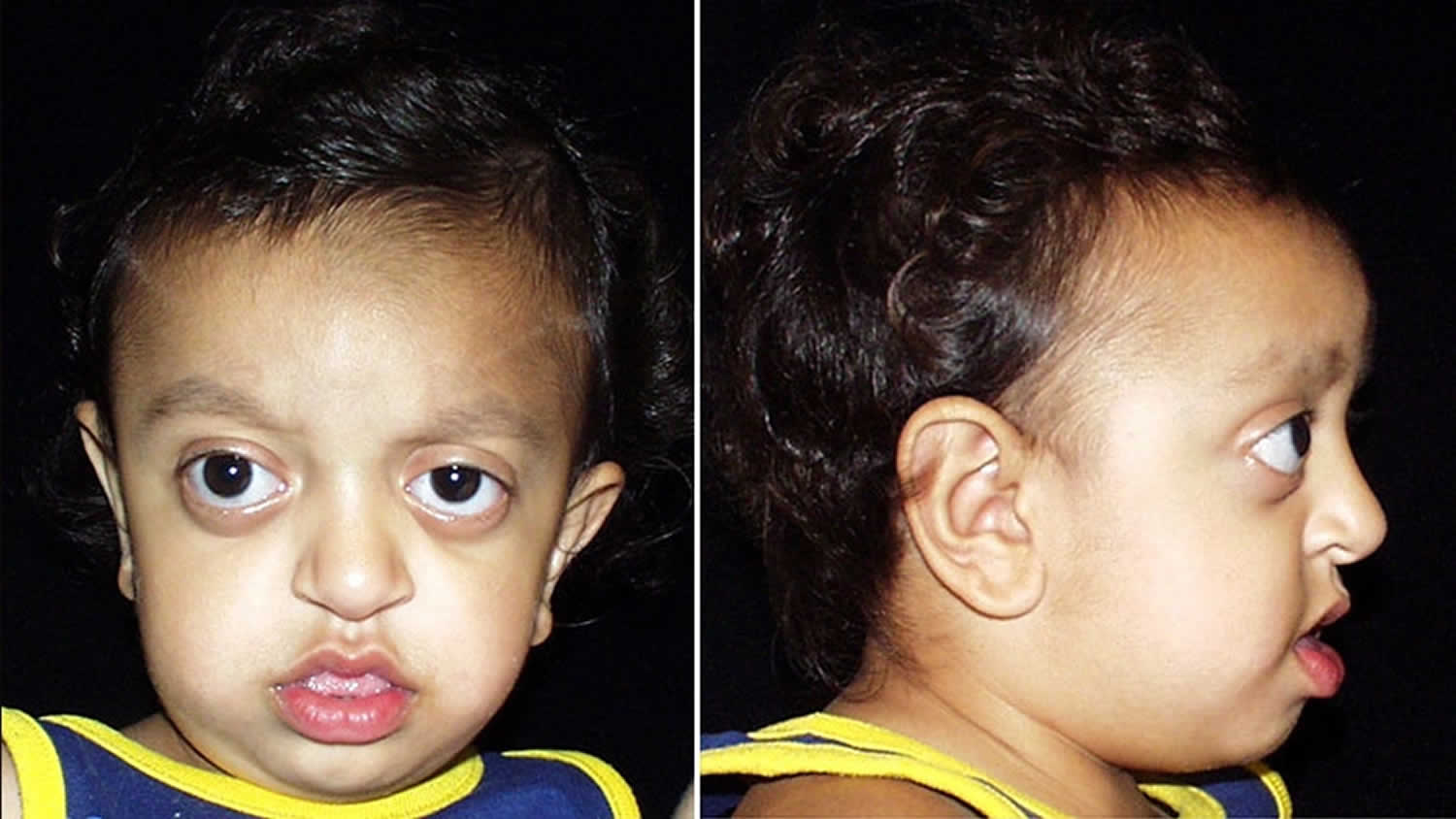
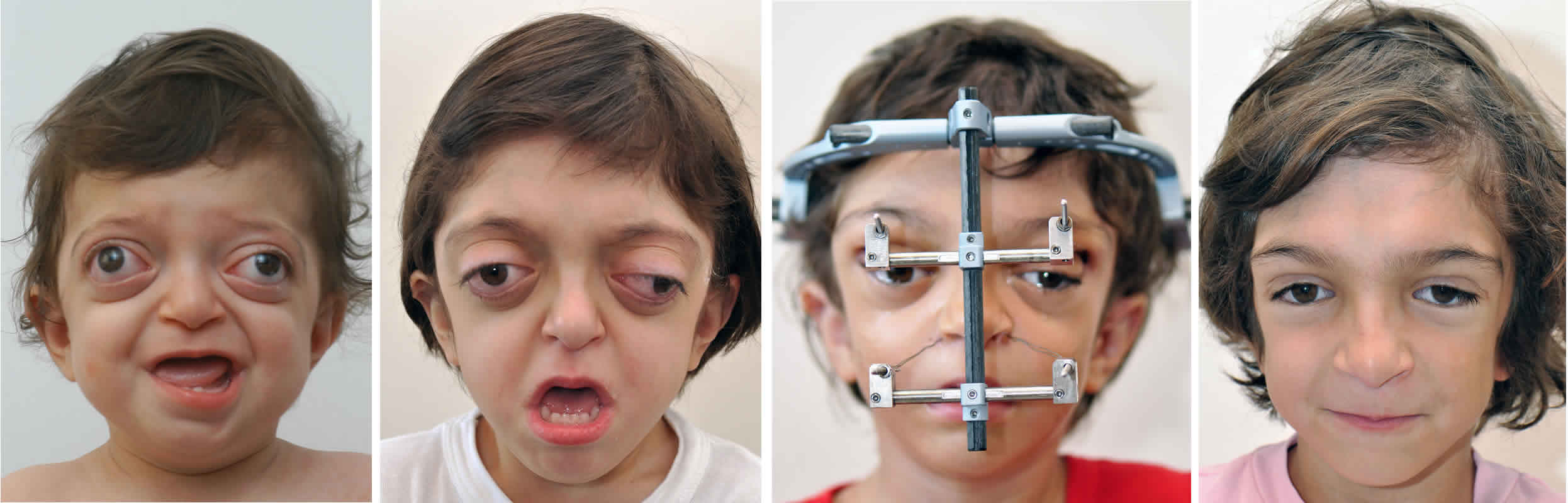
The main sign and symptoms are facial asymmetry (one side of the face is different from the other), a partially formed ear (microtia) or totally absent ear (anotia), noncancerous (benign) growths of the eye (ocular dermoid cysts), and spinal abnormalities. Goldenhar syndrome may also affect the heart, lungs, kidneys, and central nervous system 3). The first case of Goldenhar syndrome was described in the following case report published in 1952 4). Goldenhar syndrome is due to problems that occur when the fetus is forming within the womb of the mother, in structures known as the first and second brachial arch. These structures will develop to form the neck and the head. The cause of Goldenhar syndrome is still unknown 5). Goldenhar syndrome is part of a group of conditions known as craniofacial microsomia. It is not known whether the conditions included in the group really are different conditions or part of the same problem with different degrees of severity. Treatment is age-dependent, with interventions at appropriate stages during the growth and development of the skull and face 6).

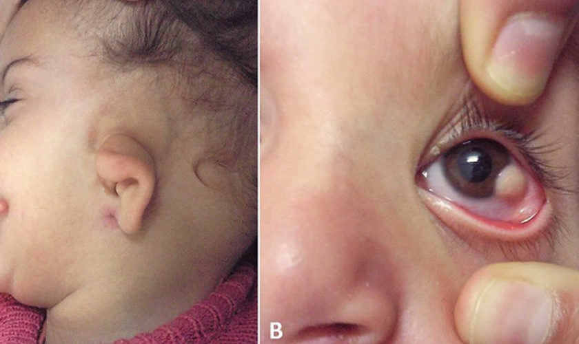
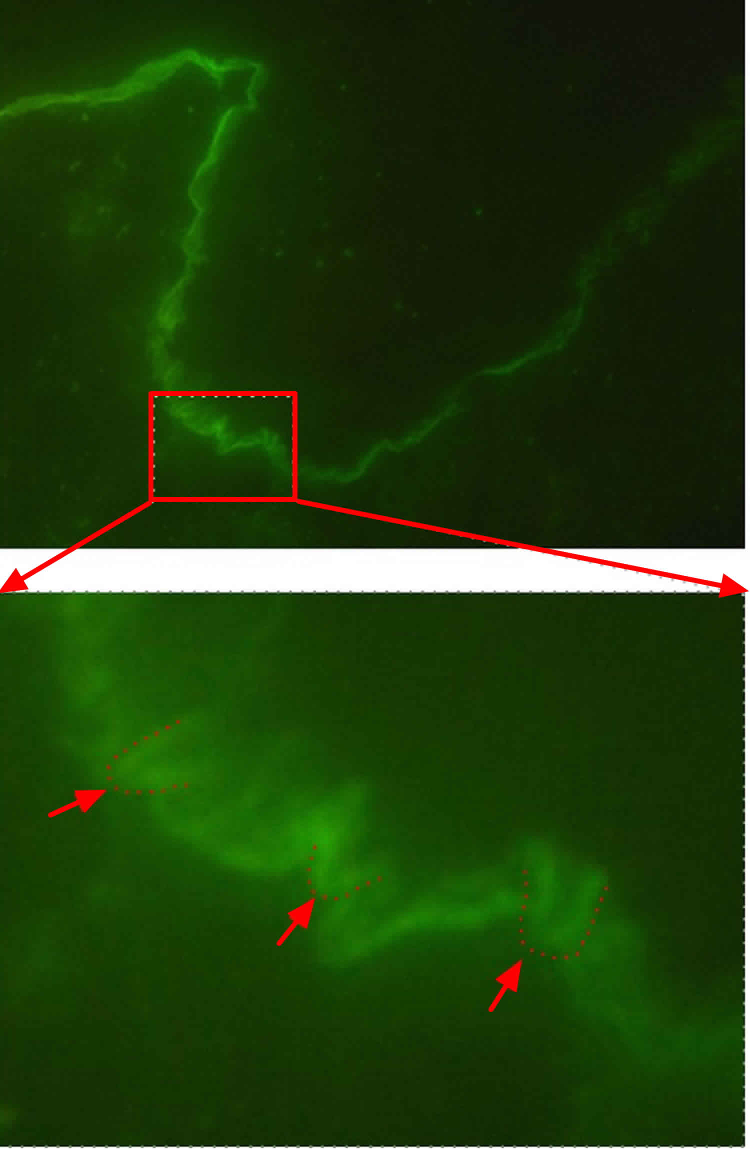
Figure 1. Goldenhar syndrome

goldenhar syndrome

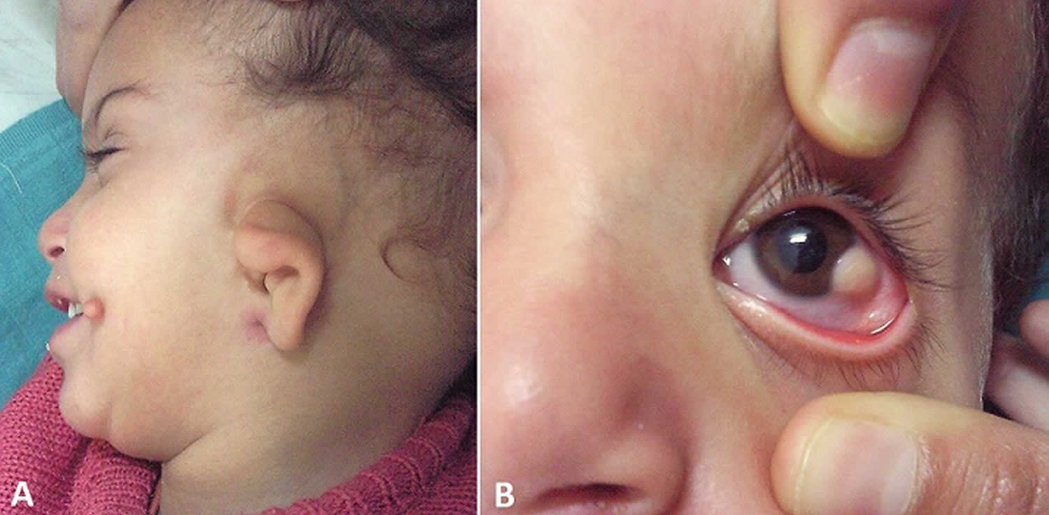
Footnote: (A) Microtia and preauricular skin tag; (B) limbal dermoid in the same child.

Will Goldenhar syndrome happen to children I have in the future?

The chances of having another child with Goldenhar syndrome is less than 1% or less. Your child has about a 3% chance of passing it on to his or her children 7).

How many people have Goldenhar syndrome and bilateral Goldenhar syndrome in the United States and worldwide?

Unfortunately, for rare diseases, there is often not a calculated incidence or prevalence because there is no official method for tracking them. However, it has been estimated that the frequency of Goldenhar disease ranges between 1 case per 3,500 births and 1 case per 25,000 births 8). Given these data, a very rough estimate for the number of people in the United States with Goldenhar syndrome ranges from 13,000-56,000 and estimates for the number of people worldwide with the syndrome ranges from 300,000-1,300,000. Currently, scientists are unaware of estimates regarding bilateral (affecting both sides of the body) disease, specifically.

Is there any research or information on people with Goldenhar syndrome having mental illness or multiple personalities?

There is one case report of a single patient diagnosed with Goldenhar syndrome who had schizophreniform disorder 9). Case reports document clinic findings associated with individual cases. It is important to keep in mind that the clinical findings documented in these case reports are based on specific individuals and may differ from one affected person to another.

Goldenhar syndrome causes

The underlying cause of Goldenhar syndrome is poorly understood. Most cases occur sporadically with no apparent explanation. Some researchers suspect that problems with blood flow or other disruptions during fetal development may contribute to the development of the condition 10). Animal models demonstrate that vascular disruption and hematoma formation affect the developing structures of the jaw and the ear regions in utero. Other hypotheses suggest that disturbance of the population of the neural crest cells occurs at 30-45 days’ gestation.

Approximately 1-2% of affected people have other family members with the condition, which suggests that genes may play a role in some cases 11).

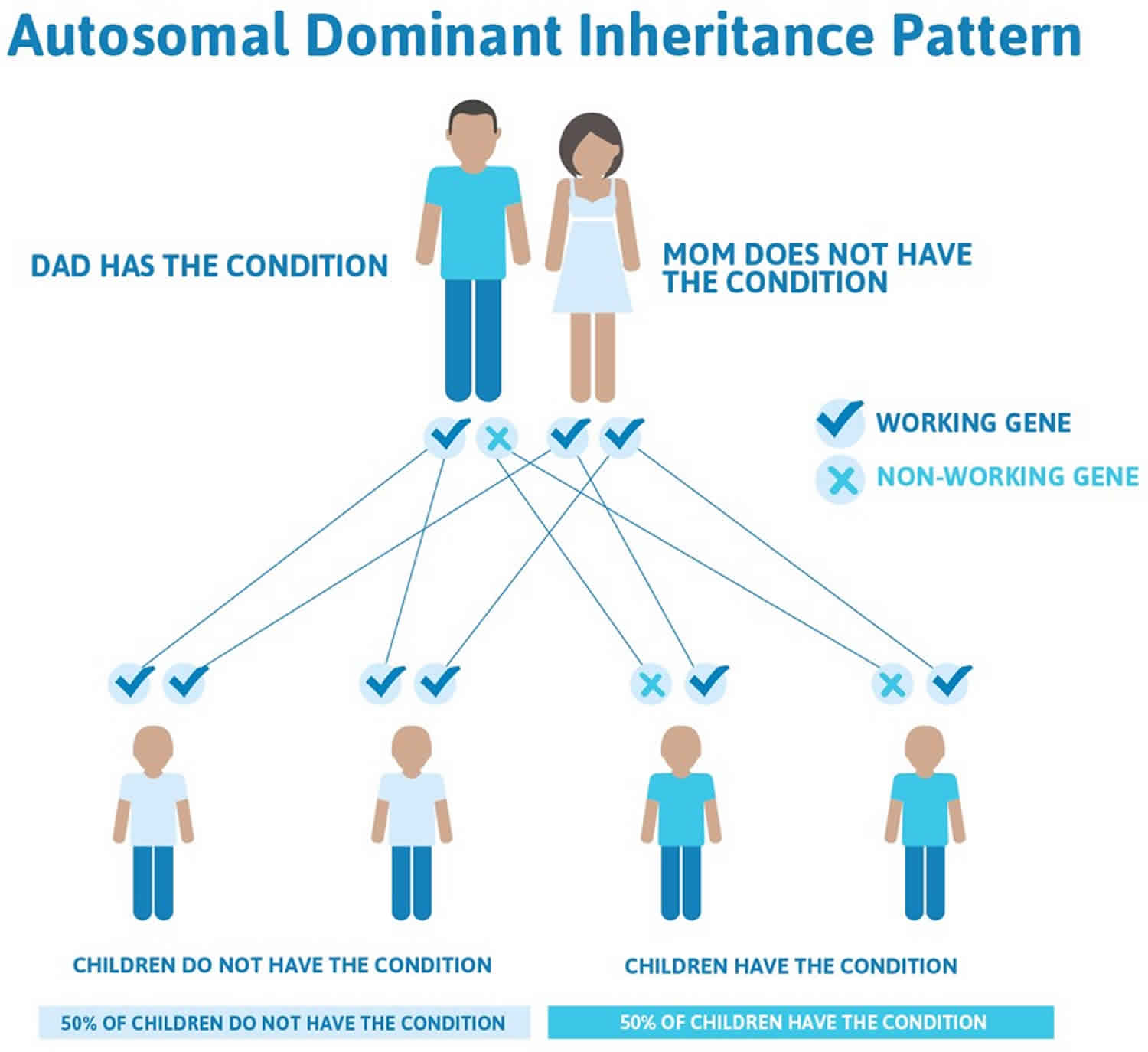
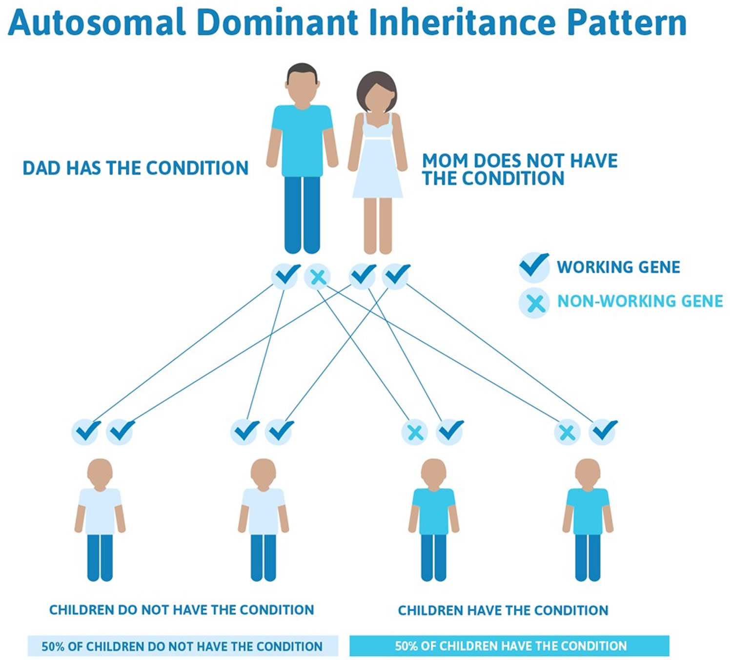
Goldenhar syndrome inheritance pattern

Most cases of Goldenhar syndrome occur sporadically in people with no family history of the condition 12). Rarely (approximately 1-2% of affected people), more than one family member can be affected. In these cases, the condition appears to be passed down through the family in an autosomal dominant manner 13).

People with specific questions about genetic risks or genetic testing for themselves or family members should speak with a genetics professional.

Resources for locating a genetics professional in your community are available online:

Goldenhar syndrome symptoms

The signs and symptoms of Goldenhar syndrome vary significantly from person to person. Common signs and symptoms of the condition include 14):

  • Microtia (a partially formed or completely absent ear) and other ear abnormalities
  • Underdeveloped facial muscles which may be associated with weakness
  • Underdeveloped jaw, cheekbone and/or temple bone
  • Cleft lip and/or palate
  • Abnormalities of the eyes, such as anophthalmia/microphthalmia, epibulbar tumors (noncancerous growths in the eyes), retinal abnormalities, and vision loss
  • An unusually large or small mouth
  • Dental abnormalities

Goldenhar syndrome deformities divided into groups according to the region they affect 15):

  • Ocular symptoms: Epibulbar dermoids, microphthalmia, anophthalmia, eyes asymmetry/dysmorphy, cleft eyelid, exophthalmia, strabismus
  • Auricular symptoms: dacryocystitis, preauricular appendages, preauricular fistulas, ear asymmetry, microtia, atresia of the externalauditory canal
  • Craniofacial deformities: cleft face, cleftlip, cleft palate, macrostomia, bifidtongue, hypoplasia of the mandible, hypoplasia of the maxilla, asymmetry of the mandible and maxilla, malocclusion, tooth discrepancies, agenesis of third molars and second premolars, supernumerary teeth, enamel and dentinmal formations, delay in tooth development
  • Skeletal abnormalities: cleft spine, microcephaly, dolichocephaly, plagiocephaly, vertebral defects
  • Abnormalities of internalorgans:
    • heart (atrial and ventricularseptal defects, conotruncal defects, out flow tract abnormalities, persistent truncusarteriosus);
    • kidneys (ectopic and/or fused kidneys, renalagenesis, multicystic kidney);
    • central nervous system (diffuse cerebralhypoplasia, dilated lateral cerebral ventricles or asymptomatic hydrocephalus, corpus callosum dysgenesis, frontal hypodensities, microcephaly, asymmetrical lateral ventricles, hydrocephalus due to aqueduct of Sylvius stenosis, corpus callosum lipoma, absence of septumpellucidum, diffuse cerebralhypodensity, hypothalamichamartoma).

In most cases, only one side of the face is affected, although approximately 10-33% of people with the condition have bilateral (both sides) involvement 16).

Some people with Goldenhar syndrome may also experience hearing loss; hydrocephalus (with or without intellectual disability); heart, kidneys, and lung problems; spinal abnormalities; and/or limb malformations 17).

Goldenhar syndrome diagnosis

A diagnosis of Goldenhar disease is based on the presence of characteristic signs and symptoms. These clinical features may be observed on physical examination or may require specialized testing such as imaging studies (i.e. CT scan, X-ray, echocardiogram, ultrasound). Additional testing including certain genetic tests may also be recommended to rule out conditions that are associated with similar features 18).

Goldenhar syndrome treatment

The treatment of Goldenhar syndrome is based on the signs and symptoms present in each person. Ideally, affected children should be managed by an experienced multidisciplinary craniofacial team. Treatment is age dependent and certain interventions may be recommended at different stages of growth and development 19).

The following are examples of medical issues that may need to be addressed in a person affected by Goldenhar syndrome 20):

  • Feeding issues – some people affected by Goldenhar syndrome may have feeding difficulties caused by the associated craniofacial abnormalities. Interventions may include special bottles, supplemental nasogastric feedings, and gastrostomy tube placement.
  • Breathing problems – affected people with an underdeveloped lower jaw may have difficulty breathing or develop sleep apnea. In these cases, referral to appropriate medical specialists is recommended so appropriate care can be provided.
  • Hearing loss – a hearing evaluation is recommended in all children with Goldenhar syndrome by 6 months of age. In those with hearing impairment, hearing aids or other treatments may be recommended.
  • Epibulbar tumors (noncancerous growths in the eyes) – these tumors may need to be surgically removed if they are particularly large or interfere with vision.
  • Craniofacial abnormalities (i.e. cleft lip and/or palate), congenital heart defects, kidney problems, and/or spine abnormalities – some of the characteristic symptoms associated with Goldenhar syndrome may require surgical repair.
  • Speech – people affected by Goldenhar syndrome are at an increased risk for a variety of speech problems due to the many associated craniofacial abnormalities. A speech evaluation and/or speech therapy may, therefore, be recommended in some affected people.

Will my child need surgery?

Depending on the severity of Goldenhar syndrome, your child may have some or all of the following surgeries:

  • lowering of the jaw on the affected side
  • lengthening of the lower jaw
  • 3 to 4 operations to rebuild the outer ear
  • addition of bone to build up the cheeks
  • soft tissue may need to be added to the face

New advances in procedures to correct the symptoms of Goldenhar syndrome are constantly being developed.

References   [ + ]

read more

Beckwith Wiedemann syndrome

Beckwith-Wiedemann-syndrome

What is Beckwith Wiedemann syndrome

Beckwith-Wiedemann syndrome is a condition that affects many parts of the body. It is classified as an overgrowth syndrome, affected infants are considerably larger than normal (macrosomia) and tend to be taller than their peers during childhood. Growth begins to slow by about age 8, and adults heights with Beckwith-Wiedemann syndrome are typically in the normal range. In some children with Beckwith-Wiedemann syndrome, specific parts of the body on one side or the other may grow abnormally large, leading to an asymmetric or uneven appearance. This unusual growth pattern, which is known as hemihyperplasia, usually becomes less apparent over time. Beckwith-Wiedemann syndrome was first recognised in 1963-64 by Dr J. Bruce Beckwith, a paediatric pathologist in America and, independently, by Dr H.E. Wiedemann, a German geneticist 1). Each had found a similar set of congenital abnormalities in children, which could not be found in any other disorders – in other words, a new syndrome.

Beckwith-Wiedemann syndrome affects an estimated 1 in 13,700 newborns worldwide 2). Beckwith-Wiedemann syndrome may actually be more common than this estimate because some people with mild symptoms are never diagnosed.

Approximately 85 percent of people with Beckwith-Wiedemann syndrome have genetic changes that appear to occur randomly (sporadically). Familial transmission occurs in approximately 10-15 percent of people with Beckwith-Wiedemann syndrome. Researchers have determined that Beckwith-Wiedemann syndrome results from various abnormalities affecting the proper expression of certain genes that control growth within a specific region of chromosome 11.

The signs and symptoms of Beckwith-Wiedemann syndrome vary among affected individuals. Some children with Beckwith-Wiedemann syndrome are born with an opening in the wall of the abdomen (an omphalocele) that allows the abdominal organs to protrude through the belly-button. Other abdominal wall defects, such as a soft out-pouching around the belly-button (an umbilical hernia), are also common. Some infants with Beckwith-Wiedemann syndrome have an abnormally large tongue (macroglossia), which may interfere with breathing, swallowing, and speaking. Other major features of this condition include abnormally large abdominal organs (organomegaly), creases or pits in the skin near the ears, low blood sugar (hypoglycemia) in infancy, and kidney abnormalities. Beckwith-Wiedemann syndrome may also be associated with other facial abnormalities, abnormal enlargement of one side or structure of the body (hemihyperplasia/hemihypertrophy) resulting in unequal (asymmetric) growth.

Children with Beckwith-Wiedemann syndrome are at an increased risk of developing several types of cancerous and noncancerous tumors, particularly a form of kidney cancer called Wilms tumor, a form of liver cancer called hepatoblastoma, adrenal carcinoma, and rhabdomyocsarcoma. Tumors develop in about 10 percent of people with Beckwith-Wiedemann syndrome and almost always appear in childhood. In infancy, Beckwith-Wiedemann syndrome has a mortality rate of approximately 20% 3).

Infants with low blood sugar may be treated with fluids given through a vein (intravenous, IV). Some infants may need medicine or other management if low blood sugar continues.

Defects in the abdominal wall may need to be repaired. If the enlarged tongue makes it hard to breathe or eat, surgery may be needed. Children with overgrowth on one side of the body should be watched for a curved spine (scoliosis). The child also must be watched closely for the development of tumors. Tumor screening includes blood tests and abdominal ultrasounds.

Beckwith-Wiedemann syndrome symptoms

Signs and symptoms of Beckwith-Wiedemann syndrome include:

  • Beckwith-Wiedemann syndrome children are usually born prematurely but are larger and heavier than one would expect, given the shorter length of gestation.
  • Large size for a newborn
  • Red birth mark on forehead or eyelids (nevus flammeus). This usually fades in the first few years.
  • Creases in ear lobes
  • Large tongue (macroglossia)
  • Low blood sugar (hypoglycemia) occurs in approximately 40% of Beckwith-Wiedemann syndrome children shortly after birth. Brain damage and other complications can result if it is not diagnosed and treated.
  • Abdominal wall defect (umbilical hernia or omphalocele)
  • Enlargement of some abdominal organs, usually the kidneys, liver, spleen, adrenals and pancreas (visceromegaly)
  • Overgrowth of one side of the body or of one limb (hemihyperplasia/hemihypertrophy) while the rest of the body grows at a normal rate.
  • Tumor growth, such as Wilms tumors (kidney cancer) and hepatoblastomas (liver tumors).
  • Enlarged heart or heart defects. These are relatively uncommon and may resolve without treatment.
  • Psychomotor development is usually normal except in cases of undiagnosed hypoglycemia or other complications.

The symptoms of Beckwith-Wiedemann syndrome vary greatly from person to person. Diagnosis of Beckwith-Wiedemann syndrome can be challenging because the patients are often mosaic (with the genetic changes occurring in some cells or parts of the body but not others), however external appearance is not necessarily predictive of internal effects. This results in some individuals appearing mildly affected, while others appear more significantly affected. The wide range of potential symptoms (clinical spectrum) can affect many different organs of the body. Affected individuals may not have all of the symptoms listed below. Many clinical features of Beckwith-Wiedemann syndrome become less evident with increasing age and many adults experience normal growth and appearance. Intelligence is usually unaffected in Beckwith-Wiedemann syndrome, unless associated with prolonged, untreated neonatal hypoglycemia or a chromosomal duplication.

Some infants with Beckwith-Wiedemann syndrome are born prematurely, but still have an excessive birth weight (large for gestational age). Many infants with Beckwith-Wiedemann syndrome are above the 97th percentile in weight for gestational age. Overgrowth continues throughout childhood (macrosomia) and slows around 7 or 8 years of age. Abnormal enlargement of one side or structure of the body (hemihyperplasia/hemihypertrophy) may occur, resulting in unequal (asymmetric) growth. Hemihyperplasia refers specifically to an increase in number of cells (proliferation) resulting in asymmetric overgrowth. A related term, hemihypertrophy, refers to overgrowth due to abnormally large cell size.

Abdominal wall defects include an omphalocele (also known as exomphalos), in which part of an infant’s intestines and abdominal organs protrude or stick out through the belly button. The intestines and other organs are covered by a thin membrane. Less severe defects can include protrusion of part of the intestines through an abnormal opening in the muscular wall of the abdomen near the umbilical cord (umbilical hernia), or weakness and separation of the left and right muscles (rectus muscles) of the abdominal wall (diastasis recti). The internal organs of affected individuals can become abnormally enlarged (organomegaly). Any or all of the following organs may be affected: liver, spleen, pancreas, kidneys, or adrenal glands.

Some newborns with Beckwith-Wiedemann syndrome may have low blood sugar (neonatal hypoglycemia or hyperinsulinism) due to overgrowth and excessive secretion of the hormone insulin by pancreatic islets. Insulin functions to help regulate blood glucose levels by promoting the movement of glucose into cells. Most infants with neonatal hypoglycemia associated with Beckwith-Wiedemann syndrome have mild and transient symptoms. However, without proper detection and appropriate treatment, neurological complications may result.

Macroglossia (enlarged tongue) is reported to occur in approximately 82%- 97% of cases and is one of the most common features of Beckwith-Wiedemann syndrome. Children with an enlarged tongue (macroglossia) can have difficulties in speaking, feeding, and breathing. In addition to an enlargement of the tongue (macroglossia), Beckwith-Wiedemann syndrome may be characterized by other abnormalities of the skull and facial (craniofacial) region. Such features may include distinctive slit-like linear grooves or creases in the ear lobes and indentations on the back rims of the ears (pits), prominent eyes with relative underdevelopment of the bony cavity of the eyes (intraorbital hypoplasia), and/or a prominent back region of the skull (occiput). Some infants may have flat, pale red or reddish purple facial lesions at birth, most commonly on the eyelids and forehead, which consist of abnormal clusters of small blood vessels (capillary nevus flammeus). Such lesions typically become less apparent during the first year of life. In children with hemihyperplasia/hemihypertrophy, one side of the face may appear larger than the other. Due to the mosaic nature of Beckwith-Wiedemann syndrome, some children have eyes with multiple colors. In addition, in some affected children, there may be improper contact of the teeth of the upper and lower jaws (malocclusion) and abnormal protrusion of the lower jaw (mandibular prognathism), features that may occur secondary to abnormal largeness of the tongue.

A variety of kidney (renal) abnormalities have occurred in individuals with Beckwith-Wiedemann syndrome, including abnormally large kidneys (nephromegaly), improper development of the innermost tissues of the kidney (renal medullary dysplasia), and the formation of calcium deposits in the kidney (nephrocalcinosis), which could potentially impair kidney function. Additional abnormalities include duplication of the series of tubes and ducts through which the kidneys reabsorb water and sodium (duplicated collecting system), widening of some of the small tubes and collecting ducts (medullary sponge kidney), and the presence of small pouches (diverticula) on the kidneys.

Children with Beckwith-Wiedemann syndrome may have an increased risk of developing certain childhood cancers, particularly Wilms tumor (nephroblastoma), which is a malignancy of the kidney, and tumors involving the liver (hepatoblastoma). Less commonly, other malignancies have been reported (e.g., neuroblastoma, rhabdomyosarcoma). The risk of malignancy is greatest before the age of 8. Around 7.5% of Beckwith-Wiedemann syndrome children will develop Wilms Tumour. Because of the aggressiveness of these tumors, abdominal ultrasound scans should take place every three months up to the age of 7 or 8 years. A baseline MRI scan may also be performed. The susceptibility to these tumors diminishes and is not usually a problem after the age of 8.

Liver tumors (hepatoblastoma) risk diminishes after the age of 3 years. Liver tumors (hepatoblastoma) can also be detected by abdominal ultrasound but, as not all the liver can be viewed, alpha-fetoprotein (AFP) levels in the blood may also be monitored 3 monthly. Screening is recommended by alpha-fetoprotein (AFP) every six weeks – three months until age 4 years and abdominal ultrasounds every 3 months until age 8 years.

Beckwith Wiedemann syndrome life expectancy

Most children and adults with Beckwith-Wiedemann syndrome do not have serious medical problems associated with the condition. Their life expectancy is usually normal. Children with Beckwith-Wiedemann syndrome are at an increased risk of developing several types of cancerous and noncancerous tumors, particularly a form of kidney cancer called Wilms tumor, a form of liver cancer called hepatoblastoma, adrenal carcinoma, and rhabdomyocsarcoma. Tumors develop in about 10 percent of people with Beckwith-Wiedemann syndrome and almost always appear in childhood. In infancy, Beckwith-Wiedemann syndrome has a mortality rate of approximately 20% 4). Because of the aggressiveness of these tumors, abdominal ultrasound scans should take place every three months up to the age of 7 or 8 years. A baseline MRI scan may also be performed. The susceptibility to these tumors diminishes and is not usually a problem after the age of 8.

Beckwith-Wiedemann syndrome causes

The genetic causes of Beckwith-Wiedemann syndrome are complex. The condition usually results from the abnormal regulation of genes in a particular region of chromosome 11 5). People normally inherit one copy of this chromosome from each parent. For most genes on chromosome 11, both copies of the gene are expressed, or “turned on,” in cells. For some genes, however, only the copy inherited from a person’s father (the paternally inherited copy) is expressed. For other genes, only the copy inherited from a person’s mother (the maternally inherited copy) is expressed. These parent-specific differences in gene expression are caused by a phenomenon called genomic imprinting. Abnormalities involving genes on chromosome 11 that undergo genomic imprinting are responsible for most cases of Beckwith-Wiedemann syndrome.

At least half of all cases result from changes in a process called methylation. Methylation is a chemical reaction that attaches small molecules called methyl groups to certain segments of DNA. In genes that undergo genomic imprinting, methylation is one way that a gene’s parent of origin is marked during the formation of egg and sperm cells. Beckwith-Wiedemann syndrome is often associated with changes in regions of DNA on chromosome 11 called imprinting centers. Imprinting centers control the methylation of several genes that are involved in normal growth, including the CDKN1C, H19, IGF2, and KCNQ1OT1 genes. Abnormal methylation disrupts the regulation of these genes, which leads to overgrowth and the other characteristic features of Beckwith-Wiedemann syndrome.

About twenty percent of cases of Beckwith-Wiedemann syndrome are caused by a genetic change known as paternal uniparental disomy. Paternal uniparental disomy causes people to have two active copies of paternally inherited genes rather than one active copy from the father and one inactive copy from the mother. People with paternal uniparental disomy are also missing genes that are active only on the maternally inherited copy of the chromosome. In Beckwith-Wiedemann syndrome, paternal uniparental disomy usually occurs early in embryonic development and affects only some of the body’s cells. This phenomenon is called mosaicism. Mosaic paternal uniparental disomy leads to an imbalance in active paternal and maternal genes on chromosome 11, which underlies the signs and symptoms of the disorder.

Less commonly, mutations in the CDKN1C gene cause Beckwith-Wiedemann syndrome. This gene provides instructions for making a protein that helps control growth before birth. Mutations in the CDKN1C gene prevent this protein from restraining growth, which leads to the abnormalities characteristic of Beckwith-Wiedemann syndrome.

About 1 percent of all people with Beckwith-Wiedemann syndrome have a chromosomal abnormality such as a rearrangement (translocation), abnormal copying (duplication), or loss (deletion) of genetic material from chromosome 11. Like the other genetic changes responsible for Beckwith-Wiedemann syndrome, these abnormalities disrupt the normal regulation of certain genes on this chromosome.

Beckwith-Wiedemann syndrome inheritance pattern

In about 85 percent of cases of Beckwith-Wiedemann syndrome, only one person in a family has been diagnosed with the condition. However, parents of one child with Beckwith-Wiedemann syndrome may be at risk of having other children with the disorder. This risk depends on the genetic cause of the condition.

Another 10 to 15 percent of people with Beckwith-Wiedemann syndrome are part of families with more than one affected family member. In most of these families, the condition appears to have an autosomal dominant pattern of inheritance. Autosomal dominant inheritance means that one copy of an altered gene in each cell is typically sufficient to cause the disorder. In most of these cases, individuals with Beckwith-Wiedemann syndrome inherit the genetic change from their mothers. Occasionally, a person who inherits the altered gene will not have any of the characteristic signs and symptoms of the condition.

Rarely, Beckwith-Wiedemann syndrome results from changes in the structure of chromosome 11. Some of these chromosomal abnormalities are inherited from a parent, while others occur as random events during the formation of reproductive cells (eggs and sperm) or in the earliest stages of development before birth.

Adults with Beckwith-Wiedemann syndrome must ensure they have had genetic testing to confirm their molecular subtype of the condition before they have children. This will allow them to receive the correct advice on the risk of the condition occurring in their children. The risk is low for most cases but is higher in some (see above). If you have not been tested or have not received recent advice, your should ask your family doctor to refer you to your local Clinical Genetics Service.

Genetic counseling may help you understand the risks of passing Beckwith-Wiedemann syndrome on to any children you have.

People with specific questions about genetic risks or genetic testing for themselves or family members should speak with a genetics professional.

Resources for locating a genetics professional in your community are available online:

Beckwith-Wiedemann syndrome diagnosis

Beckwith-Wiedemann syndrome may be diagnosed or confirmed shortly after birth based on a thorough clinical evaluation, detection of characteristic physical findings (e.g., increased weight and length, macroglossia, abdominal wall defects and careful methylation testing and chromosomal (cytogenetic) analysis of the Beckwith-Wiedemann syndrome region (i.e., chromosome 11p15)). Note that in around 20% of Beckwith-Wiedemann syndrome children there is no detectable molecular cause i.e. the blood test will not confirm Beckwith-Wiedemann syndrome.

In some cases, certain procedures may be performed before birth (prenatally). For example, ultrasound imaging may allow assessment of organ size and overall size of the developing fetus and potentially reveal other findings that may be suggestive of Beckwith-Wiedemann syndrome, such as increased amniotic fluid surrounding the fetus (hydramnios), enlarged placenta, omphalocele, enlarged abdominal circumference, and/or other abnormalities. If Beckwith-Wiedemann syndrome is suspected, prenatal testing is available.

Beckwith Wiedemann syndrome treatment

The treatment of Beckwith-Wiedemann syndrome is directed toward the specific symptoms that are apparent in each individual. Treatment may require the coordinated efforts of a team of specialists. Geneticists, pediatricians, plastic surgeons, kidney specialists, dental specialists, speech pathologists, pediatric oncologists, and other healthcare professionals may need to systematically and comprehensively plan an affected child’s treatment.

In newborns with Beckwith-Wiedemann syndrome, regular monitoring of blood glucose levels should be performed to ensure prompt detection and treatment of hypoglycemia. Although neonatal hypoglycemia is usually mild and temporary, its prompt detection and treatment is essential in preventing associated neurologic complications. Treatment measures may include the administration of intravenous glucose, frequent feedings, certain medications (e.g., diazoxide or octreotide), and/or surgical intervention in some cases.

In many infants with umbilical hernia, the defect may spontaneously disappear by the age of approximately one year. Surgery usually is not required unless an umbilical hernia becomes progressively larger, does not spontaneously resolve (e.g., by about three or four years of age), and/or is associated with certain complications. However, in newborns with omphalocele, surgical repair of the defect is typically required shortly after birth.

Children with macroglossia should undergo feeding evaluation and sleep studies in addition to consultations with plastic surgeons and pulmonologists if needed. Feeding difficulties caused by an abnormally large tongue (macroglossia) may be treated by the use of specialized nipples or the temporary insertion of a nasogastric tube. Some affected children may undergo tongue reduction surgery. Such surgery is performed if macroglossia causes dentoskeletal defects, psychosocial problems, upper airway obstruction, or difficulties swallowing, feeding or speaking. Macroglossia may also correct itself without medical intervention.

Orthopedic evaluation is recommended for patients with hemihyperplasia or hemihypertrophy.

In addition, infants and children with Beckwith-Wiedemann syndrome should undergo regular abdominal and kidney (renal) ultrasounds, and serum alpha-fetoprotein (AFP) levels as recommended to enable early detection and treatment of certain malignancies that may occur in association with Beckwith-Wiedemann syndrome (e.g., Wilms tumor, hepatoblastoma). Alpha-fetoprotein (AFP) is a protein produced by the liver. Alpha-fetoprotein (AFP) levels typically decline during infancy; however, alpha-fetoprotein (AFP) may be abnormally elevated in blood serum if certain malignancies are present. The trend in alpha-fetoprotein (AFP) levels over time should be followed in children with Beckwith-Wiedemann syndrome. Screening is recommended by AFPs every six weeks – three months until age 4 years and abdominal ultrasounds every 3 months until age 8 years.

If malignancies develop in association with Beckwith-Wiedemann syndrome (e.g., Wilms tumor, hepatoblastoma), the appropriate treatment measures vary depending upon the specific malignancy present, grade and/or extent of disease, and/or other factors. Treatment methods may include surgery, use of certain anticancer drugs (chemotherapy), radiation therapy, and/or other measures.

Children with cardiac, gastrointestinal, and renal abnormalities may require certain medications, surgery, or other medical interventions. These children should be referred to appropriate specialists. Genetic counseling may be of benefit for affected individuals and their families. Other treatment is symptomatic and supportive.

References   [ + ]

read more

Crouzon syndrome

crouzon syndrome

What is Crouzon syndrome

Crouzon syndrome also known as craniofacial dysostosis, is a genetic disorder characterized by the premature fusion of certain skull bones (craniosynostosis). This early fusion of certain skull bones prevents the skull from growing normally and affects the shape of the head and face. Crouzon syndrome is the most common type of craniosynostosis syndrome. Crouzon syndrome is seen in about 1.6 per 100,000 newborns.

Crouzon syndrome shares many of the same features as Apert syndrome due to the result of the premature fusion of the skull bones, including abnormal development of the eye sockets, prominent and widely spaced eyes, bulging eyes and vision problems caused by shallow eye sockets, eyes that do not point in the same direction (strabismus); a beaked nose; and an underdeveloped upper jaw and slow development of the midface. In addition, people with Crouzon syndrome may have dental problems and hearing loss, which is sometimes accompanied by narrow ear canals. A few people with Crouzon syndrome have an opening in the lip and the roof of the mouth (cleft lip and palate). The severity of these signs and symptoms varies among affected people.

In addition to the physical characteristics common to Crouzon syndrome, your child may have the following problems:

  • dental problems due to crowded teeth and a narrow palate
  • poor vision
  • ear disease and hearing loss in about 50% of children
  • difficulty breathing due to small airway
  • possible fluid on the brain (hydrocephalus)

The severity of signs and symptoms can vary among affected people, even within a family. Intelligence is usually normal, but intellectual disability may be present 1). Crouzon syndrome is caused by changes (mutations) in the FGFR2 gene and is inherited in an autosomal dominant manner.

The child with Crouzon syndrome usually enters a programme of care involving many different clinical specialities, which often continues from birth to the later teenage years. Treatment may involve surgeries to prevent complications, improve function, and aid in healthy psychosocial development 2).

Depending on the severity of Crouzon syndrome, your child may have some or all of the following surgeries:

  • frontal orbital advancement to allow the skull to grow properly and to increase the size of the eye sockets
  • jaw surgery
  • orthodontics work
  • surgical advancement of the mid-face

New advances in procedures to correct Crouzon syndrome are constantly being developed. It is important for patients with Crouzon syndrome to be treated by a multidisciplinary care team that specializes in caring for children with these complex disorders.

Figure 1. Crouzon syndrome

Crouzon syndrome

Footnote: 18-month-old with Crouzon syndrome and characteristic rounded forehead, sunken midface and prominent eyes.

[Source 3) ]

What is Crouzon syndrome with acanthosis nigricans?

Crouzon syndrome with acanthosis nigricans is found in an estimated 5-10% of all Crouzon cases, it is very rare. Crouzon syndrome with acanthosis nigricans has an estimated prevalence of 1 per 1,000,000 newborns. Fewer than 70 cases have been described in the medical literature. A female-to-male sex ratio of 2.4:1 has been reported. In addition to the facial characteristics, Crouzon syndrome with acanthosis nigricans includes some of the following:

  • Darkened, rough patches of skin found in the folds of the body (armpits, neck, groin, elbows, knees, chin/mouth area, eye area, or stomach).
  • Signs of this begin between the ages of 2-4.
  • The acanthosis nigricans generally does not advance after the age of 12. So, the texture and color stay the same from then on.

Crouzon syndrome with acanthosis nigricans is caused by a specific p.Ala391Glu mutation in the fibroblast growth-factor receptor 3 FGFR3 gene (4p16.3), involved in regulation of cell proliferation, differentiation and apoptosis. Acanthosis nigricans is associated with inadequate stimulation of various fibroblast growth-factor receptors.

Normal newborn skull anatomy and physiology

It is important to have an understanding of skull anatomy and growth in order to understand craniosynostosis. An infant’s skull has 7 bone plates that relate to each other through specialized joints called “cranial sutures”. Sutures are made of tough, elastic fibrous tissue and separate the bones from one another. Sutures meet up (intersect) at two spots on the skull called fontanelles, which are better known as an infant’s “soft spots”. Although there are several major and minor sutures, the sutures that potentially have the most clinical significance are the singular metopic and sagittal sutures, as well as the paired (right and left) coronal and lambdoid sutures (see Figure 2). The seven bones of an infant’s skull normally do not fuse together until around age two or later. The sutures normally remain flexible until this point. In infants with primary craniosynostosis, the sutures abnormally stiffen or harden causing one or more of the bones of the skull to prematurely fuse together. This in turn, may lead to asymmetric skull growth.

Cranial sutures are very unique and specialized joints (syndesmosis joints). Their primary purpose is to grow bone in response to the rapidly developing brain within the protective skull compartment (cranial cavity or calvarium). The skull not only needs to be firm to protect the brain from accidental blows, but should also be expansile to accommodate its rapid growth. The brain doubles in volume in the first year of life and almost triples in volume by the age of three. The sutures of the skull allow for this important but almost contradictory balance of protection and growth.

Each cranial suture is designed to generate growth in the skull in a very specific area and configuration, ultimately reflecting the size and shape of the underlying brain structure. The overall bone development from cranial sutures occurs in a direction perpendicular to the long axis of the suture. Understanding these two facts, it makes sense that a fusion of each suture independently would cause a unique head shape.

The greatest increase in brain volume (brain growth) occurs from 0 to 14 months of age. The size of a child’s brain typically reaches 80% of adult size by the age of 2. The growth in head circumference after that age is more related to growth in the thickness of the skull and scalp but not actual brain growth. “Hat size” increases but not necessarily “brain size”.

The various cranial sutures close at different ages. The metopic suture closes earliest, around 6 months to 2 years. The rest of the sutures stay open into the 20’s and 30’s. The brain and fluid cavities of the brain do continue to grow in volume as you go into early adulthood, albeit not nearly as rapidly as the first couple years of life. Since the skull is much firmer (calcified or “rock-like”) and thicker, the skull needs the sutures to grow bone for any increase in volume.

Interestingly, there is a lot of variability here. Doctors have operated on adults in their 30’s for reasons unrelated to their skull sutures and have coincidentally found open metopic sutures. They have also seen young adults with closed coronal, lambdoid, and sagittal sutures, but with normal head shapes and often, no indication or symptoms of high pressure.

Scientists have learned that a cranial suture’s purpose is to grow bone to accommodate a growing brain, and that most brain growth occurs in the first two years of life. The brain reaches 85% of adult size by age 3 years (see Figure 3. Brain size vs. age diagram).

So it makes sense that the sutures are vitally important in the first two years of life. The earlier the fusion, the more severe the restriction in growth and, consequently, volume provided to the brain.

Figure 2. Normal skull of a newborn

Normal skull of a newborn

Figure 3. Brain size versus age diagram

Brain size versus age diagram

How is Crouzon syndrome inherited?

Crouzon syndrome is inherited in an autosomal dominant manner. This means that having a change (mutation) in only one copy of the responsible gene in each cell is enough to cause features of the condition.

There is nothing that either parent can do, before or during a pregnancy, to cause a child to be born with Crouzon syndrome.

In some cases, an affected person inherits the mutated gene from an affected parent. In other cases, the mutation occurs for the first time in a person with no family history of the condition. This is called a de novo mutation.

When a person with a mutation that causes an autosomal dominant condition has children, each child has a 50% (1 in 2) chance to inherit that mutation.

Figure 4. Crouzon syndrome autosomal dominant inheritance pattern

Crouzon syndrome autosomal dominant inheritance pattern

Is there a way to prevent having a child with Crouzon syndrome?

With advanced planning and appropriate testing, it may be possible to prevent having a child with Crouzon syndrome.

During a pregnancy:

If the genetic change (mutation) in an affected family member has been identified, prenatal genetic testing may be possible during pregnancy. Genetic testing may be performed on a sample obtained by chorionic villus sampling (at about 10 to 12 weeks gestation), or by amniocentesis (usually performed at about 15 to 18 weeks gestation). If the condition is confirmed in the fetus by either method, planning for an affected child and/or pregnancy management options, may be discussed with a health care provider.

Before a pregnancy:

As an alternative to prenatal diagnosis during the pregnancy, preimplantation genetic diagnosis before a pregnancy may be an option if the mutations in the family are known. Preimplantation genetic diagnosis is done after in vitro fertilization (IVF) to diagnose a genetic condition in an embryo before it is introduced into the uterus. When having preimplantation genetic diagnosis, only embryos known to be unaffected are introduced into the uterus for a possible pregnancy.

People interested in genetic testing, prenatal diagnosis, and/or preimplantation genetic diagnosis should speak with a genetic counselor or other genetics professional regarding their testing options. A genetics professional can help by:

  • thoroughly evaluating the family history
  • addressing questions and concerns
  • assessing recurrence risks
  • facilitating genetic testing if desired
  • discussing reproductive options

Can an unaffected sibling of someone with Crouzon syndrome have an affected child?

Almost all people with a mutation known to cause Crouzon syndrome have features of the condition. However, in at least one family, it appeared that the responsible mutation did not affect every family member who had it 4). For this reason, Crouzon syndrome has been described by some as having variable penetrance and expressivity, even within families 5). When a condition has variable (or reduced) penetrance, it means that not every person with a mutation in the responsible gene will have apparent signs and symptoms of the condition. When a condition has variable expressivity, it means that not all people who do have features will be affected the same way – there is a range of possible features and severity. While extremely uncommon, it may be possible for family members with a mutation to “appear” unaffected, with very mild or unnoticeable features 6). Therefore, whether an unaffected sibling is at risk to have an affected child may depend on whether the sibling has a mutation in the responsible gene.

If an apparently unaffected sibling is confirmed to not have the mutation that is present in an affected family member, he/she is not considered at risk to have an affected child. In other words, a person cannot pass on a mutation that he/she does not have. Based on most reports in the literature, people without features of Crouzon syndrome typically do not carry a mutation in the responsible gene.

If an apparently unaffected sibling does have the mutation that is present in an affected family member, he/she has a 50% (1 in 2) chance to pass the mutation on to each child. In this case, even though the sibling does not have apparent features of the condition, it would not be possible to predict whether a child that inherits the mutation will be affected, or how severely he/she may be affected.

If the specific mutation causing Crouzon syndrome has been identified in an affected family member, other family members can have genetic testing to determine whether they carry the mutation and are at risk for passing the mutation on to their children.

People with personal questions about genetic risks and genetic testing are encouraged to speak with a genetic counselor or other genetics professional. A genetics professional can help by:

  • thoroughly evaluating the family history
  • addressing questions and concerns
  • assessing recurrence risks
  • facilitating genetic testing if desired
  • discussing reproductive options

Resources for locating a genetics professional in your community are available online:

If you have a health condition that has not been diagnosed, you may be interested in the Undiagnosed Diseases Network (https://undiagnosed.hms.harvard.edu/). They have information about how to apply for this multicenter research study.

Crouzon syndrome causes

Mutations in the FGFR2 gene cause Crouzon syndrome. This gene provides instructions for making a protein called fibroblast growth factor receptor 2. Among its multiple functions, this protein signals immature cells to become bone cells during embryonic development. Mutations in the FGFR2 gene probably overstimulate signaling by the FGFR2 protein, which causes the bones of the skull to fuse prematurely.

Crouzon syndrome is inherited in an autosomal dominant pattern, which means one copy of the altered gene in each cell is sufficient to cause the disorder. When a person with a mutation that causes an autosomal dominant condition has children, each child has a 50% (1 in 2) chance to inherit that mutation.

Crouzon syndrome symptoms

Crouzon syndrome predominantly affects the appearance of the head and face. Crouzon syndrome presents many of the same associated issues as Apert syndrome, including airway compromise, sleep apnea, hydrocephalus and eye exposure issues. In patients with Crouzon syndrome, the forehead generally appears more rounded with a more normally shaped nose, and the eyes tend to be more prominent. Another key difference is that patients with Crouzon syndrome do not have associated hand or feet anomalies present in other syndromes.

Common features:

  • regressed mid-face and shallow orbits (eye sockets), which may be present at birth or become more evident; seen from the side the face has a concave appearance and the shallow orbits result in prominent eyeballs (proptosis); the arrangement of the teeth (dentition) is also affected; and
  • the abnormal skull shape may result in raised intracranial pressure and require surgery to protect the restricted brain.

Crouzon syndrome may involve any combination of cranial sutures, most commonly including the coronal and sagittal sutures.

Cranial and facial malformations may vary, ranging from mild to potentially severe, including among members of the same family (kindred).

For example, the degree of cranial malformation is variable and depends on the specific cranial sutures involved as well as the order and rate of progression. In most affected individuals, there is premature fusion of the sutures (i.e., coronal and sagittal sutures) between bones forming the forehead (frontal bone) and the upper sides of the skull (parietal bones). In addition, the suture between the back and the sides of the skull (i.e., lambdoidal suture) or other sutures may be involved in some people. In most individuals with Crouzon syndrome, early sutural fusion causes the head to appear unusually short and broad (brachycephaly). In other cases, the head may appear long and narrow (scaphocephaly) or triangular (trigonocephaly). Rarely, premature closure of multiple sutures (known as Kleeblattschadel type craniosynostosis) causes the skull to be abnormally divided into three lobes (cloverleaf skull deformity). In those with Crouzon syndrome, craniosynostosis typically begins during the first year of life and progresses until approximately age two to three. However, craniosynostosis may sometimes be apparent at birth or, more rarely, may not be noted during early childhood.

In most individuals, there is unusual shallowness of the orbits or the bony cavities of the skull that accommodate the eyeballs. As a result, the eyeballs appear to protrude or bulge forward (proptosis). Due to such abnormalities, affected individuals are unusually susceptible to developing inflammation of the front, transparent regions of the eyes (i.e., exposure keratitis) as well as the membranes that line the inner surfaces of the eyelids and cover the whites of the eyes (exposure conjunctivitis). Crouzon syndrome is also often associated with additional eye abnormalities including eyes that are spaced apart wider than usual (hypertelorism) and eyes that are crossed or do not point in the same direction (strabismus). Sometimes, the various eye abnormalities can lead to a loss in vision.

Crouzon syndrome is associated with additional craniofacial abnormalities. Affected individuals often have a prominent forehead (frontal bossing); a curved nose; unusually flat or underdeveloped mid-facial regions (midface hypoplasia); and a short upper lip. In addition, a small, underdeveloped upper jaw (hypoplastic maxilla) with protrusion of the lower jaw (relative mandibular prognathism) may also occur. Clefting of the lip and/or palate (incomplete closure of the palate or an abnormal groove in the upper lip) can occur rarely. Typical dental problems include a highly arched narrow palate with crowded teeth, and upper and lower teeth that don’t meet when biting (malocclusion).

Approximately 30% of individuals with Crouzon syndrome develop hydrocephalus, a condition which is characterized by impaired flow or absorption of the fluid (i.e., cerebrospinal fluid [CSF]) that circulates through cavities (ventricles) of the brain and the spinal canal, potentially leading to increasing fluid pressure within the skull (intracranial pressure) and the brain and other associated findings.

Some affected individuals have hearing impairment due to an inability to transmit sound impulses to the brain (sensorineural hearing loss). In some infants, breathing problems may occur in infancy due to various abnormalities of the face and upper airway. In severe instances, this can lead to life-threatening breathing complications.

Crouzon syndrome diagnosis

Crouzon syndrome is usually diagnosed at birth or during infancy based upon a thorough clinical evaluation, identification of characteristic physical findings, and a variety of specialized tests. Such testing may include advanced imaging techniques, such as computerized tomography (CT) scanning or magnetic resonance imaging (MRI), or other imaging studies.

Clinical Testing and Workup

  • CT scanning and MRIs are used to help detect or characterize certain abnormalities that may be associated with the disorder (e.g., craniosynostosis, other skeletal abnormalities, etc.). During CT scanning, a computer and x-rays are used to create a film showing cross-sectional images of internal structures. During MRI, a magnetic field and radio waves create detailed cross-sectional images of certain organs and tissues.
  • Molecular genetic testing can confirm a diagnosis of Crouzon syndrome in some people. Molecular genetic testing can detect mutations in the FGFR2 gene known to cause the disorder, but is available only as a diagnostic service at specialized laboratories.

After a general craniofacial examination, a treatment plan is established and other specialists begin certain examinations. Photographs are also made. Basic exams include the following:

  • Dental impressions.
  • X-rays including a panorex for the lower jaw position, cephalograms to assess the relationship of the upper and lower jaws, CT (computed tomography) scan to assess skull growth, orbital size and jaw relationships. These scans can be converted into vivid, three-dimensional images of the skull and facial bones.
  • Hearing tests when possible since patients with Crouzon syndrome tend to have ear problems.
  • An eye examination.

It is important for a geneticist to meet with the family to discuss whether the condition runs in the family. It is important for the child to be evaluated by a craniofacial team to provide support and treatment for patients and their families.

Crouzon syndrome treatment

The treatment of Crouzon syndrome is dependent upon both functional and appearance-related needs, and should be addressed immediately after your child is born. Because of the complex issues that can be associated with Crouzon syndrome, your child should be treated at a medical center where he/she will have access to pediatric specialists across the many clinical areas he/she may need.

Surgery is the main form of therapy for affected children, but not all children will require surgery. Surgery is performed to create and ensure that there is enough room within the skull for the developing brain to grow; to relieve intracranial pressure (if present); and to improve the appearance of an affected child’s head.

Affected children should be seen at craniofacial clinics, which are often affiliated with major pediatric hospitals or medical centers. These clinics have a team of physicians and other healthcare providers who are experienced in treating craniofacial disorders. A team of specialists will work together to plan and carry out a child’s treatments. Such specialists include pediatricians, neurosurgeons, plastic surgeons, otolaryngologists, audiologists, ophthalmologists, dental specialists, social workers, and other healthcare professionals. Genetic counseling may be of benefit for affected individuals and their families. Psychosocial support for the entire family is essential as well.

As your child grows, he/she should also have access to psychosocial support services to address any mental, social or psychological issues that accompany these conditions.

Immediately after birth, your child should be assessed for the following:

  • Respiratory function. If there is a breathing difficulty that continues after initial support in the intensive care unit then it may be required tracheostomy.
  • Eyelids closure capacity. If the newborn cannot close his/her eyelids because of exophthalmos (eyes bulging out of their sockets), it may need to perform a temporary tarsorrhaphy (where stitches are carefully placed at the corners of the eyelid opening (palpebral fissure) to narrow it) so as to protect the cornea up to solve the problem permanently.
  • Intracranial pressure (ICP). Rarely need to take immediate measures to confront it. The cases in which is required to perform an emergency surgery are extremely serious and nearly incompatible with life.

Crouzon syndrome surgery

Crouzon syndrome surgery

Every patient with Crouzon syndrome has unique problems, the timing and course of surgical treatment is highly individualized. It is important to see a surgeon with expertise in pediatric plastic and reconstructive surgery who specializes in treating these rare conditions.

Some children will need ear drainage tubes inserted. In addition, if there is incomplete closure of lids causing the eyes to be severely exposed, a surgical closure may be performed by an ophthalmic surgeon. However, this is rare.

Table 1. Crouzon syndrome surgical options

Operation Age Indication
Cranioplasty Infancy Skull expansion and remodelling, for cosmetic benefit and to relieve
Shunt surgery Childhood Neurosurgical operation to reduce intracranial pressure
Facial advancement Childhood & Adolescence To protect the eyes, protect against breathing difficulty, and provide cosmetic benefit. Often preceded and followed by a choanal dilation, grommet insertion and bone-anchored hearing aid
Choanal dilation, Grommet insertion and Bone-anchored hearing aid Childhood ENT procedures to improve the airway and treat chronic ear infection and hearing
Squint surgery and Tarsorrhaphy Childhood To correct ocular squint and improve vision. Tarsorrhaphy may be used to protect against exposure damage to surface of eye.

If CT scans show the skull is not growing fast enough for brain expansion and if the soft spot is closing, the head must be enlarged. The neurosurgeon and the craniofacial surgeon perform this operation. Because almost always there is a limitation of the cranial capacity and the brain undergoes some pressure, this pressure should be alleviated in infancy with expansion (enlargement) of the cranial cavity. It is done under general anesthesia. An incision is made in the scalp in a zigzag fashion from behind one ear across the head to the other ear. This method allows the cut to be hidden in the hair.

During the surgery, the neurosurgeon removes the front part of the skull. The craniofacial surgeon removes the upper part of the orbits. Then the brain is able to expand. The two portions of the bone that are removed are then joined together with small plates. The plates will dissolve later.

The bony complex is put back in place but is advanced to increase the skull size to allow brain expansion. It is fixed in the new position again with plates which will dissolve. In some patients the front of the skull is of a strange shape and other cuts may be necessary to make this more normal. Then the scalp is closed and tubes are left in to drain the blood. If necessary, a bulky bandage is placed on the head and the child goes to the Intensive Care Unit (ICU). In most cases, normal nursing care is sufficient after a day or two, and the child will leave the hospital in three to four days. Frequently, no further skull surgery is required. If there are no indications for early correction, then the skull surgery can be done in combination with upper jaw surgery.

Usually, the initial surgery is performed on the back of the cranial area i.e. to the parietal-occipital region (Occipitoparietal Decompression). A few months later (if necessary), it follows a decompression in the frontal part of the skull (Frontal Orbital Advancement). In this surgery, it is formed the orbital area and the exophthalmos is somewhat reduced. Finally, at age of 10-12 years it is achieved a complete and permanent correction of malformation and functional problems moving the frontal part of the skull and face forward (Frontal Facial Advancement). This surgery can be performed at an earlier age, if necessary due to the functional problems that require immediate solution (obstructive apnea, increased intracranial pressure that causes vision impairment).

Although, all these problems seem to be overwhelming and it is very natural to be a source of significant anxiety for parents, however, with regular visits in a well-organized Craniofacial Center, their treatment is accomplished properly and with the minimal discomfort for the child and his/her family.

Where is the best place to have my child treated?

Crouzon syndrome is a complex condition. It requires the expert skill of several different specialists working together. Craniofacial teams experienced in the management of these patients best treat these. Centers with craniofacial teams working together have the advantage of greater experience. This definitely leads to better results and fewer complications. In addition, ongoing research at these centers offers patients the latest break-throughs in treatment. As there are only a few experienced centers in the country, it is quite common for families to travel quite some distance to get the best care. Children who are treated locally by inexperienced teams or by individual physicians not working together as a team, are more likely to have unsatisfactory results. It sometimes requires two or three additional operations to correct what has been done. Another advantage of traveling to busy centers is the opportunity to meet other families and children affected with similar problems who can offer advice. These families often share their experiences, which provides moral support.

References   [ + ]

read more

Chorionic villus sampling

Chorionic villus sampling

What is chorionic villus sampling

Chorionic villus sampling (CVS) is a prenatal test carried out during pregnancy to detect specific birth defects and genetic abnormalities in an unborn baby. A sample of cells is taken from the placenta (the organ that links the mother’s blood supply with her unborn baby’s) and tested for genetic defects. Genetic abnormalities are changes in the genes that are passed down to a baby from mom or dad. These genetic changes can cause health problems for a baby.

You can get chorionic villus sampling early in pregnancy, between 10 and 13 weeks. In special circumstances, the chorionic villus sampling might be carried out later, but it should not be done before 10 weeks of pregnancy. Chorionic villus sampling isn’t given to all pregnant women because there’s a small chance of miscarriage after the test.

The risk of chorionic villus sampling causing complications, such as miscarriage or birth defects in the baby, would be higher if it were carried out before week 10 of the pregnancy.

Chorionic villus sampling is offered in pregnancies where there is a high risk of the baby having a serious inherited condition. You may want to have chorionic villus sampling if you’re at risk for having a baby with a genetic abnormality. These risks include:

  • You are over 35 or older: The risk of having a baby with certain birth defects or genetic abnormalities, such as Down syndrome, increases as you get older.
  • You have had a previous pregnancy where the baby had problems such as a chromosome abnormality or another serious health condition. If you had a child or a pregnancy with a birth defect in the past, your provider should offer you testing.
  • You have a family history of a genetic health problem: If you or your partner has a certain genetic disease (a health condition that gets passed down to a baby from mom or dad), or a close family member with a disease, such as cystic fibrosis, muscular dystrophy, thalassemia or sickle cell anemia, you may want to have chorionic villus sampling.
  • An antenatal screening test, such as the combined first trimester screening test or the non-invasive prenatal test, has suggested that the baby may have a health problem e.g., Down’s syndrome, Edwards’ syndrome or Patau’s syndrome. If you had abnormal results from a pregnancy screening test, your provider should discuss chorionic villus sampling with you. Chorionic villus sampling can provide specific information to confirm if there is an abnormality in the baby. Most babies with abnormal screening test results don’t have problems and are born healthy.

Chorionic villus sampling is different from another prenatal test called amniocentesis (also called amnio). Amniocentesis is performed a little later in pregnancy. Talk to your doctor about having chorionic villus sampling, amniocentesis or other prenatal tests.

How does chorionic villus sampling work?

Early in pregnancy, the embryo divides into two parts. One part develops into the baby; the other part develops into the placenta.

The part of the embryo that forms the placenta starts out as finger-like sections known as ‘chorionic villi’. These burrow into the wall of the womb to get close to the mother’s blood vessels.

The chorionic villi are formed when the fertilised egg divides, which means they have exactly the same DNA as the embryo, including any possible genetic abnormality. Any defect in the chorionic villi will also be present in the fetus.

What conditions can chorionic villus sampling detect?

Chorionic villus sampling test can help detect hundreds of genetic disorders. Abnormal results may be due to many different genetic conditions, including:

  • Down’s syndrome – a condition that typically causes some level of learning disability and a characteristic range of physical features
  • Edwards’ syndrome and Patau’s syndrome – conditions that can result in miscarriage, stillbirth or (in babies that survive) severe physical problems and learning disabilities
  • Cystic fibrosis – a condition where the lungs and digestive system become clogged with thick, sticky mucus
  • Duchenne muscular dystrophy – a condition that causes progressive muscle weakness and disability
  • Tay-Sachs disease
  • Thalassemia – a condition that affects the red blood cells, which can cause anaemia, restricted growth and organ damage
  • Sickle-cell disease – where the red blood cells develop abnormally and are unable to carry oxygen around the body properly
  • Phenylketonuria – where your body can’t break down a substance called phenylalanine, which can build up to dangerous levels in the brain

Chorionic villus sampling can’t detect neural tube defects. These are birth defects affecting the brain and the spinal cord, such as spina bifida, which can usually be detected with an ultrasound scan.

What if I’m not sure about having chorionic villus sampling?

It’s important to remember that you don’t have to have chorionic villus sampling if it’s offered. It’s up to you to decide whether you want it.

Choosing to have chorionic villus sampling is a personal decision. Talking with genetic counselors, your health care provider, and religious and spiritual leaders can help you make decisions about testing for birth defects during pregnancy.

Ask your provider about other prenatal test options and how you can find a doctor who is trained and experienced in offering specific tests. Learn as much as you can about any prenatal tests your provider recommends to make the right decisions for you and your baby.

Reasons to have chorionic villus sampling:

Chorionic villus sampling will usually tell you whether your baby will be born with any of the conditions that were tested for.

If no problem is found, it may be reassuring. If a condition is detected, you’ll have plenty of time to decide how you want to proceed with your pregnancy.

Read more about the results of chorionic villus sampling for more information.

Reasons not to have chorionic villus sampling:

There’s a 0.5 to 1% chance you could have a miscarriage after the procedure. You may feel this risk outweighs the potential benefits of the test.

Some women decide they don’t want to know if there’s a problem with their baby until later on. You may choose to have an alternative test called amniocentesis later in your pregnancy instead, or you might just want to find out when your baby is born.

Is chorionic villus sampling painful?

Chorionic villus sampling is usually described as being uncomfortable, rather than painful.

In most cases, an injection of local anesthetic will be given before transabdominal chorionic villus sampling to numb the area where the needle is inserted, but you may have a sore tummy afterwards.

Transcervical chorionic villus sampling feels similar to a Pap screening test.

Chorionic villus sampling vs Amniocentesis

Chorionic villus sampling is an alternative to amniocentesis, where a sample of the mother’s amniotic fluid is taken for testing. Chorionic villus sampling can be carried out earlier than amniocentesis, which is usually carried out between weeks 15 and 20 of pregnancy.

Results from amniocentesis can take 2 to 3 weeks to come through. This may mean that your pregnancy is at a more advanced stage, around 20 weeks or more, before you can consider the results.

If you are at risk of passing a genetic condition on to your child, your doctor or midwife will be able to discuss the tests with you and explain why they might be necessary. They can also help you make a decision, if needed, about whether to continue with your pregnancy.

In some cases, you may be referred to a genetic counselor (a healthcare professional trained in genetics). They will discuss your risk of passing on certain genetic conditions and can offer you advice about what to do when you get the results of chorionic villus sampling.

Preparing for chorionic villus sampling

You won’t usually need to do anything special to prepare for chorionic villus sampling. You can eat and drink as normal beforehand.

In some cases, you may be asked to avoid going to the toilet for a few hours before having chorionic villus sampling, because it’s sometimes easier to do the test when your bladder is full. Your doctor or midwife will tell you about this before you attend your appointment.

You may want to bring a partner, friend or family member for support when you have the test.

Chorionic villus sampling procedure

A health care provider with expertise in performing chorionic villus sampling takes a tiny piece of tissue from the placenta, which has cells from your baby, to check for problems. The placenta grows with your baby in your uterus (womb). It gives your baby food and oxygen through the umbilical cord.

During chorionic villus sampling, a sample of cells, called ‘chorionic villi cells’, is taken from your placenta using one of the procedures below:

  1. Transabdominal chorionic villus sampling — Your abdomen is cleaned with antiseptic before a local anesthetic injection is used to numb it. A needle is inserted through your abdomen, observed at all times by ultrasound. The needle does not enter the amniotic sac or go near the baby. The procedure is performed under local anesthetic. A syringe is attached to the needle, which is used to take a small sample of cells from the chorionic villi. After the sample is removed, the needle is removed.
  2. Transcervical chorionic villus sampling — A tube is inserted through your cervix (the neck of the womb) and observed by ultrasound. You don’t need an anesthetic — it’s similar to having a Pap smear. The tube gently sucks in a tiny sample of the placenta tissue. Some women who have testing through the cervix say it feels like having a Pap smear.

The test takes about 5 minutes, although the whole consultation will take about 30 to 45 minutes. Some women find that chorionic villus sampling is painless. Others described chorionic villus sampling as uncomfortable rather than painful, and there may be some cramps afterwards which are similar to menstrual cramps.

After chorionic villus sampling, relax for the rest of the day. You may have spotting or cramping for a few hours after the test. Call your health care provider right away if you have heavy bleeding, fever or contractions.

Chorionic villus sampling does involve a small risk of miscarriage. The American College of Obstetricians and Gynecologists reports that 1 in 100 (1 percent) women has a miscarriage following testing.

Which method will be used?

In most cases, the transabdominal method is preferred in most cases, because it’s often easier to carry out.

Transcervical chorionic villus sampling is also more likely to cause vaginal bleeding immediately after the procedure, which occurs in about 1 in 10 women who have this procedure. There is, however, no difference in the rate of miscarriages between the 2 methods.

Transcervical chorionic villus sampling may be preferred to transabdominal chorionic villus sampling if it’s easier to reach your placenta this way.

Chorionic villus sampling recovery

After having chorionic villus sampling, it’s normal to have cramps similar to period pain and light vaginal bleeding called “spotting” for a day or two.

You can take painkillers you can buy in a pahrmacy or shop, such as paracetamol (but not ibuprofen or aspirin), if you experience any discomfort.

You may wish to avoid any strenuous activity for the rest of the day.

Contact your midwife or the hospital where the procedure was carried out for advice as soon as possible if you develop any of the following symptoms after the procedure:

  • persistent or severe pain
  • a high temperature of 100.4 °F (38 °C) or more
  • chills or shivering
  • heavy vaginal bleeding
  • discharge of clear fluid from your vagina
  • contractions

Getting the chorionic villus sampling results

After chorionic villus sampling has been carried out, the sample of chorionic villi will be taken to a laboratory so that the cells can be examined under a microscope. The number of chromosomes in the cells can be counted, and the structure of the chromosomes can be checked for any abnormalities. If the chorionic villus sampling is being carried out to test for a specific genetic disorder, the cells in the sample can also be tested for this. Chorionic villus sampling is estimated to be about accurate in 99 cases out of 100.

However, chorionic villus sampling cannot test for every birth defect, and it may not give conclusive results. In about 1 of every 100 cases, the results of chorionic villus sampling cannot be completely certain that the chromosomes in the fetus are normal. If this happens, it may be necessary to have further tests, such as examining the chromosomes in cells from the parents. It may also be necessary to have amniocentesis (an alternative test in which a sample of amniotic fluid is taken from the mother) to confirm a diagnosis.

The first result should be available within a few days, and this will tell you whether a major chromosome problem has been discovered. The full results, including smaller, rarer conditions, can take 2 to 3 weeks to come back, although if the test is looking for a specific disorder the results may take up to a month. For most women who have chorionic villus sampling, the results of the procedure will be ‘normal’. This means the baby will not have any of the disorders that were tested for.

It is occasionally possible to have a normal result but then the baby is born either with the condition that was tested for or with another genetic condition. This is because a normal test result cannot exclude every possible genetic disorder. If your test is ‘positive’, your baby has the disorder that was being tested for. You will then be able to discuss the implications fully. There is no cure for most chromosomal conditions. A baby born with one of these conditions will always have the condition, so you’ll need to consider your options carefully. Therefore, you need to consider your options carefully.

These will include:

  • continuing with your pregnancy, while gathering information about the condition so that you are prepared for caring for your baby
  • terminating the pregnancy

If you are considering termination, talk to your doctor or midwife. They can give you important information and advice and can refer you to a counselor if you like. This can be a very difficult decision, but you don’t have to make it on your own.

Talk to your doctor about the meaning of your specific test results. Ask your doctor:

  • How the condition or defect may be treated either during or after the pregnancy
  • What special needs your child may have after birth
  • What other options you have about maintaining or ending your pregnancy

As well as discussing it with specialist healthcare professionals, talk things over with your partner and speak to close friends and family, if you think it might help

Chorionic villus sampling risks

Chorionic villus sampling increases your risk of miscarriage, but only 1 in every 100 pregnancies that end in miscarriage would be directly due to chorionic villus sampling. This risk is in addition to the ‘background risk’ of miscarriage that all women have in early pregnancy due to natural causes. It is important to discuss your risk of miscarriage with your doctor, midwife or genetic counselor.

The specific figure for the risk of a miscarriage depends on the experience of the doctor doing the test and the difficulty he or she has in obtaining the sample. It is therefore important that a chorionic villus sampling test is only carried out by a doctor experienced in this technique and it should be done after the 11th week of pregnancy.

Possible complications chorionic villus sampling include:

  • Bleeding
  • Infection
  • Miscarriage (in up to 1 in 100 women)
  • Rh incompatibility in the mother
  • Rupture of membranes

You may have some vaginal bleeding after a chorionic villus sampling. Talk to your doctor about what you can expect after the testing and what symptoms to look out for.

If your blood is Rh negative, you may receive a medicine called Rho(D) immune globulin (RhoGAM and other brands) to prevent Rh incompatibility.

You will receive a follow-up ultrasound 2 to 4 days after the procedure to make sure your pregnancy is proceeding normally.

read more

Epidermolysis bullosa

Epidermolysis bullosa

What is epidermolysis bullosa

Epidermolysis bullosa is a rare group of inherited genetic connective tissue disorders that cause painful blisters to form on the skin. These blisters can cause serious problems if they become infected. Anyone can have epidermolysis bullosa. Epidermolysis bullosa affects 1 out of every 20,000 births in the United States (approximately 200 children a year are born with epidermolysis bullosa). Skin blisters are the major symptom of epidermolysis bullosa and symptoms usually first appear in babies or toddlers.

There are many genetic and symptomatic variations of epidermolysis bullosa, but all share the prominent symptom of extremely fragile skin that blisters and tears from minor friction or trauma. Internal organs and bodily systems can also be seriously affected by the disease. Some people develop blisters inside the body—in places such as the mouth, the stomach, the tube between the throat and stomach, and the bladder.

There are different forms of epidermolysis bullosa, depending upon where the blistering occurs within the different skin layers. Some people with the illness have a mild form with few blisters. Others have many blisters on the skin.

Epidermolysis bullosa is always painful, often pervasive and debilitating and in some cases lethal before the age of 30. Epidermolysis bullosa affects both genders and every racial and ethnic background equally.

Dermatologists can identify the disease by taking a small piece of skin and looking at it under a microscope. Other tests can identify defective genes in epidermolysis bullosa patients and their family members.

There is no cure for epidermolysis bullosa, although there are medicines to help prevent infection and to reduce discomfort. Treatment includes daily proper skin care to prevent blisters, pain management, treating blisters and infections, and a good diet. Surgery may be needed in more severe cases.

While many who live with milder forms of epidermolysis bullosa can lead long and productive lives, the list of manifestations and secondary complications in the more severe forms is lengthy and requires multiple interventions from a range of medical specialists. Those forms of epidermolysis bullosa result in disfigurement, disability, and in some cases early death.

Figure 1. Epidermolysis bullosa

Epidermolysis bullosa

Epidermolysis bullosa

Epidermolysis bullosa

Epidermolysis bullosa

Figure 2. Skin structure

Skin structure

Figure 3. Structure and skin cells of the Epidermis

Structure and skin cells of the Epidermis

Figure 4. Epidermolysis bullosa autosomal dominant inheritance pattern

Epidermolysis bullosa autosomal dominant inheritance pattern

Figure 5. Epidermolysis bullosa autosomal recessive inheritance pattern

Epidermolysis bullosa autosomal recessive inheritance pattern

Figure 6. Epidermolysis bullosa blister formation

Epidermolysis bullosa blister formation

Epidermolysis bullosa types

Epidermolysis bullosa has been categorized as encompassing 4 major types (Simplex, Junctional, Dystrophic & Aquisita) and 31 subtypes, therefore it is commonly referred to as a group of disorders. Other manifestations of epidermolysis bullosa include: anemia, cardiomyopathy, syndactyly (fusion of the fingers and toes), renal insufficiency, dysphagia (difficulty swallowing), malnourishment, cancer, constipation, osteoporosis, muscular dystrophy, and pyloric atresia.

Although the 4 major types differ in severity, their features overlap significantly, and they are caused by mutations in the same genes. Most researchers now consider the major forms of epidermolysis bullosa to be part of a single disorder with a range of signs and symptoms.

The skin is made up of an outer layer (epidermis) and an underlying layer (dermis). The area where the layers meet is called the basement membrane. The various types of epidermolysis bullosa are largely defined by which layer the blisters form in.

Epidermolysis bullosa simplex

This is the most common form. It develops in the outer layer of skin and mainly affects the palms and the feet. The blisters usually heal without scarring. Although epidermolysis bullosa simplex is considered a non-scarring form of epidermolysis bullosa, secondary infection may cause scarring.

The mildest form of epidermolysis bullosa simplex, known as the localized type epidermolysis bullosa simplex (formerly called the Weber-Cockayne type), is characterized by skin blistering that begins anytime between childhood and adulthood and is usually limited to the hands and feet in response to friction. This type of epidermolysis bullosa simplex usually does not involve nails or mucous membranes. Most individuals seem to be more prone to blisters in warmer climates and during periods of strenuous activity such as jogging, marching or walking. With trauma or friction rarely the blistering can be (generalized) or appear on other parts of the body. Later in life, skin on the palms of the hands and soles of the feet may thicken and harden (hyperkeratosis).

The Dowling-Meara type is the most severe form of epidermolysis bullosa simplex. Extensive, severe blistering can occur anywhere on the body, including the inside of the mouth, gastrointestinal tract and rarely, the upper respiratory tree and blisters may appear in clusters. Blistering is present from birth and tends to improve with age. Since Dowling-Meara type epidermolysis bullosa simplex is the most severe form of epidermolysis bullosa simplex, the widespread blistering may lead to death in infancy. However, blistering tends to become smaller and less problematic for most patients as they grow older. Affected individuals also experience abnormal nail growth and hyperkeratosis of the palms and soles. If the thickening is severe enough it may limit the range of motion of a joint. In such cases, consultation from a surgeon may be necessary to determine the best course of treatment. Heat may exacerbate blistering. Milia (tiny cysts on skin) may be present after blisters have healed. Nail thickening and discoloration is a common feature.

Another form of epidermolysis bullosa simplex, known as the other generalized type epidermolysis bullosa simplex (formerly called the Koebner type), is associated with widespread blisters that appear at birth or in early infancy. There may be mild involvement of mucous membranes. Fingernails and toenails are sometimes involved. Localized thickening of the skin (keratoderma) on the soles of the feet and the palms of the hands may occur especially as one gets older. The blistering tends to be less severe than in the Dowling-Meara type. Though it is not a common feature of this type of epidermolysis bullosa simplex to scar on rare occasions it does happen.

Epidermolysis bullosa simplex with mottled pigmentation is characterized by patches of darker skin on the trunk, arms, and legs that fade in adulthood. This form of the disorder also involves skin blistering from early infancy, hyperkeratosis of the palms and soles, and abnormal nail growth.

In addition to the four major types described above, researchers have identified another skin condition related to epidermolysis bullosa simplex, which they call the Ogna type. It is caused by mutations in a gene that is not associated with the other types of epidermolysis bullosa simplex. It is unclear whether the Ogna type is a subtype of epidermolysis bullosa simplex or represents a separate form of epidermolysis bullosa.

Several other variants of epidermolysis bullosa simplex have been proposed, but they appear to be very rare.

The exact prevalence of epidermolysis bullosa simplex is unknown, but this condition is estimated to affect 1 in 30,000 to 50,000 people. The localized type is the most common form of the condition.

Epidermolysis bullosa simplex causes

The four major types of epidermolysis bullosa simplex can result from mutations in either the KRT5 or KRT14 gene. These genes provide instructions for making proteins called keratin 5 and keratin 14. These tough, fibrous proteins work together to provide strength and resiliency to the outer layer of the skin (the epidermis). Mutations in either the KRT5 or KRT14 gene prevent the keratin proteins from assembling into strong networks, causing cells in the epidermis to become fragile and easily damaged. As a result, the skin is less resistant to friction and minor trauma and blisters easily. In rare cases, no KRT5 or KRT14 gene mutations are identified in people with one of the four major types of epidermolysis bullosa simplex.

Mutations in another gene, PLEC, have been associated with the rare Ogna type of epidermolysis bullosa simplex. The PLEC gene provides instructions for making a protein called plectin, which helps attach the epidermis to underlying layers of skin. Researchers are working to determine how PLEC gene mutations lead to the major features of the condition.

Some precipitating factors that may cause an outbreak of blistering may include the following:

  • Physical stress
  • Emotional stress
  • Warmer climates
  • Infections
  • Sexual maturation

Even though some forms of epidermolysis bullosa simplex are localized it is important to know that all skin cells are affected. Therefore, all skin surfaces are prone to develop generalized blistering.

Epidermolysis bullosa simplex inheritance pattern

Epidermolysis bullosa simplex is usually inherited in an autosomal dominant pattern, which means one copy of the altered gene in each cell is sufficient to cause the disorder (see Figure 4 above). Some affected people inherit the mutation from one affected parent. Other cases result from new mutations in the gene and occur in people with no history of the disorder in their family.

In rare cases, epidermolysis bullosa simplex is inherited in an autosomal recessive pattern (see Figure 5 above). Autosomal recessive inheritance means the condition results when two copies of the gene in each cell are altered. The parents of an individual with an autosomal recessive disorder typically each carry one copy of the altered gene, but do not show signs and symptoms of the disorder.

Junctional epidermolysis bullosa

Junctional epidermolysis bullosa is one of the major forms of epidermolysis bullosa. This type may be severe, with blisters beginning in infancy. A baby with junctional epidermolysis bullosa may develop a hoarse-sounding cry from continual blistering and scarring of the vocal cords.

Researchers classify junctional epidermolysis bullosa into two main types: Herlitz junctional epidermolysis bullosa and non-Herlitz junctional epidermolysis bullosa. Although the types differ in severity, their features overlap significantly, and they can be caused by mutations in the same genes. Both types of junctional epidermolysis bullosa are rare, affecting fewer than 1 per million people in the United States.

Herlitz junctional epidermolysis bullosa is the more severe form of the condition. From birth or early infancy, affected individuals have blistering over large regions of the body. Blistering also affects the mucous membranes, such as the moist lining of the mouth and digestive tract, which can make it difficult to eat and digest food. Some babies develop a hoarse cry and breathing difficulties which indicates internal involvement as well. As a result, many affected children have chronic malnutrition and slow growth. Skin blistering and ulcerations can occur spontaneously on the arms, hands, finger tips, back of the head, neck, shoulders, trunk, buttocks, legs and feet and toes (generalized distribution). Nails may be ulcerated or dystrophic. Warmer climates can exacerbate blistering. Blistering is noted on perioral (around the mouth) and mucosal surfaces as well. The extensive blistering leads to scarring and the formation of red, bumpy patches called granulation tissue. Granulation tissue bleeds easily and profusely, making affected infants susceptible to serious infections and loss of necessary proteins, minerals, and fluids. Additionally, a buildup of granulation tissue in the airway can lead to a weak, hoarse cry and difficulty breathing.

Other complications of Herlitz junctional epidermolysis bullosa can include fusion of the fingers and toes, abnormalities of the fingernails and toenails, joint deformities (contractures) that restrict movement, and hair loss (alopecia). Because the signs and symptoms of Herlitz junctional epidermolysis bullosa are so severe, these infants often die during infancy due to overwhelming infection (sepsis), malnutrition, dehydration, electrolyte imbalance or complications resulting from blistering in the respiratory, gastrointestinal or genitourinary tract.

The milder form of junctional epidermolysis bullosa is called non-Herlitz junctional epidermolysis bullosa. The blistering associated with non-Herlitz junctional epidermolysis bullosa may be limited to the hands, feet, knees, and elbows, and it often improves after the newborn period. Other characteristic features of this condition include alopecia, malformed fingernails and toenails, and irregular tooth enamel. Most affected individuals do not have extensive scarring or granulation tissue formation, so breathing difficulties and other severe complications such as infection, dehydration, electrolyte imbalances, respiratory, gastrointestinal, and/or genitourinary tract involvement are rare. These complications may lead to death. Non-Herlitz junctional epidermolysis bullosa is typically associated with a normal lifespan.

Junctional epidermolysis bullosa with Pyloric Atresia

Some infants are born with junctional epidermolysis bullosa and have been observed to have pyloric atresia, in which the opening between the stomach and the intestines fails to form. Surgery is necessary to repair the anomaly.

Generalized blistering, ulcerations of skin and mucous membranes is usually evident at birth. Blistering may be mild to severe. Erosions on finger and toenails, nail dystrophy or absence of nails may be evident. Erosions and loss of hair (alopecia ) upon the scalp and granulation tissue around mouth and nares may occur. There may be some scarring and thinning of the skin on affected areas (atrophic scarring). Warmer climates can exacerbate blistering.

The infant may suffer complications such as infection, dehydration, electrolyte imbalances, respiratory, gastrointestinal, and/or genitourinary tract involvement. These complications may lead to death.

Electron microscopic evaluation of the structure of the skin of a person affected with junctional epidermolysis bullosa-pyloric atresia reveals skin separation at the level of the lamina lucida, small hemidesmosomal plaques and reduced amount of keratin filaments with hemidesmosomes.

Mutations in junctional epidermolysis bullosa-pyloric atresia are within the genes encoding either alpha 6 or its partner beta 4 integrin. These components of the hemidesmosome are found both in skin and the stomach, explaining the failure of formation of the first part of the intestine (the pylorus).

Since epidermolysis bullosa varies in severity these manifestations may or may not be experienced by the individual affected.

Junctional epidermolysis bullosa causes

Junctional epidermolysis bullosa results from mutations in the LAMA3, LAMB3, LAMC2, and COL17A1 genes. Mutations in each of these genes can cause Herlitz junctional epidermolysis bullosa or non-Herlitz junctional epidermolysis bullosa. LAMB3 gene mutations are the most common, causing about 70 percent of all cases of junctional epidermolysis bullosa.

The LAMA3, LAMB3, and LAMC2 genes each provide instructions for making one part (subunit) of a protein called laminin 332. This protein plays an important role in strengthening and stabilizing the skin by helping to attach the top layer of skin (the epidermis) to underlying layers. Mutations in any of the three laminin 332 genes lead to the production of a defective or nonfunctional version of this protein. Without functional laminin 332, cells in the epidermis are fragile and easily damaged. Friction or other minor trauma can cause the skin layers to separate, leading to the formation of blisters.

The COL17A1 gene provides instructions for making a protein that is used to assemble type XVII collagen. Collagens are molecules that give structure and strength to connective tissues, such as skin, tendons, and ligaments, throughout the body. Type XVII collagen helps attach the epidermis to underlying layers of skin, making the skin strong and flexible. Mutations in the COL17A1 gene prevent the normal formation of collagen XVII. As a result, the skin is less resistant to friction and minor trauma and blisters easily. Most COL17A1 gene mutations cause non-Herlitz junctional epidermolysis bullosa, although a few individuals with mutations in this gene have had the more severe Herlitz junctional epidermolysis bullosa.

Junctional epidermolysis bullosa inheritance pattern

Both types of junctional epidermolysis bullosa are inherited in an autosomal recessive pattern (see Figure 5 above), which means both copies of the gene in each cell have mutations. The parents of an individual with an autosomal recessive condition each carry one copy of the mutated gene, but they typically do not show signs and symptoms of the condition.

Dystrophic epidermolysis bullosa

Dystrophic epidermolysis bullosa is one of the major forms of epidermolysis bullosa. Dystrophic epidermolysis bullosa is related to a flaw in the gene that helps produce a type of collagen that provides strength to the pig-skin like dermis layer of the skin. If this substance is missing or doesn’t function, the layers of the skin won’t join properly. The signs and symptoms of this condition vary widely among affected individuals. In mild cases, blistering may primarily affect the hands, feet, knees, and elbows. Severe cases of this condition involve widespread blistering that can lead to vision loss, disfigurement, and other serious medical problems.

Researchers classify dystrophic epidermolysis bullosa into three major types. Although the types differ in severity, their features overlap significantly and they are caused by mutations in the same gene.

  1. Recessive Dystrophic Epidermolysis Bullosa – Hallopeau-Siemens type
  2. Recessive Dystrophic Epidermolysis Bullosa – Non-Hallopeau-Siemens type
  3. Autosomal Dominant Dystrophic Epidermolysis Bullosa (Dominant Dystrophic Epidermolysis Bullosa)

Autosomal recessive dystrophic epidermolysis bullosa, Hallopeau-Siemens type is the most severe, classic form of the condition. Affected infants are typically born with widespread blistering and areas of missing skin, often caused by trauma during birth. Most often, blisters are present over the whole body and affect mucous membranes such as the moist lining of the mouth and digestive tract. As the blisters heal, they result in severe scarring. Scarring in the mouth and esophagus can make it difficult to chew and swallow food, leading to chronic malnutrition and slow growth. Additional complications of progressive scarring can include fusion of the fingers and toes, loss of fingernails and toenails, joint deformities (contractures) that restrict movement, and eye inflammation leading to vision loss. Additionally, young adults with the classic form of dystrophic epidermolysis bullosa have a very high risk of developing a form of skin cancer called squamous cell carcinoma, which tends to be unusually aggressive and is often life-threatening.

A second type of autosomal recessive dystrophic epidermolysis bullosa is known as the non-Hallopeau-Siemens type. This form of the condition is somewhat less severe than the classic type and includes a range of subtypes. Blistering is limited to the hands, feet, knees, and elbows in mild cases, but may be widespread in more severe cases. Affected people often have malformed fingernails and toenails. Non-Hallopeau-Siemens dystrophic epidermolysis bullosa type involves scarring in the areas where blisters occur, but this form of the condition does not cause the severe scarring characteristic of the classic type.

The third major type of dystrophic epidermolysis bullosa is known as the autosomal dominant dystrophic epidermolysis bullosa type (Dominant Dystrophic Epidermolysis Bullosa). The signs and symptoms of this condition tend to be milder than those of the autosomal recessive forms, with blistering often limited to the hands, feet, knees, and elbows. The blisters heal with scarring, but it is less severe. Most affected people have malformed fingernails and toenails, and the nails may be lost over time. In the mildest cases, abnormal nails are the only sign of the condition.

Considered together, the incidence of all types of dystrophic epidermolysis bullosa is estimated to be 6.5 per million newborns in the United States. The severe autosomal recessive forms of this disorder affect fewer than 1 per million newborns.

Dystrophic epidermolysis bullosa causes

Mutations in the COL7A1 gene cause all three major forms of dystrophic epidermolysis bullosa. This gene provides instructions for making a protein that is used to assemble type VII collagen. Collagens are molecules that give structure and strength to connective tissues, such as skin, tendons, and ligaments, throughout the body. Type VII collagen plays an important role in strengthening and stabilizing the skin. It is the main component of structures called anchoring fibrils, which anchor the top layer of skin, called the epidermis, to an underlying layer called the dermis.

COL7A1 mutations alter the structure or disrupt the production of type VII collagen, which impairs its ability to help connect the epidermis to the dermis. When type VII collagen is abnormal or missing, friction or other minor trauma can cause the two skin layers to separate. This separation leads to the formation of blisters, which can cause extensive scarring as they heal. Researchers are working to determine how abnormalities of type VII collagen also underlie the increased risk of skin cancer seen in the severe form of dystrophic epidermolysis bullosa.

Dystrophic epidermolysis bullosa inheritence pattern

The most severe types of dystrophic epidermolysis bullosa are inherited in an autosomal recessive pattern. Autosomal recessive inheritance means that both copies of the COL7A1 gene in each cell have mutations. Most often, the parents of an individual with an autosomal recessive condition each carry one copy of the mutated gene, but do not show signs and symptoms of the condition.

A milder form of dystrophic epidermolysis bullosa has an autosomal dominant pattern of inheritance. Autosomal dominant inheritance means that one copy of the altered gene in each cell is sufficient to cause the disorder. About 70 percent of all people with autosomal dominant dystrophic epidermolysis bullosa have inherited an altered COL7A1 gene from an affected parent. The remaining 30 percent of affected people have the condition as a result of a new mutation in the COL7A1 gene. These cases occur in people with no history of the disorder in their family.

Epidermolysis bullosa acquisita

Epidermolysis bullosa acquisita is a chronic mucocutaneous autoimmune skin blistering disease 1). The pathogenic relevance of autoantibodies targeting type VII collagen (COL7) has been well-documented. Therefore, epidermolysis bullosa acquisita is a prototypical autoimmune disease with a well-characterized pathogenic relevance of autoantibody binding to the target antigen. Epidermolysis bullosa acquisita is a rare disease with an incidence of 0.2 new cases per million and per year 2).

Epidermolysis bullosa acquisita is distinguished from other epidermolysis bullosa diseases on the basis of distinctive clinical and histological features diagnostic criteria for the disease. Specifically, these included (i) clinical lesions resembling dystrophic epidermolysis bullosa, (ii) adult onset of disease, (iii) a negative family history of dystrophic epidermolysis bullosa and (iv) exclusion of other bullous diseases 3). The diagnosis of epidermolysis bullosa acquisita is based on the clinical presentation, the detection of tissue-bound antibodies by direct immunofluorescence microscopy, and the detection of circulating antibodies directed against COL7 and/or a u-serrated pattern in direct immunofluorescence microscopy. Additional tests such as transmission electron microscopy or antigen mapping may be performed in unclear cases. The detection of a linear IgG and/or IgA deposition along the basement membrane in a perilesional skin lesion from the patient is observed in almost all epidermolysis bullosa acquisita cases. In detail, these deposits can be detected in at least 93% of all epidermolysis bullosa acquisita patients. If the direct immunofluorescence sections are observed at high magnification, n- and u-serrated patterns can be differentiated. The n-serrated pattern is seen in several subepidermal autoimmune bullous dermatosis, but the u-serrated pattern is unique to epidermolysis bullosa acquisita (Figure 7). If this criterion is included for epidermolysis bullosa acquisita diagnosis, the diagnostic sensitivity can be greatly increased 4).

Figure 7. Epidermolysis bullosa acquisita u-serrated pattern in direct immunofluorescence microscopy

Epidermolysis bullosa acquisita

The cutaneous manifestations in epidermolysis bullosa acquisita patients are heterogeneous. However, epidermolysis bullosa acquisita patients can be classified into two major clinical subtypes: noninflammatory (classical or mechanobullous) and inflammatory epidermolysis bullosa acquisita, which is characterized by cutaneous inflammation resembling bullous pemphigoid, linear IgA disease, mucous membrane pemphigoid, or Brunsting-Perry pemphigoid 5). The clinical presentation of an individual epidermolysis bullosa acquisita patient may change during the course of the disease, or the same patients may present with two different forms simultaneously.

The current treatment of epidermolysis bullosa acquisita relies on general immunosuppressive therapy, which does not lead to remission in all cases.

Due to the low prevalence, no controlled clinical trials on the treatment of epidermolysis bullosa acquisita have been performed. Current recommendations for epidermolysis bullosa acquisita treatment are therefore solely based on the clinical expertise by clinicians specialized in autoimmune bullous dermatoses 6). In addition, the clinical phenotype and the disease severity have to be taken into account when selecting treatment for epidermolysis bullosa acquisita patients. However, the increased understanding of the diseases’ pathogenesis has identified several potential therapeutic targets. However, and of note, the treatment of epidermolysis bullosa acquisita is challenging. In a cohort of 30 epidermolysis bullosa acquisita patients, who were initially treated with a combination of methylprednisolone, dapsone, and colchicine, remission was achieved after a median of 9 months on therapy. Long-term followup of these patients showed complete remission in 46% and incomplete remission in another 46% of the patients 6 years after initiation of treatment 7). Overall, most experts recommend colchicine as first line treatment, as it has fewer adverse events than most of the other medications used for epidermolysis bullosa acquisita treatment.

Epidermolysis bullosa acquisita treatment

Colchicine

Colchicine has been used in well over 40 epidermolysis bullosa acquisita patients 8). As most reports state beneficial effects of colchicine, it should be used as first line treatment for patients with (inflammatory-type) epidermolysis bullosa acquisita.

Corticosteroids

Most epidermolysis bullosa acquisita patients are treated with systemic corticosteroids. Initial doses range from 0.5 to 1.5 mg/kg per day 9). In most cases, corticosteroid treatment is combined with other immunosuppressive/modulatory agents to lower the corticosteroid dose. These include almost all of the treatment modalities described below. As steroid treatment often leads to improvement of epidermolysis bullosa acquisita, it has to be considered as effective. Furthermore, corticosteroid treatment is also effective in antibody transfer-induced epidermolysis bullosa acquisita in mice 10). However, due to its known high number of adverse events, other treatment modalities should be taken into consideration before systemic corticosteroid treatment whenever possible. Furthermore, it is tempting to speculate that patients with inflammatory-type epidermolysis bullosa acquisita respond better to corticosteroids compared to patients with noninflammatory epidermolysis bullosa acquisita.

Methotrexate and Azathioprine

Both compounds are used as steroid-sparing agents. There are no reports on single use of these compounds in epidermolysis bullosa acquisita patients. Furthermore, no data is available documenting the effectiveness of either methotrexate or azathioprine as steroid-sparing agents in epidermolysis bullosa acquisita. Therefore, both compounds should be used only in the treatment of refractory cases.

Cyclosporine

Since its first use in 1987 for epidermolysis bullosa acquisita 11), cyclosporine has been used in a total of 11 epidermolysis bullosa acquisita patients 12). In all patients, the use of cyclosporine was reported to have improved epidermolysis bullosa acquisita. Of note, in at least 3 patients, cyclosporine was used as monotherapy, leading to remission of epidermolysis bullosa acquisita. Hence, cyclosporine is most likely an effective treatment of epidermolysis bullosa acquisita.

High Dose Intravenous Immunoglobulin (IVIG)

Immunoglobulin IgG preparations, isolated from human serum, have been used as substitution treatment for patients with antibody deficiencies and severe infections for many decades 13). Later, intravenous application of high doses of IgG (IVIG) has been established as an effective therapy for many autoimmune diseases, including immune thrombocytopenia, Guillain-Barre syndrome, multiple sclerosis, myasthenia gravis, pemphigus disease, and Kawasaki disease 14). Until 2010, 12 patients with extensive treatment-resistant epidermolysis bullosa acquisita had been treated with IVIG, and the reported response was usually favorable 15). In 2012, results on additional 10 IVIG-treated epidermolysis bullosa acquisita patients were reported. These patients received IVIG in addition to the previous medication that had failed to control the disease. These earlier drugs were withdrawn over a 5–9-month period, and IVIG was continued as monotherapy for a total duration of 30–52 months. In all 10 patients, a satisfactory clinical response was observed, and during followup (29–123 months), no relapse was observed 16). Collectively, this data strongly supports the assumption that IVIG is an effective treatment option for epidermolysis bullosa acquisita.

Dapsone

Several case reports have stated the use of dapsone in epidermolysis bullosa acquisita 17). Of note, dapsone monotherapy has been shown to be effective in one epidermolysis bullosa acquisita patient 18). Further data, like for all applied treatments in epidermolysis bullosa acquisita, is needed before final conclusions of the efficacy of dapsone can be drawn.

Cyclophosphamide

Cyclophosphamide has been rarely used in epidermolysis bullosa acquisita. Therefore, no interpretation on its efficacy for controlling epidermolysis bullosa acquisita can be drawn 19).

Rituximab

Anti-CD20 treatment with rituximab has been increasingly used for the treatment of autoimmune bullous dermatoses 20). Regarding epidermolysis bullosa acquisita, 12 cases have been reported with rituximab treatment. In most cases, a favorable outcome was reported. Most patients treated with rituximab, several previous treatments had failed. In most cases, anti-CD20 was administered in an adjuvant setting. One patient, unresponsive to previous treatments, was treated with immunoadsorption to lower antibody titers rapidly, followed by rituximab monotherapy. This led to rapid and lasting clinical remission. Another patient was treated with additional rituximab as azathioprine monotherapy had not improved epidermolysis bullosa acquisita. Approximately 2 weeks later, the patient died of bacterial pneumonia 21). Hence, anti-CD20 treatment should be taken into account when selecting treatments for patients with either severe and/or relapsing epidermolysis bullosa acquisita.

Plasmapheresis and Immunoadsorption

Although several reports and case report series have documented a beneficial effect of plasmapheresis and immunoadsorption in autoimmune bullous dermatoses—especially in pemphigus 22), only very limited experience with this treatment modality has been described in epidermolysis bullosa acquisita. Taken together, 3 cases of either plasmapheresis or immunoadsorption in epidermolysis bullosa acquisita patients have been published so far, and in all cases, plasmapheresis and immunoadsorption have been used in addition to other treatments 23). Despite the improvement that was reported in all 3 cases, the currently available data does not allow drawing a final conclusion on the effectiveness of these methods for epidermolysis bullosa acquisita patients.

Extracorporeal Photochemotherapy

In principle, similar conclusions like for plasmapheresis and immunoadsorbtion can be drawn for extracorporeal photochemotherapy. Currently, data on 8 extracorporeal photochemotherapy patients successfully treated with extracorporeal photochemotherapy has been reported 24). Hence, this method seems promising for the management of treatment-refractory patients.

Kindler syndrome

Kindler syndrome is a rare subtype of inherited epidermolysis bullosa, is characterized by a mixed pattern of blistering on multiple levels within and/or beneath the basement membrane zone. Skin fragility and acral blister formation beginning at birth, diffuse cutaneous atrophy, photosensitivity (which is most prominent during childhood and usually decreases after adolescence), poikiloderma, diffuse palmoplantar hyperkeratosis, and pseudosyndactyly. Mucosal manifestations are also common and include hemorrhagic mucositis and gingivitis, periodontal disease, premature loss of teeth, and labial leukokeratosis. Other mucosal findings can include ectropion, esophageal strictures/stenosis, anal stenosis, colitis, urethral stenosis/strictures, and severe phimosis. Severe long-term complications of Kindler syndrome include periodontitis, mucosal strictures, and aggressive squamous cell carcinomas. Manifestations can range from mild to severe.

Kindler syndrome is inherited as an autosomal recessive disorder (see Figure 5 above). This means that an abnormal gene must be inherited from each parent. On average, one-in-four children in a family are affected, and the familial nature of the disorder may be unnoticed.

Clinical features of Kindler syndrome

  • Blistering and photosensitivity beginning in infancy or early childhood
  • Gradual poikiloderma (altered pigmentation) and cutaneous atrophy (wasting)
  • Trauma related blistering on hands and feet
  • Can also develop mucosal involvement, ophthalmic and dental abnormalities
  • Early development of actinic keratoses

The diagnosis of Kindler syndrome is established in the index case with characteristic clinical findings and identification of either biallelic FERMT1 gene mutation variants on molecular genetic testing or suggestive histologic findings and/or immunolabeling on skin biopsy. Mutational analysis (blood testing of genes), although not currently considered the first-line diagnostic test, is also available in some countries.

Kindler syndrome treatment: See treatment of epidermolysis bullosa below.

Epidermolysis bullosa causes

Epidermolysis bullosa is usually inherited. Epidermolysis bullosa can result from a genetic mutation in one of 18 genes. These mutations, or errors in the genetic code, do not allow the body to either produce an essential protein or produce a working form of the protein thus resulting in extremely fragile skin. The disease gene may be passed on from one parent who has the disease (autosomal dominant inheritance). Or it may be passed on from both parents (autosomal recessive inheritance) or arise as a new mutation in the affected person that can be passed on.

Epidermolysis bullosa can also be an autoimmune disease in which the body produces antibodies to the structural components of the skin.

The severity of epidermolysis bullosa is generally dependent upon many factors including type, subtype, and inheritance pattern.

Epidermolysis bullosa prevention

It’s not possible to prevent epidermolysis bullosa. But you can take steps to help prevent blisters and infection.

  • Handle your child gently. Your infant or child needs cuddling, but be very gentle. To pick up your child, place him or her on soft material, such as cotton, and support under the buttocks and behind the neck. Don’t lift your child from under his or her arms.
  • Take special care with the diaper area. If your child wears diapers, remove the elastic bands and avoid cleansing wipes. Line the diaper with a nonstick dressing or spread it with a thick layer of zinc oxide paste.
  • Keep the home environment cool. Set your thermostat so that your home remains cool and the temperature remains steady.
  • Keep the skin moist. Gently apply lubricants, such as petroleum jelly.
  • Dress your child in soft clothes. Use soft clothing that’s simple to get on and off. It may help to remove labels and put clothing on seam-side out to minimize scratching. Try sewing foam pads into the lining of clothing by elbows, knees and other pressure points. Use soft special shoes, if possible.
  • Prevent scratching. Trim your child’s fingernails regularly. Consider putting mittens on him or her at bedtime to help prevent scratching and infection.
  • Encourage your child to be active. As your child grows, encourage him or her to be involved in activities that don’t cause skin injury. Swimming is a good option. For children with mild forms of epidermolysis bullosa, they can protect their skin by wearing long pants and sleeves for outdoor activities.
  • Cover hard surfaces. For example, place sheepskin on car seats and line the bathing tub with a thick towel.

Epidermolysis bullosa complications

Complications of epidermolysis bullosa may include:

  • Infection. Blistering skin is vulnerable to bacterial infection.
  • Sepsis. Sepsis occurs when bacteria from a massive infection enter the bloodstream and spread throughout the body. Sepsis is a rapidly progressing, life-threatening condition that can cause shock and organ failure.
  • Fusion of fingers and changes in the joints. Severe forms of epidermolysis bullosa can cause fusion of fingers or toes and abnormal bending of joints (contractures). This can affect the function of the fingers, knees and elbows.
  • Problems with nutrition. Blisters in the mouth can make eating difficult and lead to malnutrition and anemia (such as low iron levels in the blood). Problems with nutrition can also cause delayed wound healing and, in children, slowed growth. It is important to work with a nutritionist experienced in the care of special needs patients. Treatment for iron deficiency anemia is often necessary. Other patients have selenium and carnitine or vitamin D deficiencies which may predispose them to cardiomyopathy and osteoporosis. Many patients develop failure to thrive and require feeding gastrostomies.
  • Constipation. Difficulty passing stool may be due to painful blisters in the anal area. It can also be caused by not ingesting enough liquids or high-fiber foods, such as fruits and vegetables.
  • Dental problems. Tooth decay and problems with tissues inside the mouth are common with some types of epidermolysis bullosa.
  • Skin cancer. Adolescents and adults with certain types of epidermolysis bullosa are at high risk of developing a type of skin cancer known as squamous cell carcinoma. Squamous cell carcinoma is the leading cause of death in epidermolysis bullosa usually occurring after the 2nd decade of life. Patients with recessive dystrophic epidermolysis bullosa and junctional epidermolysis bullosa are at increased risk of developing skin cancers during their lifetimes. It is very important that all epidermolysis bullosa patients have at least yearly examination of all skin areas.
  • Death. Infants with a severe form of junctional epidermolysis bullosa are at high risk of infections and loss of body fluids from widespread blistering. Their survival also may be threatened because of blistering, which may hamper their ability to eat and breathe. Many of these infants die in childhood.

Epidermolysis bullosa symptoms

Epidermolysis bullosa signs and symptoms vary depending on type. They include:

  • Fragile skin that blisters easily, especially on the hands and feet
  • Nails that are thick or don’t form
  • Blisters inside the mouth and throat
  • Thickened skin on the palms and soles of the feet
  • Scalp blistering, scarring and hair loss (scarring alopecia)
  • Thin-appearing skin (atrophic scarring)
  • Tiny white skin bumps or pimples (milia)
  • Dental problems, such as tooth decay from poorly formed enamel
  • Difficulty swallowing (dysphagia)
  • Itchy, painful skin

Epidermolysis bullosa blisters may not appear until a toddler first begins to walk or until an older child begins new physical activities that trigger more intense friction on the feet.

Epidermolysis bullosa diagnosis

A doctor may suspect epidermolysis bullosa from the appearance of the affected skin. He or she will likely have your child undergo laboratory tests to confirm the diagnosis. They may include:

  • Skin biopsy for immunofluorescent mapping. With this technique, a small sample of affected skin is removed and examined with a microscope and reflected light to identify the layer or layers of skin involved. This test also identifies whether the proteins needed for skin growth are functioning.
  • Genetic testing. Genetic testing is sometimes used to confirm the diagnosis because most forms of epidermolysis bullosa are inherited. A small sample of blood is taken and sent to a lab for analysis.
  • Prenatal testing. Families with a history of epidermolysis bullosa may want to consider prenatal testing and genetic counseling.

Epidermolysis bullosa treatment

There is no cure for epidermolysis bullosa, although there are medicines to help prevent infection and to reduce discomfort.

Goals of treatment include preventing blisters, caring for blistered skin, treating infection, and treating nutritional problems.

Dietitians can find recipes for food that is nutritious and easy to eat, and recommend diets to prevent stomach problems, constipation, or diarrhea.

Medications

Medications can help control pain and itching and treat complications such as infection in the bloodstream (sepsis). The doctor may prescribe oral antibiotics if the wounds show signs of widespread infection (fever, weakness, swollen lymph glands).

Surgery

Surgery may be necessary in some cases:

  • If the tube from your mouth to stomach has narrowed because of scarring, your doctor may suggest surgery to make it larger.
  • If you are unable to eat, your doctor may suggest a feeding tube so that food can go right into your stomach.
  • If blisters have caused your fingers or toes to join together, your doctor may suggest surgery to separate them.
  • If scarring has affected the function of the hand, the doctor may suggest a skin graft.

Rehabilitation therapy

Working with a rehabilitation specialist (physical therapist, occupational therapist) can help ease the limitations on motion caused by scarring and contractures.

Home remedies

Coping with the symptoms of epidermolysis bullosa can be difficult. You do not have to handle the disease alone. There are doctors, nurses, social workers, clergy members, psychologists, dietitians (people who study food and nutrition), and support groups that can help.

Preventing blisters. There are a few things you can do to protect your skin from blistering. These steps will also help protect your baby’s skin during cuddling.

  • Avoid getting too hot by keeping rooms at an even temperature.
  • Apply lotion to the skin to reduce rubbing and keep the skin moist.
  • Wear soft clothing.
  • Use sheepskin on car seats and other hard surfaces.
  • Wear mittens at bedtime to help prevent scratching.

Treating blisters. Talk to your doctor about how to treat blisters when they appear. Steps your doctor might take include:

  • Explaining how to safely break a new blister.
  • Prescribing a mild painkiller so that changing bandages won’t hurt as much.
  • Recommending special bandages that will help the blisters to heal, lessen the pain, and prevent infection.

Caring for blisters

Your doctor can show you how to care for blisters properly and advise you on ways to prevent them. Talk to your doctor about safe ways to break and drain blisters before they get too large. Your doctor can also recommend products to help keep the affected areas moist, which helps promote heling and prevent infection.

In general, take these steps:

  • Wash your hands. Wash your hands before touching your child’s blisters or changing dressings.
  • Control pain. About 30 minutes before a dressing change or other painful procedure, older children and adults may take a prescription-strength pain medication. For people who don’t respond to pain relievers, other options include anti-seizure drugs such as gabapentin and pregabalin.
  • Cleanse skin daily. To cleanse a wound, soak it for five to 10 minutes in a mild solution of salt and water. Other options are mild solutions of diluted vinegar or bleach. Soaking loosens stuck bandages and helps reduce the pain of changing bandages. Rinse with lukewarm water.
  • Puncture new blisters. This prevents them from spreading. Use a sterile needle to puncture each new blister in two spots. But leave the roof of the blister intact to allow for drainage while protecting the underlying skin.
  • Apply treated dressings. Spread petroleum jelly or other moisturizing substance on a nonstick bandage (Mepilex, Telfa, Vaseline gauze). Then gently place the bandage on the wound. Secure the pad with rolled gauze if needed.
  • Wrap blistered hands and feet daily. With some severe forms of this condition, daily wraps help prevent contractures and fusion of the fingers and toes. Special wraps and gauze dressings are useful for this treatment.
  • Watch for signs of infection. If you notice redness, heat, pus or a red line leading from the blister, talk with your doctor about prescription antibiotics.
  • Keep cool. Blistering is often worsened by heat and warm conditions.

Dressing Basics

After lancing and gently draining epidermolysis bullosa blisters, dressings are applied. In most cases, dressings consist of three layers:

  • The first or primary layer is non-adherent and the products used are variable and primarily based on individual tolerance and family preference.
  • The secondary layer stabilizes the primary layer and provides padding and protection.
  • The third layer is used to maintain the dressing in place. It is usually somewhat elastic in nature.

First or Contact Layer Dressings

The contact layer sits on top of a wound protect against trauma during dressing changes and allow for the passage of drainage through the holes onto an outer secondary dressing.

One of the benefits of using contact layers is the fact that they can stay on the wound bed for several days without disturbing the newly healed skin. Depending on the condition of the wound, the surrounding skin and the presence of infection, the physician may modify the frequency of dressing changes.

These dressings are made up of a single layer, non-adherent woven mesh like material. Examples include:

  • Mepitel is made of a silicone safetac material
  • N-Terface is made of a high density polyethylene
  • Conformant 2 is made of a high density polyethylene
  • Restore Contact Layer is a non-occlusive, fine polyester mesh with a petrolatum-based formula

Hydrogels

Hydrogels may be in the form of either a gel, impregnated into gauze or in the form of a hydrated sheet dressing. Basically made up of glycerin or water, they hydrate wounds providing a moist environment enhancing re-epithelialization. These dressings may be cooled in the refrigerator to provide a soothing effect on painful wounds.

Hydrogels may be helpful as primary or secondary dressings to a varied number of wounds. They are not usually recommended for wounds with heavy exudate. Please note in some instances hydrogels may dehydrate if not covered appropriately. If this happens, the dressing will need to be moistened for removal. Please review product application carefully.

  • Vigilon
  • DermaGuaze
  • Normlgel (Note: Normlgel contains normal saline, has been known to sting.)
  • Restore Hydrogel

Secondary Dressings

If a contact layer product is not being used a secondary dressing will go directly on the wound area

Impregnated Gauze:

The gauze dressings listed below are impregnated with Vaseline, Aquaphor or Zinc Oxide. They can be used as primary dressings. Adding additional ointment will help prevent the gauze from drying out and hardening.

  • Vaseline Gauze
  • Aquaphor Gauze
  • Viscopaste Zinc Oxide Paste Gauze

Basic Non-Adherent Gauze:

Provides a non-stick covering for the wounds. Can be used with or without a contact layer.

  • Telfa
  • Release
  • Non Adhesive Pads

Foams:

These dressings are non-adherent and absorbent.

Most foams provide insulation and a moist environment. Foam dressings are used on wounds with light to heavy amounts of drainage. Please keep in mind if the dressing becomes saturated with drainage, it could irritate the surrounding skin.

Foams can also be used for heavier draining, non-infected wounds and can be used over a contact layer.

Depending on the condition of the wound, the surrounding skin and the presence of infection, the physician may modify the frequency of dressing changes.

  • Allevyn
  • Cutinova Foam
  • Gentleheal
  • Mepilex
  • Mepilex Transfer
  • Mepilex Lite
  • Mepilex Border
  • Mepilex Border Lite
  • PolyMem
  • Restore Foam

Specialty Absorptive Dressings:

These dressings provide a non shearing, non-adherent, absorbent environment that allows for the wicking away of drainage. They can be utilized on wounds with mild or heavy drainage and can be used to provide a cushion/padding for areas of the skin that can come into contact with pressure such as crib, playpen, high chair and play areas. Special use: some hospitals will use a large sheet of Exu-dry under a newborn.

Exu-dry: Two non-adherent wound contact layers which helps to prevent friction and shear. The drainage is wicked away into an absorbent layer.

Hydrofiber Wound Dressing:

When applied to a wound it creates a gel after coming into contact with wound exudate. This provides a moist environment and helps reduce damage of newly forming tissue upon removal and should only be used on moderately to heavily draining wounds. These dressings are not indicated for dry (non-draining) wounds. The gel can be rinsed or soaked away with saline, allowing removal of the dressing without re-traumatizing the wound bed and minimizing pain. Depending on the condition of the wound, the surrounding skin and the presence of infection, the physician may modify the frequency of dressing changes.

  • Aquacel: A sterile, absorbent pad made from sodium carboxymethylcellulose fibers
  • Restore Calcium Alginat: Alginates are made of soft non-woven fibers which are derived from seaweed

Gauze:

Cotton mesh interwoven material

Rolled gauze: can be used to hold dressings in place. Available in both sterile and non-sterile packaging, in various length and sizes by many venders
Kerlix

Gauze pads can be used for cleansing and covering. It is available from many vendors and comes in various widths.

Retention Gauze:

The outermost layer, these help hold the dressings in place. These mesh dressings are packaged as a roll, and are cut to length based on the bandaged area. They are washable and can be used more than once. There are many options:

  • Promed Elastic Net: This is packaged as a roll, and is cut to length based on the bandaged area
  • Surilast
  • Tubifast: This is also available in garments, though in most cases these are unlikely to be covered by insurance

Similar in look to a roller bandage, these options come in many colors and the layers will adhere to itself, which helps it stay in place.

  • Coban: Similar in look to a roller bandage, this comes in many colors and will adhere to itself, which helps it stay in place
  • Coflex: Similar in look to a roller bandage, this comes in many colors and will adhere to itself, which helps it stay in place

Adhesive removers:

While it is better to avoid having adhesives and tape applied to epidermolysis bullosa skin, accidents happen. These are some suggestions of products that can help remove things that stick to skin and wounds.

  • AllKare Adhesive Remover Wipes
  • Uni-Solve Adhesive Remover
  • Detachol
  • Trio Niltac Medical Adhesive Remover (currently not available in the USA or Canada)

Advanced Wound Care

For non-healing wounds, your medical provider may decide to use a specialized product to help close the wounds. With the advancement of science there are many useful wound care products that are either collagen based or even live cells. All of these products are expensive and should only be used in those individuals with chronic wound that will not heal with simple good wound care. Your healthcare provider, or a specialist in wound care, can help you determine if one of these advance wound care products are right for a non-healing wound.

Treating infection. Infections may develop even when blisters are treated. Signs of infection are:

  • Redness and heat around open parts of the skin.
  • Pus.
  • Crusting on top of the sore.
  • A red line or streak under the skin that spreads away from blister.
  • A sore that does not heal.
  • Fever or chills.

If you get an infection, your doctor may treat it with:

  • A soaking liquid.
  • An antibiotic cream or pill.
  • A special covering (for sores that don’t heal).

Wound Care Products

Wound care is part of the supportive care given to those with epidermolysis bullosa. It is important to note that no one product works for every person. Wound healing is primary goal.

Cleansers and Topical Products:

Cleansers (mild):

Some cleansers used successfully in epidermolysis bullosa include:

  • Cetaphil mild non soap cleanser
  • Dove mild soap
  • Restore Wound Cleanser

It is important to use gentle cleaners and wash the wounds and skin routinely. We have found that if bathing is painful, the addition of one pound of pool salt in a tub of water really decrease the pain with bathing.

Topical:

1)Moisturizers:

  • Emu oil : purified from the refined fat of an Emu
  • Restore Dimethicreme: dimethicone-based protective layer
  • A&D ( Schering-Plough HealthCare Products, Inc)
  • Aquaphor (Beiersdorf, Inc.)
  • White petroleum

These are some of the non-medicated topicals that when used alone, or on a dressing, help maintain a moist environment on wound bed. When used on dressings, it helps to prevent sticking to the wound.

2) Antibiotic Ointments:

A) Over the counter (OTC) antibiotic ointments:

  • Bacitracin
  • Polysporin

Apply topical antibiotics to lesions: Mild, over-the-counter antibiotics (Polysporin, Bacitracin) are effective in preventing infection and may be rotated every month or two to discourage bacterial resistance.

It is important to note that some data suggests that white petrolatum is a better antibacterial agent than those listed above and it does not cause allergic contact dermatitis. There are epidermolysis bullosa providers who advocate its use for that reason.

B) Prescription antibiotic ointments:

  • Bactroban (Mupricin) (GlaxoSmithKline)
  • Centany (Mupricin) (OrthoNeutrogena)

Mupricin should be used only when infection is present. (Signs of infection include increased redness, swelling, pain and warmth).

Prolonged use of antibiotics has been associated with the development of resistant Staph infections.

3) Antimicrobial Topicals – Honey:

MediHoney (DermaScience)

Recently approved for use in the USA, these products can be helpful in cleaning up draining wounds. The ointment can be applied to the dressings already used by the family.

*It is very important to remember that you must use medical grade honey and that honey form your local grocery store is not to be used under any circumstances.*.

4) Antimicrobial Topicals – Silver (AG) Products:

  • Silvadine cream 1% ( silver sulfadiazine ) – a soft, white, water-miscible cream
  • Restore Silver contact layer and foam
  • Mepilex AG
  • Acticoat
  • Aquacel AG
  • SilvaSorb
  • Silverlon
  • Contreet

Different modes of silver delivery can lead to varying rates of absorption.

There are many unanswered questions about the safety of long-term use of silver products and there is not consensus among epidermolysis bullosa providers regarding its use in epidermolysis bullosa. Until clinical trials are conducted that prove these products are safe to use on a regular long-term basis, individuals should use the products cautiously and for limited periods of time.

With improper use there have been a few documented cases in the laboratory of bacteria becoming resistant to silver.

5) Additional Antimicrobial Dressings:

Xeroform contains a medication called Bismuthtribromophenate. Please consult physician prior to using. It may not be appropriate for infants and children.

Providing good nutrition

A varied, nutritious diet promotes growth and development in children and helps wounds heal. If blisters in the mouth or throat make it difficult for your child to eat, here are some suggestions:

  • For babies, try bottle nipples designed for premature infants, a syringe or a rubber-tipped medicine dropper.
  • For older children, serve nutritious, soft foods that are easy to swallow, such as vegetable soup and fruit smoothies. Puree solid foods with broth or milk.
  • Serve food and beverages lukewarm, at room temperature or cold.
  • Talk with a dietitian or doctor about using supplements to minimize nutrient and vitamin deficiencies.

Preventing nutritional problems. In some people with epidermolysis bullosa, blisters may appear in the mouth and in the tube leading from the mouth to the stomach. This makes it hard to chew and swallow and can lead to nutritional problems. Because nutrition is so important for proper growth and development, it is important that children with the disease eat well.

Steps you can take to help prevent nutritional problems in children with the disease include:

  • Feed infants using a bottle with a special nipple, an eyedropper, or a syringe.
  • Add extra liquid to finely mashed food to make it easier to swallow.
  • Give your children soups, milk drinks, mashed potatoes, custards, and puddings.
  • Never serve food that is too hot.

Dietitians can help by:

  • Finding recipes for food that is nutritious and easy to eat.
  • Suggesting you take certain vitamins.
  • Recommending diet changes to prevent stomach problems, constipation, or diarrhea.

References   [ + ]

read more

Umbilical cord care

umbilical cord care

How to care for umbilical cord

In the womb, the umbilical cord delivers the oxygen and nutrients needed to allow your baby to grow in the womb. After birth, the umbilical cord is clamped and cut, leaving a umbilical cord stump. This eventually falls off, healing to form the umbilicus (belly button). There are ways for you to prevent problems during healing.

After birth, the doctor or midwife cuts your baby’s umbilical cord from the placenta and puts a clamp on the remaining umbilical cord stump to pinch it off. When the umbilical cord is cut at birth, a small umbilical cord stump is left attached to baby’s tummy. Umbilical cord stumps are initially glistening blue or white in color. Upon contact with air they quickly dry and within twenty-four (24) hours become stiff and a dull yellow-brown. Later it may turn black-brown and shrivel considerably. After a day or two, once the umbilical cord has dried, you can take the clamp off. The umbilical cord stump separates from the umbilical area by a process of dry gangrene. This usually occurs five (5) to ten (10) days after birth. The baby belly button is what’s left of the umbilical cord stump after it falls off in about 5 to 15 days after birth.

During the first few days after birth, the umbilical cord stump will get darker and shrivel, and will eventually fall off. Sometimes it takes a week or two. If the umbilical cord stump hasn’t fallen off after more than two weeks, you can check with your child’s doctor and family health nurse.

While the umbilical cord stump is drying up and just after it falls off, you might notice some oozing around the baby belly button. This might be clear, sticky or brownish, and it might leave a mark on your baby’s clothes or nappy. It might also smell a little. This is a normal part of the healing process.

Note: Never attempt to remove the umbilical cord forcefully

Procedure for Umbilical Cord Care – cleaning and caring for your baby’s belly button

  1. Remove nappy.
  2. Wash your hands before handling the cord stump, and avoid touching it whenever possible.
  3. Use water to keep your baby’s belly button area clean. You don’t usually need to use soap, creams, antiseptics or alcohol to clean it, and you don’t need to bandage the belly button.
  4. Observe the cord area, after bathing and with each nappy change.
  5. Make sure the umbilical cord stump dries properly after bathing. The stump will dry and heal much faster if you expose it to air as much as possible. Try not to cover it with plastic pants and nappies. Fold nappies down and away from the stump if you can.
  6. After each bath dry the skin/cord junction thoroughly with a cotton bud.
    • Note: Parents should be advised to dry baby after a bath with a towel and around the umbilical area. Cotton buds are not used – the skin/cord junction is not disturbed to allow natural separation to occur.
  7. If the area is soiled, e.g. with urine or poop, wash it off using clean water and a pH-neutral cleanser. Look for ‘pH-neutral’ on product labels, or ask your pharmacist or child and family health nurse to recommend a product. It can be hard to clean poop off with just water because baby poo has a lot of fat in it.
  8. Replace nappy, leaving umbilical cord area exposed.
    • If skin/cord junction has a heavy discharge or is foul smelling notify the medical officer, and obtain a dry swab.
    • Clean the skin/cord junction with 0.5% aqueous chlorhexidine solution using cotton buds, repeat this every four hours until the area is clean and dry
  9. Never try to pull the umbilical cord stump off yourself, even if it looks like it’s ready to fall off.

What is the umbilical cord?

The umbilical cord is a tube that connects you to your baby during pregnancy. It has three blood vessels: one vein that carries food and oxygen from the placenta to your baby and two arteries that carry waste from your baby back to the placenta. A substance called Wharton’s jelly cushions and protects these blood vessels. The umbilical cord starts to form at about 4 weeks of pregnancy and usually grows to be about 22 inches long.

How long does the newborn umbilical cord stay attached for?

The newborn umbilical cord stump usually stays attached for 5 to 15 days. Over this time, the newborn umbilical cord dries, shrinks and turns black. Sometimes, especially in the day or so before it falls off, the umbilical cord stump can ooze a little and may leave marks on your baby’s clothes.

Do not pull the umbilical cord stump off, even if it looks like it will come off easily, as this can prolong healing time and cause scarring. Let the cord stump fall off by itself in its own time.

When the umbilical cord stump falls off, there is sometimes a little bleeding at the stump site. This is normal and it should stop quickly.

Please see your doctor or maternal and child health nurse if you have any concerns or questions.

How to tell if the umbilical cord is infected

Signs of an infection of the belly button may include:

  • redness, swelling, stickiness or a bad smell on or around the belly button
  • fevers, poor feeding and tiredness in your baby

If you think your baby’s umbilical cord stump or belly button is infected, see your doctor as soon as possible.

Umbilical cord care after stump falls off

Wash the umbilical cord stump as part of your baby’s usual bathing routine.

Make sure you wash your hands first. Use only water and cotton pads, and dry it carefully. If urine (pee) or poop (stools) gets on the umbilical cord stump, you can use a mild soap to help clean it off. You don’t need to use antiseptics and alcohol.

Let the umbilical cord sit out of the nappy so it dries out in the air; this can be done by folding the nappy under the umbilical cord stump. There is no need to cover the umbilical cord stump with Band-Aids or bandages, as this stops airflow around the stump.

If you’re not washing the umbilical cord stump, try not to handle it.

How long does the belly button take to heal?

After the umbilical cord falls off, the belly button may bleed or ooze a little, but it should heal completely in days. If there is continuous stickiness or discharge, it may be infected, and you should show your doctor or maternal and child health nurse.

Sometimes the belly button does not heal completely and moist red tissue forms over the stump site, often with a lump present. This is called a ‘granuloma’. It is usually harmless, but you should ask your doctor or child and family nurse to have a look at it.

Umbilical granuloma

Once the umbilical cord stump has fallen off, some babies can develop an umbilical granuloma. This usually looks like a small pink or red lump where the belly button should be. Sometimes the lump might be oozing.

An umbilical granuloma is usually harmless, but you should ask your doctor or child and family nurse to have a look at it.

Umbilical hernia

If your baby develops a bulge, lump or swelling near his belly button, it might be an umbilical hernia. An umbilical hernia is often more noticeable when your baby cries or strains to do a poop. This usually isn’t dangerous, and it doesn’t hurt your baby.

An umbilical hernia will probably close on its own, but you should still see your doctor or child and family health nurse about it.

An umbilical hernia is very common in children. It appears as a small soft lump near the umbilicus (navel or belly-button). It usually causes no problems and goes away as the child grows.

A hernia is the lump that appears when part of the body pushes through an opening or weak spot in a muscle wall. Hernia happens most often around the abdomen (tummy area):

  • an inguinal hernia (in the groin)
  • a hiatus hernia (where part of the stomach pushes up through the diaphragm into the chest area),
  • an umbilical hernia.

An umbilical hernia happens when the muscles around the umbilicus (belly button) have a gap between them, so that part of the gut or other tissue in the abdomen can poke through particularly when pressure in the tummy rises (such as when a child cries or coughs). This can be pushed easily back into the tummy, but it does not matter if some usually stays pushed through the hernia.

An umbilical hernia is quite common in young children, when the muscles are relatively weak, but a lot less common in older children and young adults because the muscles become stronger, closing off the gap.

Some adults develop an umbilical hernia when the pressure in the tummy is greater than the strength of the muscles which close off the gap (eg during pregnancy or in an older, overweight person). There can be health problems for adults, and an operation may be needed.

Problems from an umbilical hernia

  • Umbilical hernias in babies very rarely cause problems and are best left alone.
  • The main problem with an umbilical hernia is the appearance of a bulge or lump. Young children do not have any discomfort and are not worried by the appearance, but many parents would prefer that the bulge was not there.
  • Some hernias – but not umbilical ones – can cause problems when part of the bowel gets caught in the hernia. Inguinal hernias are usually operated on as soon as they are found, because it is quite common for bowel to get trapped in them.

What treatment is needed for umbilical hernia?

  • If a baby has an umbilical hernia, nothing needs to be done, as almost will close by themselves.
  • As children get older and their tummy wall muscles get stronger, the gap will usually close, and the bulge goes away. While this often happens before the age of one, it may not happen until the child is four or five years old.
  • If it does not close by itself, the gap can be closed by an operation if the appearance causes distress to children when they begin to be self aware (usually not until they are around 6 years or older). The operation is simple and quick, and usually the child will not need to stay in hospital for more than a few hours. But still it is usually recommended that it is done when the child is old enough to be able to understand what is happening, and when an anaesthetic can be given very safely (when the child is at least 4 years old).
  • If the umbilical hernia does cause problems (it becomes painful and it cannot be gently pushed back into the tummy), the child needs to be checked urgently by a doctor. This is rare.

Treatment that does NOT work for umbilical hernia

In the past some people recommended strapping the tummy or placing a coin on the hernia and then putting on strapping. These seemed to work, because the hernia did eventually go away, but it has been shown by research that the gap which causes the hernia will close just as quickly without strapping.

Umbilical infection

If there is a sticky discharge or pus or a leak for longer than the first few days after the umbilical cord comes off, check with your doctor. There may be a mild infection that does need treatment with an antiseptic or antibiotic cream or other treatment.

At any stage if you notice redness, heat, or swelling of the skin around the umbilicus, especially if the baby is unwell (feverish, poor feeding), see a doctor as soon as possible because antibiotics or other treatments may need to be given quickly.

read more

Pneumonia in kids

pneumonia in kids

Pneumonia in kids

Pneumonia is an infection of one or both of the lungs caused by bacteria, viruses, or fungi and is sometimes called a chest infection. Pneumonia can be mild or serious. Pneumonia is generally more common in children younger than 5 years old. Because of the infection, the small airways in the lungs become swollen and make more mucus (sticky fluid). The mucus blocks the airways and reduces the amount of oxygen that is able to get into the body.

Pneumonia often comes after another respiratory infection, such as a cold. Sometimes there may be complications associated with pneumonia, but these are not common, and your doctor will monitor your child for these.

Pneumonia can be:

  • Lobar pneumonia affects one or more sections (lobes) of the lungs.
  • Bronchial pneumonia affects patches throughout both lungs. It is also called bronchopneumonia.

A child is more likely to get pneumonia if he or she has:

  • Weak immune system, such as from cancer
  • Ongoing (chronic) health problem, such as asthma or cystic fibrosis
  • Problems with the lungs or airways

In addition, children younger than 1 year old are at risk if they are around secondhand tobacco smoke. This is especially true if their mother smokes.

Pneumonia in kids key points to remember:

  • You should take your child to see your doctor if you think your child has pneumonia.
  • If your child has pneumonia he/she will need to rest and drink small amounts of fluid often to prevent dehydration.
  • It is very important for your child to complete the full course of antibiotics if they have been prescribed.
  • Cough medicines do not help children with pneumonia.
When to see a doctor

You should see your child’s doctor if you think your child has pneumonia. Usually your child won’t need any tests, but sometimes a doctor will order a blood test or X-ray to help diagnose pneumonia.

See your child’s healthcare provider if your child’s symptoms get worse. Or if he or she has:

  • A cough and a high fever (temperature higher than 100.4 °F [38°C]). A normal temperature range for children is 97.7 °F – 100.4 °F (36.5°C to 38°C).
  • A fever for more than a few days
  • Breathing problems or difficulty breathing
  • New symptoms, such as neck stiffness or swollen joints
  • Trouble drinking enough fluids to stay well hydrated
  • Vomiting and not able to drink much
  • Your young baby is listless and breathing rapidly.

Call an ambulance immediately if your child’s lips look blue.

What causes pneumonia in a child?

There are more than 30 different causes of pneumonia, but pneumonia is most often caused by bacteria or viruses. Some of these bacteria and viruses can be spread by direct contact with a person who is already infected with them.

The main types of pneumonia are:

  • Bacterial pneumonia. This type is caused by various bacteria. Bacterial pneumonia often occurs when the body is weakened in some way. This may be from illness, poor nutrition, older age, or impaired immunity. Then the bacteria can work their way into the lungs. Bacterial pneumonia can affect all ages. But you are at greater risk if you abuse alcohol, smoke, are weak, have just had surgery, have a respiratory disease or viral infection, or have a weakened immune system.
    • Common bacteria that may cause pneumonia are:
      • Streptococcus pneumoniae (pneumococcus). Streptococcus pneumoniae bacteria is the most common bacteria that causes bacterial pneumonia (pneumococcal pneumonia). Pneumococcal infections can range from ear and sinus infections to pneumonia and bloodstream infections. There are vaccines to help prevent pneumococcal disease.
      • Mycoplasma pneumoniae. This often causes a mild form of the illness called walking pneumonia.
      • Group B streptococcus
      • Staphylococcus aureus
  • Viral pneumonia. This type is caused by different viruses, including the flu. Viral pneumonia is responsible for about one-third of all pneumonia cases. You may be more likely to get bacterial pneumonia if you have viral pneumonia.
    • Common viruses that may cause pneumonia are:
      • Respiratory syncytial virus (RSV). This is most often seen in children younger than 5 years old.
      • Parainfluenza virus
      • Influenza virus
      • Adenovirus

Pneumonia may sometimes be caused by fungi.

Walking pneumonia in kids

Walking pneumonia also called atypical pneumonia, is a less serious form of the lung infection pneumonia, since symptoms tend to be milder than pneumonia caused by other germs 1). Walking pneumonia is caused by Mycoplasma pneumoniae bacteria, and causes cold-like symptoms, a low-grade fever, and a hacking cough.

Most kids with ‘atypical pneumonia’ will not feel sick enough to stay at home — hence, the name “walking” pneumonia. But even a child who feels fine needs to stay at home for a few days until antibiotic treatment kicks in and symptoms improve.

In general, illness caused by Mycoplasma pneumoniae is mild with symptoms that appear and get worse over a period of 1 to 4 weeks. These bacteria can cause several types of infections.

The most common type of illness, especially in children, is tracheobronchitis, commonly known as a chest cold. Common symptoms of a chest cold include:

  • Sore throat
  • Fatigue (being tired)
  • Fever
  • Slowly worsening cough that can last for weeks or months
  • Headache

Experts have estimated that up to 1 in 10 people who get ill from Mycoplasma pneumoniae get pneumonia. Common symptoms of pneumonia include:

  • Cough that may produce mucus
  • Fever and chills
  • Shortness of breath
  • Chest pain
  • Fatigue

Children younger than 5 years old often do not run a fever when they have an illness caused by Mycoplasma pneumoniae. Instead they may have signs that appear more like a cold than pneumonia. They sometimes wheeze, vomit, or have diarrhea.

All mycoplasmas lack a cell wall and, therefore, all are inherently resistant to beta-lactam antibiotics (e.g., penicillin) 2). There are several types of antibiotics available to treat pneumonia caused by Mycoplasma pneumoniae. If a doctor diagnoses you or your child with an Mycoplasma pneumoniae illness, your doctor will explain how to treat it.

Mycoplasma pneumoniae key facts:

  • Mycoplasma pneumoniae is a small bacterium that infects the lungs and other parts of the respiratory tract. People can spread these bacteria to others by coughing or sneezing.
  • Pneumonia caused by Mycoplasma pneumoniae is considered an atypical bacterial pneumonia (walking pneumonia) because:
    • It tends to cause milder illness but symptoms tend to last longer
    • It can appear differently on an x-ray
    • The antibiotics commonly used in the past to treat bacterial pneumonia did not work against it
  • Symptoms of illness caused by Mycoplasma pneumoniae usually start gradually. It can take someone between 1 to 4 weeks to get sick after they are exposed to the bacteria.
  • Most Mycoplasma pneumoniae infections are mild and get better on their own without treatment, but serious complications can occur.
  • An estimated 2 million cases of Mycoplasma pneumoniae infections occur each year in the United States 3). The true number is unknown and likely underestimated.
  • Mycoplasma pneumoniae is the second most common cause of pneumonia-related hospitalization in adults with community-acquired pneumonia (lung infections developed outside of a hospital) 4).
  • Mycoplasma pneumoniae may cause between 1 and 10 in every 50 cases of community-acquired pneumonia in the United States 5).

What causes walking pneumonia?

Walking pneumonia is caused by Mycoplasma pneumoniae bacteria that spread from person to person by coughing and sneezing. People without symptoms may carry the bacteria in their nose or throat at one time or another. People spread Mycoplasma pneumoniae by coughing or sneezing, which creates small respiratory droplets in the air that contain the bacteria. Other people then breathe in the bacteria.

Most people who spend a short amount of time with someone who is sick with Mycoplasma pneumoniae do not become ill. However, it is common for the bacteria to spread between people who live together. Mycoplasma pneumoniae infections usually have long incubation periods (the time between breathing in the bacteria and developing symptoms). The incubation period is usually between 1 to 4 weeks 6).

Mycoplasma pneumoniae outbreaks occur mostly in crowded settings like schools, college residence halls, military barracks, nursing homes, and hospitals. During school-based outbreaks, if people in the community get sick it is usually family members of ill school children 7).

People at risk

Mycoplasma pneumoniae infections are most common in young adults and school-aged children, but can affect anyone. People at increased risk include those who live or work in crowded settings, such as:

  • Schools
  • College residence halls
  • Military barracks
  • Nursing homes
  • Hospitals

People at increased risk for serious infections include people:

  • Recovering from a respiratory illness
  • With a weakened immune system.

Mycoplasma pneumoniae prevention

Like many respiratory germs, Mycoplasma pneumoniae spread by coughing and sneezing. Some tips to prevent the spread of Mycoplasma pneumoniae include:

  • Cover your mouth and nose with a tissue when you cough or sneeze.
  • Put your used tissue in a waste basket.
  • If you don’t have a tissue, cough or sneeze into your upper sleeve or elbow, not your hands.
  • Wash your hands often with soap and water for at least 20 seconds.
  • If soap and water are not available, use an alcohol-based hand rub.

There is no vaccine to prevent Mycoplasma pneumoniae infections.

Walking pneumonia signs and symptoms

Colds that last longer than 7 to 10 days or respiratory illnesses like respiratory syncytial virus (RSV) can develop into walking pneumonia. Symptoms can come on suddenly or take longer to appear. Those that start slowly tend to be more severe.

Here’s what to look for:

  • a fever of 101°F (38.5°C) or below
  • headache, chills, sore throat, and other cold or flu-like symptoms
  • fast breathing or breathing with grunting or wheezing sounds
  • labored breathing that makes the rib muscles retract (when muscles under the ribcage or between ribs draw inward with each breath)
  • hacking cough
  • ear pain
  • chest pain or stomach pain
  • malaise (feeling of discomfort)
  • vomiting
  • loss of appetite (in older kids) or poor feeding (in infants)
  • rash
  • joint pain

Symptoms usually depend on where the infection is concentrated. A child whose infection is in the top or middle part of the lungs will probably have labored breathing. Another whose infection is in the lower part of the lungs (near the belly) may have no breathing problems, but may have an upset stomach, nausea, or vomiting.

Walking pneumonia complications

While Mycoplasma pneumoniae usually causes mild disease, severe complications can occur, resulting in needing care in a hospital. Mycoplasma pneumoniae infections can cause or worsen the following complications 8):

  • Serious pneumonia
  • Asthma attacks or new asthma symptoms
  • Encephalitis (swelling of the brain)
  • Hemolytic anemia (too few red blood cells, which means fewer cells to deliver oxygen in the body)
  • Renal dysfunction (kidney problems)
  • Skin disorders (Stevens-Johnson syndrome, erythema multiforme, toxic epidermal necrolysis)

Walking pneumonia diagnosis

Walking pneumonia is usually diagnosed through a physical examination. Your doctor will check your child’s breathing and listen for a hallmark crackling sound that often indicates walking pneumonia.

If needed, a chest X-ray or tests of mucus samples from the throat or nose might be done to confirm the diagnosis.

Walking pneumonia treatment

Antibiotics are an effective treatment for walking pneumonia. A 5- to 10-day course of oral antibiotics is usually recommended. If your doctor prescribes antibiotics, make sure your child takes them on schedule for as long as directed to recover more quickly.

Clinicians treat walking pneumonia with macrolide, tetracycline, or fluoroquinolone classes of antibiotics, taking age of the patient and local antibiotic resistance patterns into consideration:

  • Macrolides (e.g., azithromycin): Children and adults
  • Tetracyclines (e.g., doxycycline): Older children and adults
  • Fluoroquinolones: Adults

Clinicians should NOT prescribe fluoroquinolones and tetracyclines for young children under normal circumstances. Macrolides are generally considered the treatment of choice. However, clinicians should practice prudent use of macrolide drugs due to the emergence of macrolide-resistant strains of Mycoplasma pneumoniae 9).

Once on antibiotics, your child has a minimal risk of passing the illness on to other family members. But encourage everyone in your household to wash their hands well and often.

Don’t let your child share drinking glasses, eating utensils, towels, or toothbrushes. Wash your hands after touching any used tissues. Also make sure that your kids are up to date on their immunizations to help protect them from other infections.

Antibiotic resistance

Resistance to macrolides has been emerging in Mycoplasma pneumoniae since 2000 10). This issue is especially troubling in Asia, where resistance rates have been as high as 90%. The United States and Europe have also reported macrolide resistance. Current data suggest that the prevalence of macrolide resistance in Mycoplasma pneumoniae is probably rising in the United States 11). The increased use of azithromycin in recent years to treat a number of illnesses could help explain this rise.

Studies have reported that patients with infections due to macrolide-resistant strains may have fever and cough for a longer duration than patients infected with macrolide-sensitive strains 12).

Experts need to learn more about the extent of macrolide resistance and its clinical implications to inform prescribing decisions.

Pneumonia in kids prevention

Pneumococcal pneumonia caused by Streptococcus pneumoniae bacteria can be prevented with a vaccine that protects against 13 types of pneumococcal pneumonia. Doctors recommend that children get a series of shots beginning at age 2 months. Talk with your child’s healthcare provider about this vaccine. Another vaccine is available for children older than 2 years who are at increased risk for pneumonia. Talk with your child’s healthcare to see if it is recommended for your child. Also make sure your child is up-to-date on all vaccines, including the yearly flu shot. Pneumonia can occur after illnesses such as whooping cough and the flu.

Two pneumococcal vaccines used in the United States help protect against pneumococcal disease:

  • Pneumococcal conjugate vaccine (PCV13)
  • Pneumococcal polysaccharide vaccine (PPSV23)

These vaccines are good at preventing severe pneumococcal disease, which often require treatment in the hospital and can be deadly. However, these vaccines will not prevent all infections.

The Centers for Disease Control and Prevention (CDC) recommends PCV13 (pneumococcal conjugate vaccine) for:

  • All children younger than 2 years old
  • People 2 years or older with certain medical conditions

In addition, adults 65 years or older may discuss and decide, with their clinician, to receive PCV13.

CDC recommends PPSV23 (pneumococcal polysaccharide vaccine) for:

  • All adults 65 years or older
  • People 2 through 64 years old with certain medical conditions
  • Adults 19 through 64 years old who smoke cigarettes

Some groups may need multiple or booster doses. Talk with your or your child’s doctor about what is best for your specific situation.

You can also help your child prevent pneumonia with good hygiene. Teach your child to cover their nose and mouth when coughing or sneezing. Your child should also wash their hands often. These measures can help prevent other infections, too.

Your child can be vaccinated against pneumococcal pneumonia. There are 2 types of vaccines that can help prevent pneumococcal disease. The vaccine that is right for your child depends on their age and risk factors. Talk with your child’s healthcare provider about which vaccine is best for your child and when they should get it.

Signs and symptoms of pneumonia in kids

Signs and symptoms of pneumonia may be a bit different for each child and depend on your child’s age and may also depend on what is causing the pneumonia.

Children with pneumonia often have one or more of the following:

  • high fever (temperature higher than 100.4 °F [38°C]). A normal temperature range for children is 97.7 °F – 100.4 °F (36.5°C to 38°C).
  • fast and/or difficult breathing – your child’s breathing will become hard work, and you may see the ribs or skin under the neck ‘sucking in’ or nostrils flaring when they are breathing;
  • younger babies may bob their heads when breathing
  • cough or moist cough
  • irritability or more tired than usual
  • might complain of sharp chest pains when she breathes deeply or coughs
  • abdominal (tummy) aches or pain
  • vomiting.

Most children also lose their appetite and might not want to drink. A younger baby might just look very ill and breathe rapidly, without showing any of the other symptoms above.

Young babies and children with severe pneumonia are at risk of dehydration.

Cases of bacterial pneumonia tend to happen suddenly with these symptoms:

  • Cough that produces mucus
  • Cough pain
  • Vomiting or diarrhea
  • Loss of appetite
  • Tiredness (fatigue)
  • Fever

Early symptoms of viral pneumonia are the same as those of bacterial pneumonia. But with viral pneumonia, the breathing problems happen slowly. Your child may wheeze and the cough may get worse. Viral pneumonia may make a child more at risk for bacterial pneumonia.

In addition to the symptoms listed above, your child may have:

  • Chills
  • Fast or hard breathing
  • Headache
  • Fussiness

Some symptoms give important clues about which germ is causing the pneumonia. For example:

  • In older kids and teens, pneumonia due to Mycoplasma (also called walking pneumonia) is very common. It causes a sore throat, headache, and rash in addition to the usual symptoms of pneumonia.
  • In babies, pneumonia due to chlamydia may cause conjunctivitis (pinkeye) with only mild illness and no fever.
  • When pneumonia is due to whooping cough (pertussis), a child may have long coughing spells, turn blue from lack of air, or make the classic “whoop” sound when trying to take a breath. Fortunately, the pertussis vaccine can help protect kids against whooping cough.

The length of time between exposure to the germ and when someone starts feeling sick varies, depending on which germ caused the pneumonia (for instance, 4 to 6 days for RSV, but just 18 to 72 hours for the flu).

The symptoms of pneumonia may look like other health problems. Make sure your child sees his or her healthcare provider for a diagnosis.

Pneumonia in kids possible complications

Pneumonia can be a life-threating illness. Children with pneumonia may have these complications:

  • Severe breathing problems
  • Bacteria that enters the blood (bacteremia)

Pneumonia in kids diagnosis

Your child’s doctor can often diagnose pneumonia with a full health history and physical exam. He or she may include these tests to confirm the diagnosis:

  • Chest X-ray. This test makes images of internal tissues, bones, and organs.
  • Blood tests. A blood count looks for signs of an infection. An arterial blood gas test looks at the amount of carbon dioxide and oxygen in the blood.
  • Sputum culture. This test is done on the mucus (sputum) that is coughed up from the lungs and into the mouth. It can find out if your child has an infection. It’s not routinely done because it is hard to get sputum samples from children.
  • Pulse oximetry. An oximeter is a small machine that measures the amount of oxygen in the blood. To get this measurement, the provider tapes a small sensor onto a finger or toe. When the machine is on, a small red light can be seen in the sensor. The sensor is painless and the red light does not get hot.
  • Chest CT scan. This test takes images of the structures in the chest. It is very rarely done.
  • Bronchoscopy. This procedure is used to look inside the airways of the lungs. It is very rarely done.
  • Pleural fluid culture. This test takes a sample of fluid from the space between the lungs and chest wall (pleural space). Fluid may collect in that area because of the pneumonia. This fluid may be infected with the same bacteria as the lung. Or the fluid may just be caused by the inflammation in the lung.

Pneumonia in children treatment

Pneumonia in children treatment may include antibiotics for bacterial pneumonia. No good treatment is available for most viral pneumonias. They often get better on their own. Flu-related pneumonia may be treated with an antiviral medicine.

Other treatments can ease symptoms. They may include:

  • Plenty of rest
  • Getting more fluids
  • Cool mist humidifier in your child’s room
  • Acetaminophen (paracetamol) for fever and discomfort
  • Medicine for cough

Some children may be treated in the hospital if they are having severe breathing problems.

Children might need treatment in a hospital if the pneumonia causes a lasting high fever, breathing problems, or if they:

  • need oxygen therapy
  • have a lung infection that may have spread to the bloodstream
  • have a chronic illness that affects the immune system
  • are vomiting so much that they cannot take medicine by mouth
  • keep getting pneumonia
  • might have whooping cough

While in the hospital, treatment may include:

  • Antibiotics by IV (intravenous) or by mouth (oral) for bacterial infection
  • IV fluids if your child is unable to drink well
  • Oxygen therapy
  • Frequent suctioning of your child’s nose and mouth to help get rid of thick mucus
  • Breathing treatments, as ordered by your child’s healthcare provider.

More severe cases might be treated in the intensive care unit (ICU).

Care at home

After a doctor has diagnosed your child with mild pneumonia, you can treat most children with pneumonia at home:

  • Your child will need a lot of rest.
  • It is important to give your child fluids frequently to prevent dehydration. Offer small sips of water, and offer babies breastmilk or formula more often.
  • Most children refuse to eat when they have pneumonia. This is not a problem, as long as they are drinking fluids.
    • If your breastfed child is younger than six months, offer extra breastfeeds.
    • If your formula-fed child is younger than six months, offer her usual amount of formula. You might need to feed her smaller amounts more frequently if she’s unwell.
    • If your child is older than six months, keep breastfeeding or bottle-feeding. You can also offer your child clear fluids, like water. If your child isn’t hungry while he has a fever, that’s OK.
  • If your child’s pneumonia is caused by bacteria, your doctor might prescribe antibiotics, which your child will need to take for a week or so. Follow your doctor’s instructions for giving antibiotics, if they have been prescribed. Antibiotics don’t work for pneumonia caused by a virus. Sometimes it can be hard to tell the difference between viral and bacterial pneumonia.
  • It may be more comfortable for older children to sleep propped up on a couple of pillows, rather than laying completely flat.
  • If your child has chest pains or a fever and is feeling miserable, they may need some pain relief, such as acetaminophen (paracetamol) or ibuprofen. Do not give ibuprofen to children under three months old or to children who are dehydrated. Never give aspirin to children.
  • Do not give cough medicines. They do not help children with pneumonia.
  • Do not allow anyone to smoke in the home or around your child.
  • Your doctor will tell you if you need to go back for review of your child’s recovery.

Smoke can make pneumonia worse, so keep your home smoke free.

If your child is very ill, or is less than one year old, he’ll probably need to go to hospital for special treatment. In hospital, your child will have antibiotics through a drip (intravenously) if the pneumonia is bacterial. Some children also need oxygen to ease their breathing. Your child might also get extra fluids through a drip.

You should go back to see your doctor if your child has pneumonia and:

  • their breathing becomes more difficult, or they develop a grunt when they breathe
  • they become more drowsy or sleepy, or are hard to wake
  • they begin vomiting and are unable to drink much
  • you are worried about your child at any stage during the illness or you have other questions.

Bacterial pneumonia

If your child’s pneumonia is caused by bacteria, they will be prescribed antibiotics. In mild cases of bacterial pneumonia, this medicine can be taken orally at home. Children with bacterial pneumonia usually improve within 48 hours of starting antibiotics. It is very important to complete the whole course of antibiotics, even if your child seems much better. Treatment will continue for 3 to 7 days. Your child may continue to cough for up to three weeks after treatment, but this is nothing to worry about if they are otherwise getting better.

Children who are very unwell with bacterial pneumonia may be admitted to hospital for antibiotics given directly into a vein through a drip (intravenous or IV therapy). Some children may also need oxygen or extra fluids.

Viral pneumonia

Viral pneumonia is usually not as severe as bacterial pneumonia. However, recovery can be slower, taking up to four weeks. Antibiotics do not cure viruses and are not given for viral pneumonia.

Antiviral medicine is now available too, but is reserved for the flu when found early in the course of illness.

Child pneumonia recovery time

Childhood pneumonia recovery time depends on the child’s age, health condition and the type of pneumonia. Children with bacterial pneumonia usually improve within 48 hours of starting antibiotics. It is very important to complete the whole course of antibiotics, even if your child seems much better. Treatment will continue for 3 to 7 days. Your child may continue to cough for up to three weeks after treatment, but this is nothing to worry about if they are otherwise getting better. Viral pneumonia is usually not as severe as bacterial pneumonia. However, recovery can be slower, taking up to four weeks.

References   [ + ]

read more

Rotten teeth kids

child-rotten-teeth

Rotten teeth kids

Rotten teeth or tooth decay is the breakdown or destruction of tooth enamel that affects the teeth of infants and young children. Tooth decay is a diet-related disease that damages teeth. Tooth decay may also be called nursing caries or baby bottle tooth decay. Rotten teeth is found usually on the upper front teeth but other teeth may also be affected. Enamel is the hard outer surface of a tooth (see Figure 1). Tooth decay can lead to cavities or dental caries. These are holes in the teeth.

Baby teeth are important. If baby teeth are lost too early, the teeth that are left may move and not leave any room for adult teeth to come in. Also, if tooth decay is not prevented, it can be costly to treat, cause pain, and lead to life-threatening infections.

Tooth decay can occur when acid is produced from plaque, which is a sticky, slimy substance made up mostly of the bacteria that builds up on your child’s teeth. If plaque is allowed to build up, it can lead to further problems, such as dental caries (holes in the teeth), gum disease or dental abscesses, which are collections of pus at the end of the teeth or in the gums. Bacteria that live in your child’s mouth thrive on the sugars and starches in the food and drinks your child eat and drink. These germs feed on sugars in food and drinks and produce an acid that damages the tooth surface. Over time, this acid eats away at the surface of the tooth, creating holes or ‘cavities’.

Children are not born with decay-causing bacteria in the mouth. These bacteria are passed to the child by their parents or caregivers through food tasting and/or through cleaning a dummy or teat in their own mouths. By keeping their own mouth healthy, parents can reduce the risk of decay in themselves and their children.

Oral hygiene should start from birth, even before teeth erupt (come through). Start by cleaning your child’s gums after feeds and then start cleaning teeth as soon as they come through.

Plaque builds up due to frequent snacking, sipping sugary drinks and not cleaning your teeth well. When plaque clings to your child’s teeth the bacteria in their mouth make acids that can eat away at the outermost layer of the tooth, called the enamel. The result is a cavity or a hole that can grow bigger and deeper over time. Once cavities have formed in the enamel, the plaque and bacteria can reach the dentine (the softer, bone-like material underneath the enamel). As the dentine is softer than the enamel, the process of tooth decay speeds up. If your child have a tooth cavity it’s important to get it repaired as soon as possible.

Without treatment, bacteria will enter the pulp (the soft center of the tooth that contains nerves and blood vessels). At this stage, the tooth nerves will be exposed to bacteria, usually making your child’s tooth painful. The bacteria can cause a dental abscess in the pulp and the infection could spread into the bone, causing another type of abscess.

Plaque also causes gingivitis, which is gum disease that can make your child’s gums red, swollen, and sore. Your child’s gums are those soft pink tissues in your mouth that hold their teeth in place. If your child doesn’t take care of his/her teeth, cavities and unhealthy gums will make their mouth very, very sore. Eating meals will be difficult.

Cavities are decayed areas of your child’s teeth that develop into tiny openings or holes. The three types of cavities are shown here in Figure 1. Smooth surface cavities occur on the smooth sides of your child’s teeth, while root cavities develop on the surface over the roots. Pit and fissure cavities occur on the chewing surface of your child’s teeth. Remember, not cleaning your child’s teeth well, frequent snacking and sipping sugary drinks are the main culprits behind cavities. The first sign of tooth cavity or decay may be a sensation of pain when your child eat something sweet, very cold or very hot. Sometimes decay will show as a brown or white spot on the tooth.

Tooth decay may not cause any pain. However, if your child has dental caries he/she might have:

  • toothache – either continuous pain keeping your child awake or occasional sharp pain without an obvious cause
  • tooth sensitivity – your child may feel tenderness or pain when eating or drinking something hot, cold or sweet
  • grey, brown or black spots appearing on their teeth
  • bad breath
  • an unpleasant taste in their mouth

Dental caries (tooth decay) remains the world’s most common chronic disease in both children and adults, even though it is largely preventable. They’re especially common in children, teenagers and older adults. But anyone who has teeth can get cavities, including infants.

If your child doesn’t go to the dentist to have the cavities treated, the acids can continue to make their way through the enamel, the cavity will get larger and affect deeper layers of your child’s teeth and the inside parts of their tooth where the nerve endings are can begin to decay. They can lead to a severe toothache, infection and tooth loss. Regular dental visits and good brushing and flossing habits are your best protection against cavities and tooth decay.

Although tooth decay is a common problem, it’s often entirely preventable. The best way to avoid tooth decay is to keep your child’s teeth and gums as healthy as possible.

Key points about tooth decay in children:

  • Tooth decay is the breakdown of tooth enamel. It can lead to holes in the teeth called cavities.
  • Tooth decay is caused by bacteria in the mouth. These bacteria make a sticky substance called plaque that can eat away at a tooth’s enamel.
  • Poor oral hygiene can raise your child’s risk for tooth decay.
  • Tooth decay can cause pain or infection. It can also affect your child’s growth and development.
  • Signs of decay include white patches or brown spots on teeth, red or swollen gums, holes in teeth or broken teeth.
  • Prevent decay with good dental care, healthy eating and drinking, and regular dental check-ups.
  • If you think your child has tooth decay, see your dentist.
  • A dentist can diagnose tooth decay with an exam and X-rays.
  • Treatment requires removing the decayed part of the tooth and replacing it with a filling.

Figure 1. Tooth decay 

rotten teeth

When does tooth decay start?

Decay can start as soon as the tooth appears in the mouth.

Whitish marks on the tooth surface close to the gum line maybe an early sign of tooth decay. At this early ‘white spot’ stage, the decay process can be stopped and/or reversed by the use of fluoride.

If it is left untreated it can quickly progress to become a hole that will need dental treatment. This more advanced stage of decay will have a yellow-brown or black appearance on teeth.

How do young children get dental decay?

Decay is more likely to occur in infants or toddlers who:

  • fall asleep sucking a bottle filled with a sugary liquid
  • fall asleep sucking a dummy dipped in a sweet substance such as honey
  • have prolonged (more than one year) on-demand breastfeeding
  • have poor oral hygiene
  • have a diet high in sugar, with lots of snacks.

Falling asleep while sucking a bottle or dummy dipped in a sweet substance is particularly damaging to the teeth because less saliva is produced during sleep. Saliva has an important role in washing away the harmful plaque acids.

Frequent snacking can also contribute to tooth decay because there is less time between eating to allow teeth to recover from plaque acid attacks.

Will a dummy or thumb sucking harm my child’s teeth?

No, but they will encourage an open bite, which is when teeth move to make space for the dummy or thumb. They may also affect speech development. That’s why you should avoid using dummies after 12 months of age.

Thumb sucking won’t cause permanent problems, as long as the habit stops by the time your child gets their second teeth, but it can be a hard habit to break.

Discourage your children from talking or making sounds with their thumb or a dummy in their mouth, and don’t dip dummies in anything sweet, such as sugar or jam.

Why do children need fluoride?

Fluoride is an important mineral for all children. Bacteria in the mouth combine with sugars and produce acid that can harm tooth enamel and damage teeth. Fluoride protects teeth from acid damage and helps reverse early signs of decay. Make sure your children are drinking plenty of water and brushing with toothpaste that has fluoride in it.

Is fluoridated water safe for my children?

Yes. The American Academy of Pediatrics, along with the American Dental Association and the Centers for Disease Control and Prevention (CDC), agree that water fluoridation is a safe and effective way to prevent tooth decay.

What is dental fluorosis and will fluoridated water mixed with infant formula increase the risk?

Although using fluoridated water to prepare infant formula might increase the risk of dental fluorosis, most cases are mild.

Fluorosis usually appears as very faint white streaks on the teeth. Often it is only noticeable by a dental expert during an exam. Mild fluorosis is not painful and does not affect the function or health of the teeth.

Once your child’s adult teeth come in (usually around age 8), the risk of developing fluorosis is over.

Should I mix infant formula with fluoridated water?

According to the American Dental Association, it is safe to use fluoridated water to mix infant formula. The risk if mixing infant formula with fluoridated water is mild fluorosis (see below for more information on this condition). However, if you have concerns about this, talk with your pediatrician or dentist.

What if I prefer not to use fluoridated water for infant formula?

If you prefer not to use fluoridated water with formula, you can:

  • Breastfeed your baby.
  • Use bottled or purified water that has no fluoride with the formula.
  • Use ready-to-feed formula that does not need water to be added.

What if we live in a community where the water is not fluoridated? What can we do?

Check with your local water utility agency to find out if your water has fluoride in it. If it doesn’t, ask your pediatrician or dentist if your child is at HIGH risk for dental caries (also known as tooth decay or a cavity). He or she may recommend you buy fluoridated water or give you a prescription for fluoride drops or tablets for your child.

How else can my child get fluoride?

There are many sources of fluoride. Fluoridated water and toothpaste are the most common. It is also found in many foods and beverages. So making sure your child eats a balanced diet with plenty of calcium and vitamin D is a great way to keep teeth healthy. Your dentist or pediatrician may also recommend a topical fluoride treatment during well child or dental visits at various stages of your child’s development.

When should my child start using fluoride toothpaste?

The American Academy of Pediatrics and the American Dental Association recommend using a “smear” of toothpaste on children once the first tooth appears and until your child is 3. Once your child has turned 3, a pea-sized amount can be used.

What causes tooth decay in kids?

Tooth decay is caused by bacteria and other things. It can happen when foods containing carbohydrates (sugars and starches) are left on the teeth. Such foods include milk, soda, raisins, candy, cake, fruit juices, cereals, and bread. Bacteria that normally live in the mouth change these foods, making acids. The combination of bacteria, food, acid, and saliva form a substance called plaque that sticks to the teeth. Over time, the acids made by the bacteria eat away at the tooth enamel, causing cavities.

Baby teeth

A baby’s first tooth usually appears at around 6 months, however this can vary greatly. By the age of 3 a child should have a full set of 20 baby teeth (10 in each jaw). It is not uncommon for babies to experience some discomfort when new teeth break through the gums.

Healthy baby teeth allow a toddler to enjoy a nutritious diet, aid in proper speech development, enhance the child’s appearance and contribute to good self-esteem.

Baby teeth also help to maintain the space for permanent teeth, guiding them into their correct position.

Early loss of a baby tooth can reduce the space for the permanent tooth, resulting in crowded permanent teeth.

Table 1: General guide of when baby teeth appear

Tooth type Age
Central incisors 6 to 10 months
Lateral incisors 10 to 16 months
Canine 17 to 23 months
1st baby molar 14 to 18 months
2nd baby molar 23 to 31 months

Figure 2. Baby teeth

Baby teeth

Figure 3. Baby teeth coming in order

Baby teeth coming in order

Abbreviations: mos = age in months; yrs = age in years

How to care for baby teeth

Baby teeth can start to decay as soon as they appear in the mouth. Frequent exposure to sugary liquids can destroy baby teeth.

You should wipe your baby’s gums with a wet facecloth or a clean gauze pad after each feed. You can brush your baby’s first tooth as soon as it appears with a soft toothbrush and a little water.

Fluoride should be added to your child’s diet at 6 months of age. Fluoride is a mineral that helps prevent tooth decay by hardening the enamel of teeth. The good news is that fluoride is often added to tap water. Give your baby a few ounces of water in a sippy or straw cup when you begin him or her on solid foods (about 6 months of age). The American Academy of Pediatrics, along with the American Dental Association (ADA) and the Centers for Disease Control and Prevention (CDC), agree that water fluoridation is a safe and effective way to prevent tooth decay. Speak with your pediatrician to see if your tap water contains fluoride or whether your child needs fluoride supplements. Fluoride is not typically found in most bottled water.

According to the American Dental Association, it is safe to use fluoridated water to mix infant formula. The risk if mixing infant formula with fluoridated water is mild fluorosis (see below for more information on this condition). However, if you have concerns about this, talk with your pediatrician or dentist.

Older children should be supervised while they are cleaning their teeth. Children over 18 months can use a pea-sized amount of children’s low-fluoride toothpaste and should be taught not to swallow it. They should rinse with water after brushing.

To reduce the risk of tooth decay:

  • never allow your baby to fall asleep with a bottle containing milk, formula, fruit juice or sweetened liquid
  • don’t dip a dummy in sugar or honey
  • clean the dummy before you give it to your baby
  • visit your dentist by about 12 months.

If you are worried about your baby’s tooth development, call your dentist.

Cleaning baby’s mouth

Start cleaning gums before the teeth appear.

  • At first, use a clean damp cloth to wipe the gums, and when a few teeth are present use a small, soft toothbrush with no toothpaste.
  • When your child is 18 months, you can use a small pea-sized amount of low strength fluoride toothpaste.
  • If your child is 6 or over, use a small amount of standard strength fluoride toothpaste and encourage your child to spit out, not swallow and not rinse after brushing.

Causes of tooth decay in babies

Tooth decay develops when a baby’s mouth is infected by acid-producing bacteria. Parents and caregivers can pass bacteria to babies through saliva. For example, bacteria is spread by sharing saliva on spoons or cups, testing foods before feeding them to babies, and cleaning off a pacifier in the parent’s or caregiver’s mouth.

Tooth decay also develops when the child’s teeth and gums are exposed to any liquid or food other than water for long periods or frequently throughout the day. Natural or added sugars in the liquid or food are changed to acid by bacteria in the mouth. This acid then dissolves the outer part of the teeth, causing them to decay.

The most common way this happens is when parents put their children to bed with a bottle of formula, milk, juice (even when mixed with water), soft drinks (soda, pop), sugar water, or sugared drinks. It can also occur when children are allowed to frequently drink anything other than water from a sippy cup or bottle during the day or night. Milk should be served only with meals and not offered throughout the day, at nap time or at bedtime. Although extended and frequent breastfeeding alone does not cause tooth decay, all breastfeeding mothers should be aware of and follow oral hygiene, fluoride, preventive dental care, and healthy diet recommendations.

Which children are at risk for tooth decay?

All children have bacteria in their mouth. So all children are at risk for tooth decay. But the following may raise your child’s risk for it:

  • High levels of the bacteria that cause cavities
  • A diet high in sugars and starches
  • Water supply that has limited or no fluoride in it
  • Poor oral hygiene
  • Less saliva flow than normal

Tooth decay prevention for children

Most children want sweets, but you can help to prevent problems by making sure they don’t have a large amount or very often, and particularly not before bed, when saliva flow lessens. Try not to give sweets or sweet drinks as rewards.

The best snacks for your child are fruit and raw vegetables. Try tangerines, bananas, pieces of cucumber or carrot sticks. Other good snacks include toast, rice cakes and plain popcorn.

Dried fruit is high in sugar and can be bad for teeth, so only ever give it to children with meals – for instance, as a dessert – and never as a snack between meals.

Fizzy drinks can contain large amounts of sugar, which will increase the risk of tooth decay. Fizzy drinks (both those containing sugar and sugar-free or “diet” versions) also contain acids that can erode the outer surface of the tooth. The best drinks for children over one year old are plain still water or plain milk.

Even unsweetened juices and smoothies contain sugars and acids. Restrict your child to no more than one small glass (about 150ml) of fruit juice or smoothie each day and only at mealtimes.

Teeth are at most risk at night because there is less saliva in the mouth to protect them. Water is the best drink to give at bedtime, but if you do give milk, don’t add anything to it. Chocolate-flavored drinks and milkshake powder usually contain sugars, which will increase the risk of decay.

A regular teeth-cleaning routine is essential for good dental health. Follow these tips and you can help keep your kids’ teeth decay-free.

From brushing their first tooth to their first trip to the dentist, here’s how to take care of your children’s teeth.

You can help prevent tooth decay in your child with these simple steps:

  • Start brushing your child’s teeth as soon as the first one appears. Brush the teeth, tongue, and gums twice a day with a fluoride toothpaste. Or watch as your child brushes his or her teeth.
  • For children younger than 3 years old, use only a small amount of toothpaste, about the size of a grain of rice. Starting at age 3, your child can use a pea-sized amount of toothpaste.
  • Floss your child’s teeth daily after age 2.
  • Make sure children don’t eat or lick toothpaste from the tube.
  • Make sure your child eats a well-balanced diet. Limit snacks that are sticky and high in sugars. These include chips, candy, cookies, and cake.
  • Prevent the transfer of bacteria from your mouth to your child’s. Don’t share eating utensils. And don’t clean your baby’s pacifier with your saliva.
  • If your child uses a bottle at bedtime, only put water in it. Juice or formula contain sugars that can lead to tooth decay.
  • Do not allow a bottle containing milk or sweetened liquids to remain in your child’s mouth after they have fallen asleep.
  • If you child does need a bottle for comfort or for sleep, only provide cooled, boiled water in the bottle.
  • Do not give cordial or juices in the bottle. Water is the best thirst quencher.
  • Replace the bottle with a cup when your child is 6 to 12 months old.
  • Do not dip your child’s dummy in any sweetened substances.
  • Avoid sweet and sticky snacks.
  • Talk with your child’s dentist about using a fluoride supplement if you live in an area without fluoridated water. Also ask about dental sealants and fluoride varnish. Both are put on the teeth.
  • Schedule routine dental cleanings and exams for your child every 6 months.

Toothbrushing tips

  • Brush your child’s teeth for about two minutes twice a day: once just before bedtime and at least one other time during the day.
  • Encourage them to spit out excess toothpaste, but not to rinse with lots of water. Rinsing with water after tooth brushing will wash away the fluoride and make it less effective.
  • Supervise tooth brushing until your child is seven or eight years old, either by brushing their teeth yourself or, if they brush their own teeth, by watching how they do it. From the age of seven or eight, they should be able to brush their own teeth, but it’s still a good idea to watch them now and again to make sure they brush properly and for about two minutes.

How to help children brush their teeth properly

  • Guide your child’s hand so they can feel the correct movement.
  • Use a mirror to help your child see exactly where the brush is cleaning their teeth.
  • Make tooth brushing as fun as possible by using an egg timer to time it for about two minutes.
  • Don’t let children run around with a toothbrush in their mouth, as they may have an accident and hurt themselves.

Dentist visit

Maintaining good oral hygiene involves going to the dentist every 6-12 months. The dentist is able to:

  • Recommend cleaning techniques and products;
  • Clean plaque and calculus from the teeth;
  • Fill cavities that could lead to further tooth decay;
  • Administer fluoride treatments;
  • Treat mild gingivitis before it turns into periodontitis;
  • Take radiographs; and
  • Reinforce oral hygiene instruction over long term.

Fluoride varnish and fissure sealants

  • Fissure sealants can be done once your child’s permanent back teeth have started to come through (usually at the age of about six or seven) to protect them from decay. This is where the chewing surfaces of the back teeth are covered with a special thin plastic coating to keep germs and food particles out of the grooves. The sealant can last for as long as 5 to 10 years.
  • Fluoride varnish can be applied to both baby teeth and adult teeth. It involves painting a varnish that contains high levels of fluoride on to the surface of the tooth every six months to prevent decay. It works by strengthening tooth enamel, making it more resistant to decay.
  • From the age of three, children should be offered fluoride varnish application at least twice a year. Younger children may also be offered this treatment if your dentist thinks they need it.

Ask your dentist about fluoride varnish or fissure sealing.

How to brush your child’s teeth

Brush your child’s teeth by placing the tip of the toothbrush bristles towards the gum line and gently jiggling the brush, or moving it in tiny circles over the teeth and gums.

  • Repeat the same brushing method on the inside surfaces of all teeth. For the chewing surfaces, use a light backward and forward motion.
  • Remember plaque is soft, use the toothbrush gently as there is no need to scrub.
  • Jiggle the toothbrush or move in tiny circles on the outside surfaces of the teeth and gums.
  • Repeat the same method on the inside surfaces of the teeth.
  • Use a light backward and forward motion on the chewing surfaces of the teeth.

What type of toothpaste should I use for my child?

It’s important to use a toothpaste with the right concentration of fluoride. Check the packaging to find out how much fluoride each brand contains.

  • Children don’t need to use special “children’s toothpaste”. Children of all ages can use family toothpaste, as long as it contains 1,350-1,500 ppm (parts per million) fluoride.
  • Children aged six and under who don’t have tooth decay can use a lower-strength children’s toothpaste, but make sure it contains at least 1,000 ppm fluoride.
  • Below the age of three, children should use just a smear of toothpaste. Children aged three to six years should use a pea-sized blob of toothpaste. Make sure children don’t lick or eat toothpaste from the tube.

Your dentist may advise you or your child to use a toothpaste with a higher concentration of fluoride, if you need it.

How to prevent tooth decay in babies

  • Whether you choose to breastfeed or bottle-feed, it is important to take good care of your baby’s teeth.
    • Birth to 12 months: Keep your baby’s mouth clean by gently wiping the gums with a clean baby washcloth. Once you see the first teeth, gently brush using a soft baby toothbrush and a smear (grain of rice) of fluoride toothpaste.
    • 12 to 36 months: Brush your child’s teeth 2 times per day for 2 minutes. Use a smear of fluoride toothpaste until your child’s third birthday. The best times to brush are after breakfast and before bed.
  • Never put your child to bed with a bottle or food. This not only exposes your child’s teeth to sugars but can also put your child at risk for ear infections and choking.
  • Do not use a bottle or sippy cup as a pacifier or let your child walk around with or drink from one for long periods. If your child wants to have the bottle or sippy cup in between meals, fill it with only water.
  • Check to see if your water is fluoridated. Your child will benefit from drinking water with fluoride in it. If your tap water comes from a well or another non-fluoridated source, your child’s doctor or dentist may want to have a water sample tested for natural fluoride content. If your tap water does not have enough fluoride, your child’s doctor or dentist may prescribe a fluoride supplement. He or she may also apply fluoride varnish to your child’s teeth to protect them from decay.
  • Teach your child to drink from a regular cup as soon as possible, preferably by 12 to 15 months of age. Drinking from a cup is less likely to cause liquid to collect around the teeth. Also, a cup cannot be taken to bed.
  • If your child must have a bottle or sippy cup for long periods, fill it with water only. During car rides, offer only water if your child is thirsty.
  • Limit the amount of sweet or sticky foods your child eats, such as candy, gummies, cookies, Fruit Roll-Ups, or cookies. Sugar is in foods like crackers and chips too. These foods are especially bad if your child snacks on them a lot. They should be eaten only at mealtime. Teach your child to use his tongue to clean food immediately off the teeth.
  • Serve juice only during meals or not at all. The American Academy of Pediatrics does not recommend juice for babies younger than 6 months. If juice is given to babies between 6 to 12 months, it should be limited to 4 ounces per day and should be diluted with water (half water, half juice). For children 1 to 6 years, any juice served should be limited to 4 to 6 ounces per day.
  • Make an appointment to have your child see the dentist before the age of 1. If you have concerns, the dentist can see your child sooner. Find a pediatric dentist in your area on the American Academy of Pediatric Dentistry Web site (https://www.aapd.org/publications/find-a-pd) or Insurekidsnow.gov​ (https://www.insurekidsnow.gov/find-a-dentist/index.html). If no dentist is available to see your child by age 1, your pediatrician can look inside of your child’s mouth, apply fluoride varnish, and talk with you about how to keep her healthy.

What are the symptoms of tooth decay in a child?

Tooth decay may be a bit different for each child. Here is a common way that teeth develop decay and cavities.

  • White spots begin to form on the teeth in areas affected. These spots mean that the enamel is starting to break down. They may lead to early sensitivity in the teeth.
  • An early cavity is seen on the tooth. It has a light brown color.
  • The cavity becomes deeper. It turns a darker shade of brown to black.

The symptoms of tooth decay and cavities vary from child to child. Cavities don’t always cause symptoms. Sometimes children don’t know they have one until their dentist finds it. But your child may feel:

  • Pain in the area around the tooth
  • Sensitivity to certain foods, such as sweets and hot or cold drinks.

Signs of tooth decay in babies

Tooth decay might first appear as white spots at the gum line on the upper front teeth. These spots are hard to see at first—even for a child’s doctor or dentist—without proper equipment. A child with tooth decay needs to be examined and treated early to stop the decay from spreading and to prevent further damage.

How is tooth decay diagnosed in a child?

Your child’s dentist can often diagnose tooth decay based on:

  • A complete history of your child
  • An exam of your child’s mouth
  • Dental X-rays

How is tooth decay treated in a child?

Treatment will depend on your child’s symptoms, age, and general health. It will also depend on how severe the condition is.

In most cases, treatment requires removing the decayed part of the tooth and replacing it with a filling. Fillings are materials placed in teeth to repair damage caused by tooth decay. They are also called restorations. There are different types of fillings:

  • Direct restorations. These need a single visit to place a filling directly into a prepared hole. These fillings may be made out of silver, fine glass powders, acrylic acids, or resin. They are often tooth-colored.
  • Indirect restorations. These may require 2 or more visits. They include inlays, onlays, veneers, crowns, and bridges. These are constructed with gold, base metal alloys, ceramics, or composites. Many of these materials can look like natural tooth enamel.
read more

Sinus infection in kids

sinus infection in kids

What is sinusitis

Sinusitis is an inflammation of the mucous membrane (the lining of the sinuses) of one or more paranasal sinuses. The sinuses are small, air-filled cavities in bones around your nose, eyes and in the forehead. The mucus produced by your sinuses usually drains into your nose through small channels. In sinusitis, these channels become blocked because the sinus linings are inflamed (swollen). Sinusitis is more common in children over 10 years old.

Sinusitis may be caused by a microbial infection (virus, bacterium, or fungus), allergic reactions, nasal polyps, or a severely deviated nasal septum. However, sinusitis is usually caused by a viral infection and often improves within two or three weeks without the need for medical treatment. If the inflammation or an obstruction blocks the drainage of mucus into the nasal cavity, fluid pressure builds up in the paranasal sinuses, and a sinus headache may develop. Other symptoms may include nasal congestion, inability to smell, fever, and cough.

Sinusitis is a common symptom after colds and the flu. If you have sinusitis, your symptoms will usually get worse after five days, or will last for more than 10 days. In some cases, there is an infection in the sinuses caused by bacteria.

There are several types of sinusitis, including:

  • Acute sinusitis, symptoms come on suddenly, which lasts up to 4 weeks
  • Subacute sinusitis, symptoms which lasts 4 to 12 weeks
  • Chronic sinusitis, symptoms last more than 12 weeks and can continue for months or even years
  • Recurrent sinusitis, with several attacks within a year

Acute sinusitis often starts as a cold, which then turns into a bacterial infection. Allergies, nasal problems, and certain diseases can also cause acute and chronic sinusitis.

Symptoms of sinusitis can include fever, weakness, fatigue, cough, and congestion. There may also be mucus drainage in the back of the throat, called postnasal drip. Your health care professional diagnoses sinusitis based on your symptoms and an examination of your nose and face. You may also need imaging tests. Treatments include antibiotics, decongestants, and pain relievers. Using heat pads on the inflamed area, saline nasal sprays, and vaporizers can also help.

Sinusitis key points:

  • A sinus infection, also known as sinusitis, is an inflammation of the lining of the sinuses.
  • Most often, sinus infections are caused by a virus and do not need antibiotics.
  • Sinusitis is more common in children over age 10.
  • Sinus infection may require antibiotics if symptoms are severe, worsening or last longer than 10 days.

How you can help your child

Monitor the fever and complete the antibiotics prescribed by your doctor.

The fever usually improves 48 to 72 hours after starting antibiotics. Complete the course of antibiotic therapy as prescribed. This is important to prevent a recurrence, antibiotic resistance and complications of the disease. Acetaminophen or ibuprofen can be used to treat fever or pain.

Offer your child liquids, often. Give plenty of liquids. Sipping with a straw or sippy cup may help.

When to see your doctor

See your doctor if:

  • your symptoms are severe or getting worse
  • your symptoms haven’t started to improve after around 7-10 days
  • you have frequent episodes of sinusitis.

Your doctor may prescribe medicines including regular pain relief, a saline nasal spray or a nasal decongestant. In some cases, your doctor may decide to give you inhaled steroids or an antibiotic. If you often get sinusitis, it could be due to an allergy so they may refer you to an ear, nose and throat (ENT) specialist.

Tell your doctor if you develop bleeding from the nose, a stiff neck, swelling, or problems with your vision.

If symptoms worsen or if they recur with clearing between episodes, physicians should reevaluate the patient to confirm the diagnosis of acute bacterial rhinosinusitis, exclude other causes of illness, and detect complications 1). People who are immunocompromised, seriously ill, or continue to deteriorate despite an extended course of antibiotics should be referred to a subspecialist 2). Complications of acute bacterial rhinosinusitis are estimated at approximately one in 1,000 episodes 3). Although rare in the United States, sinonasal cancer should also be included in the differential diagnosis 4). The box below summarizes indications for subspecialist referral in patients with acute bacterial rhinosinusitis 5).

Indications for Subspecialist Referral for Acute Bacterial Sinusitis
  • Anatomic defects causing obstruction
  • Complications, such as orbital cellulitis, subperiosteal abscess, intraorbital abscess, altered mental status, meningitis, cavernous sinus thrombosis, intracranial abscess, Pott puffy tumor (osteomyelitis of frontal bone)
  • Evaluation of immunotherapy for allergic rhinitis
  • Frequent recurrences (three to four episodes per year)
  • Fungal sinusitis, granulomatous disease, or possible neoplasm
  • Immunocompromised host
  • Nosocomial infection
  • Severe infection with persistent fever greater than 102°F (39°C)
  • Treatment failure after extended antibiotic courses
  • Unusual or resistant bacteria

The sinuses

The paranasal sinuses are spaces in the bones of the skull which are mainly full of air. The paranasal sinuses include:

  • the sphenoid sinus – in the centre of the head;
  • the maxillary sinuses – situated in the cheekbones;
  • the frontal sinuses – above the eyes; and
  • the ethmoidal sinuses – on either side of the nose.

The lining of the sinuses produces clear fluid – mucus – which cleans them. This fluid passes through narrow drainage passages into the back of the nose and throat, from where it is swallowed. This happens continually, although we are usually unaware of it.

Because the drainage holes from the sinuses are narrow, they block up easily. So any excess mucus production can cause a blockage, and pressure builds up in the sinuses.

The 2 main causes of excess mucus production are:

  • Infection; and
  • Allergy.

Figure 1. Paranasal sinuses

Paranasal sinuses

Paranasal-sinuses-medial-aspect

Sinusitis symptoms

The main symptoms of sinusitis include:

  • feeling of pressure inside the face;
  • facial pain, particularly when leaning forward;
  • either blocked nose or runny nose and/or postnasal drip (mucus running down the back of the throat causing irritation); and
  • reduced sense of smell or taste.

Additional symptoms may include:

  • nasal discharge that persists for five to six days and then worsens;
  • a green or yellow discharge from your nose for at least three days;
  • facial tenderness;
  • fever 38° C (100.4° F) or more many days into an illness;
  • tiredness;
  • bad breath (halitosis);
  • headache that worsens when bending forward;
  • swelling around the eyes;
  • swelling of the face;
  • pain in the upper jaw and/or teeth; A sinus infection (sinusitis) or inflammation can cause a toothache — specifically in the upper rear teeth, which are close to the sinuses.
  • cough; daytime cough that can be worse at night
  • nausea;
  • generally feeling unwell.

Symptoms of chronic sinusitis are similar to those of acute sinusitis, but last longer. Fever is generally not a feature of chronic sinusitis.

A sensation of partial deafness or blockage in the ear may also occur. This is because the Eustachian tube, which connects the middle ear to the back of the nose, blocks up in just the same way as the nearby sinus openings.

Sinusitis complications

Sometimes sinus infections can cause skin infections or spread into the area around the eye or into the bones of the face. Severe infections can also cause infections in the brain. These complications are very rare, and symptoms may include:

  • swelling or redness of the skin around the eye
  • pain with eye movements or limited eye movements
  • decreased vision
  • seizures
  • very severe headache
  • photophobia (pain when looking at light)

Sinusitis causes

Sinus infections almost always start from a viral infection. Even when the sinus infection is caused by a bacterial infection, the infection often clears up on its own in a week or so.

If symptoms last longer than 10 days, symptoms start to improve and then worsen, or are very severe, the sinus infection could be getting worse and may be caused by bacteria. In this case, your child may need antibiotics.

Sinusitis is caused by too much mucus, or a swelling of the lining of the sinuses and nose, which can block the narrow channels. This can occur during a cold, or may be due to allergy (for example, hayfever) or irritation of the linings of the sinuses (for example, from chlorine in a swimming pool). Bacteria (germs) then grow inside the sinuses, causing pain, headache and sometimes fever. Mucus from infected sinuses can be yellow or green. Some people get sinusitis with most colds, while others get it rarely.

In the first three to four days of illness, viral rhinosinusitis cannot be differentiated from early acute bacterial rhinosinusitis 6). Figure 2 summarizes the natural course of signs and symptoms associated with rhinovirus infections 7). A pattern of initial improvement followed by a worsening of symptoms—called double sickening—between days 5 and 10 of the illness is consistent with acute bacterial rhinosinusitis 8).

Physicians should not rely solely on colored nasal drainage as an indication for antibiotic therapy because it does not predict the likelihood of sinus infection (positive likelihood ratio = 1.5; negative likelihood ratio = 0.5) 9). Local sinus pain with unilateral predominance in addition to purulent rhinorrhea had an overall reliability of 85% for diagnosing sinusitis according to one study 10). Another study showed that four signs and symptoms with a high likelihood ratio for acute bacterial sinusitis are double sickening, purulent rhinorrhea, an erythrocyte sedimentation rate greater than 10 mm per hour, and purulent secretion in the nasal cavity. A combination of at least three of these four signs and symptoms has a specificity of 81% and sensitivity of 66% for acute bacterial rhinosinusitis 11). Diagnosis of acute bacterial rhinosinusits is indicated when signs or symptoms of acute rhinosinusitis persist without evidence of improvement for at least 10 days beyond the onset of upper respiratory symptoms 12). After 10 days of upper respiratory symptoms, the probability of bacterial rhinosinusitis is 60% 13).

Figure 2. Natural course of symptom prevalence for rhinovirus infections

Natural-course-of-symptom-prevalence-for-rhinovirus-infections

[Source 14)]

Causes of infective sinusitis include:

  • viral infections such as the common cold;
  • bacterial infections (usually a complication of viral sinusitis);
  • fungal infections (usually seen in people with an underlying problem with their sinuses or immune system).

Risk factors for sinusitis

Factors that can increase your risk of developing sinusitis include:

  • obstruction in the nose or sinuses (due to problems such as nasal polyps – small benign growths in the nose, or a deviated septum after a fractured nose);
  • allergy (hay fever, or allergic rhinitis);
  • a medical condition such as cystic fibrosis;
  • problems with your immune system.

Sinusitis diagnosis

Your doctor will usually be able to diagnose sinusitis based on your symptoms and physical examination. Imaging of the sinuses with X-rays or CT scans are usually not required. In some cases, tests may be necessary if the sinusitis is persistent.

During the past decade, expert panels have created evidence-based guidelines for the diagnosis (Table 1) and management of acute rhinosinusitis in adults 15).

Table 1. Summary of Guidelines for the Diagnosis of Acute Rhinosinusitis in Adults

Guideline Signs and symptoms Diagnostic criteria
Clinical Practice Guideline (update): Adult Sinusitis 16) Purulent nasal discharge with nasal obstruction, facial pain, or facial pressure Presumed acute viral rhinosinusitis:
Symptoms last less than seven days and do not worsen
Presumed acute bacterial rhinosinusitis:
Severe symptoms in first three to four days of illness; symptoms persist seven days or longer after initial presentation; symptoms worsen within seven days of initial presentation
European Position Paper on Rhinosinusitis and Nasal Polyps 2012 17) Inflammation of the nasal cavity and paranasal sinus, characterized by either nasal congestion or obstruction or nasal discharge with or without facial pain or pressure with or without decreased sense of smell Presumed acute viral rhinosinusitis:
Symptoms last less than 10 days and do not worsen
Presumed acute bacterial rhinosinusitis:
Symptoms persist more than 10 days after start of URI; symptoms worsen after five days
IDSA Clinical Practice Guideline 2011 18) Two major symptoms or one major and more than two minor symptoms Presumed acute viral rhinosinusitis:
Major symptoms: Purulent nasal discharge, nasal congestion or obstruction, facial congestion or fullness, facial pain or pressure, decreased sense of smell, fever For mild symptoms, watchful waiting for first three days of illness
Minor symptoms: Headache; ear pain, pressure, fullness; halitosis; dental pain; cough; fever; fatigue Presumed acute bacterial rhinosinusitis:
Severe symptoms in first three to four days of illness; symptoms persist more than 10 days after start of URI; symptoms worsen after three to four days
Joint Task Force on Practice Parameters 2005 19) Nasal congestion, purulent rhinorrhea, facial-dental pain, postnasal drainage, headache, cough, tenderness over sinuses, dark circles under eyes Presumed acute viral rhinosinusitis:
Symptoms last less than 10 days and do not worsen
Presumed acute bacterial rhinosinusitis:
Symptoms persist more than 10 to 14 days
Severe symptoms: fever with purulent nasal discharge, facial pain or tenderness, periorbital swelling
Rhinosinusitis Initiative 2004 20) Two or more major symptoms or one major and two or more minor symptoms (see IDSA symptom lists above) Presumed acute bacterial rhinosinusitis:
Severe symptoms in first three to four days of illness; symptoms persist more than 10 days after start of URI; symptoms worsen within 10 days of initial improvement

Abbreviations: IDSA = Infectious Diseases Society of America; URI = upper respiratory infection

[Source 21)]

Tests that can help diagnose sinusitis and determine the cause include:

  • Imaging tests, such as CT scan or MRI (magnetic resonance imaging) of the paranasal sinuses, are sometimes used when symptoms are severe or chronic sinusitis is not improving.
  • Nasal endoscopy, where a narrow lighted tube with a camera is used to look at the nasal cavity and sinuses. It can show excess mucus, inflammation and swelling of the inner lining of the nose and sinuses, and problems such as polyps.
  • Microbiology tests: a nasal swab is sent for testing to try to confirm if there is an infection and what type. This type of test is only performed when sinusitis is not responding to treatment.
  • Allergy tests, to determine whether allergies are contributing to your sinusitis.

LABORATORY TESTS

Erythrocyte sedimentation rate and C-reactive protein (CRP) are somewhat useful tests for diagnosing acute bacterial maxillary sinusitis. In a study of 173 patients using antral puncture as the reference standard, 30 of 38 (79%) of those with a CRP level greater than 49 mg per L (466.7 nmol per L) had acute maxillary sinusitis compared with 37 of 61 (61%) of those with a CRP level of 11 to 49 mg per L (104.8 to 466.7 nmol per L) and only 25 of 74 (34%) of those with a CRP level less than 11 mg/L 22).

IMAGING

Radiography is not recommended in the evaluation of acute uncomplicated rhinosinusitis 23). It is somewhat helpful in ruling out the presence of fluid when the results are negative. Positive results are not helpful because they cannot differentiate between viral and bacterial sinusitis.

For patients with recurrent acute or chronic rhinosinusitis, computed tomography (CT) of the sinuses without contrast media is the imaging method of choice 24). CT of the sinuses should be performed only after completing maximal medical therapy. CT is primarily used to define the anatomy of the sinuses before surgery, as well as to assess the extent, pattern, and a probable mechanical cause of the recurrent or chronic rhinosinusitis. A thickened mucosa of 5 mm or greater on CT is a significant sign of sinus infection 25). Antral puncture data from patients with CT evidence of fluid in the maxillary sinus were associated with demonstrated pus or mucopus by sinus lavage in 90% of patients reviewed 26).

Sinusitis treatment

Treatments for sinusitis include:

  • Self-care measures such as rest, drinking plenty of fluids, steam inhalations, and applying a warm compress to your forehead.
  • Pain relievers such as paracetamol and non-steroidal anti-inflammatory drugs (NSAIDs).
  • Saline nasal sprays and nasal irrigation are useful in relieving nasal congestion.
  • Corticosteroid nasal sprays can also help improve symptoms. Oral corticosteroid tablets are also occasionally used to treat acute and chronic sinusitis.
  • Decongestants: decongestant nasal sprays can provide relief but should not be used for longer than 3 days or in young children. Decongestant tablets can also be used to ease symptoms, but should not be used for more than a few days.
  • Hay fever treatment, including antihistamine nasal spray or tablets, is recommended when allergic rhinitis is contributing to sinusitis. Immunotherapy for ongoing hay fever is another treatment option.

Watchful waiting

Watchful waiting is appropriate in place of antibiotics for seven to 10 days after upper respiratory symptoms appear when there is assurance of follow-up 27). Antibiotic therapy should be considered if the patient’s condition does not improve by seven to 10 days after initial presentation or if symptoms worsen at any time 28). Symptomatic therapies should be offered to patients who are under observation. Physicians may also provide these patients with a safety net antibiotic prescription (also called a delayed prescription), with instructions describing when to fill the prescription 29). Watchful waiting is supported by four meta-analyses that were published after the 2007 American Academy of Otolaryngology–Head and Neck Surgery clinical practice guideline was released 30), 31), 32), 33). These studies did not provide evidence confirming or disproving the American Academy of Otolaryngology–Head and Neck Surgery position that patients with more severe acute bacterial rhinosinusitis should initially be treated with antibiotics.

A Cochrane review showed that the symptoms of bacterial rhinosinusitis had resolved in 47% of all patients after seven days with or without antibiotic therapy (Lemiengre MB, van Driel ML, Merenstein D, Young J, De Sutter AI. Antibiotics for clinically diagnosed acute rhinosinusitis in adults. Cochrane Database Syst Rev. 2012;(10):CD006089.()). In addition, approximately 70% of patients improved within two weeks without antibiotics 34). Antibiotic therapy did increase cure rates for patients between seven and 15 days compared with placebo; however, the clinical benefit was small, with only 5% of patients having a faster cure 35). Moreover, analysis of individual patient data found that it was not possible to identify which patients with rhinosinusitis would benefit from antibiotics based on the presence of common clinical signs and symptoms 36). The number needed to treat was estimated to be 11 to 15.1,22,24 Adverse effects occurred more often with antibiotics than with placebo (number needed to harm = 8) 37). The risk of harm outweighs the potential benefit of antibiotic therapy. As a result of this research, the 2015 American Academy of Otolaryngology–Head and Neck Surgery guideline recommends offering watchful waiting to patients regardless of illness severity 38).

Sinusitis Antibiotics

Antibiotics are sometimes needed to treat sinusitis when a bacterial infection is suspected. Acute bacterial sinusitis is suspected when:

  • symptoms are getting worse after initial improvement;
  • you have a high fever, symptoms are severe and have lasted for more than 3 days; or
  • symptoms of rhinosinusitis have lasted for longer than 7 days.

Chronic sinusitis may also sometimes need antibiotic treatment.

Antibiotics for the treatment of acute bacterial rhinosinusitis are outlined in Table 2. Most guidelines recommend amoxicillin with or without clavulanate as a first-line antibiotic for adults because of its safety, effectiveness, low cost, and narrow microbiologic spectrum 39). Amoxicillin/clavulanate (Augmentin) should be used in patients who are at high risk of bacterial resistance or who have comorbid conditions, as well as in those with moderate to severe infection 40). No significant differences in acute bacterial rhinosinusitis cure rates are noted between different antibiotic classes 41).

Table 2. Oral Antibiotics for the Treatment of Acute Bacterial Sinusitis

Antibiotic Clinical effectiveness (%) Dosage
First line for most patients
Amoxicillin (regular dose) 83 to 88 500 mg every eight hours for five to 10 days
or
875 mg every 12 hours for five to 10 days
First line for coverage of beta lactamase–producing Haemophilus influenzae and Moraxella (Branhamella) catarrhalis
Amoxicillin/clavulanate (Augmentin; regular dose) 90 to 92 500 mg/125 mg every eight hours for five to 10 days
or
875 mg/125 mg every 12 hours for five to10 days
For possible Streptococcus pneumoniae resistance (e.g., child in household who attends day care)
Amoxicillin (high dose) 83 to 88 1 g every eight hours for five to 10 days
or
1 g four times per day for five to 10 days
For moderate to severe disease, high risk of resistance, recent antibiotic use, or treatment failure
Amoxicillin/clavulanate, extended release (Augmentin XR) 90 to 92 2,000 mg/125 mg twice per day for 10 days
For patients with penicillin allergy or as second-line antibiotic
Doxycycline 77 to 81 100 mg twice per day or 200 mg once per day for five to 10 days
Levofloxacin (Levaquin)‡ 90 to 92 500 mg per day for 10 to 14 days
or
750 mg every day for five days
Moxifloxacin (Avelox)‡ 90 to 92 400 mg per day for 10 days
Non–type I penicillin allergy or second-line antibiotic
Clindamycin 77 to 81 300 mg three times per day for five to10 days
plus
Cefixime (Suprax) 83 to 88 400 mg per day for 10 days
or
Cefpodoxime 83 to 88 200 mg twice per day for 10 days

Footnote: ‡ Fluoroquinolones should be reserved for those who do not have alternative treatment options

[Sources 42), 43)]

Respiratory fluoroquinolones are not recommended as first-line antibiotics because they conferred no benefit over beta-lactam antibiotics and are associated with a variety of adverse effects 44). According to a recent U.S. Food and Drug Administration safety alert, fluoroquinolones should be reserved for patients who do not have other treatment options 45). Macrolides, including azithromycin (Zithromax), trimethoprim/sulfamethoxazole, and second- or third-generation cephalosporins, are no longer recommended as initial therapy for acute bacterial rhinosinusitis because of high rates of resistance in S. pneumoniae and H. influenzae 46).

The recommended duration of therapy for uncomplicated acute bacterial rhinosinusitis is five to 10 days for most adults 47). This recommendation is based primarily on findings from a meta-analysis of 12 randomized controlled trials 48). A shorter treatment course (median of five days) may be just as effective as a longer course of treatment (median of 10 days) and is associated with fewer adverse effects 49). Regardless, clinicians should assess disease and symptom response before stopping antibiotic therapy prematurely, especially in older adults and in patients with underlying disease.

Treatment failure occurs when a patient’s symptoms do not improve by seven days after diagnosis or when they worsen at any time, with or without antibiotic therapy 50). If treatment failure occurs following initial antibiotic therapy, an alternative antibiotic with a broader spectrum is required. High-dose amoxicillin/clavulanate, a respiratory fluoroquinolone, or the combination of clindamycin plus a third-generation oral cephalosporin may be considered 51).

Adjunctive symptomatic treatment

Current guidelines consider analgesics, intranasal corticosteroids, and saline nasal irrigation to be options for the management of rhinosinusitis symptoms 52). They are recommended for use within the first 10 days but may be continued if antibiotics are initiated. Selection of interventions should be based on shared decision making. Decongestants, antihistamines, and guaifenesin are not recommended for patients with acute bacterial rhinosinusitis because of their unproven effectiveness, potential adverse effects, and cost 53). Table 2 summarizes adjunctive therapies for acute rhinosinusitis 54), 55), 56).

Table 3. Adjunctive Therapies for Acute Rhinosinusitis

Treatment Evidence summary Comment
Intranasal corticosteroids Two systematic reviews of randomized controlled trials showed minimal benefit and symptom relief occurred late (at 15 to 21 days) 57) Some agents available over the counter
Saline nasal irrigation One small randomized controlled trial found hypertonic saline decreased nasal symptoms 58) Available in a low-pressure method using a spray or squeeze bottle or a gravity-flow method using a vessel with a nasal spout
Another study found no significant difference between groups receiving no treatment vs. those treated with saline nasal solutions, topical decongestants, and intranasal corticosteroids 59)
Oral decongestants No clinical trial evidence for patients with acute sinusitis Significant adverse effects; phenylephrine is similar in effectiveness to placebo for nasal congestion due to seasonal allergic rhinitis 60)
Topical decongestants One comparative trial found no significant difference in improvement among groups receiving no treatment vs. those treated with saline nasal solutions, topical decongestants, and intranasal corticosteroids 61) May cause rebound congestion when used for prolonged periods

Intranasal Corticosteroids

Intranasal corticosteroids may be helpful in reducing mucosal swelling of inflamed tissue and facilitating sinus drainage because of an anti-inflammatory effect 62). However, data on intranasal corticosteroids as monotherapy for symptomatic relief in patients with acute sinusitis are limited 63). Several studies suggest that they provide additional benefit in symptom improvement when used with antibiotics in patients with acute sinusitis 64). A meta-analysis of six trials 65) concluded that intranasal corticosteroids offered a modest therapeutic benefit in patients with acute sinusitis (number needed to treat = 13).

Similarly, a 2013 Cochrane review found that patients receiving intranasal corticosteroids were more likely to experience symptom improvement after 15 to 21 days compared with those receiving placebo (73% vs. 66.4%; number needed to treat = 15) 66). Higher doses of intranasal corticosteroids had a greater effect on symptom relief than lower doses. Even though their benefits are only marginal,3 intranasal corticosteroids are often used as an adjunct to antibiotic therapy in the symptomatic treatment of acute bacterial sinusitis. They are likely most beneficial in patients with concurrent allergic rhinitis 67).

Saline Nasal Irrigation

Intranasal irrigations with either physiologic or hypertonic saline have been shown to improve mucociliary clearance and may be beneficial for the treatment of acute rhinosinusitis 68). One randomized controlled trial of 76 patients with frequent sinusitis found that daily hypertonic saline nasal irrigation decreases nasal symptoms and medication use 69). A Cochrane review concluded that saline nasal irrigation may relieve symptoms of upper respiratory infection 70). Despite limited evidence regarding effectiveness, saline nasal irrigation can be a safe and inexpensive adjunctive therapy for symptom relief in acute rhinosinusitis. It is important to inform patients to prepare irrigations using distilled, boiled, or filtered water because there have been reports of amebic encephalitis due to contaminated tap water rinses 71).

Decongestants

Oral decongestants have been shown to be modestly effective for short-term relief of symptoms in adults with the common cold by reducing nasal airway resistance 72). However, the effect of decongestants in the nasal cavity does not extend to the paranasal sinuses 73). There are currently no randomized controlled trials evaluating the effectiveness of decongestants in patients with sinusitis. Oral decongestants should be used with caution in patients with hypertension or cardiovascular disease. Topical decongestants should not be used for longer than 72 hours, owing to the potential for rebound congestion (rhinitis medicamentosa).

Antihistamines

Antihistamines are often used to relieve symptoms of excessive secretions and sneezing. However, there are no studies to support the effectiveness of antihistamine in acute rhinosinusitis 74). Antihistamines may also worsen congestion by overdrying the nasal mucosa. Current guidelines do not recommend antihistamines as an adjunctive therapy for acute bacterial rhinosinusitis, except in patients with a history of allergy 75).

Sinusitis Surgery

Sometimes surgery is necessary to drain a chronically blocked sinus, or to enlarge the drainage passage. This is most commonly done using an endoscope (a thin, flexible tube with an attached light), which is passed through the nose.

How to treat sinusitis at home yourself

If you are looking after yourself, the tips below may help relieve the symptoms:

  • Decongestant medicines – available as tablets, nasal sprays or drops – may be helpful, but do not take them for longer than instructed.
  • Always follow the manufacturer’s instructions when taking or giving someone else any medicines.
  • It is important to stay well hydrated so drink plenty of water. If you have an existing medical condition check with your doctor about how much water is right for you.
  • Gently blow your nose one nostril at a time.
  • Place a warm or cool cloth, whichever helps, over the aching area.
  • Use a saline nasal spray. Saline washes or sprays can remove thick secretions and allow the sinuses to drain.
  • Use a neti pot. A neti pot is a container designed to rinse debris or mucus from your nasal cavity. Neti pots are often available in pharmacies and health food stores, as well as online. Talk to your doctor to see if nasal rinsing is right for you.
  • Rest and avoid heavy activity until symptoms go away.
  • Keep the room at a comfortable temperature.
  • Smoking or breathing in other people’s smoke can make symptoms worse. Try to avoid being around people who are smoking. If you are a smoker, try to cut down or quit. For advice on quitting smoking, visit our quit smoking article.
  • Find advice on suitable medicines for pain.
  • Find out more about self-care tips if you have a high temperature (fever).

Most people with acute sinusitis get better without antibiotics. However, if your symptoms are severe or last longer than a few days, talk to your doctor.

Making your own saline nasal irrigation solution

There are many over-the-counter saline (salt water) solutions available, but you can make your own saline solution at home:

  • 1 quart (4 cups) boiled or distilled water
  • 1 teaspoon baking soda
  • 1 teaspoon non-iodized salt

If you make your own rinse, use water that’s contaminant-free — distilled, sterile, previously boiled and cooled, or filtered using a filter with an absolute pore size of 1 micron or smaller — to make up the irrigation solution. Also be sure to rinse the irrigation device after each use with contaminant-free water, and let air-dry.


Over-the-counter medicines for pain

An over-the-counter analgesic, such as acetaminophen or a nonsteroidal anti-inflammatory drug, is often sufficient to relieve pain or fever in acute rhinosinusitis. Narcotics are not recommended because of potential adverse effects 76).

There are a range of pain relief medicines that can be bought without prescription as over-the-counter pain relievers, including paracetamol (Tylenol, others), ibuprofen (Advil, Motrin IB, others) and aspirin. Always use over-the-counter products as directed.

Use caution when giving aspirin to children or teenagers. Children and teenagers recovering from chickenpox or flu-like symptoms should never take aspirin. This is because aspirin has been linked to Reye’s syndrome, a rare but potentially life-threatening condition, in such children.

Just because they are available over-the-counter does not mean that they are completely free of side effects and you should always check with your pharmacist or doctor if you are unsure whether these drugs are safe for you or not.

If you have allergies, chronic illness or are on any other medicines always check first before taking these medicines. The pharmacist can help in giving medication advice in these cases. Always if your pain persists or you are concerned see your doctor.

Paracetamol (acetaminophen)

Paracetamol (acetaminophen) is effective for mild to moderate pain, if used correctly. When you take paracetamol, check that none of your other medicines contain the same active ingredient, as it can cause serious liver damage if taken in larger doses than recommended. Arthritis medicines and over-the-counter cold and flu medicines can contain paracetamol.

Paracetamol (acetaminophen) can be given to children from the age of one month for pain and symptoms of fever. Paracetamol for children can only be bought at pharmacies. Make sure you’ve got the right strength for your child’s age and weight as overdosing can be dangerous. Read and follow the directions on the label carefully. If you are not sure, check with your doctor or pharmacist.

Ibuprofen

Non-steroidal anti-inflammatory (NSAIDs) drugs, such as ibuprofen, are effective against mild to moderate pain. Use these at the lowest dose that improves your symptoms and only use them for a short time. These medicines may not be suitable for people with stomach troubles, heart problems, kidney impairment, high blood pressure or asthma. See your doctor if you need to take these drugs for more than one week. The use of oral NSAIDs in older people is not recommended.

Ibuprofen can be given for pain and symptoms of fever in children aged three months and over who weigh more than 5kg. Ibuprofen for children can only be bought at pharmacies. Make sure you’ve got the right strength for your child’s age and weight as overdosing can be dangerous. Read and follow the directions on the label carefully. If you are not sure, check with your doctor or pharmacist. Avoid ibuprofen if your child has asthma, unless advised by your doctor.

If you have a high temperature there are a number of things you can do to help manage the condition:

  • it’s important to stay well hydrated so drink plenty of clear non-alcoholic fluids
  • ice blocks or iced drinks may be soothing
  • wear lightweight clothing and avoid using blankets or quilts in bed as this may make you too hot and increase your temperature
  • keep the room at a comfortable temperature
  • rest and avoid heavy activity until your symptoms go away
  • Rinse your nasal passages. Use a specially designed squeeze bottle (Sinus Rinse, others) or neti pot. This home remedy, called nasal lavage, can help clear your sinuses. If you make your own rinse, use water that’s contaminant-free — distilled, sterile, previously boiled and cooled, or filtered using a filter with an absolute pore size of 1 micron or smaller — to make up the irrigation solution. Also be sure to rinse the irrigation device after each use with contaminant-free water and leave open to air-dry.
  • Moisten your sinus cavities. Drape a towel over your head as you breathe in the vapor from a bowl of hot water. Keep the vapor directed toward your face. Or take a hot shower, breathing in the warm, moist air. This will help ease pain and help mucus drain.
  • Apply warm compresses to your face. Place warm, damp towels around your nose, cheeks and eyes to ease facial pain.
  • Sleep with your head elevated. This will help your sinuses drain, reducing congestion.
  • Smoking or breathing in other people’s smoke can make symptoms worse. Try to avoid being around people who are smoking. If you are a smoker, try to cut down or quit. For advice on quitting smoking, visit our quit smoking article.
  • avoid cool baths or sponging – this can actually make you or your child feel more uncomfortable
  • see your doctor if your fever persists or you are concerned.

Medicines you can take if you have a high temperature

There is no need to take medicines for fever unless you are experiencing discomfort.

If you are experiencing discomfort consider:

  • paracetamol (acetaminophen)
  • ibuprofen.

Before taking any medicine, you should check that it’s safe to take them:

  • with any medical conditions that you may have
  • with any other medicines that you are taking
  • if you are pregnant or breastfeeding.

You can do this by reading the information leaflet inside the packet or asking a pharmacist.

References   [ + ]

read more

Strep throat toddler

strep throat toddler

What is Strep throat

Strep throat is an infection of the throat caused by bacteria called Streptococcus pyogenes, also known as group A streptococcus. Most sore throats are caused by viruses, but some are caused by streptococcus. There are different types of streptococci. The type that causes serious sore throats and needs to be treated with antibiotics is a Group A streptococcus (group A strep). Group A strep live in the nose and throat and can easily spread to other people.

How do you get strep throat

When someone who is infected with strep throat coughs or sneezes, the bacteria travel in small droplets of water called respiratory droplets. You can get sick if you breathe in those droplets or if you touch something that has the droplets on it and then touch your mouth or nose. You could also become ill if you drink from the same glass or eat from the same plate as a sick person. It is possible to get strep throat from touching sores on the skin caused by group A strep (impetigo). Although rare, group A strep can be spread through food if it is not handled properly. Pets or household items, like toys, are not known to spread these bacteria.

  • Strep bacteria are spread through direct contact with mucus from the nose or throat of persons who are infected, or through contact with infected wounds or sores on the skin.

Group A streptococcus (group A strep) is a bacterium that can cause many different infections, including strep throat, scarlet fever, impetigo, and others. The bacteria live in the nose and throat. When someone who is infected coughs or sneezes, the bacteria travel in small droplets of water called respiratory droplets. One of the ways you can get sick is if you breathe in those droplets or if you touch something that has the droplets on it and then touch your mouth, nose, or eyes.

Strep throat infections are very contagious. They are usually passed directly from person to person.

Strep throat is most common in children between ages 5 and 15, although anyone can get it. Children under 2 years old and adults not exposed to children are much less likely to get strep throat.

  • Between 4 and 6 out of every 20 children with a sore throat have strep throat
  • Only 1 to 3 out of every 20 adults with a sore throat have strep throat

Is strep throat contagious?

Strep throat is very contagious. Anybody can get it, but most cases are in school-age kids and teens. Infections happen most often during the school year when big groups of kids and teens are in close quarters.

You can pass the strep infection to other people until you have been treated with an antibiotic for 1 to 3 days. Children who have strep throat should not go back to school or day care until their fever has gone away and they have taken an antibiotic for at least 24 hours.

Here are more things you can do to prevent spreading strep throat:

  • Frequently wash your hands.
  • Cover your mouth when you cough or sneeze.
  • Avoid sharing food utensils (such as drinking glasses) while you are sick.

How long is strep throat contagious?

You can be contagious for 2 to 3 weeks if you’re not treated. But if you take antibiotics, you will stop being contagious after about 24 hours. The symptoms rarely last longer than 5 days.

Strep throat complications

Complications can occur after a strep throat infection. This can happen if the bacteria spread to other parts of the body. Complications can include abscesses (pockets of pus) around the tonsils, swollen lymph nodes in the neck, and sinus or ear infections. Other complications can affect the heart (rheumatic fever) or kidneys (post-streptococcal glomerulonephritis).

Inflammatory reactions

Strep infection may lead to inflammatory illnesses, including:

  • Scarlet fever, a streptococcal infection characterized by a prominent rash
  • Inflammation of the kidney (poststreptococcal glomerulonephritis)
  • Rheumatic fever, a serious inflammatory condition that can affect the heart, joints, nervous system and skin

Researchers are investigating a possible link between strep infection and a rare condition called pediatric autoimmune neuropsychiatric disorder associated with group A streptococci (PANDAS). Pediatric autoimmune neuropsychiatric disorder associated with group A streptococci is a term used to describe certain children whose symptoms of neuropsychiatric conditions, such as obsessive-compulsive disorder or tic disorders, are worsened by strep infection.

When to see a doctor

See your doctor if you or your child has any of these signs and symptoms:

  • A sore throat accompanied by tender, swollen lymph glands
  • A sore throat that lasts longer than 48 hours
  • A fever higher than 101 °F (38.3 °C) in older children, or fever lasting longer than 48 hours
  • A sore throat accompanied by a rash
  • Problems breathing or swallowing
  • If strep has been diagnosed, a lack of improvement after taking antibiotics for 48 hours

What does strep throat look like

Figure 1. Strep throat (note the moderate redness of the oropharynx, i.e., soft palate, and tonsillitis)

Strep throat

Figure 2. Strep throat (note the inflammation of the oropharynx and petechiae, or small red spots on the soft palate)

strep throat toddler

Figure 3. Strep throat (note the inflammation of the oropharynx and petechiae, or small red spots on the soft palate)

strep throat in toddlers

Figure 4. Strep throat rash

Strep throat rash

Strep throat causes

The cause of strep throat is bacteria known as Streptococcus pyogenes, also known as group A streptococcus.

Streptococcal bacteria are highly contagious. They can spread through airborne droplets when someone with the infection coughs or sneezes, or through shared food or drinks. You can also pick up the bacteria from a doorknob or other surface and transfer them to your nose, mouth or eyes.

Risk Factors for strep throat

Anyone can get strep throat, but there are some factors that can increase your risk of getting this common infection.

Strep throat is more common in children than adults. It is most common in children 5 through 15 years old. It is rare in children younger than 3 years old. Parents of school-aged children and adults who are often in contact with children will have a higher risk for strep throat than adults who are not around children very often.

Close contact with another person with strep throat is the most common risk factor for illness. For example, if someone has strep throat, it often spreads to other people in their household.

Infectious illnesses tend to spread wherever large groups of people gather together. Crowded conditions — such as those in schools, daycare centers, or military training facilities — can increase the risk of getting a group A strep infection.

Strep throat prevention

The following suggestions may help you prevent spread of your strep infection to others:

  • Avoid close contact with other people until you have been taking the antibiotic for 24 to 48 hours so they will not be exposed to the strep bacteria.
  • Use tissues when you cough and dispose of them carefully.
  • Hand washing is the best method of prevention. Wash your hands before you touch food, dishes, glasses, silverware, napkins, etc.
  • Use an alcohol-based hand rub if soap and water are not available.
  • Wash your hands after you cough and before you touch certain things, like food, dishes, glasses, silverware or napkins.
  • Use paper cups, or separate cups, and paper towels in bathrooms instead of shared drinking cups and hand towels.
  • Do not share food and eating utensils with others.
  • Do not prepare food for others.

People with strep throat should not go to childcare, preschool, school or work until they are no longer contagious.

Antibiotics

Someone with strep throat is usually not able to spread the bacteria to others after they have taken the correct antibiotic for 24 hours or longer. If you are diagnosed with strep throat, you should stay home from work, school, or daycare until you no longer have a fever and have taken antibiotics for at least 24 hours so you don’t spread the infection to others.

Strep throat signs and symptoms

The symptoms of a strep throat infection may include:

  • sore, red throat
  • painful swallowing
  • fever
  • chills
  • headaches
  • muscle aches and pains
  • feeling tired
  • swollen, tender lymph nodes (“glands”) in the neck
  • loss of appetite.

Other symptoms may include a headache, stomach pain, nausea, or vomiting — especially in children. Cough, runny nose, hoarseness (changes in your voice that makes it sound breathy, raspy, or strained), and conjunctivitis (also called pink eye) are not symptoms of strep throat and suggest that a virus is the cause of the illness. Someone with strep throat may also have a rash known as scarlet fever (also called scarlatina).

It usually takes two to five days for someone exposed to group A strep to become ill.

Note: It’s possible for you or your child to have many of these signs and symptoms but not have strep throat. The cause of these signs and symptoms could be a viral infection or some other illness. That’s why your doctor generally tests specifically for strep throat.

It’s also possible for you to be exposed to a person who carries strep but shows no symptoms.

Strep throat diagnosis

Your doctor will conduct a physical exam, look for signs and symptoms of strep throat. Since sore throats can be caused by many viruses and bacteria, it is very important to determine if group A strep is the cause. A rapid strep test or a throat culture is needed. A doctor cannot tell if you have strep throat just by looking at the throat.

A rapid strep test involves swabbing the throat and running a test on the swab to quickly see if group A strep is causing the illness. If the test is positive, doctors can prescribe antibiotics (medicine that kills bacteria in the body). If the rapid strep test is negative, but a doctor still strongly suspects strep throat, then they can take a throat culture swab to see if bacteria grow from the sample. A culture test requires more time to get the results but can be important to use in children and teens because they are at risk of getting rheumatic fever if their strep throat infection is not treated. For adults, it is usually not necessary to do a throat culture following a negative rapid strep test since there is little risk of adults getting rheumatic fever following a strep throat infection.

  • Rapid antigen test. Your doctor will likely first perform a rapid antigen test on a swab sample from your throat. This test can detect strep bacteria in minutes by looking for substances (antigens) in the throat. If the test is negative but your doctor still suspects strep, he or she might do a throat culture.
  • Throat culture. A sterile swab is rubbed over the back of the throat and tonsils to get a sample of the secretions. It’s not painful, but it may cause gagging. The sample is then cultured in a laboratory for the presence of bacteria, but results can take as long as two days.

Strep throat treatment

If you or your child has strep throat, your doctor will likely prescribe an oral antibiotic. If taken within 48 hours of the onset of the illness, antibiotics reduce the duration and severity of symptoms, as well as the risk of complications and the likelihood that infection will spread to others.

Children taking an antibiotic who feel well and don’t have a fever often can return to school or child care when they’re no longer contagious — usually 24 hours after beginning treatment. But be sure to finish all the medicine. Stopping early can lead to recurrences and serious complications, such as rheumatic fever or kidney inflammation.

Someone who has no symptoms but tests positive for strep throat is known as a “carrier.” Carriers usually do not need antibiotics. They are less likely to spread the bacteria to others and very unlikely to get complications. If a carrier gets a sore throat illness caused by a virus, the rapid strep test can be positive even though the illness is not caused by the bacteria that cause strep throat. If someone keeps getting a sore throat after testing positive for strep throat and being treated with the right antibiotics, this may be a clue that the person is a strep carrier. Talk to your healthcare professional if you think you or your child may be a strep carrier.

Antibiotics for strep throat

Unlike sore throats caused by viruses, strep throat is treated with antibiotics. Either penicillin or amoxicillin are recommended as a first choice for people who are not allergic to penicillin, but other antibiotics can be used to treat strep throat in people who are allergic to penicillin (learn more about sore throats and antibiotic use). Antibiotics help shorten how long someone is sick, prevent spreading the disease to others, and prevent getting complications like rheumatic fever.

The symptoms of strep throat may go away as soon as 24 hours after you start treatment. The symptoms rarely last longer than 5 days.

With antibiotic treatment, you or your child should start feeling better in a day or two. Call your doctor if there’s no improvement after taking antibiotics for 48 hours.

Strep throat in toddlers home remedies

To relieve throat pain and reduce fever, try over-the-counter pain relievers, such as ibuprofen (Advil, Motrin IB, others) or acetaminophen (Tylenol, others).

Use caution when giving aspirin to children or teenagers. Though aspirin is approved for use in children older than age 3, children and teenagers recovering from chickenpox or flu-like symptoms should never take aspirin. This is because aspirin has been linked to Reye’s syndrome, a rare but potentially life-threatening condition, in such children.

If you have a strep throat, there are a number of different ways that might help ease your symptoms:

  • Make sure you have enough fluids. Drink clear soup, cold drinks, and other clear, nutritious liquids. If eating hurts your throat, don’t force yourself to eat solid food. When you are able to eat more foods, choose healthy food to give you strength and to help fight the infection.
  • Eat soothing foods. Easy-to-swallow foods include broths, soups, applesauce, cooked cereal, mashed potatoes, soft fruits, yogurt and soft-cooked eggs. You can puree foods in a blender to make them easier to swallow. Cold foods, such as sherbet, frozen yogurt or frozen fruit pops also may be soothing. Avoid spicy foods or acidic foods such as orange juice.
  • Do not smoke. Do not breathe second-hand smoke. Cigarette smoke can irritate a sore throat and increase the likelihood of infections such as tonsillitis. Avoid fumes from paint or cleaning products, which can irritate throats and lungs.
  • Gargle with salt water (children shouldn’t try this).
  • Suck on lozenges or hard candy.
  • Don’t talk a lot. Rest your voice.
  • Use a humidifier or vaporizer to add moisture to the air. Adding moisture to the air can help ease discomfort. Choose a cool-mist humidifier and clean it daily because bacteria and molds can flourish in some humidifiers. Saline nasal sprays also help to keep mucous membranes moist.
  • Put warm compresses on your neck.

Get plenty of rest. Sleep helps your body fight infection. If you have strep throat, stay home from work if you can. If your child is ill, keep him or her at home until there’s no sign of fever, and he or she feels better and has taken an antibiotic for at least 24 hours.

How to gargle with salt water

  • Dissolve half a teaspoon of salt in a glass of warm water – warm water helps salt dissolve.
  • Gargle with the solution then spit it out – don’t swallow it.
  • Repeat as often as you like.

If you have a fever, rest and limit your activities until the fever is gone. Ask your doctor or pharmacist if you can take paracetamol, or ibuprofen to reduce your fever and to relieve pain.

Make sure you don’t pass strep throat to anyone else by washing your hands and covering your nose and mouth when you cough or sneeze.

read more