Uncategorized

Smith Lemli Opitz syndrome

smith-lemli-opitz-syndrome

Smith Lemli Opitz syndrome

Smith-Lemli-Opitz syndrome is a variable genetic disorder (multiple congenital anomaly or intellectual disability syndrome) that affects many parts of the body caused by a defect in cholesterol synthesis 1). Smith-Lemli-Opitz syndrome is caused by a mutation in the DHCR7 (7-dehydrocholesterol reductase) gene on chromosome 11. This gene codes for a 3 beta-hydroxysterol-delta 7-reductase enzyme (7-dehydrocholesterol-delta 7-reductase [DHCR7]) the final enzyme in the sterol synthetic pathway that converts 7-dehydrocholesterol (7DHC) to cholesterol. People who have Smith-Lemli-Opitz syndrome are unable to make enough cholesterol to support normal growth and development.

Cholesterol is an essential component of the cell membrane and tissues of the brain. A person who can’t make enough cholesterol will therefore experience poor growth, developmental delays, and mental retardation. People with Smith-Lemli-Opitz syndrome may also have a range of physical malformations (such as extra fingers or toes) and problems with internal organs (such as the heart or kidney).

Smith-Lemli-Opitz syndrome is characterized by distinctive facial features, small head size (microcephaly), intellectual disability or learning problems, and behavioral problems. Many affected children have the characteristic features of autism, a developmental condition that affects communication and social interaction. Malformations of the heart, lungs, kidneys, gastrointestinal tract, and genitalia are also common. Infants with Smith-Lemli-Opitz syndrome have weak muscle tone (hypotonia), experience feeding difficulties, and tend to grow more slowly than other infants. Most affected individuals have fused second and third toes (syndactyly), and some have extra fingers or toes (polydactyly).

The signs and symptoms of Smith-Lemli-Opitz syndrome vary widely. Mildly affected individuals may have only minor physical abnormalities with learning and behavioral problems. Severe cases can be life-threatening and involve profound intellectual disability and major physical abnormalities.

Affected individuals usually have low plasma cholesterol levels and invariably have elevated levels of cholesterol precursors, including 7DHC (7-dehydrocholesterol). The most severely affected individuals (those with the condition formerly referred to as Smith-Lemli-Opitz syndrome type II) have multiple congenital malformations and are often miscarried or stillborn or die in the first weeks of life. Dysmorphic facial features, microcephaly, second-toe and third-toe syndactyly, other malformations, and intellectual disability are typical. Mildly affected individuals may have only subtle dysmorphic features and, often, learning and behavioral disabilities.

Smith-Lemli-Opitz syndrome affects an estimated 1 in 20,000 to 60,000 newborns 2). Smith-Lemli-Opitz syndrome is most common in whites of European ancestry, particularly people from Central European countries such as Slovakia and the Czech Republic. Smith-Lemli-Opitz syndrome is very rare among African and Asian populations.

Smith-Lemli-Opitz syndrome is usually suspected clinically, but biochemical studies (and/or genetic studies) are necessary for diagnosis. Currently, no treatment has proven effective long-term for patients with the Smith-Lemli-Opitz syndrome 3). With the right medical care and proper diet a person with Smith-Lemli-Opitz syndrome can experience a normal life expectancy, although independent living is unlikely due to mental retardation. Sadly, children with the most severe cases of Smith-Lemli-Opitz syndrome (produce almost no cholesterol) often die a few months after birth.

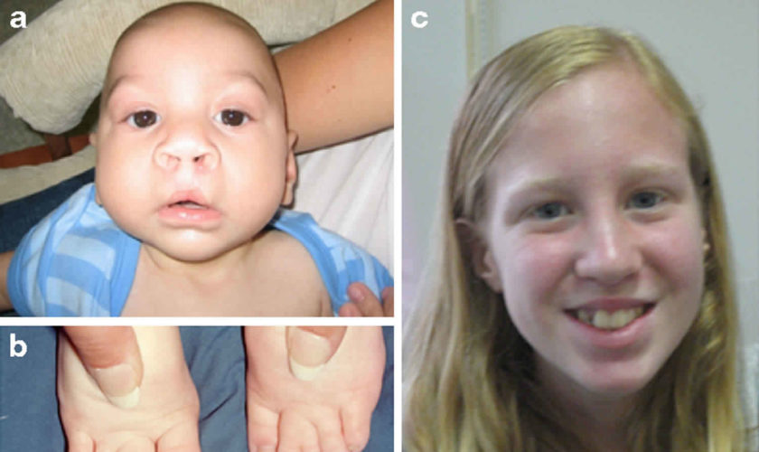
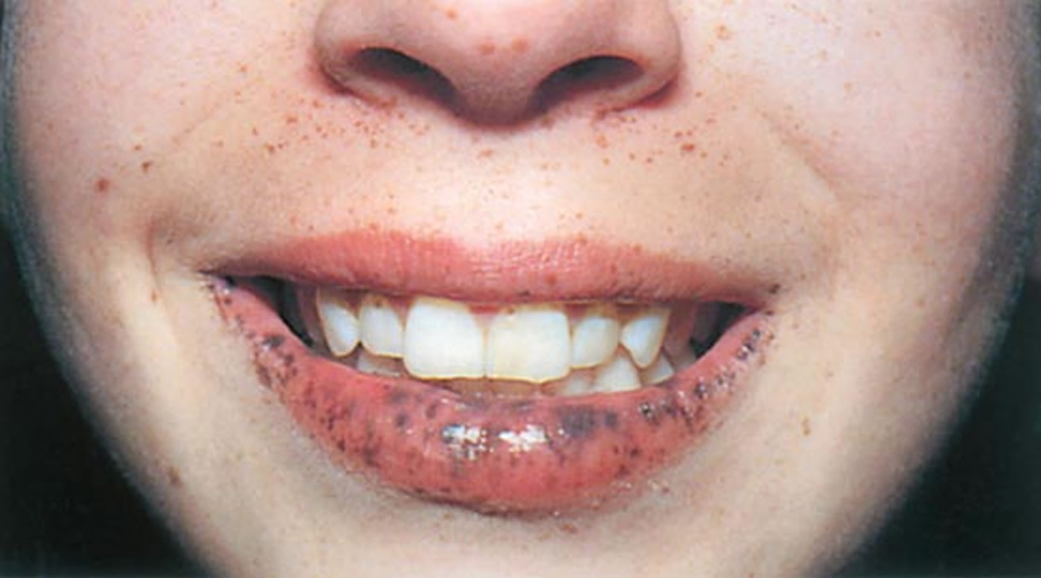
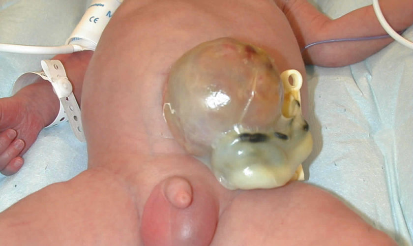
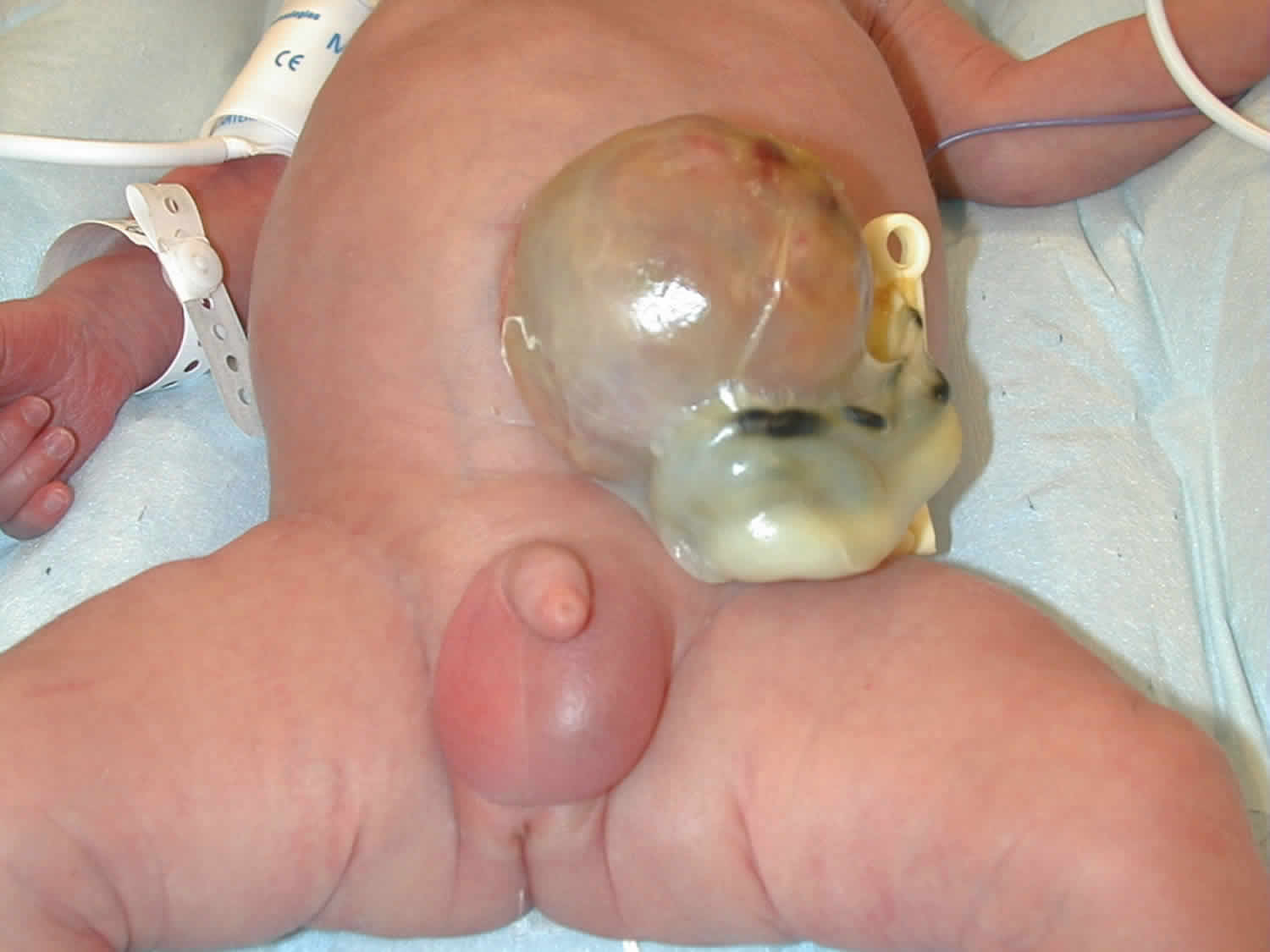
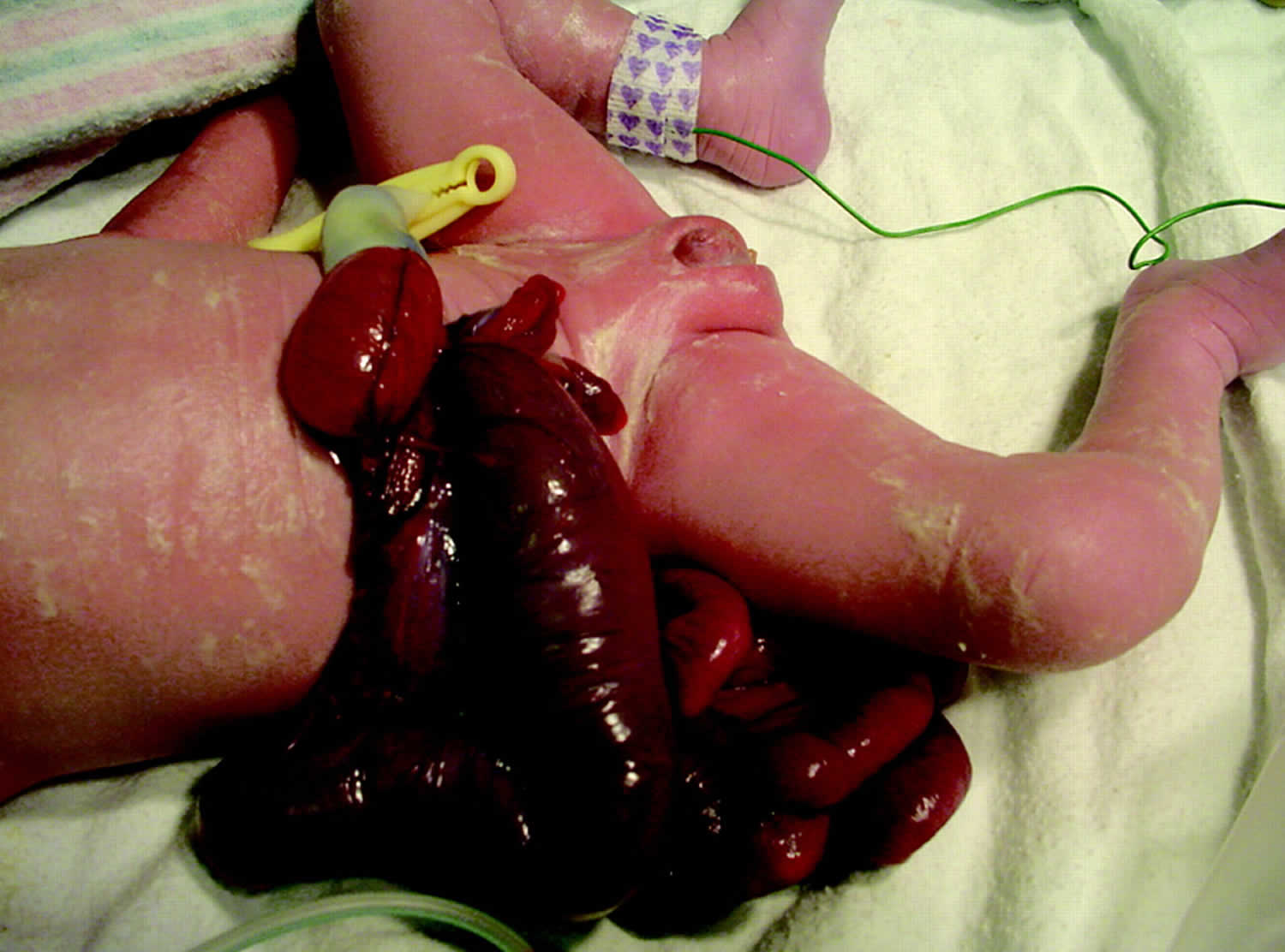
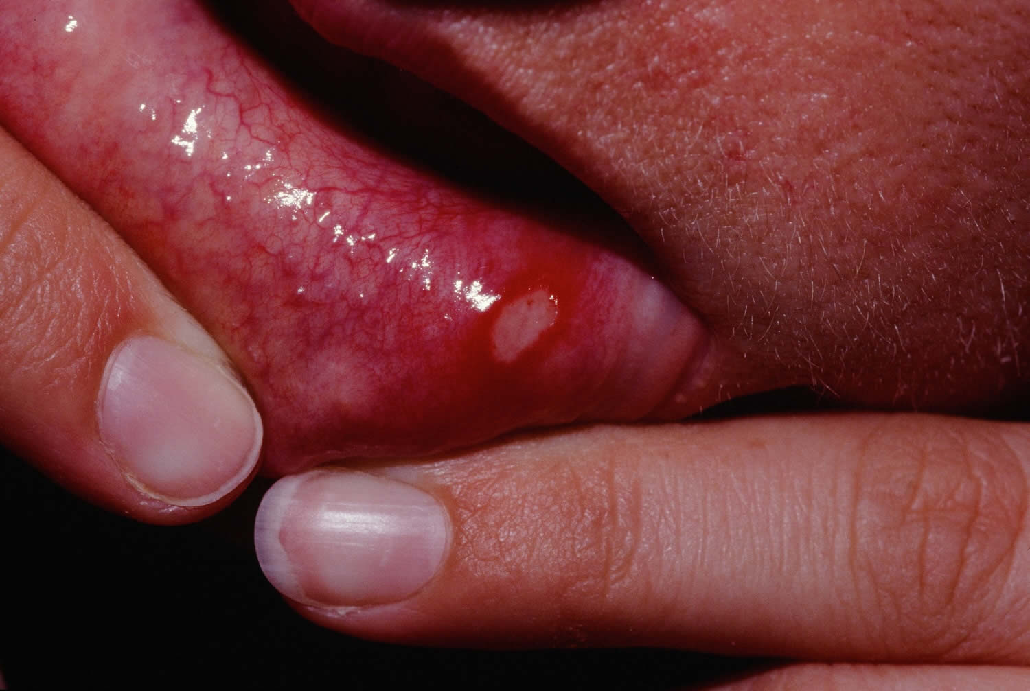
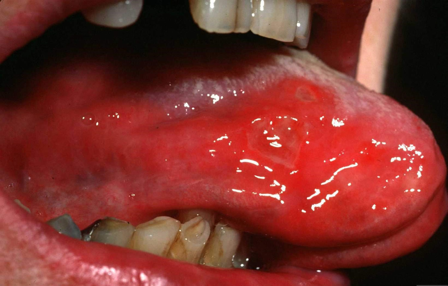
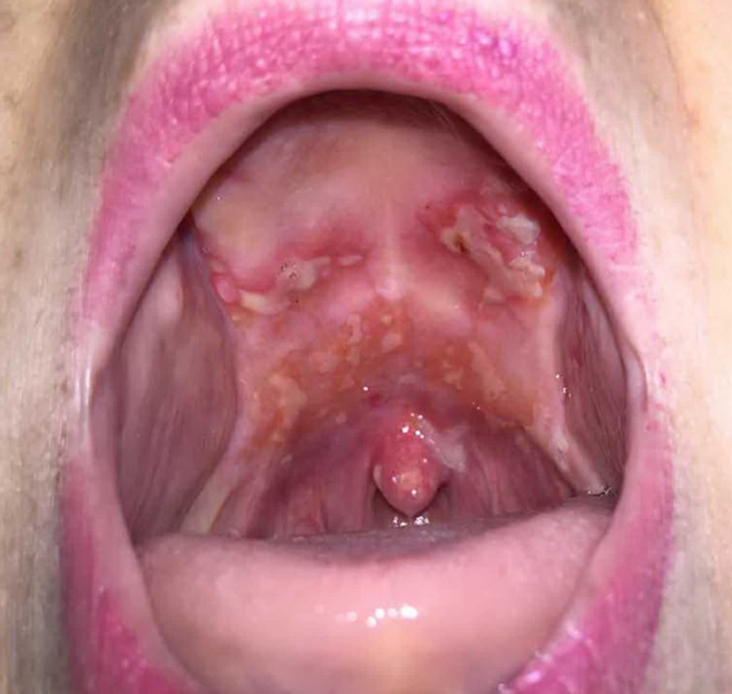
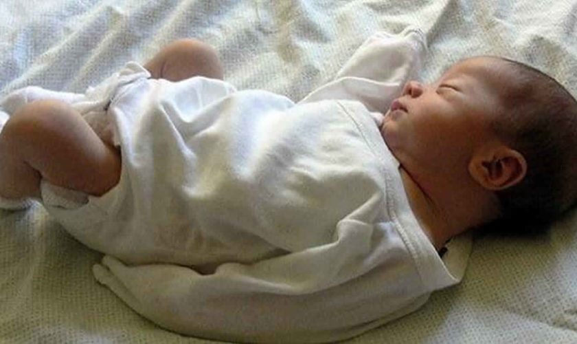
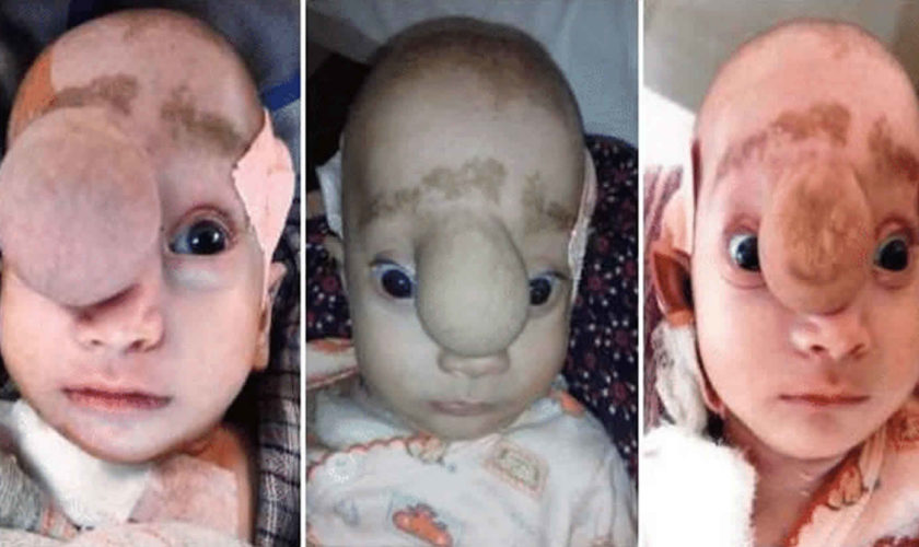
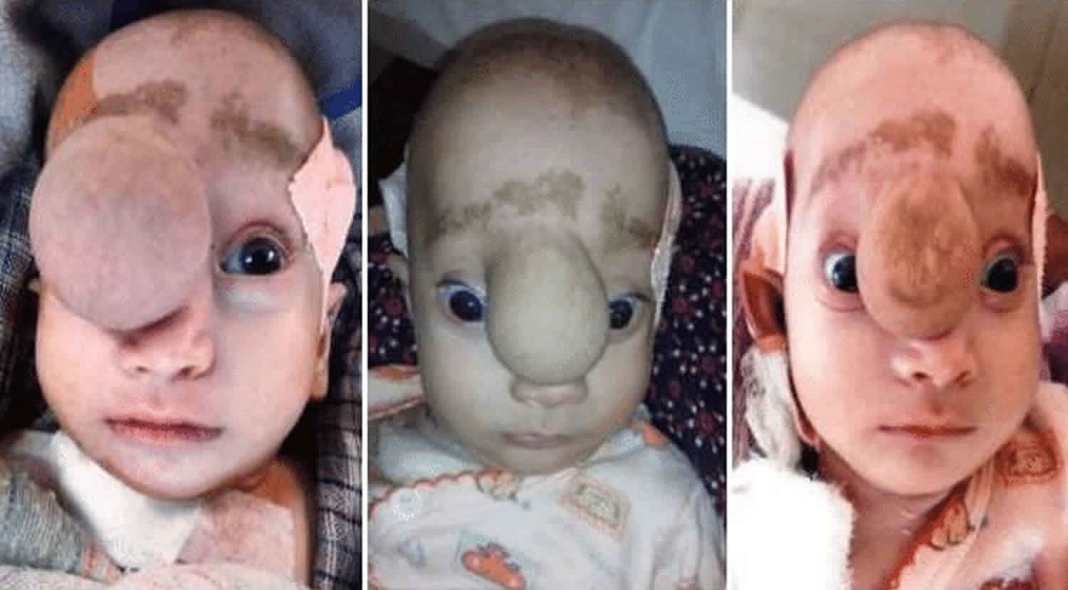
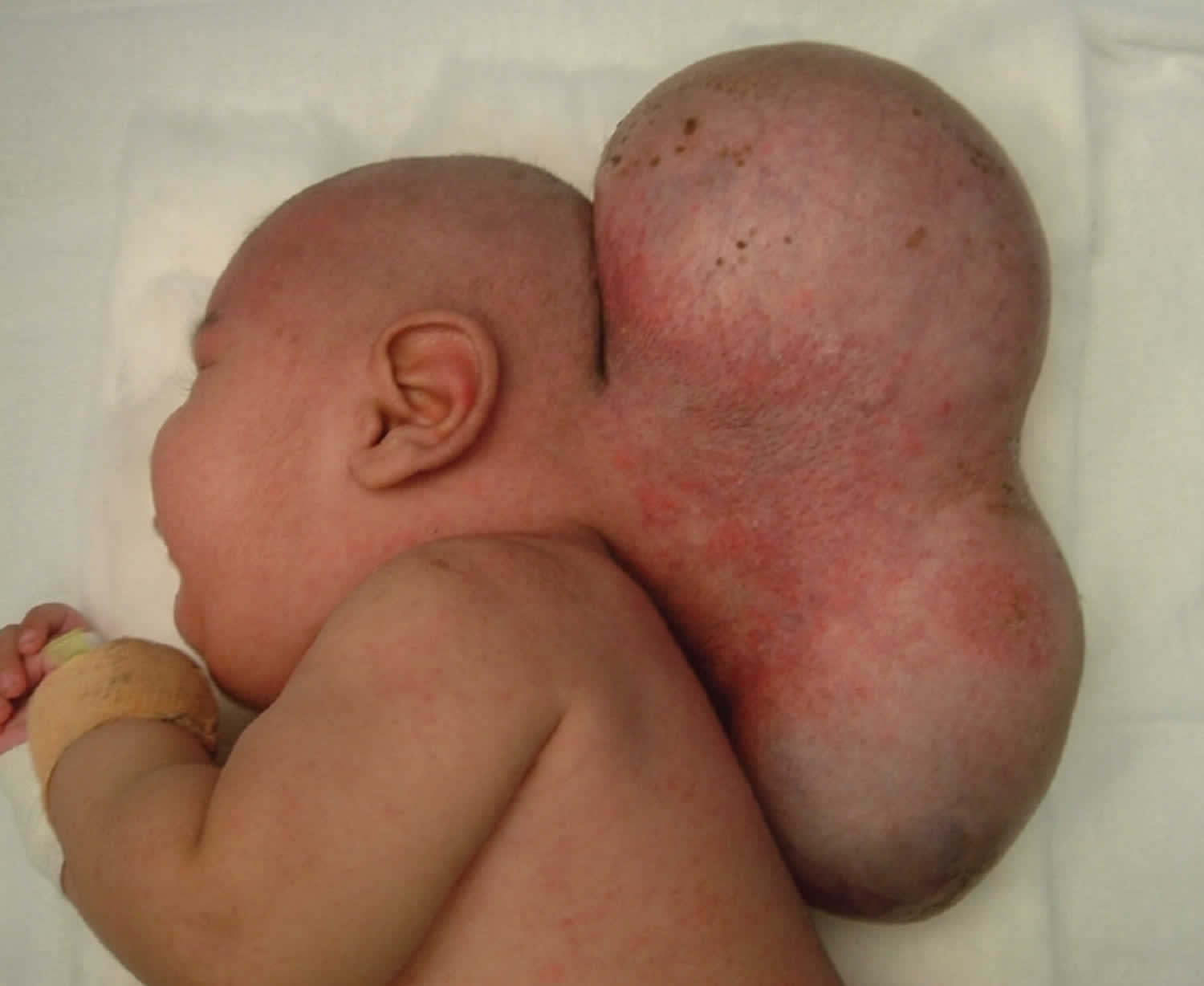
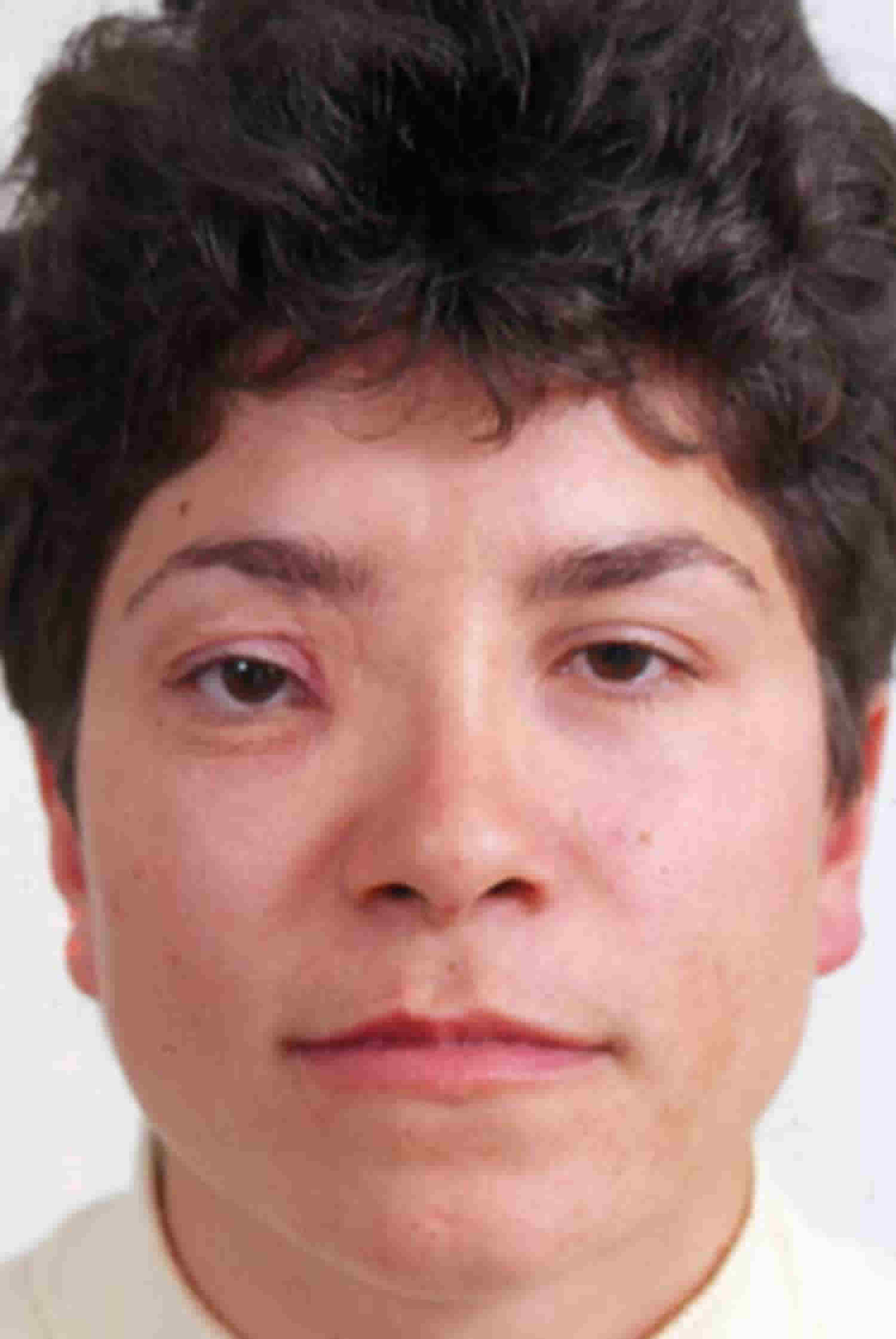
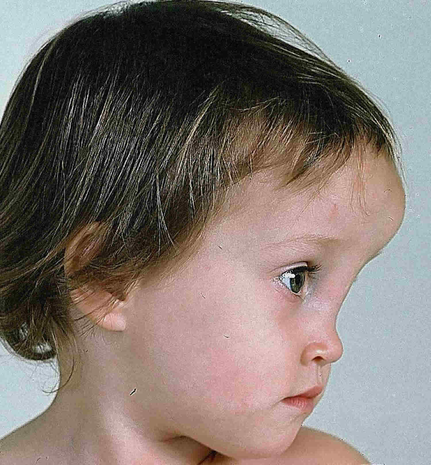
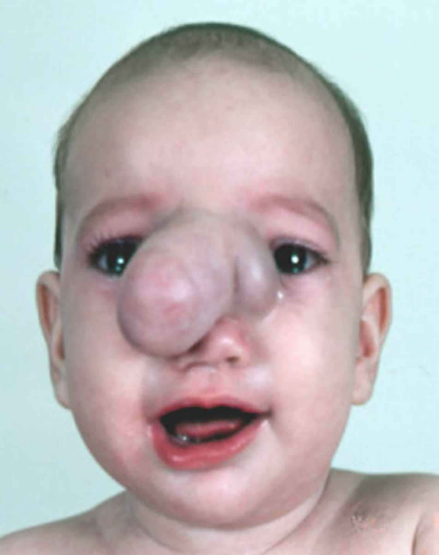
Figure 1. Smith-Lemli-Opitz syndrome

Smith Lemli Opitz syndrome

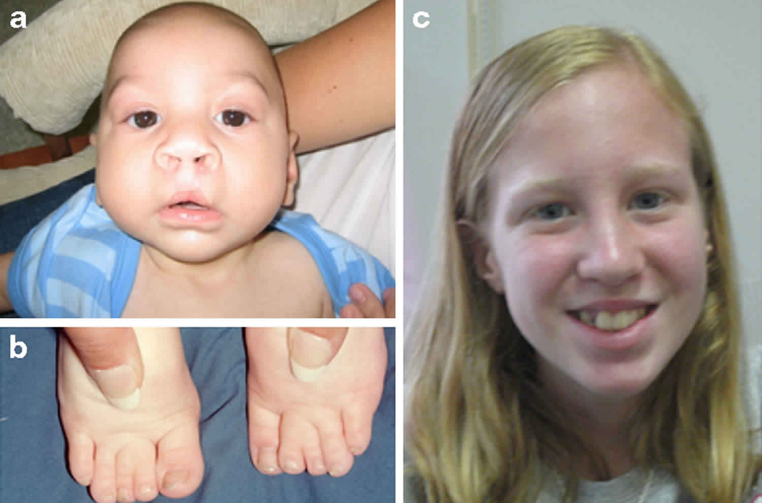
Footnote: Clinical features of Smith–Lemli–Opitz syndrome. (a) Typical facial features of Smith-Lemli-Opitz syndrome: microcephaly, bitemporal narrowing, ptosis, short nasal root, anteverted nares and micrognathia. (b) Toe syndactyly in Smith-Lemli-Opitz syndrome. (c) Smith-Lemli-Opitz syndrome with mild phenotype.

[Source 4) ]

Smith-Lemli-Opitz syndrome causes

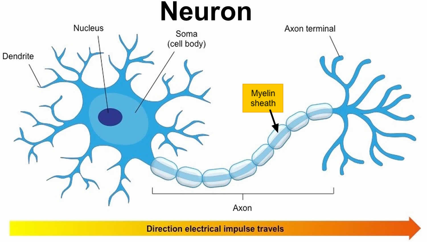
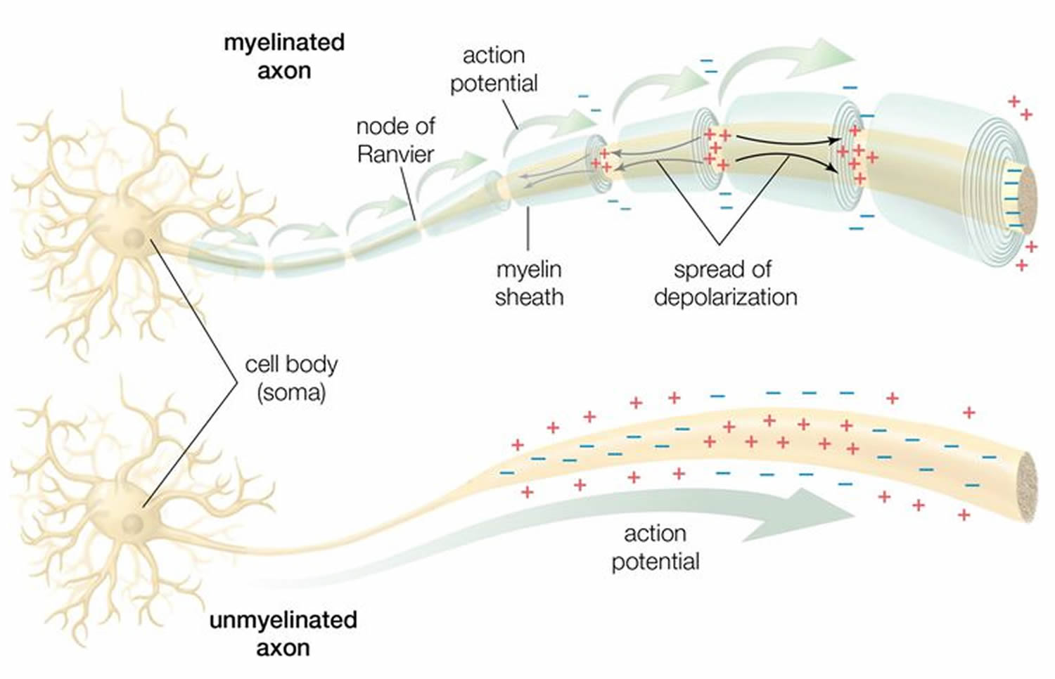
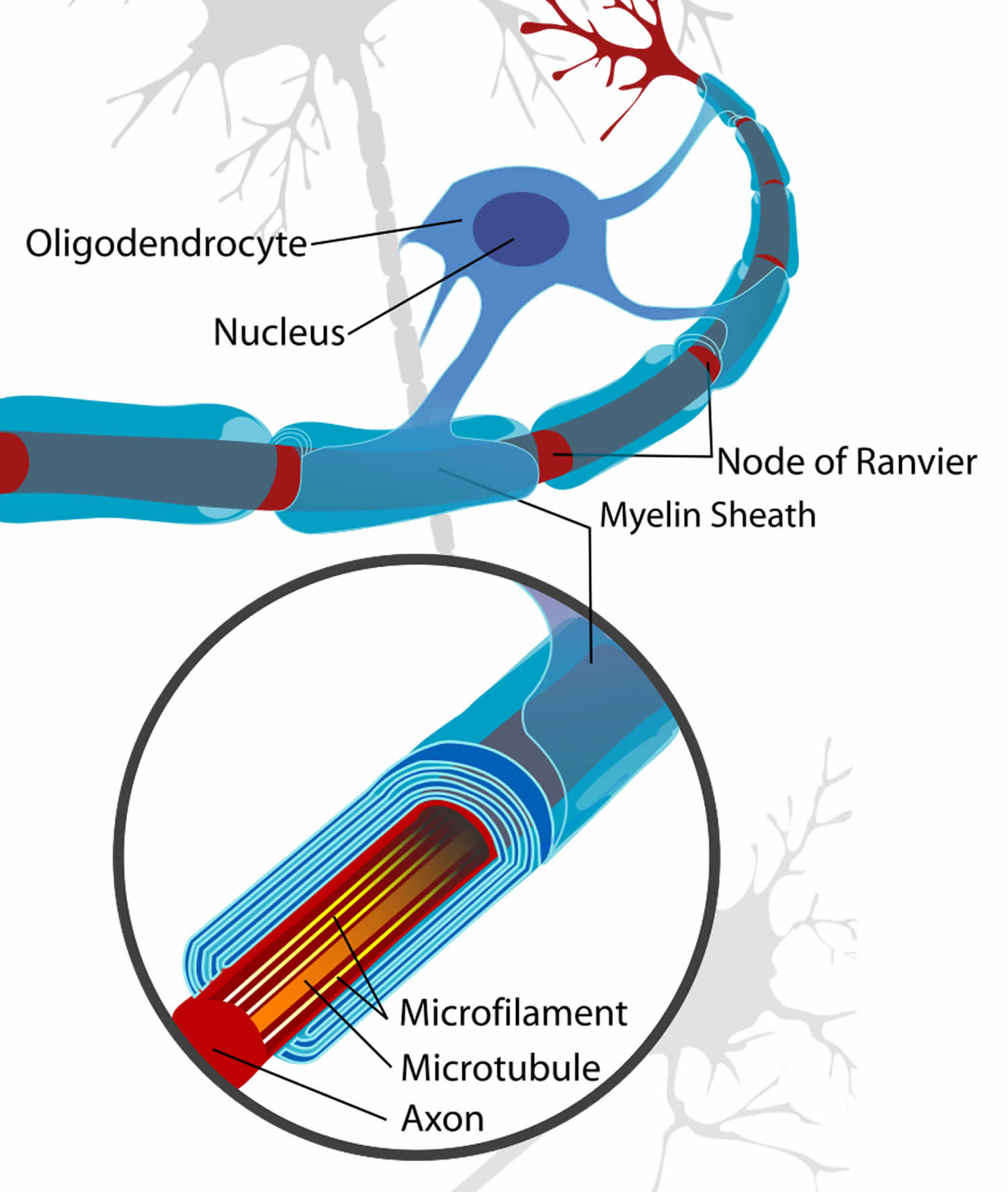
Mutations in the DHCR7 gene cause Smith-Lemli-Opitz syndrome. The DHCR7 gene provides instructions for making an enzyme called 7-dehydrocholesterol reductase. This enzyme is responsible for the final step in the production of cholesterol. Cholesterol is a waxy, fat-like substance that is produced in the body and obtained from foods that come from animals (particularly egg yolks, meat, poultry, fish, and dairy products). Cholesterol is necessary for normal embryonic development and has important functions both before and after birth. It is a structural component of cell membranes and the protective substance covering nerve cells (myelin). Additionally, cholesterol plays a role in the production of certain hormones and digestive acids.

Mutations in the DHCR7 gene reduce or eliminate the activity of 7-dehydrocholesterol reductase, preventing cells from producing enough cholesterol. A lack of this enzyme also allows potentially toxic byproducts of cholesterol production to build up in the blood, nervous system, and other tissues. The combination of low cholesterol levels and an accumulation of other substances likely disrupts the growth and development of many body systems. It is not known, however, how this disturbance in cholesterol production leads to the specific features of Smith-Lemli-Opitz syndrome.

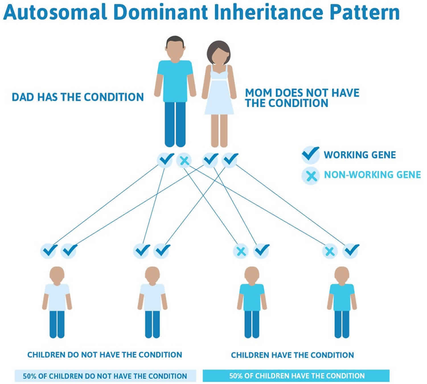
Smith-Lemli-Opitz syndrome inheritance pattern

This condition is inherited in an autosomal recessive pattern, which means both copies of the gene in each cell have mutations. The parents of an individual with an autosomal recessive condition each carry one copy of the mutated gene, but they typically do not show signs and symptoms of the condition.

It is rare to see any history of autosomal recessive conditions within a family because if someone is a carrier for one of these conditions, they would have to have a child with someone who is also a carrier for the same condition. Autosomal recessive conditions are individually pretty rare, so the chance that you and your partner are carriers for the same recessive genetic condition are likely low. Even if both partners are a carrier for the same condition, there is only a 25% chance that they will both pass down the non-working copy of the gene to the baby, thus causing a genetic condition. This chance is the same with each pregnancy, no matter how many children they have with or without the condition.

  • If both partners are carriers of the same abnormal gene, they may pass on either their normal gene or their abnormal gene to their child. This occurs randomly.
  • Each child of parents who both carry the same abnormal gene therefore has a 25% (1 in 4) chance of inheriting a abnormal gene from both parents and being affected by the condition.
  • This also means that there is a 75% ( 3 in 4) chance that a child will not be affected by the condition. This chance remains the same in every pregnancy and is the same for boys or girls.
  • There is also a 50% (2 in 4) chance that the child will inherit just one copy of the abnormal gene from a parent. If this happens, then they will be healthy carriers like their parents.
  • Lastly, there is a 25% (1 in 4) chance that the child will inherit both normal copies of the gene. In this case the child will not have the condition, and will not be a carrier.

These possible outcomes occur randomly. The chance remains the same in every pregnancy and is the same for boys and girls.

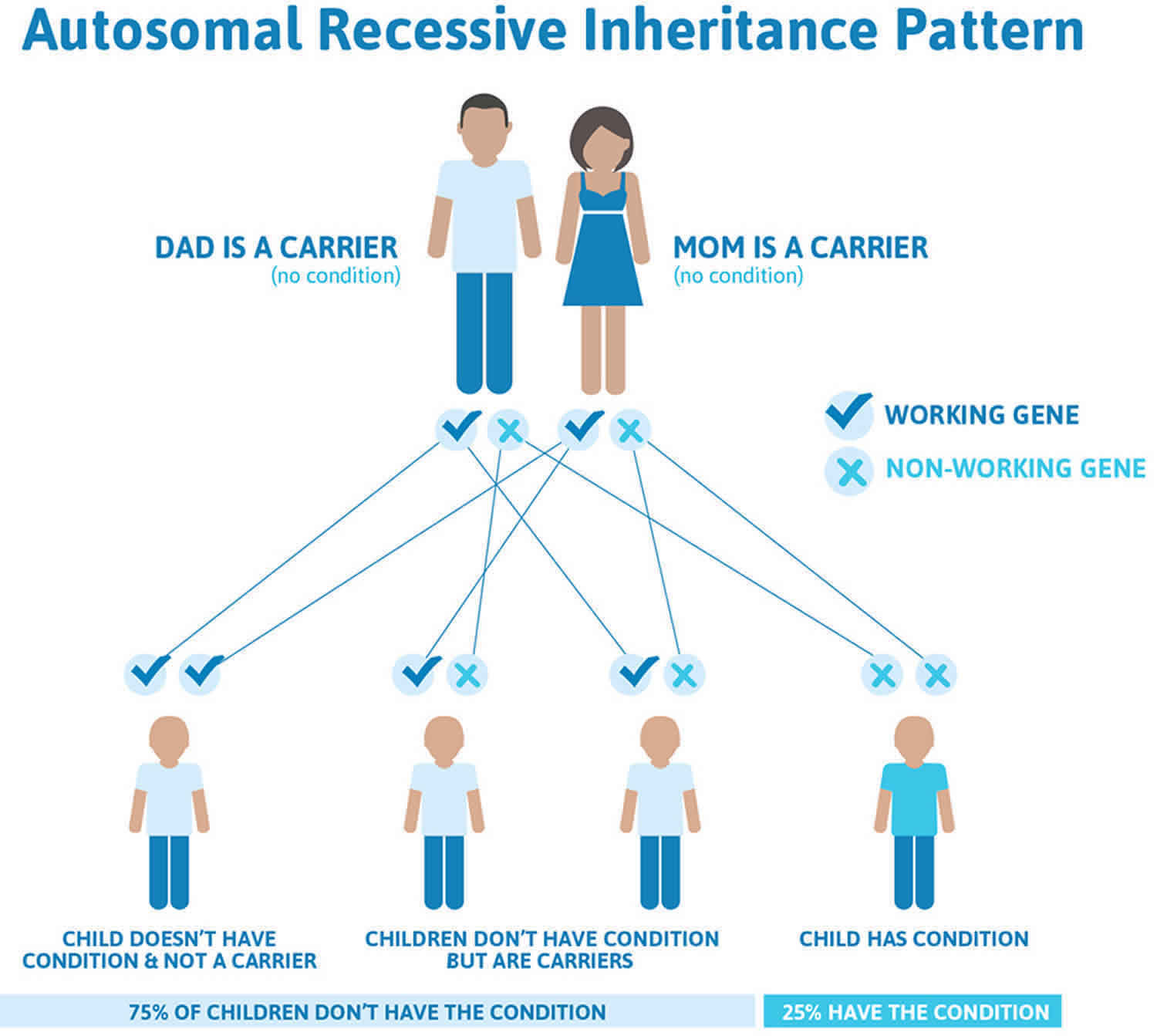
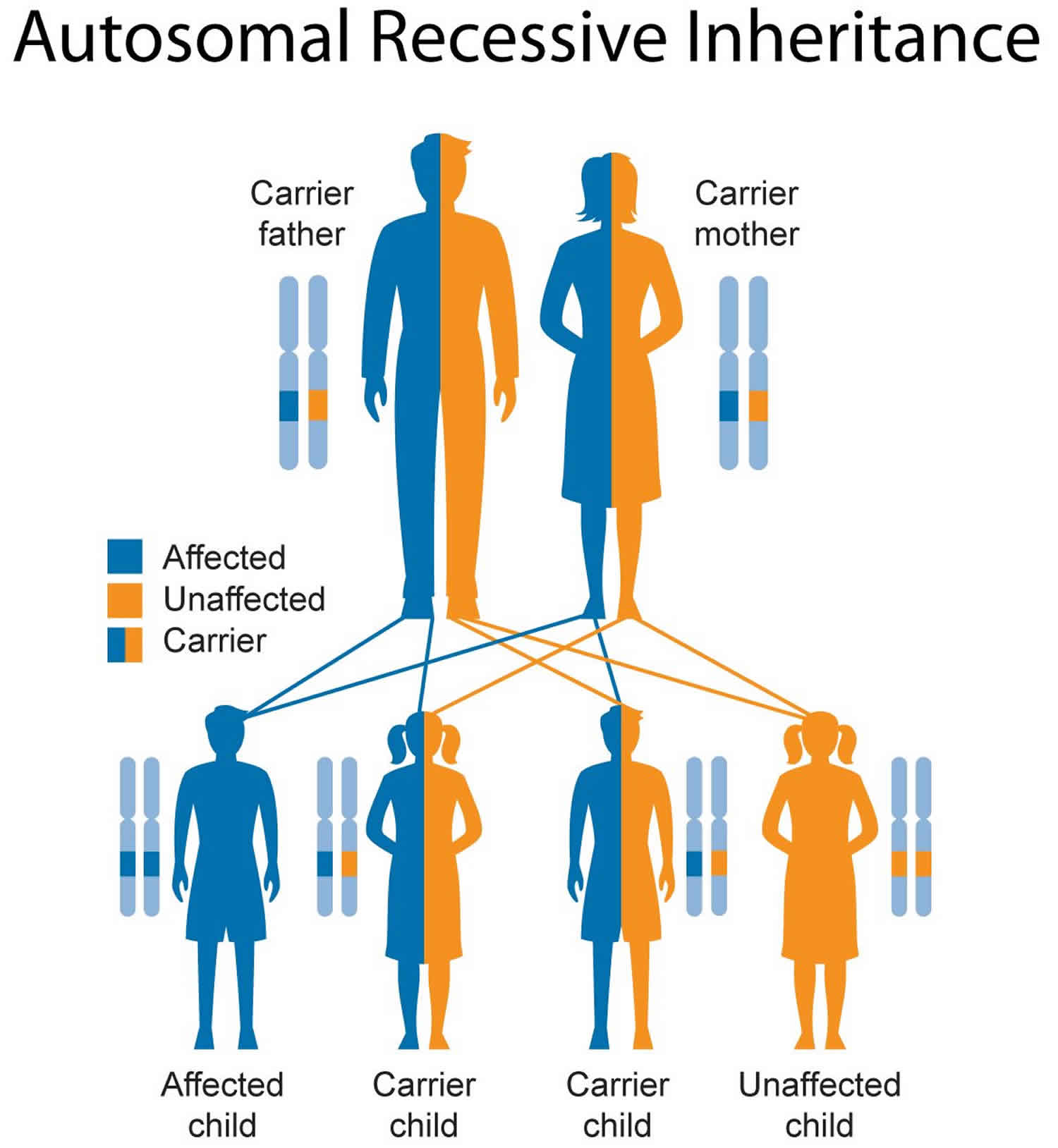
Figure 2 illustrates autosomal recessive inheritance. The example below shows what happens when both dad and mum is a carrier of the abnormal gene, there is only a 25% chance that they will both pass down the abnormal gene to the baby, thus causing a genetic condition.

Figure 2. Smith-Lemli-Opitz syndrome autosomal recessive inheritance pattern

Smith-Lemli-Opitz syndrome autosomal recessive inheritance pattern

People with specific questions about genetic risks or genetic testing for themselves or family members should speak with a genetics professional.

Resources for locating a genetics professional in your community are available online:

Smith-Lemli-Opitz syndrome symptoms

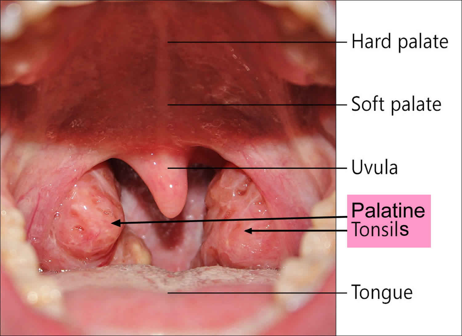
Smith-Lemli-Opitz syndrome symptoms vary from person to person, depending upon the amount of cholesterol they can produce. In addition to mental retardation and poor growth, common physical signs of Smith-Lemli-Opitz syndrome are a cleft palate (a split upper lip), malformed genitals (in males), and polydactyly (extra fingers or toes).

Other symptoms that may be present at birth include: microcephaly (small head), webbing between the second and third toes, drooping eyelids, heart defects, hearing or sight loss, and difficulties feeding.

The following signs and symptoms may be noted in Smith-Lemli-Opitz syndrome:

  • Lethargy
  • Obtundation or coma
  • Respiratory failure
  • Hearing loss
  • Visual loss
  • Vomiting
  • Feeding difficulties
  • Failure to thrive
  • Constipation
  • Cyanosis
  • Congestive heart failure
  • Photosensitivity

Neuropsychiatric and neurodevelopmental abnormalities are common and include variable intellectual disability, aberrant behavior, and autism.

Smith-Lemli-Opitz syndrome diagnosis

The diagnosis of Smith-Lemli-Opitz syndrome is based on physical findings and detection of an elevated concentration of 7-dehydrocholesterol (7-DHC) in blood serum or an elevated 7-dehydrocholesterol:cholesterol ratio. Molecular genetic testing for mutations in the DHCR7 gene is available and is mainly used for carrier testing and prenatal diagnosis.

Smith-Lemli-Opitz syndrome treatment

There is no cure for Smith-Lemli-Opitz syndrome. Cholesterol therapy, which comes in several forms and can improve growth and development, is the recommended treatment. The results, however, vary and not every family sees significant change. Other possible treatments such as simvastatins and antioxidants are currently being investigated through clinical trials. Simvastatin has been successful in reducing cholesterol levels in individuals with Smith-Lemli-Opitz syndrome but must be used with caution because of the possible risk of liver damage. Surgery may be necessary to correct some of the physical deformities (cleft palate, heart defects) associated with the disorder.

Medical treatment for Smith-Lemli-Opitz syndrome is based on the specific problems that are present in the affected child. It is important that the child be evaluated for the range of conditions associated with Smith-Lemli-Opitz syndrome including eye, heart, musculoskeletal, genitourinary and gastrointestinal disorders and that a physician familiar with Smith-Lemli-Opitz syndrome oversees the care. Severely affected individuals may require surgery to correct cleft palate, heart defects and genital anomalies. Cholesterol supplementation (one or two egg yolks), sometimes in combination with bile acids, appears to improve growth and reduce photosensitivity in individuals with Smith-Lemli-Opitz syndrome with no harmful side effects.

Genetic counseling is recommended for the parents of an affected child.

Surgical care

  • Consider repair of congenital heart defects in cases of Smith-Lemli-Opitz syndrome type I.
  • Repair of polydactyly is best performed early.
  • Consider cleft palate repair as well as pyloromyotomy in a timely fashion in cases of pyloric stenosis.
  • Rectal biopsy for evaluation of ganglion cells may be useful when Hirschsprung disease is suspected and surgical management for Hirschsprung disease may be needed.
  • Gastrostomy placement, with or without fundoplication, may be necessary when feeding difficulties or gastrointestinal reflux is present.

Smith-Lemli-Opitz syndrome life expectancy

A person with Smith-Lemli-Opitz syndrome who has appropriate medical care and follows a proper diet has the potential for a normal life expectancy. Independent living is unlikely, however, due to the presence of intellectual disability. Children with the most severe cases of Smith-Lemli-Opitz syndrome (those who produce little or no cholesterol) often die within a few months of birth 5).

References   [ + ]

read more

Peutz Jeghers syndrome

Peutz-Jeghers-syndrome

Peutz Jeghers syndrome

Peutz-Jeghers syndrome is a rare inherited disease that is characterized by the development of noncancerous growths called hamartomatous polyps (benign tumors made up of a mixture of mature cells normally found in that tissue) in the gastrointestinal tract particularly the stomach and intestines. Patients with Peutz-Jeghers syndrome have an estimated 15-fold increased risk of developing intestinal cancer compared to the general population 1). Approximately 50% of patients with Peutz Jeghers syndrome develop and die from cancer by 57 years of age. The overall risk of Peutz-Jeghers syndrome patients developing a cancer over adult life is 93%. Cancers are not only located on the gastrointestinal tract but can occur on many other sites including the breast, ovary, testicle, pancreas, uterus, esophagus and lung.

Children with Peutz-Jeghers syndrome often develop small, dark-colored spots on the lips, around and inside the mouth (buccal mucosa), near the eyes and nostrils, and around the anus (perianal). These spots may also occur on the hands and feet. They appear during childhood and often fade as the person gets older. In addition, most people with Peutz-Jeghers syndrome develop multiple polyps in the stomach and intestines during childhood or adolescence. Polyps can cause health problems such as recurrent bowel obstructions, chronic bleeding, and abdominal pain. Around half of patients with Peutz Jeghers syndrome have to undergo surgery by age 18 because of polyps-related complication. Polyps most often tend to develop in the small intestine (in the jejunum, specifically) but can also arise in the stomach and large intestine. Rarely, polyps can grow outside the gastrointestinal tract and affect the ureters, bladder, lungs, bronchi, and gallbladder. Gastrointestinal polyps can cause abdominal pain, vomiting, diarrhea, intestinal obstruction and rectal bleeding, which can lead to anemia. They can also provoke folding of the intestine into itself (intussusception), which can lead to severe abdominal pain and emergency surgery.

People with Peutz-Jeghers syndrome have a high risk of developing cancer during their lifetimes. Cancers of the gastrointestinal tract, pancreas, cervix, ovary, and breast are among the most commonly reported tumors. Individuals that develop cancer are usually affected around their fifth decade of life (age 40-49). Affected females have an increased risk for a benign ovarian tumor called SCTAT (sex cord tumors with annular tumors) for which symptoms may include irregular or heavy periods or early puberty. Usually before age 20, affected males can develop a tumor in the testes, called Sertoli cells carcinoma that secretes estrogen and can lead to breast development (gynecomastia).

Peutz Jeghers syndrome is caused by changes (mutations) in the STK11 gene and is inherited in an autosomal dominant manner 2). Some people with Peutz Jeghers syndrome do not have mutations in the STK11 gene. In these cases, the cause is unknown 3).

The prevalence of Peutz Jeghers syndrome is uncertain; estimates range from 1 in 25,000 to 300,000 individuals 4).

As there is no cure for Peutz Jeghers syndrome, treatment is mostly focused on surveillance and control of symptoms. People with Peutz Jeghers syndrome should be monitored by a health care provider and checked regularly for cancerous polyp changes. Surgery may be needed to remove polyps that cause long-term problems. Iron supplements help counteract blood loss.

Figure 1. Peutz Jeghers syndrome lips

Peutz Jeghers syndrome lips

Peutz Jeghers syndrome causes

Mutations in the STK11 gene (also known as LKB1) cause most cases of Peutz-Jeghers syndrome. The STK11 gene is a tumor suppressor gene, which means that it normally prevents cells from growing and dividing too rapidly or in an uncontrolled way. A mutation in this gene alters the structure or function of the STK11 protein, disrupting its ability to restrain cell division. The resulting uncontrolled cell growth leads to the formation of noncancerous polyps and cancerous tumors in people with Peutz-Jeghers syndrome.

A small percentage of people with Peutz-Jeghers syndrome do not have mutations in the STK11 gene. In these cases, the cause of the disorder is unknown.

Peutz Jeghers syndrome inheritance pattern

Peutz-Jeghers syndrome is inherited in an autosomal dominant pattern, which means one copy of the altered gene in each cell is sufficient to increase the risk of developing noncancerous polyps and cancerous tumors. In about half of all cases, an affected person inherits a mutation in the STK11 gene from one affected parent. The remaining cases occur in people with no history of Peutz-Jeghers syndrome in their family. These cases appear to result from new (de novo) mutations in the STK11 gene. For the individual with the condition, the chance of their children inheriting it will be 50%. However, other family members are generally not likely to be at increased risk.

In cases where the autosomal dominant condition does run in the family, the chance for an affected person to have a child with the same condition is 50% regardless of whether it is a boy or a girl. These possible outcomes occur randomly. The chance remains the same in every pregnancy and is the same for boys and girls.

  • When one parent has the abnormal gene, they will pass on either their normal gene or their abnormal gene to their child. Each of their children therefore has a 50% (1 in 2) chance of inheriting the changed gene and being affected by the condition.
  • There is also a 50% (1 in 2) chance that a child will inherit the normal copy of the gene. If this happens the child will not be affected by the disorder and cannot pass it on to any of his or her children.

Figure 2 illustrates autosomal dominant inheritance. The example below shows what happens when dad has the condition, but the chances of having a child with the condition would be the same if mom had the condition.

Figure 2. Peutz Jeghers syndrome autosomal dominant inheritance pattern

Peutz Jeghers syndrome autosomal dominant inheritance pattern

People with specific questions about genetic risks or genetic testing for themselves or family members should speak with a genetics professional.

Resources for locating a genetics professional in your community are available online:

Peutz Jeghers syndrome symptoms

Symptoms of Peutz Jeghers syndrome are:

  • Brownish or bluish-gray spots on the lips, gums, inner lining of the mouth, and skin
  • Clubbed fingers or toes
  • Cramping pain in the belly area
  • Dark freckles on and around the lips of a child
  • Blood in the stool that can be seen with the naked eye (sometimes)
  • Vomiting

Symptoms usually appear during the first decade of life and begin with spots of dark skin freckling (melanocytic macules) around the mouth, eyes, nostrils, fingers as well as inside the mouth (oral mucosa) and around the anus (perianal). Multiple benign polyps called hamartomas also begin to grow in the gastrointestinal tract of affected individuals around that age. These polyps are located throughout the gastrointestinal tract and can cause nausea, vomiting, abdominal pain, intestinal obstruction and rectal bleeding. Abdominal surgery or endoscopic procedures might be necessary to remove polyps (polypectomy) to prevent polyps-related complications, such as folding of the intestine into itself (intussusception).

Clinical features of Peutz Jeghers syndrome can be divided into two types, cutaneous and gastrointestinal.

Cutaneous features

The most noticeable cutaneous feature of Peutz Jeghers syndrome is the appearance of melanocytic macules (pigmented spots) in 95% of patients. Tan, dark brown, or bluish black flat patches 1 to 5 mm in size are seen around the mouth, lips, gums, inner lining of the mouth, eyes, hands and feet, fingers and toes, anus and genital areas. Pigmentation usually appears before 5 years of age and may fade after puberty.

Gastrointestinal features

Gastrointestinal polyps occur later on in life and are rare in childhood. The polyps may cause bleeding and abdominal pain. They have a high chance of becoming malignant.

Small intestine intussusception (when one portion of an intestine protrudes into another) and intestinal obstruction are also fairly common complications of Peutz-Jeghers syndrome.

Peutz Jeghers syndrome diagnosis

A diagnosis of Peutz-Jeghers syndrome is based on the presence of characteristic signs and symptoms. In people with a clinical diagnosis of Peutz Jeghers syndrome, genetic testing of the STK11 gene confirms the diagnosis in approximately 100% of people who have a positive family history and approximately 90% of people who have no family history of Peutz Jeghers syndrome.

The polyps develop mainly in the small intestine, but also in the large intestine (colon). An exam of the colon called a colonoscopy will show colon polyps. The small intestine is evaluated in two ways. One is a barium x-ray (small bowel series). The other is a capsule endoscopy, in which a small camera is swallowed and then takes many pictures as it travels through the small intestine.

A clinical diagnosis of Peutz Jeghers syndrome can be made when any one of the following criteria is present:

  • Presence of at least two Peutz Jeghers syndrome polyps
  • Any number of Peutz Jeghers syndrome polyps and at least one close relative diagnosed with Peutz Jeghers syndrome Characteristic dark pigmented spots (melanocytic macules) and at least one close relative diagnosed with Peutz Jeghers syndrome
  • Any number of Peutz Jeghers syndrome polyps and characteristic dark pigmented spots
  • Gynecomastia in males as a result of estrogen-producing Sertoli cell testicular tumors
  • History of intussusception, especially in a child or young adult

Additional exams may show:

  • Part of the intestine folded in on itself (intussusception)
  • Benign (noncancerous) tumors in the ear

Laboratory tests may include:

  • Complete blood count — may reveal anemia
  • Stool guaiac, to look for blood in stool
  • Total iron-binding capacity (TIBC) — may be linked with iron-deficiency anemia

Peutz Jeghers syndrome treatment

Individuals with Peutz Jeghers syndrome have an increased risk for intestinal and other cancers. Frequent medical examination and testing is necessary to allow early detection of polyps and cancer. Peutz Jeghers syndrome management typically includes high-risk screening for associated polyps and cancers 5). There is no specific treatment for Peutz Jeghers syndrome but the main goal is to manage and prevent associated problems of intestinal obstruction and intussusception, and cancer development.

Management of Peutz Jeghers syndrome patients should include:

  • Annual complete blood count (CBC). The polyps of Peutz-Jeghers syndrome may ulcerate and be a source of blood loss and anemia; gastrointestinal bleeding may be massive but also microscopic, with subsequent iron deficiency, therefore, cell counts and iron studies should be monitored.
  • Iron studies
  • Fecal occult blood: Hemoccult should be performed to check for occult blood in the stool
  • Carcinoembryonic antigen (CEA): This test has been used by some clinicians for screening and monitoring of cancer degeneration.
  • Cancer antigen (CA)–19-9 and CA-125: CA-125 testing is indicated every year starting at age 18 years, and CA 19-9 is indicated every 1-2 years starting at age 25 years 6)
  • Annual physical examination of breasts, abdomen, pelvis and testes
  • Repeated removal of bleeding or large polyps (>5 mm) by endoscopic polypectomy
  • Laparotomy and resection as required for repeated or persistent intestinal intussusception, obstruction, or persistent bleeding
  • Surgical removal of cancers as they are diagnosed

After initial diagnosis, it is recommended that individuals older than 8 years or having symptoms undergo endoscopic and small bowel examination. The latter can be done with magnetic resonance imaging of the intestines (magnetic resonance enterography) or by swallowing a capsule that records internal images from inside the gastrointestinal tract (video capsule endoscopy). Gynecologic and breast examination are also recommended for women older than 18 years. Testicular examination is recommended for men.

Following initial workup after the diagnosis, endoscopy, colonoscopy, and small bowel examination should be performed every 2-3 years to detect polyps and potential tumors. An annual mammogram is recommended for women. Testicular ultrasound can be done every two years for men.

As Peutz Jeghers syndrome increases the risk of breast, uterine, and ovarian cancer, it is possible for affected women to undergo preventive mastectomy, hysterectomy or salpingo-oophorectomy (surgical removal of the breasts, uterus, and fallopian tubes and ovaries, respectively).

Polyps over 1 cm in size are removed with endoscopic techniques to avoid polyps-related complications such as bleeding and intussusception. These complications might require surgical interventions to be corrected. If a patient undergoes surgery, endoscopic removal of polyps (polypectomy) is performed at the same time as surgery to reduce the risk of recurrence of complications and surgery.

The freckling may be less obvious with careful sun protection. In some cases, the pigmentation may be lessened by cosmetic treatment. Cosmetic camouflage may also be useful. In cases where dark pigmented spots (melanocytic macules) have a greatly negative psychological impact on affected individuals, they can be partially removed with laser treatment.

DNA screening may be offered to family members to see if they have inherited the gene mutation. If so, they should also undergo regular screening for disease; however, not all families with Peutz-Jeghers syndrome map to the STK11/LKB1 locus 7). Thus, a negative genetic test does not exclude the diagnosis.

References   [ + ]

read more

Omphalocele

omphalocele

What is omphalocele

An omphalocele is also called exomphalos, is a birth defect in which an infant’s intestine, liver and other abdominal organs are outside of the body because of a hole in the belly button (navel) area at the base of the umbilical cord insertion. The intestines are covered only by a thin layer of tissue (omphalocele sac) that hardly ever is open or broken and can be easily seen. Omphalocele is a life-threatening condition. It needs to be treated soon after birth so that the baby’s organs can develop and be protected in the belly. Surgical repair is performed primarily in stages, or after a period of waiting which can last several months.

Because some or all of the abdominal (belly) organs are outside of the body, babies born with an omphalocele can have other problems. The abdominal cavity, the space in the body that holds these organs, might not grow to its normal size. Also, infection is a concern, especially if the sac around the organs is broken. Sometimes, an organ might become pinched or twisted, and loss of blood flow might damage the organ.

One-third of all babies with omphalocele have liver herniation, which is often associated with a small belly size and small lungs (known as pulmonary hypoplasia), two factors that can affect treatment and long-term outcomes. Up to one-third can also have a heart defect which can also affect long-term outcome.

Omphalocele is a rare birth defect that occurs in 1 in 4,000 — 7,000 live births. The Centers for Disease Control and Prevention (CDC) estimates that each year about 775 babies in the United States are born with an omphalocele 1). Many babies born with an omphalocele also have other birth defects, such as heart defects, neural tube defects, and chromosomal abnormalities 2). Therefore, all babies born with an omphalocele should have chromosome testing. This will help parents understand the risk for this disorder in future pregnancies.

Omphalocele occurs very early in pregnancy when the abdominal cavity fails to form normally. The abdominal cavity is normally formed at three to four weeks gestation when the disk-like embryo undergoes infolding. A large or “giant omphalocele” forms when there is a failure of lateral infolding of the embryo, resulting in an inadequate abdominal cavity with containment of the abdominal organs only by a thin clear membrane called the omphalocele sac.

Smaller omphaloceles, also referred to as “hernia of the cord,” form later (eight to 11 weeks gestation) after normal infolding of the embryo occurs (resulting in a formed abdominal cavity), when the umbilical ring fails to close around the umbilical cord resulting in a small defect that usually contains only intestine. Small omphaloceles are more likely to be associated with chromosomal defects or syndromes.

Omphalocele differs from gastroschisis in that the protruding organs are covered by the omphalocele sac. Gastroschisis has no sac and is likely caused by a rupture of a hernia of the cord, resulting in extrusion of intestine through the small umbilical defect. In contrast to gastroschisis, a ruptured giant omphalocele has all of the organs, including liver, outside the abdomen without a covering membrane.

In addition, compared to gastroschisis, giant omphaloceles are frequently associated with small lung size. Finally, whereas gastroschisis often develops in the first pregnancy of young mothers, omphaloceles typically develop in the pregnancies of older women.

If the omphalocele is identified before birth, the mother should be closely monitored to make sure the unborn baby remains healthy.

Plans should be made for careful delivery and immediate management of the problem after birth. The baby should be delivered in a medical center that is skilled at repairing abdominal wall defects. Babies are likely to do better if they do not need to be taken to another center for further treatment.

For more information about the diagnosis, delivery and treatment of babies with omphalocele, watch the educational video series about abdominal wall defects below.

Figure 1. Omphalocele

omphalocele

Omphalocele vs Gastroschisis

Omphalocele looks similar to gastroschisis. An omphalocele is a birth defect in which the infant’s intestine or other abdominal organs protrude through a hole in the belly button area and are covered with a membrane. In gastroschisis, there is no covering membrane (the omphalocele sac). Presence of liver tissue within the herniated sac is more common finding in omphalocele than gastroschisis. Gastroschisis has no sac and is likely caused by a rupture of a hernia of the umbilical cord, resulting in extrusion of intestine through the small umbilical defect. In contrast to gastroschisis, a ruptured giant omphalocele has all of the organs, including liver, outside the abdomen without a covering membrane.

Gastroschisis is a congenital anterior abdominal wall defect, adjacent and usually to the right of the umbilical cord insertion 3). Gastroschisis occurs as a small, full-thickness periumbilical cleft either immediately adjacent to the umbilicus or separated from it by a strip of skin. This results in herniation of the abdominal contents into the amniotic sac, usually just the small intestine, but sometimes also the stomach, colon, and ovaries (Figure 2). The abdominal wall defect is relatively small compared with the size of the eviscerated bowel, which often develops walls that are matted and thickened with a fibrous peel. Gastroschisis has no covering sac and no associated syndromes. Babies with gastroschisis usually do not have other related birth defects. This differentiates it from an omphalocele, which usually is covered by a membranous sac and more frequently is associated with other structural and chromosomal anomalies (Table 1). In addition, although gastroschisis may be associated with gastrointestinal anomalies such as intestinal atresia, stenosis, and malrotation, it has a much better prognosis than omphalocele.

Mothers with the following may be at higher risk of having babies with gastrochisis:

  • Younger age
  • Fewer resources
  • Poor nutrition during pregnancy
  • Use illegal substances

If gastroschisis is found before birth, the mother will need special monitoring to make sure her unborn baby remains healthy.

Table 1. Differences Between Omphalocele and Gastroschisis

Gastroschisis Omphalocele
Incidence 1 in 10,000 (now increasing) 1 in 5,000
Defect Location Right paraumbilical Central
Covering Sac Absent Present (unless sac ruptured)
Description Free intestinal loops Firm mass including bowel, liver, etc
Associated With Prematurity 50% to 60% 10% to 20%
Necrotizing Enterocolitis Common (18%) Uncommon
Common Associated Anomalies Gastrointestinal (10% to 25%)

  • Intestinal atresia
  • Malrotation
Trisomy syndromes (30%)
Cardiac defects (20%)
Beckwith-Weidemann syndrome
Cryptorchidism (31%) Bladder extrophy
Prognosis Excellent for small defect Varies with associated anomalies
Mortality 5% to 10% Varies with associated anomalies (80% with cardiac defect)
[Source 4) ]

Figure 2. Gastroschisis

Gastroschisis

[Source 5) ]

Gastroschisis treatment

Treatment for gastroschisis is surgery to repair the defect. The surgeon will put the bowel back into the abdomen and close the defect, if possible. If the abdominal cavity is too small, a mesh sack is stitched around the borders of the defect and the edges of the defect are pulled up. The sack is called a silo. Over 5 to 7 days, the intestine returns into the abdominal cavity and the defect can be closed.

Other treatments for the baby include nutrients by IV and antibiotics to prevent infection. The baby’s temperature must be carefully controlled, because the exposed intestine allows a lot of body heat to escape.

Gastroschisis prognosis

The baby has a good chance of recovering if there are no other problems and if the abdominal cavity is large enough. A very small abdominal cavity may result in complications that require more surgeries.

Plans should be made for careful delivery and immediate management of the problem after birth. The baby should be delivered in a medical center that is skilled at repairing abdominal wall defects. Babies are likely to do better if they do not need to be taken to another center for further treatment.

Gastroschisis possible complications

A small number of babies with gastroschisis (about 10%) may have parts of the intestines that did not develop normally in the womb. With these babies, their intestines may not work normally even after the organs are put back inside the abdominal cavity.

The increased pressure from the misplaced abdominal contents can decrease blood flow to the intestines and kidneys. It can also make it difficult for the baby to expand the lungs, leading to breathing problems.

Another complication is bowel death necrosis. This occurs when intestinal tissue dies due to low blood flow or infection.

This condition is apparent at birth and will be detected in the hospital at delivery if it has not already been seen on routine fetal ultrasound exams during pregnancy. If you have given birth at home and your baby appears to have this defect, call the local emergency number (such as 911) right away.

Omphalocele causes

The causes of omphalocele among most infants are unknown. Some babies have omphalocele because of a change in their genes or chromosomes. Omphalocele might also be caused by a combination of genes and other factors, such as the things the mother comes in contact with in the environment or what the mother eats or drinks, or certain medicines she uses during pregnancy.

Omphalocele is considered an abdominal wall defect (a hole in the abdominal wall). Disturbance of organogenesis during the embryonic period results in omphalocele 6). Around weeks six through ten of pregnancy, the intestines get longer and push out from the abdominal cavity and protrude at the base of the umbilical cord. This event is known as physiologic midgut herniation and is easily identified in prenatal ultrasound between the 9 and 11 weeks of gestation 7). The liver is never present in the physiologic midgut herniation. By 11th to 12th weeks of gestation, the intestines normally go back into the belly. If this does not happen, an omphalocele occurs. Omphalocele occurs when the gut contents fail to rotate and return to the abdominal cavity. It can occasionally contain the liver in the presence of a large abdominal wall defect. The omphalocele can be small, with only some of the intestines outside of the belly, or it can be large, with many organs outside of the belly.

Recently, the Centers for Disease Control and Prevention (CDC) researchers have reported important findings about some factors that can affect the risk of having a baby with an omphalocele:

  • Alcohol and tobacco: Women who consumed alcohol or were heavy smokers (more than 1 pack a day) were more likely to have a baby with omphalocele 8)
  • Certain medications: Women who used selective serotonin-reuptake inhibitors (SSRIs) during pregnancy were more likely to have a baby with an omphalocele 9)
  • Obesity: Women who were obese or overweight before pregnancy were more likely to have a baby with an omphalocele 10)

Omphalocele looks similar to gastroschisis. An omphalocele is a birth defect in which the infant’s intestine or other abdominal organs protrude through a hole in the belly button area and are covered with a membrane. In gastroschisis, there is no covering membrane.

Abdominal wall defects develop as a baby grows inside the mother’s womb. During development, the intestines and other organs (liver, bladder, stomach, and ovaries or testes) develop outside the body at first and then usually return inside. In babies with omphalocele, the intestines and other organs remain outside the abdominal wall, with a membrane covering them. The exact cause for abdominal wall defects is not known.

Infants with an omphalocele often have other birth defects. Defects include genetic problems (chromosomal abnormalities), congenital diaphragmatic hernia, and heart and kidney defects. These problems also affect the overall outlook (prognosis) for the baby’s health and survival.

Omphalocele associated anomalies

Associated anomalies are high (27-91% 7) and are thought to be even commoner with smaller omphalocoele containing bowel only 11).

Omphalocele can be associated with several syndromes; the most common is Beckwith-Wiedemann syndrome 12). Beckwith-Wiedemann syndrome is an overgrowth syndrome characterized by macrosomia, enlarged tongue, neonatal hypoglycemia, ear creases, and pits, hemihypertrophy, visceromegaly, umbilical hernia, embryonal tumors, omphalocele, nephrocalcinosis, medullary sponge kidney disease, cardiomegaly, and nephromegaly. Traditionally, the macrosomia, macroglossia, and hypoglycemia are noted in the neonatal period. Hemihyperplasia is noted in segmental regions of the body or specific organs 13). Developmental and cognitive outcomes are typically normal. Patients with Beckwith-Wiedemann syndrome have an increased risk of cancer during the first eight years of life with embryonal tumors such as neuroblastoma, hepatoblastoma and Wilms tumor. These embryonal tumors have a higher cure rate when diagnosed early, making screening paramount for prevention. Screening for hepatoblastoma is performed by measuring serum alpha fetoprotein every 3 months until 4 years of age and screening for Wilms tumor is done every 3 months through 8 years of age with a complete abdominal ultrasonography 14).

Associated anomalies include:

  • Chromosomal anomalies: can occur in 20-50% of cases; the risk of an associated chromosomal anomaly gets higher when the omphalocele is detected earlier in gestation
    • Trisomy 18 (Edwards syndrome): considered the most common associated chromosomal anomaly. Dolichocephaly, external ear anomalies, micrognathia, short palpebral fissures, small face, clenched fist with overriding fingers, hypotonia and rocker bottom feet 15)
    • Trisomy 13 (Patau syndrome). Small eyes, cleft lip and palate, microcephaly, cryptorchidism, polydactyly, hypertelorism, micrognathia, cutis aplasia and external ears anomalies 16)
    • Trisomy 21 (Down syndrome). Hypotonia, upslanting palpebral fissures, brachycephaly, low set ears, single palmar crease, flat nasal bridge, brushfield spots around the iris, in-curved fifth digits and a gap between the first and second toes 17)
    • Turner syndrome
    • Klinefelter syndrome
    • Pallister-Killian syndrome
  • Other syndromic associations
    • Beckwith-Wiedemann syndrome
    • Carpenter syndrome: Kleeblattschadel skull deformity (trilobed cloverleaf skull) from pancraniosynostosis, syndactyly in the hands and feet, and mental retardation 18)
    • Marshall-Smith syndrome: Prominent forehead, shallow orbits, blue sclerae, depressed nasal bridge, micrognathia, accelerated skeletal maturation, respiratory difficulties, mental retardation 19)
    • Meckel-Gruber syndrome: Occipital encephalocele, cleft lip and palate, microcephaly, microphthalmia, abnormal genitalia, polycystic kidneys, and polydactyly 20)
    • Pentalogy of Cantrell. Ectopia cordis, midline supraumbilical abdominal defect, sternal cleft and intracardiac defect 21)
    • Shprintzen-Goldberg syndrome: Craniosynostosis, dolichocephaly, hypertelorism, exophthalmos, strabismus, elongated fingers and limbs, umbilical and abdominal hernias 22)
    • Charge syndrome: Coloboma, heart defect, choanal atresia, growth or developmental retardation, genital abnormality, and ear anomalies 23)
    • OEIS complex: omphalocele, bladder/cloacal exstrophy, imperforate anus, spinal anomalies 24)
    • Lethal omphalocele- cleft palate syndrome
  • Other fetal gastrointestinal anomalies: which confer a poor prognosis
  • Fetal CNS anomalies
  • Fetal cardiac anomalies: can occur in 50% of cases
  • Fetal genitourinary anomalies
    • Bladder exstrophy
    • Cloacal exstrophy
  • Fetal skeletal anomalies
    • Omphalocele-radial ray (ORR) complex

Omphalocele symptoms

An omphalocele can be clearly seen. This is because the abdominal contents stick out (protrude) through the belly button area.

There are different sizes of omphaloceles. In small ones, only the intestines remain outside the body. In larger ones, the liver or other organs may be outside as well.

Omphalocele diagnosis

Omphalocele is usually seen on prenatal ultrasounds before the baby is born. The diagnosis of omphalocele is usually made by ultrasound in the middle or second trimester of pregnancy (about 20 weeks). An amniocentesis is recommended to evaluate for chromosomal abnormalities or genetic syndromes.

After an omphalocele is found, your baby will be followed very closely to make sure he or she is growing.

Your baby should be delivered at a hospital that has a neonatal intensive care unit (NICU) and a pediatric surgeon. A NICU is set up to handle emergencies that occur at birth. A pediatric surgeon has special training in surgery for babies and children. Delivery of babies with omphalocele may be vaginal or cesarean (C-section) depending on the size and contents of the omphalocele. However, most babies who have omphalocele are delivered by cesarean section in order to protect the omphalocele and prevent organs from rupturing or bleeding, which can be life-threatening.

Testing is often not necessary to diagnose omphalocele. However, babies with an omphalocele should be tested for other problems that often go with it. This includes ultrasounds of kidneys and heart (fetal echocardiogram), blood tests for genetic disorders, among other tests.

Ultrafast fetal MRI — an additional imaging technique that shows the omphalocele and the entire fetus. The MRI is used to confirm ultrasound findings and evaluate for the presence of any other anatomic abnormalities, especially central nervous system anomalies. Lung volumes are determined and compared to normal values at that gestational age (this comparison is called the observed-to-expected lung volume ratio, or O/E ratio).

Figure 3. Omphalocele ultrasound

Omphalocele ultrasound

Footnote: Routine 2nd trimester antenatal care at 17 weeks of gestation. Ultrasound image and correlated doppler scanning revealed abnormal herniation of the bowel loops outside the abdominal cavity along with herniation of liver tissue. No evidence of rupture. The mean gestational age was equal to 17 weeks.

[Source 25) ]

Omphalocele possible complications

Omphalocele complications can be categorized according to their time of occurrence. Prenatally and during delivery, the omphalocele may rupture, and in case of a giant omphalocele, the liver can be injured. Postnatally and after surgical repair, the complications consist of feeding difficulties, failure to thrive, inguinal hernias, gastroesophageal reflux, and occasionally esophagitis.

Another complication is bowel death (necrosis). This occurs when intestinal tissue dies due to low blood flow or infection.

The increased pressure from the misplaced abdominal contents can decrease blood flow to the intestine and kidneys. It can also make it difficult for the baby to expand the lungs, leading to breathing problems.

This condition is apparent at birth and will be detected in the hospital at delivery if it has not already been seen on routine fetal ultrasound exams during pregnancy. If you have given birth at home and your baby appears to have this defect, call the local emergency number right away.

Omphalocele survival rate

Omphalocele survival rate is close to 80%, and it is directly related to the severity of the associated anomalies as infants with isolated omphalocele have a higher survival rate (90%) 26).

Omphalocele prognosis

The prognosis for a baby with an omphalocele largely depends upon the size of the herniation and the presence or absence of other birth defects. Complete recovery is expected after surgery for an omphalocele. However, omphaloceles often occur with other birth defects. How well a child does depends on which other conditions the child has.

More than half of all babies born with omphalocele have other birth defects, including brain, spine, heart, gastrointestinal issues, genitourinary problems or Pentalogy of Cantrell. Other syndromes more commonly seen with small omphaloceles include chromosomal abnormalities such as trisomy 18 (Edwards syndrome) or genetic syndromes such as Beckwith-Wiedemann syndrome.

An omphalocele is associated with a higher morbidity and mortality than a gastroschisis, primarily due to a higher incidence of associated congenital anomalies. Smaller omphaloceles are thought to carry a worse prognosis due to increased risk of associated abnormalities.

Mortality rates can approach 80% when associated anomalies are present and increase to ~100% when chromosomal or cardiovascular anomalies exist. However, if found in isolation, then the associated mortality rate decreases to ~10% 27).

Parents should consider testing the baby, and possibly family members, for other genetic problems that are associated with this condition.

Omphalocele long-term outlook

Babies who have had small omphaloceles receive follow-up through their pediatrician and the pediatric surgeon. Those without associated defects generally have good long-term outcomes. Babies with giant omphaloceles typically need to be followed more closely by a multidisciplinary team as part of ongoing omphalocele treatment. The pulmonary hypoplasia (small lungs) associated with giant omphalocele can affect not only breathing, but also heart function, ability to feed, and overall development. This represents a significant long-term health issue.

The multidisciplinary team that follows children throughout infancy and well into school age includes:

  • Pediatric surgeons
  • Pediatric pulmonologists
  • Pediatric cardiologists
  • Developmental pediatricians
  • Developmental psychologists
  • Dieticians
  • Audiologists
  • Social workers
  • Other specialties as needed, including gastroenterology, orthopedics, urology, physical therapy and occupational therapy.

Omphalocele treatment

Surgical repair of the omphalocele takes place after birth. The overall health of your baby, especially his/her respiratory status, the size of the omphalocele and the degree of liver involvement, determine the type of omphalocele treatment. Babies with small omphaloceles are monitored closely until they are ready to undergo primary repair. This means the herniated organs are placed back into the abdominal cavity and the defect is completely closed in one operation.

For babies with giant omphalocele that contain the liver and other organs, a staged repair (involving several steps, also called the Schuster procedure) is needed to gradually return the abdominal contents to the belly. This gradual process provides time for the abdominal wall to stretch to accommodate the viscera, and ensures that the lungs can continue to grow and expand without immediate pressure of surgical closure.

In a staged repair, a mesh fabric is sewn to the fascia (connective tissue) and muscle on each side of the omphalocele defect. The two pieces of fabric are then sewn together over the defect, and the omphalocele sac remains intact. Your baby returns to the NICU, where his organs are gradually returned to the abdominal cavity and the mesh is continuously tightened over the course of days or weeks. Once all of his organs are back in his belly, your child’s surgeons can remove the mesh and safely perform the final closure. Babies are monitored very closely throughout this process.

In some cases of babies with giant omphaloceles, the amount of organs protruding may be so large that there isn’t enough room in your baby’s body to fit them all inside, preventing omphalocele closure in the neonatal period. Small lung size may also delay closure. If this is the case, surgery may be postponed for months to allow the lungs and body to grow. During this time, a technique called “paint and wait” is used. The sac covering the omphalocele is painted with an antibiotic cream and covered with elastic gauze. Your baby’s skin will grow over the sac with time.

Some babies do not need to remain hospitalized during the paint and wait treatment. Your healthcare team will teach you how to do this technique so that you can bring your baby home. When all of the contents of the omphalocele are covered with skin and the lungs have had a chance to grow, your child’s surgeon will talk with you about options for surgically closing the remaining hole.

NICU stays for babies with omphalocele can range from several days to several months, depending on your baby’s lung function, the size of the defect and timing of surgical repair. Infants are monitored for common complications of omphalocele, such as feeding difficulties, bowel obstruction and gastroesophageal reflux. Babies with omphalocele also frequently have inguinal hernias, another condition that requires surgical repair.

Before your baby is ready to go home, he will need to gradually meet certain milestones, including:

  • breathing on his own (may need supplemental oxygen)
  • full enteral feedings (by mouth or feeding tube)
  • maintaining his own temperature
  • gaining weight

Another important milestone is making sure you and any other caretakers are ready to take care of your child at home. Parents are an integral part of your baby’s healthcare team and play an important role in caring for your baby from the start. During the stay in the NICU, a specialized team of surgeons, nurses, speech therapists (for feeding therapy), lactation consultants, respiratory therapists and social workers are available as needed to help educate your family about what you can do during the hospital stay, as well as caring for your baby after discharge. The nursing staff teaches you special feeding techniques and other specialized care that your child might need.

Omphalocele repair

Omphalocele repair is a procedure done on an infant to correct a birth defect in the wall of the belly (abdomen) in which all or part of the small intestine, liver, and large intestine stick out of the belly button (navel) in a thin sac.

The goal of the omphalocele repair procedure is to place the organs back into the baby’s belly and fix the defect. Repair may be done right after the baby is born. This is called primary repair. Or, the repair is done in stages. This is called staged repair.

Surgery for primary repair is most often done for a small omphalocele:

  • Right after birth, the sac with the organs outside the belly is covered with a sterile dressing to protect it.
  • When the doctors determine your newborn is strong enough for surgery, your baby is prepared for the operation.
  • Your baby receives general anesthesia. This is medicine that allows your baby to sleep and be pain-free during the operation.
  • The surgeon makes a cut (incision) to remove the sac around the organs.
  • The organs are examined closely for signs of damage or other birth defects. Unhealthy parts are removed. The healthy edges are stitched together.
  • The organs are placed back into the belly.
  • The opening in the wall of the belly is repaired.

Staged repair is done when your baby isn’t stable enough for primary repair. Or, it is done if the omphalocele is very large and the organs can’t fit into the baby’s belly. The repair is performed the following way:

  • Right after birth, a plastic pouch (called a silo) or a mesh-type of material is used to contain the omphalocele. The pouch or mesh is then attached to the baby’s belly.
  • Every 2 to 3 days, the doctor gently tightens the pouch or mesh to push the intestine into the belly.
  • It may take up to 2 weeks or more for all of the organs to be back inside the belly. The pouch or mesh is then removed. The opening in the belly is repaired.

Omphalocele repair risks

Risks for anesthesia and surgery in general are:

  • Allergic reactions to medicines
  • Breathing problems
  • Bleeding
  • Infection

Risks for omphalocele repair are:

  • Breathing problems. The baby may need a breathing tube and breathing machine for a few days or weeks after surgery.
  • Inflammation of the tissue that lines the wall of the abdomen and covers the abdominal organs.
  • Organ injury.
  • Problems with digestion and absorbing nutrients from food, if a baby has a lot of damage to the small bowel.

After the omphalocele repair procedure

After surgery, your baby will receive care in the neonatal intensive care unit (NICU). Your baby will be placed in a special bed to keep your baby warm.

Your baby may need to be on a breathing machine until organ swelling has decreased and the size of the belly area has increased.

Other treatments your baby will probably need after surgery are:

  • Antibiotics
  • Fluids and nutrients given through a vein
  • Oxygen
  • Pain medicines
  • A nasogastric (NG) tube placed through the nose into the stomach to drain the stomach and keep it empty

Feedings are started through the nasogastric tube as soon as your baby’s bowel starts working after surgery. Feedings by mouth will start very slowly. Your baby may eat slowly and may need feeding therapy, lots of encouragement, and time to recover after a feeding.

How long your baby stays in the hospital depends on whether there are other birth defects and complications. You may be able to take your baby home once he or she is taking all foods by mouth and gaining weight.

Omphalocele repair prognosis

After you go home, your child may develop a blockage in the intestines (bowel obstruction) due to a kink or scar in the intestines. The doctor can tell you how this will be treated.

Most of the time, surgery can correct omphalocele. How well your baby does depends on how much damage or loss of intestine there was, and whether your child has other birth defects.

Some babies have gastroesophageal reflux after surgery. This condition causes food or stomach acid to come back up from the stomach into the esophagus.

Some babies with large omphaloceles may also have small lungs and may need to use a breathing machine.

References   [ + ]

read more

Leukodystrophy

leukodystrophy

What is leukodystrophy

Leukodystrophies make up a group of rare heritable myelin disorders affecting the white matter of the central nervous system (the brain and spinal cord) with or without peripheral nervous system (the rest of the neurons in the body) myelin involvement 1). The word leukodystrophy comes from the Greek words leuko (meaning white), dys (meaning ill), and trophy (meaning growth). Adding these pieces together, the word leukodystrophy describes a disease that affects the growth or maintenance of the white matter (myelin). Leukodystrophies disrupt the growth or maintenance of the myelin sheath, which insulates nerve cells. Leukodystrophies are progressive, meaning that they tend to worsen throughout the life of the patient. Involvement of the white matter tracts almost universally leads to motor involvement that manifests as hypotonia in early childhood and progresses to spasticity over time. This may lead to variable motor impairment, from mild spastic diplegia to severe spastic quadriplegia that limits purposeful movement. In addition, motor dysfunction is likely to significantly impair vital functions including swallowing, chewing, and (in some cases) respiration. Other findings that vary by disorder include extrapyramidal movement disorders (e.g., dystonia and/or dyskinesias), ataxia, seizures, and delay in cognitive development or change in cognitive function over time.

All leukodystrophies are a result of problems with the growth or maintenance of the myelin sheath. There are many genes that are important in this process. For example, some genes are involved with the synthesis of the proteins needed for the myelin, while others are required for the proper transport of these proteins to their final location in the myelin sheath that covers the axons. Defects in any of the genes (called a mutation) may lead to a leukodystrophy; however, the symptoms of the individual leukodystrophies may vary because of the differences in their genetic causes.

Leukodystrophies are mostly inherited disorders, meaning that they pass from parent to child. Leukodystrophy may be inherited in an autosomal dominant manner, an autosomal recessive manner, or an X-linked recessive manner; other inheritance patterns may be identified as more genetic causes of leukodystrophy are discovered and depends on the type of leukodystrophy.

There are some leukodystrophies that do not appear to be inherited, but rather arise spontaneously. They are still caused by a mutation in a particular gene, but it means that the mutation was not inherited. In this case, the birth of one child with the disease does not necessarily increase the likelihood of a sibling having the disease.

Genetic counseling regarding risk to family members depends on accurate diagnosis, determination of the mode of inheritance in each family, and results of molecular genetic testing. Prenatal testing for pregnancies at increased risk is possible for some types of leukodystrophy if the pathogenic variant(s) in the family are known. Many leukodystrophies are still without an identified genetic cause; once a genetic cause is identified, other inheritance patterns may emerge.

Genetic counseling may help you understand the risks of passing leukodystrophy on to any children you have.

People with specific questions about genetic risks or genetic testing for themselves or family members should speak with a genetics professional.

Resources for locating a genetics professional in your community are available online:

Treatment is symptomatic and ideally occurs in a multidisciplinary setting by specialists experienced in the care of persons with a leukodystrophy. Pharmacologic agents are used to manage muscle tone and block neuronal signaling to muscle (chemodenervation). Intensive physical therapy is used to improve mobility and function. Pharmacologic treatment of dystonia and dyskinesias may result in significant functional improvement. Treatment of ataxia, seizures, and cognitive issues is provided in the usual manner, depending on the needs of the individual.

Prevention of primary manifestations: In a few leukodystrophies primary disease manifestations can be prevented by hematopoietic stem cell transplantation (HSCT) or bone marrow transplantation (BMT) early in the disease course.

Surveillance: Routine assessment of growth and nutritional status; physical examination and/or serial x-rays of the hips and spine to monitor for orthopedic complications; and routine history re signs and symptoms of seizures.

Agents/circumstances to avoid: Mild head injuries and infection as these may exacerbate disease manifestations.

Evaluation of relatives at risk: When primary prevention of a leukodystrophy is possible (e.g., by HSCT or BMT), it is appropriate to offer testing to asymptomatic at risk relatives who would benefit from early diagnosis and consideration of early treatment.

Are the leukodystrophies related to multiple sclerosis?

The leukodystrophies do share some common features with multiple sclerosis (MS). Like the leukodystrophies, MS is caused by the loss of myelin from the axons; however, the cause is different. Whereas leukodystrophies are generally caused by a defect in one of the genes involved with the growth or maintenance of the myelin, MS is thought to be caused by an attack on the myelin by the body’s own immune system.

What is myelin?

Myelin is a lipid-rich (fatty) substance, sometimes referred to as “white matter” because of its white, fatty appearance, protects and insulates the axons. Myelin consists of a protective sheath of many different molecules that include both lipids (fatty molecules) and proteins. Myelin is an electrical insulator that insulates nerve cell axons to increase the speed at which information (encoded as an electrical signal) travels from one nerve cell body to another (as in the central nervous system) or from a nerve cell body to a muscle (as in the peripheral nervous system). The myelinated axon can be likened to an electrical wire (the axon) with insulating material (myelin) around it. However, unlike the plastic covering on an electrical wire, myelin does not form a single long sheath over the entire length of the axon. Rather, each myelin sheath insulates the axon over a single section and in general, each axon comprises multiple long myelinated sections separated from each other by short gaps called the nodes of Ranvier. Nodes of Ranvier are the short (~1 micron) unmyelinated regions of the axon between adjacent long (~0.2 mm – >1 mm) myelinated internodes 2). Each myelin sheath is formed by the concentric wrapping of an oligodendrocyte or Schwann cell process around the axon. Each myelin-generating cell (oligodendrocyte in the CNS or Schwann cell in the peripheral nervous system) furnishes myelin for only one segment of any given axon. The periodic interruptions where short portions of the axon are left uncovered by myelin, the nodes of Ranvier, are critical to the functioning of myelin.

With the protective myelin coat, neurons can transmit signals at speeds up to 60 meters per second. When the coat is damaged, the maximum speed can decrease by ten-fold or more, since some of the signal is lost during transmission. This decrease in speed of signal transmission leads to significant disruption in the proper functioning of the nervous system.

In myelinated axons, the excitable axonal membrane is exposed to the extracellular space only at the nodes of Ranvier; this is the location of sodium channels 3). When the membrane at the node of Ranvier is excited, the local circuit generated cannot flow through the high-resistance sheath and, therefore, flows out through and depolarizes the membrane at the next node, which might be 1 mm or farther away (Figure 3). The low capacitance of the myelin sheath means that little energy is required to depolarize the remaining membrane between the nodes of Ranvier, which results in local circuit spreading at an increased speed. Active excitation of the axonal membrane jumps from node to node; this form of impulse propagation is called saltatory conduction (Latin saltare, “to jump”). This saltatory conduction whereby the action potential “jumps” from one node of Ranvier, over a long myelinated stretch of the axon called the internode, before ‘recharging’ at the next node of Ranvier, and so on, until it reaches the axon terminal. Once it reaches the axon terminal, this electrical signal provokes the release of a chemical message or neurotransmitter that binds to receptors on the adjacent post-synaptic cell (e.g. nerve cell in the CNS or muscle cell in the peripheral nervous system) at specialized regions called synapses.

Furthermore, such movement of the wave of depolarization is much more rapid in myelinated nerve fibers than in unmyelinated fibers, because only the nodes of Ranvier are excited during conduction in myelinated fibers, Na+ (sodium) flux into the nerve is much less than in unmyelinated fibers, where the entire membrane is involved. An example of the advantage of myelination is obtained by comparison of two different nerve fibers, both of which conduct at 25 m/sec at 20°C. The 500-mm diameter unmyelinated giant axon of the squid requires 5,000 times as much energy and occupies about 1,500 times as much space as the 12-mm diameter myelinated nerve in the frog.

In another word, myelin speeds the transmission of electrical impulses called action potentials along myelinated axons by insulating the axon and reducing axonal membrane capacitance. Conduction velocity in myelinated fibers is proportional to the diameter, while in unmyelinated fibers it is proportional to the square root of the diameter 4). Thus, differences in energy and space requirements between the two types of fiber are exaggerated at higher conduction velocities. If nerves were not myelinated and equivalent conduction velocities were maintained, the human spinal cord would need to be as large as a good-sized tree trunk. Myelin, then, facilitates conduction while conserving space and energy 5).

This “insulating” role for myelin is essential for normal motor function (i.e. movement such as walking), sensory function (e.g. hearing, seeing or feeling the sensation of pain) and cognition (e.g. acquiring and recalling knowledge), as demonstrated by the consequences of disorders that affect it, such as the genetically determined leukodystrophies 6), the acquired inflammatory demyelinating disorder, multiple sclerosis 7) and the inflammatory demyelinating peripheral neuropathies 8). Due to its high prevalence, multiple sclerosis, which specifically affects the central nervous system (brain, spinal cord and optic nerve), is the best known disorder of myelin.

Figure 1. Neuron with myelin sheath

Neuron with myelin sheath

Neuron with myelin sheath

Figure 2. How electrical impulses travel down a neuron (myelinated and unmyelinated)

How electrical impulses travel down a neuron

Figure 3. Formation of myelin sheath in the central nervous system (CNS)

Formation of myelin sheath in the central nervous system

Leukodystrophy types

Known types of leukodystrophy and leukoencephalopathy (in alphabetical order) 9)

  • 18q Syndrome with Deficiency of Myelin Basic Protein
  • Acute Disseminated Encephalomyeolitis (ADEM)
  • Acute Disseminated Leukoencephalitis
  • Acute Hemorrhagic Leukoencephalopathy
  • Adrenoleukodystrophy (ALD) – See X-linked Adrenoleukodystrophy
  • Adrenomyeloneuropathy (AMN)
  • Adult Onset Autosomal Dominant Leukodystrophy (ADLD)
  • Adult Polyglucosan Body Disease
  • Aicardi-Goutieres Syndrome
  • Alexander Disease
  • Autosomal Dominant Diffuse Leukoencephalopathy with Neuroaxonal Spheroids (HDLS)
  • Autosomal Dominant Late-Onset Leukoencephalopathy
  • Canavan Disease
  • Childhood Ataxia with Diffuse CNS Hypomyelination (CACH or Vanishing White Matter Disease)
  • Cerebral Autosomal Dominant Arteriopathy with Subcortical Infarcts and Leukoencephalopathy (CADASIL)
  • Cerebroretinal Micro-Angiography with Calcifications and Cysts
  • Cerebrotendinous Xanthomatosis (CTX)
  • Childhood Ataxia with Central Nervous System Hypomyelination (CACH) – See Vanishing White Matter Disease
  • Craniometaphysical Dysplasia with Leukoencephalopathy
  • Cystic Leukoencephalopathy (RNASET2 related)
  • Elongation of Very Long-Chain Fatty Acids-4 (ELOVL4; Pseudo-Sjogren-Larsson)
  • Extensive Cerebral White Matter Abnormality without Clinical Symptoms
  • Familial Adult-Onset Leukodystrophy Manifesting as Cerebellar Ataxia and Dementia
  • Familial Leukodystrophy with Adult Onset Dementia and Abnormal Glycolipid Storage
  • Fatty Acid 2-Hydroxylase Deficiency
  • Fucosidosis
  • Fukuyama Congential Muscular Dystrophy
  • Galactosialidosis
  • Globoid Cell Leukodystrophy (Krabbe Disease)
  • GM1 Gangliosidosis
  • GM2 Gangliosidosis (Tay-Sachs Disease)
  • Hereditary Adult Onset Leukodystrophy Simulating Chronic Progressive Multiple Sclerosis
  • Herditary Diffuse Leukoencephalopathy with Axonal Spheroids (HDLS)
  • Hypomyelination with Atrophy of the Basal Ganglia and Cerebellum (H-ABC)
  • Hypomyelination, Hypogonadotropic, Hypogonadism and Hypodontia (4H Syndrome)
  • Leukoencephalopathy with Brain Stem and Spinal Cord Involvement and Lactate Elevation (LBSL)
  • Lipomembranous Osteodysplasia with Leukodystrophy (Nasu Disease)
  • Metachromatic Leukodystrophy (MLD)
  • Megalencephalic Leukodystrophy with subcortical Cysts (MLC)
  • Neuroaxonal Leukoencephalopathy with axonal spheroids (Hereditary diffuse leukoencephalopathy with spheroids – HDLS)
  • Neonatal Adrenoleukodystrophy (NALD)
  • Oculodetatoldigital Dysplasia with Cerebral White Matter Abnormalities
  • Orthochromatic Leukodystrophy with Pigmented Glia
  • Ovarioleukodystrophy Syndrome
  • Pelizaeus Merzbacher Disease (X-linked spastic paraplegia)
  • Refsum Disease
  • Sjogren-Larsson Syndrome
  • Sudanophilic Leukodystrophy – See Adrenoleukodystrophy – ALD
  • Van der Knaap Syndrome (Vacuolating Leukodystrophy with Subcortical Cysts or MLC)
  • Vanishing White Matter Disease (VWM) or Childhood Ataxia with Diffuse Central Nervous System Hypomyelination (CACH)
  • X-linked Adrenoleukodystrophy (X-ALD)
  • Zellweger Spectrum: Zellweger Syndrome, Neonatal Adrenoleukodystrophy, and Infantile Refsum Disease

Table 1. Leukodystrophies Meeting Strict Diagnostic Criteria

Name of Disorder Mode of Inheritance Gene 1 Biochemical Testing / Other
18q deletion syndrome Most often de novo deletion; may be inherited Chromosome analysis for 18q microdeletion involving MBP
Adult polyglucosan body disease (APBD) AR GBE1 Histopathologic examination of muscle, nerve, axillary skin: pathologic polyglucosan accumulation
Aicardi-Goutières syndrome (AGS) Usually AR; may be AD TREX1
RNASEH2A
RNASEH2B
RNASEH2C
SAMHD1
ADAR
CSF analysis: lymphocytosis, ↑ interferon-α, ↑ pterins
Alexander disease AD GFAP
AD adult-onset leukodystrophy (ADLD) AD LMNB1
Cerebroretinal microangiopathy w/calcifications & cysts (CRMCC) 2 Likely AR Clinical & neuroradiologic features
Canavan disease AR ASPA In urine, plasma, CSF, & amniotic fluid: ↑ N-acetylaspartic acid in urine;
In skin fibroblasts: deficient aspartoacylase enzyme activity
Cerebrotendinous xanthomatosis (CTX) AR CYP27A1 In plasma & CSF: ↑ cholestanol concentration, ↓ chenodeoxycholic acid;
In bile, urine, plasma: ↑ concentration bile alcohols & glyconjugates;
In fibroblasts, liver, leukocytes: ↓ sterol 27-hydroxylase activity
Childhood ataxia w/CNS hypomyelination / vanishing white matter (CACH/VWM) AR EIF2B1-5
Free sialic acid storage disorders 3 AR SLC17A5 In urine, fibroblast, lysosomes: ↑ free sialic acid
Fucosidosis AR FUCA1 On urinary oligosaccharide assay: ↑ fucose-containing glycoconjugates;
In leukocytes or fibroblasts: deficient α-fucosidase activity
Hypomyelination w/atrophy of the basal ganglia & cerebellum (H-ABC) Likely AD TUBB4A Clinical & neuroradiologic features
Hypomyelination and congenital cataract (HCC) AR FAM126A
Krabbe disease AR GALC
See footnote 4
In leukocytes or fibroblasts: deficient galactocerebrosidase activity
L-2-hydroxyglutaric aciduria AR L2HGDH In plasma, urine, CSF: ↑ concentration of L-2-hydroxyglutaric acid (and lysine)
Leukoencephalopathy w/brain stem & spinal cord involvement & lactate elevation (LBSL) AR DARS2
Leukoencephalopathy w/thalamus and brain stem involvement & lactate elevation (LTBL) AR EARS2
Megalencephalic leukodystrophy w/subcortical cysts (MLC) AR MLC1
HEPACAM (MLC2)
Metachromatic leukodystrophy (MLD) AR ARSA In leukocytes, fibroblasts: ↓ arylsulfatase A activity;
In urine: ↑ sulfatides
PSAP-related MLD 5 PSAP In leukocytes, fibroblasts: normal arylsulfatase A activity;
In urine: ↑ sulfatides
Multiple sulfatase deficiency (MSD) SUMF1 ↓ activity of other sulfatases;
In urine: ↑ mucopolisaccharides, ↑ urinary sulfatides
Hereditary diffuse leukoencephalopathy w/spheroids (HDLS) 6 AD CSF1R
Oculodentodigital dysplasia (ODDD) Usually AD; may be AR GJA1
Pelizaeus-Merzbacher disease (PMD) XL PLP1
Pelizaeus-Merzbacher-like disease 1 (PMLD1) AR GJC2
Zellweger spectrum disorder (PBD, ZSD) 7 AR PEX genes Plasma VLCFA, phytanic & pristanic acid, plasma & urine concentration of pipecolic acid & bile acids aid to distinguish different forms of peroxisomal disorders
Pol III-related leukodystrophies 8 AR >POLR3A
POLR3B
RNAse T2-deficient leukoencephalopathy AR RNASET2
Single-enzyme deficiencies of peroxisomal fatty acid beta oxidation 9 AR Dibifunctional protein deficiency: HSD17B4 Plasma VLCFA, phytanic & pristanic acid, plasma & urine concentration of pipecolic acid & bile acids aid to distinguish different forms of peroxisomal disorders
Peroxisomal acyl-CoA-oxidase deficiency: ACOX1
SCPx deficiency: SCP2
Sjögren-Larsson syndrome AR ALDH3A2 In urine: abnormal metabolites of leukotriene B4;
In cultured skin fibroblasts, leukocytes: deficiency of fatty aldehyde dehydrogenase activity (FALDH) and/or of fatty alcohol:NAD oxidoreductase (FAO)
SOX10-associated disorders AD SOX10
X-linked adrenoleukodystrophy (X-ALD) XL ABCD1 On plasma VLCFA assay: C26:0, ↑ ratio of C24:0 to C22:0, ↑ ratio of C26:0 to C22:0

Footnotes:

Disorders listed in alphabetic order.

  1. Genetic testing is available for many of these genes.
  2. This disorder now appears to be distinct from Coats plus caused by pathogenic variants in CTC1, encoding conserved telomere maintenance component 1.
  3. Includes Salla disease; infantile sialic acid storage disease, intermediate form.
  4. Defects in PSAP causing a deficiency in the activator protein of SapA-d essential for the action of GALC have been reported.
  5. Pathogenic variants in PSAP result in deficiency in SapB-d, an activator protein essential for ARSA activity.
  6. Also known as adult-onset leukodystrophy w/ neuroaxonal spheroids & pigmented glia; may include hereditary diffuse; pigmentary type of orthochromatic leukodystrophy w/pigmented glia (POLD).
  7. Includes neonatal adrenoleukodystrophy; infantile Refsum disease.
  8. Includes hypomyelination, hypodontia, hypogonadotropic hypogonadism (4H syndrome); ataxia, delayed dentition, and hypomyelination (ADDH); tremor-ataxia with central hypomyelination (TACH); leukodystrophy with oligodontia (LO); and hypomyelination with cerebellar atrophy and hypoplasia of the corpus callosum (HCAHC).
  9. Includes D-bifunctional protein (DBP) deficiency; sterol carrier protein-2 (SCPx) deficiency; peroxisomal acyl-CoA-oxidase deficiency

Abbreviations: AD = autosomal dominant; AR = autosomal recessive; XL = X-linked; VLCFA = very long-chain fatty acid

[Source 10) ]

Adult-onset autosomal dominant leukodystrophy

Adult-onset autosomal dominant leukodystrophy results from tandem duplication of the LMNB1 gene, which encodes the nuclear lamina protein lamin B1. Symptoms begin in the fourth to fifth decade with autonomic dysfunction including bowel and bladder dysfunction and orthostatic hypotension with lightheadedness. This is followed by slowly progressive motor and balance difficulties. The MRI of the brain shows extensive white matter involvement with relative sparing of the periventricular white matter. The spinal cord develops atrophy which may precede the motor difficulties.

Aicardi-Goutieres syndrome

Aicardi-Goutieres syndrome is an autosomal recessive condition, presenting with an early encephalopathy followed by stabilization of neurologic symptoms. At least six different genes have been described. Neuroimaging reveals leukoencephalopathy with calcifications and cerebral atrophy. Cerebrospinal fluid analysis reveals chronic lymphocytosis (elevated white blood cell count), elevated INF-a, and neopterin.

Alexander disease

Alexander disease is a rare, progressive, leukodystrophy that usually becomes apparent during infancy or early childhood but juvenile and adult onset forms have also been reported. Alexander disease is characterized by degenerative changes of the white matter of the brain caused by a lack of normal amounts of myelin. The disorder is also associated with the formation of abnormal, fibrous deposits known as “Rosenthal fibers” in the astrocytic processes around small blood vessels and astrocytic cell bodies in certain regions of the brain and spinal cord. The disease is caused by a dominant gain of function mutation in the glial fibrillary acidic protein (GFAP) (Chromosome 17q21). Treatment for Alexander’s disease is currently symptomatic consisting of anticonvulsants for seizures, orthopedic and pharmacologic management of spasticity, and nutritional support. Strategies for future treatment include decreasing the expression of GFAP.

CADASIL

CADASIL is a rare genetic disorder with dominant inheritance caused by a mutation in the NOTCH3 receptor gene. This condition presents with migraine headaches and multiple strokes in adults, even young adults, often without cardiovascular risk factors. CADASIL often progresses to cause cognitive impairment and dementia. The symptoms of CADASIL result from damage of various small blood vessels, especially those within the brain. The age of onset, severity, specific symptoms and disease progression varies greatly from one person to another, even among members of the same family. CADASIL is an acronym that stands for:

  • (C)erebral – relating to the brain
  • (A)utosomal (D)ominant – a form of inheritance in which one copy of an abnormal gene is necessary for the development of a disorder
  • (A)rteriopathy – disease of the small arteries (blood vessels that carry blood away from the heart)
  • (S)ubcortical – relating to a specific area of the deep brain that is involved in higher functioning (e.g., voluntary movements, reasoning, memory)
  • (I)nfarcts – tissue loss in the brain caused by lack of oxygen to the brain, which occurs when blood flow in the small arteries is blocked or abnormal
  • (L)eukoencephalopathy – destruction of the myelin, that covers and protects nerve fibers in the central nervous system

Canavan disease

Canavan disease is a rare inherited neurological disorder characterized by spongy degeneration of the brain and spinal cord (central nervous system). Physical symptoms that appear in early infancy may include progressive mental decline accompanied by the loss of muscle tone, poor head control, an abnormally large head (macrocephaly), and/ or irritability. Physical symptoms appear in early infancy and usually progress rapidly. Canavan disease is caused by an abnormality in the ASPA gene (Chromosome 17p13-ter0) that leads to a deficiency of the enzyme aspartoacylase. Canavan disease is inherited as an autosomal recessive genetic disorder. There are two common mutations among the Ashkenazi Jewish individuals that account for over 97% of the alleles in Jewish patients with Canavan disease.

CARASIL

CARASIL is rare autosomal recessive disorder that is caused by mutations in cerebral small-vessel disease protein HTRA1 that controls the amount of TGF-B1 via cleavage of proTGF-B1b. Individuals with CARASIL are at risk of developing multiple strokes, even if they do not have cardiovascular risk factors. The symptoms of CARASIL result from damage to various small blood vessels, especially those within the brain. Individuals with CARASIL may develop a variety of symptoms relating to white matter involvement or leukoaraiosis (changes in deep white matter in the brain, which are observed on MRI). Such symptoms include an increasing muscle tone (spasticity), pyramidal signs, and pseudo bulbar palsy beginning between 20 and 30 years of age. Pseudo bulbar palsy is a group of neurologic symptoms including difficulties with chewing, swallowing and speech. Eventually, cognitive impairment and dementia may result. About half of cases have a stroke-like episode. The age of onset is 20 to 50 years old. CARASIL is an acronym that stands for:

  • (C)erebral – relating to the brain or the cerebellum, which is the part of the brain that controls balance and muscular coordination
  • (A)utosomal (R)ecessive – a form of inheritance in which two copies (one from each parent) of an abnormal gene is necessary for the development of a disorder
  • (A)rteriopathy – disease of the small arteries (blood vessels that carry blood away from the heart)
  • (S)ubcortical – relating to a specific area of the deep brain that is involved in higher functioning (e.g., voluntary movements, reasoning, memory)
  • (I)nfarcts – tissue loss in the cerebellum caused by lack of oxygen to the brain, which occurs when blood flow in the small arteries is blocked or abnormal
  • (L)eukoencephalopathy – destruction of the myelin, an oily substance that covers and protects nerve fibers in the central nervous system

Cerebrotendinous xanthomatosis

Cerebrotendinous xanthomatosis (CTX) is an autosomal recessive genetic disorder due to mutations in the sterol 27-hydroxylase gene (CYP27A1), resulting in a deficiency of the mitochondrial enzyme sterol 27-hydroxylase. The lack of this enzyme prevents cholesterol from being converted into a bile acid called chenodexoycholic acid. Lipid rich deposits containing cholestanol and cholesterol accumulate in the nerve cells and membranes, and cause damage to the brain, spinal cord, tendons, lens of the eye and arteries. Affected individuals experience cataracts during childhood and benign, fatty tumors (xanthomas) of the tendons during adolescence. The disorder leads to progressive neurologic problems in adulthood such as paralysis, ataxia and dementia. Coronary heart disease is common. More than 300 patients with cerebrotendinous xanthomatosis (CTX) have been reported to date worldwide and about 50 different mutations identified in the CYP27A1 gene. Almost all mutations lead to the absent or inactive form of the sterol 27-hydroxylase. Dietary therapy with the bile acid, chenodeoxycholic acid, does correct many of the symptoms of cerebrotendinous xanthomatosis (CTX); however, early diagnosis of the disorder with early therapy leads to a better clinical outcome. The activity of cholesterol 7 alpha-hydroxylase, the rate limiting enzyme in bile acid synthesis, is normalized by this diet therapy and there is a reduction in the development of xanthomas.

Childhood ataxia with cerebral hypomyelination

Childhood ataxia with cerebral hypomyelination (CACH), also known as vanishing white matter disease (VWMD), is an autosomal recessive leukodystrophy that is characterized by progressive deterioration in motor function and speech during the first five years of life. Clinical symptoms typically begin in the first few years of life, following a normal to mildly delayed early development. Common presenting symptoms include ataxia and seizures. The course is chronic and progressive with episodic decline following fever, head trauma, or periods of fright. Patients usually survive only a few years past the clinical onset, though the course is variable even among patients with mutations in the same eIF2B subunit. In the rare reports of adult-onset vanishing white matter disease (VWMD), the typical presentation consists of cognitive deterioration, pseudo bulbar palsy and progressive spastic paraparesis. An important association between vanishing white matter disease (VWMD) and ovarian failure has been described, termed ‘ovarioleukodystrophy’. Vanishing white matter disease (VWMD) may be one of the more common inherited leukoencephalopathies, though its exact incidence is not yet known.

Vanishing white matter disease (VWMD) is caused by mutations in one of the 5 subunits of eukaryotic initiation factor 2B (eIF2B). eIF2B is a highly conserved, ubiquitously expressed protein that plays an essential role in the initiation of protein synthesis by catalyzing the GDP-GTP exchange on eIF2 to enable binding of methionyl-transfer-RNA to the ribosome. Despite the essential role of eIF2B in all cells, its defect curiously causes selective damage of white matter and in some cases damage to the ovaries alone. The ability of glia to regulate eIF2 activity may represent a critical protective mechanism in response to stress conditions.

Fabry disease

Fabry disease is a progressive X-linked lysosomal disorder due to a deficiency of the enzyme alpha-galactosidase A, leading to an accumulation of glycosphingolipids, mainly globotriaosylceramide GL-3 in lysosomes. This accumulation triggers tissue ischemia and fibrosis. The classic form of the disease presenting in males with no detectable enzyme activity, is characterized by angiokeratomas, acroparesthesia, hyperhidrosis, corneal opacity in childhood or adolescence and progressive vascular disease of the heart, kidneys, and central nervous system. MRI findings include white matter abnormalities and vertebrobasilar stroke. In contrast, patients with mild forms of Fabry disease (female carriers and males with residual alpha-galactosidase activity) may remain asymptomatic until late adulthood. The incidence of Fabry disease is estimated to be 1/100,000; however, with the advent of newborn screening the true incidence will be determined. Recently enzyme replacement therapy and pharmacological chaperone therapy have been introduced to lower the GL-3 accumulation in the lysosome.

Fucosidosis

Fucosidosis is a rare autosomal recessive disorder characterized by deficiency of the lysosomal enzyme alpha-L-fucosidase, which is required to break down (metabolize) certain complex compounds (e.g., fucose-containing glycolipids or fucose-containing glycoproteins). Fucose is a type of the sugar required by the body to perform certain functions (essential sugar). The inability to breakdown fucose-containing compounds results in their accumulation in various tissues in the body. Fucosidosis results in progressive neurological deterioration, skin abnormalities, delayed growth, skeletal disease and coarsening of facial features. The symptoms and severity of fucosidosis are highly variable and the disorder represents a disease spectrum in which individuals with mild cases have been known to live into the third or fourth decades. Individuals with severe cases of fucosidosis can develop life-threatening complications early in childhood. Hypomyelination is present on the MRI scans.

The disorder belongs to a group of diseases known as lysosomal storage disorders. Lysosomes are particles bound in membranes within cells that function as the primary digestive units within cells. Enzymes within lysosomes break down or digest particular nutrients, such as certain fats and carbohydrates. Low levels or inactivity of the alpha-L-fucosidase enzyme leads to the abnormal accumulation of fucose-containing compounds in the tissues of individuals with fucosidosis.

GM1 gangliosidosis

GM1 gangliosidosis is an autosomal recessive disorder due to deficiency of the lysosomal enzyme ß-galactosidase associated with mutations in the GLB1 gene. More than 100 mutations have been described. ß-galactosidase hydrolyses the ß-galactosyl residue from GM1 ganglioside, glycoproteins, and glycosaminoglycans. Deficiency of ß-galactosidase results in lysosomal storage of these substances, particularly in the central nervous system (CNS). Three types of GM1 gangliosidosis have been described. Type 1 or infantile GM1 gangliosidosis has its onset before 6 months of age with rapidly progressive hypotonia (low body tone) and CNS deterioration resulting in death by 1 to 2 years of age. Type II or late-infantile/ juvenile GM1 gangliosidosis presents with delay in cognitive and motor development between 7 months and 3 years of age with slow progression. Adult-onset GM1 gangliosidosis presents between 3 to 30 years of age with a progressive extrapyramidal disorder. MRI findings include delayed myelination, diffuse white matter abnormalities and abnormal signal in the basal ganglia.

L-2-hydroxyglutaric aciduria

L-2-hydroxyglutaric aciduria is a rare autosomal recessive disorder. Mutations in both copies of the L2HDGH gene result in deficiency of L-2-hydroxyglutarate dehydrogenase activity. L-2 hydroxyglutarate dehydrogenase is an FAD-linked mitochondrial enzyme that converts L-2 hydroxyglutarate to a-ketoglutarate. Biochemically, L-2-hydroxyglutaric aciduria presents with significantly elevated levels of L-2-hydroxyglutaric acid in the urine and CSF. Plasma amino acids reveal elevated lysine. Clinically, L-2 hydroxyglutaric aciduria presents with variable degrees of psychomotor and speech delay followed by a slowly progressive neurodegenerative disorder with cognitive decline. The MRI demonstrate a complex but characteristic pattern of abnormal signal intensity in the subcortical white matter bilaterally with frontal predominance and involvement of the globus pallidus, caudate and putamen bilaterally as well as the dentate nucleusAn increased risk of brain tumors has been described.

Megalencephalic leukoencephalopathy with subcortical cysts

Megalencephalic leukoencephalopathy with subcortical cysts (MLC) is an autosomal recessive condition which initially presents with macrocephaly (enlarged head size). Mild motor delay is followed by gradual motor deterioration with ataxia and spasticity. Cognitive abilities are relatively spared but seizures may occur in this classical form. Recessive MLC1 mutations are observed in 80% of patients with megalencephalic leukoencephalopathy with subcortical cysts (MLC). Other patients with the classical, deteriorating phenotype have two mutations in the HEPACAM gene. An improving phenotype has been described in patients with only one mutation in HEPACAM. Most parents with a single mutation had macrocephaly, indicating dominant inheritance. In some families with dominant HEPACAM mutations, the clinical picture and magnetic resonance imaging normalized, indicating that HEPACAM mutations can cause benign familial macrocephaly. In other families with dominant HEPACAM mutations, patients had macrocephaly and intellectual disability with or without autism. Diffuse white matter abnormalities on MRI are accompanied by anterior temporal cysts.

Multiple sulfatase deficiency

Multiple sulfatase deficiency (MSD) is a very rare leukodystrophy in which all of the known sulfatase enzymes (thought to be seven in number) are deficient or inoperative due to mutations in the SUMF1 gene. Major symptoms include mildly coarsened facial features, deafness, and an enlarged liver and spleen (hepatosplenomegaly). Abnormalities of the skeleton may occur, such as curvature of the spine (lumbar kyphosis) and the breast bone. The skin is usually dry and scaly (ichthyosis). Before symptoms are noticeable, children with this disorder usually develop more slowly than normal. They may not learn to walk or speak as quickly as other children.

Similar to metachromatic leukodystrophy, multiple sulfatase deficiency patients exhibit neurodegenerative disease in early childhood due to central nervous system (CNS) and peripheral demyelination with loss of sensory and motor functions. They also develop intellectual disability, hepatosplenomegaly, coarse facies, and corneal clouding as seen in patients with mucopolysaccharidoses. Ichthyosis and skeletal changes reflect enzyme deficiencies of steroid sulfatase (X-linked ichthyosis) and arylsulfatase E (chondrodysplasia punctata), respectively. The unique combination of neurodegeneration, coarse facial features, hepatosplenomegaly, and ichthyosis is not seen in other neuro-ichthyotic disorders. However, the sequential appearance of these clinical signs often delays the diagnosis of multiple sulfatase deficiency (MSD).

Pelizaeus-Merzbacher disease

Pelizaeus-Merzbacher disease (PMD), also known as X-linked spastic paraplegia, is a rare inherited disorder affecting the central nervous system that is associated with a lack of myelin sheath. Many areas of the central nervous system may be affected, including the deep portions of the cerebrum (subcortical), cerebellum, and/or brain stem. Symptoms may include the impaired ability to coordinate movement (ataxia), involuntary muscle spasms (spasticity) that result in slow, stiff movements of the legs, delays in reaching developmental milestones, loss of motor abilities, and the progressive deterioration of intellectual function. The symptoms of Pelizaeus-Merzbacher disease are usually slowly progressive. Several forms of the disorder have been identified, including classical X-linked Pelizaeus-Merzbacher disease; acute infantile (or connatal) Pelizaeus-Merzbacher disease; and adult-onset (or late-onset) Pelizaeus-Merzbacher disease. Various types of mutations of the X-linked proteolipid protein 1 gene (PLP1) that include copy number changes, point mutations, and insertions or deletions of a few bases lead to a clinical spectrum from the most severe connatal Pelizaeus-Merzbacher disease, to the least severe spastic paraplegia 2 (SPG2). The most common form of Pelizaeus-Merzbacher disease is caused by a duplication of the PLP1 gene and affects males. Signs of Pelizaeus-Merzbacher disease include nystagmus, hypotonia, tremors, titubation, ataxia, spasticity, athetotic movements and cognitive impairment; the major findings in SPG2 are leg weakness and spasticity. Supportive therapy for patients with PMD/SPG2 includes medications for seizures and spasticity; physical therapy, exercise, and orthotics for spasticity management; surgery for contractures and scoliosis; gastrostomy for severe dysphagia; proper wheelchair seating, physical therapy, and orthotics to prevent or ameliorate the effects of scoliosis; special education; and assistive communication devices.

An autosomal recessive condition clinically resembling classical Pelizaeus-Merzbacher disease, Pelizaeus-Merzbacher disease-like disease, has been described due to mutations in gap junction protein (GJA12). This condition affects both males and females.

Pol III-Related Leukodystrophies

The Pol III-related leukodystrophies comprise a group of 5 overlapping clinically defined hypomyelinating leukodystrophies including: Hypomyelination, hypodontia, hypogonadotropic hypogonadism (4H syndrome); Ataxia, delayed dentition, and hypomyelination (ADDH); Tremor-ataxia with central hypomyelination (TACH);Leukodystrophy with oligodontia (LO); and Hypomyelination with cerebellar atrophy and hypoplasia of the corpus callosum (HCAHC). These conditions present with varying combinations of motor dysfunction, abnormal teeth and hypogonadotropic hypogonadism. The MRI scan of the brain demonstrates hypomyelination. The condition is associated with autosomal recessive mutations in POLR3A or POLR3B.

Refsum disease

Refsum disease, also called hereditary sensory motor neuropathy type IV, is an autosomal recessive leukodystrophy in which the myelin sheath fails to grow. The disorder is caused by the accumulation of a methyl branched chain fatty acid (phytanic acid) in blood plasma and tissues due to mutations in the PHYH gene that encodes the peroxisomal enzyme phytanoyl-CoA hydroxylase that is responsible for the a-oxidation of phytanic acid. 90% of patients with Refsum disease have a mutation in the PHYH gene; whereas the remaining 10% have a mutation in the peroxisomal gene, Pex7, which is necessary for import of phytanoyl-CoA hydroxylase into peroxisomes. Refsum disease is characterized by progressive loss of vision (retinitis pigmentosa); degenerative nerve disease (peripheral neuropathy); failure of muscle coordination (ataxia); and dry, rough, scaly skin (ichthyosis). Treatment with a diet low in phytanic acid and avoidance of foods such as cold water fish, dairy and ruminant meats that contain phytanic acid can be beneficial. Plasmapheresis and the intestinal lipase inhibitor, Orlistat have shown some efficacy in lowering phytanic acid levels. However these therapies, while successful at diminishing the neurological symptoms do not prevent the slow progression of retinitis pigmentosa.

Salla disease

Salla disease is a rare autosomal recessive disorder due to deficiency of the sialic acid transporter, SLC17A5. Free sialic acid (N-acetylneuraminic acid) accumulates in lysosomes in various tissues. The severe form, infantile free sialic acid storage disorder, results in early death. Salla’s disease, which is more common in patients of Finnish descent, has wide clinical variability. Most children present between 3 and 9 months of age with hypotonia, ataxia, delayed motor milestones, and transient nystagmus. Cognitive delay and slow motor decline occurs after the second to third decade. Peripheral neuropathy may be present and contribute to motor disability. MRI findings are consistent with hypomyelination with minimal or extremely slow myelination. Myelin is present in the internal capsule and is usually normal in the cerebellum. The corpus callosum is usually thin. Treatment for Salla’s disease is supportive.

Sjögren-Larsson syndrome

Sjögren-Larsson syndrome (SLS) is caused by mutations in the ALDH3A2 gene that codes for fatty aldehyde dehydrogenase is located on chromosome 17p11.2. More than 70 different mutations in the ALDH3A2 gene have been identified in Sjögren-Larsson syndrome patients originating from about 120 different families. Fatty aldehyde dehydrogenase is necessary for the oxidation of long-chain aldehydes and alcohols to fatty acids. Deficiency of this enzyme leads to accumulation of these lipids leading to increased inflammatory lipids, the leukotrienes, in skin and brain, which are thought to be directly responsible for the symptoms of ichthyosis and delay in myelination. About 70% of Sjögren-Larsson syndrome patients are born preterm most likely due to the fetal excretion of abnormal lipids and leukotrienes causing inflammation and early labor. During early childhood (1–2 years of age) intellectual and motor disabilities gradually become clear, however, the typical MRI and H-MRS abnormalities, as well as crystalline maculopathy, may be absent, and normal radiologic and ocular findings do not exclude Sjögren-Larsson syndrome at this stage. Later on in childhood (from 3 years of age), the full-blown phenotype of Sjögren-Larsson syndrome with the classical triad of ichthyosis, spasticity, and intellectual disability is present with the typical findings of ophthalmological and MRI/H-MRS studies. Therapies consist of preventing skin lesions through application of special creams and urea-containing emollients and physical therapy and bracing to diminish contractures. Therapies to reduce the levels of leukotrienes, to prevent the skin lesions and improve neurological functioning are being studied.

X-linked adrenoleukodystrophy

X-linked adrenoleukodystrophy (ALD) is the most common leukodystrophy and affects the myelin or white matter of the brain and the spinal cord as well as the adrenal cortex. The gene for X-linked adrenoleukodystrophy, the ABCD1 gene, is located at Xq28 and encodes a peroxisomal protein belonging to the ATPase Binding Cassette proteins. There have been more than 1000 mutations reported in the ABCD1 gene (www.x-ald.nl). X-linked adrenoleukodystrophy is a progressive disease characterized by an accumulation of very long chain fatty acids, mainly of 26 carbons in chain length. There are several phenotypes of X-linked adrenoleukodystrophy, each distinguished by the age of onset and by the features that are present. All phenotypes can occur in the same kindred with 31-35% of affected males having the demyelinating childhood cerebral form (CCER) with typical onset between 4 and 8 yrs. Boys develop normally until the onset of cognitive decline and progressive neurologic deficits which lead to a vegetative state, blindness, seizures and death often within 3 yrs. Forty to 46% of males with X-linked adrenoleukodystrophy present in early adulthood with slowly progressive paraparesis (weakness and spasticity), sensory, and sphincter disturbances involving spinal cord long tracts. This form is called adrenomyeloneuropathy (AMN). At least 30% of men with adrenomyeloneuropathy (AMN) develop cerebral involvement that is similar to demyelinating childhood cerebral form (CCER). Fifty per cent of heterozygous females (carriers) develop overt neurologic disturbances resembling adrenomyeloneuropathy (AMN), with a mean age of onset of 37 yrs. The minimum frequency of hemizygotes (i.e., affected males) identified in the United States is estimated at 1:21,000 and that of hemizygotes plus heterozygotes (i.e., carrier females) 1:16,800.

Untreated adrenal insufficiency can be fatal and occurs independent of neurological symptoms. Earlier onset of demyelinating childhood cerebral form (CCER) correlates with more severe, rapidly progressive clinical manifestations. Boys with parieto-occipital lobe disease demonstrate visual and/or auditory processing abnormalities, impaired communication skills and gait disturbances prior to death. Boys with frontal lobe involvement have signs/symptoms similar to ADHD and are often misdiagnosed prior to death. The extent of demyelination can be quantitated using the MRI severity score of Loes.

X-linked adrenoleukodystrophy (ALD) in boys can be diagnosed by analysis of the very long chain fatty acids in plasma and if positive, mutation analysis of the ABCD1 gene is recommended. For females at risk of X-linked adrenoleukodystrophy, the most accurate test is targeted analysis of the family mutation in the ABCD1 gene as the plasma very long chain fatty acid test for females has a 20% false negative rate due to lyonization (selective X-inactivation) of the X-chromosome. It is important to screen all at-risk relatives for X-linked adrenoleukodystrophy as the males with X-linked adrenoleukodystrophy are at risk for Addison disease which is treatable with life-saving hormone therapy. Dietary therapy with Lorenzo’s oil if started early before MRI abnormalities occur and if plasma levels of very long chain fatty acids are normalized, has shown to statistically lower the development of demyelinating childhood cerebral form (CCER). Over one third of X-linked adrenoleukodystrophy boys will develop demyelinating childhood cerebral form (CCER) thus X-linked adrenoleukodystrophy boys who are diagnosed before neurological symptoms occur should be followed by a pediatric neurologist and have MRI every 6 months. At first signs of progressive white matter abnormalities on MRI, bone marrow transplantation, or hematopoetic cell transplantation, is recommended as the only effective long-term treatment for demyelinating childhood cerebral form (CCER); however, to achieve optimal survival and clinical outcomes, hematopoetic cell transplantation must occur prior to manifestations of symptoms. Gene therapy experimental treatment has been shown to be safe and efficacious.

With the development of a newborn screening test for X-linked adrenoleukodystrophy (ALD) all boys with X-linked adrenoleukodystrophy (ALD) will be diagnosed at an age before Addison disease and brain dysfunction occur. Thus life-saving therapies can be implemented early and other at risk relatives identified.

Zellweger syndrome spectrum disorders

Zellweger syndrome spectrum disorders, also known as peroxisomal biogenesis disorders (PBDs), are characterized by a deficiency or absence of peroxisomes in the cells of the liver, kidneys, and brain. Peroxisomes are very small, membrane-bound structures within the cytoplasm of cells that function as part of the body’s waste disposal system. In the absence of the enzymes normally found in peroxisomes, waste products, especially very long chain fatty acids, accumulate in the cells of the affected organ. The accumulation of these waste products has profound effects on the development of the fetus. Peroxisomal biogenesis disorders are inherited as autosomal recessive disorders and have two clinically distinct subtypes: the Zellweger syndrome spectrum (ZSS) disorders and rhizomelic chondrodysplasia punctata (RCDP) type 1. Peroxisomal biogenesis disorders are caused by defects in any of at least 14 different PEX genes, which encode proteins involved in peroxisome assembly and proliferation. There is genetic heterogeneity among peroxisomal biogenesis disorders and this is present in all defective PEX genes. The peroxisomal biogenesis disorders with the mildest phenotype are known by the clinical names, neonatal adrenoleukodystrophy and infantile Refsum’s disease. A range of symptoms are seen including developmental delay, sensorineural hearing loss, visual abnormalities, adrenal insufficiency and liver dysfunction. MRI scans may show developmental abnormalities of the brain and progressive white matter changes may develop. Diagnosis of peroxisomal biogenesis disorders is made by finding an increase in the plasma very long chain fatty acids and the branched chain fatty acids, phytanic and pristanic. Additional biochemical laboratory tests are the measurement of red blood cell plasmalogens.

Krabbe leukodystrophy

Krabbe disease also called globoid cell leukodystrophy is a severe neurological condition. It is part of a group of disorders known as leukodystrophies, which result from the loss of myelin (demyelination) in the nervous system. Krabbe disease (globoid cell leukodystrophy) is also characterized by abnormal cells in the brain called globoid cells, which are large cells that usually have more than one nucleus.

In the United States, Krabbe disease (globoid cell leukodystrophy) affects about 1 in 100,000 individuals. A higher incidence (6 cases per 1,000 people) has been reported in a few isolated communities in Israel 11).

The most common form of Krabbe disease (globoid cell leukodystrophy), called the infantile form, usually begins before the age of 1. Initial signs and symptoms typically include irritability, muscle weakness, feeding difficulties, episodes of fever without any sign of infection, stiff posture, and delayed mental and physical development. As the disease progresses, muscles continue to weaken, affecting the infant’s ability to move, chew, swallow, and breathe. Affected infants also experience vision loss and seizures. Because of the severity of the condition, individuals with the infantile form of Krabbe disease rarely survive beyond the age of 2.

Less commonly, Krabbe disease begins in childhood, adolescence, or adulthood (late-onset forms). Vision problems and walking difficulties are the most common initial symptoms in these forms of the disorder, however, signs and symptoms vary considerably among affected individuals. Individuals with late-onset Krabbe disease may survive many years after the condition begins.

Krabbe leukodystrophy causes

Mutations in the galactocerebrosidase (GALC) gene cause Krabbe disease. This gene provides instructions for making an enzyme called galactosylceramidase, which breaks down certain fats called galactolipids. One galactolipid broken down by galactosylceramidase, called galactosylceramide, is an important component of myelin. Breakdown of galactosylceramide is part of the normal turnover of myelin that occurs throughout life. Another galactolipid, called psychosine, which is formed during the production of myelin, is toxic if not broken down by galactosylceramidase.

GALC gene mutations severely reduce the activity of the galactosylceramidase enzyme. As a result, galactosylceramide and psychosine cannot be broken down. Excess galactosylceramide accumulates in certain cells, forming globoid cells. The accumulation of these galactolipids causes damage to myelin-forming cells, which impairs the formation of myelin and leads to demyelination in the nervous system. Without myelin, nerves in the brain and other parts of the body cannot transmit signals properly, leading to the signs and symptoms of Krabbe disease.

Krabbe leukodystrophy inheritance pattern

This condition is inherited in an autosomal recessive pattern, which means both copies of the gene in each cell have mutations. The parents of an individual with an autosomal recessive condition each carry one copy of the mutated gene, but they typically do not show signs and symptoms of the condition.

Genetic counseling may help you understand the risks of passing leukodystrophy on to any children you have.

People with specific questions about genetic risks or genetic testing for themselves or family members should speak with a genetics professional.

Resources for locating a genetics professional in your community are available online:

Figure 4. Krabbe leukodystrophy autosomal recessive inheritance pattern

Krabbe leukodystrophy autosomal recessive inheritance pattern

Krabbe leukodystrophy symptoms

The majority of cases of Krabbe disease appear within the first year of life. The patients rapidly regress to a condition with little to no brain function, and generally die by age 2, though some have lived longer. Death generally occurs as a result of a respiratory infection or brain fever. Symptoms that might be encountered in the infantile form of Krabbe disease include:

  • Developmental delay
  • Seizures
  • Limb stiffness
  • Optic atrophy: wasting of a muscle of the eye, resulting in vision diffculties
  • Neurosensoral deafness
  • Extreme irritability
  • Spasticity: presence of spasms
  • Ataxia: loss of the ability to control muscular movement
  • Progressive psychomotor decline: progressive decline in the coordination of movement

Although the majority of Krabbe Disease patients show symptoms within the first year of life, there have been cases diagnosed at all ages, through late adulthood. In general, the earlier the diagnosis, the more rapid the progression of the disease. Those who first show symptoms at ages 2-14 will regress and become severely incapacitated, and generally die 2-7 years following diagnosis. Some patients who have been diagnosed in the adolescent and adult years have symptoms that remain confined to weakness without any intellectual deterioration, while others may become bedridden and deteriorate both mentally and physically.

Krabbe leukodystrophy diagnosis

Krabbe Disease can be diagnosed by a biochemical assay that measures the galactocerebrosidase (GALC) activity from a blood sample or skin biopsy. It should be noted that the absolute level of GALC activity is not an indicator of prognosis; that is, a particularly low GALC activity does not necessarily predict a more rapid progression of disease than a somewhat higher GALC activity.

The genetic basis for Krabbe disease is known, so it also may be possible to perform DNA sequencing of the gene in order to confirm the diagnosis of Krabbe disease. In conjunction with genetic counseling, this knowledge may also allow relatives of patients with Krabbe disease to be tested for the presence of the genetic mutation responsible, allowing them to make informed decisions about having children.

Krabbe leukodystrophy treatment

Most treatment of Krabbe Disease is supportive. However, one medical treatment that has been demonstrated to have some effect is hematopoietic stem cell transplant. This method appears to be of some benefit in cases of later onset or in infantile patients who have been diagnosed before or at birth. The clinical course of patients who have received the transplants seems to be less severe, and an improvement in the pathology of the disease can be seen by MRI. However, HSCT does not appear to be beneficial in the case of infantile patients who have already begun displaying the symptoms of Krabbe Disease. Hematopoietic stem cell transplant has been attempted on three fetuses with Krabbe Disease, and failed in all cases, presumably because the donor cells were not sufficiently engrafted.

One promising treatment is genetic therapy, where the deficient gene (GALC) is delivered in a harmless virus. Another promising method of treatment is stem cell therapy, which can provide healthy cells with GALC activity to allow for remyelination. However, none of these methods have yet been attempted on human subjects.

Metachromatic leukodystrophy

Metachromatic leukodystrophy is an inherited disorder characterized by the accumulation of fats called sulfatides in cells 12). Metachromatic leukodystrophy gets its name from the way cells with an accumulation of sulfatides appear when viewed under a microscope. The sulfatides form granules that are described as metachromatic, which means they pick up color differently than surrounding cellular material when stained for examination. This accumulation especially affects cells in the nervous system that produce myelin, the substance that insulates and protects nerves. Nerve cells covered by myelin make up a tissue called white matter. Sulfatide accumulation in myelin-producing cells causes progressive destruction of white matter (leukodystrophy) throughout the nervous system, including in the brain and spinal cord (the central nervous system) and the nerves connecting the brain and spinal cord to muscles and sensory cells that detect sensations such as touch, pain, heat, and sound (the peripheral nervous system).

In people with metachromatic leukodystrophy, white matter damage causes progressive deterioration of intellectual functions and motor skills, such as the ability to walk 13). Affected individuals also develop loss of sensation in the extremities (peripheral neuropathy), incontinence, seizures, paralysis, an inability to speak, blindness, and hearing loss. Eventually they lose awareness of their surroundings and become unresponsive. While neurological problems are the primary feature of metachromatic leukodystrophy, effects of sulfatide accumulation on other organs and tissues have been reported, most often involving the gallbladder.

Metachromatic leukodystrophy is reported to occur in 1 in 40,000 to 160,000 individuals worldwide 14). Metachromatic leukodystrophy is more common in certain genetically isolated populations: 1 in 75 in a small group of Jews who immigrated to Israel from southern Arabia (Habbanites), 1 in 2,500 in the western portion of the Navajo Nation, and 1 in 8,000 among Arab groups in Israel.

There are three forms of metachromatic leukodystrophy, defined by the age of onset of the disease. The late infantile form of metachromatic leukodystrophy is the most common, and produces symptoms between the ages of 1 and 2. The juvenile form generally becomes apparent between the ages of 4 and 12, and the adult form occurs after age 14. As with all the leukodystrophies, the symptoms can vary widely, although in all cases there is a progressive loss of physical and intellectual function over a relatively extended period of time. In general, the earlier the onset, the more rapid the progression of the disease.

Late infantile metachromatic leukodystrophy

The most common form of metachromatic leukodystrophy, affecting about 50 to 60 percent of all individuals with this disorder, is called the late infantile form 15). Late infantile form of metachromatic leukodystrophy usually appears in the second year of life. After a period of apparently normal growth and development, skills such as walking and speech may begin to deteriorate. Once clinical symptoms become noticeable, they often appear to progress rapidly over a period of several months, with alternating periods of stabilization and decline. The child eventually becomes bedridden, unable to speak or feed independently. There may be seizures at this stage, which eventually disappear. Contractures are common and apparently painful. The child is still able to smile and respond to parents at this stage, but eventually may become blind and largely unresponsive. Swallowing eventually becomes difficult and a feeding tube becomes necessary. Individuals with the late infantile form of metachromatic leukodystrophy typically do not survive past childhood 16). With modern treatment and care, the child may survive for 5-10 years.

Death generally occurs as the result of an infection such as pneumonia, as opposed to being a direct result of the metachromatic leukodystrophy. Other symptoms that may be encountered are listed below.

  • Developmental delay
  • Hypotonia: decreased muscle tone
  • Esotropia: cross-eyed
  • Psychomotor regression
  • Clumsiness
  • Spasticity: increased reflexes
  • Nystagmus: type of abnormal eye movement
  • Weakness
  • Decreased speech
  • Seizures
  • Ataxia: loss of the ability to coordinate muscular movement
  • Quadriplegia: paralysis from the neck down
  • Eventual absence of voluntary functions

Juvenile metachromatic leukodystrophy

In 20 to 30 percent of individuals with metachromatic leukodystrophy, onset occurs between the age of 4 and adolescence. In this juvenile form, the first signs of the disorder may be behavioral problems and increasing difficulty with schoolwork. Progression of the juvenile form is slower than in the late infantile form, and affected individuals may survive for about 20 years after diagnosis 17). Symptoms may include incontinence, difficulties in walking and slurred speech. As symptoms advance, children may develop seizures, abnormal postures, tremor, and eventually lose the ability to walk. The final stages of the disease are similar to the late infantile form. An increasing number of patients are living into adulthood.

Adult metachromatic leukodystrophy

The adult form of metachromatic leukodystrophy affects approximately 15 to 20 percent of individuals with metachromatic leukodystrophy 18). In this form, the first symptoms appear during the teenage years (14 years) or later as late as 50 years 19). Often behavioral problems such as alcoholism, drug abuse, or difficulties at school or work are the first symptoms to appear. The affected individual may experience psychiatric symptoms such as delusions or hallucinations. People with the adult form of metachromatic leukodystrophy may survive for 20 to 30 years after diagnosis. During this time there may be some periods of relative stability and other periods of more rapid decline.

Metachromatic leukodystrophy causes

Most individuals with metachromatic leukodystrophy have mutations in the arylsulfatase A (ARSA) gene, which provides instructions for making the enzyme arylsulfatase A (also called sulfatide sulfatase). This enzyme is located in cellular structures called lysosomes, which are the cell’s recycling centers. Within lysosomes, arylsulfatase A helps break down sulfatides, also called glycolipid- cerebroside sulfates, which are fats present in myelin. When arylsulfatase A (ARSA) is deficient, the sulfatides build up in the myelin to high levels, disrupting the myelin structure and causing demyelination to occur in both the central nervous system and in the peripheral nervous system. The sulfatides will also build up in the visceral organs (such as the kidneys), and will be excreted at high levels in the urine.

A few individuals with metachromatic leukodystrophy have mutations in the prosaposin (PSAP) gene. This gene provides instructions for making a protein called prosaposin that is broken up (cleaved) into smaller proteins (saposin A, B, C, and D) that assist enzymes in breaking down various fats. One of these smaller proteins is called saposin B; this protein works with arylsulfatase A arylsulfatase A (ARSA) to break down sulfatides.

Mutations in the arylsulfatase A (ARSA) or prosaposin (PSAP) genes result in a decreased ability to break down sulfatides, resulting in the accumulation of these substances in cells. Excess sulfatides are toxic to the nervous system. The accumulation gradually destroys myelin-producing cells, leading to the impairment of nervous system function that occurs in metachromatic leukodystrophy.

In some cases, individuals with very low arylsulfatase A (ARSA) activity show no symptoms of metachromatic leukodystrophy. This condition is called pseudoarylsulfatase deficiency.

Metachromatic leukodystrophy inheritance pattern

Metachromatic leukodystrophy is inherited in an autosomal recessive pattern, which means both copies of the gene in each cell have mutations. The parents of an individual with an autosomal recessive condition each carry one copy of the mutated gene, but they typically do not show signs and symptoms of the condition.

Genetic counseling may help you understand the risks of passing leukodystrophy on to any children you have.

Figure 5. Metachromatic leukodystrophy autosomal recessive inheritance pattern

Metachromatic leukodystrophy autosomal recessive inheritance pattern

Metachromatic leukodystrophy diagnosis

If a child displays some of the symptoms described previously, a series of biochemical evaluations and brain imaging studies can be performed.

Biochemical evaluations

Because the most common cause of metachromatic leukodystrophy is a deficiency of arylsulfatase A (ARSA), a blood sample or skin punch biopsy may be taken, and arylsulfatase A (ARSA) activity can be measured; a low activity is suggestive of metachromatic leukodystrophy. However, it should be noted that low arylsulfatase A (ARSA) activity does not necessarily indicate metachromatic leukodystrophy. There is a mutation in the arylsulfatase A (ARSA) gene known as the “pseudodeficiency allele” that results in a lowering of arylsulfatase A (ARSA) activity. However, this pseudodeficiency allele does not directly cause metachromatic leukodystrophy. Roughly 10% of the population carries this pseudodeficiency allele, so biochemical results should be interpreted in conjunction with other tests. Other studies that may be performed include measurement of sulfatides in urine, a test for elevated cerebrospinal fluid protein, slowed nerve conduction, and changes in electrical potential that may be indicative of leukodystrophy.

Prenatal diagnosis for metachromatic leukodystrophy is available.

Brain imaging studies

An MRI (Magnetic Resonance Imaging) may be performed to look for white matter disturbances characteristic of metachromatic leukodystrophy.

Metachromatic leukodystrophy treatment

Currently, the only treatment for metachromatic leukodystrophy is bone marrow transplantation; this means that cells that produce normal arylsulfatase A (ARSA) are introduced into the patient, and the normal arylsulfatase A (ARSA) protein is then taken up into the deficient cells, allowing sulfatides in those cells to be broken down. However, this is only useful for those who are pre-symptomatic or those with very mild neurological manifestations. This highlights the importance of testing asymptomatic brothers and sisters of patients who have metachromatic leukodystrophy. This treatment can slow the disease progress and increase the quality of life for the patient 20).

Leukodystrophy prognosis

The prognosis for the leukodystrophies varies according to the specific type of leukodystrophy.

Leukodystrophy life expectancy

Life expectancy for leukodystrophies varies according to the specific type of leukodystrophy.

Leukodystrophy symptoms

Involvement of the white matter tracts almost universally leads to motor involvement that manifests as hypotonia in early childhood and progresses to spasticity over time. This may lead to variable motor impairment, from mild spastic diplegia to severe spastic quadriplegia that limits purposeful movement. In addition, motor dysfunction is likely to significantly impair vital functions including swallowing, chewing and (in some cases) respiration. Spasticity may result in orthopedic complications such as scoliosis and large joint luxation.

Significant pyramidal dysfunction (i.e., spasticity) may sometimes mask or overshadow the presence of extrapyramidal movement disorders such as dystonia and/or dyskinesias. For example, in MCT8-specific thyroid hormone cell transporter deficiency dystonia is a prominent finding.

Ataxia is a predominant finding in some leukodystrophies and can be disabling; for example, childhood ataxia with central nervous system hypomyelination/vanishing white matter and hypomyelination with hypogonadotropic hypogonadism and hypodontia (4H syndrome).

Seizures are an often late manifestation of leukodystrophies, with the exception of rare leukodystrophies (e.g., Alexander disease) in which they are often a presenting feature.

Delay in cognitive development or change in cognitive function over time, while far less pronounced than motor dysfunction, can be common in the child or adult with leukodystrophy. Because progressive loss of cognitive function is slow in the majority of leukodystrophies, dementia is not an early feature.

Leukodystrophy treatment

Although the underlying mechanisms of leukodystrophies are diverse, many symptoms are similar across this group of disorders. In the great majority of cases, primary treatment is not possible, but management of symptoms can improve the comfort and care of individuals with these complex disorders.

Ideally, the child or adult with a leukodystrophy is managed in a multidisciplinary setting by providers experienced in the care of persons with a leukodystrophy.

Spasticity

  • Pharmacologic agents are used to manage muscle tone and block neuronal signaling to muscle (chemodenervaton). Intensive physical therapy is used to improve mobility and function.

Extrapyramidal manifestations

  • Dystonia and dyskinesias may cause significant disability; pharmacologic treatment may result in significant functional improvement.

Ataxia

  • No specific treatment of ataxia exists, although rehabilitative measures can be of great assistance.

Seizures

  • Seizures should be treated with typical anticonvulsants and are rarely refractory, except on occasion at the end of life.

Cognitive developmental delay/encephalopathy

  • It is important to advocate for persons with a leukodystrophy in school or at work to avoid limitations related to their motor disabilities. Augmentative communication may be used to address speech deficits. Accommodations for cognitive delays and fine motor disabilities should be used as needed.

Orthopedic

  • Attention should be given to the prevention and treatment of orthopedic problems, such as hip dislocation and scoliosis.

Feeding

Swallowing dysfunction and pulmonary problems resulting from the increased risk of aspiration are common as the disease progresses. Decreased nutritional intake and failure to thrive may also occur. The decision to place a gastrostomy tube for nutrition is based on the overall health status of the individual, expected disease course, and family and patient wishes.

Prevention of Primary Manifestations

Primary disease manifestations can be prevented in a few of the leukodystrophies: in X-linked adrenoleukodystrophy, Krabbe disease, and metachromatic leukodystrophy, for example, hematopoietic stem cell transplantation or bone marrow transplantation may be beneficial if performed early in the disease course. Patients with these disorders should be referred to specialized centers for consideration of hematopoietic stem cell transplantation or bone marrow transplantation 21).

Surveillance

Standard surveillance includes the following:

  • Routine measurement of weight and height to assess growth and nutritional status
  • Physical examination and/or serial x-rays of the hips and spine to monitor for orthopedic complications
  • Routine history on signs and symptoms of seizures

Certain disorders require specialized surveillance, for example monitoring for the development of hydrocephalus in Alexander disease.

Agents/Circumstances to Avoid

In a number of leukodystrophies anecdotal evidence suggests episodic worsening of manifestations with mild head injuries and infection. While this has been clearly documented only for childhood ataxia with central nervous system hypomyelination/vanishing white matter, it appears prudent to avoid these triggers when possible.

Genetic Counseling

Genetic counseling is the process of providing individuals and families with information on the nature, inheritance, and implications of genetic disorders to help them make informed medical and personal decisions. People with specific questions about genetic risks or genetic testing for themselves or family members should speak with a genetics professional.

Genetic counseling may help you understand the risks of passing leukodystrophy on to any children you have.

Resources for locating a genetics professional in your community are available online:

References   [ + ]

read more

Snoring in children

snoring in children

Snoring in children

It’s common for children to snore when they have a cold, but frequent snoring in young children and toddlers is not normal, it’s a sign of a serious medical problem that should be treated by a doctor. Snoring disturbs your child’s sleep which over time can decrease their ability to focus, and cause behavioral issues.

Snoring happens when a person can’t move air freely through his or her nose and mouth during sleep. That annoying sound is caused by certain structures in the mouth and throat — the tongue, upper throat, soft palate, uvula, as well as big tonsils and adenoids — vibrating against each other (see Figure 1).

Sleep apnea or obstructive sleep apnea (OSA) is a common problem that affects an estimated 2% of all children, including many who are undiagnosed. If not treated, sleep apnea can lead to a variety of problems. These include heart, behavior, learning, and growth problems.

Symptoms of sleep apnea include:

  • Frequent snoring
  • Problems breathing during the night
  • Sleepiness during the day
  • Difficulty paying attention
  • Behavior problems

If you notice any of these symptoms, let your child’s doctor or pediatrician know as soon as possible. Your pediatrician may recommend an overnight sleep study called a polysomnogram. Overnight polysomnograms are conducted at hospitals and major medical centers. During the study, medical staff will watch your child sleep. Several sensors will be attached to your child to monitor breathing, oxygenation, and brain waves (electroencephalogram or EEG).

The results of the study will show whether your child suffers from sleep apnea. Other specialists, such as pediatric pulmonologists, otolaryngologists, neurologists, and pediatricians with specialty training in sleep disorders, may help your pediatrician make the diagnosis.

Figure 1. Throat anatomy

Throat anatomy

When to take your child to see a doctor

You should take your child to a doctor if:

  • his snoring has been going on for a long time
  • he’s tired or falls asleep during the day
  • you’re worried about your child’s snoring.

Causes of snoring in children

You’ll notice your child snoring when their breathing is blocked. Here are some of the most common causes of snoring:

  • Seasonal allergies can make some people’s noses stuffy and cause them to snore.
  • Blocked nasal passages or airways (due to a cold or sinus infection) can cause a rattling snore.
  • A deviated septum is when the septum (the tissue and cartilage separating the two nostrils in your nose) is crooked. Some people with a very deviated septum have surgery to straighten it out. This also helps them breathe better — not just stop snoring.
  • Enlarged or swollen tonsils or adenoids may cause a person to snore. Tonsils and adenoids (adenoids are glands located inside of your head, near the inner parts of your nasal passages) help trap harmful bacteria, but they can become very big and swollen all of the time. Many kids who snore have this problem.
  • Being overweight can cause narrowing of the air passages. Many people who are very overweight snore.

Snoring is also one symptom of a serious sleep disorder known as obstructive sleep apnea (OSA). Obstructive sleep apnea causes periods of decreased airflow and pauses in breathing. When a person has obstructive sleep apnea (OSA), his or her breathing is irregular during sleep. Typically, someone with obstructive sleep apnea (OSA) will actually stop breathing for short amounts of time 30 to 300 times a night! As a result, your child may experience drops in oxygen while sleeping. It can be a big problem if the person doesn’t get enough oxygen. When left untreated obstructive sleep apnea can have long-term effects, including elevated blood pressure.

People with obstructive sleep apnea often wake up with bad headaches and feel exhausted all day long. They may be very drowsy and have difficulty staying awake while having a conversation or even while driving. Kids affected by obstructive sleep apnea may be irritable and have difficulty concentrating, particularly in school and with homework.

What is obstructive sleep apnea?

Sleep apnea is when a person stops breathing during sleep. It usually happens because something obstructs, or blocks, the upper airway. This is called obstructive sleep apnea. Sleep apnea is a common problem that affects an estimated 2% of all children, including many who are undiagnosed.

Obstructive sleep apnea (OSA) can make the body’s oxygen levels fall and interrupt sleep. This can make kids miss out on healthy, restful sleep. Untreated obstructive sleep apnea can lead to learning, behavior, growth, and heart problems.

What causes obstructive sleep apnea?

When you sleep, your muscles relax. This includes the muscles in the back of the throat that help keep the airway open. In obstructive sleep apnea, these muscles can relax too much and collapse the airway, making it hard to breathe.

This is especially true if someone has enlarged tonsils or adenoids (germ-fighting tissues at the back of the nasal cavity), which can block the airway during sleep.

Other things that can make a child likely to have it include:

  • a family history of obstructive sleep apnea
  • being overweight
  • medical conditions such as Down syndrome or cerebral palsy
  • problems of the mouth, jaw, or throat that narrow the airway
  • a large tongue, which can fall back and block the airway during sleep

What are the signs and symptoms of obstructive sleep apnea?

When breathing stops, oxygen levels in the body drop and carbon dioxide levels rise. This usually triggers the brain to wake you up to breathe. Most of the time, this happens quickly and you go right back to sleep without knowing you woke up.

This pattern can repeat itself all night in obstructive sleep apnea. So people who have obstructive sleep apnea don’t reach a deeper, more restful level of sleep.

Signs of obstructive sleep apnea in kids include:

  • snoring, often with pauses, snorts, or gasps
  • heavy breathing while sleeping
  • very restless sleep and sleeping in unusual positions
  • bedwetting (especially if a child had stayed dry at night)
  • daytime sleepiness or behavior problems
  • sleepwalking or night terrors

Because it’s hard for them to get a good night’s sleep, kids might:

  • have a hard time waking up in the morning
  • be tired or fall asleep during the day
  • have trouble paying attention or be hyperactive

As a result, obstructive sleep apnea can hurt school performance. Teachers and others may think a child has ADHD or learning problems.

How is obstructive sleep apnea diagnosed?

Talk to your doctor if your child:

  • snores regularly
  • is a restless sleeper
  • falls asleep during the day
  • has other signs of sleep apnea

Your doctor might refer you to a sleep specialist or recommend a sleep study.

A sleep study also called a polysomnogram, can help doctors diagnose sleep apnea and other sleep disorders. Sleep studies are painless and risk-free, but kids usually need to spend the night in a hospital or sleep center.

During a sleep study, doctors check:

  • eye movements
  • heart rate
  • breathing patterns
  • brain waves
  • blood oxygen levels
  • carbon dioxide levels
  • snoring and other noises
  • body movements and sleep positions

How is obstructive sleep apnea treated?

When obstructive sleep apnea is mild, doctors might check a child’s sleep for a while to see if symptoms improve before deciding on treatment.

Many children with sleep apnea have larger tonsils and adenoids. The most common way to treat sleep apnea is to remove your child’s tonsils and adenoid.

When big tonsils cause sleep apnea, doctors will refer families to an ear, nose, and throat doctor (ENT). The ENT (otolaryngologists) might recommend:

  • removing the tonsils (a tonsillectomy)
  • removing large adenoids (adenotonsillectomy)

These surgeries often are effective treatments for obstructive sleep apnea.

For other causes, a doctor may recommend continuous positive airway pressure (CPAP) therapy. In CPAP therapy, the child wears a mask while he sleeps. The mask may cover the nose only or the nose and mouth. It’s connected to a machine that pumps air to open the airways. The mask delivers steady air pressure through the child’s nose, allowing him to breathe comfortably. Continuous positive airway pressure is usually used in children who do not improve after tonsillectomy and adenoidectomy, or who are not candidates for tonsillectomy and adenoidectomy.

Children born with other medical conditions, such as Down syndrome, cerebral palsy, or craniofacial (skull and face) abnormalities, are at higher risk for sleep apnea. Children with these conditions may need additional treatments.

Overweight children are also more likely to suffer from sleep apnea. Most overweight children will improve if they lose weight, but may need to use CPAP until the weight is lost.

When excess weight causes obstructive sleep apnea, it’s important to work with a doctor on diet changes, exercise, and other safe weight-loss methods.

Signs your child’s snoring may be something more

If you are concerned about your child snoring, here are some things to pay attention to at night and during the day.

At night:

  • Does your child snore on a regular basis?
  • Are there gasps or pauses when your child wakes up?
  • Does your child ever seem to stop breathing while asleep?
  • Do you notice heavy sweating during sleep?

During the day:

  • Is your child difficult to wake up?
  • Is your child irritable and cranky?
  • Do they have frequent headaches or fall asleep during the day?
  • Is your child experiencing behavioral problems?

Observe your child’s habits and discuss what you see with your child’s primary care provider. In some cases, he or she may refer you to a sleep specialist at Children’s Hospital of Philadelphia to evaluate your child.

Snoring in children diagnosis

Your doctor will look in your child’s throat to check out her tonsils. Your child’s doctor might send your child to an ear, nose and throat (ENT) specialist if there’s a chance that your child’s adenoids or tonsils or a nose obstruction is causing the snoring, or if the doctor thinks that obstructive sleep apnea (OSA) might be the problem.

The doctor might recommend that your child uses an oximeter. This tool measures your child’s oxygen levels during the night to find out whether there are any times when he stops breathing.

Snoring in children treatment

Snoring that isn’t related to obstructive sleep apnea (OSA) and other conditions is usually more of a nuisance than a danger. Encouraging your child to sleep on her side, rather than her back, might help with this kind of snoring.

If a blocked nose is causing the snoring, the doctor might get your child to try a corticosteroid nasal spray.

If your child has recently gained some weight, your doctor might suggest a gentle exercise and weight loss program.

If your child’s snoring is linked to obstructive sleep apnea (OSA) caused by enlarged adenoids or tonsils or a nose obstruction, your doctor might refer you to a surgeon or ear, nose and throat specialist for advice about having surgery.

read more

Headaches in children

Headaches in children

Headaches in children

Headaches are common in children and adolescents. A headache is a symptom of pain in the area of the head, face or neck. Headaches can happen once in a while. Or they may happen often. Headaches can be caused by many things, including colds, stress, hunger, dehydration, lack of sleep, eye problems (e.g. straining to read), infections, caffeine, medicines, hormonal changes, stress, allergies, head injury, meningitis, brain aneurysm and brain tumor. Fortunately, most headaches in children are not due to a serious underlying problem, but they can be upsetting for the child and have an impact on schooling, sport and play activities.

Headaches are often divided into 2 groups, based on what causes them:

  1. Primary headaches. These are not linked to another health condition. They are usually caused by tight muscles, widened (dilated) blood vessels, changes in nerve signals, or swelling (inflammation) in parts of the brain.
  2. Secondary headaches. These are a less common type of headache. They are caused by a problem in the brain, or another health condition or disease.

Types of primary headaches include:

  • Tension headache. These are the most common type of headache. Stress and mental or emotional conflict can trigger tension headaches.
  • Migraine. Migraines may start early in childhood. Researchers estimate that nearly 1 in 5 teens has migraine headaches. The average age they can start is 7 years old for boys and 10 years old for girls. There is often a family history of migraines. Some girls may have migraines that happen with their menstrual periods.
  • Cluster headaches. Cluster headaches usually occur in a series that may last weeks or months. This series of headaches may return every 1 to 2 years. These headaches are much rarer than tension headaches or migraines. They can start in children older than age 10. They are more common in teen boys.

Some primary headaches and their symptoms are:

  • Tension headache – feels like a tight band around the head. A tension headache is usually a dull, steady ache felt on both sides of the head, but may be felt at the front and back of the head.
  • Migraine headaches – often described as a throbbing feeling, which may be on one side of the head. Migraines are sometimes accompanied by symptoms of dizziness, nausea, vomiting and visual disturbances.
  • Cluster headaches less common, and can be associated with sudden, one sided, facial pain, and nasal congestion or lots of tears (tears without actually crying).

If your child’s headaches are severe and persistent, and cause them to miss school or activities more often than once a month, they should be checked by their doctor.

Headaches in children key points to remember:

  • Headaches are common in children and generally not serious.
  • A headache is pain or discomfort in one or more areas of the head, neck or face. In addition to head pain, your child may have nausea or vomiting.
  • Headaches can happen once in a while. Or they may happen often.
  • Headaches can have an impact on schooling, sport and play activities.
  • Keeping a headache diary can help identify the things that trigger your child’s headaches, so you can try to avoid them.
  • Regular healthy meals, and enough sleep and exercise are important to help prevent headaches.
  • Some headaches can be serious, so see your doctor if you are concerned.
  • Primary headaches are not linked to another health condition. They are usually caused by tight muscles, widened (dilated) blood vessels, changes in nerve signals, or swelling (inflammation) in parts of the brain.
  • Secondary headaches are the least common type of headaches. They are caused by a problem in the brain, or another health condition or disease.
  • Your child may have an magnetic resonance imaging (MRI) or a CT scan to help diagnose what may be causing a headache.
  • Treatment may include resting, taking medicines, managing stress, getting more sleep, and not having certain foods or drinks.
When to see a doctor

These headaches need medical care right away:

  • A headache in a child who has had a blow to the head or a recent history of head trauma. This is especially true if the headache is steadily getting worse.
  • A headache with fever, nausea or vomiting, confusion, significant sleepiness or loss of consciousness after the headache starts, stiff neck, changes in vision, seizures or fainting episodes, or skin rash.
  • A headache that comes on quickly and seems to be the worst headache the child can possibly imagine having. Watch for this, especially if the child has a history of never having headaches.

Some headaches can be serious, so if you are concerned, see your child’s doctor. Your child should see the doctor if:

  • the headaches are getting worse
  • they are having a headache more than once a week
  • the headache wakes your child from sleep or the headache is worse in the morning
  • the headache is associated with vision changes, vomiting or high fevers
  • the headaches begin to disrupt your child’s school, home or social life
  • you identify that stress is causing your child’s headaches but cannot manage it without further help.

For severe, recurrent headaches, your child may be prescribed medication that is stronger than over-the-counter acetaminophen (paracetamol) or ibuprofen. If the headaches happen a lot, the doctor may suggest a daily prophylactic (preventative) medicine to help prevent the headaches. In cases where psychological stress is identified as a trigger for headaches, a referral to a child psychologist may be helpful.

Which children are at risk for headaches?

A child is more at risk for headaches if he or she has any of the following:

  • Stress
  • Poor sleep
  • Head injury
  • Family history of headaches or migraines.

Causes of headaches in children

Researchers don’t fully understand the exact cause of headaches. Children and adolescents who experience primary headaches often have other family members who get headaches. Common triggers for headaches in children with migraine or tension headaches are not getting enough sleep or being stressed. In rare cases, exercise can also trigger these types of headaches.

Many headaches may be caused by tight muscles and widened (dilated) blood vessels in the head. Stress and mental or emotional conflict can trigger tension headaches. Migraine headaches may be caused by changes in brain chemicals or nerve signals.

Other headaches may be caused by a change in pain signals from nerves in the head, face, and neck. Lack of sleep and poor sleep quality are often the cause of chronic headaches.

Common causes of secondary headaches are viral infections such as colds, sinusitis, or ear infections. Rare causes of secondary headaches are brain tumors or intracranial (inside the skull) bleeding.

Each type of headache may be treated differently. A detailed history and physical exam help your pediatrician figure out what kind of headache your child has. Based on your child’s diagnosis, your pediatrician will create a plan with you on how to best relieve your child’s pain.

Table 1. Common types of headaches

Common symptoms: Call pediatrician if your child has: Seek emergency care if your child has:
Tension headaches
  • Mild or moderate headache
  • Typically develops during the middle of the day
  • Constant, dull or achy pain
  • Sensation of tightness that feels like a band or circle around the head
  • Pain located in the forehead or on both sides of the head
  • Neck pain
  • Fatigue
  • Daily headaches
  • Headaches caused by straining from coughing, sneezing, running or having a bowel movement.
  • Headaches that occur along with pain in the eye or ear, confusion, nausea or vomiting, sensitivity to light and sound, or numbness.
  • Headaches that keep coming back and get worse.
  • Headaches following a head injury that don’t go away after a week.
  • Headaches severe enough to wake from sleep.

​​

  • Sudden, severe head pain happening for the first time–especially if your child has double vision, seems confused, sleepy, hard to wake up, has numbness or projectile vomiting.
  • Headache with a stiff neck, or complaints of neck pain, especially with a fever.

​​

Migraine headaches
  • Throbbing pain that is often on one side of head, but can be on both sides–particularly in children
  • Light and/or noise sensitivity
  • Fatigue
  • Nausea and vomiting
  • Mood changes
  • An aura: flashes of light, zig-zag lines or other odd vision changes that may appear before or during a migraine

Note: Some symptoms of a migraine may be slightly different in children than adults.

  • Daily headaches
  • Headaches caused by straining from coughing, sneezing, running or having a bowel movement.
  • Headaches that occur along with pain in the eye or ear, confusion, nausea or vomiting, sensitivity to light and sound, or numbness.
  • Headaches that keep coming back and get worse.
  • Headaches following a head injury that don’t go away after a week.
  • Headaches severe enough to wake from sleep.

​​

  • Sudden, severe head pain happening for the first time–especially if your child has double vision, seems confused, sleepy, hard to wake up, has numbness or projectile vomiting.
  • Headache with a stiff neck, or complaints of neck pain, especially with a fever.

​​

Congestion headaches
  • Pain and pressure above a sinus, often above the eyebrow, behind the eye, and under the cheekbone
  • Pain may be on just one side of the face/head
  • Blocked or runny nose
  • Daily headaches
  • Headaches caused by straining from coughing, sneezing, running or having a bowel movement.
  • Headaches that occur along with pain in the eye or ear, confusion, nausea or vomiting, sensitivity to light and sound, or numbness.
  • Headaches that keep coming back and get worse.
  • Headaches following a head injury that don’t go away after a week.
  • Headaches severe enough to wake from sleep.

​​

  • Sudden, severe head pain happening for the first time–especially if your child has double vision, seems confused, sleepy, hard to wake up, has numbness or projectile vomiting.
  • Headache with a stiff neck, or complaints of neck pain, especially with a fever.

​​

Medication overuse (analgesic rebound headaches)
  • Pain starting behind the eyes and moving up the front of the head, or dull ache around the forehead
  • Grogginess
  • Irritability
  • Flu-like aches and pains
  • May start after pain reliever use ends
  • Caffeine in sodas and energy drinks can also be a culprit
  • Daily headaches
  • Headaches caused by straining from coughing, sneezing, running or having a bowel movement.
  • Headaches that occur along with pain in the eye or ear, confusion, nausea or vomiting, sensitivity to light and sound, or numbness.
  • Headaches that keep coming back and get worse.
  • Headaches following a head injury that don’t go away after a week.
  • Headaches severe enough to wake from sleep.

​​

  • Sudden, severe head pain happening for the first time–especially if your child has double vision, seems confused, sleepy, hard to wake up, has numbness or projectile vomiting.
  • Headache with a stiff neck, or complaints of neck pain, especially with a fever.

​​

Headaches after a head injury
  • Pain that feels like pressure inside the head
  • Dizziness
  • Mental fogginess
  • Nausea
  • Fatigue
  • Moodiness
  • Blurry vision
  • Daily headaches
  • Headaches caused by straining from coughing, sneezing, running or having a bowel movement.
  • Headaches that occur along with pain in the eye or ear, confusion, nausea or vomiting, sensitivity to light and sound, or numbness.
  • Headaches that keep coming back and get worse.
  • Headaches following a head injury that don’t go away after a week.
  • Headaches severe enough to wake from sleep.

​​

  • Sudden, severe head pain happening for the first time–especially if your child has double vision, seems confused, sleepy, hard to wake up, has numbness or projectile vomiting.
  • Headache with a stiff neck, or complaints of neck pain, especially with a fever.

​​

Tension headache

This is the most common type of headache in children. The most likely causes are emotional upsets or stress. Your child may describe the pain as widespread or like a tight band around the head. This type of headache does not often cause nausea and vomiting. It is also not tied to other symptoms, such as fever, change in mental status, or other physiologic changes.

Tension headaches are almost always linked to stressful situations at school, competition, family friction, or too many demands by parents. The healthcare provider needs to also find out whether anxiety or depression may be present.

These headaches are often easily treatable with over-the-counter medicine, such as acetaminophen or ibuprofen. Your healthcare provider will tell you how to give these medicines safely. It is also important to identify likely triggers and make lifestyle changes in diet, sleep patterns, exercise, and study habits.

Migraine headaches

A migraine headache is sometimes one-sided and throbbing. It is sometimes accompanied by nausea and vomiting, or sensitivity to light, noise, or both. Some migraines are preceded by aura, which are often one-sided sensory changes that point to the start of a migraine. Children who have a family history of migraines have a greater chance of getting migraines themselves. The younger the child, the harder it is to make the diagnosis of migraine headaches. Fortunately, migraines may go away in some children several years after they appear. But many children who get migraine headaches will go on to have them during the rest of their lives. Research has shown that symptoms will have happened in about a fourth of migraine sufferers before the age of 5, and in about half before the age of 20.

Migraines often develop in stages:

  1. Premonitory or warning phase: tiredness, stiff neck, mood changes (can last up to 24 hours).
  2. Aura: seeing spots, squiggly lines, dizziness, weakness, numbness and/or confusion. These symptoms, which don’t happen with all types of migraines, may last up to an hour.
  3. Headache or attack: severe, throbbing/pulsating pain with nausea, vomiting and light sensitivity.
  4. Resolution: sleep ends the headache pain for some children.
  5. Recovery: feeling tired (lasts hours to days).

It is important to realize that a migraine headache may happen after a head injury, especially after injury in sporting activities like football and baseball. The child will often recover fully over time.

There are two ways to treat migraine headaches. There are medicines used to stop an acute migraine headache. There are also others used to prevent frequently occurring headaches. Your healthcare provider will advise you on the proper medicines you can give to best control the symptoms of your child’s migraine headaches.

What is the difference between a migraine and a hea​dache?

Migraine pain usually is more severe. It often includes throbbing on one side of the head that often worsens with activity. Migraine headaches also tend to strike with other symptoms, such as nausea, vomiting, vision problems (seeing spots or flashing lights, for example), light and sound sensitivity, and tingling.

At what age can children get m​igraines?

Any child can get a migraine. About 10% of children age 5-15 and up to 28% of teens get them. Half of people who get migraines have their first attack before age of 12. Migraines have even been reported in children as young as 18 months!

What are some migraine causes, risk factors and ​triggers?

  • Family history. Migraines tend to run in fam​ilies. If one parent has migraines, there is roughly 50% chance that their child will too. If both parents have them, the chance is close to 90%.
  • Gender. Before puberty, boys have more migraines than girls. That flips in the teen years and by age 17, as many as 8% of boys and 23% of girls have had a migraine. For adults, migraines are more common in women.
  • Stress and sleep. Irregular sleep schedules – getting too much or too little sleep – can be migraine triggers. So are changes in stress levels.
  • Exercise. While exercise can sometimes trigger migraines, regular exercise may help prevent or reduce the number of attacks.
  • Food and fluids. Skipping meals and eating certain foods and additives can set off migraines. Common triggers include aged cheeses and meats, chocolate, citrus fruits, red and yellow food dyes, monosodium glutamate (MSG), and the artificial sweetener aspartame. Too much caffeine and spicy foods can also trigger migraines, but sometimes help headaches because they act as vasodilators and expand blood vessels. Not drinking enough water and other beverages can cause dehydration, another migraine trigger.
  • Weather. Stormy weather with changes in barometric pressure, extreme heat or cold, bright sunlight and glare, high humidity or very dry air all can be triggers.

How is a m​igraine diagnosed?

The diagnosis of a migraine​ is usually based on a thorough medical history along with physical and neurological exams. Occasionally, tests like bloodwork, MRI or lumbar punctures may be recommended.

How are mig​​raines treated?

  • Lifestyle changes. Keeping healthy, regular routines can help prevent or reduce the frequency and severity of migraines:
  • Sleep hygiene. Children, especially those with migraines, should get 8-10 hours of sleep daily. If your child has trouble sleeping, your pediatrician may recommend tests to monitor for snoring or sleep disorders, which have been linked to migraines. Make sure TVs, cell phones, tablets and other media devices are turned off an hour before bedtime, since they can interfere with sleep.
  • ​Healthy diet. Eat three regular meals each day at consistent times. Avoid heavily processed foods, which tend contain more migraine triggers like additives and artificial colors and sweeteners. Drink plenty of water and other healthy beverages to stay hydrated.

Medications

Acute medications. Your child’s doctor may recommend or prescribe medications that can help during a migraine attack. These work best when taken at the first sign of an attack. Keep in mind that medication overuse headaches may start if these are used daily or frequently. Examples of medicines that can help during a migraine include:

  • Analgesic pain medicines such as acetaminophen and products that combine ​acetaminophen, aspirin and caffeine, and nons​teroidal anti-inflammatory medicines such as ibuprofen and naproxen.
  • Triptans, a category of drugs called selective serotonin receptor agonists. Evidence shows that combination sumatriptan/naproxen tablets and zolmitriptan nasal spray can stop headache pain within two hours.

Preventive medications. There are some medications that when taken daily can help reduce the severity and/or frequency of migraines. These tend to be “off-label,” meaning they are not approved by the U.S. Food and Drug Administration (FDA) for migraines. Their risks and benefits should be discussed with your doctor. Options include:

  • Cardiovascular drugs: propranolol
  • Antidepressants drugs: amitriptyline
  • Anti-seizure drugs: topiramate
  • Antihistamines: cyproheptadine

Are there any alternative therapies​​ shown to help migraines?

There are some alternative or natural and non-pharmaceutical approaches to migraine treatment that may help. These include:

Cognitive behavioral therapy (CBT), which focuses on coping skills, positive thinking, sticking to healthy habits, and relaxation techniques to help ease migraine pain. Research also has found that CBT combined with migraine medications is more helpful in treating migraines that medication alone.

Herbs, vitamins and minerals. Certain extracts and supplements may help with migraines, although some should be avoided for safety reasons. Talk with your child’s doctor before using any herbal or vitamin supplements. Common supplements include:

  • Feverfew: this plant contains parthenolide, which some small studies suggest may help prevent migraines in some people. However, the evidence remains mixed.
  • Riboflavin (vitamin B-2), coenzyme Q10 and magnesium supplements may decrease the frequency of migraines.
  • Butterbur extract: plant containing petasins is NOT recommended because of long-term liver disease risk.

Do Botox treatments help children with Migraines?

​​​Although botulinum toxin (Botox) is approved and shown to be effective for adults who get chronic migraines, a recent American Academy of Neurology report found that it is not effective for children and teens.

Headaches in children prevention

Headaches in a child may be prevented by things such as:

  • Taking medicines recommended by your child’s healthcare provider
  • Learning how to manage stress
  • Staying away from foods and drinks that trigger headaches
  • Getting enough sleep
  • Not skipping meals
  • Making changes to your child’s diet
  • Getting exercise.

Dietary supplements for headache prevention

Certain nutritional and herbal dietary supplements have been studied for prevention or decreasing the pain that comes with headaches. All dietary supplements should be discussed with the child’s pediatrician before use.

Vitamin B2 (riboflavin)

Riboflavin is a B-vitamin that may reduce the number of headaches and pain. Rare side effects may include diarrhea, increased amount of urination, and yellowish discoloration of urine.

Magnesium

Magnesium supplements may also help reduce the number of headaches if taken for several months. Magnesium deficiency is related to factors that promote headaches. Teens who get migraines may have lower levels of magnesium in their bodies than those who do not. The typical diet of an American teenager may be deficient in magnesium-rich foods such as dark green leafy vegetables, beans, seeds, nuts, and whole grains. Magnesium supplements can cause diarrhea and may interact with some medications. They should be used only under the supervision of your child’s pediatrician.

Coenzyme Q10 (CoQ10)

Coenzyme Q10 (CoQ10) is an antioxidant present in each cell of our bodies; however, it was found to be deficient in one third of children with migraines. Taken as a dietary supplement, it may help lower the frequency of headaches. It is generally well-tolerated by children without significant side effects. Rare and mild GI symptoms such as nausea, vomiting, diarrhea, decreased appetite, and heartburn have occurred.

Butterbur

Butterbur is an herb extract that may reduce the number and severity of migraine headaches. The most common side effects include fatigue, belching, nausea, diarrhea, heartburn, itchy eyes or skin, and allergic reaction for those children allergic to ragweed, chrysanthemums, marigolds or daisies. Raw butterbur contains chemicals called pyrrolizidine alkaloids (PAs). Pyrrolizidine alkaloids can damage the liver and kidneys and result in serious illness. Only butterbur products that are certified as pyrrolizidine alkaloid-free should be used.

Headaches in children signs and symptoms

Children are affected by many different types of headaches, and they can range in severity from a mild ache to severe pain. Symptoms can occur a bit differently in each child.

Headaches can be thought of as primary headaches and secondary headaches.

  1. Primary headaches. These are not linked to another health condition. They are usually caused by tight muscles, widened (dilated) blood vessels, changes in nerve signals, or swelling (inflammation) in parts of the brain.
  2. Secondary headaches. These are a less common type of headache. They are caused by a problem in the brain or another health condition or disease commonly a mild illness (e.g. a viral infection) or dehydration.

Types of primary headaches include:

  • Tension headache. These are the most common type of headache. Stress and mental or emotional conflict can trigger tension headaches.
  • Migraine. Migraines may start early in childhood. Researchers estimate that nearly 1 in 5 teens has migraine headaches. The average age they can start is 7 years old for boys and 10 years old for girls. There is often a family history of migraines. Some girls may have migraines that happen with their menstrual periods.
  • Cluster headaches. Cluster headaches usually occur in a series that may last weeks or months. This series of headaches may return every 1 to 2 years. These headaches are much rarer than tension headaches or migraines. They can start in children older than age 10. They are more common in teen boys.

Symptoms of tension headaches can include:

  • Pain that starts slowly
  • Head hurting on both sides
  • Pain that is dull
  • Pain that feels like a band around the head
  • Pain in the back part of the head or neck
  • Pain mild to moderate, but not severe
  • Change in the child’s sleep habits

Signs and symptoms of migraines can include:

  • Premigraine symptoms (an aura) such as seeing flashing lights, a change in vision, or funny smells
  • Pain on one or both sides of the head
  • Pain that may be throbbing or pounding
  • Sensitivity to light or sound
  • Nausea and vomiting
  • Belly pain discomfort
  • Sweating
  • Child looking pale and being quiet

Symptoms of cluster headaches can include:

  • Severe pain on one side of the head, usually behind one eye
  • The eye that is affected may have a droopy lid, small pupil, or redness and swelling of the eyelid
  • Runny nose or congestion
  • Swelling of the forehead

Symptoms of a secondary headache may include:

  • Headaches that start very early in the morning
  • Pain that is made worse by coughing or sneezing
  • Sudden onset of pain
  • Severe pain
  • Headache that is becoming more severe or continuous
  • Personality changes along with headache
  • Changes in vision
  • Weakness in the arms or legs, or balance problems
  • Seizures or epilepsy
  • Recurrent episodes of vomiting without nausea or other signs of a stomach virus
  • A very young child with a headache
  • A child that is awakened by the pain of a headache

The symptoms of headaches can be like other health conditions. Make sure your child sees his or her healthcare provider for a diagnosis.

Headaches in children possible complications

Headaches of any type that come back again and again (recurrent) can cause:

  • Behavior problems
  • Problems with grades at school
  • Depression

Headaches in children diagnosis

Your child’s doctor will ask about your child’s symptoms and health history. He or she may also ask about your family’s health history. He or she will give your child a physical exam. The physical exam may include a neurological exam.

Your child may be asked questions, such as:

  • When do headaches happen?
  • What do they feel like?
  • Where is the pain?
  • How long does the pain last?
  • Do changes in position such as sitting up cause the headache?

You may be asked questions about your child, such as:

  • Does your child have changes in walking?
  • Does your child have changes in behavior or personality?
  • Is your child having trouble sleeping?
  • Does your child have a history of emotional stress?
  • Is there a history of injury to your child’s head or face?

If a more serious condition is suspected ,your child may also have tests, such as:

  • Magnetic resonance imaging (MRI). This test uses large magnets and a computer to make detailed images of organs and tissues in the body.
  • CT scan. This test uses X-rays and a computer to make detailed images of the body. A CT scan shows detailed images of any part of the body, including the bones, muscles, fat, and organs. CT scans are more detailed than standard X-rays.
  • Spinal tap (lumbar puncture). This test measures cerebrospinal fluid (CSF) pressure. It may also be used to check for an infection in the CSF.

Imaging of the brain, blood tests and invasive procedures are NOT required to diagnose headaches. In some cases, brain imaging or a lumbar puncture is needed if a more serious condition is suspected. If necessary, the pediatrician will discuss what further tests are needed and why. The pediatrician may also recommend that your child see a pediatric neurologist who can help provide further recommendations for your child’s headache.

Headaches in children treatment

Treatment will depend on your child’s symptoms, age, and general health. It will also depend on what type of headache your child has and how severe it is.

Treating an occasional headache can be as simple as having something to eat and drink, and a lie down to rest and relax. A cool, wet cloth placed on the forehead may help relieve the headache, and massaging or stretching the head and neck muscles if they are tight or tender may also help.

If these strategies don’t work, it may help to give your child some non-prescription pain medicine, such as paracetamol or ibuprofen (see our fact sheet Pain relief for children). Follow the instructions on the packet and do not use pain medication for more than two days in a week without advice from your GP. Overuse of pain medication can cause additional problems.

To try to prevent headaches, make sure your child is getting enough sleep and rest, regular exercise and balanced nutrition. Balanced nutrition means your child should eat plenty of vegetables and fruit, lean meats and dairy products, and limit processed foods that are high in fats and sugars.

The goal of treatment is to stop the headache from occurring. Treatment may include:

  • Resting in a quiet, dark environment
  • Taking medicines recommended by your child’s healthcare provider
  • Learning how to manage stress
  • Staying away from foods and drinks that trigger headaches
  • Getting enough sleep
  • Not skipping meals
  • Making changes to your child’s diet
  • Getting exercise

Migraine headaches may be treated with medicine, such as:

  • Abortive medicines. These prescription medicines act on specific receptors in blood vessels in the head. They can stop a headache in progress during a migraine attack. These work best when taken at the first sign of an attack. Keep in mind that medication overuse headaches may start if these are used daily or frequently. Examples of medicines that can help during a migraine include:
    • Triptans, a category of drugs called selective serotonin receptor agonists. Evidence shows that combination sumatriptan/naproxen tablets and zolmitriptan nasal spray can stop headache pain within two hours.
  • Rescue medicines. These are over-the-counter medicines such as acetaminophen and products that combine ​acetaminophen, aspirin and caffeine, and nons​teroidal anti-inflammatory medicines such as ibuprofen and naproxen that stop a headache.
  • Preventive medicines. These prescription medicines are taken daily to reduce severe the severity and/or frequency of migraine headaches. These tend to be “off-label,” meaning they are not approved by the U.S. Food and Drug Administration (FDA) for migraines. Their risks and benefits should be discussed with your doctor. Options include:
    • Cardiovascular drugs: propranolol
    • Antidepressants drugs: amitriptyline
    • Anti-seizure drugs: topiramate
    • Antihistamines: cyproheptadine

In some cases, a headache may need medical attention right away. Your child may need to stay overnight in the hospital to be watched. He or she may need testing or surgery.

Talk with your child’s healthcare providers about the risks, benefits, and possible side effects of all treatments.

Use caution with over-the-counter pain medicine

Don’t be tempted to turn to over-the-counter pain medication every time your child complains of head pain. If you do, be sure to always read the label for any medication to determine the right dose based on your child’s weight.

  • Acetaminophen (Tylenol): You can give your child one dose to help reduce headache. Do not exceed the maximum dosage and frequency for children, 22-33 milligrams per pound within a 4-hour period.
  • Ibuprofen (Motrin/Advil): If acetaminophen does not initially work, you can also give one dose of ibuprofen. Do not exceed the maximum dosage and frequency for children, 13-22 milligrams per pound within a 12-hour period.
  • Keep track of how often you are giving these medications. If you are using more than 3 doses total per week, consult your pediatrician to determine if other medication is required.
  • Using more than three doses per week can also lead to medication overuse headaches (also known as analgesic rebound headaches). Children and teenagers can get these types of headaches from taking pain medicine too often, and therefore, being dependent on the medication. The over-the-counter medications no longer work to reduce pain, and the headaches become more frequent and more painful.

Keep a headache diary

There are a number of types of headaches, as well as potential causes. So, it can be helpful to see if a pattern develops. There are many different apps and online tools available to help you and/or your child. Your pediatrician will use this information to determine the best course of treatment.

Keeping a headache diary is a good way to try to identify what triggers (causes) your child’s headaches. The diary will also be helpful for your child’s doctor in determining the best way to manage and treat your child’s headaches in the future. If you find there are any specific triggers that seem to cause your child’s headaches, you can work to avoid them.

The headache diary should include:

  • when the headache started and what seemed to trigger it
  • how long it lasted
  • which part of the head hurt
  • how bad it was on a scale of 1 (mild) to 10 (severe)
  • if anything helped to soothe the headache
  • the time of going to bed the night before
  • if there were any other symptoms with the headache
  • if school was missed because of the headache.

Natural therapies for children with chronic headaches

If you are exploring natural therapies to treat your child’s headaches, it is important for you to educate yourself fully on the pros and cons of each approach and discuss the options thoroughly with your child’s pediatrician before you take any action. Your child’s pediatrician may also be able to assist in evaluating your child’s response to that treatment.

Massage

Massage therapy may be helpful for a child with chronic daily headaches and includes a variety of techniques in which practitioners manipulate the soft tissues of the body. Massage therapy can be used in conjunction with a healthy diet, regular exercise, stress management, and avoidance of headache triggers. There are relatively few side effects when massage is performed by a trained practitioner, but its use should be discussed with the child’s pediatrician to be sure they know every therapy your child is receiving.

Acupuncture

Acupuncture may also benefit a child with headaches. This ancient Chinese remedy involves a practitioner inserting thin needles through the skin, which releases endorphins, and reduces the perception of pain. Treatment usually occurs one or two times a week for 4 to 6 weeks. There are few side effects and many children tolerate acupuncture well with a practitioner trained in treating children. Due to needle insertion, rarely mild bleeding and bruising can be seen. Infection is very rare.

Biofeedback

Biofeedback is one of the treatments researched most extensively for migraines. It measures body functions so that the child can learn to control them. For example, a biofeedback device may show tension in a child’s neck muscles in the back of the head that are causing the headaches. By watching how these measurements change, the child becomes more aware of when his or her muscles are tense and learns to relax them. Several biofeedback programs and devices are available in clinics or at medical centers, but also available for home use. Biofeedback is generally safe to use and does not have any harmful side effects.

Guided imagery

Guided imagery, self-hypnosis, or relaxation can be helpful for preventing headaches. Children are often great at this technique, as it uses their imagination and mental images to promote relaxation. Some pediatricians are trained in these relaxation skills or may refer you to another trained practitioner who work with your child.

read more

Aphthous stomatitis

aphthous stomatitis

Aphthous stomatitis

Aphthous stomatitis is a painful superficial oral ulcer that forms on the mucous membranes. Aphthous stomatitis are often found inside the lips, on the back part of the roof of the mouth (soft palate), on the cheeks, or on the tongue. Aphthous stomatitis is also called aphthae, aphthous ulcers, aphthosis or canker sores. Aphthous stomatitis often come in crops. Trauma may induce them. The majority of cases arise spontaneously with unknown cause. An aphthous ulcer is typically a recurrent round or oval sore or ulcer inside the mouth on an area where the skin is not tightly bound to the underlying bone, such as on the inside of the lips and cheeks or underneath the tongue. Aphthous ulcers can also affect the genitalia in males and females.

Aphthous stomatitis are often first seen in children and teens between ages 10 and 19. For about 3 in 10 children affected, canker sores come back for years after the first outbreak. They can’t be spread from one child to another.

Aphthous ulcers that keep coming back may be linked to celiac disease, inflammatory bowel disease, or HIV infection.

Recurrent aphthous ulcers are mostly a minor nuisance, but they are associated with significant health problems in some people. Some have suggested that recurrent aphthous stomatitis may have an immunogenetic background owing to cross-reactivity with Streptococcus sanguis. Recurrent aphthous stomatitis typically has its onset in from 10-30 years of age. A small percentage of patients have a hematinic deficiency (e.g., iron, folate, vitamin B-12) 1). Case reports of isolated causes include zinc deficiency and fluoride allergy.

Anyone can get an aphthous ulcer; 20% of the population have one or more, at least occasionally. They usually first appear in childhood or adolescence, and more commonly affect females than males.

Aphthous ulcer triggers

Many triggers have been reported including spicy foods, citrus, walnuts, pineapple, trauma (e.g., from the toothebrush, self-biting, dental procedures), menstruation, pregnancy, menopause and stress.

Aphthous ulcer is classified into three types:

  1. Recurrent minor aphthous ulcer (80%). This is less than 5 mm in diameter and heals within 1–2 weeks.
  2. Major aphthous ulcer, which is large (often more than 10 mm) and takes weeks or months to heal and leaves a scar.
  3. Herpetiform ulcers, which are multiple pinpoint ulcers that heal within a month. These are most commonly on the tongue.

There is no cure for aphthous ulcer. Most recurrent minor aphthous ulcers heal within 1–2 weeks without any treatment. The main goal of treatment is to lessen pain and discomfort, and promote healing.

Aphthous ulcer treatment will depend on your child’s symptoms, age, and general health. It will also depend on how severe the condition is.

The goal of treatment is to help ease symptoms. Treatment may include:

  • Drinking more fluids
  • Taking acetaminophen for any fever or pain
  • Getting proper oral hygiene
  • Using medicines on the skin to help ease the pain of the sores
  • Using mouth rinses to help with the pain

Your child may feel better by not eating spicy, salty, or acidic foods. These foods may make the mouth more irritated.

Key points about aphthous stomatitis in children

  • Aphthous ulcers are small white or gray sores with a red border that are seen in the mouth.
  • They are often found inside the lips, on the cheeks, or on the tongue.
  • Experts don’t know the exact cause. But they may be linked to things such as food allergies, stress, poor nutrition, or certain medicines.
  • The sores are different from other sores. They are often diagnosed simply with a physical exam.
  • Treatment may include oral medicine or medicine for the skin to ease pain. Antibiotics may be prescribed for secondary infections.

Figure 1. Aphthous stomatitis

aphthous stomatitis

Figure 2. Aphthous ulcer tongue

Aphthous ulcer tongue

Figure 3. Major aphthous stomatitis

Major aphthous stomatitis

When should my child see a doctor?

See your child’s healthcare provider if the sores:

  • Are very painful
  • Last more than a few weeks
  • Are very large in size
  • Keep coming back

Aphthous stomatitis symptoms

Each child may feel symptoms a bit differently. Aphthous stomatitis or aphthous ulcers appear as white or gray oval areas with a bright red surrounding erythema in the oral cavity. Aphthous ulcers most commonly occur on the buccal and labial mucosa.

Below are the most common symptoms of aphthous stomatitis:

  • Painful sores in the mouth, often inside the lips, on the cheeks, or on the tongue
  • Sores that are white or gray with a red border
  • Trouble eating or talking because of the sores
  • No fever (in most cases)

People may experience a single ulcer or multiple ulcers. Multiple ulcers tend to be widely distributed throughout a person’s mouth.

Occasionally, patients may have a more severe presentation with larger or more persistent lesions. The term major aphthous stomatitis has been used and is defined as ulcers greater than 1 cm that are present for more than two weeks and often heal with scarring.

Aphthous stomatitis often heal in 7 to 14 days. They tend to come back.

Recurrent aphthous ulcer usually begins as a round yellowish elevated spot surrounded by a red halo. This then breaks down into a punched-out ulcer, which is covered with a loosely attached white, yellow or greyish membrane. Surrounding tissue is healthy and unaffected. The ulcer can be painful, particularly if irritated by movement or eating certain types of food such as citrus fruit.

Aphthous stomatitis causes

The exact reason why aphthous stomatitis develops is not yet clearly defined. Approximately 40% of people who get aphthous ulcers have a family history of aphthous ulcer. Current thinking is that the immune system is disturbed by some external factor and reacts abnormally against a protein in mucosal tissue.

When aphthous ulcer occur in children, PFAPA syndrome (periodic fever, aphthous stomatitis, pharyngitis and cervical adenitis syndrome) should be considered (high fever and aphthous ulcer occurring every 4 weeks).

Factors that seem to trigger outbreaks of aphthous ulcers include:

  • Emotional stress and lack of sleep
  • Mechanical trauma, for example self-inflicted bite
  • Irritation from orthodontic braces
  • Weakened immune system
  • Nutritional deficiency, particularly of vitamin B, iron, and/or folic acid
  • Allergies to food, such as coffee, chocolate, cheese, nuts, and citrus fruits
  • Certain toothpastes; this may relate to sodium laureth sulfate (the foaming component of toothpaste)
  • Menstruation
  • Certain medications, including nicorandil, given for angina
  • Viruses or bacterial infections.

Other causes of mouth ulcer should also be considered, including:

  • Herpes simplex
  • Herpangina
  • Erythema multiforme
  • Fixed drug eruption.

How are aphthous stomatitis diagnosed in a child?

Your child’s healthcare provider can often make a diagnosis with a full health history and a physical exam. But your child may also need these tests to rule out other causes:

  • Blood tests
  • Cultures of the sores
  • Biopsy of the sore (taking a small piece of tissue from the sore and checking the cells under a microscope)

What tests should be done in aphthous stomatitis?

Most people affected by occasional minor aphthous ulceration do not require tests. Blood tests are undertaken if there are recurrent attacks of multiple or severe oral aphthous ulcers, or complex aphthosis.

Blood work for HIV, iron, zinc, folate and vitamin B12 may be measured. The history may be reviewed for inflammatory bowel disease or any signs of other skin problems, e.g. vasculitis, genital ulcerations (Behcets Syndrome).

Blood tests may include:

  • Blood count, iron, vitamin B12, zinc and folate studies
  • Blood work for HIV
  • Gluten antibody tests for celiac disease
  • Fecal calprotectin test for Crohn’s disease

Swabs for microbiology evaluate the presence of Candida albicans, Herpes simplex virus and Vincent’s organisms.

Aphthous stomatitis treatment

There is no cure for aphthous stomatitis. Most recurrent minor aphthous ulcers heal within 1–2 weeks without any treatment. The main goal of treatment is to lessen pain and discomfort, and promote healing.

General measures:

  • Protective pastes that form a barrier over the ulcer so that exposure to irritating substances is reduced.
  • Superficial tissue cauterization using silver nitrate stick
  • Local anaesthetics benzocaine and lignocaine (lidocaine) to reduce pain
  • Medicated toothpaste without sodium laureth sulfate
  • Antibacterial mouthwashes to reduce secondary infection.
  • Avoidance of foods that trigger or exacerbate the ulcers.
  • Dietary supplements of vitamins or minerals, if diet is deficient.
  • Reduction in stress
  • Gentle dental care, e.g., use soft toothbrush
  • Vitamin B12, e.g., 1000 mcg sublingual nightly
  • Amlexanox
  • Magic Mouthwash

Aphthous stomatitis medications

Topical prescription medicines include:

  • Tetracycline suspension as mouthwash
  • Topical corticosteroids as lotions, creams or paste, often triamcinolone in dental paste
  • Calcineurin inhibitors: topical pimecrolimus or tacrolimus.

In severe cases, particularly if there are systemic symptoms, anti-inflammatory oral medications may be considered (off-label use):

  • Tetracycline, e.g. doxycycline 50-100mg daily for 3-6 months or longer.
  • Dapsone
  • Colchicine
  • Potent topical steroids (and even intralesional) along with:
    • Colchicine
    • Dapsone
    • Combination colchicine and dapsone
    • Apremilast
  • Oral prednisone short course
  • Immunosuppressive agents such as azathioprine, methotrexate, ciclosporin
  • Tumor necrosis factor (TNF) antagonists (adalimumab, etanercept, infliximab)
  • Thalidomide (e.g. 50-100 mg at bedtime).

Vitamin B12

Vitamin B12 (cobalamin) has been reported effective in several studies for recurrent aphthous stomatitis. It may be taken the standard way as a pill that is swallowed. However, because of concerns about gut absorption, multiple other delivery methods are available including a sublingual tablet (e.g., 1000 mcg/day), lozenge or oral spray, intranasal spray, and by prescription as a subcutaneous weekly injection. In one double blind placebo controlled clinical trial of one sublingual vitamin B12 tablet (1000 mcg of vitamin B12) at bedtime, there was a significant reduction of the number of lesions at 6 months, 74.1% vs 32.0% “aphthous ulcer free” for one month 2). This intervention was beneficial regardless of the Vitamin B level. In one double blind placebo controlled clinical trial, topical vitamin B12 greatly reduced pain compared to placebo after 2 days 3).

Topical Steroids

What follows is off label and the patient should be informed as such and they must accept that this is “experimental”. A high-potency topical steroid (e.g., clobetasol ointment) may be applied 3-5 times per day directly to the ulcer. The patient should massage the steroid into the ulcer for 30-60 seconds and then not eat or drink for 30 minutes. There is an increased risk of Candida and the patient should be monitored. If the patient finds it hard to keep the topical steroid on the lesion, applying the steroid to a gauze and applying to the lesion for ten minutes several times a day may be done. Alternatively, Kenalog in Orobase applied every night at bedtime and every morning may be tried. Other options include applying a steroid pill directly onto the ulcer and allowing it to dissolve; spraying an asthma steroid inhaler directly onto the ulcer; gargle with tacrolimus solution for 30 seconds and spit; do the same with cyclosporin oral solution; or 5 drops of clobetasol solution in a capful of over-the-counter Biotene oral rinse, swish 3-5 minutes, then spit.

Oral Steroids

A short course of prednisone e.g., 40 mg/day x 4 days can rapidly heal ulcers.

Antibiotics

50 mg penicillin G potassium troches (Cankercillin) speeded healing time in a double blind placebo controlled clinical trial 4). Alternatively, the contents of a 250 mg capsule of tetracycline mixed in water, swished and held in the mouth for 2-3 minutes three times a day may be tried. In one double blind placebo controlled clinical trial 5), the contents of a single crushed doxycycline tablet was applied with denture adhesive and a few drops of saline directly to the ulcer(s). Just one application sped healing and reduced pain.

Magic Mouthwash

The Magic Mouthwash can be quite soothing of any oral ulcerations. Various recipes exist. Key ingredients include Maalox, viscous lidocaine, diphenhydramine elixir and dexamethasone. In the case of aphthous ulcers, the contents of tetracycline or doxycycline capsules may be added.

Miscellaneous

Apremilast cleared one patient with recalcitrant disease completely after 6 weeks 6).

Good oral hygiene and the use of a low allergenic over-the-counter toothpaste has been recommended.

One patient noted with certainty that sweets (e.g., chocolate, cookies) in the diet were positively correlated with outbreaks. Avoiding sweets nearly prevented outbreaks.

Thalidomide is the treatment of choice for severe disease. It may be given at a dose of 100-200 mg/day and 2-3 months may be needed to see an effect. Others start at 300 mg/day. Once controlled, the thalidomide may be tapered to alternate day to every third day dosage as possible. Alternative oral medications that have been tried include azathioprine, cyclosporin, colchicine, pentoxifylline and dapsone.

A double blind placebo controlled clinical trial of a multivitamin as treatment to prevent recurrent aphthous ulcers did not show any benefit 7).

References   [ + ]

read more

Newborn sleep patterns

newborn sleep patterns

Newborn sleep patterns

Newborns usually sleep for around 16 hours in every 24 hours. Newborns usually sleep in short bursts through the day and night. Each sleep usually lasts 2-3 hours. Some newborns sleep for up to four hours at a time. But all babies are different, and their sleep patterns can vary a lot. The average newborn sleeps much of the day and night, waking only for feedings every few hours. It’s often hard for new parents to know how long and how often a newborn should sleep. Unfortunately, there is no set schedule at first, and many newborns have their days and nights confused. Newborns don’t know that people sleep at night. They think they are supposed to be awake at night and sleep during the day. When newborns are awake, they’re usually feeding. After feeding, your baby will probably want to go back to sleep. This means that ‘playtime’ at this age is very short.

Sleep needs for babies vary depending on their age. Newborns do sleep much of the time. But their sleep is in very short segments. As a baby grows, the total amount of sleep slowly decreases. But the length of nighttime sleep increases.

Generally, newborns sleep a total of about 8 to 9 hours in the daytime and a total of about 8 hours at night. Newborns may not sleep more than 1 to 2 hours at a time, because they have a small stomach, they must wake every few hours to eat.

Most babies don’t start sleeping through the night (6 to 8 hours) without waking until at least 3 months of age or until they weigh 12 to 13 pounds. About two-thirds of babies are able to sleep through the night on a regular basis by age 6 months. But this can vary a lot. Some babies don’t sleep through the night until closer to 1 year. In most cases, your baby will wake up and be ready to eat at least every 3 hours. How often your baby will eat depends on what he or she is being fed and his or her age. Make sure you talk with your healthcare provider to figure out if you need to wake your baby for feedings.

Watch for changes in your baby’s sleep pattern. If your baby has been sleeping consistently, and suddenly is waking more often, there may be a problem. Or your baby may be going through a growth spurt and need to eat more often. Some sleep disturbances are simply due to changes in development or because of overstimulation.

Babies are little individuals so they are all different. The information below is a general guide and your baby might be different. Try not to spend too much time comparing how your baby sleeps with other babies.

Babies also have different sleep cycles than adults. Babies spend much less time in rapid eye movement (REM) sleep (which is dream time sleep). And the cycles are shorter. The following are the usual nighttime and daytime sleep needs for newborns through 2 years old:

Table 1. Newborn baby sleep patterns

Age Total sleep hours Total hours of nighttime sleep Total hours of daytime sleep
Newborn 16 hours 8 to 9 8
1 month 15.5 hours 8 to 9 7
3 months 15 hours 9 to 10 4 to 5
6 months 14 hours 10 4
9 months 14 hours 11 3
1 year 14 hours 11 3
1.5 years 13.5 hours 11 2.5
2 years 13 hours 11 2

Newborn sleep cycle

Newborns have two different kinds of sleep – active sleep and quiet sleep.

During active sleep, newborns move around a lot and make noises. They can be woken easily during active sleep.

During quiet sleep, newborns are still. Their breathing is deep and regular. They’re less likely to wake during quiet sleep.

When newborns sleep, they go through sleep cycles. Each newborn sleep cycle has both active sleep and quiet sleep, and takes about 40 minutes.

At the end of each cycle, newborns wake up for a little while. When your newborn wakes, he might grizzle, groan or cry. If your baby wakes at the end of a sleep cycle, you might need to help him settle for the next sleep cycle.

Birth to three months

  • Newborns sleep on and off through the day and night.
  • The total sleep varies between babies — it can be from around 8 to 18 hours a day.
  • They tend to sleep only in short stretches because they need to be fed and changed regularly.
  • Newborns generally sleep very lightly: they spend half of their sleeping time in active sleep.
  • Also, a newborn has not learnt to sleep when it is dark. They usually start to learn this rhythm of day and night when they are about 6 weeks old. You can help your newborn to learn to sleep more at night by exposing them to light and playing with them during the day, and providing a dim and quiet environment at night.

Three to six months

  • At this age, your baby might have 3 daytime naps of up to 2 hours each.
  • Most will sleep 14-15 hours of sleep in total a day, with some babies sleeping up to 8 hours at night.
  • The amount of active sleep starts to reduce and they begin to enter quiet sleep at the beginning of their sleep cycles.
  • They still tend to wake up at least once during the night.

Six to 12 months

  • From about 6 months old, your baby’s sleep patterns are more like yours.
  • At this age, babies sleep an average of about 13 hours in total a day. They tend to sleep the longest period at night, averaging about 11 hours.
  • Your baby will start dropping their number of daytime naps to about 2. Their naps are usually about 1 to 2 hours.
  • In general, babies may wake up less frequently during the night because they don’t need to be fed as often.
  • Most babies will wake only once during the night and need settling back to sleep. Some will still wake up more often.
  • At this age, babies may start to worry about being away from their parent or carer. This may make it longer for babies to fall asleep and may temporarily increase night wakings.
  • Regular daytime and bedtime routines may help your baby to fall and stay asleep.

After 12 months

From 12 months old, babies tend to sleep better. As they approach their first birthday, babies tend to sleep longer, wake up less often, take a nap once or twice during the day and sleep more at night. By the time they turn one year old, babies are likely to be sleeping 8 to 12 hours a night, waking only once or twice in that time.

At night newborn sleep and waking

In the first few months, it’s common for newborns to wake several times a night for feeds.

Between one and three months, your baby will probably start waking less often and have a longer period of sleep at night.

By the time your baby is around three months old, she might regularly be having a longer sleep at night – for example, around 4-5 hours. But up until six months of age, many babies still need feeds at night and help to settle.

If your baby is premature or low birth weight, your paediatrician or child and family health nurse might recommend that you let him sleep for only a certain amount of time at night before you wake him for a feed.

Normal baby sleep versus adult sleep

Babies under 1 are naturally lighter sleepers compared with adults. They spend more of their sleeping time in ‘active sleep’ instead of ‘quiet sleep’.

In active sleep, babies breathe shallowly and twitch their arms and legs. Their eyes flutter under their eyelids. Babies can be easily woken up from active sleep.

By comparison, adults and adolescents tend to have more quiet sleep, where they lie still and breathe deeply.

Everybody has a cycle, where their sleep varies from light to deep. Adults’ sleep cycles are usually about 90 minutes. Babies’ sleep cycles are usually about 40 minutes, so they tend to wake up more often.

Stages of newborn sleep

Sleep patterns in newborns are different from those in older children and adults. For newborns, sleep is about equally divided between rapid eye movement (REM) and non-REM sleep and follows these stages:

  • Stage 1: Drowsiness, in which the baby starts to fall asleep.
  • Stage 2: REM sleep (also referred to as active sleep), in which the baby may twitch or jerk her arms or legs, and her eyes move under her closed eyelids. Breathing is often irregular and may stop for 5 to 10 seconds—a condition called normal periodic breathing of infancy—then start again with a burst of rapid breathing at the rate of 50 to 60 breaths a minute for 10 to 15 seconds, followed by regular breathing until the cycle repeats itself. The baby’s skin color does not change with the pauses in breathing and there is no cause for concern (in contrast with apnea). Babies generally outgrow periodic breathing by about the middle of the first year.
  • Stage 3: Light sleep, in which breathing becomes more regular and sleep becomes less active.
  • Stages 4 and 5: Deep non-REM sleep (also referred to as quiet sleep). Twitching and other movements cease, and the baby falls into sleep that becomes progressively deeper. During these stages, the baby may be more difficult to awake.

Preemie sleep patterns

Don’t expect your preterm baby to sleep through the night for many months. Unlike a term baby, who might sleep a full 6 to 8 hours at night by 4 months of age, your baby may not accomplish this task until 6 to 8 months or later.

During this transition period, play with your baby during daytime awake periods. Keep night feedings as quiet and as businesslike as possible, with minimal or soft lighting. This will help your baby learn the difference between day and night and may help you get much-needed sleep at appropriate hours. But remember, it may take several weeks before your baby gets her days and nights straight.

Following a routine

Babies vary in how easily they settle down to sleep. Follow the same steps each time you put your baby down to sleep to help her learn a personal going-to-sleep routine. At first, you’ll probably jump up and go to your baby at the first crying sound. But as you get to know each other and as you notice your baby’s self-comforting skills, you need to allow your baby to console herself and go back to sleep on her own.

Self-comforting

Self-comforting is an important skill for your baby. Beginning early to teach your baby to fall asleep on her own will ease you through the later developmental stage (at 6–9 months corrected age) when sleep problems may emerge once again.

Setting the mood for sleep

To help your baby rest, try playing the radio softly or placing a ticking clock in the room for those first few weeks at home. In addition, a soft night-light may be reassuring to you both. Let your baby suck on her fist or a pacifier if this seems calming.

What are the signs of newborn baby sleep problems?

Once a baby begins to regularly sleep through the night, parents are often unhappy when the baby starts to wake up at night again. This often happens at about 6 months old. This is often a normal part of development called separation anxiety. This is when a baby does not understand that separations are short-term (temporary). Babies may also start to have trouble going to sleep because of separation anxiety. Or because they are overstimulated or overtired.

Common responses of babies having these night awakenings or trouble going to sleep may include the following:

  • Waking and crying one or more times in the night after sleeping through the night
  • Crying when you leave the room
  • Refusing to go to sleep without a parent nearby
  • Clinging to the parent at separation

Sleep problems may also happen with illness. Talk with your baby’s healthcare provider if your baby begins having trouble going to sleep or staying asleep, especially if this is a new pattern.

Helping your baby sleep

Babies may not be able to form their own sleeping and waking patterns, especially in going to sleep. Surprisingly, not all babies know how to put themselves to sleep. And not all babies can go back to sleep if they are awakened in the night. When it is time for bed, many parents want to rock or breastfeed a baby to help him or her fall asleep. Creating a bedtime routine is a good idea. But don’t let your baby fall asleep in your arms. This may become a pattern. And your baby may begin to expect to be in your arms in order to fall asleep. When your baby briefly wakes up during a sleep cycle, they may not be able to go back to sleep on their own.

You can help your baby sleep by knowing the signs of sleep readiness, teaching him or her to fall asleep on his or her own, and providing the right environment for comfortable and safe sleep.

Babies who feel secure are better able to handle separations, especially at night. Cuddling and comforting your baby during the day can help him or her feel more secure. Other ways to help your baby learn to sleep include:

  • Allowing time for naps each day as needed for your baby’s age.
  • Not having any stimulation or activity close to bedtime.
  • Creating a bedtime routine, such as bath, reading books, and rocking.
  • Playing soft music while your baby is getting sleepy.
  • Offering a transitional object that your baby can take to bed. This may be a small blanket or a soft toy. But don’t do this before your baby is old enough. Your baby should be able to roll and sit. This will prevent the risk of suffocation.
  • Tucking your baby into bed when he or she is drowsy, but before going to sleep.
  • Comforting and reassuring your baby when he or she is afraid.
  • For night awakenings, comfort and reassure your baby by patting and soothing. Don’t take your baby out of bed.
  • If your baby cries, wait a few minutes, then return and reassure with patting and soothing. Then say goodnight and leave. Repeat as needed.
  • Being consistent with the routine and your responses.

How can you help your baby fall asleep?

Not all babies know how to put themselves to sleep. When it’s time for bed, many parents want to rock their baby to sleep. Newborns and younger infants will fall asleep while breastfeeding. Having a routine at bedtime is a good idea. But if an older baby falls asleep while eating or in your arms, this may become a pattern. Your baby may then start to expect to be in your arms to fall asleep. When your baby briefly awakens during a sleep cycle, he or she may not be able to go back to sleep on his or her own.

After the newborn period, most experts recommend allowing your baby to become sleepy in your arms, then placing him or her in the bed while still awake. This way your baby learns how to go to sleep on his or her own. Playing soft music while your baby is getting sleepy is also a good way to help create a bedtime routine.

What are the signs of your baby’s sleep readiness?

You can help your baby sleep by recognizing signs of sleep readiness, teaching him or her to fall asleep on his own, and comforting him or her with awakenings.

Your baby may show signs of being ready for sleep when you see the following signs:

  • Rubbing eyes
  • Yawning
  • Looking away
  • Fussing

Alert phases of a newborn

Babies are also different in how alert they are during the time they are awake.

Quiet alert phase

When a newborn wakes up at the end of the sleep cycle, there is typically a quiet alert phase. This is a time when the baby is very still, but awake and taking in the environment. During the quiet alert time, babies may look or stare at objects, and respond to sounds and motion. This phase usually progresses to the active alert phase. This is when the baby is attentive to sounds and sights, and moves actively.

Crying phase

After the quiet alert phase is a crying phase. The baby’s body moves erratically, and he or she may cry loudly. Babies can easily be overstimulated during the crying phase. It’s usually best to find a way of calming the baby and the environment. Holding your baby close or wrapping your baby snugly in a blanket (swaddling) may help calm a crying baby.

It’s usually best to feed babies before they reach the crying phase. During the crying phase, they can be so upset that they may refuse the breast or bottle. In newborns, crying is a late sign of hunger.

Reducing the risk for sudden infant death syndrome (SIDS) and other sleep-related infant deaths

Here are recommendations from the American Academy of Pediatrics on how to reduce the risk for SIDS and sleep-related deaths from birth to 1 year old:

  • Have your baby immunized. An infant who is fully immunized may reduce his or her risk for SIDS.
  • Breastfeed your baby. The American Academy of Pediatrics recommends breastmilk only for at least 6 months.
  • Place your baby on their back for all sleep and naps until they are 1 year old. This can reduce the risk for SIDS, breathing in food or a foreign object (aspiration), and choking. Never place your baby on their side or stomach for sleep or naps. If your baby is awake, give your child time on their tummy as long as you are watching. This can reduce the chance that your child will develop a flat head.
  • Always talk with your baby’s healthcare provider before raising the head of the crib if your baby has been diagnosed with gastroesophageal reflux.
  • Offer your baby a pacifier for sleeping or naps. If your baby is breastfeeding, don’t use a pacifier until breastfeeding has been fully established.
  • Use a firm mattress that is covered by a tightly fitted sheet. This can prevent gaps between the mattress and the sides of a crib, a play yard, or a bassinet. That can reduce the risk of the baby getting stuck between the mattress and the sides (entrapment). It can also reduce the risk of suffocation and SIDS.
  • Share your room instead of your bed with your baby. Putting your baby in bed with you raises the risk for strangulation, suffocation, entrapment, and SIDS. Bed sharing is not recommended for twins or other multiples. The American Academy of Pediatrics recommends that infants sleep in the same room as their parents, close to their parents’ bed. But babies should be in a separate bed or crib appropriate for infants. This sleeping arrangement is recommended ideally for the baby’s first year. But it should at least be maintained for the first 6 months.
  • Don’t use infant seats, car seats, strollers, infant carriers, and infant swings for routine sleep and daily naps. These may lead to blockage of an infant’s airway or suffocation.
  • Don’t put infants on a couch or armchair for sleep. Sleeping on a couch or armchair puts the baby at a much higher risk of death, including SIDS.
  • Don’t use illegal drugs and alcohol, and don’t smoke during pregnancy or after birth. Keep your baby away from others who are smoking and places where others smoke.
  • Don’t overbundle, overdress, or cover your baby’s face or head. This will prevent them from getting overheated, reducing the risk for SIDS.
  • Don’t use loose bedding or soft objects (bumper pads, pillows, comforters, blankets) in your baby’s crib or bassinet. This can help prevent suffocation, strangulation, entrapment, or SIDS.
  • Don’t use home cardiorespiratory monitors and commercial devices (wedges, positioners, and special mattresses) to help reduce the risk for SIDS and sleep-related infant deaths. These devices have never been shown to reduce the risk of SIDS. In rare cases, they have caused infant deaths.
  • Always place cribs, bassinets, and play yards in places with no dangling cords, wires, or window coverings. This can reduce the risk for strangulation.

Caution on swaddling

Swaddling means wrapping newborn babies snugly in a blanket to keep their arms and legs from flailing. This can make a baby feel safe and help him or her fall asleep. You can buy a special swaddling blanket designed to make swaddling easier.

But don’t use swaddling if your baby is 2 months or older, or if your baby can roll over on his or her own. Swaddling may raise the risk for SIDS (sudden infant death syndrome) if the swaddled baby rolls onto his or her stomach.

When you swaddle, give your baby enough room to move his or her hips and legs. The legs should be able to bend up and out at the hips. Don’t place your baby’s legs so that they are held together and straight down. This raises the risk that the hip joints won’t grow and develop correctly. This can cause a problem called hip dysplasia and dislocation.

Also be careful of swaddling your baby if the weather is warm or hot. Using a thick blanket in warm weather can make your baby overheat. Instead use a lighter blanket or sheet to swaddle the baby.

read more

Newborn reflexes

newborn reflexes

Newborn reflexes

Baby reflexes are involuntary movements or actions. Some movements are spontaneous and occur as part of the baby’s normal activity. Others are responses to certain actions. Healthcare providers check reflexes to determine if the brain and nervous system are working well. Some reflexes occur only in specific periods of development.

The following are some of the normal reflexes you will see your baby perform during her first weeks. Not all infants acquire and lose these reflexes at exactly the same time, but this table will give you a general idea of what to expect.

Table 1. Newborn reflexes

Reflex Age when reflex appears Age when reflex disappears
Moro Reflex Birth 2 months
Walking/Stepping Birth 2 months
Rooting Birth 4 months
Tonic neck reflex Birth 5-7 months
Palmar grasp Birth 5-6 months
Plantar grasp Birth 9-12 Months
When to contact a medical professional

Your health care provider will often discover abnormal infant reflexes during an exam that is done for another reason. Reflexes that remain longer than they should may be a sign of a nervous system problem.

Parents should talk to their child’s doctor if:

  • They have worries about their child’s development.
  • They notice that baby reflexes continue in their child after they should have stopped.

Rooting reflex in babies

Rooting reflex starts when the corner of the baby’s mouth or baby’s cheek is stroked or touched. The baby will turn his or her head and open his or her mouth to follow and root in the direction of the stroking. This helps the baby find the breast or bottle to start feeding. The rooting reflex is present at birth and assists in breastfeeding, disappearing at around four months of age as it gradually comes under voluntary control. A newborn infant will turn their head toward anything that strokes their cheek or mouth, searching for the object by moving their head in steadily decreasing arcs until the object is found. After becoming used to responding in this way (if breastfed, approximately three weeks after birth), the infant will move directly to the object without searching. Rooting reflex lasts about 4 months.

Suck reflex

Rooting helps the baby get ready to suck. When the roof of the baby’s mouth is touched, the baby will start to suck. Sucking reflex, which triggers an infant to forcibly suck on any object put in the mouth or an area around mouth is touched, doesn’t start until about the 32nd week of pregnancy and is not fully developed until about 36 weeks. Premature babies may have a weak or immature sucking ability because of this. Because babies also have a hand-to-mouth reflex that goes with rooting and sucking reflex, they may suck on their fingers or hands.

Moro reflex baby

Moro reflex is often called a startle reflex. That’s because it usually occurs whenever the baby has been startled by a loud noise, bright light, strong smell, sudden movement, or other stimulus. In response to the sound, the baby throws back his or her head, extends out his or her arms and legs, cries, then pulls the arms and legs back in. A baby’s own cry can startle him or her and trigger this reflex. Moro reflex or Startle reflex lasts until the baby is about 2 months old.

Tonic neck reflex

Tonic neck reflex also called asymmetrical tonic neck reflex (ATNR) occurs when the head of a child who is relaxed and lying face up is moved to the side. The arm on the side where the head is facing reaches away from the body with the hand partly open. The opposite arm (the arm on the side away from the face) bends up at the elbow and the fist is clenched tightly. Turning the baby’s face in the other direction reverses the position. The tonic neck position is often described as the fencer’s position because it looks like a fencer’s stance. This is often called the fencing position.

Tonic neck reflex lasts until the baby is about 5 to 7 months old.

Grasp reflex

Grasp reflex also called Palmar grasp reflex occurs if you place a finger on the infant’s open palm or stroking the palm of a baby’s hand causes the baby to close his or her fingers in a grasp. Trying to remove the finger causes the grip to tighten. Newborn infants have strong grasps and can almost be lifted up if both hands are grasping your fingers.

The grasp reflex lasts until the baby is about 5 to 6 months old. A similar reflex in the toes lasts until 9 to 12 months.

Stepping reflex

Step reflex also called the walking or dance reflex because a baby appears to take steps or dance when held upright with his or her feet touching a solid surface. The walking or stepping reflex is present at birth; though infants this young can not support their own weight, when the soles of their feet touch a flat surface they will attempt to ‘walk’ by placing one foot in front of the other. Stepping reflex disappears as an automatic response in about 2 months and reappears as a voluntary behavior at around eight months to a year old.

Babinski reflex

The Babinski reflex appears when the side of the foot is stroked, causing the toes to fan out and the hallux to extend. The Babinski reflex is caused by a lack of myelination in the corticospinal tract in young children. Babinski reflex is often confused with the plantar reflex, the Babinski reflex is also present at birth and fades around the first year.

The Babinski reflex is a sign of neurological abnormality in adults.

Placing reflex

Placing reflex is when the baby extends his leg when sole of his foot is touched

Galant reflex

The galant reflex also known as Galant’s infantile reflex, is present at birth and fades between the ages of four to six months. When the skin along the side of an infant’s back is stroked, the infant will swing towards the side that was stroked. If the reflex persists past six months of age, it is a sign of pathology.

Plantar grasp or Plantar reflex

The plantar reflex or plantar grasp is present at birth and fades around the infant’s first birthday. The plantar reflex causes the infant’s toes to curl up tightly when something rubs the ball of their foot.

Truncal incurvation or galant reflex

This reflex occurs when the side of the infant’s spine is stroked or tapped while the infant lies on the stomach. The infant will twitch their hips toward the touch in a dancing movement.

Parachute reflex

Parachute reflex occurs in slightly older infants when the child is held upright and the baby’s body is rotated quickly to face forward (as in falling). The baby will extend his arms forward as if to break a fall, even though this reflex appears long before the baby walks.

Symmetrical tonic neck reflex

Symmetrical tonic neck reflex (STNR) is a primitive reflex that is characterized by upper extremity extension and lower extremity flexion with neck extension, and by upper extremity flexion and lower extremity extension with neck flexion 1). Symmetrical tonic neck reflex (STNR) consists of two phases: flexion (inward movement) and extension (outward movement). When the child is positioned on their hands and knees, flexion or lowering of the head causes the arms to bend and the legs to extend. When the head is extended or raised, the arms extend and the legs bend. Although often seen in children with cerebral palsy (CP), it is an uncommon finding in term neonates and infants. Symmetrical tonic neck reflex is developed after the asymmetrical tonic neck reflex (ATNR) and allows the infant to defy gravity on their hands and knees, and is a precursor to creeping 2). This reflex helps the infant learn to rise up onto the hands and knees. The symmetrical tonic neck reflex emerges between 6 and 9 months of life, and should be integrated by 9 to 12 months of life. This is a short-lived reflex that primarily helps the baby to learn to get up off the floor and onto their hands and knees. However, if this reflex is retained, the baby will not be able to move forward by crawling or creeping but will do a “bear walk”, scoot on their bottoms, or skip crawling, and just stand up and walk.

Glabellar reflex

Glabellar reflex also known as the “glabellar tap sign”, nasopalpebral reflex or blinking reflex, is a primitive reflex elicited by repetitive light tapping on the forehead over the glabella produces a reflex blinking of both eyes 3). Subjects blink in response to the first several taps. If the blinking persists, this is known as Myerson’s sign and is abnormal and a sign of frontal release; it is often seen in people who have Parkinson’s disease 4). However, the glabella tap reflex may also be positive as a release phenomenon as a result of diffuse (probably frontal lobe) damage 5) and in one report was positive in 36% of patients with no intracranial pathology 6). As the glabella tap reflex is neither sensitive forthe presence of intracerebral pathology, nor specific for parkinsonism, its role in modern clinical practice is question-able.

Tonic labyrinthine reflex

Tonic labyrinthine reflex is a primitive reflex found in newborn humans. With this reflex, tilting the head back while lying on the back causes the back to stiffen and even arch backwards, the legs to straighten, stiffen, and push together, the toes to point, the arms to bend at the elbows and wrists, and the hands to become fisted or the fingers to curl. The presence of this reflex beyond the newborn stage is also referred to as abnormal extension pattern or extensor tone. The presence of the tonic labyrinthine reflex as well as other primitive reflexes such as the asymmetrical tonic neck reflex (ATNR) beyond the first six months of life may indicate that the child has developmental delays and/or neurological abnormalities 7). For example, in people with cerebral palsy, the reflexes may persist and even be more pronounced. As abnormal reflexes, both the tonic labyrinthine reflex and the asymmetrical tonic neck reflex can cause problems for the growing child. The tonic labyrinthine reflex and asymmetrical tonic neck reflex (ATNR) both hinder functional activities such as rolling, bringing the hands together, or even bringing the hands to the mouth. Over time, both the tonic labyrinthine reflex and asymmetrical tonic neck reflex (ATNR) can cause serious damage to the growing child’s joints and bones, causing the head of the femur to partially slip out of the acetabulum (subluxation) or completely move out of the acetabulum (dislocation).

Landau reflex

The Landau reflex assists with posture development and technically isn’t a primitive reflex as it isn’t present at birth. It is when a baby lifts his head up causing the entire trunk to flex and typically emerges at around 3 months of age. It is fully integrated by one year. If the landau reflex persists beyond this point, children may experience short term memory problems, poor motor development and low muscle tone.

References   [ + ]

read more

Encephalocele

encephalocele

What is encephalocele

Encephaloceles are rare neural tube birth defects associated with skull defects characterized by partial lacking of bone fusion leaving a gap through which a portion of the brain sticks out (protrudes). In some cases, cerebrospinal fluid or the membranes that cover the brain (meninges) may also protrude through this opening in the skull. The portion of the brain that sticks outside the skull is usually covered by skin or a thin membrane so that the defect resembles a small sac. Protruding tissue may be located on any part of the head, but most often affects the back of the skull (occipital encephalocele). Occurrence of occipital encephalocele is common in western hemisphere where as anterior encephaloceles are found more often in south East Asia 1). The Centers for Disease Control and Prevention (CDC) estimates that approximately 375 babies are born each year in the United States with an encephalocele. That would be approximately 1 in 10,000 babies each year. Females are more likely to have an encephalocele in the back (occipital area) of the skull, while males are more likely to have one in the front of the skull. In Western populations, encephaloceles are more common in the back of the skull. In Southeast Asia, they are more common in the front of the skull.

Incidence of encephalocele is not uncommon in developing countries. The incidence ranges from 1-3 per 1000 live births worldwide 2). Anterior encephalocele is a rare entity among encephalocele. Incidence of anterior encephalocele in the western countries varies from 1/35,000 to 1/40,000 live births, but in Asian countries its incidence increases to 1/5000 live births 3). Encephaloceles occurs commonly in the mid sagittal plane anywhere from frontonasal region to the occiput 4). Occurrence of encephalocele in occipital region (75%), followed by frontoethmoidal (13% to 15%), parietal (10% to 12%) or sphenoidal.

The functional problems that may arise depending on the location and the size of encephalocele. These are the following:

  • Hydrocephalus (a condition in which excess cerebrospinal fluid in the skull causes pressure on the brain)
  • Neurological problems
  • Vision problems
  • Growth problems

It may be emphasized, however, that many cases are not accompanied by functional problems and the mental development may be entirely normal.

Encephaloceles are classified as neural tube defects. The neural tube is a narrow channel in the developing fetus that allows the brain and spinal cord to develop. The neural tube folds and closes early during pregnancy (third or fourth week) to complete the formation of the brain and spinal cord. A neural tube defect occurs when the neural tube does not close completely, which can occur anywhere along the head, neck or spine. The lack of proper closing of the neural tube can lead to a herniation process which appears as a pedunculated (having a stalk-like base) or sessile (attached directly to its base without a stalk) cystic lesion protruding through a defect in the cranial vault referred as encephalocele. They may contain herniated meninges and brain tissue (encephalocele or meningoencephalocele) or only meninges (cranial meningocele). Encephaloceles containing tissue from the brain and spinal cord are called encephalomyeloceles.

Most encephaloceles are large and significant birth defects that are diagnosed before birth. However, in extremely rare cases, some encephaloceles may be small and go unnoticed. The exact cause of encephaloceles is unknown, but most likely the disorder results from the combination of several factors (multifactorial). There is a genetic component to the condition; it often occurs in families with a history of spina bifida and anencephaly in other family members 5).

Encephaloceles defects are caused by failure of the neural tube to close completely during fetal development. The result is a groove down the midline of the upper part of the skull, or the area between the forehead and nose, or the back of the skull. When located in the back of the skull, encephaloceles are often associated with neurological problems. Encephaloceles are usually dramatic deformities diagnosed immediately after birth; but occasionally a small encephalocele in the nasal and forehead region can go undetected.

Generally, surgery is performed during infancy to place the protruding tissues back into the skull, remove the sac, and correct the associated craniofacial abnormalities. Even large protrusions can often be removed without causing major functional disability. Hydrocephalus associated with encephaloceles may require surgical treatment with a shunt. Other treatment is symptomatic and supportive.

Figure 1. Frontal encephalocele

Frontal encephalocele

Figure 2. Occipital encephalocele (giant occipital encephalocele associated with microcephaly and micrognathia)

Occipital encephalocele

Footnote: 5-month-old male second child with a progressively increasing swelling at the back of head, since birth. He was delivered by caesarean section. Antenatal ultrasound picked a swelling at back of neck with no other abnormality. The child was taking feeds normally and moving all four limbs equally. Child had small jaw, receding chin with no breathing problem. Tongue was normal. His weight was 6 kg, and the head circumference was 30 cm with a bulging anterior fontanelle. There was a large occipital swelling which was tense, cystic measuring 22΄ 13 cm arising from posterior part of head. Lower part of swelling was extended up to mid-dorsal region. The child was taken up for surgery after obtaining informed consent. Before intubation, the encephalocele was aspirated slowly and about 150 ml of fluid was aspirated. The child was intubated in supine position with head supported from below by two people, over the edge of OT table. After intubation, the child was placed in prone position for surgery. To facilitate painting and draping, encephalocele was pulled with thick thread toward the roof. Excision of the encephalocele sac was done with the herniated glial tissue. Primary water tight closure of dura was done. The cranial defect was covered with spongistone (Gelfoam® ) for later cranioplasty. On third postoperative day, anterior fontanelle was tense. Non-contrast CT (NCCT) head revealed hydrocephalus, and a medium pressure ventriculo peritoneal shunt was done. Postoperative period was uneventful and the child was discharged on thirteen postoperative day. Cranioplasty for the bony defect was advised but parents refused due to personal reasons and took discharge for surgery at later date.

[Source 6) ]

Encephalocele location and classification

Most of the encephaloceles are located at the posterior part of the skull (occipital encephalocele). Occipital encephaloceles have the worst prognosis and often fetus dies before pregnancy is completed.

The rest types of encephaloceles that constitute the smaller percentage have much better prognosis, they are located in the inferior part of the skull, and depending on their exact projection, they are characterized as:

  • Nasofrontal when projecting between the frontal and nasal bones.
  • Naso-Orbital when projecting in the orbit.
  • Nasoethmoidal when projecting below the nasal bones (between nasal bones and nasal cartilages).

Figure 3. Nasofrontal encephalocele in adult

Nasofrontal encephalocele

Figure 4. Naso-orbital encephalocele

Naso-orbital encephalocele

Figure 5. Nasoethmoidal encephalocele

Nasoethmoidal encephalocele

Encephalocele causes

The exact underlying cause of an encephalocele is unknown. Most cases occur sporadically. Most researchers believe that multiple factors are required for the development of an encephalocele including both genetic and environmental factors.

Encephaloceles are more common in individuals who have a family history of neural tube defects such as spina bifida or anencephaly. In such cases, individuals might have a genetic predisposition to developing a neural tube defect and may develop an encephalocele. A person who is genetically predisposed to certain disorders may carry a gene (or genes) for the disease, which may not necessarily be expressed unless it is triggered or “activated” under certain circumstances, such as the exposure to particular environmental factors.

No specific environmental factors have been confirmed as contributing to the development of an encephalocele. Researchers speculate that certain toxins or infections may be involved.

An encephalocele may occur as part of more than 30 different syndromes, including Meckel syndrome, Fraser syndrome, Roberts syndrome, and Walker-Warburg syndrome. Amniotic band syndrome can also be associated with an encephalocele.

Encephalocele symptoms

The symptoms of an encephalocele can vary from one individual to another depending upon many different factors including size, location and the amount and kind of brain tissue protruding from the skull. Encephaloceles are congenital malformations i.e. present at birth. The location of the encephaloceles is very important since there are distinct clinical implications for treatment and prognosis for anterior and posterior encephaloceles. Posterior encephaloceles are more often associated with neurological problems. Encephaloceles toward the front of the skull usually do not contain brain tissue and generally have a better prognosis.

Most registries and epidemiological studies classify encephaloceles using broad categories like frontal, parietal, occipital and sphenoidal.

The most common area of skull for the development of an encephalocele is the upper portion from the forehead to the lower back of the skull in the area of the occipital bone. Encephaloceles can also occur near the sinuses, forehead and nose or near the base of the skull.

Symptoms that can develop include delays in reaching developmental milestones, intellectual disability, learning disabilities, growth delays, seizures, vision impairment, uncoordinated voluntary movements (ataxia), and hydrocephalus, a condition in which excess cerebrospinal fluid in the skull causes pressure on the brain. Hydrocephalus can result in a variety of symptoms. Some affected individuals develop microcephaly, a condition that indicates that head circumference is smaller than would be expected for an infant’s age and sex. Also in some cases, affected individuals experience progressive weakness and loss of strength in the arms and legs due to increased muscle tone and stiffness (spastic paraplegia). However it is important to note that not all affected individuals may have the symptoms discussed above and some children may have normal intelligence, while others experience intellectual disability. Parents should talk to their child’s physician and medical team about their specific case, associated symptoms and overall prognosis.

In some cases, encephaloceles occur in association with other neurological conditions such as Dandy-Walker malformation or Chiari malformation. Dandy-Walker malformation is a brain malformation with partial or complete absence of the cerebellar vermis and enlargement of the fourth ventricle. Chiari malformation of the brain is characterized by a downward displacement of the cerebellar tonsils through the opening at the base of the skull (foramen magnum) which may result in the obstruction of cerebrospinal fluid (CSF) circulation leading sometime to a non-communicating hydrocephalus. (For more information on these disorders, choose the specific disorder name as your search term in the Rare Disease Database.)

Encephalocele prognosis

The prognosis for individuals with encephaloceles varies depending on the type of brain tissue involved, the location of the sacs, and the size of encephalocele as well as the brain malformations that may be coexist. In many cases, the brain tissue that projecting is not functional and therefore it can be removed without any problems.

According to data from the Metropolitan Atlanta Congenital Defects Program 7), the majority of deaths of children with encephalocele occurred during the first day of life and the estimated survival probability to 20 years of age was 67.3%, In addition, factors associated with increased mortality were low birth weight, presence of multiple defects instead of single defect, and Black or African American ancestry.

Encephalocele diagnosis

Most encephaloceles are diagnosed on a routine prenatal ultrasound or seen right away when a baby is born. In some cases, small encephaloceles may initially go unnoticed. These encephaloceles are usually located near the baby’s nose or forehead.

Clinical testing and work-up

An ultrasound exam is a routine examination in which reflected sound waves are used to create an image of the developing fetus. An encephalocele may appear as a cyst on an ultrasound examination. If an encephalocele is diagnosed prenatally, further tests may be recommended to detect whether additional anomalies are present. Such tests can include a prenatal magnetic resonance imaging (fetal MRI).

Encephalocele treatment

Surgical intervention is usually necessary for children with an encephalocele. The surgical intervention is usually performed during the neonatal or infancy period. Some factors such as if the encephalocele is covered by skin or not as well as the general condition of the newborn play a decisive role for the exact time of the surgery. Surgery is usually performed sometime between birth and 4 months of age depending upon the size, location and associated complications as well as whether a layer of skin covers the encephalocele. If a layer of skin is present and acts as a protective cover, surgery can be delayed for a few months. If no layer of skin protects an encephalocele, surgery might be recommended shortly after birth.

Surgery is done to put the protruding part of the brain that is outside the skull back into place and close the opening. The encephaloceles concerning the posterior part of the skull fall exclusively in the specialty of Neurosurgery, while the anterior encephaloceles require the combination of Neurosurgical and Craniofacial techniques.

Encephalocele repair

The basic principle of encephalocele repair is the excision of the area (part) that is projected and the definitive closure of the deficit, after a detailed assessment of the possible consequences of the excision. The repair of craniofacial deformity that accompanies each case, it may be performed during the initial surgery or if this is not possible, at a second time.

The neurosurgeon will cut and remove a portion of the skull (craniotomy), allowing access to the brain. Then, a neurosurgeon will cut through the dura mater, the tough outer covering of the brain.

Next, the neurosurgeon will relocate any herniated portion of the brain, meninges and fluid back into the skull and will remove the surrounding sac. Afterward, the dura mater is closed and the skull is repaired either by replacing the piece of the skull that was initially removed or using an artificial replacement. Surgical correction of an encephalocele can be achieved without causing any further functional disability, even in cases of large encephaloceles.

Additional treatment is based on the specific symptoms present in each individual case. Craniofacial abnormalities or additional abnormalities of the skull are treated surgically. Hydrocephalus may be treated by surgically implanting a shunt that allows excess cerebrospinal fluid to be drained.

Services that may be beneficial to the patient may include special remedial education, and other medical, social, and/or vocational services. Genetic counseling may be of benefit for affected individuals and their families. Other treatment is symptomatic and supportive.

Studies have shown that adding folic acid (a form of B vitamin) to the diet of women who might become pregnant can lower the risk of some neural tube defects. The CDC and other health agencies have advocated that women of childbearing age should have 400 micrograms of folic acid daily.

References   [ + ]

read more