Uncategorized

Ambiguous genitalia

ambiguous genitalia

What is ambiguous genitalia

Ambiguous genitalia encompasses a variety of disorders labeled as disorders of sex development or alternatively, differences in sex development 1). Ambiguous genitalia is a rare condition in which an infant’s external genitals don’t appear to be clearly either male or female. In a baby with ambiguous genitalia, the genitals may be incompletely developed or the baby may have characteristics of both sexes. The external sex organs may not match the internal sex organs or genetic sex.

Ambiguous genitalia isn’t a disease, it’s a disorder of sex development. Usually, ambiguous genitalia is obvious at or shortly after birth, and it can be very distressing for families. Your medical team will look for the cause of ambiguous genitalia and provide information and counseling that can help guide decisions about your baby’s gender and any necessary treatment.

Disorders of sex development have been defined as congenital conditions in which development of chromosomal, gonadal or anatomical sex is atypical 2), and comprise a wide set of metabolic and anatomic defects and variants that often can result in atypical genital appearance and may lead to emotional and psychological distress if not sensitively addressed.

Early in fetal development, the tissue that will become the gonads (ovaries or testes) is undifferentiated and has the potential to become either ovaries or testes. Gonadal development is the result of a complex interaction between chromosomes and hormones. Humans have 46 chromosomes in each cell of their bodies, or 23 pairs. The 23rd pair determines our gender; females usually have two X chromosomes, while males usually have one X and one Y chromosome. The chromosome complement (karyotype) in humans is usually written: 46 XX or 46 XY.

There is a gene located on the short arm (top half) of the Y chromosome, called “SRY,” which, if present, will cause the undifferentiated gonad to become testes around the sixth week of fetal life. At the same time, shrinkage of what would have been the female reproductive tract occurs. As the testes produce testosterone, the phallus (penis), scrotum and urethra form. Later, during the seventh to eighth month of the pregnancy, the testes will descend into the scrotum.

If there is no SRY gene, the gonad will usually differentiate into an ovary. Likewise, the female reproductive tract will continue to develop, forming the uterus and fallopian tubes. At the same time, shrinkage of what would have become the male reproductive organs occurs.

A variety of genetic, hormonal, and environmental factors may influence development and lead to ambiguous genitalia. There is great variability in how the genitals may appear.

In most cases, ambiguous genitalia in genetic females (babies with 2 X chromosomes) has the following features:

  • An enlarged clitoris that looks like a small penis.
  • The urethral opening (where urine comes out) can be anywhere along, above, or below the surface of the clitoris.
  • The labia may be fused and look like a scrotum.
  • The infant may be thought to be a male with undescended testicles.
  • Sometimes a lump of tissue is felt within the fused labia, further making it look like a scrotum with testicles.

In a genetic male (1 X and 1 Y chromosome), ambiguous genitalia most often include the following features:

  • A small penis (less than 2 to 3 centimeters, or 3/4 to 1 1/4 inches) that looks like an enlarged clitoris (the clitoris of a newborn female is normally somewhat enlarged at birth).
  • The urethral opening may be anywhere along, above, or below the penis. It can be located as low as the perineum, further making the infant appear to be female.
  • There may be a small scrotum that is separated and looks like labia.
  • Undescended testicles commonly occur with ambiguous genitalia.

With a few exceptions, ambiguous genitalia is most often not life-threatening. However, it can create social problems for the child and family. For this reason, a team of experienced specialists, including neonatologists, geneticists, endocrinologists, and psychiatrists or social workers will be involved in the child’s care.

There are a few key terms that are frequently used in describing ambiguous genitalia and disorders of sex development 3):

  1. Micropenis is defined as penile length <2 standard deviations below the mean, and is based on age. For term infants, the commonly used cutoff is <2.0 cm for stretched penile length. References are available for different ages and gestational ages 4). Stretched penile length is measured along the dorsal surface of the penis, from pubic symphysis to the tip of the glans, with gentle traction. One method involves use of a firm, flat object, such as a tongue depressor, which is pressed against the pubic bone while the penis is gently stretched along the flat surface. A mark is made where the tip of the glans reaches and this length can then be directly measured.
  2. Clitoromegaly results from hypertrophy of the clitoris and is seen secondary to androgen exposure (virilization). A commonly used definition is a length >1 cm in term female infants, although width is often measured as well 5).
  3. Hypospadias is a ventral defect in the penile urethra, and is one of the most common genital defects. Incomplete fusion of the urethral folds will lead to hypospadias. This finding can be described by the location of the defect: penile, penoscrotal, scrotal, perineal, etc.
  4. Labioscrotal fusion describes the fusion or closure of the labioscrotal folds, and is also seen secondary to androgen exposure (virilization) in the fetus. This phenotype can vary as the fusion effect “zippers” up from posterior (inferior) to anterior (superior), reflecting the magnitude of androgen exposure.
  5. Bifid scrotum can be a more severe variant of labioscrotal fusion with a clear separation of left and right hemi-scrotum, on each side of the midline scrotal raphe.
  6. Cryptorchidism, also known as undescended testes, can be unilateral or bilateral, and along with hypospadias, are among the most common genital defects. There are two phases of descent: the first phase (intra-abdominal/inguinal, at 10–15 weeks gestation) is androgen-independent and the second phase (inguinoscrotal, at 26–40 weeks gestation) requires testosterone and dihydrotestosterone (DHT) 6). Bilaterality can be a clue to more severe hormonal defect or hypogonadism, though unilateral cryptorchidism can be seen in ovotesticular disorders of sex development or mixed gonadal dysgenesis and chromosomal mosaicism.

There are a number of different causes of ambiguous genitalia, with the most common described below. The cause, in many cases, is not known and the disorder appears to occur by chance.

Causes for ambiguous genitalia includes:

Children who are born with ambiguous genitalia may fall into one of the following groups:

46 XX (genetic females) – children who have:

Normal female internal structures (uterus, ovaries, fallopian tubes), but masculinized external genitalia. The most common cause of 46 XX DSD is congenital adrenal hyperplasia (CAH). Congenital adrenal hyperplasia (CAH) is a serious medical condition which can be associated with severe electrolyte (such as sodium) imbalances.

46 XX disorder of sex development can also result from exposure of the fetus to high levels of male hormones while in utero. This can occur if hormones enter the placenta, such as when the mother receives progesterone to prevent a miscarriage or has a hormone-producing tumor.

46 XY (genetic males) – children with a 46 XY karyotype and one of the following conditions:

  • Testes with normal female external genitalia. This is called Androgen Insensitivity Syndrome (AIS) because the baby is not responsive to androgens (testosterone)
  • Testes with ambiguous genitalia. This may be caused by a condition called 5-alpha-reductase deficiency. The enzyme 5-alpha reductase is deficient; therefore, it cannot carry out its task of converting testosterone into dihydrotestosterone (DHT), which is necessary for complete masculinization of a male fetus

Ovotesticular disorder of sex development – children who have:

  • Both ovarian and testicular tissue
  • External genitalia that are partially ambiguous
  • Chromosomes that are either 46 XX, 46 XY, or a mixture (referred to as “mosaic”) of the two (46XX/46XY)

Gonadal dysgenesis – children who have:

  • An undeveloped gonad
  • Internal sex organs that are usually female
  • External genitals that may vary between normal female and normal male, with the majority female
  • Chromosomes that are 45 X, 46 XY, 46 XX, or a mosaic (such as 45X/46XX)

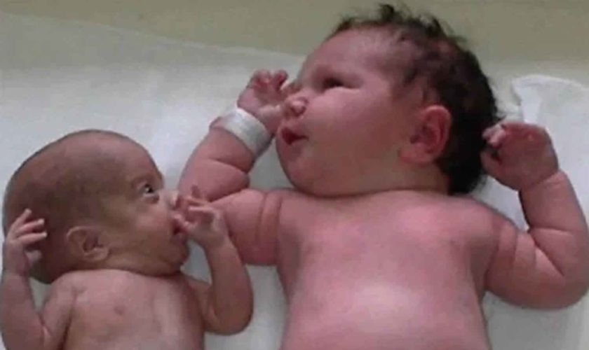
Figure 1. Ambiguous genitalia baby girl

Ambiguous genitalia baby girl

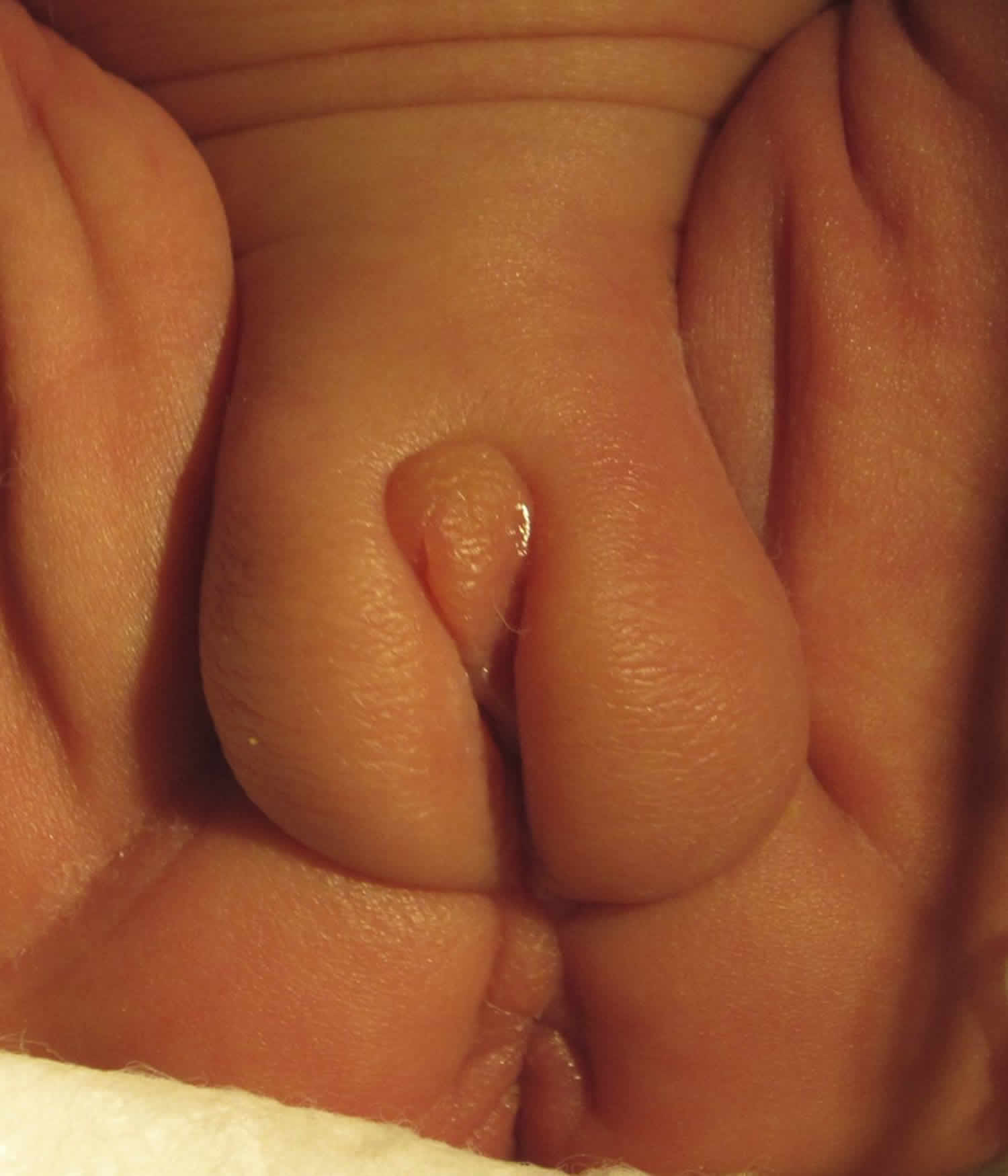
Footnote: A 53-day-old, 27-weeks’premature female baby girl presents with genitalia that have become more noticeably ambiguous over the preceding several weeks.

[Source 7) ]

Normal sex development

The genetic sex of a child is determined at conception. The mother’s egg cell contains an X chromosome, while the father’s sperm cell contains either an X or a Y chromosome. These X and Y chromosomes determine the child’s genetic sex.

Normally, an infant inherits 1 pair of sex chromosomes, 1 X from the mother and 1 X or one Y from the father. The father “determines” the genetic sex of the child. A baby who inherits the X chromosome from the father is a genetic female and has 2 X chromosomes. A baby who inherits the Y chromosome from the father is a genetic male and has 1 X and 1 Y chromosome.

The male and female reproductive organs and genitals both come from the same tissue in the fetus. Ambiguous genitalia can develop if the process that causes this fetal tissue to become “male” or “female” is disrupted. This makes it hard to easily identify the infant as male or female. The extent of the ambiguity varies. Very rarely, the physical appearance may be fully developed as the opposite of the genetic sex. For example, a genetic male may have developed the appearance of a normal female.

How sex organs form in the womb

A baby’s genetic sex is established at conception, based on the sex chromosomes. The mother’s egg contains an X chromosome, and the father’s sperm contains either an X or a Y chromosome. A baby who inherits the X chromosome from the father is a genetic female (two X chromosomes). A baby who inherits the Y chromosome from the father is a genetic male (one X and one Y chromosome).

Male and female sex organs develop from the same tissue. Whether this tissue becomes male organs or female organs depends on the chromosomes and the presence or absence of male hormones.

  • In males, a region on the Y chromosome triggers the development of testicles, which produce male hormones. Male genitals develop in response to male hormones from the fetal testicles.
  • In a fetus without a Y chromosome — without the effects of male hormones — the genitals develop as female.

Occasionally, a chromosomal abnormality may make determination of genetic sex complex.

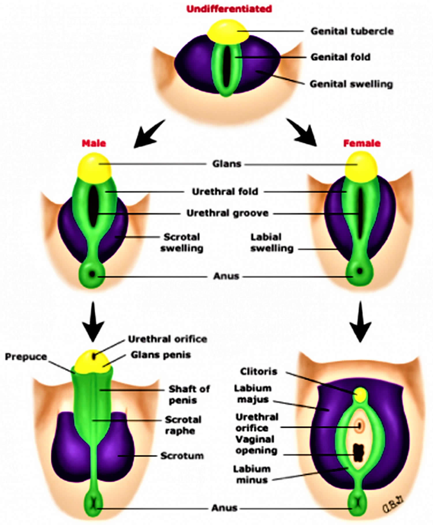
It is important to realize that the gonads (male testis or female ovaries) as well as internal and external genital structures all originate from the same bipotential embryologic tissues, and only through well-choreographed, well-coordinated and localized expression of genes and their gene products does normal sex differentiation and development progress.

The first migration of bipotential primordial germ cells from the yolk sac endoderm to the urogenital ridges leads to the undifferentiated gonad beginning at 4–6 weeks 8). The first 7 weeks of gestation have been considered the indifferent stage, as male and female fetuses would appear grossly indistinguishable from one another, though histologically, changes are well underway. A clear “switch” or decision point signaling the sex determination then occurs leading to the initiation of testicular or ovarian gonadal differentiation. The young bipotential gonad is able to differentiate towards a dedicated male or female pathway, either testis or ovary, respectively, based initially on factors that reside on the Y chromosome (the SRY gene is the prototypical example). Gonadal differentiation then triggers hormonal responses and along with precise and timely gene activation lead to normal/typical external and internal genital development. Scientists have learned about the role of timely and appropriate gene action in normal sex development through gene defects discovered along the pathways associated with disorders of sex development 9).

There is no true “default” pathway; however, androgen exposure determines the external genitalia phenotype. For example, androgen exposure from testosterone and its more potent metabolite dihydrotestosterone (DHT) leads to phallic enlargement and the formation of the penis and the fusion of the labioscrotal folds into a scrotum. The spectrum of phenotypes relating to androgen exposure is seen in many disorders of sex development with varying degrees of genital ambiguity. Different scales or scores have been developed to assess this spectrum, though the most commonly used is the Prader Scale 10), with some alternative scales being developed more recently 11).

With regard to the internal genitalia, the mesonephric duct develops into the Wollfian duct and the internal male genital organs, and the paramesonephric duct develops into the Müllerian duct and the internal female genital organs. In the presence of anti-Müllerian hormone (AMH) secreted by the Sertoli cells of the newly developed testes, the Müllerian structures regress, leaving only the Wollfian structures-epididymis, vas deferens, seminal vesicles, and ejaculatory ducts. Otherwise, the Müllerian structures persist, developing into the fallopian tubes, uterus, cervix, and upper vagina, while the Wollfian structures regress without testosterone trophic effect.

Figure 2. Development of external genitalia

Development of external genitalia

Ambiguous genitalia causes

Ambiguous genitalia primarily occurs when hormone abnormalities during pregnancy interrupt or disturb the fetus’s developing sex organs.

A disruption of the steps that determine sex can result in a mismatch between the appearance of the external genitals and the internal sex organs or the genetic sex (XX or XY).

  • A lack or deficiency of male hormones in a genetic male fetus can cause ambiguous genitalia, while exposure to male hormones during development results in ambiguous genitalia in a genetic female.
  • Mutations in certain genes can influence fetal sex development and cause ambiguous genitalia.
  • Chromosomal abnormalities, such as a missing sex chromosome or an extra one, also can cause ambiguous genitalia.
  • In some cases, the cause of ambiguous genitalia may not be determined.
  • Ambiguous genitalia can also be a feature of certain rare, complex syndromes that affect many organ systems.

The causes of ambiguous genitalia in newborn period are listed below. The most common cause of ambiguous genitalia is congenital adrenal hyperplasia (CAH). Congenital adrenal hyperplasia (CAH) comprises a collection of different adrenal steroid biosynthetic disorders which can result in wide-ranging signs and symptoms from salt-wasting adrenal crisis in the infant to virilization in young females, delayed puberty in adolescents and even polycystic ovary syndrome (PCOS)-like symptoms (i.e., acne, irregular menses, hirsutism, infertility) in young women. Congenital adrenal hyperplasia (CAH) should be the primary consideration in any virilized infant with non-palpable gonads. Adrenal crisis can lead to shock and death if this condition is not promptly recognized and treated.

In addition to congenital adrenal hyperplasia (CAH) being often recognized as the most common cause of ambiguous genitalia in infants and neonates, other notable causes include partial androgen insensitivity syndrome (Partial AIS or PAIS). Androgen insensitivity syndrome is caused by mutations in the androgen receptor (AR) gene on the X chromosome, and dictates response to androgen such as testosterone or DHT. Complete androgen insensitivity syndrome (CAIS) refers to a complete defect in androgen receptor (AR) and thus no response to androgen, whereas partial androgen insensitivity syndrome (PAIS) refers to a partial defect and thus partial activity in the androgen receptor (AR) and the androgen response. Complete androgen insensitivity syndrome (CAIS) often presents with primary amenorrhea in adolescent female, and since there is no androgen effect it does not lead to genital ambiguity. Complete androgen insensitivity syndrome (CAIS) cases are characterized by female external genitalia but with 46XY karyotype and elevated levels of testosterone, illustrating the complete androgen resistance resulting from the inactive androgen receptor (AR). Partial androgen insensitivity syndrome, being an intermediate and partial mutation, leads to some androgen effect and thus partial virilization. This phenotype then results in ambiguous genitalia in a 46XY infant.

Unfortunately, a great number of ambiguous genitalia cases do not have an identifiable cause, particularly among 46XY disorder of sex development, which highlights the importance of an experienced team with multidisciplinary focus 12). Please note that congenital malformations can also cause ambiguous genitalia, many of which have a primary genetic cause, and others which have a more structural or developmental cause and do not have any hormonal basis. These include VATER/VACTERL associations or cloacal exstrophy 13). VACTERL stands for vertebral defects, anal atresia, cardiac defects, tracheo-esophageal fistula, renal anomalies, and limb abnormalities. People diagnosed with VACTERL association typically have at least three of these characteristic features. Affected individuals may have additional abnormalities that are not among the characteristic features of VACTERL association.

(1) Virilization in 46XX infant

  • Excess androgen production
  • Congenital adrenal hyperplasia (CAH)
    • 21 hydroxylase deficiency
    • 11-beta hydroxylase deficiency
    • 3-beta hydroxysteroid dehydrogenase (3βHSD) deficiency
    • P450 oxidoreductase (POR) deficiency
  • Defects in androgen metabolism or signaling
  • Placental aromatase deficiency
  • Exogenous: hyperandrogenism from maternal source
  • Luteoma of pregnancy
  • Uncontrolled maternal congenital adrenal hyperplasia (CAH)
  • Adrenal virializing tumor

(2) Undervirilization in 46XY infant

  • Defects in testosterone metabolism/synthesis
  • Smith-Lemli-Opitz syndrome (7-dehydrocholesterol reductase deficiency)
  • 5-alpha reductase deficiency
  • Leydig cell hypoplasia/agenesis (LH-hCG Receptor defect)
  • Congenital adrenal hyperplasia (CAH)
    • 3-beta hydroxysteroid dehydrogenase (3βHSD) deficiency
    • 17-alpha hydroxylase/17,20 lyase deficiency
    • 17-beta hydroxysteroid dehydrogenase (ketosteroid reductase) deficiency
    • Steroid acute regulatory (StAR) protein deficiency
  • Defects in testosterone action
  • Androgen insensitivity syndrome-partial (PAIS)
  • Gonadal dysgenesis genetic syndromes (associated with developmental/skeletal anomalies)
  • Denys-Drash Syndrome-Frasier syndrome spectrum (WT1)
  • Campomelic dysplasia (SOX9)
  • Others: SRY, SF1, DAX1, Wnt4
  • Exogenous estrogen/progestin exposure

(3) Sex chromosome differences of sex development/other

  • Partial and mixed gonadal dysgenesis (usually with mosaicism, such as 45X/46XY)
  • Ovotesticular disorder of sex development (karyotype can vary, but most often 46XX)
  • Congenital/complex urogenital abnormalities
  • Incomplete development of sex anatomy (e.g., vaginal agenesis, gonadal agenesis, or aphallia)
  • VATER/VACTERL
  • Bladder/cloacal exstrophy
  • Aphallia
  • Others

(4) Transient

  • Prematurity/persistent fetal adrenal
  • Preterm ovarian hyperstimulation syndrome
  • Genital swelling/edema

Possible causes in genetic females

Causes of ambiguous genitalia in a genetic female may include:

  • Congenital adrenal hyperplasia (CAH). Certain forms of this genetic condition cause the adrenal glands to make excess male hormones (androgens).
  • Prenatal exposure to male hormones. Certain drugs that contain male hormones or that stimulate production of male hormones in a pregnant woman can cause developing female genitals to become more masculine. A developing baby also may be exposed to excess male hormones if the mother has a disease or condition that causes hormone imbalance.
  • Tumors. Rarely, a tumor in the mother can produce male hormones.

Possible causes in genetic males

Causes of ambiguous genitalia in a genetic male may include:

  • Impaired testicle development. This may be due to genetic abnormalities or unknown causes.
  • Androgen insensitivity syndrome (AIS). In this condition, developing genital tissues don’t respond normally to male hormones made by the testes.
  • Abnormalities with testes or testosterone. Various abnormalities can interfere with the testes’ activity. This may include structural problems with the testes, problems with production of the male hormone testosterone or problems with cellular receptors that respond to testosterone.
  • 5-alpha-reductase deficiency. This 5-alpha-reductase enzyme defect impairs normal male hormone production.
  • Sex chromosome anomalies (e.g., Turner Syndrome, Klinefelter Syndrome, sex chromosome mosaicism)

Risk factors for developing ambiguous genitalia

Family history may play a role in the development of ambiguous genitalia, because many disorders of sex development result from genetic abnormalities that can be inherited. Possible risk factors for ambiguous genitalia include a family history of:

  • Unexplained deaths in early infancy
  • Infertility, absent menstrual periods or excess facial hair in females
  • Genital abnormalities
  • Abnormal physical development during puberty
  • Congenital adrenal hyperplasia (CAH), a group of inherited genetic disorders that affect the adrenal glands

If your family has a history of these risk factors, consider seeking medical advice before trying to conceive. You may also benefit from genetic counseling.

Ambiguous genitalia symptoms

Your medical team will likely be the first to recognize ambiguous genitalia soon after your baby is born. Occasionally, ambiguous genitalia may be suspected before birth (prenatally). Characteristics can vary in severity, depending on when during genital development the problem occurred and the cause of the disorder.

Babies who are genetically female (with two X chromosomes) may have:

  • An enlarged clitoris, which may resemble a penis
  • Closed labia, or labia that include folds and resemble a scrotum
  • Lumps that feel like testes in the fused labia

Babies who are genetically male (with one X and one Y chromosome) may have:

  • A condition in which the narrow tube that carries urine and semen (urethra) doesn’t fully extend to the tip of the penis (hypospadias)
  • An abnormally small penis with the urethral opening closer to the scrotum
  • The absence of one or both testicles in what appears to be the scrotum
  • Undescended testicles and an empty scrotum that has the appearance of a labia with or without a micropenis

Ambiguous genitalia complications

Complications of ambiguous genitalia may include:

  • Infertility. Whether people with ambiguous genitalia can have children depends on the specific diagnosis. For example, genetic females with congenital adrenal hyperplasia usually can get pregnant if they so choose.
  • Increased risk of certain cancers. Some disorders of sex development are associated with an increased risk of certain types of cancer.

Ambiguous genitalia diagnosis

Ambiguous genitalia is usually diagnosed at birth or shortly after. Doctors and nurses who help with delivery may notice the signs of ambiguous genitalia in your newborn.

History and physical exam assessment in infants with atypical genitalia

History

  • Family history (other cases of atypical genitalia or sudden infant death; could point to genetic cause)
  • Birth/maternal history (medications/exposures or maternal virilization)

Physical exam

  • Overall appearance: vital signs, sick or well appearing (adrenal crisis?)
  • Dysmorphic features (genetic syndromes)
  • Skeletal malformations (campomelic dysplasia)
  • Anatomic malformations (VATER/VACTERL, cloacal exstrophy)

Genital exam

  • Phallic structure (micropenis vs. clitoromegaly)
  • Gonads (bilateral, unilateral or non-palpable)
  • Urethral orifice (hypospadias)
  • Labioscrotal folds (symmetry, presence of gonads)
  • Anogenital distance (androgen exposure)
  • Skin exam: hyperpigmentation (possible ACTH elevation and adrenal failure)

Determining the cause

If your baby is born with ambiguous genitalia, the doctors will work to determine the underlying cause. The cause helps guide treatment and decisions about your baby’s gender. Your doctor will likely begin by asking questions about your family and medical history. He or she will do a physical exam to check for testes and evaluate your baby’s genitalia.

Diagnostic procedures may include a newborn screening test for CAH, hormonal studies and a biopsy of the reproductive organs.

Your medical team will likely recommend these tests:

  • A pelvic ultrasound to check for the presence of female reproductive organs
  • A genitourethrogram to look at the urethra and vagina if present
  • A chromosomal analysis to help determine genetic sex: 46 XX or 46 XY
  • Blood tests to measure hormone levels
    • 17 hydroxy-progesterone
    • Testosterone
    • Gonadotropins (LH, FSH)
  • Blood tests to analyze chromosomes and determine the genetic sex (XX or XY) or tests for single gene disorders
  • Ultrasound of the pelvis and abdomen to check for undescended testes, uterus or vagina
  • X-ray studies using a contrast dye to help clarify anatomy

In certain cases, minimally invasive surgery may be necessary to collect a tissue sample of your newborn’s reproductive organs.

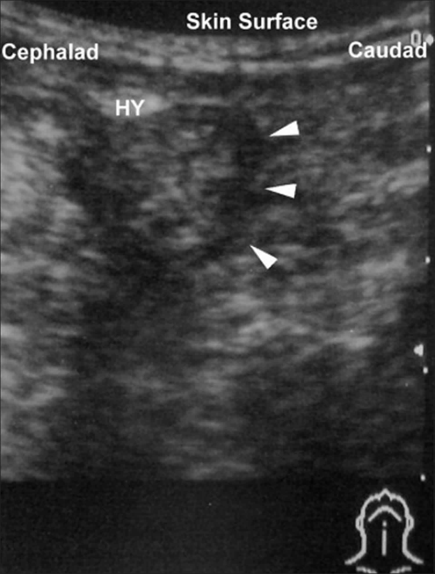
Figure 3. Ambiguous genitalia ultrasound (hypospadias)

Ambiguous genitalia ultrasound

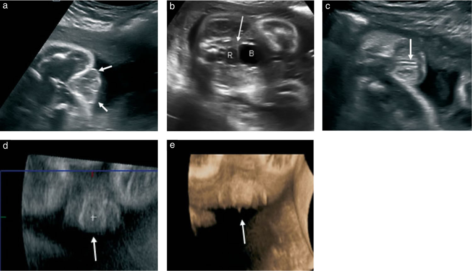
Footnote: Imaging in a case of hypospadias at 29 weeks’ gestation in a male fetus with severe early onset intrauterine growth restriction. Addition of an internal genitalia view in combination with the pregnancy history helped to confirm male gender. At delivery, micturitionoccurred at the penoscrotal junction, consistent with Grade 3 hypospadias with penoscrotal transposition. (a – c) Transverse ultrasoundimages demonstrating ambiguous genitalia. (a) Three structures create the ‘tulip’ sign, withthe short penile shaft nestled between the bifidscrotum (arrows). (b) The thin echogenic line (arrow) is the short rectovesical distance creatinga flat interface with the bladder (B) consistent with male internal genitalia. R, rectum. (c) The urethra is visualizedas an echogenic line (arrow) ending along the base/shaft of the shortpenis, consistent with hypospadias. (d) Coronal reconstruction image demonstrating the micropenis with bulbous ending and downward direction between the bifid scrotum (arrow). (e) Volume-rendered coronal image demonstrating downward direction of the micropenis between the bifid scrotum (arrow).

[Source 14) ]

Determining your baby’s gender

Using the information gathered from these tests, your doctor may suggest a gender for your baby. The suggestion will be based on the cause, genetic sex, anatomy, future reproductive and sexual potential, probable adult gender identity, and discussion with you.

In some cases, a family may make a decision within a few days after the birth. However, it’s important that the family wait until test results are completed. Sometimes gender assignment can be complex and the long-term impact can be difficult to predict. Parents should be aware that as the child grows up, he or she may make a different decision about gender identification.

Be thoughtful with the sex assignment: do not rush!

Assigning a sex of rearing is a great responsibility and obligation and should not be rushed. Many factors must be taken into account, and thus it is essential to collect all relevant information while still being expeditious. This includes clinical information (genital appearance as reflecting androgen exposure and future sexual function), laboratory (and cytogenetic) data, as well as imaging, as discussed above. Note also that karyotype data alone is insufficient, and can lead the team astray if used in isolation. Parental views and possible cultural practices must also be respected. Additional considerations include prenatal androgen exposure effects, the future need for hormone replacement, surgical options and fertility potential, even if considered remote 15). Scientists now know that the brain should be considered a “sex organ”: early androgen exposure can have long-lasting effects, and the degree of androgen exposure in utero is believed to be predictive of future masculine behavior and gender identity, though evidence is limited 16). All of these factors named above must be collectively integrated to formulate the most accurate working diagnosis prior to assigning a sex and gender 17).

Ultimately, an individualized approach is essential since long term outcome data are still quite limited. And though uncertainty in the diagnosis is common, there are some general concepts that are widely accepted. For example, 46XX congenital adrenal hyperplasia (CAH) infants should generally be raised as female, unless markedly virilized, in which case the literature is not clear on outcomes. Also, in general 46XY infants with genital ambiguity should generally be assigned as male, especially if there is reasonable evidence of testicular function and androgen responsiveness. To assign such an individual as female constitutes risk 18). Evidence based guidelines are badly needed and future studies will help give better guidance for specific conditions and clinical scenarios, though multicenter networks are making progress 19).

Ambiguous genitalia treatment

Treatment for ambiguous genitalia depends on the type of condition and may include diagnostic, reconstructive surgery, hormone replacement therapy (HRT) or other treatments are used to treat conditions that can cause ambiguous genitalia.

Treatment is decided on a case-by-case basis through discussions with the family. Our team of providers will help each family decide what makes the most sense for their child.

Sometimes, the parents must choose whether to raise the child as male or female (regardless of the child’s chromosomes). This choice can have a big social and psychological impact on the child, so counseling is most often recommended.

In general, most individuals that are 46 XY disorder of sex development make up identify as males in adulthood, and should be raised as such. Exceptions include androgen insensitivity syndrome (AIS) and complete gonadal dysgenesis.

It is often technically easier to treat (and therefore raise) the child as female. This is because it is easier for a surgeon to make female genitalia than it is to make male genitalia. Therefore, sometimes this is recommended even if the child is genetically male. However, this is a difficult decision. You should discuss it with your family, your child’s provider, the surgeon, your child’s endocrinologist, and other health care team members.

For many children with disorder of sex development, long-term medical and surgical care may not be necessary. Common exceptions are children with congenital adrenal hyperplasia (CAH) who will require lifelong endocrine care and children who will require hormone replacement.

All children with disorder of sex development, however, may be at greater risk for psychosocial distress over time. For that reason doctors recommend ongoing, routine follow-up with your psychologist at least yearly to identify concerns early, address the child’s questions throughout childhood and adolescence, and provide needed support about body image, gender identity and peer interactions (e.g., dating), as appropriate.

With regard to psychosexual development, it is now becoming more understood that sex and gender, while having much overlap, is not the same. In addition, the concept of gender itself is being broadened, and reference has been made to a spectrum of gender expression and identity. The so-called “genderbread person”, an infographic and learning tool popularized on social media is one model to understand this wide phenotypic variation 20). While designed as a free online resource for understanding and conceptualizing differences and variations in gender identity, gender expression, and anatomical sex, it can also help facilitate a discussion of gender vs. sex either in the context of transgender medical care or in disorder of sex developments 21). Cases of either dissatisfaction with assigned gender or gender dysphoria are not rare, and these require the care of specialists experienced in managing gender dysphoria. Key points of advice for health care providers are summarized in Table 1.

Table 1. Top five key points in the early management of ambiguous genitalia or atypical genitalia

(I) Exam: check for gonads; assess androgen effect
(II) Diagnostics: check labs for hormone/androgen levels, gonadotropins
❖ The first several days of life are a critical time period for capturing hormone levels while they are still high yield. Delaying will miss this window
(III) Genetics: check chromosomes/karyotype
❖ While not a perfectly specific test, chromosomes are still very helpful in working through the differential diagnosis
(IV) Assigning sex (sex of rearing): engage the family. Stay calm. Do not rush
❖ Experts know that open, bidirectional discussion is of great value for families
❖ First impressions matter: set a positive, respectful and sensitive tone
❖ Allow enough time for discussion and questions
(V) Mobilize your multidisciplinary gender/disorder of sex development team*
❖ A multidisciplinary care team can help minimize psychological trauma for families
❖ Team should be comprised of various members with expertise and experience in disorder of sex development care, and may include the following disciplines:
⬥ Core group
Endocrinology
Urology/surgery
Psychology/psychiatry
Social work
⬥ Additional resources
Genetics
Gynecology
Neonatology
Medical ethics
Nursing

Footnote: *communication with primary care physician should be ongoing.

[Source 22) ]

Once you and your doctor have chosen a gender for your baby, you may choose to begin treatment for ambiguous genitalia. The goal of treatment is long-term psychological and social well-being, as well as to enable sexual function and fertility to the greatest extent possible. When to begin treatment depends on your child’s specific situation.

Ambiguous genitalia is uncommon and complex, and it may require a team of experts. The team might include a pediatrician, neonatologist, pediatric urologist, pediatric general surgeon, endocrinologist, geneticist, and psychologist or social worker.

Medications

Hormone medications may help correct or compensate for the hormonal imbalance. For example, in a genetic female with a slightly enlarged clitoris caused by a minor to moderate case of congenital adrenal hyperplasia, proper levels of hormones may reduce the size of the tissue. Other children may take hormones around the time they would normally experience puberty.

Psychological issues

Optimizing psychosocial and psychosexual well-being

The need for broad and longitudinal psychological support cannot be overstated. Psychological issues can be dynamic along the age scale through infant, childhood, adolescent and adult developmental phases. Beginning in infancy, the confusion surrounding the diagnosis and sex assignment is long lasting and can be traumatic. Then later in childhood and progressing through adolescence and adulthood, themes of stigma, secrecy, and shame are common and intervention by experienced mental health providers for individual and family can provide much needed support.

Perhaps the ultimate goal of the sex assignment is that it eventually aligns with the individual’s gender identity, in order to foster a happy, productive and well-adjusted adult. Ideally this goal should also include optimized fertility potential and adult sexual function. Fertility prognosis has generally been dismal for many disorder of sex developments, though assisted reproductive technologies continue to improve and a recent study suggested that germ cells may be present in many infants with a variety of disorder of sex developments 23). Further, sexual function has often been compromised, sometimes thought to be related to surgical intervention. In addition to issues of stigmatization, these factors have contributed to high rates of dissatisfaction with medical and surgical care, leading to mistrust as well as increased depression and other mental health concerns. Parental stress has also been increasingly identified as a critical factor as well 24). For these reasons psychological support is crucial. In addition to resources within the multidisciplinary clinic team, there are many support groups available 25).

Ambiguous genitalia surgery

Though the history of surgery in disorder of sex development is filled with controversy and regret, surgical intervention has an important role in certain circumstances. The major indications for surgery in disorder of sex development are gonadectomy (generally to mitigate increased risk of malignancy) and genital reconstructive surgery, and both carry significant controversy. Additional goals of surgery, however, are to optimize future sexual function, facilitate options for future reproduction, minimize risk of urinary tract infection, and to avoid stigmatization related to atypical anatomy 26), though many of these points carry some controversy as well.

In children with ambiguous genitalia, surgery may be used to:

  • Preserve normal sexual function
  • Create genitals that appear more typical

The timing of surgery will depend on your child’s specific situation. Some doctors prefer to postpone surgery done solely for cosmetic reasons until the person with ambiguous genitalia is mature enough to participate in the decision about gender assignment.

For girls with ambiguous genitalia, the sex organs may work normally despite the ambiguous outward appearance. If a girl’s vagina is hidden under her skin, surgery in childhood can help with sexual function later. For boys, surgery to reconstruct an incomplete penis may normalize appearance and make erections possible. Surgery to reposition the testes into the scrotum may be required.

Results of surgery are often satisfying, but repeat surgeries may be needed later. Risks include a disappointing cosmetic result or sexual dysfunction, such as an impaired ability to achieve orgasm.

Gonadectomy

There is increased risk of neoplasm in dysgenetic gonads carrying Y chromosome material, and in sex chromosome disorder of sex developments such as mixed/partial gonadal dysgenesis. Malignant transformation can occur if resection is not pursued in a timely manner. While resection of the gonads to prevent the formation of these gonadal tumors (i.e., gonadoblastoma or dysgerminoma) is a reasonable consideration, there is believed to be time to deliberate since they usually do not occur in the first few years of life. In addition, potential function of that gonad (from a hormone production and fertility perspective) should be considered. In some cases, for example, waiting until adolescence can allow for spontaneous puberty to occur and avoid the need for pubertal induction hormone therapy. Importantly though, there may be future ethical or medicolegal implications, since it can be viewed as a sterilization procedure 27).

Reconstructive surgery

This type of surgery includes feminizing procedures for CAH, such as clitoral reduction and vaginoplasty, but also includes corrective repair in the male, for hypospadias and cryptorchidism. Previous guidelines (including the 2005 Chicago consensus) emphasized that feminizing reconstructive surgery should only be considered for more severe cases of virilization (Prader scores III, IV and V) 28). However, more recently some human rights groups have called for a moratorium on genital surgery 29), with the general recommendation to defer surgery until the individual is of age to decide for themselves. This complex topic will continue to foster controversy, since there is limited evidence for both sides of the debate, and this highlights the importance of open and transparent dialogue between the individual, family and team 30).

Prior to undergoing any procedure, it is important to have a thorough and open dialogue with the family (and patient, whenever possible) to avoid unnecessary and irreversible surgery. In addition, clear communication is needed to prevent unrealistic expectations regarding the timing of surgery and of anatomy following reconstruction 31).

Surgery for purely cosmetic reasons should be deferred until the individual has the knowledge to make an educated decision on their gender identity and external genitalia.

Vaginoplasty if the child is reared as female may be beneficial because of improved functional outcomes in adulthood if the surgery is performed early. Clitoroplasy can be delayed until the child can provide input, and often because the CAH improves as the child ages.

Coping and support

If your baby is diagnosed with ambiguous genitalia, you may worry about your child’s future. Mental health professionals can help you deal with this difficult and unexpected challenge. Ask your child’s doctor for a referral to a mental health professional who has experience helping people in your situation.

In addition to ongoing counseling for your family and your child, you may benefit from a support group, either in person or online. Your child can benefit from ongoing counseling by mental health professionals and participation in support groups into adulthood.

Not knowing the gender of your newborn immediately can turn a hoped-for celebration into a stressful crisis. Your medical team will provide you with updates and information as quickly as possible to help settle any questions about your child’s health.

Consider delaying a formal announcement of the birth until testing is complete and you’ve developed a plan with advice from your medical team. Give yourself some time to learn and think about the issue before answering difficult questions from family and friends.

References   [ + ]

read more

Choanal atresia

Choanal-atresia

Choanal atresia

Choanal atresia is a congenital (present at birth) occlusion of the back of the nasal cavity by bone, soft tissue, or both, due to failed recanalization of the nasal fossae during fetal development that causes difficulty breathing 1). Choanal atresia is rare, occurring in approximately 1 in 7,000 live births, and is seen more often in females than in males (females to males ratio 2:1). Choanal atresia is often associated with other developmental anomalies such as CHARGE syndrome, Treacher Collins syndrome, and Tessier syndrome.

If your child is diagnosed with choanal atresia, it may take two different forms. It can can be bilateral, meaning it blocks both nasal passages, or unilateral, meaning only one nasal passage is blocked. The blockage can be either completely bony (30 percent of cases) or be composed of both bone and membranes (70 percent of cases).

  1. Bilateral choanal atresia is life-threatening and symptoms appear immediately after birth. Babies breathe only through their noses when they are very young, so the blocked nasal passages will cause extreme difficulty breathing. Establishing an airway is an acute otolaryngologic emergency 2).
  2. Unilateral choanal atresia is more common (60% of case), less serious, and sometimes appears later in childhood because the child has been able to manage while breathing through only one side of the nasal passage.

The treatment of choanal atresia is essentially surgical. The objectives are to restore choanal patency, not to interfere with the patient normal craniofacial development, to minimize invasiveness, and to avoid recurrences.

Unilateral choanal atresia does not require surgical treatment as urgently as the bilateral case and can be postponed until school age when the anatomy of the region is more similar to that in adults. However, it needs to be closely monitored for any signs of a breathing problem. Using a nasal saline spray can also help to keep the nasal route clear.

Figure 1. Choanal atresia

Choanal atresia

Footnote: (A) Unilateral choanal atresia; (B) Bilateral choanal atresia.

Choanal atresia causes

Choanal atresia is a developmental abnormality. Nasal choanae (paired openings that connect the nasal cavity with the nasopharynx) develop between the third and seventh embryonic weeks, following rupture of the vertical epithelial fold between the olfactory groove and the roof of the primary oral cavity (stomodeum) 3). Choanal atresia is present at birth and can be associated with other developmental abnormalities.

There is no known specific cause of choanal atresia. Most believe that choanal atresia occurs when the tissue that separates the nose and mouth area during fetal development remains after birth. The following theories have been proposed to explain the pathogenesis of choanal atresia: the persistence of the buccopharyngeal membrane, the persistence of the nasobuccal membrane of Hochstetter, the incomplete resorption of the nasopharyngeal mesoderm, and the local misdirection of neural crest cell migration. These theories are associated with molecular and genetic studies to give further insights into the pathogenesis of choanal atresia 4).

Choanal atresia symptoms

The symptoms of choanal atresia include:

  • Cyclic respiratory distress relieved with crying
  • Noisy breathing
  • Feeding difficulties
  • Inability to pass a 5/6 French suction catheter into the nasopharynx
  • Nasal drainage

Babies with unilateral choanal atresia often show symptoms later during infancy and childhood with signs of unilateral nasal obstruction (discharge, mouth breathing, inability to move air on one side) and/or a history of chronic sinusitis. Since one side is open at birth, it is rare that these infants have difficulty breathing when they are first born. In some cases, the correct diagnosis is not reached until adulthood due to the non-specific symptoms of unilateral nasal obstruction.

Babies with bilateral choanal atresia often have respiratory distress with cyanosis episodes immediately after birth. These are relieved only when the baby begins to cry and with the return of cyanosis with rest (paradoxical cyanosis). Feeding difficulty can be the initial alerting event in which the infants can present with progressive airway obstruction and choke episodes during feeding because of their inability to breathe and feed at the same time. The child can not breathe during normal circumstances and can only breath when they open their airway to cry. Because of this, infants with bilateral choanal atresia often require intubation immediately after delivery. Once the airway is secured, the baby can be evaluated for other developmental abnormalities. CHARGE syndrome is the most common of these and consists of coloboma, heart disease, atresia choanae, growth and mental retardation, genital hypoplasia, and ear anomalies 5).

Choanal atresia diagnosis

The diagnosis of choanal atresia should be done immediately after birth. The initial clinical evaluation includes placing a laryngeal mirror under to nostril to check for fogging and introducing a suction catheter through each nostril and into the child’s oral cavity. The clinical suspicion of choanal atresia can be confirmed by examination with a flexible nasal endoscope in a newborn with proper preparation, such as nasal decongestion and mucous suctioning, allowing direct visualization of the possible obstruction in the nasal passage. To confirm the diagnosis of choanal atresia a CT scan should be done to further delineate characteristics of the malformation, such as the anatomy of the atretic area, including the thickness of the atretic plate and the presence and thickness of a bony plate. Besides delineating the nature and severity of choanal atresia, CT is also useful in differentiating other causes of nasal obstruction from choanal atresia. Differential diagnoses include pyriform aperture stenosis, nasolacrimal duct cysts, turbinate hypertrophy, septal dislocation and deviation, antrochoanal polyp, or nasal neoplasm.

Choanal atresia treatment

Choanal atresia treatment depends on the severity of the anomaly (unilateral or bilateral), how much it affects the child’s breathing and eating, and what other medical conditions the patient has.

In August 2019, the International Pediatric Otolaryngology Group published consensus recommendations, including the following, with regard to pediatric choanal atresia diagnosis and care 6):

  • Nasal endoscopy or fiberscopy and computed tomography (CT) scanning are recommended diagnostic modalities
  • Whenever possible, unilateral choanal atresia repair should wait until the patient is over age 6 months
  • Treatment with transnasal endoscopic repair is preferred
  • Patient follow-up for at least 1 year, via nasofiberscopy or rigid endoscopy (but without systematic imaging), is recommended

Treatment for mild choanal atresia cases

Sometimes, a child with unilateral choanal atresia has no respiratory distress or significant feeding issues. These patients may initially be managed with close observation and occasionally supplemental oxygen. Nasal saline can help to keep the nasal linings healthy and free of discharge. Parents with children diagnosed with unilateral choanal atresia may wait until the child grows older before deciding to repair the condition.

During surgery, the nasal passage is opened. A small plastic tube called a nasal stent is placed in the nostril to keep the nasal passage open while it is healing. Nasal stents are usually left in the nasal passage for about three months. When your child is ready, the stent will be removed in the hospital under a general anaesthetic.

Aero-digestive evaluation

If your infant has choanal atresia, he should be evaluated for both airway and feeding issues. Management of the airway often requires a combination of supportive, medical and surgical care. Feeding and swallowing issues are very common in children with these conditions and often need to be addressed by speech pathologists and gastroenterologists. Children born with syndromes often need other subspecialty evaluations (cardiology, ophthalmology, etc.) and benefit greatly from the coordinated care provided in the multidisciplinary setting.

Treatment for severe choanal atresia cases

If your child is diagnosed with bilateral choanal atresia the condition must be treated as soon as possible. The acute care of infants with bilateral choanal atresia in asphyxia consists of endotracheal intubation (when available, may be utilized a McGovern nipple to maintain an adequate oral airway, consisting in an intraoral nipple with a large opening by cutting its end off, secured in the mouth with ties around the infant’s occiput) followed by perforation of the atresia plate.

Surgery should be performed as soon as the patient is stable and has been evaluated for other anomalies. The major goal of the surgical repair is to open the nasal airway sufficiently, allowing the infant to breathe on his own. There are five different surgical approaches available (i.e., transpalatal, transeptal, sublabial, transantral. and transnsal) to accomplish this goal, all of which are performed in the operating room under general anesthesia.

Choanal atresia repair

The growing experience in both instrumentation and technique in endoscopic sinus surgery have led many surgeons to make more frequent use of the endoscopic endonasal technique for the repair of choanal atresia, which has provided better results and fewer surgical complications than in traditional procedures.

  • Transnasal endoscopic approach: Currently, surgeons prefer to treat children with choanal atresia using the transnasal endoscopic approach. It can be done safely on infants just a few days old. The posterior nasal airway is opened using a combination of dilators, sinus instruments, ear curettes and drills. In some cases, a piece of endotracheal tube is placed inside the nasal passage as a stent to maintain the opening while the area heals. The stent will be removed a few weeks after the surgery at the bedside or during an office visit. Sometimes stenting is not required.
  • The transseptal technique consists of making a window in the septum anterior to the atretic plate.
  • Transpalatal approach: The transpalatal approach was the most frequently used until the advent, in the last decades, of the endoscopic endonasal approach. The increased blood loss and the possible occurrence of palatal fistula, palatal dysfunction, and maxillofacial growth disturbance are complications of this procedure. In rare circumstances where the skull base is abnormally developed (other craniofacial anomalies are present), the surgeon may need to use a transpalatal approach to reach the area of choanal atresia through the roof of the mouth.

The use of choanal stenting and mitomycin C as an adjunct therapy to prevent restenosis are a controversial topic in the management of choanal atresia as there is no clear-cut evidence on the effectiveness of using stents or mitomycin after choanal atresia repair 7).

Follow-up

Following surgical repair of choanal atresia, patients may require operative debridement or periodic dilatations. Periodic dilations can sometimes be performed as an outpatient procedure with local decongestant and topical anesthesia using urethral sounds.

Bedwell et al 8) described the use of balloon dilation to repair choanal atresia in a series of patients with membranous stenoses, postrepair granulation, and scar tissue in revision cases. They concluded that balloon dilation is an effective adjuvant tool for use in the repair of choanal atresia and stenosis.

References   [ + ]

read more

Schizophrenia in children

childhood schizophrenia

Schizophrenia in children

Childhood schizophrenia also called “early-onset” schizophrenia when it occurs before the age of 18, is an uncommon but severe mental disorder (major psychiatric illness) in which children interpret reality abnormally. Schizophrenia involves a range of problems with thinking (cognitive), behavior or emotions. It may result in some combination of hallucinations, delusions, and extremely disordered thinking and behavior that impairs your child’s ability to function.

Childhood schizophrenia is essentially the same as schizophrenia in adults, but it occurs early in life and has a profound impact on a child’s behavior and development. With childhood schizophrenia, the early age of onset presents special challenges for diagnosis, treatment, education, and emotional and social development.

Schizophrenia can cause:

  • visual hallucinations of people and objects that are not actually there
  • auditory hallucinations of voices, music or other sounds that are not real
  • delusions of threats that have no basis in reality
  • severe difficulty making friends and maintaining relationships
  • impaired speech and other communication skills
  • inappropriate and damaging behavior

Though scientists are still learning the specifics of how schizophrenia affects the brain, it is believed that schizophrenia may be linked to:

  • A below-normal amount of gray matter—cell material that transmits sensory and movement messages throughout the central nervous system–in the brain’s temporal lobe (the part of the brain’s cerebral cortex that is responsible for hearing) and frontal lobe (the front portion of the brain’s cerebral hemisphere, responsible for processing emotions, retaining memories, making decisions and measuring social responses)
  • Related loss of gray matter in the parietal lobe (part of the brain that processes information from the senses, makes mathematical calculations and controls how we handle objects)

Schizophrenia is a major psychiatric illness that requires careful, often complex and lifelong treatment. Identifying and starting treatment for childhood schizophrenia as early as possible may significantly improve your child’s long-term outcome.

A combination of therapies is usually necessary to effectively manage childhood schizophrenia.

Since there is no known cure for schizophrenia,treatment is aimed at reducing the severity of schizophrenia’s impact on early life and helping the child manage symptoms. Treatment is most successful when symptoms are addressed early on.

Schizophrenia key points:

  • has no known, exact cause
  • often appears to be inherited, passing down from generation to generation
  • affects boys slightly more often than girls when it develops in childhood
  • affects men and women equally when it develops in adolescence and adulthood
  • has no known cure, but can be managed well when caught early and treated with effective therapy, medications and support

Suicidal thoughts
  • Suicidal thoughts and behavior are common among people with schizophrenia. If you have a loved one who is in danger of attempting suicide or has made a suicide attempt, make sure someone stays with that person. Call your local emergency number immediately.

A person with schizophrenia may have thoughts of self-harm or suicide. If you think a person is in immediate danger from suicide, call your local emergency number immediately. Or if you think you can do so safely, take the person to the nearest hospital emergency department.

What are the early warning signs of schizophrenia?

The behavioral changes caused by schizophrenia in children can be difficult to identify in the earliest stages of the disease. Symptoms may emerge slowly, develop over time or occur suddenly, as though “out of the blue.”

The following list of possible warning signs for schizophrenia is not definitive. Many of these symptoms may be caused by a condition other than schizophrenia; some will occur in children who do not have any disorder. However, it’s important to take note of any of these behaviors in your child as soon as they arise—especially if you have a family history of schizophrenia—and, if the behaviors persist, to contact a mental health professional as soon as possible. Typically a child and adolescent psychiatrist will make the diagnosis following a comprehensive evaluation with you and your child. During the assessment, a child and adolescent psychiatrist will ask you to describe your child’s symptoms and provide an overview of your child’s family history, medical history, school life and social interactions.

Typically, a child is diagnosed with schizophrenia if he or she:

  • displays positive or negative symptoms for a period of at least one month
  • is experiencing a worsening decrease in the ability to function on a day-to-day basis

Possible early warning signs in infants

  • abnormal listlessness or extensive periods of inactivity
  • overly relaxed or “floppy” arms or legs
  • unnaturally still, flat posture when lying down
  • unusual sensitivity to bright lights or rapid movements

Possible early warning signs in toddlers

  • chronic high fevers
  • fixation on repeating behaviors, even play, according to a specific regimen
  • persistent state of distraction, anxiety or distress
  • pronounced and sustained fear of certain events, situations or objects (note: while nearly all children experience specific fears as a normal developmental stage, children with early-onset schizophrenia tend to experience an extreme degree of fear that does not subside)
  • weak and slumping posture

Possible early warning signs in school-aged children

  • auditory hallucinations (the perception of sounds that others do not hear); most often, these hallucinations manifest as loud noises, whispers or collective murmuring
  • claims that someone or something is “in my head” or “telling me to do things”
  • extreme sensitivity to sounds and lights
  • frequent self-talk (note: while many children will go through phases of having an “imaginary friend” or occasionally talking to themselves, children with possible early-onset schizophrenia spend the majority of their time conversing and laughing with themselves while shutting out real people and surroundings)
  • tendency to be very “closed off” from others
  • visual hallucinations (seeing things that are not actually there); common examples include streaks or swirls of light or flashing patches of darkness

Possible early warning signs in adolescents and teens

  • a persistently vacant facial expression (known as “blank affect”)
  • awkward, contorted or unusual movements of the face, limbs or body
  • complaints and suspicions of threats, plots or conspiracies (for example, “someone has been sent to spy on me”)
  • dwelling excessively on perceived slights, failures or past disappointments
  • extreme irritability or angry outbursts that are unprovoked or disproportionate to the situation
  • extreme or unwarranted resentment and accusations against others (“I know my parents have been stealing from me”)
  • inability to follow a single train of thought
  • inability to read nonverbal “cues” (failing to understand and respond appropriately to other people’s tone of voice, facial expressions or body language)
  • inappropriate behavior and responses to social situations (for example, laughing out loud during a sad moment)
  • incoherent speech
  • irrational thinking, including:
    • assignment of “special meaning” to events and objects with no personal significance (for example, watching a famous person on television and believing they are conveying a secret message with their words or gestures)
    • assumption of extravagant religious, political or other authority (“I am God”)
    • belief that another person or entity is controlling one’s body, thoughts or movements
    • belief that an evil force, spirit or entity has “possessed” the body or mind
  • lapses in personal hygiene practices
  • long periods of staring without blinking or difficulty focusing on objects
  • rapidly fluctuating moods
  • seeing or hearing things that others do not
  • sudden, painful sensitivity to light and noise
  • sudden, significant changes in sleep patterns—either inability to fall or stay asleep (insomnia), or excessive sleepiness and listlessness (called catatonia)
  • talking aloud to oneself, often repeating or rehearsing conversations with others (real or imaginary)
  • tendency to rapidly shift topics during a single conversation
  • use of “nonsense” or made-up words
  • withdrawal from friendships and activities

It is important to note that, in the case of all of the above warning signs, a child or adolescent with schizophrenia is not aware that these behaviors pose a problem. A schizophrenic child does not have a sense of becoming ill or that something is wrong. The gravity of the situation is only apparent to outside observers.

What symptoms do people with schizophrenia develop as the disease progresses?

As the disease progresses, people with schizophrenia display symptoms that are grouped into four categories: positive symptoms, negative symptoms, disorganized speech and disorganized or catatonic behavior.

Positive symptoms

Positive symptoms of schizophrenia involve the onset and acquisition of certain feelings, traits, and behaviors. These can include:

  • beliefs that someone, or something, poses a threat or is causing some type of harm (for example, a sense of being followed by a person or group)
  • confused thinking (for example, confusing what is happening on television with what is occurring in reality)
  • hallucinations (seeing, hearing or feeling things that are not real; for example, hearing voices giving commands or seeing people, animals or objects that are not really there)
  • delusions (ideas, situations or threats that seem real but are not actually based in reality; for example, believing a surveillance device has been installed in the body, home or car). Children with schizophrenia tend to experience hallucinations, but not delusions, until they reach early adulthood.
  • problems distinguishing dreams from reality
  • regressive behavior (for example, an older child suddenly acting like a much younger child and clinging to parents)
  • severe anxiety
  • severe changes in behavior (for example, becoming noticeably withdrawn)
  • suddenly struggling with schoolwork; inability to comprehend material that was previously familiar
  • vivid, detailed and bizarre thoughts and ideas

Negative symptoms

Negative symptoms of schizophrenia involve the lack or loss of certain capabilities and traits, such as:

  • failure to demonstrate appropriate emotional responses (for example, laughing during a somber event or an upsetting conversation)
  • inability to sustain existing friendships and relationships
  • lack of emotional expression when speaking or interacting with others (having what is known as a “blank affect” on the face or failing to make eye contact)
  • severe difficulty making friends

Disorganized speech

Schizophrenia often causes spoken and written communication that is garbled, nonsensical or otherwise impossible for others to follow. Examples of this disorganized speech may include:

  • using words and sentences that do not fit together
  • inventing words or terms that make no sense to others
  • inability to stay “on track” in a conversation

Disorganized or catatonic behavior

Schizophrenia may lead to impaired behaviors that have a drastic impact on daily functions and activities. These disorganized or catatonic behaviors include:

  • engaging in inappropriate activities or speech (for example, making obscene gestures or comments in public)
  • extreme moodiness and irritability
  • failure to dress in accordance with the weather (for example, wearing layers of heavy clothing on a sweltering summer day)
  • failure to practice personal hygiene (for example, not bathing or brushing teeth)
  • suddenly becoming confused or agitated, followed by sitting and staring in place as though “frozen” (this is called a catatonic state)

Your child may be diagnosed with schizophrenia if these symptoms are present for a period of at least one month.

What are the major similarities and differences between schizophrenia in adults and childhood-onset schizophrenia?

As is the case for adults with schizophrenia, children who are schizophrenic are also likely to:

  • display limited or impaired emotional responses
  • fail to practice adequate personal hygiene or other aspects of self-care (such as dressing weather-appropriately)
  • have great difficulty in day-to-day functioning
  • “live in their heads,” closing themselves off from other people and their surroundings
  • suffer from hallucinations (both visual and auditory) or delusions (impressions or perceptions of situations that are not real)
  • struggle to make and maintain friendships

Unlike adults with schizophrenia, children with schizophrenia tend to:

  • experience a gradual appearance and progression of symptoms, as opposed to a sudden and severe onset
  • display difficulty meeting age-appropriate developmental milestones in motor skills, memory and reasoning and speech and language before developing symptoms of schizophrenia.

Do people with schizophrenia really have multiple personalities?

Although this is a common misconception about schizophrenia, it’s not true. What many people refer to as “multiple personality disorder” is altogether different and is now known as dissociative identity disorder.

A schizophrenic person does not experience memory “blackouts” and alternate identities. Instead, an individual with schizophrenia experiences a separation from reality that is characterized by:

  • visual and auditory hallucinations
  • false and irrational ideas and perceptions
  • impaired or incoherent thinking and speech
  • problems initiating and maintaining relationships
  • difficulty processing social cues and non-verbal communication
  • inability to recognize and adhere to appropriate social behaviors or personal hygiene practices
  • oversensitivity to external stimulation, such as sounds and lights
  • withdrawal from the outside world

Is it possible my child has bipolar disorder, not schizophrenia?

There are certain similarities between early-onset schizophrenia and pediatric bipolar disorder, particularly in the shared tendency to erupt in sudden and often unpredictable emotional outbursts.

The differentiating factor is what triggers these episodes: A child with bipolar disorder will become angry or inconsolable in response to a specific event or action). A child with early schizophrenia, by contrast, will have outbursts seemingly “out of nowhere,” with no obvious cause. In these cases, schizophrenic children are usually reacting to an overwhelming onslaught of sensation, such as sudden, unbearable sensitivity to noise in a room. They may also be frustrated by a sudden inability to communicate, think clearly or even stand or walk properly.

If my child is diagnosed with schizophrenia, what happens next?

Your clinician will explain the particulars of schizophrenia, including its possible causes and effects and long-term repercussions. Specifically, you will be given a thorough overview of your child’s individual symptoms and prognosis.

The next step is developing a mutually agreed-upon treatment plan—incorporating psychotherapy, medication and school and community support—that works for you, your child and your family.

You and your child (if old enough) will always have the opportunity to ask questions. Throughout the duration of treatment, you will be encouraged to bring up any and all concerns, worries and fears so that the clinical team can provide the information and support you need.

Helping someone who may have schizophrenia

Family and friends can help their loved ones with schizophrenia by helping them get treatment and encouraging them to stay in treatment. Supporting a loved one with schizophrenia can be hard. Being respectful, supportive, and kind without tolerating dangerous behavior is the best way to help someone with schizophrenia.

If you think someone you know may have symptoms of schizophrenia, talk to him or her about your concerns. Although you can’t force someone to seek professional help, you can offer encouragement and support and help your loved one find a qualified doctor or mental health professional.

If your loved one poses a danger to self or others or can’t provide his or her own food, clothing or shelter, you may need to call your local emergency number or other emergency responders for help so that your loved one can be evaluated by a mental health professional.

In some cases, emergency hospitalization may be needed. Laws on involuntary commitment for mental health treatment vary by state. You can contact community mental health agencies or police departments in your area for details.

Are people with schizophrenia violent?

Most people with schizophrenia are not violent; however, the risk of violence is greatest when schizophrenia is untreated. It is important to help a person with schizophrenia symptoms get treatment as quickly as possible. People with schizophrenia are much more likely to harm themselves than others.

If you suspect you or someone you know may be experiencing the symptoms of schizophrenia, see a doctor as soon as possible.

Childhood schizophrenia causes

It’s not known what causes childhood schizophrenia, but it’s thought that it develops in the same way as adult schizophrenia does. Researchers believe that a combination of genetics, brain chemistry and environment contributes to development of the disorder. It’s not clear why schizophrenia starts so early in life for some and not for others.

Problems with certain naturally occurring brain chemicals, including neurotransmitters called dopamine and glutamate, may contribute to schizophrenia. Neuroimaging studies show differences in the brain structure and central nervous system of people with schizophrenia. While researchers aren’t certain about the significance of these changes, they indicate that schizophrenia is a brain disease.

Genetics

While there is no single known cause for schizophrenia, experts believe that the disease has a strong genetic component—specifically, an inherited chemical imbalance in the brain.

A combination of genes passed down by both parents can lead to schizophrenia. If a parent has schizophrenia, a child has an estimated 10 to 15 percent chance of developing schizophrenia; if a sibling is schizophrenic, a child has an estimated 7 to 8 percent chance of developing schizophrenia. The risk significantly increases if more than one family member has schizophrenia.

Environmental stresses during pregnancy

Though data is not conclusive, some experts believe a child’s schizophrenia may be linked to certain environmental factors that affect the mother during pregnancy, such as:

  • drug or alcohol use
  • exposure to particular hormonal or chemical agents
  • exposure to certain viruses or infections
  • extreme stress
  • poor nutritional health

Risk factors for schizophrenia in kids

Although the precise cause of schizophrenia isn’t known, certain factors seem to increase the risk of developing or triggering schizophrenia, including:

  • Having a family history of schizophrenia
  • Increased immune system activation, such as from inflammation or autoimmune diseases
  • Older age of the father
  • Some pregnancy and birth complications, such as malnutrition or exposure to toxins or viruses that may impact brain development
  • Taking mind-altering (psychoactive or psychoactive) drugs during teen years.

Childhood schizophrenia prevention

While there is no way to prevent schizophrenia, a close look at your family history and careful observation of your child’s behavior can help predict the likelihood of him developing schizophrenia. The earlier you seek treatment, the better chance you have to improve your child’s quality of life. Early identification and treatment may help get symptoms of childhood schizophrenia under control before serious complications develop. Early treatment is also crucial in helping limit psychotic episodes, which can be extremely frightening to a child and his or her parents. Ongoing treatment can help improve your child’s long-term outlook.

If you suspect your child is displaying symptoms of schizophrenia, the most important step you can take is scheduling an immediate mental professional evaluation.

Childhood schizophrenia signs and symptoms

Schizophrenia involves a range of problems with thinking, behavior or emotions. Signs and symptoms may vary, but usually involve delusions, hallucinations or disorganized speech, and reflect an impaired ability to function. The effect can be disabling.

Schizophrenia symptoms generally start in the mid- to late 20s. It’s uncommon for children to be diagnosed with schizophrenia. Early-onset schizophrenia occurs before age 18. Very early-onset schizophrenia in children younger than age 13 is extremely rare.

Symptoms can vary in type and severity over time, with periods of worsening and remission of symptoms. Some symptoms may always be present. Schizophrenia can be difficult to recognize in the early phases.

Early signs and symptoms

The earliest indications of childhood schizophrenia may include developmental problems, such as:

  • Language delays
  • Late or unusual crawling
  • Late walking
  • Other abnormal motor behaviors — for example, rocking or arm flapping

Some of these signs and symptoms are also common in children with pervasive developmental disorders, such as autism spectrum disorder. So ruling out these developmental disorders is one of the first steps in diagnosis.

Symptoms in teenagers

Schizophrenia symptoms in teenagers are similar to those in adults, but the condition may be more difficult to recognize in this age group. This may be in part because some of the early symptoms of schizophrenia in teenagers are common for typical development during teen years, such as:

  • Withdrawal from friends and family
  • A drop in performance at school
  • Trouble sleeping
  • Irritability or depressed mood
  • Lack of motivation
  • Strange behavior
  • Substance use

Compared with schizophrenia symptoms in adults, teens may be:

  • Less likely to have delusions
  • More likely to have visual hallucinations

Later signs and symptoms

As children with schizophrenia age, more typical signs and symptoms of the disorder begin to appear. Signs and symptoms may include:

  • Delusions. These are false beliefs that are not based in reality. For example, you think that you’re being harmed or harassed; that certain gestures or comments are directed at you; that you have exceptional ability or fame; that another person is in love with you; or that a major catastrophe is about to occur. Delusions occur in most people with schizophrenia.
  • Hallucinations. These usually involve seeing or hearing things that don’t exist. Yet for the person with schizophrenia, hallucinations have the full force and impact of a normal experience. Hallucinations can be in any of the senses, but hearing voices is the most common hallucination.
  • Disorganized thinking. Disorganized thinking is inferred from disorganized speech. Effective communication can be impaired, and answers to questions may be partially or completely unrelated. Rarely, speech may include putting together meaningless words that can’t be understood, sometimes known as word salad.
  • Extremely disorganized or abnormal motor behavior. This may show in several ways, from childlike silliness to unpredictable agitation. Behavior is not focused on a goal, which makes it hard to do tasks. Behavior can include resistance to instructions, inappropriate or bizarre posture, a complete lack of response, or useless and excessive movement.
  • Negative symptoms. This refers to reduced or lack of ability to function normally. For example, the person may neglect personal hygiene or appear to lack emotion ― doesn’t make eye contact, doesn’t change facial expressions, speaks in a monotone, or doesn’t add hand or head movements that normally occur when speaking. Also, the person may have reduced ability to engage in activities, such as a loss of interest in everyday activities, social withdrawal or lack ability to experience pleasure.

Symptoms may be difficult to interpret

When childhood schizophrenia begins early in life, symptoms may build up gradually. The early signs and symptoms may be so vague that you can’t recognize what’s wrong, or you may attribute them to a developmental phase.

As time goes on, symptoms may become more severe and more noticeable. Eventually, your child may develop the symptoms of psychosis, including hallucinations, delusions and difficulty organizing thoughts. As thoughts become more disorganized, there’s often a “break from reality” (psychosis) frequently requiring hospitalization and treatment with medication.

Suicidal thoughts and behavior

Suicidal thoughts and behavior are common among people with schizophrenia. If you have a child or teen who is in danger of attempting suicide or has made a suicide attempt, make sure someone stays with him or her. Call your local emergency number immediately. Or if you think you can do so safely, take your child to the nearest hospital emergency room.

When to see a doctor

It can be difficult to know how to handle vague behavioral changes in your child. You may be afraid of rushing to conclusions that label your child with a mental illness. Your child’s teacher or other school staff may alert you to changes in your child’s behavior.

Seek medical advice if your child:

  • Has developmental delays compared with other siblings or peers
  • Has stopped meeting daily expectations, such as bathing or dressing
  • No longer wants to socialize
  • Is slipping in academic performance
  • Has strange eating rituals
  • Shows excessive suspicion of others
  • Shows a lack of emotion or shows emotions inappropriate for the situation
  • Has strange ideas and fears
  • Confuses dreams or television for reality
  • Has bizarre ideas, behavior or speech
  • Has violent or aggressive behavior or agitation

These general signs and symptoms don’t necessarily mean your child has childhood schizophrenia. These could indicate a phase, another mental health disorder such as depression or an anxiety disorder, or a medical condition. Seek medical care as soon as possible if you have concerns about your child’s behavior or development.

Childhood schizophrenia complications

Left untreated, childhood schizophrenia can result in severe emotional, behavioral and health problems. Complications associated with schizophrenia may occur in childhood or later, such as:

  • Suicide, suicide attempts and thoughts of suicide
  • Self-injury
  • Anxiety disorders, panic disorders and obsessive-compulsive disorder (OCD)
  • Depression
  • Abuse of alcohol or other drugs, including tobacco
  • Family conflicts
  • Inability to live independently, attend school or work
  • Social isolation
  • Health and medical problems
  • Being victimized
  • Legal and financial problems, and homelessness
  • Aggressive behavior, although uncommon

Childhood schizophrenia diagnosis

Diagnosis of childhood schizophrenia involves ruling out other mental health disorders and determining that symptoms aren’t due to substance abuse, medication or a medical condition. The process of diagnosis may involve:

  • Physical exam. This may be done to help rule out other problems that could be causing symptoms and to check for any related complications.
  • Tests and screenings. These may include tests that help rule out conditions with similar symptoms, and screening for alcohol and drugs. The doctor may also request imaging studies, such as an MRI or CT scan.
  • Psychological evaluation. This includes observing appearance and demeanor, asking about thoughts, feelings and behavior patterns, including any thoughts of self-harm or harming others, evaluating ability to think and function at an age-appropriate level, and assessing mood, anxiety and possible psychotic symptoms. This also includes a discussion of family and personal history.
  • Diagnostic criteria for schizophrenia. Your doctor or mental health professional may use the criteria in the Diagnostic and Statistical Manual of Mental Disorders (DSM-5), published by the American Psychiatric Association. Diagnostic criteria for childhood schizophrenia are generally the same as for adult schizophrenia.

Typically a child and adolescent psychiatrist will make the diagnosis following a comprehensive evaluation with you and your child. During the assessment, a child and adolescent psychiatrist will ask you to describe your child’s symptoms and provide an overview of your child’s family history, medical history, school life and social interactions.

Typically, a child is diagnosed with schizophrenia if he or she:

  • displays positive or negative symptoms for a period of at least one month
  • is experiencing a worsening decrease in the ability to function on a day-to-day basis

Challenging process

The path to diagnosing childhood schizophrenia can sometimes be long and challenging. In part, this is because other conditions, such as depression or bipolar disorder, can have similar symptoms.

A child psychiatrist may want to monitor your child’s behaviors, perceptions and thinking patterns for six months or more. As thinking and behavior patterns and signs and symptoms become clearer over time, a diagnosis of schizophrenia may be made.

In some cases, a psychiatrist may recommend starting medications before an official diagnosis is made. This is especially important for symptoms of aggression or self-injury. Some medications can help limit these types of behavior and restore a sense of normalcy.

Diagnostic criteria for Schizophrenia

Criteria for schizophrenia include signs and symptoms of at least six months’ duration, including at least one month of active-phase positive and negative symptoms (Table 1) 1). Delusions, hallucinations, disorganized speech, and disorganized behavior are examples of positive symptoms. Negative symptoms include a decrease in the range and intensity of expressed emotions (i.e., affective flattening) and a diminished initiation of goal-directed activities (i.e., avolition).

Table 1. Diagnostic Criteria for Schizophrenia

A. Two (or more) of the following, each present for a significant portion of time during a 1-month period (or less if successfully treated). At least one of these must be (1), (2), or (3):
1. Delusions
2. Hallucinations
3. Disorganized speech (e.g., frequent derailment or incoherence)
4. Grossly disorganized or catatonic behavior
5. Negative symptoms (i.e., diminished emotional expression or avolition)
B. For a significant portion of the time since the onset of the disturbance, level of functioning in one or more major areas, such as work, interpersonal relations, or self-care, is markedly below the level achieved prior to the onset (or when the onset is in childhood or adolescence, there is failure to achieve expected level of interpersonal, academic, or occupational functioning)
C. Continuous signs of the disturbance persist for at least 6 months. This 6-month period must include at least 1 month of symptoms (or less if successfully treated) that meet Criterion A (i.e., active-phase symptoms) and may include periods of prodromal or residual symptoms. During these prodromal or residual periods, the signs of the disturbance may be manifested by only negative symptoms or by two or more symptoms listed in Criterion A present in an attenuated form (e.g., odd beliefs, unusual perceptual experiences)
D. Schizoaffective disorder and depressive or bipolar disorder with psychotic features have been ruled out because either: (1) no major depressive or manic episodes have occurred concurrently with the active-phase symptoms, or (2) if mood episodes have occurred during active-phase symptoms, they have been present for a minority of the total duration of the active and residual periods of the illness
E. The disturbance is not attributable to the physiological effects of a substance (e.g., a drug of abuse, a medication) or another medical condition
F. If there is a history of autism spectrum disorder or a communication disorder of childhood onset, the additional diagnosis of schizophrenia is made only if prominent delusions or hallucinations, in addition to the other required symptoms of schizophrenia, are also present for at least 1 month (or less if successfully treated)
[Source 2)]

Child schizophrenia test

Early onset schizophrenia in children presents symptoms such as hallucinations, strange thoughts or feelings, and abnormal behavior that inhibit his/her ability to function and maintain relationships. Answer the questions below to see if your child or teenager may be suffering from schizophrenia.

Below is a list of questions that relate to life-experiences common among children and adolescents who have been diagnosed with schizophrenia.

  1. Does your child claim to hear or see things that others cannot?
  2. Does your child claim that others are controlling their thoughts and emotions?
  3. Does your child struggle to keep up with daily living tasks such as showering, changing clothes, doing homework, etc.?
  4. Does your child speak in a monotone voice or not make facial expressions that match their emotions?
  5. Does your child respond to questions with unrelated or confusing responses?
  6. Does your child ever claim to have powers that other people cannot understand or appreciate?
  7. Does your child feel that they are being tracked, followed, or watched at home or outside?
  8. Does your child have difficulty distinguishing fiction from reality?
  9. Does your child have disorganized or abnormal motor behavior, such as strange posture or excessive movement of their body?
  10. Does your child have developmental delays compared to their peers?

Childhood schizophrenia treatment

Schizophrenia in children requires lifelong treatment, even during periods when symptoms seem to go away. Treatment is a particular challenge for children with schizophrenia.

Treatment team

Childhood schizophrenia treatment is usually guided by a child psychiatrist experienced in treating schizophrenia. The team approach may be available in clinics with expertise in schizophrenia treatment. The team may include, for example, your:

  • Psychiatrist, psychologist or other therapist
  • Psychiatric nurse
  • Social worker
  • Family members
  • Pharmacist
  • Case manager to coordinate care

Main treatment options

The main treatments for childhood schizophrenia are:

  • Medications
  • Psychotherapy
  • Life skills training
  • Hospitalization

Medications

Most of the antipsychotics also called neuroleptics used in children are the same as those used for adults with schizophrenia. Antipsychotic drugs are often effective at managing symptoms such as delusions, hallucinations, loss of motivation and lack of emotion.

In general, the goal of treatment with antipsychotics is to effectively manage symptoms at the lowest possible dose. Over time, your child’s doctor may try combinations, different medications or different doses. Depending on the symptoms, other medications also may help, such as antidepressants or anti-anxiety drugs. It can take several weeks after starting a medication to notice an improvement in symptoms.

Antipsychotics (neuroleptics):

  • are primarily used to treat the pervasive, intrusive, and disturbing thoughts caused by schizophrenia
  • are designed to minimize the severity of hallucinations and delusions
  • must be taken exactly as prescribed
  • may require adjustments of dosage or type over time to maintain their effectiveness.

Traditionally prescribed neuroleptics include:

  • Stelazine (Trifluoperazine)
  • Flupenthixol (Fluanxol)
  • Loxapine (Loxapac, Loxitane)
  • Perphenazine (Etrafon, Trilafon)
  • Chlorpromazine (Thorazine)
  • Haldol (Haloperidol)
  • Prolixin (Fluphenazine Decanoate, Modecate, Permitil)

Newer and less commonly prescribed medications that have proven effective in treating symptoms of schizophrenia include:

  • Aripiprazole (Abilify)
  • Clozaril (clozapine)
  • Geodon (ziprasidone)
  • Risperdal (resperidone)
  • Seroquel (Quetiapine)
  • Zyprexa (olanzapine)

Your child’s physician and other healthcare team members will work with parents and family members—to determine the best medications for your child, incorporate the medication regimen into the child’s overall treatment plan and monitor the effectiveness of the drugs over the long term.

Second-generation antipsychotics

Newer, second-generation medications are generally preferred because they have fewer side effects than do first-generation antipsychotics. However, they can cause weight gain, high blood sugar, high cholesterol and heart disease.

Examples of second-generation antipsychotics approved by the Food and Drug Administration (FDA) to treat schizophrenia in teenagers age 13 and older include:

  • Aripiprazole (Abilify)
  • Olanzapine (Zyprexa)
  • Quetiapine (Seroquel)
  • Risperidone (Risperdal)

Paliperidone (Invega) is FDA-approved for children 12 years of age and older.

First-generation antipsychotics

These first-generation medications are usually as effective as second-generation antipsychotics in controlling delusions and hallucinations. In addition to having side effects similar to those of second-generation antipsychotics, first-generation antipsychotics also may have frequent and potentially significant neurological side effects. These can include the possibility of developing a movement disorder (tardive dyskinesia) that may or may not be reversible.

Because of the increased risk of serious side effects with first-generation antipsychotics, they often aren’t recommended for use in children until other options have been tried without success.

Examples of first-generation antipsychotics approved by the FDA to treat schizophrenia in children and teens include:

  • Chlorpromazine for children 13 and older
  • Haloperidol for children 3 years and older
  • Perphenazine for children 12 years and older

First-generation antipsychotics are often cheaper than second-generation antipsychotics, especially the generic versions, which can be an important consideration when long-term treatment is necessary.

Medication side effects and risks

All antipsychotic medications have side effects and possible health risks, some life-threatening. Side effects in children and teenagers may not be the same as those in adults, and sometimes they may be more serious. Children, especially very young children, may not have the capacity to understand or communicate about medication problems.

Talk to your child’s doctor about possible side effects and how to manage them. Be alert for problems in your child, and report side effects to the doctor as soon as possible. The doctor may be able to adjust the dose or change medications and limit side effects.

Also, antipsychotic medications can have dangerous interactions with other substances. Tell your child’s doctor about all medications and over-the-counter products your child takes, including vitamins, minerals and herbal supplements.

What is the “black label warning” I keep hearing about when it comes to certain psychiatric medications?

Since 2004, the U.S. Food and Drug Administration has placed a black warning label on antidepressant medications. The warning label states, in part:

“Antidepressants increased the risk of suicidal thinking and behavior (suicidality) in short-term studies in children and adolescents with Major Depressive Disorder (MDD) and other psychiatric disorders. Anyone considering the use of [Drug Name] or any other antidepressant in a child or adolescent must balance this risk with the clinical need. Patients who are started on therapy should be observed closely for clinical worsening, suicidality, or unusual changes in behavior.”

Your clinician will carefully go over the specifics of any medication prescribed for your child’s schizophrenia, as well as any potential side effects you should watch for. Our team has years of experience in managing the use of psychiatric medications in children of all ages and with a wide variety of conditions. We will closely monitor your child for any sign of a negative response to the medication, and are always here to answer your questions and address any concerns you may have.

Psychotherapy

In addition to medication, psychotherapy, sometimes called talk therapy, counseling, psychosocial therapy or, simply, therapy, can help manage symptoms and help you and your child cope with the disorder.

Psychotherapy is a general term for treating mental health problems by talking with a psychiatrist, psychologist or other mental health provider.

During psychotherapy, you learn about your condition and your moods, feelings, thoughts and behaviors. Psychotherapy helps you learn how to take control of your life and respond to challenging situations with healthy coping skills.

There are many types of psychotherapy, each with its own approach. The type of psychotherapy that’s right for you depends on your individual situation.

Psychotherapy may include:

  • Individual therapy. Psychotherapy, such as cognitive behavioral therapy, with a skilled mental health professional can help your child learn ways to deal with the stress and daily life challenges brought on by schizophrenia. Therapy can help reduce symptoms and help your child make friends and succeed at school. Learning about schizophrenia can help your child understand the condition, cope with symptoms and stick to a treatment plan.
  • Family therapy. Your child and your family may benefit from therapy that provides support and education to families. Involved, caring family members who understand childhood schizophrenia can be extremely helpful to children living with this condition. Family therapy can also help you and your family to improve communication, work out conflicts and cope with stress related to your child’s condition.

Life skills training

Treatment plans that include building life skills can help your child function at age-appropriate levels when possible. Skills training may include:

  • Social and academic skills training. Training in social and academic skills is an important part of treatment for childhood schizophrenia. Children with schizophrenia often have troubled relationships and school problems. They may have difficulty carrying out normal daily tasks, such as bathing or dressing.
  • Vocational rehabilitation and supported employment. This focuses on helping people with schizophrenia prepare for, find and keep jobs.

Specialized educational and/or structured activity programs

Children and adolescents with schizophrenia may reap significant benefits from specialized programs offered at schools, in medical centers or in the community. Examples of these programs might include:

  • customized, smaller classroom settings, with educators who have specialized training in teaching children and adolescents with psychiatric disorders
  • social skills training to:
    • develop healthy personal interaction techniques (such as maintaining good eye contact and determining fitting topics of conversation)
    • create a checklist for good hygiene
    • learn how to manage everyday tasks like balancing a checkbook or preparing a meal
  • vocational training to help young adults find jobs and volunteer opportunities
  • speech and language therapy to improve verbal and written communication

Hospitalization

During crisis periods or times of severe symptoms, hospitalization may be necessary. This can help ensure your child’s safety and make sure that he or she is getting proper nutrition, sleep and hygiene. Sometimes the hospital setting is the safest and best way to get symptoms under control quickly.

Partial hospitalization and residential care may be options, but severe symptoms are usually stabilized in the hospital before moving to these levels of care.

Lifestyle and home remedies

Although childhood schizophrenia requires professional treatment, it’s critical to be an active participant in your child’s care. Here are ways to get the most out of the treatment plan.

  • Follow directions for medications. Try to make sure that your child takes medications as prescribed, even if he or she is feeling well and has no current symptoms. If medications are stopped or taken infrequently, the symptoms are likely to come back and your doctor will have a hard time knowing what the best and safest dose is.
  • Check first before taking other medications. Contact the doctor who’s treating your child for schizophrenia before your child takes medications prescribed by another doctor or before taking any over-the-counter medications, vitamins, minerals, herbs or other supplements. These can interact with schizophrenia medications.
  • Pay attention to warning signs. You and your child may have identified things that may trigger symptoms, cause a relapse or prevent your child from carrying out daily activities. Make a plan so that you know what to do if symptoms return. Contact your child’s doctor or therapist if you notice any changes in symptoms, to prevent the situation from worsening.
  • Make physical activity and healthy eating a priority. Some medications for schizophrenia are associated with an increased risk of weight gain and high cholesterol in children. Work with your child’s doctor to make a nutrition and physical activity plan for your child that will help manage weight and benefit heart health.
  • Avoid alcohol, street drugs and tobacco. Alcohol, street drugs and tobacco can worsen schizophrenia symptoms or interfere with antipsychotic medications. Talk to your child about avoiding drugs and alcohol and not smoking. If necessary, get appropriate treatment for a substance use problem.

Coping and support

Coping with childhood schizophrenia can be challenging. Medications can have unwanted side effects, and you, your child and your whole family may feel angry or resentful about having to manage a condition that requires lifelong treatment. To help cope with childhood schizophrenia:

  • Learn about the condition. Education about schizophrenia can empower you and your child and motivate him or her to stick to the treatment plan. Education can help friends and family understand the condition and be more compassionate with your child.
  • Join a support group. Support groups for people with schizophrenia can help you reach out to other families facing similar challenges. You may want to seek out separate groups for you and for your child so that you each have a safe outlet.
  • Get professional help. If you as a parent or guardian feel overwhelmed and distressed by your child’s condition, consider seeking help from a mental health professional.
  • Stay focused on goals. Dealing with childhood schizophrenia is an ongoing process. Stay motivated as a family by keeping treatment goals in mind.
  • Find healthy outlets. Explore healthy ways your whole family can channel energy or frustration, such as hobbies, exercise and recreational activities.
  • Take time as individuals. Although managing childhood schizophrenia is a family affair, both children and parents need their own time to cope and unwind. Create opportunities for healthy alone time.
  • Begin future planning. Ask about social service assistance. Most individuals with schizophrenia require some form of daily living support. Many communities have programs to help people with schizophrenia with jobs, affordable housing, transportation, self-help groups, other daily activities and crisis situations. A case manager or someone on your child’s treatment team can help find resources.

Clinical course of schizophrenia

Patients with schizophrenia have a varied clinical course that may include remission, exacerbations, or a more persistent chronic illness. Among patients who remain ill despite therapy, some have a stable clinical course, whereas others experience worsening symptoms and functioning. Factors that predict the clinical course and prognosis of these patients are not understood, and there is no reliable way to predict outcomes. Approximately 20% of patients can be expected to have a positive outcome 3).

Suicide is a concern when treating patients with schizophrenia. The risk of suicide is 13 times greater in persons diagnosed with schizophrenia compared with the general public, with a lifetime risk of about 5% 4). Patients with auditory hallucinations, delusions, substance abuse, or a history of suicide attempts are at higher risk. Adequate treatment of schizophrenia and its comorbidities, along with diligent screening for risk factors, reduces the likelihood of suicide 5). The overall mortality rate for patients with schizophrenia is two to three times higher than that of the general public 6). Most deaths are related to an increased rate of cardiovascular and respiratory diseases, stroke, cancer, and thromboembolic events 7).

In the past, schizophrenia was viewed as a disease with a poor prognosis. Currently, the disease course and response to treatment are marked by heterogeneity; differences in treatment response, disease course, and prognosis are to be expected 8). Despite adequate treatment, one-third of patients will remain symptomatic. Although most patients need some form of support, most are able to live independently and actively participate in their lives 9).

Prognosis of schizophrenia

If schizophrenia is detected and treated early, and if medications and therapies are successful, the disease has an excellent treatment rate. Lifelong monitoring by a qualified health professional is a must for anyone diagnosed with schizophrenia.

While there is no cure, children and adolescents with the disease can achieve normal—and even extraordinary—milestones at school, at work and in their personal lives. With proper treatment, many children with schizophrenia are able to go to college, hold jobs and have families as adults.

The following factors are critical in successfully treating schizophrenia:

  • building a foundation of family and school awareness and support
  • remaining under a clinician’s care for therapeutic treatment and regular monitoring
  • seeking professional treatment as soon as symptoms emerge
  • taking prescribed medications exactly as directed and for as long as directed (often long-term or throughout the lifetime)

Your treating clinician can give you specific information about your child’s condition, symptoms and recommended treatment plan.

References   [ + ]

read more

Oppositional defiant disorder

oppositional-defiant-disorder

What is oppositional defiant disorder

Oppositional defiant disorder is a pattern of disobedient, hostile behavior and defiant behavior toward authority figures with frequent loss of temper, arguing, becoming angry or vindictive, or other negative behaviors. The essential feature of oppositional defiant disorder is a frequent and persistent pattern of angry or irritable mood, argumentative or defiant behavior, or vindictiveness. It is not unusual for individuals with oppositional defiant disorder to show the behavioral features of the disorder without problems of negative mood. However, individuals with oppositional defiant disorder who show the angry or irritable mood symptoms typically show the behavioral features as well.

The prevalence of oppositional defiant disorder ranges from 1% to 11 %, with an average prevalence estimate of around 3.3%. The rate of oppositional defiant disorder may vary depending on the age and gender of the child. The disorder appears to be somewhat more more common in boys than in girls (1.4:1) prior to adolescence. This male predominance is not consistently found in samples of adolescents or adults. Some studies have shown that oppositional defiant disorder affects 20% of school-age children. However, most experts believe this figure is high due to changing definitions of normal childhood behavior. It may also possibly have racial, cultural, and gender biases. Oppositional defiant disorder typically starts by age 8. However, it may start as early as the preschool years. Oppositional defiant disorder is thought to be caused by a combination of biological, psychological, and social factors.

Symptoms of oppositional defiant disorder include:

  • Actively does not follow adults’ requests
  • Angry and resentful of others
  • Argues with adults
  • Blames others for own mistakes
  • Has few or no friends or has lost friends
  • Is in constant trouble in school
  • Loses temper
  • Is spiteful or seeks revenge
  • Is touchy or easily annoyed

To fit this diagnosis, the pattern must last for at least 6 months and must be more than normal childhood misbehavior. The pattern of behaviors must be different from those of other children around the same age and developmental level. The behavior must lead to significant problems in school or social activities.

The symptoms of oppositional defiant disorder may be confined to only one setting, and this is most frequently the home. Individuals who show enough symptoms to meet the diagnostic threshold, even if it is only at home, may be significantly impaired in their social functioning. However, in more severe cases, the symptoms of oppositional defiant disorder are present in multiple settings. Given that the pervasiveness of symptoms is an indicator of the severity of the oppositional defiant disorder, it is critical that the individual’s behavior be assessed across multiple settings and relationships. Because these behaviors are common among siblings, they must be observed during interactions with persons other than siblings. Also, because symptoms of the oppositional defiant disorder are typically more evident in interactions with adults or peers whom the individual knows well, they may not be apparent during a clinical examination.

Oppositional defiant disorder is typically diagnosed around early elementary school ages and stops being diagnosed around adolescence. Kids who have oppositional defiant disorder have a well-established pattern of behavior problems. Symptoms include:

  • Being unusually angry and irritable
  • Frequently losing their temper / Unusually quick to lose his temper
  • Being easily annoyed
  • Arguing with authority figures
  • Refusing to follow rules
  • Ignores or rebels against rules, at home or at school
  • Deliberately annoying people and be easily annoyed himself
  • Blaming others for mistakes or misbehavior
  • Being vindictive
  • Disruptive behavior appears to be intentional rather than impulsive
  • Refuses to cooperate reflexively—even before he knows what is being asked

All children can have these symptoms from time to time. What distinguishes oppositional defiant disorder from normal oppositional behavior is how severe it is, and how long it has been going on for. A child with oppositional defiant disorder will have had extreme behavior issues for at least six months.

Another hallmark of oppositional defiant disorder is the toll it takes on family relationships. Regular daily frustrations — ignored commands, arguments, explosive outbursts — build up over time, and these negative interactions damage the parent-child bond and reinforce hostile patterns of behavior.

Kids who have behavioral issues push parents towards the extremes. They push parents to become permissive and they push parents to become hyper-coercive in the hope that a larger amount of control will get the kid to listen. Neither of these extremes make for ideal parenting. It is never a parent’s intention to reinforce bad behavior, and we often don’t realize when we’re doing it. Here are two common scenarios:

  • You tell your child to stop playing a game and get ready for bed. He ignores your first two requests. By the third time you ask, you’re so mad that you yell.
  • You tell your child to stop playing a game and get ready for bed. He throws a tantrum because he wants to keep playing. You don’t want him to be so worked up before bedtime, so you back down and say he can play for another ten minutes — but then he has to go to bed.

In the first scenario, your child learns that yelling is an acceptable way to get a message across. More subtly, he might also be learning that he can continue ignoring those first few requests — when you escalate the situation is when he knows you’re serious.

In the second scenario, your child has learned that throwing a tantrum might give him something that he wants, so he’ll be more likely to do it again in the future.

Both of these scenarios can set families up for future conflicts, and the more they are repeated the more they become familiar patterns of behavior that are harder to break out of. Your child doesn’t have to have oppositional defiant disorder for these scenarios to happen, but repeated negative interactions like these make diagnosing a behavior disorder much more likely.

And just like parents aren’t necessarily to blame, neither are the kids. Through no conscious effort of the child, he/she learns through hundreds of trials that this is a way to continue getting what he/she wants.

This also explains why kids who have oppositional defiant disorder might act out more at home. Kids who have oppositional defiant disorder are likely to be more oppositional with people they know well, partly because the pathways are so well worn. Whereas in a place like school, where a kid has less control in general over their environment, the types of behaviors that are common to oppositional defiant disorder may not pay off as much.

Children with symptoms of this disorder should be evaluated by a psychiatrist or psychologist. In children and adolescents, the following conditions can cause similar behavior problems and should be considered as possibilities:

  • Anxiety disorders
  • Attention-deficit/hyperactivity disorder (ADHD)
  • Bipolar disorder
  • Depression
  • Learning disorders
  • Substance abuse disorders

There is a very high overlap in kids who have ADHD who are also diagnosed with oppositional defiant disorder. Depending on the study, the overlap could be 30 to 50 percent of kids with ADHD also have oppositional defiant disorder.

Kids with ADHD are biologically loaded to be distractible, to be impulsive, to have difficulty staying in one place for a little while. So kids with ADHD start off doing things that parents perceive as off limits. And then when those kids get negative feedback they start to become even more negatively oriented towards adults. These repeated patterns of negative interactions can lead to developing oppositional defiant disorder.

But another pathway into developing oppositional defiant disorder has more to do with a child’s temperament and might be apparent early on. Children who had a lot of difficulty soothing themselves as toddlers and continue to struggle with an age-appropriate ability to control their emotions in the face of disappointment or frustration can sometimes develop oppositional defiant disorder. The adults in their environment might be more inclined to accommodate their demands in order to keep the family functioning as harmoniously as possible.

Kids who have experienced a lot of life stress and trauma are also more likely to develop oppositional defiant disorder.

It’s important to get treatment to improve the parent-child relationship, which is crucial to the health and happiness of the entire household. It is also important for your child’s future. Some children will grow out of oppositional defiant disorder, but others will continue to have behavior issues, which could lead to peer rejection and difficulty forming healthy relationships, not to mention continued family discord.

They’ll also be less likely to achieve their potential. If something doesn’t go their way, they might think it’s anyone’s fault but theirs. They might also retreat to the places where they know they can get what they want. That might mean that they try even less, push even more on the people who are closest to them, who they actually care about the most, causing even more frayed relationships.

A small percentage of kids with oppositional defiant disorder go on to develop something called conduct disorder, which is a more severe behavior disorder that includes criminal acts like stealing, setting fires and hurting people. Getting treatment sooner rather than later improves a child’s trajectory.

Oppositional defiant disorder is treatable, usually with behavioral therapy or a combination of behavioral intervention and medication.

Psychosocial management of oppositional defiant disorder involves learning skills to help build positive family interactions and to manage problematic behaviors. These include parental management training, school-based training, functional family therapy/brief strategic family therapy, and cognitive behavior therapy. Management of severe aggression and treatment of co-morbid mental health disorders are indications for pharmacotherapy. Medicines are not specifically indicated for oppositional defiant disorder, but may be administered for co-occurring conditions such as ADHD, or to help the child get the most out of therapy sessions.

Oppositional defiant disorder possible complications

In many cases, children with oppositional defiant disorder grow up to have conduct disorder as teenagers or adults. In some cases, children may grow up to have antisocial personality disorder.

Children and teenagers with oppositional defiant disorder may have trouble at home with parents and siblings, in school with teachers, and at work with supervisors and other authority figures. Children with oppositional defiant disorder may struggle to make and keep friends and relationships.

Oppositional defiant disorder may lead to problems such as:

  • Poor school and work performance
  • Antisocial behavior
  • Impulse control problems
  • Substance use disorder
  • Suicide

Many children and teens with oppositional defiant disorder also have other mental health disorders, such as:

  • Attention-deficit/hyperactivity disorder (ADHD)
  • Conduct disorder
  • Depression
  • Anxiety
  • Learning and communication disorders

Treating these other mental health disorders may help improve oppositional defiant disorder symptoms. And it may be difficult to treat oppositional defiant disorder if these other disorders are not evaluated and treated appropriately.

Oppositional defiant disorder causes

There’s no known clear cause of oppositional defiant disorder. Contributing causes may be a combination of inherited and environmental factors, including:

  • Genetics — a child’s natural disposition or temperament and possibly neurobiological differences in the way nerves and the brain function
  • Environment — problems with parenting that may involve a lack of supervision, inconsistent or harsh discipline, or abuse or neglect

Risk factors for oppositional defiant disorder

Oppositional defiant disorder is a complex problem. Possible risk factors for oppositional defiant disorder include:

  • Temperament — a child who has a temperament that includes difficulty regulating emotions, such as being highly emotionally reactive to situations or having trouble tolerating frustration
  • Parenting issues — a child who experiences abuse or neglect, harsh or inconsistent discipline, or a lack of parental supervision
  • Other family issues — a child who lives with parent or family discord or has a parent with a mental health or substance use disorder
  • Environment — oppositional and defiant behaviors can be strengthened and reinforced through attention from peers and inconsistent discipline from other authority figures, such as teachers

Oppositional defiant disorder prevention

There’s no guaranteed way to prevent oppositional defiant disorder. However, positive parenting and early treatment can help improve behavior and prevent the situation from getting worse. The earlier that oppositional defiant disorder can be managed, the better.

Treatment can help restore your child’s self-esteem and rebuild a positive relationship between you and your child. Your child’s relationships with other important adults in his or her life — such as teachers and care providers — also will benefit from early treatment.

Oppositional defiant disorder symptoms

Sometimes it’s difficult to recognize the difference between a strong-willed or emotional child and one with oppositional defiant disorder. It’s normal to exhibit oppositional behavior at certain stages of a child’s development.

Signs of oppositional defiant disorder generally begin during preschool years. Sometimes oppositional defiant disorder may develop later, but almost always before the early teen years. These behaviors cause significant impairment with family, social activities, school and work.

Angry and irritable mood:

  • Often and easily loses temper
  • Is frequently touchy and easily annoyed by others
  • Is often angry and resentful

Argumentative and defiant behavior:

  • Often argues with adults or people in authority
  • Often actively defies or refuses to comply with adults’ requests or rules
  • Often deliberately annoys or upsets people
  • Often blames others for his or her mistakes or misbehavior

Vindictiveness:

  • Is often spiteful or vindictive
  • Has shown spiteful or vindictive behavior at least twice in the past six months

Opositional defiant disorder can vary in severity:

  • Mild. Symptoms occur only in one setting, such as only at home, school, work or with peers.
  • Moderate. Some symptoms occur in at least two settings.
  • Severe. Some symptoms occur in three or more settings.

For some children, symptoms may first be seen only at home, but with time extend to other settings, such as school and with friends.

Oppositional defiant disorder diagnosis

To determine whether your child has oppositional defiant disorder, the mental health professional will likely do a comprehensive psychological evaluation.

To distinguish symptoms of oppositional defiant disorder from normal childhood or adolescent rebellion, the mental health professional will likely do a comprehensive psychological evaluation inclduing a detailed history of behaviors in various situations. For children younger then 5, the behaviors should occur on most days for at least 6 months; for those who are 5 or older, they should occur once a week for 6 months. Since children with oppositional defiant disorder may show symptoms only in one setting—usually at home—and are more likely to be defiant in interactions with adults and peers they know well, the symptoms may not be in evidence in the clinician’s office. Furthermore, oppositional defiant disorder often occurs along with other behavioral or mental health problems, and symptoms of oppositional defiant disorder may be difficult to distinguish from those related to other problems.

Your child’s evaluation will likely include an assessment of:

  • Overall health
  • Frequency and intensity of behaviors
  • Emotions and behavior across multiple settings and relationships
  • Family situations and interactions
  • Strategies that have been helpful — or not helpful — in managing problem behaviors
  • Presence of other mental health, learning or communication disorders

Oppositional defiant disorder DSM 5 diagnostic criteria

The Diagnostic and Statistical Manual of Mental Disorders (DSM-5), published by the American Psychiatric Association, lists criteria for diagnosing oppositional defiant disorder 1). The DSM-5 criteria include emotional and behavioral symptoms that last at least six months.

  • A). A pattern of angry/irritable mood, argumentative/defiant behavior, or vindictiveness lasting at least 6 months as evidenced by at least four symptoms from any of the following categories, and exhibited during interaction with at least one individual who is not a sibling.
    • Angry / Irritable Mood
      1. Often loses temper.
      2. Is often touchy or easily annoyed.
      3. Is often angry and resentful .
    • Argumentative / Defiant Behavior
      • 4. Often argues with authority figures or, for children and adolescents, with adults.
      • 5. Often actively defies or refuses to comply with requests from authority figures or with rules.
      • 6. Often deliberately annoys others.
      • 7. Often blames others for his or her mistakes or misbehavior.
    • Vindictiveness
      • 8. Has been spiteful or vindictive at least twice within the past 6 months.
      • Note: The persistence and frequency of these behaviors should be used to distinguish a behavior that is within normal limits from a behavior that is symptomatic. For children younger than 5 years, the behavior should occur on most days for a period of at least 6 months unless otherwise noted (Criterion A8) . For individuals 5 years or older, the behavior should occur at least once per week for at least 6 months, unless otherwise noted (Criterion A8) . While these frequency criteria provide guidance on a minimal level of frequency to define symptoms, other factors should also be considered, such as whether the frequency and intensity of the behaviors are outside a range that is normative for the individual’s developmental level, gender, and culture .
  • B). The disturbance in behavior i s associated with distress in the individual o r others in his or her immediate social context (e.g., family, peer group, work colleagues), or it impacts negatively on social, educational, occupational, or other important areas of functioning.
  • C). The behaviors do not occur exclusively during the course of a psychotic, substance use, depressive, or bipolar disorder. Also, the criteria are not met for disruptive mood dysregulation disorder.

Specify current severity:

  • Mild: Symptoms are confined to only one setting (e.g., at home, at school, at work, with peers).
  • Moderate: Some symptoms are present in at least two settings.
  • Severe: Some symptoms are present in three or more settings.

It is not uncommon for individuals with oppositional defiant disorder to show symptoms only at home and only with family members. However, the pervasiveness of the symptoms is an indicator of the severity of the disorder.

The symptoms of oppositional defiant disorder can occur to some degree in individuals without this disorder. There are several key considerations for determining if the behaviors are symptomatic of oppositional defiant disorder. First, the diagnostic threshold of four or more symptoms within the preceding 6 months must be met. Second, the persistence and frequency of the symptoms should exceed what is normative for an individual’s age, gender, and culture. For example, it is not unusual for preschool children to show temper tantrums on a weekly basis. Temper outbursts for a preschool child would be considered a symptom of oppositional defiant disorder only if they occurred on most days for the preceding 6 months, if they occurred with at least three other symptoms of the disorder and if the temper outbursts contributed to the significant impairment associated with the disorder (e.g., led to destruction of property during outbursts, resulted in the child being asked to leave a preschool) .

The symptoms of oppositional defiant disorder often are part of a pattern of problematic interactions with others. Furthermore, individuals with this disorder typically do not regard themselves as angry, oppositional, or defiant. Instead, they often justify their behavior as a response to unreasonable demands or circumstances. Thus, it can be difficult to disentangle the relative contribution of the individual with the disorder to the problematic interactions he or she experiences. For example, children with oppositional defiant disorder may have experienced a history of hostile parenting, and it is often impossible to determine if the child’s behavior caused the parents to act in a more hostile manner toward the child, if the parents’ hostility led to the child’s problematic behavior, or if there was some combination of both. Whether or not the clinician can separate the relative contributions of potential causal factors should not influence whether or not the diagnosis is made. In the event that the child may be living in particularly poor conditions where neglect or mistreatment may occur (e.g., in institutional settings), clinical attention to reducing the contribution of the environment may be helpful.

Oppositional defiant disorder treatment

The best treatment for the child is to talk with a mental health professional in individual and possibly family therapy, but it may include other types of psychotherapy and training for your child — as well as for parents. The parents should also learn how to manage the child’s behavior. Some children respond well to treatment, while others do not.

Treatment often lasts several months or longer. It’s important to treat any co-occurring problems, such as a learning disorder, because they can create or worsen oppositional defiant disorder symptoms if left untreated.

Medications alone generally aren’t used for oppositional defiant disorder unless your child also has another mental health disorder. If your child has coexisting disorders, such as ADHD, childhood psychosis, anxiety or depression, medications may help improve these symptoms.

Psychosocial management: A popular evidence-based treatment is a type of behavior therapy called parent-child interaction therapy. The parent and child work together through a set of exercises while a therapist coaches parents through an ear bud. Parents learn to increase positive interactions with the child and to set consistent consequences for undesirable behavior. Children learn to rein in behavior and enjoy a more supportive relationship with parents.

Pharmacological treatment: Medicines are not specifically indicated for oppositional defiant disorder. However, as many children with oppositional defiant disorder have co-occurring conditions such as ADHD, they may be on medications for those other disorders. In addition, some children are so troubled by their own aggression, and their difficulties managing their painfully low frustration tolerance, that a clinician may recommend medication—like psychostimulants used to treat ADHD to help them control those responses and benefit more from behavioral therapy.

Treatment options for oppositional defiant disorder

Psychosocial management is the mainstay. Active components are as follows:

  • Parental management training
  • In schools, teachers’ training for behavioral interventions
  • Peer group support and peer-mediated intervention
  • Social awareness and supervision
  • Cognitive behavior therapy: perspective taking, problem-solving skills training, assertive training, anger management training
  • Functional family therapy or brief strategic family therapy

Parents play a key role in treatment for oppositional defiant disorder. This might be surprising, since children are the ones given the diagnosis, but in oppositional defiant disorder the parent-child relationship needs to be repaired, which means both parties need to make changes to get back on track.

All programs have certain goals in common, like helping parents find the middle ground between being too authoritative and too permissive. A behavioral therapist helps parents learn how to train their child’s behavior through setting clear expectations, praising kids when they follow through and using effective consequences when they don’t. Parents also learn to use these strategies consistently — one reason why behavior management strategies sometimes don’t work is because parents try different, conflicting techniques, or don’t stick to one program long enough to see gains. Parents and children will also learn problem solving skills they can rely on when they run into issues.

Pharmacotherapy

For management of comorbidities:

  • According to the existing protocol for comorbidities
  • Methylphenidate has best evidence followed by atomoxetine, and guanfacine SR, clonidine for ADHD with behavior problems

For management of aggression:

  • Antipsychotic (risperidone followed by aripiprazole has the best evidence, followed by other atypical and typical antipsychotics)
  • In cases of partial response, mood stabilizer can be added
  • For no response, antipsychotic can be changed

The cornerstones of treatment for oppositional defiant disorder usually include:

  • Parent training. A mental health professional with experience treating oppositional defiant disorder may help you develop parenting skills that are more consistent, positive and less frustrating for you and your child. In some cases, your child may participate in this training with you, so everyone in your family develops shared goals for how to handle problems. Involving other authority figures, such as teachers, in the training may be an important part of treatment.
  • Parent-child interaction therapy. During parent-child interaction therapy, a therapist coaches parents while they interact with their child. In one approach, the therapist sits behind a one-way mirror and, using an “ear bug” audio device, guides parents through strategies that reinforce their child’s positive behavior. As a result, parents learn more-effective parenting techniques, the quality of the parent-child relationship improves, and problem behaviors decrease.
  • Individual and family therapy. Individual therapy for your child may help him or her learn to manage anger and express feelings in a healthier way. Family therapy may help improve your communication and relationships and help members of your family learn how to work together.
  • Cognitive problem-solving training. This type of therapy is aimed at helping your child identify and change thought patterns that lead to behavior problems. Collaborative problem-solving — in which you and your child work together to come up with solutions that work for both of you — can help improve oppositional defiant disorder-related problems.
  • Social skills training. Your child may also benefit from therapy that will help him or her be more flexible and learn how to interact more positively and effectively with peers.

As part of parent training, you may learn how to manage your child’s behavior by:

  • Giving clear instructions and following through with appropriate consequences when needed
  • Recognizing and praising your child’s good behaviors and positive characteristics to promote desired behaviors

Although some parenting techniques may seem like common sense, learning to use them consistently in the face of opposition isn’t easy, especially if there are other stressors at home. Learning these skills will require routine practice and patience.

Most important in treatment is for you to show consistent, unconditional love and acceptance of your child — even during difficult and disruptive situations. Don’t be too hard on yourself. This process can be tough for even the most patient parents.

The basics of parent training programs

Despite the dizzying number of parenting books out there, sometimes parents need more support and coaching than a manual can provide. In part that’s because some kids are just more prone to challenging behaviors, and tougher to manage, than others.

Even the most charming of children can get out of control, with parents stuck in ineffectual ruts and the level of frustration and conflict mounting in the family. Sometimes behavior problems are associated with ADHD and other developmental or emotional challenges.

Kids can be noncompliant, ignoring instructions (and even direct orders) no matter how many times they’re reminded. They can be impulsive, oblivious to warnings and the rules they’re breaking. They can be oppositional and may argue back regularly. They may melt down or tantrum when asked to do something they aren’t keen on doing. All of this hurts the parent-child relationship and adds to stress on both sides.

These parent training programs are offered by psychologists and social workers, and they’ve been tested to determine exactly what techniques are most effective. Ideally, all the adults who spend a significant amount of time with the child should take the training, acknowledging that that isn’t always possible, so clinicians work with as many family members and other caregivers as they can.

What all the programs have in common is that they teach parents how to use praise, or positive reinforcement, more effectively, to encourage the behaviors they want to encourage. And they teach parents how to deploy consistent consequences when kids don’t comply. The result is that kids learn to modulate their behavior to meet expectations and enjoy much more positive interactions with their parents.

Where the programs differ is in how instruction is delivered, how parents practice the skills they’re learning, and the pace at which they’re expected to master these new skills. The programs involve 10 or more sessions, and they target different age groups of children.

These programs include:

  • Parent-Child Interaction Therapy (PCIT)
  • Parent Management Training (PMT)
  • Defiant Teens
  • Positive Parenting Program (Triple P)
  • The Incredible Years.

All endorse using praise and attention to focus on positive behaviors, and all use time out in one way or another, for consequences. Regardless of the pace at which the skills are presented, the good news is that all of them have a ton of evidence behind them working.

It’s helpful for parents to understand the differences among the programs so they can pick the one they feel will work best for their family. These treatments can be matched to a child’s age, symptoms and degree of impairment.

Here’s a rundown of these types of training, what makes them different and which families they may work best for.

Parent-Child Interaction Therapy

Parent-Child Interaction Therapy works with parents and children together, teaching them skills to interact in a positive, productive way. It is effective for kids between the ages of 2 and 7, and usually requires 14 to 17 weekly sessions.

In Parent-Child Interaction Therapy, parents receive live coaching (via a bug in the ear) from a therapist who watches from behind a one-way mirror as they and their child perform a series of tasks, and parents practice specific responses to both desired and undesired behavior.

Parent-Child Interaction Therapy is the most practice-intensive, as parents demonstrate mastery of each skill before going on to the next one. It starts out with positive interactions, then waits till parents reach mastery of these skills before moving on to discipline strategies to improve oppositional behavior.

Parent-Child Interaction Therapy is recommended for parents who need a lot of one-on-one attention in terms of how they’re interacting with their child, and especially if the therapist feels like there have been a lot of coercive negative interactions. Sometimes it’s very important for parents, to learn how to be with their kid in a positive way.

Parent-Child Interaction Therapy is not just related to the child’s age range, but it’s related to the situation. If there needs to be a really strong dose of positive interactions as the first thing and the child is under 7, therapist usually lean more toward Parent-Child Interaction Therapy.

Parent Management Training

In Parent Management Training, which is for children ages 3 to 13, parents are usually seen without the child present, although children may be asked to participate in some sessions. Skills to deal more effectively with challenging behaviors are taught and modeled by the therapist and then role-played with parents. After each session, parents are expected to practice the skills at home. Families usually participate in at least 10 sessions.

In Parent Management Training there’s a new skill that’s taught at every module, so if the family really goes home and implements what was talked about in session, parent could could them all the important skills that they need to know within four or five sessions. From that point on it’s kind of tinkering, refining and tightening up everything.

Parent Management Training is usually recommended if there’s a premium on “more change more quickly.” This can be because the situation seems particularly urgent, such as really severe tantrums or aggressive behaviors.

Since Parent Management Training is appropriate for all ages, it’s a good choice when kids are too old for Parent-Child Interaction Therapy. But therapists also recommend it for a 4-year-old if they feels that the parent-child interaction is actually pretty good, but there might be other things going on, such as anxiety, extreme impulsiveness or explosive anger.

However, Parent Management Training, which establishes a token economy with point charts used to reinforce positive behaviors, can be tougher for younger kids to grasp, especially if they’re cognitively delayed. It’s sometimes hard for children to understand that they’re earning points that they can then spend on things. But if a kid is able to make the connection that they’re earning these stars for good behavior and that these stars get them a reward, then it’s appropriate.

Defiant Teens

The first half of this program involves only parents, and focuses on teaching more effective tools for interacting with their teenager, specifically for handling noncompliance or defiant behavior. But since teenagers are more autonomous than younger children and less influenced by their parents’ guidance, the program also includes training for the adolescent to help him become a participant in changing the family dynamic. In the second half, parents and teenagers are both trained in problem-solving communication. The aim is to provide family behavioral resources to help each family member develop more effective problem-solving, negotiation and communication skills and to correct any unreasonable beliefs that might be impeding their interactions.

Positive Parenting Program (Triple P)

Triple P’s focus is on equipping parents with information and skills to increase confidence and self-sufficiency in managing child behavior. It can be utilized with a wide age range of children from toddlerhood through adolescence.

There are different levels of intervention depending on how severe the case is, with more of a whole blanket system of improving parenting on many different levels.

First, for parenting basics, there’s a four-session treatment that can be given in doctor’s offices that include social workers or other mental health professionals for integrated care.

But for families with more severe behavior problems, there’s a 12-session parent-and-child-focused treatment. It incorporates the best of Parent Management Training and Parent-Child Interaction Therapy by having some sessions in which the clinician is meeting one-on-one with parents and talking about skills and strategies, and some sessions where kids are included and the therapist can do live coaching.

The Incredible Years

The Incredible Years offers small-group-based training for parents of kids from infants through age 12. The programs are broken into four age groups (baby, toddler, preschool and school age) and they range from 12 to 20 weeks.

There is also specialized training designed for high-risk socioeconomically disadvantaged families, and for families with children diagnosed with ADHD and oppositional defiance disorder (ODD).

The Incredible Years is in between Parent Management Training, where you’re given you everything up front, and Parent-Child Interaction Therapy, where skills are added only after earlier ones have been mastered.

There’s a different topic for parents each session. The program starts with a focus on improving parent-child relationships and positive attachment before moving on to consistent routines, rules, and limit-setting. Finally it covers child management strategies such as ignoring, redirection, logical and natural consequences, time to calm down and problem solving.

For children from four to eight years old, Incredible Years offers children’s groups that focus on helping them acquire emotion regulation strategies and social skills.

Research shows that the kids’ group works well at improving pro-social behavior and decreasing disruptive behaviors, but when you add the parenting part it really boosts the effectiveness of it.

One of the great things for parents is that you’ll learn not only from therapists but from each other. Parents will hear what others tried. It also normalizes what you’re seeing in your kids.

Oppositional defiant disorder strategies

At home, you can begin chipping away at problem behaviors of oppositional defiant disorder by practicing these strategies:

  • Recognize and praise your child’s positive behaviors. Be as specific as possible, such as, “I really liked the way you helped pick up your toys tonight.” Providing rewards for positive behavior also may help, especially with younger children.
  • Model the behavior you want your child to have. Demonstrating appropriate interactions and modeling socially appropriate behavior can help your child improve social skills.
  • Pick your battles and avoid power struggles. Almost everything can turn into a power struggle, if you let it.
  • Set limits by giving clear and effective instructions and enforcing consistent reasonable consequences. Discuss setting these limits during times when you’re not confronting each other.
  • Set up a routine by developing a consistent daily schedule for your child. Asking your child to help develop that routine may be beneficial.
  • Build time together by developing a consistent weekly schedule that involves you and your child spending time together.
  • Work together with your partner or others in your household to ensure consistent and appropriate discipline procedures. Also enlist support from teachers, coaches and other adults who spend time with your child.
  • Assign a household chore that’s essential and that won’t get done unless the child does it. Initially, it’s important to set your child up for success with tasks that are relatively easy to achieve and gradually blend in more important and challenging expectations. Give clear, easy-to-follow instructions.
  • Be prepared for challenges early on. At first, your child probably won’t be cooperative or appreciate your changed response to his or her behavior. Expect behavior to temporarily worsen in the face of new expectations. Remaining consistent in the face of increasingly challenging behavior is the key to success at this early stage.

With perseverance and consistency, the initial hard work often pays off with improved behavior and relationships.

Managing Problem Behavior at Home

One of the biggest challenges parents face is managing difficult or defiant behavior on the part of children. Whether they’re refusing to put on their shoes, or throwing full-blown tantrums, you can find yourself at a loss for an effective way to respond.

For parents at their wits end, behavioral therapy techniques can provide a roadmap to calmer, more consistent ways to manage problem behaviors problems and offers a chance to help children develop gain the developmental skills they need to regulate their own behaviors.

ABC’s of behavior management at home

To understand and respond effectively to problematic behavior, you have to think about what came before it, as well as what comes after it. There are three important aspects to any given behavior:

  • Antecedents: Preceding factors that make a behavior more or less likely to occur. Another, more familiar term for this is triggers. Learning and anticipating antecedents is an extremely helpful tool in preventing misbehavior.
  • Behaviors: The specific actions you are trying to encourage or discourage.
  • Consequences: The results that naturally or logically follow a behavior. Consequences — positive or negative — affect the likelihood of a behavior recurring. And the more immediate the consequence, the more powerful it is.

Define behaviors

The first step in a good behavior management plan is to identify target behaviors. These behaviors should be specific (so everyone is clear on what is expected), observable, and measurable (so everyone can agree whether or not the behavior happened).

An example of poorly defined behavior is “acting up,” or “being good.” A well-defined behavior would be running around the room (bad) or starting homework on time (good).

Antecedents, the good and the bad

Antecedents come in many forms. Some prop up bad behavior, others are helpful tools that help parents manage potentially problematic behaviors before they begin and bolster good behavior.

Antecedents to AVOID:

  • Assuming expectations are understood: Don’t assume kids know what is expected of them — spell it out! Demands change from situation to situation and when children are unsure of what they are supposed to be doing, they’re more likely to misbehave.
  • Calling things out from a distance: Be sure to tell children important instructions face-to-face. Things yelled from a distance are less likely to be remembered and understood.
  • Transitioning without warning: Transitions can be hard for kids, especially in the middle of something they are enjoying. Having warning gives children the chance to find a good stopping place for an activity and makes the transition less fraught.
  • Asking rapid-fire questions, or giving a series of instructions: Delivering a series of questions or instructions at children limits the likelihood that they will hear, answer questions, remember the tasks, and do what they’ve been instructed to do.

Antecedents to EMBRACE:

Here are some antecedents that can bolster good behavior:

  • Be aware of the situation: Consider and manage environmental and emotional factors — hunger, fatigue, anxiety or distractions can all make it much more difficult for children to reign in their behavior.
  • Adjust the environment: When it’s homework time, for instance, remove distractions like video screens and toys, provide a snacks, establish an organized place for kids to work and make sure to schedule some breaks — attention isn’t infinite.
  • Make expectations clear: You’ll get better cooperation if both you and your child are clear on what’s expected. Sit down with him and present the information verbally. Even if he “should” know what is expected, clarifying expectations at the outset of a task helps head off misunderstandings down the line.
  • Provide countdowns for transitions: Whenever possible, prepare children for an upcoming transition. Let them know when there are, say, 10 minutes remaining before they must come to dinner or start their homework. Then, remind them, when there are say, 2 minutes, left. Just as important as issuing the countdown is actually making the transition at the stated time.
  • Let kids have a choice: As kids grow up, it’s important they have a say in their own scheduling. Giving a structured choice — “Do you want to take a shower after dinner or before?” — can help them feel empowered and encourage them to become more self-regulating.

Creating effective consequences

Not all consequences are created equal. Some are an excellent way to create structure and help kids understand the difference between acceptable behaviors and unacceptable behaviors while others have the potential to do more harm than good. As a parent having a strong understanding of how to intelligently and consistently use consequences can make all the difference.

Consequences to AVOID

  • Giving negative attention: Children value attention from the important adults in their life so much that any attention — positive or negative — is better than none. Negative attention, such as raising your voice or spanking — actually increases bad behavior over time. Also, responding to behaviors with criticism or yelling adversely affects children’s self-esteem.
  • Delayed consequences: The most effective consequences are immediate. Every moment that passes after a behavior, your child is less likely to link her behavior to the consequence. It becomes punishing for the sake of punishing, and it’s much less likely to actually change the behavior.
  • Disproportionate consequences: Parents understandably get very frustrated. At times, they may be so frustrated that they overreact. A huge consequence can be demoralizing for children and they may give up even trying to behave.
  • Positive consequences: When a child dawdles instead of putting on his shoes or picking up his blocks and, in frustration, you do it for him, you’re increasing the likelihood that he will dawdle again next time.

EFFECTIVE consequences:

Consequences that are more effective begin with generous attention to the behaviors you want to encourage.

  • Positive attention for positive behaviors: Giving your child positive reinforcement for being good helps maintain the ongoing good behavior. Positive attention enhances the quality of the relationship, improves self-esteem, and feels good for everyone involved. Positive attention to brave behavior can also help attenuate anxiety, and help kids become more receptive to instructions and limit-setting.
  • Ignoring actively: This should used ONLY with minor misbehaviors — NOT aggression and NOT very destructive behavior. Active ignoring involves the deliberate withdrawal of attention when a child starts to misbehave — as you ignore, you wait for positive behavior to resume. You want to give positive attention as soon as the desired behavior starts. By withholding your attention until you get positive behavior you are teaching your child what behavior gets you to engage.
  • Reward menus: Rewards are a tangible way to give children positive feedback for desired behaviors. A reward is something a child earns, an acknowledgement that she’s doing something that’s difficult for her. Rewards are most effective as motivators when the child can choose from a variety of things: extra time on the iPad, a special treat, etc. This offers the child agency and reduces the possibility of a reward losing its appeal over time. Rewards should be linked to specific behaviors and always delivered consistently.
  • Time outs: Time outs are one of the most effective consequences parents can use but also one of the hardest to do correctly. Here’s a quick guide to effective time out strategies.
  • Be clear: Establish which behaviors will result in time outs. When a child exhibits that behavior, make sure the corresponding time out is relatively brief and immediately follows a negative behavior.
  • Be consistent: Randomly administering time outs when you’re feeling frustrated undermines the system and makes it harder for the child to connect behaviors with consequences.
  • Set rules and follow them: During a time out, there should be no talking to the child until you are ending the time out. Time out should end only once the child has been calm and quiet briefly so they learn to associate the end of time out with this desired behavior.
  • Return to the task: If time out was issued for not complying with a task, once it ends the child should be instructed to complete the original task. This way, kids won’t begin to see time outs as an escape strategy.

By bringing practicing behavioral tools management at home, parents can make it a much more peaceful place to be.

How to Make Time Outs Work

Time outs are one of the most popular disciplinary tools for parents. They can be a great way to let kids know when their behavior is unacceptable without escalating the situation, which can distract kids from what you want them to learn.

But, like any tool, time outs need to be used correctly if you want them to be effective. If you’ve tried using time outs and your child doesn’t seem to be getting the message, it might be time to review how (and why) they should work.

There are a number of schools of thought about the best way to deploy them, but whichever you choose, using time outs consistently can help kids understand which behaviors aren’t (and are) appropriate and that means more positive, less fraught interactions all around.

Why use time outs?

Research shows that the most effective form of parenting is both warm and firm. That means a lot of affection and positive feedback for kids, but also consistent consequences when they act inappropriately. Time outs help you communicate that behavior is unacceptable without blowing your top.

And unlike emotional confrontations, time outs give both parties the time and space they need to calm down.

The point of a time out isn’t to shame or punish your child, but to diffuse an emotional situation, to help your child switch gears and learn to manage frustration and regulate his own behavior.

Effective time outs

Here are the basics to making the most out of time outs.

  • Advance warning: Kids need to understand which behaviors are linked to which consequences. Work with your child to establish which behaviors—hitting, for instance, or not complying with instruction from you—lead to time outs so she knows what to expect.
  • A pre-determined place: Designating a special chair, or a place on the stairs, also helps a child know what to expect. It’s also a good idea to label the time out chair just that, and not “the naughty chair” or something similar. Time outs work better when they are focused on teaching children how to behave, not on punishing them.
  • A quick response: When a kid misbehaves in one of the ways you have discussed, make sure the following time out is immediate, and that you state the reason: “No hitting. Go to time out.” Be specific, brief, and unemotional. This helps ensure that the child is able to link her action with its consequence. Delayed consequences are ineffective because kids tend to feel you are just being punitive.
  • Keep it brief: A standard formula for time outs is one minute per year of age. Some experts recommend a timer so a child can see that the time is being measured
  • Keep it calm: The goal in a time out is for kids to sit quietly. Some experts recommend not starting the allotted time until your child is quiet. Others feel this is too hard for young children. They require that the child be completely quiet for 5 seconds before ending the time out. This way kids learn to associate good behaviors with the end of the time out and it sends the message to kids that yelling and screaming during a time out won’t work.
  • Pay no attention: Kids in time out should be ignored—no talking to them or about them, not even gesturing in their direction, even if they’re whining, crying or protesting. By withdrawing your attention during the time out, you’re sending the message that misbehaving is not the way to get what they want
  • Consistency is key: It’s tempting to put kids in time out whenever they’re acting inappropriately or pushing your buttons, but using time outs randomly makes it more difficult for kids to make the connection between specific misbehaviors and their consequences. Also, it is important that the time out occurs each and every time the specific target behavior occurs. If not, you are encouraging the child to think that he might be able to get away with it.
  • No rewarding stimuli: In the time out chair the child should have no access to television, electronic devices, toys, or games. If you’re away from home, pick any spot that removes the child from distracting stimulation.
  • If a child won’t stay in time out: If a child breaks the rules by leaving the time out chair too soon, put him in a backup time out area that he cannot escape from—i.e., a bedroom where there aren’t any rewarding stimuli such as television, toys, or games. Briefly explain that he must stay there for one minute and be calm and quiet before he is allowed to leave. Once he does that he should be returned to the time out chair, and the time he must stay there is restarted. If he leaves the chair again, the cycle repeats. Your child should learn quickly that it’s in his best interest to stay in the chair until the time is up.

After the time out

When kids are given time outs for not complying with your instruction, once a time out is finished, they should be asked to complete whatever task they were asked to do before the time out. This helps them understand that time outs aren’t escape routes.

Once the time out is over, you want to turn the attention back on, tuning in to whatever they are doing/working on/playing so that you can “catch them being good” and specifically praise them for a positive behavior. For example, if your child completes his time out, and then he plays gently with the dog, you’d want to let him know what he was doing right (i.e., “I love how nicely you’re playing with Lucky! You are using such nice gentle hands!”) This is reassuring your child that although he had to go to time out, he also is completely capable of doing good and positive things that make you proud and loving toward him.

How to Give Kids Effective Instructions

The first step to harmony is teaching your child to listen and follow directions.

One of the most important keys to minimizing problem behavior is making sure that kids are getting the message you’re trying to send. When it comes to parenting, sometimes the way instructions are given can be just as important as what you’re trying to communicate.

Here are ways to present information to your children to make it more likely that they’ll hear you, and comply:

  • Be direct. Make statements rather than asking questions: “Please sit down,” as opposed to “Are you ready to get out your homework?”
  • Be close. Give instructions when you are near the child, rather than calling out from across the room.
  • Use clear and specific commands. Instead of “Go ahead,” say, “Please go start your reading assignment.”
  • Give age-appropriate instructions. Speak to your child at a level he will understand. If your child is younger, keep things simple and use words you know he knows: “Please pick up the ball.” With older children, who are so often keenly aware of not being “babies anymore” it’s important to be clear without being patronizing.
  • Give instructions one at a time. Especially for kids who have attention challenges, try to avoid giving a series of instructions: “Please put on your sneakers, get your lunch off the kitchen counter, and meet me in the front hall.
  • Keep explanations simple. Giving a rationale can increase the likelihood children will listen to a command, but not if the commands gets lost in it. For instance: “Go get your coat on because it’s raining and I don’t want you to catch a cold.” Instead, try: “It’s raining and I don’t want you to catch a cold. Go get your coat on.”
  • Give kids time to process. After you give an instruction, wait a few seconds, without repeating what you said. Children then learn to listen to calm instructions given once rather than learning that they don’t need to listen because the instructions will be repeated. Watching and waiting also helps keep adults from doing what we’ve requested of our kids for them.

Dealing With Explosive Behavior

When a child—even a small child—melts down and becomes aggressive, he can pose a serious risk to himself and others, including parents and siblings.

It’s not uncommon for kids who have trouble handling their emotions to lose control and direct their distress at a caregiver, screaming and cursing, throwing dangerous objects, or hitting and biting. It can be a scary, stressful experience for you and your child, too. Children often feel sorry after they’ve worn themselves out and calmed down.

So what are you to do?

It’s helpful to first understand that behavior is communication. A child who is so overwhelmed that he is lashing out is a distressed child. He doesn’t have the skill to manage his feelings and express them in a more mature way. He may lack language, or impulse control, or problem-solving abilities.

Sometimes parents see this kind of explosive behavior as manipulative. But kids who lash out are usually unable to handle frustration or anger in a more effective way—say, by talking and figuring out how to achieve what they want.

Nonetheless, how you react when a child lashes out has an effect on whether he will continue to respond to distress in the same way, or learn better ways to handle feelings so they don’t become overwhelming. Some pointers:

  • Stay calm. Faced with a raging child, it’s easy to feel out of control and find yourself yelling at him. But when you shout, you have less chance of reaching him. Instead, you will only be making him more aggressive and defiant. As hard as it may be, if you can stay calm and in control of your own emotions, you can be a model for your child and teach him to do the same thing.
  • Don’t give in. Don’t encourage him to continue this behavior by agreeing to what he wants in order to make it stop.
  • Praise appropriate behavior. When he has calmed down, praise him for pulling himself together. And when he does try to express his feelings verbally, calmly, or try to find a compromise on an area of disagreement, praise him for those efforts.
  • Help him practice problem-solving skills. When your child is not upset is the time to help him try out communicating his feelings and coming up with solutions to conflicts before they escalate into aggressive outbursts. You can ask him how he feels, and how he thinks you might solve a problem.
  • Time outs and reward systems. Time outs for nonviolent misbehavior can work well with children younger than 7 or 8 years old. If a child is too old for time outs, you want to move to a system of positive reinforcement for appropriate behavior—points or tokens toward something he wants.
  • Avoid triggers. Dost kids who have frequent meltdowns do it at very predictable times, like homework time, bedtime, or when it’s time to stop playing, whether it’s Legos or Nintendo. The trigger is usually being asked to do something they don’t like, or to stop doing something they do like. Time warnings (“we’re going in 10 minutes”), breaking tasks down into one-step directions (“first, put on your shoes”), and preparing your child for situations (“please ask to be excused before you leave Grandma’s table”) can all help avoid meltdowns.

What kind of tantrum is it?

How you respond to a tantrum also depends on its severity. The first rule in handling nonviolent tantrums is to ignore them as often as possible, since even negative attention, like telling the child to stop, can be encouraging.

But when a child is getting physical, ignoring is not recommended since it can result in harm to others as well as your child. In this situation, putting the child in a safe environment that does not give her access to you or any other potential rewards.

If the child is young (usually 7 or younger), try placing her in a time out chair. If she won’t stay in the chair, take her to a backup area where she can calm down on her own without anyone else in the room. Again, for this approach to work there shouldn’t be any toys or games in the area that might make it rewarding.

Your child should stay in that room for one minute, and must be calm before she/he is allowed out. Then she/he should come back to the chair for time out. What this does is gives your child an immediate and consistent consequence for her/his aggression and it removes all access to reinforcing things in her/his environment.

If you have an older child who is being aggressive and you aren’t able to carry her into an isolated area to calm down, removing yourself from her vicinity. This ensures that she is not getting any attention or reinforcement from you and keeps you safe. In extreme instances, it may be necessary to call your local emergency number to ensure your and your child’s safety.

Help with behavioral techniques

If your child is doing a lot of lashing out—enough that it is frequently frightening you and disrupting your family—it’s important to get some professional help. There are good behavioral therapies that can help you and your child get past the aggression, relieve your stress and improve your relationship. You can learn techniques for managing his behavior more effectively, and he can learn to rein in disruptive behavior and enjoy a much more positive relationship with you.

  • Parent-child interaction therapy. Parent-child interaction therapy has been shown to be very helpful for children between the ages of 2 and 7. The parent and child work together through a set of exercises while a therapist coaches parents through an ear bud. You learn how to pay more attention to your child’s positive behavior, ignore minor misbehaviors, and provide consistent consequences for negative and aggressive behavior, all while remaining calm.
  • Parent Management Training. Parent Management Training teaches similar techniques as Parent-child interaction therapy, though the therapist usually works with parents, not the child.
  • Collaborative and Proactive Solutions. Collaborative and Proactive Solutions is a program based on the idea that explosive or disruptive behavior is the result of lagging skills rather than, say, an attempt to get attention or test limits. The idea is to teach children the skills they lack to respond to a situation in a more effective way than throwing a tantrum.

Figuring out explosive behavior

Tantrums and meltdowns are especially concerning when they occur more often, more intensely, or past the age in which they’re developmentally expected—those terrible twos up through preschool. As a child gets older, aggression becomes more and more dangerous to you, and the child. And it can become a big problem for him at school and with friends, too.

If your child has a pattern of lashing out it may be because of an underlying problem that needs treatment. Some possible reasons for aggressive behavior include:

  • ADHD: Kids with ADHD are frustrated easily, especially in certain situations, such as when they’re supposed to do homework or go to bed.
  • Anxiety: An anxious child may keep his worries secret, then lash out when the demands at school or at home put pressure on him that he can’t handle. Often, a child who “keeps it together” at school loses it with one or both parents.
  • Undiagnosed learning disability: When your child acts out repeatedly in school or during homework time, it could be because the work is very hard for him.
  • Sensory processing issues: Some children have trouble processing the information they are taking in through their senses. Things like too much noise, crowds and even “scratchy” clothes can make them anxious, uncomfortable, or overwhelmed. That can lead to actions that leave you mystified, including aggression.
  • Autism: Children on all points of the spectrum are often prone to major meltdowns when they are frustrated or faced with unexpected change. They also often have sensory issues that make them anxious and agitated.

Given that there are so many possible causes for emotional outbursts and aggression, an accurate diagnosis is key to getting the help you need. You may want to start with your pediatrician. She can rule out medical causes and then refer you to a specialist. A trained, experienced child psychologist or psychiatrist can help determine what, if any, underlying issues are present.

When behavioral plans aren’t enough

Professionals agree, the younger you can treat a child, the better. But what about older children and even younger kids who are so dangerous to themselves and others, behavioral techniques aren’t enough to keep them, and others around them, safe?

  • Medication. Medication for underlying conditions such as ADHD and anxiety may make your child more reachable and teachable. Kids with extreme behavior problems are often treated with antipsychotic medications like Risperdal or Abilify. But these medications should be partnered with behavioral techniques.
  • Holds. Parent training may, in fact, include learning how to use safe holds on your child, so that you can keep both him and yourself out of harm’s way.
  • Residential settings. Children with extreme behaviors may need to spend time in a residential treatment facility, sometimes, but not always, in a hospital setting. There, they receive behavioral and, most likely, pharmaceutical treatment. Therapeutic boarding schools provide consistency and structure round the clock, seven days a week. The goal is for the child to internalize self-control so he can come back home with more appropriate behavior with you and the world at large.
  • Day treatment. With day treatment, a child with extreme behavioral problems lives at home but attends a school with a strict behavioral plan. Such schools should have trained staff prepared to safely handle crisis situations.

Explosive children need calm, confident parents

It can be challenging work for parents to learn how to handle an aggressive child with behavioral approaches, but for many kids it can make a big difference. Parents who are confident, calm, and consistent can be very successful in helping children develop the skills they need to regulate their own behavior.

This may require more patience and willingness to try different techniques than you might with a typically developing child, but when the result is a better relationship and happier home, it’s well worth the effort.

Coping and support

It’s challenging to be the parent of a child with oppositional defiant disorder. Ask questions and try to effectively communicate your concerns and needs to the treatment team. Consider getting counseling for yourself and your family to learn coping strategies to help manage your own distress. Also seek and build supportive relationships and learn stress management methods to help get through difficult times.

These coping and support strategies can lead to better outcomes for your child because you’ll be more prepared to deal with problem behaviors.

References   [ + ]

read more

Epiglottis

epiglottis

What is epiglottis

The epiglottis is a leaf-shaped elastic cartilage flap behind your tongue (epiglottis means “upon the tongue”), which bends down to cover the glottis (opening) of the larynx each time you swallow. The epiglottis is a flap of elastic cartilage that sits beneath the tongue at the back of the throat. Its main function is to close over the windpipe (trachea) while you’re eating, to prevent food entering your airways. The epiglottis stalk attaches anteriorly to the internal aspect of the angle of the thyroid cartilage (Figure 1). From there, the epiglottis projects superoposteriorly and attaches to the posterior aspect of the tongue.

Figure 1. Epiglottis anatomy

epiglottis

Where is the epiglottis located

The epiglottis is located behind your tongue and it sits beneath the tongue at the back of the throat.

Figure 2. Epiglottis location

Epiglottis locationEpiglottis location

Figure 3. Epiglottis function

Epiglottis function

Epiglottis function

During swallowing, the entire larynx is pulled superiorly and anteriorly by the suprahyoid muscles, and the epiglottis tips inferiorly to cover and seal the laryngeal inlet. This action keeps food out of the lower respiratory tubes — so that food is not inhaled into your lungs. The entry into the larynx of anything other than air initiates the cough reflex, which expels the substance and prevents it from continuing into the lungs. By pulling anteriorly on the hyoid bone, suprahyoid muscles also widen the pharynx to receive the food.

The taste receptors occur in taste buds in the mucosa of the mouth and pharynx. The majority of the 10,000 or so taste buds are on the surface of the tongue; a few others occur on the epiglottis. The vagus nerve (cranial nerve 10) carries taste impulses from the few taste buds on the epiglottis and lower pharynx.

Epiglottitis

Epiglottitis is a potentially life-threatening inflammation and swelling of the epiglottis with the potential risk of fatal airway obstruction – blocking the flow of air into your lungs (see Figures 1 and 2) 1). Acute epiglottitis is known to be potentially life-threatening because the special structure and anatomical location of the epiglottis (see Figures 1 and 2) make it prone to severe edema, which can lead to dyspnea and even suffocation, resulting in death 2). In most cases, epiglottitis is caused by infection. Effective management requires rapid diagnosis, airway management, and treatment of the causative agent.

The epiglottis is a flap of tissue that sits beneath the tongue at the back of the throat. Its main function is to close over the windpipe (trachea) while you’re eating, to prevent food entering your airways.

Acute epiglottitis can occur at any age. Epidemiological studies have shown that the number of acute epiglottitis cases has decreased in children due to proper vaccinations, while the number of adult cases has increased significantly 3). Previously, 75%–90% of cases of epiglottitis were caused by Haemophilus influenza type B (Hib), the same bacterium that causes pneumonia, meningitis and infections in the bloodstream. Haemophilus influenzae type b (Hib) bacteria spreads in the same way as the cold or flu virus; the bacteria are in the tiny droplets of saliva and mucus propelled into the air when an infected person coughs or sneezes. You catch the infection by breathing in these droplets or, if the droplets have landed on a surface or object, by touching this surface and then touching your face or mouth. Because of the success of the Hib vaccination programme, epiglottitis is rare in the US, and most cases now occur in adults. Deaths from epiglottitis are also rare, occurring in less than 1 in 100 cases. But infection with group A beta-hemolytic Streptococci has become more frequent after the widespread use of Hemophilus influenzae type B (Hib) vaccination. Hib vaccination, introduced in 1985, significantly reduced the incidence of epiglottitis 4). Routine Hemophilus influenzae type B (Hib) vaccination for infants has made epiglottitis rare, but epiglottitis remains a concern. Cases presenting today show a mixed microbial etiology (other bacteria as well as fungi and virus), with a relative increased incidence in older children 5). A number of other factors can cause the epiglottis to swell — burns from hot liquids, direct injury to your throat and various infections.

Less common causes of acute epiglottitis include:

  • other bacterial infections – such as streptococcus pneumoniae (a common cause of pneumonia)
  • fungal infections – people with a weakened immune system are most at risk from these types of infection
  • viral infections – such as the varicella zoster virus (the virus responsible for chickenpox) and the herpes simplex virus (the virus responsible for cold sores)
  • trauma to the throat – such as a blow to the throat, or burning the throat by drinking very hot liquids
  • smoking – particularly illegal drugs, such as cannabis or crack cocaine

The clinical features of acute epiglottitis commonly include sore throat, odynophagia [painful swallowing], stridor [high-pitched, wheezing sound caused by obstructed airflow], fever, hoarseness, drooling, hyperpyrexia, and dyspnea [difficult or labored breathing or shortness of breath] 6).

The typical histopathological changes associated with this condition include extensive mucosal or submucosal edema accompanied by neutrophil infiltration and hemorrhage, which are frequently found microscopically. In addition, epiglottic abscess may be observed in some patients with acute epiglottitis (approximately 24% of the cases) 7).

There are differences in trends, occurrences and management of acute epiglottitis between children and adults. There is also more diversity in the cause of epiglottitis in adults 8).

In an 8-year retrospective (1998–2006) review of epiglottitis admissions, Shah et al. 9) found that epiglottitis continues to be a significant entity, with two uniquely vulnerable populations: infants (<1 year old) and the elderly (>85 years old). When examining the pediatric cohort of patients (patients <18 years of age), 34.4% were <1 year of age. This category of age <1 year seemed to have increased in frequency in representing 26.8% of pediatric patients in 1998 to 41.1% in 2006 10). A case of epiglottitis with negative cultures has been reported in a neonate within hours of birth 11).

When to see a doctor

Epiglottitis is a medical emergency. If you or someone you know suddenly has trouble breathing and swallowing, call your local emergency number for an ambulance or go to the nearest hospital emergency department. Try to keep the person quiet and upright, because this position may make it easier to breathe. Don’t try to examine the person’s throat yourself. This can make matters worse.

Croup vs epiglottitis

Croup is a viral laryngotracheobronchitis (usually caused by the Hemophilus parainfluenzae virus) that causes swelling of the windpipe (trachea), the airways to the lungs (the bronchi) and the vocal cords (larynx or voice box). This swelling makes the airway narrower, so it is harder to breathe. Croup causes difficulty breathing, a seal barking cough, and a hoarse voice. The cause is usually a virus, often parainfluenza virus. Other causes include allergies and reflux. Treatment of croup includes oxygen, steroids (dexamethasone 0.6 mg/kg PO/IM) and epinephrine (to relieve airway edema and decrease airway resistance due to swelling).

Epiglottitis is an inflammation of the epiglottis due to an infectious process. It can involve other structures such as the arytenoid, false cords and posterior tongue leading to airway obstruction. Most often epiglottitis is bacterial in origin due to Hemophilus influenzae type B (Hib), it affects children between 2 and 5, however the median age is increasing over the past decade. It presents acutely in otherwise healthy children with a fever as high as 104 °F (40 °C). Epiglottic inflammation occurs quickly with the child sitting forward to use the accessory muscles of respiration and pain in the throat. Salivation is prominent with difficulty swallowing. Treatment includes keeping a parent in attendance at all times to keep the child calm, oxygen, sitting position, immediate intubation. Diagnosis is confirmed through radiologic studies showing the steeple sign representing a uniform narrowing of the subglottic airway by inflammation.

Croup is more prevalent during the wintertime, while there is no seasonal predilection to epiglottitis. Croup also has a more gradual onset than acute epiglottitis, and is commonly associated with low-grade fever. Although both acute epiglottitis and croup share the same symptoms of inspiratory stridor, suprasternal, intercostal and substernal retractions and hoarseness, differentiation in early illness is possible by additional observation of seal barking cough and absence of drooling and dysphagia in croup and by the additional observation of drooling and dysphagia with absence of coughing in epiglottitis. Additional reliable signs of epiglottitis are a preference to sit, dysphagia and refusal to swallow 12). Unlike croup, where onset is insidious over a period of 2 to 3 days, epiglottitis patients tend to experience acute onset of symptoms. In one series, while both croup and epiglottitis were associated with acute stridor, croup was also associated with coughing and no drooling, while epiglottitis was associated with drooling and the lack of coughing 13).

Epiglottitis causes

Epiglottitis is usually caused by epiglottis infection with Haemophilus influenzae type b (Hib) bacteria.

As well as epiglottitis, Haemophilus influenzae type b (Hib) can cause a number of serious infections, such as pneumonia and meningitis.

Haemophilus influenzae type b (Hib) spreads in the same way as the cold or flu virus. The Haemophilus influenzae type b (Hib) bacteria are in the tiny droplets of saliva and mucus propelled into the air when an infected person coughs or sneezes.

You catch Haemophilus influenzae type b (Hib) infection by breathing in these droplets or, if the droplets have landed on a surface or object, by touching this surface and then touching your face or mouth.

Less common causes of epiglottitis include:

  • other bacterial infections – such as streptococcus pneumoniae (a common cause of pneumonia)
  • fungal infections – people with a weakened immune system are most at risk from these types of infection
  • viral infections – such as the varicella zoster virus (the virus responsible for chickenpox) and the herpes simplex virus (the virus responsible for cold sores)
  • trauma to the throat – such as a blow to the throat, or burning the throat by drinking very hot liquids
  • smoking – particularly illegal drugs, such as cannabis or crack cocaine

Haemophilus influenzae type b (Hib) vaccination

The most effective way to prevent your child getting epiglottitis is to make sure their vaccinations are up-to-date.

Children are particularly vulnerable to a Haemophilus influenzae type b (Hib) infection because they have an underdeveloped immune system.

Risk factors for epiglottitis

Certain factors increase the risk of developing epiglottitis, including:

  • Being male. Epiglottitis affects more males than females.
  • Having a weakened immune system. If your immune system has been weakened by illness or medication, you’re more susceptible to the bacterial infections that may cause epiglottitis.
  • Lacking adequate vaccination. Delayed or skipped immunizations can leave a child vulnerable to Hib and increases the risk of epiglottitis.

Figure 3. Swollen epiglottis

Swollen epiglottis

Epiglottitis prevention

Haemophilus influenzae type b (Hib) vaccine

The most effective way to prevent your child getting epiglottitis is to make sure their vaccinations are up to date.

Children are particularly vulnerable to a Haemophilus influenzae type b (Hib) infection, because they have an underdeveloped immune system.

Children should receive their Haemophilus influenzae type b (Hib) as part of the 5 in 1 DTaP/IPV/Hib vaccine, which also protects against diphtheria, tetanus, whooping cough and polio.

Immunization with the Haemophilus influenzae type b (Hib) vaccine is an effective way to prevent epiglottitis caused by Haemophilus influenzae type b (Hib). Children should receive three doses of the vaccine: one at two months, one when they are three months and one when they are four months old. This is followed by an additional Hib/Men C “booster” vaccine at 12 months.

In the United States, children usually receive the vaccine in three or four doses:

  • At 2 months
  • At 4 months
  • At 6 months if your child is being given the four-dose vaccine
  • At 12 to 15 months

The Haemophilus influenzae type b (Hib) vaccine is generally not given to children older than age 5 or to adults because they’re less likely to develop Hib infection. But the Centers for Disease Control and Prevention recommends the vaccine for older children and adults whose immune systems have been weakened by:

  • Sickle cell disease
  • HIV/AIDS
  • Spleen removal
  • Chemotherapy
  • Medications to prevent rejection of organ or bone marrow transplants

Haemophilus influenzae type b (Hib) vaccine side effects

  • Allergic reaction. Seek immediate medical help if you have an allergic reaction. Though rare, an allergic reaction may cause difficulty breathing, wheezing, hives, weakness, a rapid heartbeat or dizziness within minutes or a few hours after the shot.
  • Possible mild side effects. These include redness, warmth, swelling or pain at the injection site, and a fever.

Commonsense precautions

Of course, the Hib vaccine doesn’t offer guarantees. Immunized children have been known to develop epiglottitis — and other germs can cause epiglottitis, too. That’s where commonsense precautions come in:

  • Don’t share personal items.
  • Wash your hands frequently.
  • Use an alcohol-based hand sanitizer if soap and water aren’t available.

Symptoms of epiglottitis

The symptoms of epiglottitis usually develop quickly and get rapidly worse, although they can develop over a few days in older children and adults.

The typical presentation in epiglottitis includes acute occurrence of high fever, severe sore throat and difficulty in swallowing with the sitting up and leaning forward position in order to enhance airflow. There is usually drooling because of difficulty and pain on swallowing. Acute epiglottitis usually leads to generalized toxemia. The most common differential diagnosis is croup and a foreign body in the airway. A late referral to an acute care setting with its serious consequences may result from difficulty in differentiation between acute epiglottitis and less urgent causes of a sore throat, shortness of breath and dysphagia. Antibiotic therapy is usually initiated without preceding bacterial culture, with the consequence of negative cultures at admission.

Epiglottitis symptoms include:

  • a severe sore throat
  • difficulty and pain when swallowing
  • difficulty breathing, which may improve when leaning forwards
  • breathing that sounds abnormal and high-pitched (stridor)
  • a high temperature (fever) of 38 °C (100.4 °F) or above
  • irritability and restlessness
  • muffled or hoarse voice
  • drooling

The main symptoms of epiglottitis in young children are breathing difficulties, stridor and a hoarse voice. In adults and older children, the main signs are a severe sore throat, swallowing difficulties and drooling.

Difficulty in breathing and stridor are common signs of epiglottitis in children, but are less frequent in adults. The most common presenting symptom in adults is odynophagia [painful swallowing] (100%), followed by dysphagia [difficulty in swallowing] (85%) and voice change (75%) 14). In adults, stridor is regarded as a warning sign for occlusion of the upper airway. Stridor, tachycardia, tachypnea, rapid onset of symptoms and a “thumb-sign” (see Figures 3 and 4 below) present in 79% of the cases on lateral X-rays of the neck are significant predictors for imminent airway compromise with rapid clinical deterioration 15).

Symptoms in children

In children, signs and symptoms of epiglottitis may develop within a matter of hours, including:

  • Fever
  • Severe sore throat
  • Abnormal, high-pitched sound when breathing in (stridor)
  • Difficult and painful swallowing
  • Drooling
  • Anxious, restless behavior
  • Greater comfort when sitting up or leaning forward

Epiglottitis can cause a life-threatening airway emergency. Patients with epiglottitis usually present with a generalized toxemia, including high fevers, severe sore throat, and difficulty swallowing. Stridor, if present, is usually inspiratory. The patient may be sitting up and leaning forward in the sniffing position, breathing with an open mouth and a protruding tongue. The child frequently drools because of difficulty and pain on swallowing. This “tripod position” may not be present in the older child/adult presenting with epiglottitis. Findings may only include subtle signs of respiratory difficulties, such as the inability to lie flat, voice changes, and dysphagia. Epiglottitis patients tend to experience acute onset of symptoms. In one series, while both croup and epiglottitis were associated with acute stridor, croup was also associated with coughing and no drooling, while epiglottitis was associated with drooling and the lack of coughing.6

Symptoms in adults

For adults, signs and symptoms may develop more slowly, over days rather than hours. Signs and symptoms may include:

  • Severe sore throat
  • Fever
  • A muffled or hoarse voice
  • Abnormal, high-pitched sound when breathing in (stridor)
  • Difficulty breathing
  • Difficulty swallowing
  • Drooling

Supraglottitis, or inflammation of the supraglottic larynx, seen in adults, has a different presentation than epiglottitis in children. Adults with supraglottitis have predominant symptoms of odynophagia, dysphagia, and voice changes out of proportion to pharyngeal inflammation. Childhood epiglottitis—muffled voice, drooling, dyspnea, stridor, and cough—occurs in less than 50% of adults 16). Adults may also experience fever, toxic appearance, cervical lymphadenopathy, and anterior neck and chest cellulitis. Guardiani et al. 17), based on a 10-yr study of 60 adults and 1 child, described odynophagia (painful swallowing) (100% of patients) as the most common symptom, followed by difficulty swallowing (85%) and voice changes (74%). The presentation of adult supraglottitis peaks at 42 to 48 yr of age, with a male predilection of 2.5:1 18). This “tripod position” may not be present in the older child/adult presenting with epiglottitis.

Epiglottitis complications

Epiglottitis can cause a number of complications, including:

  • Respiratory failure. The epiglottis is a small, movable “lid” just above the larynx that prevents food and drink from entering your windpipe. But if the epiglottis becomes swollen — either from infection or from injury — the airway narrows and may become completely blocked. This can lead to respiratory failure — a life-threatening condition in which the level of oxygen in the blood drops dangerously low or the level of carbon dioxide becomes excessively high.
  • Spreading infection. Sometimes the bacteria that cause epiglottitis cause infections elsewhere in the body, such as pneumonia, meningitis or a blood infection (sepsis).

Epiglottitis can lead to airway loss and death. Epiglottic abscess has been found to occur in up to 24% of patients 19). The abscesses can often be detected using a computed tomography scan, while a magnetic resonance imaging shows obliteration of the surrounding fat planes. These scans require the patient to be in a supine position, and with this condition, respiratory distress can be greater when the patient is supine. If computed tomography or magnetic resonance imaging is felt to be necessary in a complicated patient, consideration should be given to securing the airway before placing these patients in the supine position in an imaging department. Likewise, naso-fiberoptic exam can also assess edema and abscesses even when performed on an intubated patient. Patients with epiglottic abscess often require drainage of the abscess in addition to intravenous antibiotics. Descending necrotizing mediastinitis has also been reported in cases of epiglottitis 20). These patients often require drainage of the mediastinum in addition to drainage of the primary abscess and airway management via tracheotomy. Immunocompromised patients are at a higher risk of these complications, and physicians must therefore use broad-spectrum antibiotics as well as debridement, as needed.

Epiglottitis diagnosis

If your doctor suspects epiglottitis, the first priority is to ensure that your or your child’s airway is open and that enough oxygen is getting through.

Initial test

A pulse oximeter is a device that estimates blood oxygen levels. This device:

  • Clips onto a finger
  • Measures an estimation of the saturation of oxygen in your blood

If oxygen saturation levels drop too low, you or your child may need help breathing.

Tests after stabilizing breathing

  • Throat examination. Using a flexible fiber-optic-lighted tube, the doctor may look down your or your child’s throat to see what’s causing the symptoms. A local anesthetic can help relieve any discomfort. In case of the diagnosis of epiglottitis, a fibreoptic nasal intubation or rigid bronchoscopy using an endotracheal tube with substantially reduced diameter is preferred. Because of the risk of inducing laryngeal spasm and/or total airway obstruction, examination of the pharynx and larynx should be attempted only in an area with adequate equipment and staff prepared to intervene should upper airway obstruction develop, ideally, in the operating room.
  • Chest or neck X-ray. Because of the danger of sudden breathing problems, children may have X-rays taken at their bedside rather than in the radiology department — but only after the airway is protected. With epiglottitis, the X-ray may reveal what looks like a thumbprint in the neck, an indication of an enlarged epiglottis.
  • Ultrasonography has been described as a way to investigate the epiglottis by visualization of the “alphabet P sign” in a longitudinal view through the thyrohyoid membrane (Figure 5) 21).
  • Throat culture and blood tests. For the culture, the epiglottis is wiped with a cotton swab and the tissue sample is checked for Hib. Blood cultures are usually taken because bacteremia — a severe bloodstream infection — may accompany epiglottitis.

In acute epiglottitis, the radiological “thumb sign” (Figures 3 and 4) is indicative of severe inflammation of the epiglottis with potential for irrevocable loss of the airway. The thumb sign in epiglottitis is a manifestation of swelling and enlarged epiglottis which is seen on lateral soft-tissue radiograph of the neck, and it suggests a diagnosis of acute infectious epiglottitis. This is the radiographic corollary of the omega sign 22).

Figure 4. Thumb sign epiglottitis (child)

Thumb sign epiglottitis in a child

Figure 5. Epiglottitis ultrasonogram – note “Alphabet P sign” formed by acoustic shadow of hyoid bone (HY), swollen epiglottis (pointed by white arrows)

Epiglottitis ultrasonogram

Epiglottitis treatment

Epiglottitis is treated in hospital!

The first thing the medical team will do is secure the person’s airways to make sure they can breathe properly.

Securing the airways

An oxygen mask will be given to deliver highly concentrated oxygen to the person’s lungs.

If this does not work, a tube will be placed in the person’s mouth and pushed past their epiglottis into the windpipe. The tube will be connected to an oxygen supply.

In severe cases where there’s an urgent need to secure the airways, a small cut may be made in the neck at the front of the windpipe so a tube can be inserted. The tube is then connected to an oxygen supply.

This procedure is called a tracheostomy and it allows oxygen to enter the lungs while bypassing the epiglottis.

An emergency tracheostomy can be carried out using local anaesthetic or general anaesthetic.

Once the airways have been secured and the person is able to breathe unrestricted, a more comfortable and convenient way of assisting breathing may be found.

This is usually achieved by threading a tube through the nose and into the windpipe.

Fluids will be supplied through a drip into a vein until the person is able to swallow.

Once this has been achieved and the situation is thought to be safe, some tests may be carried out, such as:

  • a fibreoptic laryngoscopy – a flexible tube with a camera attached to one end (laryngoscope) is used to examine the throat
  • a throat swab – to test for any bacteria or viruses
  • blood tests – to check the number of white blood cells (a high number indicates there may an infection) and identify any traces of bacteria or viruses in the blood
  • an X-ray or a CT scan – sometimes used to check the level of swelling

Any underlying infection will be treated with a course of antibiotics.

With prompt treatment, most people recover from epiglottitis after about a week and are well enough to leave hospital after 5 to 7 days.

References   [ + ]

read more

Small for gestational age

small for gestational age

Small for gestational age

Small for gestational age (SGA) means that a fetus or an infant is smaller or less developed than normal for the baby’s gender and gestational age. Small for gestational age generally describes any infant whose birth weight and/or birth length is less than the 10th percentile for babies of the same gestational age (that means the SGA baby’s weight and/or length falls below that of 90 percent of unborn babies of the same gestational age), adjusted for prematurity (gestational age) 1). This means that the baby is smaller than many other babies of the same gestational age. Many babies normally weigh more than 5 pounds, 13 ounces by the 37th week of pregnancy. Babies born weighing less than 5 pounds, 8 ounces are considered low birth weight. Gestational age is the age of a fetus or baby that starts on the first day of the mother’s last menstrual period. Severe SGA is defined as an estimated fetal weight or abdominal circumference less than the 3rd percentile 2).

Between 3% and 10% of live births each year are diagnosed as small for gestational age. In addition, when ultrasound evidence demonstrated poor fetal growth while in-utero, an infant may also be described as intrauterine growth restriction (IUGR), which means the fetus experienced intrauterine growth restriction.

SGA babies may appear physically and neurologically mature but are smaller than other babies of the same gestational age. SGA babies may be proportionately small (equally small all over) or they may be of normal length and size but have lower weight and body mass. Small for gestational age babies may be premature (born before 37 weeks of pregnancy), full term (37 to 41 weeks), or post term (after 42 weeks of pregnancy).

Although many SGA babies have low birthweight, they are not all premature and may not experience the problems of premature babies. Other SGA babies, especially those with IUGR, appear thin, pale, and with loose, dry skin. The umbilical cord is often thin, and dull-looking rather than shiny and fat.

The factors behind why an infant is born small for gestational age can be quite complex. The factors include fetal (such as genetic syndrome), maternal (such as substance use or infection), placental, and/or demographic (mother’s age, income level – these are both rare).

But setting aside these possible causes, 9 out of 10 infants born small for gestational age do experience catch-up growth by the age of 2 years, and usually by 6 months of age. Catch-up growth typically means that the child’s length curve moves upward, crossing the 3rd percentile line at a minimum and ideally getting closer to the percentile curve the child should be at based on his or her parents. It is the smaller subset of small for gestational age children, the 1 of 10 who fail to achieve catch-up growth by age 2, that are often referred to as “short small for gestational age” and who are our focus.

This group of short small for gestational age children typically include “idiopathic” small for gestational age children – children who remain small for unknown reasons…parents who are of normal height, there is a history of non-smoking/non-drinking, and lab tests have ruled out known causative factors. It can be frustrating to be the parent of such an small for gestational age child – you want answers to why your child isn’t growing. Here we hope to offer information on small for gestational age children and to answer some of the possible questions you may have.

Key points about small for gestational age babies

  • Small for gestational age means a baby is smaller than expected for the number of weeks of pregnancy.
  • Although some babies are small because their parents are small, most babies who are small for gestational age have growth problems that happen during pregnancy.
  • When the unborn baby does not get enough oxygen or nutrients during pregnancy, he or she does not grow as much as normal.
  • Small for gestational age is often suspected before birth.
  • Prenatal care is important in all pregnancies. It is especially helpful to see any growth problems of the developing baby.

Figure 1. Small for gestational age chart

small for gestational age chart

Footnote: Gestational age and birth weight of infants born at 24 to 46 weeks’ gestation. Infants are classified as large for gestational age (LGA), appropriate for gestational age (AGA), or small for gestational age (SGA). Another classification which takes in consideration only the weight and not the gestational age, is low body weight (LBW) = birth weight of less than 2500 g (5 lb 8 oz), very low birth weight (VLBW) = birth weight less than 1500 g and extremely low birth weight (ELBW) which is birth weight less than 1000 g.

Small for gestational age causes

Although some babies are small because of genetics (because their parents are small), most SGA babies are small because of fetal growth problems that occur during pregnancy. Many babies with SGA have a condition called intrauterine growth restriction (IUGR). IUGR occurs when the fetus does not receive the necessary nutrients and oxygen needed for proper growth and development of organs and tissues. IUGR can begin at any time in pregnancy. Early-onset IUGR is often due to chromosomal abnormalities, maternal disease, or severe problems with the placenta. Late-onset growth restriction (after 32 weeks) is usually related to other problems.

When the unborn baby doesn’t get enough oxygen or nutrients during pregnancy, the baby’s body and organs don’t grow as much as they should. Some of the problems that cause babies to be small for gestational age limit how much blood flows through the placenta. This can cause the baby to get less oxygen than normal. This increases the baby’s risks during pregnancy and delivery, and later. Things that can cause babies to be small for gestational age are listed below.

Some factors that may contribute to SGA and/or IUGR include the following:

  • Maternal factors:
    • High blood pressure
    • Chronic kidney disease
    • Advanced diabetes
    • Heart or respiratory disease
    • Malnutrition or anemia
    • Infection
    • Substance use (alcohol, drugs)
    • Cigarette smoking
    • Weighing less than 100 pounds
  • Factors involving the uterus and placenta:
    • Decreased blood flow in the uterus and placenta
    • Placental abruption (placenta detaches from the uterus)
    • Placenta previa (placenta attaches low in the uterus)
    • Infection in the tissues around the fetus
  • Factors related to the developing baby (fetus):
    • Multiple gestation (for example, twins, triplets, and more)
    • Infection
    • Birth defects
    • Inherited metabolic diseases
    • Genetic diseases
    • Chromosomal abnormality.

Small for gestational age prevention

Prenatal care is important in all pregnancies, and especially to identify problems with fetal growth. Stopping smoking and use of substances such as drugs and alcohol are essential to a healthy pregnancy and can reduce the risk for sudden infant death syndrome (SIDS) and other sleep-related infant deaths. Eating a healthy diet in pregnancy may also help.

Small for gestational age signs and symptoms

Small for gestational age babies may look mature, but they are smaller than other babies of the same gestational age. They may be small all over, or they may be of normal length and size but have lower weight and body mass. These babies may be born:

  • Premature. Before 37 weeks of pregnancy.
  • Full-term. Between 37 and 38 weeks (early term) through 41 weeks.
  • Post-term. After 42 weeks of pregnancy.

Many small for gestational age babies have low birth weight. But not all are premature. They may not have the same problems as premature babies. Other babies, especially those with intrauterine growth restriction, may look thin and pale, and have loose, dry skin. The umbilical cord is often thin and dull-looking rather than shiny and fat.

The typical small for gestational age child is clearly thinner and shorter than his or her peers, but the range of other characteristics can vary. A list of possible characteristics can be found below:

Characteristics seen in almost all short SGA children:

  • Low birth weight; probably low birth length
  • Inadequate catch-up growth in first 2 years
  • persistently low weight-for-height proportion
  • Lack of muscle mass and/or poor muscle tone

Other common characteristics of short SGA children:

  • Lack of interest in eating
  • Fasting hypoglycemia & mild metabolic acidosis
  • Generalized intestinal movement abnormalities:
    • Esophageal reflux resulting in movement of food up from stomach into esophagus
    • Delayed stomach emptying resulting in vomiting or frequent spitting up
    • Slow movement of the small intestine &/or large intestine (constipation)
  • Late closure of the anterior fontanel (soft spot)
  • Frequent ear infections or chronic fluid in ears
  • Congenital absence of the second premolars
  • Delay of gross and fine motor development
  • Delay of speech and oral motor development
  • Kidney abnormalities
  • Delayed bone age early, later fast advancement
  • Early pubic hair and underarm odor (adrenarche) [associated with sudden bone age advancement]
  • Early puberty or rarely true precocious puberty
  • Classical or neurosecretory growth hormone deficiency
  • ADHD and specific learning disabilities
  • Blue sclera (bluish tinge in white of eye)
  • Hypospadias (abnormal opening of the penis)
  • Cryptorchidism (undescended testicles)

Small for gestational age complications

When the fetus does not receive enough oxygen or nutrients during pregnancy, overall body and organ growth is limited, and tissue and organ cells may not grow as large or as numerous. Some of the conditions that cause SGA and IUGR restrict blood flow through the placenta. This can cause the fetus to receive less oxygen than normal, increasing the risks for the baby during pregnancy, delivery, and afterwards.

Babies with SGA and/or intrauterine growth restriction (IUGR) may have problems at birth including the following:

  • Decreased oxygen levels than normal
  • Low Apgar scores (an assessment that helps identify babies with difficulty adapting after delivery)
  • Meconium aspiration (inhalation of the first stools passed in utero) which can lead to difficulty breathing
  • Hypoglycemia (low blood sugar)
  • Low body temperature
  • Difficulty maintaining normal body temperature
  • Polycythemia (too many red blood cells)

Small for gestational age diagnosis

The baby with SGA is often identified before birth. During pregnancy, a baby’s size can be estimated in different ways. The height of the fundus (the top of a mother’s uterus) can be measured from the pubic bone. This measurement in centimeters usually corresponds with the number of weeks of pregnancy after the 20th week. If the measurement is low for the number of weeks, the baby may be smaller than expected.

Other diagnostic procedures may include the following:

  • Ultrasound. Ultrasound (a test using sound waves to create a picture of internal structures) is a more accurate method of estimating fetal size. Measurements can be taken of the fetus’ head and abdomen and compared with a growth chart to estimate fetal weight. The fetal abdominal circumference is a helpful indicator of fetal nutrition.
  • Doppler flow. Another way to interpret and diagnose IUGR during pregnancy is Doppler flow, which uses sound waves to measure blood flow. The sound of moving blood produces wave-forms that reflect the speed and amount of the blood as it moves through a blood vessel. Blood flow through blood vessels in both the fetal brain and the umbilical cord can be checked with Doppler flow studies.
  • Mother’s weight gain. A mother’s weight gain can also indicate a baby’s size. Small maternal weight gains in pregnancy may correspond with a small baby
  • Gestational assessment. Babies are weighed within the first few hours after birth. The weight is compared with the baby’s gestational age and recorded in the medical record. The birthweight must be compared to the gestational age. Some doctors use a formula for calculating a baby’s body mass to diagnose SGA.

Small for gestational age treatment

Treatment will depend on your child’s symptoms, age, and general health. It will also depend on how severe the condition is.

Babies with small for gestational age may be physically more mature than their small size would suggest. But they may be weak and less able to take large feedings or stay warm.

Specific treatment for SGA will be determined by your baby’s doctor based on:

  • Your baby’s gestational age, overall health, and medical history
  • Extent of the condition
  • Your baby’s tolerance for specific medications, procedures, or therapies
  • Expectations for the course of the condition
  • Your opinion or preference

Babies with SGA may be physically more mature than their small size indicates. But they may be weak and less able to tolerate large feedings or to stay warm. Treatment of the SGA baby may include:

  • Temperature controlled beds or incubators
  • Tube feedings (if the baby does not have a strong suck)
  • Checking for hypoglycemia (low blood sugar) through blood tests
  • Monitoring of oxygen levels

Babies who are SGA and are also premature may have additional needs including oxygen and mechanical help to breathe.

References   [ + ]

read more

Rhesus disease

Rhesus disease

Rhesus disease

Rhesus disease also called Rh incompatibility or Rh disease, is a dangerous kind of anemia. Anemia is when a person doesn’t have enough healthy red blood cells to carry oxygen to the rest of your body. Rhesus disease happens when your blood is Rh-negative and your baby’s blood is Rh-positive. This means your blood and your baby’s blood are incompatible (Rh incompatibility), so it’s not safe for them to mix together. If they do mix, your body makes Rh antibodies that may go from your body through the placenta into your baby’s body, where they attack and destroy her/his red blood cells. The placenta grows in your uterus (womb) and supplies your baby with food and oxygen through the umbilical cord.

Even though you and your baby don’t share blood, a small amount of your baby’s blood can mix with yours during pregnancy. This can happen if:

  • Your baby’s blood crosses into your blood through the placenta.
  • You have an amniocentesis (also called amnio) or chorionic villus sampling (also called CVS). These are prenatal tests that your health care provider may recommend during pregnancy.
  • You have bleeding during pregnancy.
  • Your baby’s in a breech position (feet-down instead of head-down) before labor and your provider tries to rotate (move) him around so he’s head-down.
  • You get hit in the belly during pregnancy.
  • You have a miscarriage or an ectopic pregnancy. A miscarriage is when a baby dies in the womb before 20 weeks of pregnancy. An ectopic pregnancy is when a fertilized egg implants itself outside of the uterus (womb) and begins to grow.

If you have Rh antibodies, you’re called Rh-sensitized.

What is Rh factor?

Rh factor is a protein that’s found on some people’s red blood cells. If your red blood cells have the protein, you’re Rh-positive. If your red blood cells don’t have the protein, you’re Rh-negative. Being Rh-positive or Rh-negative doesn’t affect your health. But it can affect your baby during pregnancy if you’re Rh-negative and your baby is Rh-positive.

You can find out if you’re Rh-positive or negative with a blood test. Most people in the United States are Rh-positive. If you’re Rh-negative, your partner can get tested to find out his Rh type.

Rh factor is inherited. This means Rh factor is passed from parents to children through genes. Genes are parts of your body’s cells that store instructions for the way your body grows and works.

Who gets tested for Rh factor?

You, your partner and your baby can have tests to find out if you’re Rh-positive or negative and if your baby is at risk for Rh disease. You get a blood test at your first prenatal care visit to find out if you’re Rh-positive or Rh-negative. If you’re Rh-positive, there’s no risk of Rh disease in your baby. If you’re Rh-negative:

  • You get a test called an antibody screen to see if you have Rh antibodies in your blood.
  • If you don’t have Rh antibodies, your partner gets tested to see if he’s Rh-positive or negative.
  • If your partner is Rh positive or you don’t know your partner’s Rh factor, your doctor may recommend an amniocentesis to check your baby’s Rh factor or his bilirubin level. Bilirubin is a yellow substance that can build up in your baby’s blood when his liver isn’t working right. You get a second antibody test at 28 weeks of pregnancy. If this second test shows that your baby has anemia, your provider may do a Doppler ultrasound to check the flow of blood into your baby’s head.

Rh disease baby

Your baby is at risk for Rh disease only if you’re Rh-negative and your baby is Rh-positive. Your baby is Rh-positive depending on the blood of you and your baby’s father. Here’s how it works:

  • If both you and your baby’s father are Rh-positive: Your baby’s blood is Rh-positive, and there’s no risk of Rh disease in your baby.
  • If both you and your baby’s father are Rh-negative: Your baby’s blood is Rh-negative, and there’s no risk of Rh disease for your baby.
  • If you’re Rh negative and your baby’s father is Rh-positive: Your baby’s blood may be Rh-positive. Your baby is at risk for Rh disease and needs to be checked closely.

Talk to your doctor about having your blood and your baby’s father’s blood tested to find out if your baby may be at risk.

If it’s your first pregnancy, your body usually doesn’t make enough Rh antibodies to harm your baby. But if you get pregnant again, your body produces more antibodies that can cause Rh disease in your baby.

Rh disease can cause serious problems for your baby, including:

  • Anemia
  • Brain damage
  • Heart failure
  • Jaundice. Jaundice can make your baby’s eyes and skin look yellow. A baby has jaundice when his liver isn’t fully developed or isn’t working. If jaundice is severe and isn’t treated, a baby can develop a kind of brain damage called kernicterus.
  • Stillbirth. Stillbirth is when a baby dies in the womb after 20 weeks of pregnancy.
  • Death after birth

If your baby has Rh disease, how is she treated?

If your baby has Rh disease, she can be treated to help prevent serious health problems.

If your baby has mild Rh disease, you may be able to have a full-term pregnancy. Full term means your baby is born between 39 weeks and 40 weeks, 6 days. After birth, your baby may need certain medicine, and she may need treatment for jaundice. Sometimes Rh disease is so mild that your baby doesn’t need any treatment. Most babies recover fully from mild Rh disease.

If your baby develops severe Rh disease and severe anemia before birth, you may have to give birth early, before her due date. She may need a blood transfusion with new blood to replace red blood cells that the Rh antibodies destroyed. Babies can get a blood transfusion in the womb as early as 18 weeks of pregnancy; they also can get a transfusion after birth.

If your baby is born with severe jaundice, she needs quick treatment to prevent more serious complications.

Rh disease causes

During pregnancy, red blood cells from the unborn baby can cross into the mother’s blood through the placenta.

If the mother is Rh-negative, her immune system treats Rh-positive fetal cells as if they were a foreign substance. The mother’s body makes antibodies against the fetal blood cells. These antibodies may cross back through the placenta into the developing baby. They destroy the baby’s circulating red blood cells.

When red blood cells are broken down, they make bilirubin. This causes an infant to become yellow (jaundiced). The level of bilirubin in the infant’s blood may range from mild to dangerously high.

Firstborn infants are often not affected unless the mother had past miscarriages or abortions. This would sensitize her immune system. This is because it takes time for the mother to develop antibodies. All children she has later who are also Rh-positive may be affected.

Rh incompatibility develops only when the mother is Rh-negative and the infant is Rh-positive. This problem has become less common in places that provide good prenatal care. This is because special Rho D immune globulin called RhoGAM are routinely used.

Rh disease symptoms

Rh incompatibility can cause symptoms ranging from very mild to deadly. In its mildest form, Rh incompatibility causes the destruction of red blood cells. There are no other effects.

After birth, the infant may have:

  • Yellowing of the skin and whites of the eyes (jaundice)
  • Low muscle tone (hypotonia) and lethargy

Before delivery, the mother may have more amniotic fluid around her unborn baby (polyhydramnios).

There may be:

  • A positive direct Coombs test result
  • Higher-than-normal levels of bilirubin in the baby’s umbilical cord blood
  • Signs of red blood cell destruction in the infant’s blood

Rh disease prevention

Rh disease can be prevented in your baby if you get treatment at the right times. If you haven’t developed Rh antibodies, your doctor can give you a shot of Rh immunoglobulin called Rho D immune globulin (brand name RhoGAM®). RhoGAM can prevent your body from producing Rh antibodies so your baby and future pregnancies won’t get Rh disease. RhoGAM doesn’t work if your body has already started making Rh antibodies in a previous pregnancy. This is why it’s really important to get prenatal care as early as possible in every pregnancy.

If you’re RH-negative, you get Rho D immune globulin:

  • At about 28 weeks of pregnancy
  • Within 72 hours after the birth if your baby is Rh-positive or if his Rh is unknown
  • After any situation in which your blood and your baby’s blood may mix, like amniocentesis or chorionic villus sampling, miscarriage, ectopic pregnancy or a hit to your belly.

Your provider watches your baby closely during pregnancy to check his health and for signs of anemia. In your third trimester, your provider may use amniocentesis or a special ultrasound called Doppler to check your baby. Ultrasound is a prenatal test that uses sound waves and a computer screen to show a picture of your baby inside the womb. A Doppler ultrasound helps a doctor check your baby’s heartbeat and measure the blood flow in the umbilical cord and certain blood vessels.

Rh disease treatment

Rh disease or Rh incompatibility can be prevented with the use of RhoGAM. Therefore, prevention remains the best treatment. Treatment of an infant who is already affected depends on the severity of the condition.

Infants with mild Rh incompatibility may be treated with phototherapy using bilirubin lights. IV immune globulin may also be used. For infants severely affected, an exchange transfusion of blood may be needed. This is to decrease the levels of bilirubin in the blood.

Rh disease prognosis

Full recovery is expected for mild Rh disease or Rh incompatibility.

Rh disease possible complications

Complications may include:

  • Brain damage due to high levels of bilirubin (kernicterus)
  • Fluid buildup and swelling in the baby (hydrops fetalis)
  • Problems with mental function, movement, hearing, speech, and seizures
read more

Postpartum hemorrhage

postpartum hemorrhage

Postpartum hemorrhage

Postpartum hemorrhage is heavy bleeding following the birth of a baby. The American College of Obstetricians and Gynecologists defines early postpartum hemorrhage as at least 1,000 mL total blood loss or loss of blood coinciding with signs and symptoms of hypovolemia within 24 hours after delivery of the fetus or intrapartum loss 1). The traditional definition of primary postpartum hemorrhage is the loss of 500 ml or more of blood from the genital tract within 24 hours of the birth of a baby. Primary postpartum hemorrhage may occur before delivery of the placenta and up to 24 hours after delivery of the fetus. Postpartum hemorrhage is the most common form of major obstetric hemorrhage and it occurs in up to 18 percent of births 2). Postpartum hemorrhage can be minor (500–1000 ml) or major (more than 1000 ml). Major could be divided to moderate (1000–2000 ml) or severe (more than 2000 ml). Annually, these preventable events are the cause of one-fourth of maternal deaths worldwide and 12% of maternal deaths in the United States 3).

Postpartum hemorrhage can be primary or secondary:

  1. Primary postpartum hemorrhage is when you lose 500 ml (a pint) or more of blood within the first 24 hours after the birth of your baby. Primary postpartum hemorrhage can be minor, where you lose 500–1000 ml (one or two pints), or major, where you lose more than 1000 ml (more than two pints).
  2. Secondary postpartum hemorrhage occurs when you have abnormal or heavy vaginal bleeding between 24 hours and 12 weeks after the birth.

About 1 to 5 percent of women have postpartum hemorrhage and it is more likely with a cesarean birth. Hemorrhage most commonly occurs after the placenta is delivered. The average amount of blood loss after the birth of a single baby in vaginal delivery is about 500 ml (or about a half of a quart). The average amount of blood loss for a cesarean birth is approximately 1,000 ml (or one quart). Most postpartum hemorrhage occurs right after delivery, but it can occur later as well.

Blood loss exceeding 1,000 mL is considered physiologically significant and can result in hemodynamic instability 4). Even with appropriate management, approximately 3 percent of vaginal deliveries will result in severe post-partum hemorrhage 5).

Complications from postpartum hemorrhage include orthostatic hypotension, anemia, and fatigue, which may make maternal care of the newborn more difficult. Post-partum anemia increases the risk of post-partum depression 6). Blood transfusion may be necessary and carries associated risks 7). In the most severe cases, hemorrhagic shock may lead to anterior pituitary ischemia with delay or failure of lactation (i.e., postpartum pituitary necrosis) 8). Occult myocardial ischemia, dilutional coagulopathy, and death also may occur 9). Delayed postpartum hemorrhage, bleeding after 24 hours as a result of sloughing of the placental eschar or retained placental fragments, also can occur 10).

Postpartum hemorrhage is one of the true surgical emergencies in obstetrics. The condition is best managed by an interprofessional team that also includes laboratory personnel and labor and delivery nurses.

The treatment and management of postpartum hemorrhage are focused on resuscitation of the patient while identifying and treating the specific cause. However, in many cases the cause is surgical. Maintaining hemodynamic stability of the patient is important to ensure continued perfusion to vital organs. Ample intravenous (IV) access should be obtained. Careful direct assessment of cumulative blood loss is important, and a focus should be on early initiation of protocols for the release of blood products and massive transfusion protocols. Rapid identification of the cause of postpartum hemorrhage and initiating treatment should be done simultaneously. To improve outcomes, the resuscitation should be done in an operating room setting as anesthesia assistance may be indicated for help with a difficult laceration repair, to correct uterine inversion, to help provide analgesia if needed for removal of retained products, or if surgical exploration is indicated.

Are some women more likely than others to have postpartum hemorrhage?

Yes. Things that make you more likely than others to have postpartum hemorrhage are called risk factors. Having a risk factor doesn’t mean for sure that you will have postpartum hemorrhage, but it may increase your chances. postpartum hemorrhage usually happens without warning. But talk to your health care provider about what you can do to help reduce your risk for having postpartum hemorrhage.

You’re more likely than other women to have postpartum hemorrhage if you’ve had it before. This is called having a history of postpartum hemorrhage. Asian and Hispanic women also are more likely than others to have postpartum hemorrhage. We don’t know why postpartum hemorrhage affects these groups of women more than others; researchers are working to learn more about it.

Several medical conditions are risk factors for postpartum hemorrhage. You may be more likely than other women to have postpartum hemorrhage if you have any of these conditions:

Conditions that affect the uterus

  • Uterine atony. This is the most common cause of postpartum hemorrhage. It happens when the muscles in your uterus don’t contract (tighten) well after birth. Uterine contractions after birth help stop bleeding from the place in the uterus where the placenta breaks away. The placenta grows in your uterus and supplies the baby with food and oxygen through the umbilical cord. You may have uterine atony if your uterus is stretched or enlarged (also called distended) from giving birth to twins or a large baby (more than 8 pounds, 13 ounces). It also can happen if you’ve already had several children, you’re in labor for a long time or you have too much amniotic fluid. Amniotic fluid is the fluid that surrounds your baby in the womb.
  • Uterine inversion. This is when the uterus turns inside out after birth.
  • Uterine rupture. This is when the uterus tears during labor. It happens rarely. It may happen if you have a scar in the uterus from having a c-section in the past or if you’ve had other kinds of surgery on the uterus.

Conditions that affect the placenta

  • Placental abruption. This is when the placenta separates from the wall of the uterus before birth. It can separate partially or completely.
  • Placenta accreta, placenta increta or placenta percreta. These conditions happen when the placenta grows into the wall of the uterus too deeply.
  • Placenta previa. This is when the placenta lies very low in the uterus and covers all or part of the cervix. The cervix is the opening to the uterus that sits at the top of the vagina.
  • Retained placenta. This happens if you don’t pass the placenta within 30 to 60 minutes after you give birth. Even if you pass the placenta soon after birth, your provider checks the placenta to make sure it’s not missing any tissue. If tissue is missing and is not removed from the uterus right away, it may cause bleeding.

Conditions during labor and birth

  • Having a C-section
  • Getting general anesthesia. This is medicine that puts you to sleep so you don’t feel pain during surgery. If you have an emergency c-section, you may need general anesthesia.
  • Taking medicines to induce labor. Providers often use a medicine called Pitocin to induce labor. Pitocin is the man-made form of oxytocin, a hormone your body makes to start contractions.
  • Taking medicines to stop contractions during preterm labor. If you have preterm labor, your provider may give you medicines called tocolytics to slow or stop contractions.
  • Tearing (also called lacerations). This may happen if the tissues in your vagina or cervix are cut or torn during birth. The cervix is the opening to the uterus that sits at the top of the vagina. You may have tearing if you give birth to a large baby, your baby is born through the birth canal too quickly or you have an episiotomy that tears. An episiotomy is a cut made at the opening of the vagina to help let the baby out. Tearing also can happen if your provider uses tools, like forceps or a vacuum, to help move your baby through the birth canal during birth. Forceps look like big tongs. A vacuum is a soft plastic cup that attaches to your baby’s head. It uses suction to gently pull your baby as you push during birth.
  • Having quick labor or being in labor a long time. Labor is different for every woman. If you’re giving birth for the first time, labor usually takes about 14 hours. If you’ve given birth before, it usually takes about 6 hours.

Other conditions

  • Blood conditions, like von Willebrand disease or disseminated intravascular coagulation (also called DIC). These conditions can increase your risk of forming a hematoma. A hematoma happens when a blood vessel breaks causing a blood clot to form in tissue, an organ or another part of the body. After giving birth, some women develop a hematoma in the vaginal area or the vulva (the female genitalia outside of the body). Von Willebrand’s disease is a bleeding disorder that makes it hard for a person to stop bleeding. DIC causes blood clots to form in small blood vessels and can lead to serious bleeding. Certain pregnancy and childbirth complications (like placenta accreta), surgery, sepsis (blood infection) and cancer can cause DIC.
  • Infection, like chorioamnionitis. This is an infection of the placenta and amniotic fluid.
  • Intrahepatic cholestasis of pregnancy (also called ICP). This is the most common liver condition that happens during pregnancy.
  • Obesity. Being obese means you have an excess amount of body fat. If you’re obese, your body mass index (also called BMI) is 30 or higher. BMI is a measure of body fat based on your height and weight.
  • Preeclampsia or gestational hypertension. These are types of high blood pressure that only pregnant women can get. Preeclampsia is a condition that can happen after the 20th week of pregnancy or right after pregnancy. It’s when a pregnant woman has high blood pressure and signs that some of her organs, like her kidneys and liver, may not be working properly. Signs of preeclampsia include having protein in the urine, changes in vision and severe headache. Gestational hypertension is high blood pressure that starts after 20 weeks of pregnancy and goes away after you give birth. Some women with gestational hypertension have preeclampsia later in pregnancy.

What can be done during birth to reduce my chance of a primary postpartum hemorrhage?

If you have a vaginal birth, you should be offered an injection into your thigh just as the baby is born to help reduce blood loss. This injection helps the placenta to come away from the womb. Once your placenta has been delivered, you will be examined for any tears. If the tears are bleeding heavily, they will be stitched to reduce any further blood loss.

If you have a caesarean section, the same injection will be given and your placenta will be removed through the caesarean incision.

If you are known to be at high risk for postpartum hemorrhage, you may be given additional medications to help reduce the amount you may bleed.

Will I have another postpartum hemorrhage in my future births?

If you have had a birth that was complicated by a primary postpartum hemorrhage, there is an increased risk of postpartum hemorrhage in future births. This is why you will be advised to have your baby in a consultant-led maternity unit. During pregnancy you may be advised to take iron supplements to reduce the chance of becoming anaemic. You should discuss your birth options with your healthcare team.

When you are in the hospital and in labor, you may have blood tests and a drip may be inserted into your arm so that fluids and medication can be given if needed. You will be offered medication to help the placenta come away and reduce the risk of a postpartum hemorrhage.

What happens if I have a secondary postpartum hemorrhage?

Secondary postpartum hemorrhage is often associated with infection in the womb. Occasionally it may be associated with some placental tissue remaining in your womb. It usually occurs after you have left hospital. You should contact your midwife or doctor if your bleeding is getting heavier, if your lochia has an offensive smell or if you feel unwell. You may be given a course of antibiotics to treat an infection.

If the bleeding is heavy or continues, you may need to go to hospital for further tests. You may need antibiotics which will be given through a drip. Less commonly, you may need an operation to remove any small pieces of remaining placenta from your womb. You may need to stay in hospital for a few days.

Your baby can usually stay with you if you wish, and you can continue to breastfeed.

Postpartum hemorrhage causes

Once a baby is delivered, the uterus normally continues to contract (tightening of uterine muscles) and expels the placenta. After the placenta is delivered, these contractions help compress the bleeding vessels in the area where the placenta was attached. If the uterus does not contract strongly enough, called uterine atony, these blood vessels bleed freely and hemorrhage occurs. This is the most common cause of postpartum hemorrhage. If small pieces of the placenta remain attached, bleeding is also likely.

Some women are at greater risk for postpartum hemorrhage than others. Conditions that may increase the risk for postpartum hemorrhage include the following:

  • Placental abruption. The early detachment of the placenta from the uterus.
  • Placenta previa. The placenta covers or is near the cervical opening.
  • Overdistended uterus. Excessive enlargement of the uterus due to too much amniotic fluid or a large baby, especially with birthweight over 4,000 grams (8.8 pounds).
  • Multiple pregnancy. More than one placenta and overdistention of the uterus.
  • Gestational hypertension or preeclampsia. High blood pressure of pregnancy.
  • Having many previous births
  • Prolonged labor
  • Infection
  • Obesity
  • Medications to induce labor
  • Medications to stop contractions (for preterm labor)
  • Use of forceps or vacuum-assisted delivery
  • General anesthesia

Postpartum hemorrhage may also be due to other factors including the following:

  • Tear in the cervix or vaginal tissues
  • Tear in a uterine blood vessel
  • Bleeding into a concealed tissue area or space in the pelvis which develops into a hematoma, usually in the vulva or vaginal area
  • Blood clotting disorders, such as disseminated intravascular coagulation
  • Placenta accreta. The placenta is abnormally attached to the inside of the uterus (a condition that occurs in one in 2,500 births and is more common if the placenta is attached over a prior cesarean scar).
  • Placenta increta. The placental tissues invade the muscle of the uterus.
  • Placenta percreta. The placental tissues go all the way into the uterine muscle and may break through (rupture).

Although an uncommon event, uterine rupture can be life-threatening for the mother. Conditions that may increase the risk of uterine rupture include surgery to remove fibroid (benign) tumors and a prior cesarean scar. A prior scar on the uterus in the upper part of the fundus has a higher risk of uterine rupture compared with a horizontal scar in the lower uterine segment called a lower transverse incision. It can also occur before delivery and place the fetus at risk as well.

Uterine atony

Uterine atony is the most common cause of postpartum hemorrhage 11). Brisk blood flow after delivery of the placenta unresponsive to transabdominal massage should prompt immediate action including bimanual compression of the uterus and use of uterotonic medications. Massage is performed by placing one hand in the vagina and pushing against the body of the uterus while the other hand compresses the fundus from above through the abdominal wall (see Figure 2 below).

Trauma

Lacerations and hematomas due to birth trauma can cause significant blood loss that can be lessened by hemostasis and timely repair. Episiotomy increases the risk of blood loss and anal sphincter tears; this procedure should be avoided unless urgent delivery is necessary and the perineum is thought to be a limiting factor 12).

Vaginal and vulvar hematomas can present as pain or as a change in vital signs disproportionate to the amount of blood loss. Small hematomas can be managed with ice packs, analgesia, and observation. Patients with persistent signs of volume loss despite fluid replacement, as well as those with large (greater than 3 to 4 cm) or enlarging hematomas, require incision and evacuation of the blood clot 13). The involved area should be irrigated and hemostasis achieved by ligating bleeding vessels, placing figure-of-eight sutures, and creating a layered closure, or by using any of these methods alone.

Uterine inversion is rare, occurring in only 0.04% of deliveries, and is a potential cause of postpartum hemorrhage 14). Active management of the third stage of labor (AMTSL) does not appear to increase the incidence of uterine inversion, but invasive placenta does 15). The contributions of other conditions such as fundal implantation of the placenta, fundal pressure, and undue cord traction are unclear 16). The inverted uterus usually appears as a bluish-gray mass protruding from the vagina. Patients with uterine inversion may have signs of shock without excess blood loss. If the placenta is attached, it should be left in place until after reduction to limit hemorrhage 17). Every attempt should be made to quickly replace the uterus. The Johnson method of reduction begins with grasping the protruding fundus with the palm of the hand, directing the fingers toward the posterior fornix 18). The uterus is returned to position by lifting it up through the pelvis and into the abdomen (see Figure 3 below). Once the uterus is reverted, uterotonic agents can promote uterine tone and prevent recurrence. If initial attempts to replace the uterus fail or contraction of the lower uterine segment (contraction ring) develops, the use of magnesium sulfate, terbutaline, nitroglycerin, or general anesthesia may allow sufficient uterine relaxation for manipulation 19).

Uterine rupture can cause intrapartum and postpartum hemorrhage 20). Although rare in an unscarred uterus, clinically significant uterine rupture occurs in 0.8% of vaginal births after cesarean delivery via low transverse uterine incision 21). Induction and augmentation increase the risk of uterine rupture, especially for patients with prior cesarean delivery 22). Before delivery, the primary sign of uterine rupture is fetal bradycardia 23).

Other signs and symptoms of uterine rupture are:

  • Abdominal tenderness
  • Circulatory collapse
  • Elevation of presenting fetal part
  • Fetal bradycardia (most common initial presenting sign)
  • Increasing abdominal girth
  • Loss of uterine contractions
  • Maternal tachycardia
  • Vaginal bleeding.

Retained tissue

Retained tissue (i.e., placenta, placental fragments, or blood clots) prevents the uterus from contracting enough to achieve optimal tone. Classic signs of placental separation include a small gush of blood, lengthening of the umbilical cord, and a slight rise of the uterus. The mean time from delivery to placental expulsion is eight to nine minutes.33 Longer intervals are associated with an increased risk of postpartum hemorrhage, with rates doubling after 10 minutes 24). Retained placenta (i.e., failure of the placenta to deliver within 30 minutes) occurs in less than 3% of vaginal deliveries 25). If the placenta is retained, consider manual removal using appropriate analgesia 26). Injecting the umbilical vein with saline and oxytocin does not clearly reduce the need for manual removal 27). If blunt dissection with the edge of the gloved hand does not reveal the tissue plane between the uterine wall and placenta, invasive placenta should be considered.

Invasive placenta (placenta accreta, increta, or percreta) can cause life-threatening postpartum hemorrhage 28). The incidence has increased with time, mirroring the increase in cesarean deliveries 29). In addition to prior cesarean delivery, other risk factors for invasive placenta include placenta previa, advanced maternal age, high parity, and previous invasive placenta 30). Treatment of invasive placenta can require hysterectomy or, in select cases, conservative management (i.e., leaving the placenta in place or giving weekly oral methotrexate) 31).

Coagulation defects

Coagulation defects can cause a hemorrhage or be the result of one. Coagulation defects should be suspected in patients who have not responded to the usual measures to treat postpartum hemorrhage or who are oozing from puncture sites. A coagulation defect should also be suspected if blood does not clot in bedside receptacles or red-top (no additives) laboratory collection tubes within five to 10 minutes. Coagulation defects may be congenital or acquired. Evaluation should include a platelet count and measurement of prothrombin time, partial thromboplastin time, fibrinogen level, fibrin split products, and quantitative d-dimer assay. Physicians should treat the underlying disease process, if known, and support intravascular volume, serially evaluate coagulation status, and replace appropriate blood components using an emergency release protocol to improve response time and decrease risk of dilutional coagulopathy 32).

Causes of disordered coagulation include 33):

  • Acquired oagulation defect
    • Amniotic fluid embolism
    • Consumptive coagulation secondary to excessive bleeding of any origin
    • Disseminated intravascular coagulation secondary to abruption
    • Fetal demise
    • HELLP (hemolysis, elevated liver enzyme levels, and low platelet levels) syndrome
    • Placental abruption
    • Preeclampsia with severe features
    • Sepsis
    • Use of anticoagulants such as aspirin or heparin
  • Chronic or congenital oagulation defect
    • Hemophilia
    • Idiopathic thrombocytopenic purpura
    • Thrombotic thrombocytopenic purpura
    • Von Willebrand disease

Postpartum hemorrhage risk factors

Risk factors for primary postpartum hemorrhage

  • Before the birth
    • known placenta previa – when the placenta is located lower down near the neck of the womb
    • suspected or proven placental abruption – when the placenta separates from the womb early
    • carrying twins or triplets
    • pre-eclampsia and/or high blood pressure
    • having had a postpartum hemorrhage in a previous pregnancy
    • having a BMI (body mass index) of more than 35
    • anemia
    • uterine fibroids
    • blood clotting problems
    • taking blood-thinning medication
  • In labor
    • delivery by cesarean section
    • induction of labor
    • delay in delivery of your placenta (retained afterbirth)
    • perineal tear or episiotomy (a surgical cut to help delivery)
    • forceps or ventouse delivery
    • having a long labour (more than 12 hours)
    • having a large baby (more than 4 kg or 9 lb)
    • having your first baby if you are more than 40 years old
    • having a raised temperature (fever) during labor
    • needing a general anesthetic during delivery

However, 20% of postpartum hemorrhage occurs in women with no risk factors, so physicians must be prepared to manage this condition at every delivery 34).

Often there is very little that you can do about these risk factors. However, in some cases, steps can be taken to reduce the risk of having a postpartum hemorrhage and also to reduce the likelihood of needing a blood transfusion:

  • If you are anemic during pregnancy, taking iron supplements may reduce the likelihood of needing a blood transfusion. Some women may also be offered iron supplements if they are at risk of anemia. If you are very anemic during pregnancy or find it difficult to take tablets, iron can be given intravenously (through a drip).
  • If you have had a previous caesarean section and the placenta attaches itself to the area of the previous scar, leading to placenta accreta/percreta (also known as morbidly adherent placenta), it may not come away easily after birth. This condition is uncommon but it can cause major hemorrhage. If this is suspected on your ultrasound scan, you may be offered additional scans. Your healthcare team will discuss your options with you and make a plan for your care.

Treating major hemorrhage may include having a blood transfusion (see below). If this worries you, or if you do not wish to receive blood or other blood products, you should talk to your healthcare team. It is important that your wishes are known well in advance and that they are written clearly in your notes.

Postpartum hemorrhage prevention

Identifying high-risk patients before delivery is one of the most important factors in preventing morbidity and mortality associated with postpartum hemorrhage. This allows for planning appropriate route and timing of delivery in the appropriate medical resource setting. Patients with previous cesarean delivery should have ultrasound evaluation antepartum to help determine appropriate route and place of delivery. Treatment of patients with anemia by either oral or parenteral iron supplementation should be considered, especially in patients with hematocrit less than 30%. Additionally, consideration for erythropoietin stimulating agents with hematology consultation should be undertaken in the high-risk patient, especially in those who do not accept a blood transfusion.

Standardized, multidisciplinary protocols have been used to help decrease severe maternal morbidity associated with postpartum hemorrhage that involves a focus on unit readiness, recognition and prevention, response, and reporting/systems learning. The nursing and anesthesia teams should be aware of the postpartum hemorrhage and available to assist. Simulation activities can be utilized in event training in postpartum hemorrhage and have been shown to improve outcomes.

Strategies for decreasing the morbidity and mortality associated with postpartum hemorrhage includes the choosing to deliver infants in women at high risk of postpartum hemorrhage at facilities with immediately available surgical, intensive care, and blood bank services.

The most effective strategy to prevent postpartum hemorrhage is active management of the third stage of labor (AMTSL) 35). Active management of the third stage of labor also reduces the risk of a postpartum maternal hemoglobin level lower than 9 g per dL (90 g per L) and the need for manual removal of the placenta 36). Components of this practice include: (1) administering oxytocin (Pitocin) with or soon after the delivery of the anterior shoulder; (2) controlled cord traction (Brandt-Andrews maneuver) to deliver the placenta; and (3) uterine massage after delivery of the placenta 37). Placental delivery can be achieved using the Brandt-Andrews maneuver, in which firm traction on the umbilical cord is applied with one hand while the other applies suprapubic counterpressure (see Figure 1) 38).

The individual components of active management of the third stage of labor have been evaluated and compared. Based on existing evidence, the most important component is administration of a uterotonic drug, preferably oxytocin 39). The number needed to treat to prevent one case of hemorrhage 500 mL or greater is 7 for oxytocin administered after delivery of the fetal anterior shoulder or after delivery of the neonate compared with placebo 40). The risk of postpartum hemorrhage is also reduced if oxytocin is administered after placental delivery instead of at the time of delivery of the anterior shoulder 41). Dosing instructions are provided in Table 1 below under medications.

An alternative to oxytocin is misoprostol (Cytotec), an inexpensive medication that does not require injection and is more effective than placebo in preventing postpartum hemorrhage 42). However, most studies have shown that oxytocin is superior to misoprostol 43). Misoprostol also causes more adverse effects than oxytocin—commonly nausea, diarrhea, and fever within three hours of birth 44).

The benefits of controlled cord traction and uterine massage in preventing postpartum hemorrhage are less clear, but these strategies may be helpful 45). Controlled cord traction does not prevent severe postpartum hemorrhage, but reduces the incidence of less severe blood loss (500 to 1,000 mL) and reduces the need for manual extraction of the placenta 46).

Figure 1. Brandt-Andrews maneuver

Brandt-Andrews maneuver

Figure 2. Bimanual uterine compression massage

Bimanual uterine compression massage

Footnote: Bimanual uterine compression massage. One hand is placed in the vagina and pushes against the body of the uterus while the other hand compresses the fundus from above through the abdominal wall. The posterior aspect of the uterus is massaged with the abdominal hand and the anterior aspect with the vaginal hand.

[Source 47) ]

Figure 3. Reduction of uterine inversion (Johnson method)

Reduction of uterine inversion

Footnote: (A) The protruding fundus is grasped with fingers directed toward the posterior fornix. (B) The uterus is returned to position by pushing it through the pelvis and (C) into the abdomen with steady pressure toward the umbilicus.

[Source 48) ]

Figure 4. Prevention and management of postpartum hemorrhage

Prevention and management of postpartum hemorrhage

Footnote: Algorithm for the prevention and management of postpartum hemorrhage. Many of the steps involved in diagnosing and treating postpartum hemorrhage must be undertaken simultaneously. Steps in maternal resuscitation may differ based on the actual cause.

Abbreviations: IM = intramuscularly; PPH = postpartum hemorrhage.

[Source 49) ]

Strategies to reduce morbidity and mortality from postpartum hemorrhage

  • Readiness by every unit
    • Have a hemorrhage cart with medications, supplies, checklist, and instruction cards immediately available
    • Establish a response team and know who to call when help is needed
    • Establish massive and emergency release transfusion protocols
    • Institute unit education on protocols and run unit-based drills
  • Recognition and prevention efforts for every patient
    • Antenatal assessment
      • Screen for and treat anemia antenatally
      • Screen for sickle cell disease and thalassemia in women of African, Southeast Asian, or Mediterranean descent
      • Obtain sonograms for women at high risk of invasive placenta
      • Perform delivery in facility with blood bank and in-house surgical services if the patient has a high risk of hemorrhage
      • Identify Jehovah’s Witnesses and other patients who decline blood products
  • Intrapartum management
    • Use active management of the third stage of labor in every delivery
    • Avoid routine episiotomy
    • Avoid instrumented deliveries, especially forceps
    • Use perineal warm compresses
    • Measure cumulative blood loss and track postpartum vital signs
  • Response for every hemorrhage
    • Use an emergency management plan with checklists
    • Provide support program for patients, families, and staff
  • Reporting and systems learning for every unit
    • Establish a culture of huddles and postevent debriefs
    • Complete a multidisciplinary review for systems issues
    • Establish a perinatal quality improvement committee.
[Source 50) ]

Postpartum hemorrhage signs and symptoms

The following are the most common symptoms of postpartum hemorrhage. However, each woman may experience symptoms differently. Symptoms may include:

  • Uncontrolled bleeding
  • Decreased blood pressure
  • Increased heart rate
  • Decrease in the red blood cell count (hematocrit)
  • Swelling and pain in tissues in the vaginal and perineal area, if bleeding is due to a hematoma

The symptoms of postpartum hemorrhage may resemble other conditions or medical problems. Always consult your doctor for a diagnosis.

Excessive and rapid blood loss can cause a severe drop in the mother’s blood pressure and may lead to shock and death if not treated.

Postpartum hemorrhage complications

Complications of postpartum hemorrhage range from worsening of common postpartum symptoms such as fatigue and depressed mood, to death from cardiovascular collapse.

Complications of postpartum hemorrhage include 51):

  • Anemia
  • Anterior pituitary ischemia with delay or failure of lactation (i.e., Sheehan syndrome or postpartum pituitary necrosis)
  • Blood transfusion
  • Death
  • Dilutional coagulopathy
  • Fatigue
  • Myocardial ischemia
  • Orthostatic hypotension
  • Postpartum depression

Postpartum hemorrhage diagnosis

In addition to a complete medical history and physical examination, postpartum hemorrhage diagnosis is usually based on symptoms, with laboratory tests often helping with the diagnosis. Tests used to diagnose postpartum hemorrhage may include:

  • Estimation of blood loss (this may be done by counting the number of saturated pads, or by weighing of packs and sponges used to absorb blood; 1 milliliter of blood weighs approximately one gram)
  • Pulse rate and blood pressure measurement
  • Hematocrit (red blood cell count)
  • Clotting factors in the blood

Postpartum hemorrhage treatment

The treatment and management of postpartum hemorrhage are focused on resuscitation of the patient while identifying and treating the specific cause 52). Transfer to an operating suite with anesthesia assistance may be indicated for help with a difficult laceration repair, to correct uterine inversion, to help provide analgesia if needed for removal of retained products, or if surgical exploration is indicated.

Specific treatment for postpartum hemorrhage will be determined by your doctor based on:

  • Your pregnancy, overall health, and medical history
  • Extent of the condition
  • Your tolerance for specific medications, procedures, or therapies
  • Expectations for the course of the condition
  • Your opinion or preference

The aim of treatment of postpartum hemorrhage is to find and stop the cause of the bleeding as quickly as possible.

Treatment depends on what’s causing your bleeding. It may include:

  • Getting fluids, medicine (like Pitocin to stimulate uterine contractions) or having a blood transfusion (having new blood put into your body). You get these treatments through a needle into your vein (also called intravenous or IV), or you may get some directly in the uterus.
  • Having surgery, like a hysterectomy or a laparotomy. A hysterectomy is when your provider removes your uterus. You usually only need a hysterectomy if other treatments don’t work. A laparotomy is when your provider opens your belly to check for the source of bleeding and stops the bleeding.
  • Massaging the uterus by hand (to stimulate contractions). Your provider can massage the uterus to help it contract, lessen bleeding and help the body pass blood clots.
  • Getting oxygen by wearing an oxygen mask
  • Removing any remaining pieces of the placenta from the uterus, packing the uterus with gauze, a special balloon or sponges, or using medical tools or stitches to help stop bleeding from blood vessels.
  • Examination of the uterus and other pelvic tissues
  • Bakri balloon or a Foley catheter to compress the bleeding inside the uterus. Packing the uterus with sponges and sterile materials may be used if a Bakri balloon or Foley catheter is not available.
  • Embolization of the blood vessels that supply the uterus. In this procedure, a provider uses special tests to find the bleeding blood vessel and injects material into the vessel to stop the bleeding. It’s used in special cases and may prevent you from needing a hysterectomy.
  • Tying-off of bleeding blood vessels using uterine compression sutures
  • Laparotomy. Surgery to open the abdomen to find the cause of the bleeding.
  • Hysterectomy. Surgical removal of the uterus; in most cases, this is a last resort.

Replacing lost blood and fluids is important in treating postpartum hemorrhage. Intravenous (IV) fluids, blood, and blood products may be given rapidly to prevent shock. The mother may also receive oxygen by mask.

Postpartum hemorrhage can be quite serious. However, quickly detecting and treating the cause of bleeding can often lead to a full recovery.

If you have had your baby at home or in a midwifery-led unit, your midwife will call for assistance and arrange your transfer to hospital.

If you give birth in hospital, your midwife will push the emergency bell to call other members of staff into the room to help. It can happen quickly and people rushing into the room may be frightening for you and your birth partner. Your midwife will tell you and your partner what is happening and why.

You may feel dizzy, light-headed, faint or nauseous. In the majority of cases (whether you are at home, in a midwifery-led unit or in hospital), heavy bleeding will settle with the simple measures listed below.

The midwife or doctor may:

  • massage your womb through your abdomen, and sometimes vaginally, to encourage it to contract
  • give a second injection into your thigh (or a first, if you did not have one at the time of the birth) to help your womb contract
  • put a catheter (tube) into your bladder to empty it as this may help the womb contract
  • put a drip into your arm to give you some warm fluids after taking some blood for testing
  • check to make sure that all of the placenta has come out. If there are any missing pieces still inside your womb, you may have to have them removed; this is usually done in an operating room under anesthetic
  • examine you to see whether any stitches are required.

Your blood pressure, temperature and pulse will be checked regularly and you will stay on the labour ward until the bleeding has settled. You can breastfeed if you wish.

Postpartum hemorrhage treatment

If the postpartum hemorrhage is due to uterine atony, treatment modalities include medical management with uterotonic agents, uterine tamponade, pelvic artery embolization, and surgical management.

Medical management with uterotonic and pharmacologic agents is typically the first step if uterine atony is identified. While oxytocin is given routinely by most institutions at the time of delivery (see prevention), additional uterotonic medications may be given with bimanual massage in an initial response to hemorrhage. Uterotonic agents include oxytocin, ergot alkaloids, and prostaglandins.

If bimanual massage and uterotonic medications are not sufficient to control hemorrhage, uterine tamponade may be considered. An intrauterine balloon tamponade system can be used, typically by filling an intrauterine balloon with 250 to 500 mL of normal saline. If there is not an intrauterine balloon readily available, the uterus may be packed with gauze, or multiple large Foley catheters may be placed concurrently. It is important to keep an accurate count of anything placed in the uterus to prevent retained foreign body.

Uterine artery embolization may be considered in the stable patient with persistent bleeding. Fluoroscopy is used to identify and occlude bleeding vessels. While the unstable patient is not a candidate for this modality, it has the benefit of uterine conservation and possible future fertility.

Exploratory laparotomy is typically indicated in the setting where less invasive measures for postpartum hemorrhage have failed or if the suspected reason for postpartum hemorrhage such as morbidly adherent placenta, demands it. A midline vertical abdominal incision should be considered to maximize exposure; however, if the patient had a cesarean delivery, the existing incision may be utilized. Vascular ligation sutures may be attempted to decrease pulse pressure at the uterus. Bilateral uterine artery ligation (O’Leary sutures) sutures may be placed as well as bilateral utero-ovarian ligament ligation sutures. Ligation of the internal iliac arteries may also be performed however as this entails a retroperitoneal approach, it is rarely used. Uterine compression sutures may also be used as a treatment for atony. The B-Lynch suture technique, the most commonly performed of the compression sutures, physically compresses the uterus looping from the cervix to the fundus. The definitive treatment for postpartum hemorrhage is a hysterectomy. A peripartum hysterectomy is associated not only with permanent sterility but also increased surgical risk with higher risk of bladder and ureteral injury. Supracervical hysterectomy may be performed alternately as a faster surgery with potentially fewer complicated risks.

If the postpartum hemorrhage has a cause other than atony, the treatment modality should be specifically tailored to the cause. Genital tract lacerations should be repaired or pressure/packing used. Retained products of conception should be removed manually or by dilation and curettage procedure. Hematomas can be managed by observation alone or may need fluoroscopy/embolization or surgical intervention if needed. If the uterine inversion is the cause of postpartum hemorrhage, steady pressure with the fist is used to replace the uterus to correct position. Uterine relaxants such as a halogenated anesthetic, terbutaline, magnesium sulfate, or nitroglycerine can be used during uterine repositioning, with oxytocin and other uterotonics given once the uterus is in normal anatomical position. Occasionally surgical correction of inversion must be undertaken via laparotomy. If a coagulation deficit exists, blood factor and product replacement may be used to correct the deficit.

Postpartum hemorrhage medications

Commonly used uterotonic include:

  • Oxytocin: A hormone naturally produced by the posterior pituitary works rapidly to cause uterine contraction with no contraindications and minimal side effects.
  • Methylergonovine: Semi-synthetic ergot alkaloid. Works rapidly for sustained uterine contraction. Contraindicated in patients with hypertension.
  • Carboprost (also known as 15-methyl prostaglandin F2α): Synthetic prostaglandin analogue of prostaglandin. Contraindicated in severe hepatic, renal, and cardiovascular disease, may cause bronchospasm in asthmatics.
  • Misoprostol: Prostaglandin E1 analogue. More delayed onset than above medications.

Oxytocin is the most effective treatment for postpartum hemorrhage, even if already used for labor induction or augmentation or as part of active management of the third stage of labor (AMTSL) 53). The choice of a second-line uterotonic should be based on patient-specific factors such as hypertension, asthma, or use of protease inhibitors. Although it is not a uterotonic, tranexamic acid (Cyklokapron) may reduce mortality due to bleeding from postpartum hemorrhage (but not overall mortality) when given within the first three hours and may be considered as an adjuvant therapy 54). Table 1 outlines dosages, cautions, contraindications, and common adverse effects of uterotonic medications and tranexamic acid 55).

Table 1. Medications used for Prevention and Treatment of Postpartum Hemorrhage

Medication Dosage Prevention Treatment Contraindications and cautions Mechanism of action Adverse effects Cost*
First-line agent
Oxytocin (Pitocin) Prevention: 10 IU IM or 5 to 10 IU IV bolus + + Overdose or prolonged use can cause water intoxication Stimulates the upper segment of the myometrium to contract rhythmically, constricting spiral arteries and decreasing blood flow through the uterus Rare $1 ($13) for 10 units of injectable solution
Treatment: 20 to 40 IU in 1 L normal saline, infuse 500 mL over 10 minutes then 250 mL per hour Possible hypotension with IV use following cesarean delivery
Second-line agents
Carboprost (Hemabate), a prostaglandin F2-alpha analogue 250 mcg IM or into myometrium, repeated every 15 to 90 minutes for a total dose of 2 mg + Avoid in patients with asthma or significant renal, hepatic, or cardiac disease Improves uterine contractility by increasing the number of oxytocin receptors and causes vasoconstriction Nausea, vomiting, and diarrhea NA ($270) for 250 mcg of injectable solution
Methylergonovine (Methergine) 0.2 mg IM, repeat every two to four hours + Avoid in hypertensive disorders of pregnancy, including chronic hypertension Causes vasoconstriction and contracts smooth muscles and upper and lower Segments of the uterus tetanically Nausea, vomiting, and increased blood pressure $9 (NA) for 0.2 mg of injectable solution
Use with caution in patients with human immunodeficiency virus infection who are receiving protease inhibitors
Misoprostol (Cytotec),† a prostaglandin E1 analogue Prevention: 600 mcg orally Treatment: 800 to 1,000 mcg rectally or 600 to 800 mcg sublingually or orally Use only when oxytocin is not available + Use with caution in patients with cardiovascular disease Causes generalized smooth muscle contraction Nausea, vomiting, diarrhea, pyrexia, and shivering $1 ($5) per 200-mcg tablet
Tranexamic acid (Cyklokapron)† 1 g intravenously over 10 minutes, may be repeated after 30 minutes + Use within three hours of onset of bleeding

Use with caution in patients with renal impairment and with other clotting factors such as prothrombin complex concentrate

Inhibits breakdown of fibrin and fibrinogen by plasmin May increase risk of thrombosis and cause visual defects $24 ($50)

Footnotes:

*—Estimated retail price. Generic price listed first; brand price listed in parentheses.

†—Misoprostol is not approved by the U.S. Food and Drug Administration (FDA) for use in prevention or treatment of postpartum hemorrhage.

Abbreviations: IM = intramuscularly; IV = intravenous; NA = not available.

[Source 56) ]

What happens if I continue to bleed very heavily?

If heavy bleeding continues and you have lost more than 1000 ml (two pints) of blood, a team of senior hospital staff will be involved in your care.

Medications may be given as an injection or via the back passage to help stop the bleeding. You will be given oxygen via a facemask and a second drip for extra intravenous fluids. You may be given a blood transfusion or medication to help your blood to clot.

If the bleeding continues, you may be taken to the operating theatre to find the cause of the hemorrhage. You will need an anaesthetic for this. Your partner will be kept informed about how you are and what is happening, and your baby will be cared for.

There are several procedures your doctors might use to control the bleeding:

  • A ‘balloon’ may be inserted into your womb to put pressure on the bleeding blood vessels. This is usually removed the following day.
  • An abdominal operation (laparotomy) may be performed to stop the bleeding.
  • Very occasionally, a hysterectomy (removal of the womb) is necessary to control the heavy bleeding.
  • In some situations, a procedure called uterine artery embolization may be performed to help stop the bleeding. This procedure is done by a specially trained radiologist (X-ray doctor). It involves injecting small particles via a thin tube (catheter) under X-ray guidance to block the blood supply to the womb.

Once your bleeding is under control, you will either be transferred back to the labour ward or you may be transferred to an intensive care or high-dependency unit. You will be monitored closely until you are well enough to go to the postnatal ward.

Ongoing or Severe Hemorrhage

Significant blood loss from any cause requires immediate resuscitation measures using an interdisciplinary, stage-based team approach. Physicians should perform a primary maternal survey and institute care based on American Heart Association standards and an assessment of blood loss 57). Patients should be given oxygen, ventilated as needed, and provided intravenous fluid and blood replacement with normal saline or other crystalloid fluids administered through two large-bore intravenous needles. Fluid replacement volume should initially be given as a bolus infusion and subsequently adjusted based on frequent reevaluation of the patient’s vital signs and symptoms. The use of O negative blood may be needed while waiting for type-specific blood.

Elevating the patient’s legs will improve venous return. Draining the bladder with a Foley catheter may improve uterine atony and will allow monitoring of urine output. Massive transfusion protocols to decrease the risk of dilutional coagulopathy and other postpartum hemorrhage complications have been established. These protocols typically recommend the use of four units of fresh frozen plasma and one unit of platelets for every four to six units of packed red blood cells used 58).

Uterus-conserving treatments include uterine packing (plain gauze or gauze soaked with vasopressin, chitosan, or carboprost [Hemabate]), artery ligation, uterine artery embolization, B-lynch compression sutures, and balloon tamponade 59). Balloon tamponade (in which direct pressure is applied to potential bleeding sites via a balloon that is inserted through the vagina and cervix and inflated with sterile water or saline), uterine packing, aortic compression, and nonpneumatic antishock garments may be used to limit bleeding while definitive treatment or transport is arranged 60). Hysterectomy is the definitive treatment in women with severe, intractable hemorrhage.

Follow-up of postpartum hemorrhage includes monitoring for ongoing blood loss and vital signs, assessing for signs of anemia (fatigue, shortness of breath, chest pain, or lactation problems), and debriefing with patients and staff. Many patients experience acute and posttraumatic stress disorders after a traumatic delivery. Individual, trauma-focused cognitive behavior therapy can be offered to reduce acute traumatic stress symptoms 61).

References   [ + ]

read more

Stillbirth

stillbirth

Stillbirth

Stillbirth is when a baby dies in the womb after 20 weeks of pregnancy. A miscarriage is usually defined as loss of a baby before the 20th week of pregnancy.

Stillbirth is further classified as either early, late, or term:

  • An early stillbirth is a fetal death occurring between 20 and 27 completed weeks of pregnancy.
  • A late stillbirth occurs between 28 and 36 completed pregnancy weeks.
  • A term stillbirth occurs between 37 or more completed pregnancy weeks.

Most stillbirths happen before a woman goes into labor, but a small number happen during labor and birth. Stillbirth affects about 1 in 100 pregnancies each year in the United States; this is about 1 percent of all pregnancies and about 24,000 babies 1). That is about the same number of babies that die during the first year of life and it is more than 10 times as many deaths as the number that occur from Sudden Infant Death Syndrome (SIDS) 2).

Because of advances in medical technology over the last 30 years, prenatal care (medical care during pregnancy) has improved, which has dramatically reduced the number of late and term stillbirth 3). However, the rate of early stillbirth has remained about the same over time 4).

Stillbirths are due to natural causes. They can happen before delivery or during delivery. Causes include:

  • Problems with the placenta, the organ that transports oxygen and nutrients to the fetus
  • Genetic problems with the fetus
  • Fetal infections
  • Other physical problems in the fetus

In at least half of all cases, it is not possible to tell why the baby died. Stillbirth with an unknown cause is called “unexplained stillbirth.” Having an unexplained stillbirth is more likely to occur the further along a woman is in her pregnancy. Having an autopsy on the baby and other laboratory tests is important in trying to understand why the baby died before birth. Your health care provider can share more information about this.

Stillbirth occurs in families of all races, ethnicities, and income levels, and to women of all ages. However, stillbirth occurs more commonly among certain groups of people including women who:

  • are of black race
  • are 35 years of age or older
  • are of low socioeconomic status
  • smoke cigarettes during pregnancy
  • have certain medical conditions, such as high blood pressure, diabetes and obesity
  • have multiple pregnancies such as triplets or quadruplets
  • have had a previous pregnancy loss

This does not mean that every individual of black race or older age is at higher risk for having a stillbirth. It simply means that overall as a group, more stillbirths occur among all mothers of black race or older age when compared to white mothers and mothers under 35 years of age. Differences in factors such as maternal health, income, access to quality health care, stress, social and emotional support resources and cultural factors may explain how these factors are related to having a stillbirth. More research is needed to determine the underlying cause of stillbirths in these populations.

These factors are also associated with other poor pregnancy outcomes, such as preterm birth.

If stillbirth happens before delivery, your health care provider may induce labor or perform a Cesarean section to deliver the fetus. In some cases, you can wait until you go into labor yourself. This usually happens within two weeks of stillbirth.

Counseling may help you cope with your grief. Later, if you do decide to try again, work closely with your health care provider to lower the risks. Many women who have a stillbirth go on to have healthy babies.

Stillbirth key points

  • Some women are more likely than others to have a stillbirth. Reducing your risk factors for stillbirth may help you have a healthy baby.
  • Scientists don’t know what causes all stillbirths. Common causes include infections, birth defects and pregnancy complications, like preeclampsia.
  • You can have tests to try to find out what caused your baby’s death and prevent another stillbirth in your next pregnancy.
  • Most women who have a stillbirth and get pregnant again later can have a healthy pregnancy and a healthy baby.

Is stillbirth hereditary?

There is no evidence to reflect that stillbirth is hereditary. However, because 1 in 150 babies are stillborn, women within the same extended family may experience a stillbirth. It is important to note that these related women’s stillbirths may have no connection whatsoever.

What are the chances of a woman who has had one stillbirth to have another with her subsequent pregnancies?

Although all pregnancies can be considered a possibility for stillbirth to occur, 98% of all pregnancies result in the live birth of a healthy baby.

If you’ve had a stillbirth, can you have a healthy baby in another pregnancy?

Yes. For most women, the chances of having another stillbirth are very low. Less than 1 in 100 women (less than 1 percent) who’ve had a stillbirth go on to have another stillbirth.

If you had a stillbirth and are thinking about having another baby, give yourself time to heal physically and emotionally. Your doctor may recommend that you have medical tests to try to find out more about what caused your stillbirth. You may need to wait until after you’ve had these tests to try to get pregnant again.

If you had a stillbirth that was caused by a genetic condition, a genetic counselor can help you understand the condition and the chances of you having another stillbirth. A genetic counselor is a person who is trained to help you understand about how genes, birth defects and other medical conditions run in families, and how they can affect your health and your baby’s health. Your health care provider can help you find a genetic counselor.

What if my baby is stillborn?

  • Autopsies on all stillborn babies are important in order to help us understand and prevent as many deaths as possible. Autopsies can provide a cause of death in 40-70 percent of all stillbirth deaths.
  • An examination of the placenta can also provide information about the possible cause of death. An autopsy and placental exam may provide answers to you about why your baby died.
  • Knowing the cause of death can help you understand that there was nothing you could have done differently that would have prevented your baby’s death. Finding a cause of death is also important for parents who would like to have another baby.

How is a stillbirth baby delivered?

Most mothers that experience a stillbirth death will deliver their baby vaginally, but C-sections are not uncommon. The health and well being of the mother, such as pain control options and supportive family members and birth assistants can help ease the overwhelming emotional and physical pain of stillbirth.

Will parents get to spend time with their stillborn baby?

It is encouraged that the parents and other family members spend some personal time together to hold the baby and say hello before saying goodbye. Many photographers offer their services to take photos that can be treasured in the years to come. Make decisions on what is best for you and any cultural beliefs you may have.

Should an autopsy be performed on a stillborn baby?

Medically speaking, autopsies on all stillborn babies are critical in order to obtain more definitive and comprehensive answers – and ultimately prevent as many stillbirth deaths as possible. Finding a cause of death through autopsy is also important for those parents that may consider another pregnancy.

Should parents have a funeral service for a stillborn baby?

Most states require that parents take responsibility for their child’s remains, whether that be by burial or by cremation. Having a funeral or memorial service for a stillborn baby helps many families say goodbye and brings closure that will help with the healing process.

Causes of stillbirth

Stillbirth can happen in any family. Scientists don’t always know why stillbirth affects certain women more than others. Stillbirth with an unknown cause is called “unexplained stillbirth.” Having an unexplained stillbirth is more likely to occur the further along a woman is in her pregnancy. Having an autopsy on the baby and other laboratory tests is important in trying to understand why the baby died before birth. Your health care provider can share more information about this.

Stillbirth can be caused by problems with the mother, baby, placenta or umbilical cord.

In stillbirths where a cause was identified by an autopsy, some common causes include:

Infections in the mother or baby. Some infections may not cause signs or symptoms and may not be diagnosed until they cause serious complications, like premature birth or stillbirth. Infections that can cause stillbirth include:

  • Cytomegalovirus (CMV). This is a kind of herpes virus that you can get by coming in contact with body fluid (like saliva, semen, mucus, urine or blood) from a person who carries the virus. It’s a common infection in young children, but if you get it during pregnancy, it can cause serious problems, like stillbirth.
  • Fifth disease also called erythema infectiosum. This is a common childhood illness that’s caused by a virus called parvovirus B19. It usually spreads through the air from an infected person’s cough or sneeze.
  • Genital and urinary tract infections. These infections affect the urinary tract and genitals (sex organs), like the vagina or ovaries. The urinary tract is the system of organs (like the kidneys and bladder) that helps your body get rid of waste and extra fluids. Getting infected with genital herpes for the first time during pregnancy can lead to stillbirth. Genital herpes is a sexually transmitted infection (also called STI) you can get from having sex with someone who’s infected.
  • Listeriosis. This is a type of food poisoning.
  • Syphilis. This is an sexually transmitted infection (STI).
  • Toxoplasmosis. This is an infection you can get from eating undercooked meat or touching cat poop.

Problems with the placenta or umbilical cord. Placental problems include infections, blood clots, inflammation (redness, pain and swelling), problems with blood vessels and other conditions, like placental abruption. Placental abruption is a serious condition in which the placenta separates from the wall of the uterus before birth. Placental problems cause about 24 in 100 stillbirths (24 percent). Problems with the umbilical cord include having a knot in the cord or the cord being pinched so that your baby doesn’t get enough oxygen. Problems with the umbilical cord may lead to about 10 in 100 stillbirths (10 percent).

Complications during pregnancy, including:

  • Being pregnant longer than 42 weeks
  • Diabetes, obesity, high blood pressure and preeclampsia
  • Health conditions like lupus, thrombophilias and thyroid disorders. Lupus is an autoimmune disorder. Autoimmune disorders are health conditions that happen when antibodies (cells in the body that fight off infections) attack healthy tissue just about anywhere in the body by mistake. Thyroid disorders affect your thyroid, a gland in your neck that makes hormones that help your body store and use energy from food. Thrombophilias increase your chances of making abnormal blood clots.
  • Preterm labor and preterm premature rupture of the membranes also called PPROM. Preterm labor is labor that begins early, before 37 weeks of pregnancy. PPROM is when the sac around the baby breaks early, causing labor to start.
  • Fetomaternal hemorrhage. This is when your baby’s blood mixes with your blood during pregnancy or birth.
  • Trauma or injuries (like from a car accident)

Conditions in the baby, including:

  • Birth defects and genetic conditions. About 14 in 100 stillborn babies (14 percent) have one or more birth defects, including genetic conditions like Down syndrome.
  • Fetal growth restriction
  • Not getting enough oxygen during labor and birth
  • Rh disease. Rh factor is a protein on the surface of red blood cells. Rh disease happens when a woman who doesn’t have the protein (called Rh-negative) is pregnant with a baby who does have the protein (called Rh-positive). Rh disease can be prevented.

Risk factors for stillbirth

Researchers are working to learn more about risk factors for stillbirth. Risk factors are things that make you at risk for (more likely than others to have) a condition. Having a risk factor for stillbirth doesn’t mean for sure that you will have stillbirth. But knowing about and reducing your risk factors may help prevent stillbirth from happening to your baby. Some risk factors are things you can’t change, such as having a stillbirth in a previous pregnancy. Other risk factors are things you can do something about, like quitting smoking. Talk to your health care provider about what you can do to help reduce your risk factors for stillbirth.

According to the Centers for Disease Control and Prevention (CDC) and other experts, risk factors for stillbirth include:

Medical conditions

  • Being obese. If you’re obese, you have an excess amount of body fat and your body mass index also called BMI, is 30 or higher. BMI is a measure of body fat based on your height and weight.
  • Diabetes. Diabetes is a condition in which your body has too much sugar (called glucose) in the blood.
  • High blood pressure. Blood pressure is the force of blood that pushes against the walls of your arteries. Arteries are blood vessels that carry blood away from your heart to other parts of the body.

Pregnancy conditions and pregnancy history

  • You’re pregnant with multiples (twins, triplets or more).
  • You have intrahepatic cholestasis of pregnancy. This is the most common liver condition that happens during pregnancy.
  • You had complications in a previous pregnancy, like premature birth, preeclampsia or fetal growth restriction. Premature birth is birth that happens too early, before 37 weeks of pregnancy. Preeclampsia is a condition that can happen after the 20th week of pregnancy or right after pregnancy. It’s when a pregnant woman has high blood pressure and signs that some of her organs, like her kidneys and liver, may not be working properly. Fetal growth restriction is when a baby doesn’t gain enough weight in the womb before birth.
  • You’ve never given birth before.
  • You had a miscarriage or stillbirth in a previous pregnancy. Miscarriage is when a baby dies in the womb before 20 weeks of pregnancy.

Age, race and social factors

  • You’re younger than 20 or older than 35.
  • You’re black. Black women have a higher risk for stillbirth compared to other women. African American mothers (2-3 times greater risk). African American mothers over 35 years of age (4-5 times greater risk). Scientists don’t know why stillbirth affects black women more than other women, but researchers are working to learn more about it.
  • You smoke, drink alcohol or use street drugs or prescription painkillers, like opioids.
  • You’re not married.

Other possible risk factor

A recent study says that being exposed to certain kinds of air pollution (mainly ozone) may increase your risk for stillbirth. Ozone is a gas that comes from car exhaust, gasoline and fumes from factories and chemicals. More research is needed to find out how ozone and other air pollution affects pregnancy and stillbirth. If you’re worried about air pollution and your pregnancy, talk to your healthcare provider.

How do you know if your baby is stillborn?

Signs of a condition are things someone else can see or know about you, like you have a rash or you’re coughing. Symptoms are things you feel yourself that others can’t see, like having a sore throat or feeling dizzy.

The most common symptom of stillbirth is when you stop feeling your baby moving and kicking. Others include cramps, pain or bleeding from the vagina. Call your health care provider right away or go to the emergency room if you have any of these conditions.

Your doctor uses ultrasound to see if your baby’s heart has stopped beating. An ultrasound uses sound waves and a computer screen to show a picture of your baby in the womb.

What are your options for giving birth if your baby is stillborn?

If your baby is stillborn, your doctor talks with you about options for giving birth. When and how you give birth depends on how far along you are in your pregnancy, your medical condition and what you think is best for you and your family. Some women need to give birth right away for medical reasons, but it’s often safe to wait until you go into labor on your own. Labor usually starts within 2 weeks after a baby dies in the womb.

Your doctor may recommend:

  • Inducing labor. This is when your provider gives you medicine or breaks your water (amniotic sac) to make your labor start. Most women with a stillbirth have their providers induce labor soon after they learn of their baby’s death. If you decide to wait to go into labor on your own and it doesn’t happen by 2 weeks after your baby’s death, your provider may induce labor to help prevent dangerous blood clots from developing.
  • Dilation and evacuation (also called D&E). During this surgical procedure, your provider dilates (opens) the cervix to remove tissue from the lining of the uterus. The cervix is the opening to the uterus (womb) that sits at the top of the vagina. Having a D&E may limit the information your provider can get about your baby’s condition. For example, if you have a D&E, your baby’s provider can’t do an autopsy on your baby.
  • Cesarean birth also called C-section. This is surgery in which your doctor makes a cut in your belly and uterus to deliver your baby.

What tests do you get after a stillbirth?

Your doctor checks your baby, the placenta and the umbilical cord to try to find out why your baby died. The placenta grows in your uterus and supplies the baby with food and oxygen through the umbilical cord. Your doctor may ask to do certain tests to try find out what caused the stillbirth. Tests may include:

  • Amniocentesis also called amnio. In this test, your provider takes some amniotic fluid from around your baby in the uterus. If your provider thinks a genetic condition or an infection in your baby may have caused the stillbirth, she may recommend an amnio before you give birth. Genetic conditions are health conditions that are passed from parents to children through genes.
  • Autopsy. This is a physical exam of your baby’s body after death. During an autopsy, a provider checks your baby’s organs for signs of birth defects or other conditions. This may help your provider find out what caused your baby’s death and if you may be at risk of having another stillbirth in the future. Birth defects are health conditions that are present at birth. They change the shape or function of one or more parts of the body. Birth defects can cause problems in overall health, how the body develops or how the body works.
  • Genetic tests to check for genetic conditions in your baby
  • Tests for infections on your baby or on the placenta

In addition to checking your baby for medical and genetic conditions, your doctor reviews your family health history and any problems or illnesses you had during pregnancy. Your family health history is a record of any health conditions you, your partner and members of both your families have had. Your doctor may want to test you for infections, genetic conditions and other medical conditions, like lupus or thyroid problems.

Information from tests on you and your baby may help you if you’re thinking about getting pregnant again. Test results may help your doctor find out if there’s a chance that you could have another stillbirth. Even if your doctor doesn’t find out what caused your stillbirth, having the tests may help you better understand and cope with your baby’s death. If you have questions about the tests, including their cost, talk to your health care provider.

Preventing stillbirth

At this time, there is no way to predict or prevent all stillbirths. But there are many things that pregnant mothers can do to reduce the risk of stillbirth.

Here’s what you can do help to lower your risk of having a stillbirth in another pregnancy:

  • Get a preconception checkup. Good prenatal care is important! This is a medical checkup you get before pregnancy. It helps make sure you’re healthy when you get pregnant. Start seeing a doctor as soon as you think you might be pregnant. Keep all appointments and follow your doctor’s advice.
  • Get treatment for any medical conditions you have. If you have diabetes or high blood pressure, work closely with your doctor to keep it under control.
  • Get to a healthy weight before getting pregnant. Your provider can recommend ways to get to a weight that’s right for you.
  • Don’t smoke, drink alcohol, use marijuana or other drugs that can be harmful to your pregnancy. Tell your doctor if you need help to quit.
  • If you have any pain or bleeding from the vagina during pregnancy, see your doctor right away.
  • During your last trimester, perform a ‘kick count” at the same time every day when your baby is most active. Kick counting is a great way for you and your partner to bond with your baby. By performing a kick count at the same time every day during your last trimester, you can also help your doctor monitor your baby’s health. Doctors recommend that parents start counting and keeping track of their baby’s kicks beginning with the 28th week of pregnancy (earlier for high-risk pregnancies.) By doing kick counting daily, you will get to know your baby’s normal movement pattern within a week or two. By using a kick count chart, you and your doctor will be able to tell if there are any changes in your baby’s normal pattern. Studies show that a major decrease in your baby’s normal amount of movement could be a signal that there’s a problem.
  • If you are past your due date, talk with your doctor. Pregnancies longer than 42 weeks may be at greater risk for stillbirth.
  • If you are concerned at anytime during your pregnancy, consider getting a second or third opinion.

If you get pregnant again, your doctor monitors you and your baby closely. At around 32 weeks of pregnancy, she may ask you to do kick counts to help you keep track of how often your baby moves. Your provider also may do medical tests to check your baby’s heart rate and movements.

Counting your baby’s kicks

Your baby moves many times a day. Your baby’s kicks are the best sign of his or her health. Thanks to ongoing research, kick counting is now being recognized as the first proactive strategy to help reduce your baby’s risk of stillbirth. Starting at week 28 (earlier for high-risk pregnancies) set aside time each day to count and keep track of your baby’s movements (kicks, twists, turns, swishes or rolls). This will help you know what the “normal” pattern for your baby is, so you can more easily notice any changes in the pattern of your baby’s movements. Experts agree that a change (increase or decrease) in your baby’s normal kicking pattern may be a signal that there is a problem.

  • Pick a quiet time in your day when your baby is usually most active. If you think your baby is asleep, try pushing on your tummy or getting up and walking around for a few minutes to wake him. You can also try drinking something cold. Don’t start counting until you know your baby is awake.
  • Sit with your feet up or lie on your side and start counting your baby’s movements until you reach 10 – it will take most moms less than 15 minutes to count kicks in this way.
  • Count each of your baby’s movements as one kick. Kicks include twists, turns, swishes, rolls and jabs. Don’t count hiccups!
  • Count until you reach 10 kicks and log the number of minutes it took on your kick count chart. It will take most moms less than 30 minutes to count 10 kicks, but could take up to two hours.
  • Use a kick count chart to note the length of time it takes to feel 10 kicks.
  • After a week or two, you will see that the kicking varies from day to day, but for the most part the days seem similar. This will continue to be the case for a healthy baby.
  • If you notice a sudden change (increase or decrease) in your baby’s kicks, or you do not feel 10 kicks within two hours, call your doctor immediately.
  • If you continue to be concerned do not hesitate to go to the emergency room or maternity ward or seek a second opinion.

Figure 1. Baby kick count chart

Baby kick count chart

Coping with feelings of grief after a stillbirth

After the loss, you might be stunned or shocked. You might be asking, “Why me?” You might feel guilty that you did or didn’t do something to cause your pregnancy to end. You might feel cheated and angry. Or you might feel extremely sad as you come to terms with the baby that will never be. These emotions are all normal reactions to loss. With time, you will be able to accept the loss and move on. You will never forget your baby. But you will be able to put this chapter behind you and look forward to life ahead.

Grief is all the feelings you have when someone close to you dies. Having a stillborn baby is a painful loss for a family. But there are things you can do to help you grieve, heal and remember your baby.

After birth, do what feels right for you and your family. You may want to spend time alone with your baby and other family members. You can name your baby, hold your baby, bathe and clothe her and take part in cultural or religious traditions, like baptism. Some families take pictures of their baby, make footprints or save locks of hair. You may be able to keep things from the hospital, like blankets, that were used with your baby. Keepsakes like these can help you and your family remember your baby.

Parents who have a stillbirth need time to grieve. You and your partner may cope with grief in different ways, and you may need help dealing with others as you grieve.

After you leave the hospital, certain things, like hearing names you were thinking of for your baby, seeing the baby’s nursery at home or having your breast milk come in can be painful reminders of your loss. You may need help learning how to deal with these situations and the feelings they create. Ask your health care provider to help you find a grief counselor to help you cope with your baby’s death. Or your hospital may have a loss and grief program for families. Getting counseling can be really helpful to you and your family.

Talking about your feelings with other parents who have had a stillbirth may help you deal with your grief. Sharing your story may ease your pain and help you heal.

Having a stillbirth may make you at risk for postpartum depression. Postpartum depression is a kind of depression that some women get after having a baby. Tell your provider if you have signs or symptoms of postpartum depression, like feeling depressed most of the day every day, having little interest in things you normally like to do, or having trouble eating or sleeping.

To help get you through this difficult time, try some of these ideas:

  • Turn to loved ones and friends for support. Share your feelings and ask for help when you need it.
  • Talk to your partner about your loss. Keep in mind that men and women cope with loss in different ways.
  • Take care of yourself. Eating healthy foods, keeping active, and getting enough sleep will help restore energy and well-being.
  • Join a support group. A support group might help you to feel less alone.
  • Do something in remembrance of your baby.
  • Seek help from a grief counselor, especially if your grief doesn’t ease with time.

References   [ + ]

read more

Group B strep

group-b-strep

What is Group B Strep

Group B strep or group B Streptococcus are bacteria that come and go naturally in your body. Group B Strep is very common – up to 2 in 5 people have group B strep living in their body, usually in the rectum or vagina. Most of the time the group B strep bacteria are not harmful and most people won’t realize they have it, but they can cause serious illness in people of all ages. Group B strep doesn’t usually cause problems in healthy adults. Most of the people who get sick from group B strep are newborns who are exposed to the bacteria during birth. In fact, group B strep disease is a common cause of severe infection in newborns and the rates of serious group B strep infections are higher among newborns than among any other age group. However, serious group B strep disease can occur in other age groups in both men and women.

Many people carry group B strep bacteria and don’t know it. It may never make you sick.

Group B Strep is usually only a problem if it affects:

  • pregnant woman – it could spread to the baby
  • young babies – it can make them very ill
  • elderly people or those who are already very ill – it can cause repeated or serious infections

Group B strep in adults usually doesn’t have any symptoms, but it can cause some minor infections, like a bladder or urinary tract infection (UTI).

While group B strep may not be harmful to you, it can be very harmful to your baby. If you’re pregnant, you can pass group B strep to your baby during labor and childbirth.

About 1 out of 4 healthy pregnant women (25 percent) carry group B strep bacteria in their bodies. The best way to know if you have group B strep is to get tested. If you do have group B strep, though, there’s good news: your health care provider can give you treatment during labor and birth that protects your baby from group B strep.

The rate of serious group B strep disease among non-pregnant adults increases with age. The rate of invasive disease is about 10 cases out of every 100,000 non-pregnant adults. However, 25 out of every 100,000 adults 65 years or older will get group B strep disease each year.

Group B strep facts

  • In the United States, group B strep bacteria is a leading cause of meningitis and bacteremia in a newborn’s first week of life (early-onset disease). Meningitis is an infection of the tissue covering the brain and bacteremia is a bloodstream infection.
  • About 1 in every 4 pregnant women carry group B strep bacteria in the rectum or vagina.
  • Newborns are at increased risk for group B strep disease if their mother tests positive for group B strep bacteria during pregnancy.
  • Doctors should test pregnant woman for group B strep bacteria when they are 35 to 37 weeks pregnant.
  • Doctors can prevent most early-onset group B strep disease in newborns by giving pregnant women antibiotics through the vein (IV) during labor.
  • Pregnant women should get antibiotics during labor if:
    • They test positive for group B strep bacteria during their current pregnancy
    • They have group B strep bacteria in their urine anytime during their current pregnancy
    • They had a previous baby that developed group B strep disease
  • A pregnant woman who tests positive for group B strep bacteria and gets antibiotics during labor has only a 1 in 4,000 chance of delivering a baby who will develop group B strep disease. If she does not receive antibiotics during labor, her chance of delivering a baby who will develop group B strep disease is 1 in 200.
  • The antibiotics used to prevent early-onset group B strep disease in newborns only help during labor. Pregnant women cannot take them before labor, because the bacteria can grow back quickly.
  • Group B strep bacteria may come and go in people’s bodies without symptoms.
  • The rate of serious group B strep disease increases with age. The average age of cases in non-pregnant adults is about 60 years old.

Group B strep pregnancy

About 1 out of 4 pregnant women (25 percent) carry group B strep bacteria and rarely causes any problems. Group B strep is not routinely tested for, but may be found during tests carried out for another reason, such as a urine test or vaginal swab.

If you have group B strep while you’re pregnant:

  • your baby will usually be healthy
  • there’s a small risk it could spread to your baby during labor and make your baby ill – this happens in about 1 in 1,750 pregnancies
  • there’s an extremely small risk you could miscarry or lose your baby

The best way to know if you have group B strep is to get tested. If you do have group B strep, though, there’s good news: your health care provider can give you treatment during labor and birth that protects your baby from group B strep.

A woman who has group B strep is said to be “colonized” with this germ. If you are colonized with group B strep, your baby can become infected with these germs while being born and can get sick. There is less than a 1% chance that this will happen. But because group B strep infection is so dangerous for babies, it’s important to find out if you’re colonized while you’re pregnant.

Your doctor will test you for group B strep at 35 to 37 weeks of pregnancy. Testing for group B strep is simple and painless. Your doctor takes a swab of your vagina and rectum and sends the sample to a laboratory. Your test results are usually available in 1 to 2 days.

Your doctor also can use some quick screening tests during labor to test you for group B strep. But these should not replace the regular group B strep test that you get at 35 to 37 weeks of pregnancy.

Babies with a group B strep infection can have one or more of these illnesses:

  • Meningitis, an infection of the fluid and lining around the brain
  • Pneumonia, a lung infection
  • Sepsis, a blood infection

Pneumonia and sepsis in newborns can be life-threatening.

Most babies who are treated for group B strep do fine. But even with treatment, about 1 in 20 babies (5 percent) who have group B strep die. Premature babies are more likely to die from group B strep than full-term babies (born at 39 to 41 weeks of pregnancy).

Group B strep infection may lead to health problems later in life. For example, about 1 in 4 babies (25 percent) who have meningitis caused by group B strep develop:

  • Cerebral palsy (A group of disorders that can cause problems with brain development. These problems affect a person’s ability to move and keep their balance and posture.)
  • Hearing problems
  • Learning problems
  • Seizures

How can I protect my baby from group B strep?

If your group B strep test at 35 to 37 weeks shows you have the infection, your doctor gives you medicine called an antibiotic during labor and birth through an IV (through a needle into a vein). You also may be treated if you have any risk factors for group B strep and you don’t know your group B strep test results or you haven’t been tested yet. Treatment with antibiotics helps prevent your baby from getting the infection.

Penicillin is the best antibiotic for most women. Another antibiotic called ampicillin also can be used. These medicines usually are safe for you and your baby. But some women (up to 1 in 25 women, or 4 percent) treated with penicillin have a mild allergic reaction, like a rash. About 1 in 10,000 women have a serious allergic reaction that needs to be treated right away. If you’re allergic to penicillin, your provider can treat you with a different medicine.

If your test shows you have group B strep, remind your health care providers at the hospital when you go to have your baby. This way, you can be treated quickly. Treatment works best when it begins at least 4 hours before childbirth.

If you have group B strep and you’re having a scheduled cesarean birth (c-section) before labor starts and before your water breaks, you probably don’t need antibiotics.

It’s not helpful to take oral antibiotics before labor to treat group B strep. The bacteria can return quickly, so you could have it again by the time you have your baby.

If I have group B strep, what are the chances that I can pass it to my baby?

If you have group B strep during childbirth and it’s not treated, there is a 1 to 2 in 100 chance (1 to 2 percent) that your baby will get the infection. The chances are higher if you have any of these risk factors:

  • Your baby is premature. This means your baby is born before 37 weeks of pregnancy.
  • Your water breaks (also called ruptured membranes) 18 hours or more before you have your baby.
  • You have a fever (100.4 F or higher) during labor.
  • You’ve already had a baby with a group B strep infection.
  • You had a UTI during your pregnancy that was caused by group B strep.

If you have group B strep and you’re treated during labor and birth, your treatment helps protect your baby from the infection.

If my baby gets group B strep, do signs of infection or other problems show up right after birth?

Not always. It depends on the kind of group B strep infection your baby has. There are two kinds of group B strep infections:

  1. Early-onset group B strep (<7 days old): Signs like fever, trouble breathing and drowsiness start during the first 7 days of life, usually on the first day. Early-onset group B strep can cause pneumonia, sepsis or meningitis. If you have group B strep, you can pass this kind of infection to your baby. But treatment with antibiotics during labor and birth can help prevent your baby from getting it. About half of all group B strep infections in newborns are early-onset.
  2. Late-onset group B strep (7-90 days old): Signs like coughing or congestion, trouble eating, fever, drowsiness or seizures usually start when your baby is between 7 days and 3 months old. Late-onset group B strep can cause sepsis or meningitis. If you have group B strep, you can pass this kind of infection to your baby during or after birth. Treatment with antibiotics during labor and birth does not prevent late-onset group B strep. After birth, your baby also can get group B strep from other people who have the infection.

Both can manifest as bacteremia, sepsis, pneumonia, and meningitis. In adults, severe infections can manifest as bacteremia (including sepsis) and soft tissue infections. Pregnancy-related infections include:

  • Bloodstream infections (including sepsis)
  • Amnionitis
  • Urinary tract infection
  • Stillbirth

If my baby has a group B strep infection, how is he treated?

It’s important to try and prevent a newborn from getting group B strep. But if a baby does get infected with early-onset group B strep or late-onset group B strep, he is treated with antibiotics through an IV.

If I’m treated for group B strep during labor, does my baby need special treatment?

Probably not. But if you have a uterine infection (an infection in your uterus) during labor and birth, your baby should be tested for group B strep. Your baby’s provider can treat your baby with antibiotics while you wait for the test results.

Can group B strep cause problems for mom during and after pregnancy?

Group B strep can cause a uterine infection during and after pregnancy. Symptoms of a uterine infection include:

  • Fever
  • Pain in your belly
  • Increased heart rate (During pregnancy, it also can cause your baby’s heart rate to increase.)

If you have a uterine infection, your doctor can give you antibiotics, and the infection usually goes away in a few days. Some women have no symptoms, so they don’t get treatment. Without treatment, infection during pregnancy may increase your chances of:

  • Premature rupture of the members – When the amniotic sac breaks after 37 weeks of pregnancy but before labor starts
  • Preterm labor – Labor that happens too early, before 37 weeks of pregnancy
  • Stillbirth – When a baby dies in the womb after 20 weeks of pregnancy

If you’re treated for group B strep during labor and birth, you probably won’t get a uterine infection after your baby is born.

Group B strep also can cause a urinary tract infection (UTI) during pregnancy. A urinary tract infection (UTI) can cause fever or pain and burning when you urinate. Sometimes a urinary tract infection (UTI) doesn’t have any symptoms. If you have a urinary tract infection (UTI), you may find out about it from a urine test during one of your prenatal visits.

If you have a urinary tract infection (UTI) caused by group B strep, your doctor gives you antibiotics to take by mouth during pregnancy. You also get antibiotics through an IV during labor and birth, because you may have high levels of group B strep in your body.

Group B strep UTI

Health care professionals routinely test pregnant women for bacteria in the urine because a group b strep UTI during pregnancy is more likely to become a kidney infection.

Complications from group b strep UTI are rare when you work with your health care provider to find the best treatment and complete it. If your infection is treated with antibiotics, it’s important to follow directions carefully and finish all the medicine, even after you start to feel better. If you stop taking antibiotics too soon, you may get another infection that is harder to treat.

Symptoms of an UTI may include:

  • Pain or burning when you urinate
  • Fever, tiredness, or shakiness
  • An urge to urinate often
  • Pressure in your lower belly
  • Urine that smells bad or looks cloudy or reddish
  • Pain in your back or side below the ribs

How can I prevent a UTI?

Changing some of your daily habits and lifestyle choices may help you prevent repeated bladder infections.

Drink enough liquids

Most people should try drinking six to eight, 8-ounce glasses of liquid a day. Talk with a health care professional if you can’t drink this amount due to other health problems, such as urinary incontinence, urinary frequency, or heart or kidney failure.

Be aware of your bathroom habits

Urinate often and when you first feel like you need to go. Bacteria can grow when urine stays in the bladder too long and can cause an infection. Urinate shortly after having sex to flush away bacteria that might have entered your urethra during sex.

After urinating or having a bowel movement, always wipe from front to back. This step is most important after a bowel movement to keep from getting bacteria into your urethra.

Wear loose-fitting clothing

Consider wearing cotton underwear and loose-fitting clothes so air can keep the area around the urethra dry.

Consider switching birth control methods if you have repeat bladder infections

If you have trouble with repeat bladder infections, talk with a health care professional about your birth control. Consider switching to a new form of birth control if you use diaphragms, unlubricated condoms, or spermicide, all of which can increase your chances of developing a bladder infection. Consider using lubricated condoms without spermicide or using a nonspermicidal lubricant.

Group B strep UTI treatment

Doctors usually treat group B strep UTI with penicillin or other common antibiotics.

The length of treatment depends on:

  • how severe the urinary tract infection (UTI) is
  • whether your symptoms and infection go away
  • whether you have repeated infections
  • whether you have problems with your urinary tract

Follow your health care professional’s instructions carefully and completely when taking antibiotics. Although you may feel relief from your symptoms, make sure to take the entire antibiotic treatment.

If needed, a health care professional may prescribe other medicines to relieve any pain or discomfort from your bladder infection.

At-home treatments

Drink a lot of liquids and urinate often to speed healing. Water is best. Talk with a health care professional if you can’t drink a lot of liquids due to other health problems, such as urinary incontinence, urinary frequency, or heart or kidney failure.

A heating pad on your back or abdomen may help you manage pain from a kidney or bladder infection.

How do you get group B strep?

The sources of disease caused by group B strep bacteria are unknown. Group B strep bacteria are common in the gastrointestinal, the urinary and genital tracts of men and women. The gastrointestinal tract is the part of your body, including the stomach, intestines and rectum. Since the group B strep bacteria are so common, their presence in the gastrointestinal tract could be a source of some infections.

Group B strep lives in your body naturally. As an adult, you can’t get group B strep from food, water or things you touch. You can’t catch group B strep from another person, and you can’t get group B strep from having sex.

In healthy adults, group B strep doesn’t usually cause any problems. Certain groups are more likely to have complications from group B strep:

  • Infants who have group B strep can develop serious or life-threatening infections, such as meningitis, pneumonia, or sepsis.
  • Some pregnant women who have group B strep may develop an infection of the urinary tract (also called a UTI), placenta, amniotic fluid, or bloodstream.
  • Older adults and people who have a chronic illness or a weak immune system are more likely to develop problems due to group B strep infection. These problems may include infections of the skin, bloodstream, urinary tract, lungs, bones and joints, heart valve (called endocarditis), or the fluid around the brain and spinal cord.

Risk factors for group B strep

In adults, most cases of group B strep disease are among those who have other medical conditions. Other medical conditions that put adults at increased risk include:

  • Diabetes
  • Heart disease
  • Congestive heart failure
  • Cancer or history of cancer
  • Obesity

Risk for serious group B strep disease also increases as people get older. Adults 65 years and older are at increased risk compared to adults under 65 years old.

Rates are also substantially higher among African Americans and the elderly.

For neonatal disease, risk is higher among infants born to women with:

  • Group B strep colonization
  • Prolonged rupture of membranes
  • Preterm delivery

Is there a vaccine for group B strep?

No. But researchers are making and testing vaccines to prevent group B strep infection in mothers and their babies.

Are there alternative prevention strategies?

The following strategies are NOT effective at preventing group B strep disease in babies:

  • Taking antibiotics by mouth
  • Taking antibiotics before labor begins
  • Using birth canal washes with the disinfectant chlorhexidine

To date, receiving antibiotics through the vein during labor is the only proven strategy to protect a baby from early-onset group B strep disease.

What causes group B strep?

The group B strep bacteria come and go naturally in people’s bodies. If a pregnant woman has the bacteria in her body, she can pass it to her baby during labor and delivery. A group B strep infection happens when a baby is exposed to the bacteria while it’s being born. The bacteria can cause the baby to get sick. The most common illnesses caused by the bacteria in newborns are:

  • Pneumonia (infection in the lungs)
  • Meningitis (infection of the tissue covering the brain and spinal cord)
  • Bacteremia or sepsis (infection in the blood)

There are some factors that increase a pregnant woman’s risk of having a baby who develops group B strep disease. These include:

  • Testing positive for the bacteria late in pregnancy (35-37 weeks)
  • Having the bacteria found in your urine anytime during your pregnancy
  • Having the baby early (before 37 weeks)
  • Developing a fever during labor
  • Going a long time between when your water breaks and when you deliver (18 hours or more)
  • Having had a baby before who developed group B strep disease

Can group B strep be prevented or avoided?

Pregnant women can’t avoid having group B strep in their bodies. But passing it along to their babies is preventable. The two ways to prevent your baby from getting an early-onset group B strep infection is to:

  • Get tested for the bacteria late in pregnancy (35-37 weeks).
  • Get antibiotics during labor and delivery if you are at increased risk. This includes women who tested positive for the bacteria.

Currently there is not a way to prevent your baby from getting late-onset group B strep disease.

Group B strep symptoms

Group B strep doesn’t usually cause symptoms in healthy adults and group B strep symptoms depend on the part of your body that is infected. Listed below are common diseases caused by group B strep bacteria in adults and newborns and their symptoms.

Babies who have group B strep may develop symptoms during their first week of life. This is called early-onset disease. Or they may develop symptoms from 1 week to 3 months of life. This is called late-onset disease.

The symptoms of group B strep infection in newborns may include:

  • Fever
  • Difficulty feeding
  • Lethargy (the baby is tired, hard to wake up, limp, or inactive)
  • Difficulty breathing (with severe breathing problems, the baby’s skin, lips, or nails may turn blue)

If you notice these symptoms in your newborn, call your doctor right away.

Group B strep symptoms in adults

Bacteremia (bloodstream infection) and sepsis (the body’s extreme response to an infection) symptoms include:

  • Fever
  • Chills
  • Low alertness

Pneumonia (lung infection) symptoms include:

  • Fever
  • Chills
  • Cough
  • Rapid breathing or difficulty breathing
  • Chest pain

Skin and soft-tissue infections often appear as a bump or infected area on the skin that may be:

  • Red
  • Swollen or painful
  • Warm to the touch
  • Full of pus or other drainage

People with skin infections may also have a fever.

Bone and joint infections often appear as pain in the infected area and might also include:

  • Fever
  • Chills
  • Swelling
  • Stiffness or inability to use affected limb or joint

Sometimes group B strep bacteria can cause urinary tract infection (UTI or bladder infections).

You may have a UTI if you notice

  • Pain or burning when you urinate
  • Fever, tiredness, or shakiness
  • An urge to urinate often
  • Pressure in your lower belly
  • Urine that smells bad or looks cloudy or reddish
  • Pain in your back or side below the ribs

People of any age or sex can get UTIs. But about four times as many women get UTIs as men. You’re also at higher risk if you have diabetes, need a tube to drain your bladder, or have a spinal cord injury.

Group B strep bacteria can cause meningitis in adults, but this is very uncommon. Meningitis is an infection of the tissue covering the brain and spinal cord.

Group B strep complications

Serious group B strep infections, such as bacteremia, sepsis, and pneumonia, can be deadly for adults. On average, about 1 in every 20 non-pregnant adults with serious group B strep infections die. Risk of death is lower among younger adults and adults who do not have other medical conditions.

Group B strep test

If doctors suspect an adult has a serious group B strep infection, they will take samples of sterile body fluids. Examples of sterile body fluids are blood and spinal fluid. Doctors look to see if group B strep bacteria grow from the samples (culture). It can take a few days to get these results since the bacteria need time to grow.

Doctors use a sample of urine to diagnose urinary tract infections.

If you’re pregnant, your doctor can do a test to see if you are “group B strep positive.” This test usually is done when you are 35 to 37 weeks pregnant. To perform the test, your doctor will swab your vagina and your rectum and will send the swabs to a lab to see if the strep bacteria grow.

If you have group B strep, it’s important to understand that you aren’t sick. Also, you probably will not make your baby sick. Knowing that you carry the bacteria just helps you and your doctor make decisions that can protect your baby from infection.

Diagnosis in newborns

Doctors check on all babies after birth, including babies born to women who tested positive for group B strep bacteria. If a doctor suspects a baby has group B strep disease, they can confirm the diagnosis by:

  • Taking a sample of the baby’s blood or spinal fluid
  • Ordering a chest x-ray

Doctors look to see if group B strep bacteria grow from the samples (culture). It can take a few days to get these results since the bacteria need time to grow.

Group B strep treatment

Doctors usually treat group B strep disease with penicillin or other common antibiotics. Sometimes people with soft tissue and bone infections may need additional treatment, such as surgery. Treatment will depend on the kind of infection caused by group B strep bacteria. Patients should ask their doctor about specific treatment options.

Group B strep infection during labor

If you test positive for group B strep or are at increased risk of having it, your doctor will give you antibiotics during labor. These will be given intravenously (through an IV) to kill the germs. Because the bacteria grow quickly, the antibiotics are only effective if they are given during labor. Penicillin is the most common antibiotic that doctors prescribe to treat group B strep.

If you get antibiotics while you’re in labor, the chances are very good that your baby won’t get an early-onset group B strep infection.

Pregnant women should receive antibiotics through the vein (IV) during labor if:

  • They test positive for group B strep bacteria during their current pregnancy
  • They have group B strep bacteria in their urine anytime during their current pregnancy
  • They had a previous baby who developed group B strep disease

Pregnant women who do not know if they are positive for group B strep bacteria when labor starts should receive antibiotics if they have:

  • Labor starting at less than 37 weeks (preterm labor)
  • Prolonged membrane rupture (water breaking 18 or more hours before delivery)
  • Fever during labor

Giving antibiotics to pregnant women during labor helps protect their babies from infection. The antibiotics help during labor only — because the bacteria can grow back quickly; doctors cannot give antibiotics before labor begins. Penicillin is the most common antibiotic that doctors prescribe, but they can also give other antibiotics to women who are severely allergic to penicillin. Women should tell their doctor or nurse about any allergies during a checkup and try to make a plan for delivery. When arriving at the hospital, women should remind their doctor and any staff if they have any allergies to medicines.

Penicillin is very safe and effective at preventing the spread of group B strep bacteria to newborns during birth. About 1 in every 10 women have mild side effects from receiving penicillin. There is a rare chance (about 1 in every 10,000 women) of having a severe allergic reaction that requires emergency treatment.

What if my baby has group B strep?

If your baby gets group B strep, he or she will be treated with IV penicillin or ampicillin to kill the bacteria. Your baby will stay in the hospital until your doctor is sure your baby is better.

read more