Uncategorized

Urinary frequency

urinary frequency

Urinary frequency

Urinary frequency is the need to urinate many times during the day, at night (nocturia), or both but in normal or less-than-normal volumes 1). Urinary frequency may be accompanied by a sensation of an urgent need to void (urinary urgency). How often you have to urinate is a good indicator of your body’s overall state of hydration. Most people urinate about 4 to 8 times a day, mostly in the daytime. Normally, adults pass between 3 cups (700 milliliters) and 3 quarts (3 liters) of urine a day. Urinary frequency is distinguished from polyuria, which is urine output of >3 L/day.

If you’re going more often than that, it could simply mean that you may be drinking too much fluid or consuming too much caffeine, which is a diuretic and flushes liquids out of the body.

Excessive urination can refer to:

  • An increased volume of urine (polyuria)
  • A normal volume of urine with the need to go more often (urinary frequency)
  • Both an increased volume of urine (polyuria) and normal volume of urine with the need to go more often (urinary frequency)

Many people particularly notice polyuria (urine output of >3 L/day) because they have to get up to urinate during the night (nocturia). Nocturia also can occur if people drink too much fluid too close to bedtime, even if they drink no more than normal overall.

Urinary frequency can be a sign of several more serious conditions, frequent urination usually results from disorders of the lower genitourinary tract such a bladder infection, interstitial cystitis also called painful bladder syndrome, which is a chronic inflammatory disorder of the bladder or prostate problems. Inflammation of the bladder, urethra, or both causes a sensation of the need to urinate. However, this sensation is not relieved by emptying the bladder, so once the bladder is emptied, patients continue trying to void but pass only small volumes of urine.

Frequent urination also can be a sign of a heart condition with leg swelling or a symptom of an overactive bladder, a common, easily treated condition that could be caused by several problems, including nerve damage, medications, infections, being overweight and estrogen deficiency.

If you’re a woman, the need to urinate frequently also may be a sign of poorly supported pelvic organs, such as the bladder. This is when the bladder drops into the vaginal opening because of weak pelvic floor muscles, typically following childbirth.

Some people find they need to urinate more frequently at night as they get older. That’s fairly typical, but most people after the age of 60 rarely get up more than twice a night, so more than that can be related to an overall indication of your health.

Depending on what’s causing your frequent urination, you may experience other urinary problems, such as:

  • Pain or discomfort during urination
  • A strong urge to urinate
  • Difficulty urinating
  • Loss of bladder control
  • Unusual urine color

Several factors may be linked to frequent urination, such as:

  • Infection, disease, injury or irritation of the bladder
  • Conditions that increase urine production
  • Changes in muscles, nerves or other tissues affecting bladder function
  • Certain cancer treatments
  • Drugs or beverages that increase urine production

Specific diseases, conditions or other causes of frequent urination include:

  • Anterior prolapse (cystocele)
  • Anxiety disorders
  • Benign prostatic hyperplasia (BPH)
  • Bladder stones
  • Change in kidney function
  • Diabetes insipidus
  • Diuretics (water retention relievers)
  • Excess consumption of total fluids, alcohol or caffeine
  • Interstitial cystitis (also called painful bladder syndrome)
  • Kidney infection (pyelonephritis)
  • Overactive bladder
  • Pregnancy
  • Prostatitis
  • Radiation treatment affecting the pelvis or lower abdomen
  • Type 1 diabetes
  • Type 2 diabetes
  • Urethral stricture (narrowing of the urethra)
  • Urinary incontinence
  • Urinary tract infection (UTI)
  • Vaginitis

Urinary frequency key points

  • Urinary tract infection (UTI) is the most common cause in children and women.
  • Urinary frequency in elderly men is often caused by bladder neck obstruction secondary to prostate enlargement or cancer. These patients usually require postvoid residual urine volume determination.
  • Prostate disease is a common cause in men aged > 50 years.
  • Excessive intake of caffeine can cause urinary frequency in healthy people.
When to see a doctor

Make an appointment with your doctor if you’re urinating more frequently than usual and if:

  • There’s no apparent cause, such as drinking more total fluids, alcohol or caffeine
  • The problem disrupts your sleep or everyday activities
  • You have other urinary problems or worrisome symptoms

Warning signs

See your doctor as soon as possible if you have frequent urination along with any of these signs or symptoms:

  • Weakness of the legs. Lower-extremity weakness or signs of spinal cord damage (eg, loss of sensation at a segmental level, loss of anal sphincter tone and anal wink reflex)
  • Fever and back pain
  • Pain in your side, lower abdomen or groin
  • Abrupt onset or onset during the first few years of life
  • Night sweats, cough, and weight loss, especially in a person who has an extensive smoking history
  • A mental health disorder
  • Blood in your urine
  • Red or dark brown urine
  • Painful urination
  • Difficulty urinating or emptying your bladder
  • A strong urge to urinate
  • Loss of bladder control

People who have leg weakness should go to the hospital immediately because they may have a spinal cord disorder. People who have fever and back pain should see a doctor within a day because they may have a kidney infection. People who have other warning signs should see a doctor within a day or two. People without warning signs should schedule an appointment as soon as is convenient, usually within a few days to a week, although waiting longer is usually safe if symptoms have been developing over weeks or longer and are mild.

Figure 1. Urinary bladder location

Urinary bladder location

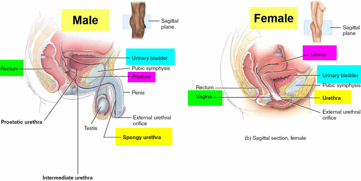
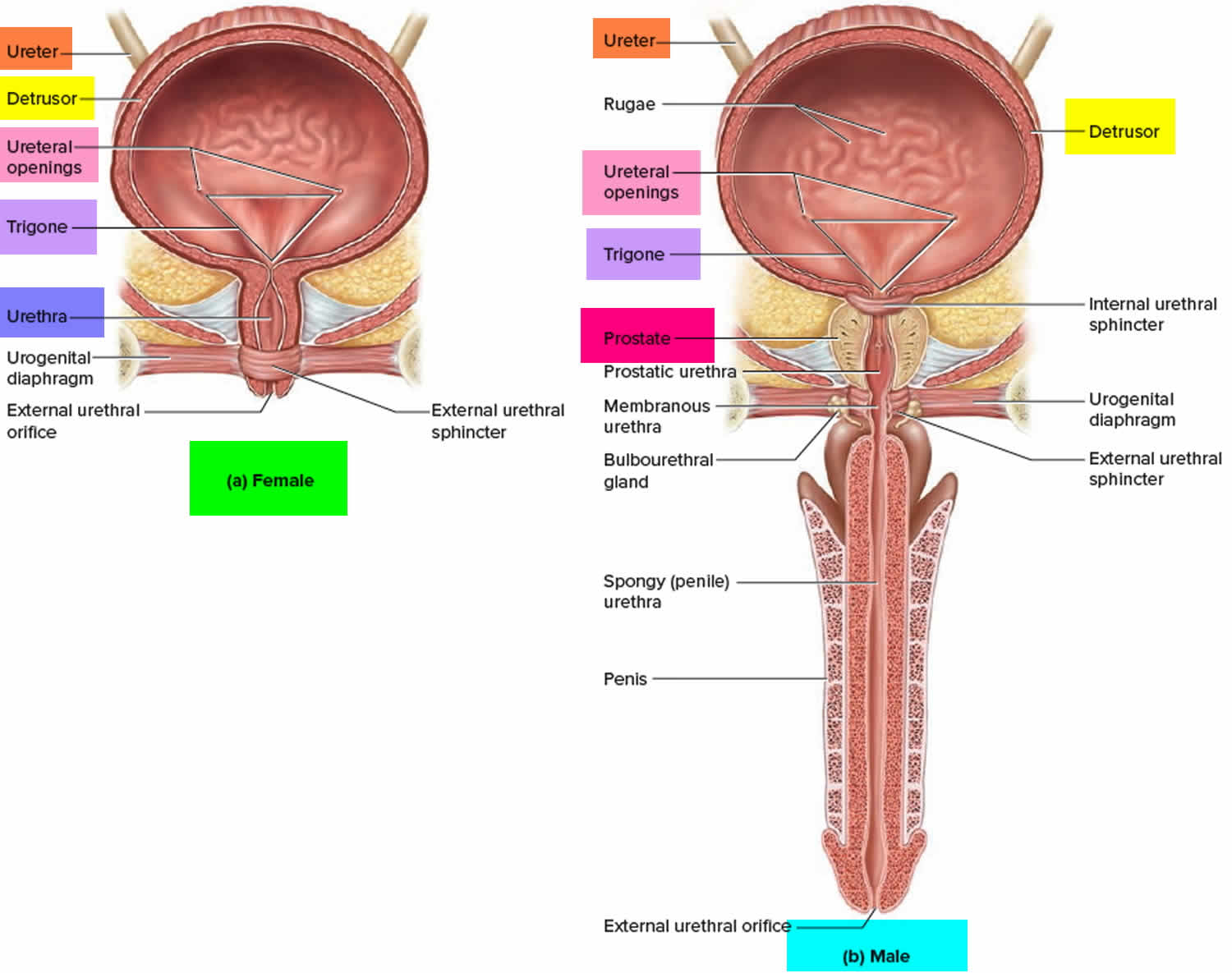
Figure 2. Urinary bladder anatomy

Urinary bladder anatomy

Causes of urinary frequency

Some of the causes of increased urine volume differ from those of too-frequent urination. However, because many people who produce excessive amounts of urine also need to urinate frequently, these two symptoms are often considered together.

The most common causes of urinary frequency are:

  • Bladder infections (the most common cause in women and children)
  • Urinary incontinence
  • Noncancerous enlargement of the prostate gland (benign prostatic hyperplasia—the most common cause in men over 50). Benign prostatic hyperplasia (BPH) is nonmalignant adenomatous overgrowth of the periurethral prostate gland. Symptoms are those of bladder outlet obstruction—weak stream, hesitancy, urinary frequency, urgency, nocturia, incomplete emptying, terminal dribbling, overflow or urge incontinence, and complete urinary retention. Diagnosis is based primarily on digital rectal examination and symptoms; cystoscopy, transrectal ultrasonography, urodynamics, or other imaging studies may also be needed. Treatment options include 5 alpha-reductase inhibitors, alpha-blockers, tadalafil, and surgery.
  • Urinary tract infections (UTIs). Urinary tract infections can be divided into upper tract infections, which involve the kidneys (pyelonephritis), and lower tract infections, which involve the bladder (cystitis), urethra (urethritis), and prostate (prostatitis). However, in practice, and particularly in children, differentiating between the sites may be difficult or impossible. Moreover, infection often spreads from one area to the other. Although urethritis and prostatitis are infections that involve the urinary tract, the term urinary tract infection (UTI) usually refers to pyelonephritis and cystitis. Most cystitis and pyelonephritis are caused by bacteria. The most common nonbacterial pathogens are fungi (usually candidal species), and, less commonly, mycobacteria, viruses, and parasites. Nonbacterial pathogens usually affect patients who are immunocompromised; have diabetes, obstruction, or structural urinary tract abnormalities; or have had recent urinary tract instrumentation. Other than adenoviruses (implicated in hemorrhagic cystitis), viruses have no major contribution to UTI in immunocompetent patients. The predominant parasitic causes of urinary tract infections are filariasis, trichomoniasis, leishmaniasis, malaria, and schistosomiasis. Of the parasitic diseases, only trichomoniasis is common in the US, usually as a sexually transmitted disease (STD). Urethritis is usually caused by an STD. Prostatitis is usually caused by a bacterium and is sometimes caused by an STD.
  • Stones in the urinary tract. Urinary calculi are solid particles in the urinary system. They may cause pain, nausea, vomiting, hematuria, and, possibly, chills and fever due to secondary infection. Diagnosis is based on urinalysis and radiologic imaging, usually noncontrast helical CT. Treatment is with analgesics, antibiotics for infection, medical expulsive therapy, and, sometimes, shock wave lithotripsy or endoscopic procedures.

The most common causes of polyuria in both adults and children are:

  • Uncontrolled diabetes mellitus (most common)
  • Drinking too much fluid (polydipsia)
  • Diabetes insipidus
  • Taking diuretic drugs or substances (which increase the excretion of urine), such as alcohol or caffeine

Diabetes insipidus causes polyuria because of problems with a hormone called antidiuretic hormone (or vasopressin). Antidiuretic hormone helps the kidneys reabsorb fluid. If too little antidiuretic hormone is produced (a condition called central diabetes insipidus) or if the kidneys are unable to properly respond to it (nephrogenic diabetes insipidus), the person urinates excessively.

People with certain kidney disorders (such as interstitial nephritis or kidney damage resulting from sickle cell anemia) may also urinate excessively because these disorders also decrease the amount of fluid reabsorbed by the kidneys.

Table 1. Some causes of urinary frequency

Cause Suggestive Findings Diagnostic Approach
Benign prostatic hyperplasia or prostate cancer Progressive onset of urinary hesitancy, incontinence, poor urine stream, a sensation of incomplete voiding Rectal examination
Ultrasonography
Cystometry
Cystocele Urinary incontinence
Sensation of vaginal fullness
Pain or urinary leakage during sexual intercourse
Pelvic examination
Voiding cystourethrography
Drugs and substances:
Caffeine
Alcohol
Diuretics
Urinary frequency in an otherwise healthy patient Empiric elimination of offending substance (to confirm that frequency resolves)
Pregnancy 3rd trimester of pregnancy Clinical evaluation
Prostatitis Urgency, dysuria, nocturia, purulent urethral discharge with fever, chills, low back pain, myalgia, arthralgia, and perineal fullness
Prostate tender to palpation
Rectal examination
Culture of secretions after prostatic massage
Radiation cystitis History of radiation therapy of the lower abdomen, prostate, or perineum for treatment of cancer Clinical evaluation
Cystoscopy and biopsy
Reactive arthritis Asymmetric arthritis of knees, ankles, and metatarsophalangeal joints
Unilateral or bilateral conjunctivitis
Small, painless ulcers on the mouth, tongue, glans penis, palms, and soles 1–2 weeks after sexual contact
STD testing
Spinal cord injury or lesion Lower-extremity weakness, decreased anal sphincter tone, absent anal wink reflex
Loss of sensation at a segmental level
Injury usually clinically obvious
MRI of the spine
Urethral stricture Hesitancy, tenesmus, reduced caliber and force of the urine stream Urethrography
Urinary incontinence Unintentional passage of urine, particularly when bending, coughing, or sneezing Cystometry
Urinary tract calculi Colicky flank or groin pain Urinalysis for hematuria
Ultrasonography or CT of the kidneys, ureters, and bladder
Urinary tract infections Dysuria and foul-smelling urine, sometimes fever, confusion, and flank pain, particularly in women and girls
Dysuria and frequency in young sexually active men (which suggests an STD)
Urinalysis and culture
STD testing
Bladder detrusor overactivity Nocturia, urge incontinence, weak urinary stream, and sometimes urinary retention Cystometry

Abbreviation: STD = sexually transmitted disease

Urinary incontinence in children

Urinary incontinence is when a child can’t control her bladder and wets herself during the day. It’s accidental, and there are a few different causes. Urinary incontinence is defined as involuntary voiding of urine ≥ 2 times/month during the day or night. Revised terminology for the time of incontinence has been suggested 2):

  1. For urinary incontinence during the day: Diurnal incontinence or diurnal wetting
  2. For urinary incontinence at night: Enuresis or bed-wetting

Diurnal (daytime) incontinence is usually not diagnosed until age 5 or 6. Nocturnal (nighttime) incontinence (that is, enuresis) is usually not diagnosed until age 7. Before this time, enuresis is typically referred to as nighttime wetting 3). These age limits are based on children who are developing typically and so may not be applicable to children with developmental delay. Both nocturnal and diurnal incontinence are symptoms—not diagnoses—and necessitate consideration of an underlying cause.

The age at which children attain urinary continence varies, but > 90% are continent during the day by age 5. Nighttime continence takes longer to achieve. Enuresis affects about 30% of children at age 4, 10% at age 7, 3% at age 12, and 1% at age 18. About 0.5% of adults continue to have nocturnal wetting episodes. Enuresis is more common among boys and when there is a family history 4).

In primary incontinence, children have never achieved urinary continence for ≥ 6 months. In secondary incontinence, children have developed incontinence after a period of at least 6 months of urinary control. An organic cause is more likely in secondary incontinence. Even when there is no organic cause, appropriate treatment and parental education are essential because of the physical and psychologic impact of urine accidents 5).

Sometimes urinary incontinence happens because children’s bladders, genitals, urinary tracts or urethras haven’t developed properly, which means they don’t work properly.

Some children have overactive bladders, which means their bladders don’t store urine the way they’re supposed to. This can make children suddenly feel like they have to do a wee, so they wet themselves.

Other children have underactive bladders, which means they can’t empty their bladders fully, so wee can dribble out before or after they go to the toilet.

Constipation and urinary tract infections (UTI) can cause temporary daytime wetting for some children.

For some children stress can cause urinary incontinence. For example, stressful life events like starting school, the birth of a new sibling, or parents separating can mean that children can’t focus on going to the toilet by themselves. They might start to accidentally wet themselves as a result.

Children who have daytime wetting often also experience bedwetting at night. Usually treatment will focus on daytime wetting first and then look at night-time wetting.

When to see your doctor about urinary incontinence

You should see your doctor for advice about treatment and management if your child is older than 5 years and:

  • is experiencing urinary incontinence more than once a month
  • has never had a period of dryness.

When you see your doctor, your doctor might start by doing a physical examination of your child’s tummy, lower back and genitals. Your doctor might also test your child’s urine.

Your doctor might refer your child to an incontinence specialist.

Symptoms of urinary incontinence in children

The main symptom of urinary incontinence is your child wetting himself during the day because he can’t control his bladder.

How often a child wets herself during the day varies. It can be:

  • continuous, which means the child has an uncontrollable dribble of wee, and has never had a period of dryness
  • intermittent, which means the child has periods of dryness during the day.

Whether urinary incontinence is continuous or intermittent depends on what’s causing the incontinence.

Treatment for urinary incontinence in children

There are many different treatment options for urinary incontinence, and the right treatment for your child will depend on what’s causing the incontinence.

Behavior modification

The most common treatment for urinary incontinence is behavior modification, which is also called urotherapy.

This involves giving your child information about how his lower urinary tract works, and also about:

  • going to the toilet regularly
  • not holding on when he needs to wee
  • recognizing the signs of needing to do a wee.

Your child might be asked to keep a diary of how often she goes to the toilet, how long in between wees, and how often she wets herself.

The doctor might also ask you to measure the amount of pee your child produces when he goes to the toilet, and how much your child drinks each day. As part of the treatment, your child will probably be asked to wee at particular times throughout the day.

Medication

In some cases, the doctor might prescribe medication to help treat daytime wetting. The type of medication depends on the cause. If your doctor prescribes medication, it’s a good idea to ask why. You can also ask about side effects.

Other treatment options

Depending on the cause of your child’s urinary incontinence, there are other treatment options. These treatment options are for more complex cases of incontinence, and your doctor will usually refer you to a specialist for advice about these options.

Urinary incontinence in adults

Urinary incontinence is involuntary loss of urine; some experts consider it present only when a patient thinks it is a problem. The disorder is greatly underrecognized and underreported. Many patients do not report the problem to their physician, and many physicians do not ask about incontinence specifically. Incontinence can occur at any age but is more common among older people and women, affecting about 30% of older women and 15% of older men.

Incontinence greatly reduces quality of life by causing embarrassment, stigmatization, isolation, and depression. Many older patients are institutionalized because incontinence is a burden to caregivers. In bedbound patients, urine irritates and macerates skin, contributing to sacral pressure ulcer formation. Older people with urgency are at increased risk of falls and fractures.

Types of urinary incontinence in adults

Incontinence may manifest as near-constant dribbling or as intermittent voiding with or without awareness of the need to void. Some patients have extreme urgency (irrepressible need to void) with little or no warning and may be unable to inhibit voiding until reaching a bathroom. Incontinence may occur or worsen with maneuvers that increase intra-abdominal pressure. Postvoid dribbling is extremely common and probably a normal variant in men. Identifying the clinical pattern is sometimes useful, but causes often overlap and much of treatment is the same.

  • Urge incontinence. Urge incontinence is uncontrolled urine leakage (of moderate to large volume) that occurs immediately after an urgent, irrepressible need to void. Nocturia and nocturnal incontinence are common. Urge incontinence is the most common type of incontinence in older people but may affect younger people. It is often precipitated by use of a diuretic and is exacerbated by inability to quickly reach a bathroom. In women, atrophic vaginitis, common with aging, contributes to thinning and irritation of the urethra and urgency.
  • Stress incontinence. Stress incontinence is urine leakage due to abrupt increases in intra-abdominal pressure (eg, with coughing, sneezing, laughing, bending, or lifting). Leakage volume is usually low to moderate. It is the 2nd most common type of incontinence in women, largely because of complications of childbirth and development of atrophic urethritis. Men can develop stress incontinence after procedures such as radical prostatectomy. Stress incontinence is typically more severe in obese people because of pressure from abdominal contents on the top of the bladder.
  • Overflow incontinence. Overflow incontinence is dribbling of urine from an overly full bladder. Volume is usually small, but leaks may be constant, resulting in large total losses. Overflow incontinence is the 2nd most common type of incontinence in men.
  • Functional incontinence. Functional incontinence is urine loss due to cognitive or physical impairments (eg, due to dementia or stroke) or environmental barriers that interfere with control of voiding. For example, the patient may not recognize the need to void, may not know where the toilet is, or may not be able to walk to a remotely located toilet. Neural pathways and urinary tract mechanisms that maintain continence may be normal.
  • Mixed incontinence. Mixed incontinence is any combination of the above types. The most common combinations are urge with stress incontinence and urge or stress with functional incontinence.

Causes of urinary incontinence in adults

The disorder tends to differ among age groups. With aging, bladder capacity decreases, ability to inhibit urination declines, involuntary bladder contractions (detrusor overactivity) occur more often, and bladder contractility is impaired. Thus, voiding becomes more difficult to postpone and tends to be incomplete. Postvoid residual volume increases, probably to ≤ 100 mL (normal < 50 mL). Endopelvic fascia weakens.

In postmenopausal women, decreased estrogen levels lead to atrophic urethritis and atrophic vaginitis and to decreasing urethral resistance, length, and maximum closure pressure.

In men, prostate size increases, partially obstructing the urethra and leading to incomplete bladder emptying and strain on the detrusor muscle. These changes occur in many normal, continent older people and may facilitate incontinence but do not cause it.

In younger patients, incontinence often begins suddenly, may cause little leakage, and usually resolves quickly with little or no treatment. Often, incontinence has one cause in younger patients but has several in older people.

Transient incontinence

There are several causes of transient incontinence see Table 2.

Table 2. Causes of Transient Incontinence

Cause Comments
Gastrointestinal disorders
Fecal impaction Mechanism may involve mechanical disturbance of the bladder or urethra. Patients usually present with urge or overflow incontinence, typically with fecal incontinence.
Genitourinary disorders
Atrophic urethritis
Atrophic vaginitis
Thinning of urethral and vaginal epithelium and submucosa may cause local irritation and decrease urethral resistance, length, and maximum closure pressure with loss of the mucosal seal. These disorders are usually characterized by urgency and occasionally by scalding dysuria.
Urinary calculi
Foreign bodies
Bladder irritation precipitates spasm.
Urinary tract infections Only symptomatic UTIs cause incontinence.
Dysuria and urgency can prevent patients from reaching the toilet before voiding.
Neuropsychiatric disorders
Delirium
Depression
Psychosis
Awareness of the need or ability to void is impaired.
Restricted mobility
Weakness
Injury
Use of physical restraints
Access to toilet is impaired.
Systemic disorders
Excess urine output due to various disorders (eg, diabetes insipidus, diabetes mellitus) Frequency, urgency, and nocturia can result.
Drugs
Alcohol Alcohol has a diuretic effect and can cause sedation, delirium, or immobility, which can result in functional incontinence.
Caffeine (eg, in coffee, tea, cola and some other soft drinks, cocoa, chocolate, and energy drinks) Urine production and output are increased, causing polyuria, frequency, urgency, and nocturia.
Alpha-adrenergic antagonists (eg, alfuzosin, doxazosin, prazosin, tamsulosin, terazosin) Bladder neck muscle in women or prostate smooth muscle in men is lax, sometimes causing stress incontinence.
Anticholinergics (eg, antihistamines, antipsychotics, benztropine, tricyclic antidepressants) Bladder contractility can be impaired, sometimes causing urinary retention and overflow incontinence. These drugs also can cause delirium, constipation, and fecal impaction.
Calcium channel blockers (eg, diltiazem, nifedipine, verapamil) Detrusor contractility is decreased, sometimes causing urinary retention and overflow incontinence, nocturia due to peripheral edema, constipation, and fecal impaction.
Diuretics (eg, bumetanide, furosemide, [not thiazides]) Urine production and output are increased, causing polyuria, frequency, urgency, and nocturia.
Hormone therapy (systemic estrogen/progestin therapy) Collagen in the paraurethral connective tissues is degraded, causing ineffective urethral closure.
Misoprostol Misoprostol relaxes the urethra and thus may cause stress incontinence.
Psychoactive drugs (eg, antipsychotics, benzodiazepines, sedative-hypnotics, tricyclic antidepressants) Awareness of the need to void is blunted, and dexterity and mobility are decreased. These drugs can precipitate delirium.

Established incontinence

Established incontinence is caused by a persistent problem affecting nerves or muscles. Mechanisms usually used to describe these problems are bladder outlet incompetence or obstruction, detrusor overactivity or underactivity, detrusor-sphincter dyssynergia, or a combination. However, these mechanisms are also involved in some transient causes.

  • Outlet incompetence. Outlet incompetence is a common cause of stress incontinence. In women, it is usually due to weakness of the pelvic floor or of the endopelvic fascia. Such weakness commonly results from multiple vaginal deliveries, pelvic surgery (including hysterectomy), age-related changes (including atrophic urethritis), or a combination. As a result, the vesicourethral junction descends, the bladder neck and urethra become hypermobile, and pressure in the urethra falls below that of the bladder. In men, a common cause is damage to the sphincter or to the bladder neck and posterior urethra after radical prostatectomy.
  • Outlet obstruction. Outlet obstruction is a common cause of incontinence in men, although most men with obstruction are not incontinent. Obstruction in men commonly results from benign prostatic hyperplasia, prostate cancer, or urethral stricture. In both sexes, fecal impaction can cause obstruction. In women, outlet obstruction can result from previous surgery for incontinence or from a prolapsed cystocele that causes the urethra to kink during straining to void. Obstruction leads to a chronically overdistended bladder, which loses its ability to contract; then the bladder does not empty completely, resulting in overflow. Obstruction also may lead to detrusor overactivity and urge incontinence. If the detrusor muscle loses its ability to contract, overflow incontinence may follow. Some causes of outlet obstruction (eg, large bladder diverticula, cystoceles, bladder infections, calculi, and tumors) are reversible.
  • Detrusor overactivity. Detrusor overactivity is a common cause of urge incontinence in older and younger patients. The detrusor muscle contracts intermittently for no apparent reason, usually when the bladder is partially or nearly full. Detrusor overactivity may be idiopathic or may result from dysfunction of the frontal micturition inhibitory center (commonly due to age-related changes, dementia, or stroke) or outlet obstruction. Detrusor overactivity (hyperactivity) with impaired contractility is a variant of urge incontinence characterized by urgency, frequency, a weak flow rate, urinary retention, bladder trabeculation, and a postvoid residual volume of > 50 mL. This variant may mimic prostatism in men or stress incontinence in women.
  • Overactive bladder. Overactive bladder is a term sometimes used to describe urinary urgency (with or without incontinence) that is often accompanied by urinary frequency and nocturia.
  • Detrusor underactivity. Detrusor underactivity causes urinary retention and overflow incontinence in about 5% of patients with incontinence. It may be caused by injury to the spinal cord or to nerve roots supplying the bladder (eg, by disk compression, tumor, or surgery), by peripheral or autonomic neuropathies, or by other neurologic disorders (see table Causes of Established Incontinence). Anticholinergics and opioids greatly decrease detrusor contractility; these drugs are common transient causes. The detrusor may become underactive in men with chronic outlet obstruction as the detrusor is replaced by fibrosis and connective tissue, preventing the bladder from emptying even when the obstruction is removed. In women, detrusor underactivity is usually idiopathic. Less severe detrusor weakness is common among older women. Such weakness does not cause incontinence but can complicate treatment if other causes of incontinence coexist.
  • Detrusor-sphincter dyssynergia. Detrusor-sphincter dyssynergia (loss of coordination between bladder contraction and external urinary sphincter relaxation) may cause outlet obstruction, with resultant overflow incontinence. Dyssynergia is often due to a spinal cord lesion that interrupts pathways to the pontine micturition center, which coordinates sphincter relaxation and bladder contraction. Rather than relaxing when the bladder contracts, the sphincter contracts, obstructing the bladder outlet. Dyssynergia causes severe trabeculation, diverticula, a “Christmas tree” deformation of the bladder seen on cystogram, hydronephrosis, and renal failure.
  • Functional impairment. Functional impairment (eg, cognitive impairment, reduced mobility, reduced manual dexterity, coexisting disorders, lack of motivation), particularly in older patients, may contribute to established incontinence but rarely causes it.

Table 3. Causes of established incontinence

Urodynamic Diagnosis Some Neurologic Causes Some Nonneurologic Causes
Bladder outlet incompetence Lower motor neuron lesion (rare). In men, radical prostatectomy* Intrinsic sphincter deficiency
Urethral hypermobility
In women, multiple vaginal deliveries, pelvic surgery (eg, hysterectomy), age-related changes (eg, atrophic urethritis)
In men, prostate surgery
Bladder outlet obstruction Spinal cord lesion causing detrusor-sphincter dyssynergia (rare) Anterior urethral stricture
Urethral diverticula (rarely) or large bladder diverticula (very rarely)
Bladder calculi
Bladder neck suspension surgery
In women, cystocele (if large)
In men, benign prostatic hyperplasia or prostate cancer
Detrusor overactivity Alzheimer disease
Spinal cord injury/dysfunction
Multiple sclerosis
Stroke
Bladder carcinoma
Cystitis
Idiopathic
Outlet obstruction or incompetence
Detrusor underactivity Autonomic neuropathy (eg, due to diabetes, alcoholism, or vitamin B12 deficiency)
Disk compression
Plexopathy
Spinal neural tube defect (less often, may cause overactivity)
Surgical damage (eg, anteroposterior resection)
Tumor
Chronic bladder outlet obstruction. Idiopathic (common among women)
Detrusor-sphincter dyssynergia Spinal cord lesion
Brain lesion affecting pathways to the pontine micturition center
Voiding dysfunction of childhood (poor relaxation of the sphincter with bladder contraction can result from the fear of bed wetting or soiling of clothes)

Footnote: * Other prostate surgical procedures rarely cause established incontinence.

Urinary frequency diagnosis

Many people are embarrassed to discuss problems related to urination with their doctor. But because some disorders that cause excessive urination are quite serious, people who urinate excessively should be evaluated by a doctor. The following information can help people know when to see a doctor and what to expect during the evaluation.

Doctors first ask questions about the person’s symptoms and medical history and then do a physical examination. What they find during the history and physical examination often suggests a cause of excessive urination and the tests that may need to be done.

Medical history

Doctors ask about:

  • Amounts of fluid drunk and urinated to determine whether the problem is related to urinary frequency or to polyuria
  • If urinary frequency is present, patients are asked about acuity of onset, presence or absence of irritative symptoms (eg, irritation, urgency, dysuria), obstructive symptoms (eg, hesitancy, poor flow, sensation of incomplete voiding, nocturia), and recent sexual contacts.
  • How long symptoms have been present
  • Whether any other urination problems are present
  • Whether the person is taking diuretics (drugs and other substances that increase urine production), including beverages that contain caffeine
  • Review of systems should cover symptoms suggestive of a cause, including fever, flank or groin pain, and hematuria (infection); missed menses, breast swelling, and morning sickness (pregnancy); and arthritis and conjunctivitis (reactive arthritis).
  • Past medical history should ask about known causes, including prostate disease and previous pelvic radiation therapy or surgeries. Drugs and diet are reviewed for the use of agents that increase urine output (eg, diuretics, alcohol, caffeinated beverages).

Some obvious findings may give clues to the cause of frequent urination. Pain or burning during urination (dysuria), fever, and back or side pain may indicate an urinary tract infection (UTI) or calculi. In a person who drinks large amounts of beverages that contain caffeine or who has just started treatment with a diuretic, the diuretic substance is a likely cause. A man who has other problems with urination, such as difficulty starting urination, a weak urine stream, and dribbling at the end of urination, may have a prostate disorder.

Prior pelvic surgery suggests incontinence. Weak urine stream, nocturia, or both suggests benign prostatic hyperplasia (BPH). Urinary frequency in an otherwise healthy young patient may be due to excessive intake of alcohol or caffeinated beverages. Gross hematuria (blood in urine) suggests UTI and calculi in younger patients and genitourinary cancer in older patients.

Some obvious findings may also give clues to the cause of polyuria. For example, polyuria that starts during the first few years of life is likely caused by an inherited disorder such as central or nephrogenic diabetes insipidus or type 1 diabetes mellitus.

Physical examination

Examination focuses on the genitourinary system.

Any urethral discharge or any lesions consistent with sexually transmitted diseases are noted. Rectal examination in men should note the size and consistency of the prostate and rectal tone; pelvic examination in women should note the presence of any cystocele. Patients should be instructed to cough while the urethra is observed for signs of urinary leakage.

The costovertebral angle should be palpated for tenderness, and the abdominal examination should note the presence of any masses or suprapubic tenderness.

Neurologic examination should test for lower-extremity weakness and loss of sensation.

In women, the physical examination usually includes a pelvic examination and the taking of samples of cervical and vaginal fluid to check for sexually transmitted diseases. In men, the penis is examined for presence of a discharge, and doctors do a digital rectal examination to examine the prostate.

Testing

All patients require urinalysis and culture, which are easily done and can detect infection and hematuria.

The need for other testing depends on what doctors find during the history and physical examination.

If doctors are not sure whether the person is actually producing more urine than normal, they may collect and measure the amount of urine produced over 24 hours. If people actually have polyuria, doctors measure the blood glucose level. If diabetes mellitus is not the cause of polyuria and no other cause, such as excess intravenous fluids, is clearly responsible, other testing is necessary. The levels of electrolytes and concentration of certain salts (osmolarity) are measured in the blood, urine, or both, often after the person is deprived of water for a time and after the person is given antidiuretic hormone.

Cytoscopy, cystometry, and urethrography can be done to diagnose cystitis, bladder outlet obstruction, and cystocele. Prostate-specific antigen level determination, ultrasonography, and prostate biopsy may be required, especially in older men, to differentiate benign prostatic hyperplasia (noncancerous enlargement of the prostate gland) from prostate cancer.

Urinary frequency treatment

Urinary frequency treatment involves treating the underlying cause or condition.

References   [ + ]

read more

Hearing loss in children

hearing loss in children

Hearing loss in children

A hearing loss can happen when any part of the ear is not working in the usual way (Figure 1 below). This includes the outer ear, middle ear, inner ear, hearing (acoustic) nerve, and auditory system. As hearing is important for language development, early detection and treatment of any hearing loss is important. In the first few years of life, hearing is a critical part of kids’ social, emotional, and cognitive development. Hearing loss can affect a child’s ability to develop speech, language, and social skills. Even a mild or partial hearing loss can affect a child’s ability to develop speech and language properly. The earlier children with hearing loss start getting services, the more likely they are to reach their full potential. If you think that a child might have hearing loss, trust your instincts and ask the child’s doctor for a hearing screening as soon as possible.

Having a hearing loss (impairment) means that a child has lost some hearing in one or both ears. Hearing loss may be partial – a child can’t hear certain sounds – or it can be complete. It may be temporary or permanent, and caused by a wide range of possible factors.

There are many types and degrees of hearing loss, and they are described according to how much hearing has been lost and which part of the ear is affected. Hearing loss is also explained as mild, moderate, moderate to severe, severe or profound. Hearing is measured in decibels (dB) and the severity of the hearing loss is graded by hearing thresholds. The normal hearing range is 0-20 decibels (dB) which equates to being able to perceive sound quieter than a whisper. Mild hearing loss corresponds to a range of 20-39 dB (a person with a mild hearing loss may hear some speech sounds but soft sounds are hard to hear), moderate 40-69 dB (a person with a moderate hearing loss may hear almost no speech when another person is talking at a normal level), severe 70-89 dB (a person with severe hearing loss will hear no speech when a person is talking at a normal level and only some loud sounds) and profound is greater than 90 dB (a person with a profound hearing loss will not hear any speech and only very loud sound).

Table 1. The table below shows a common way to classify hearing loss

Degree of hearing loss Hearing loss range (dB)
Normal –10 to 15
Slight 16 to 25
Mild 26 to 40
Moderate 41 to 55
Moderately severe 56 to 70
Severe 71 to 90
Profound 91+
[Source 1) ]

Hearing loss occurs in 1-3 newborns per 1000 births 2), with 1-2 per 1,000 suffering from permanent childhood hearing impairment 3). There is a slightly increased prevalence of hearing loss in boys compared to girls with a ratio of 1.16:1.0 4).

There are 2 main types of hearing impairment: ‘conductive’ and ‘sensorineural’. Having both types is called a ‘mixed loss’.

  1. Conductive hearing loss occurs when something interferes with sound waves travelling through the outer and middle parts of the ear to the inner ear. It can be caused by a wax blockage, middle ear infection (otitis media), a fluid build-up in the middle ear, or damage to the tiny bones in the middle ear. Conductive hearing loss may be either temporary or permanent.
  2. Sensorineural hearing loss is caused by a problem in the innermost part of the ear or in the nerve which carries hearing, the ‘auditory nerve’. It can be caused by abnormal inner ear development, a physical injury to the inner ear, damage to the ear from diseases such as meningitis and rubella, or a tumor. Despite being relatively uncommon in children as a whole, it is the primary cause of permanent hearing loss in the pediatric population. Sensorineural hearing losses are usually permanent. Sometimes the problem with the inner ear also causes problems with balance. Children with both hearing and balance problems may have delayed development of their motor skills.
  3. Mixed hearing loss occurs when there are both conductive and sensorineural components.

Ear anatomy

A hearing loss can happen when any part of the ear or auditory (hearing) system is not working in the usual way.

Outer Ear

The outer ear is made up of:

  • the part we see on the sides of our heads, known as pinna
  • the ear canal
  • the eardrum, sometimes called the tympanic membrane, which separates the outer and middle ear

Middle Ear

The middle ear is made up of:

  • the eardrum
  • three small bones called ossicles that send the movement of the eardrum to the inner ear

Inner Ear

The inner ear is made up of:

  • the snail shaped organ for hearing known as the cochlea
  • the semicircular canals that help with balance
  • the nerves that go to the brain

Auditory (ear) Nerve

This nerve sends sound information from the ear to the brain.

Auditory (Hearing) System

The auditory pathway processes sound information as it travels from the ear to the brain so that our brain pathways are part of our hearing.

Figure 1. Ear anatomy

Human ear anatomy

Ear anatomy

Types of hearing loss

There are four types of hearing loss:

  1. Conductive hearing loss: Hearing loss caused by something that stops sounds from getting through the outer or middle ear. This type of hearing loss can often be treated with medicine or surgery.
  2. Sensorineural hearing loss: Hearing loss that occurs when there is a problem in the way the inner ear or hearing nerve works.
  3. Mixed hearing loss: Hearing loss that includes both a conductive and a sensorineural hearing loss.
  4. Auditory Neuropathy Spectrum Disorder: Hearing loss that occurs when sound enters the ear normally, but because of damage to the inner ear or the hearing nerve, sound isn’t organized in a way that the brain can understand.

Hearing loss can also be described as:

  1. Unilateral or Bilateral: Hearing loss is in one ear (unilateral) or both ears (bilateral).
  2. Pre-lingual or Post-lingual: Hearing loss happened before a person learned to talk (pre-lingual) or after a person learned to talk (post-lingual)
  3. Symmetrical or Asymmetrical: Hearing loss is the same in both ears (symmetrical) or is different in each ear (asymmetrical).
  4. Progressive or Sudden: Hearing loss worsens over time (progressive) or happens quickly (sudden).
  5. Fluctuating or Stable: Hearing loss gets either better or worse over time (fluctuating) or stays the same over time (stable).
  6. Congenital or Acquired/Delayed Onset: Hearing loss is present at birth (congenital) or appears sometime later in life (acquired or delayed onset).

Hearing loss in children causes

Hearing loss can be broadly characterized as congenital or acquired in the pediatric population.

Congenital causes

Congenital hearing loss can be classified as genetic and non-genetic in origin. Genetic cause of hearing loss is responsible for 50% to 60% of congenital causes and can be due to either an autosomal dominant, recessive or sex-linked mutation 5). Genetic causes are often further subdivided into syndromic versus non-syndromic categories based on whether the patient suffers from an underlying genetic syndrome. About 70% of all mutations causing hearing loss are non-syndromic. This means that the person does not have any other symptoms. About 30% of the mutations causing hearing loss are syndromic. This means that the person has other symptoms besides hearing loss. For example, some people with hearing loss are also blind 6). The most common cause of congenital hearing loss is autosomal recessive non-syndromic hearing loss.

There are also a number of things in the environment that can cause hearing loss. 25% or more of hearing loss in babies is due to “environmental” causes such as maternal infections during pregnancy and complications after birth. Sometimes both genes and environment work together to cause hearing loss. For example, there are some medicines that can cause hearing loss, but only in people who have certain mutations in their genes.

Genes contain the instructions that tell the cells of people’s bodies how to grow and work. For example, the instructions in genes control what color a person’s eyes will be. There are many genes that are involved in hearing. Sometimes, a gene does not form in the expected manner. This is called a mutation. Some mutations run in families and others do not. If more than one person in a family has hearing loss, it is said to be “familial”. That is, it runs in the family.

The cochlea (the part of the ear that changes sounds in the air into nerve signals to the brain) is a very complex and specialized part of the body that needs many instructions to guide its development and function. These instructions come from genes. Changes in any one of these genes can result in hearing loss. The GJB2 gene is one of the genes that contains the instructions for a protein called connexin 26; this protein plays an important role in the functioning of the cochlea. In some populations about 40% of newborns with a genetic hearing loss who do not have a syndrome, have a mutation in the GJB2 gene.

TORCH organisms (toxoplasmosis, rubella, cytomegalovirus [CMV] and herpes) have been identified as key infective causative agents. Cytomegalovirus (CMV) is the most common cause of congenital non-genetic hearing loss in the developed world 7). Other congenital causes include trauma, ototoxic medications used in the antenatal period and several perinatal risk factors such as prematurity, low birth weight, and hyperbilirubinemia.

Acquired hearing loss

Otitis media with effusion is the number one cause of acquired hearing loss in children. It is beyond the scope of this article to cover this in detail, but it classically has a bimodal beak at 2 years and 5 years of age and is characterized by a conductive hearing loss associated with flattened tympanogram 8). It typically resolves without intervention as the eustachian tube matures or following the insertion of a ventilation tube in the middle ear 9). Adenoidal hypertrophy can contribute to this clinical picture 10). Infections also present another major category for acquired hearing loss, with a particularly strong link with bacterial meningitis, mumps, and measles. Other reasons include primary otological pathologies such as cholesteatoma, impacted wax and otosclerosis as well as trauma.

High-risk factors in neonates:

  • Congenital infections
  • Family history
  • Craniofacial anomalies
  • Hyperbilirubinemia
  • Birth weight 1500 g
  • Low Apgar score
  • Bacterial meningitis
  • Prolonged intubation.

Risk factors for hearing problems in children

Hearing loss can happen any time during life – from before birth to adulthood.

Following are some of the things that can increase the chance that a child will have hearing loss:

  • A genetic cause: About 1 out of 2 cases of hearing loss in babies is due to genetic causes. Some babies with a genetic cause for their hearing loss might have family members who also have a hearing loss. About 1 out of 3 babies with genetic hearing loss have a “syndrome.” This means they have other conditions in addition to the hearing loss, such as Down syndrome or Usher syndrome.
  • 1 out of 4 cases of hearing loss in babies is due to maternal infections during pregnancy, complications after birth, and head trauma. For example, the child:
    • Was exposed to infection, such as , before birth
    • Spent 5 days or more in a hospital neonatal intensive care unit (NICU) or had complications while in the NICU
    • Needed a special procedure like a blood transfusion to treat bad jaundice
    • Has head, face or ears shaped or formed in a different way than usual
    • Has a condition like a neurological disorder that may be associated with hearing loss
    • Had an infection around the brain and spinal cord called meningitis
    • Received a bad injury to the head that required a hospital stay
  • For about 1 out of 4 babies born with hearing loss, the cause is unknown.

Hearing loss in children prevention

Following are tips for parents to help prevent hearing loss in their children:

  • Have a healthy pregnancy.
  • Make sure your child gets all the regular childhood vaccines.
  • Keep your child away from high noise levels, such as from very loud toys.
  • If you think that your child might have hearing loss, ask your child’s doctor for a hearing screening as soon as possible. Don’t wait!
  • If your child does not pass a hearing screening, ask your child’s doctor for a full hearing test as soon as possible.
  • If your child has hearing loss, talk to your child’s doctor about treatment and intervention services.

Remember, hearing loss can affect a child’s ability to develop speech, language, and social skills. The earlier children with hearing loss start getting services, the more likely they are to reach their full potential. If you are a parent and you suspect your child has hearing loss, trust your instincts and speak with your child’s doctor.

Dangerous and safe noise levels

The noise chart below lists average decibel levels for everyday sounds around you.

Painful impulse noise—Not safe for any period of time

  • 150 dBP = fireworks at 3 feet, firecracker, shotgun
  • 140 dBP = firearms

Painful steady noise—Not safe for any period of time

  • 130 dBA = jackhammer
  • 120 dBA = jet plane takeoff, siren, pneumatic drill

Extremely loud—Dangerous to hearing; wear earplugs or earmuffs

  • 112 dBA = maximum output of some MP3 players, rock concert, chainsaw
  • 106 dBA = gas leaf blower, snow blower
  • 100 dBA = tractor, listening with earphones
  • 94 dBA = hair dryer, kitchen blender, food processor

Very loud—Dangerous to hearing; wear earplugs or earmuffs

  • 91 dBA = subway, passing motorcycle, gas mower

Moderate—Safe listening for any time period

  • 70 dBA = group conversation, vacuum cleaner, alarm clock
  • 60 dBA = typical conversation, dishwasher, clothes dryer
  • 50 dBA = moderate rainfall
  • 40 dBA = quiet room

Faint—Safe listening for any time period

  • 30 dBA = whisper, quiet library

Hearing loss in children signs and symptoms

The signs and symptoms of hearing loss are different for each child. If you think that your child might have hearing loss, ask the child’s doctor for a hearing screening as soon as possible. Don’t wait!

Even if a child has passed a hearing screening before, it is important to look out for the following signs.

Things to look out for, as they can be signs of possible hearing loss are:

  • loud noises do not startle your child by 4 months of age, or your child does not turn towards the source of a sound
  • your child notices you only when they see you
  • your child does not make sounds other than gargles and other vibrating noises that they can feel
  • speech development is delayed
  • your child seems to not hear when called or doesn’t respond to their name or say simple words by 14 months
  • your child can’t follow simple instructions by 24 months
  • your child hears some sounds but not others
  • your child has trouble holding their head steady, or is slow to sit up by themselves or walk

Signs in babies

  • Does not startle at loud noises.
  • Does not turn to the source of a sound after 6 months of age.
  • Does not say single words, such as “dada” or “mama” by 1 year of age.
  • Turns head when he or she sees you but not if you only call out his or her name. This sometimes is mistaken for not paying attention or just ignoring, but could be the result of a partial or complete hearing loss.
  • Seems to hear some sounds but not others.

As a guide, here’s what you’d expect in a typically developing baby:

  • At 0-4 months, your baby should startle at a loud noise, turn her head or move her eyes to locate the source of the sound. If she’s upset, she should calm down when she hears your voice.
  • At 4-8 months, your baby should notice sounds around him, smile when spoken to, babble and understand simple words like ‘bye-bye’.
  • At 8-14 months, your baby should respond to her name, say simple words like ‘mama’ and ‘dada’, copy simple sounds and use her voice to get attention from people nearby.
  • At 14-24 months, your child will start to develop vocabulary, understand and follow simple instructions, and put two words together.

If your child isn’t doing these things, it might be a good idea to talk to your child’s doctor. Even if everything seems OK but you still feel worried, you should see your doctor. After all, you know your baby best.

Signs in children

  • Speech is delayed.
  • Speech is not clear.
  • Does not follow directions. This sometimes is mistaken for not paying attention or just ignoring, but could be the result of a partial or complete hearing loss.
  • Often says, “Huh?”
  • Turns the TV volume up too high.

Babies and children should reach milestones in how they play, learn, communicate and act. A delay in any of these milestones could be a sign of hearing loss or other developmental problem.

Hearing loss in children diagnosis

Hearing loss can present in different ways depending on the age of the child. Hearing loss in neonates is almost exclusively picked up via newborn screening programme assessments. In older children, parents or other professionals such as school teachers, may notice delayed language skills, behavioral problems or listening to the television at raised volumes. In the history, it is important to ascertain whether there are any associated otological symptoms such as otorrhoea, otalgia, tinnitus, or vertigo. A thorough history is required including asking about any other neurological symptoms, medical history including drug history and precipitating events such as trauma, recent viral infections or new medications.

The examination will involve assessing the ear including the appearance of the pinna particularly inspecting for any deformities such as microtia or anotia. Otoscopic examination of the external auditory canal and tympanic membrane is crucial, with special attention on the attic for cholesteatoma. An examination should also include assessment of cranial nerves, a full neurological assessment, and assessment of balance depending on the age of the child.

With the implementation of Universal Newborn Hearing Screening program, today most patiets are identified within a few months after birth, with intervention started by 6 months.

Babies usually have their hearing tested in the first few weeks of life. Most newborns are tested before they leave hospital to help identify those who might require further hearing and middle ear function testing.

Babies with possible hearing loss are referred to a hearing specialist (audiologist) who will do more specialized testing to diagnose the type and extent of hearing loss. Understanding the cause helps determine the best treatment.

Hearing screening

Hearing screening can tell if a child might have hearing loss. Hearing screening is easy and is not painful. In fact, babies are often asleep while being screened. It takes a very short time — usually only a few minutes.

  • Babies: All babies should have a hearing screening no later than 1 month of age. Most babies have their hearing screened while still in the hospital. If a baby does not pass a hearing screening, it’s very important to get a full hearing test as soon as possible, but no later than 3 months of age.
  • Children: Children should have their hearing tested before they enter school or any time there is a concern about the child’s hearing. Children who are at risk for acquired, progressive, or delayed-onset hearing loss should have at least one hearing test by 2 to 2 1/2 years of age. Hearing loss that gets worse over time is known as acquired or progressive hearing loss. Hearing loss that develops after the baby is born is called delayed-onset hearing loss. Find out if a child may be at risk for hearing loss. Children who do not pass the hearing screening need to get a full hearing test as soon as possible.

Full hearing test

All children who do not pass a hearing screening should have a full hearing test. This test is also called an audiology evaluation. An audiologist, who is an expert trained to test hearing, will do the full hearing test. In addition, the audiologist will also ask questions about birth history, ear infection and hearing loss in the family.

There are many kinds of tests an audiologist can do to find out if a person has a hearing loss, how much of a hearing loss there is, and what type it is. The hearing tests are easy and not painful.

Hearing assessment in children is age and ability dependent and some of the tests the audiologist might use include 11):

  • Auditory Brainstem Response (ABR) Test or Brainstem Auditory Evoked Response (BAER) Test: Auditory Brainstem Response (ABR) or Brainstem Auditory Evoked Response (BAER) is a test that checks the brain’s response to sound. Because this test does not rely on a person’s response behavior, the person being tested can be sound asleep during the test.
  • Otoacoustic Emissions (OAE): Otoacoustic Emissions (OAE) is a test that checks the inner ear response to sound. Because this test does not rely on a person’s response behavior, the person being tested can be sound asleep during the test.
  • Behavioral Audiometry Evaluation: Behavioral Audiometry Evaluation will test how a person responds to sound overall. Behavioral Audiometry Evaluation tests the function of all parts of the ear. The person being tested must be awake and actively respond to sounds heard during the test.

With the parents’ permission, the audiologist will share the results with the child’s primary care doctor and other experts, such as:

  • An ear, nose and throat (ENT) doctor, also called an otolaryngologist
  • An eye doctor, also called an ophthalmologist
  • A professional trained in genetics, also called a clinical geneticist or a genetics counselor

Neonates

Otoacoustic emissions

All newborns and those who require less than 48 hours of special care in neonatal intensive care (NICU), are offered evoked otoacoustic emission testing within the first 4-5 weeks of birth as part of a Newborn Hearing Screening Program 12). Oto-acoustic emissions are outer hair vibrations that are detected in the external auditory canal in response to a click stimulus. This test is easy to perform and does not involve a general anesthetic.

Automated Auditory Brainstem Response (ABR)

This investigation is offered to all newborns who have spent over 48 hours in the neonatal intensive NICU and is also offered to those who do not pass two otoacoustic emission tests 13). It involves measuring brainstem electrophysiological responses to click stimuli using electrodes placed on the scalp. This assesses hearing throughout the entire hearing pathway; form the external ear through to the brainstem.

6-8 months

Distraction techniques

An assistant engages the child’s attention, and the tester, whilst placed behind and to the side of the child, makes sounds of different intensities. The child is assessed to see whether they turn to the side of the noise.

9 – 36 months

Visual Reinforcement Audiometry

The child is placed at a table with some toys with two speakers either side that produce sounds. If the child looks towards the speaker playing a sound they are delivered a visual reinforcement (such as a flashing light) 14).

24-60 months

Conditioned Play Audiometry

The child is conditioned to perform a task in response to an auditory stimulus such as placing a ball in a cup. Once the task is learned the sound volume is reduced in order to determine their hearing threshold.

Over 60 months

Pure Tone Audiometry

A 5 years of age most children can undergo pure tone audiometry. Hearing thresholds are determined by presenting sounds of various frequencies and at various intensities until the quietest sound is reliably detected 50% of the time. This test requires a higher level of attention and therefore is rarely done below the age of 5 years.

Other investigations

Additional investigations will be tailored to the precise clinical picture. In syndromic children, chromosomal testing is advised. There is also a role for imaging in the form of either computed tomography (CT) or magnetic resonance imaging (MRI)

Some authors advocate measurement of renal function and testing for connexin-26, which is a marker who sensorineural hearing loss. In some children, imaging studies may prove useful and detect abnormalities of the cochlea or the cochlear nerve. Finally, ECG may be useful in children with Jervell Lange Nelsen syndrome. The ECG will reveal a prolonged QT interval, which can lead to syncopal attacks and death.

Hearing loss in children treatment

Treatment for hearing loss depends on the type of hearing loss present, the underlying cause and often there is an element of patient/parent preference. Conductive hearing loss due to otitis media is treated with antibiotics. Some children may benefit from a myringotomy tube. Sensorineural loss cannot be treated with medical measures. Mild cases may be treated with amplifcation aids and speech therapy is useful. However, amplifcation of sound can result in ear pain and discomfort.

Treatment of hearing loss depends on its cause and severity but can include:

  • medication, such as antibiotics for ear infections
  • repeated ear infections may sometimes be treated with grommets
  • removal of a foreign object or wax
  • surgery, such as inserting tubes to help fluid drain out of the ears
  • hearing aids, or other technology to amplify sounds or assist hearing
  • a cochlear implant for severe or profound hearing loss
  • speech therapy
  • assistance from a specialist teacher of the deaf to help make the most of any residual hearing

The earlier that hearing loss is identified and treated, the better for a child’s language, learning and overall development.

The most important thing for your child’s development is being able to communicate. A range of options are available, including spoken language, sign language or a combination of sign and spoken language to talk.

Children with hearing loss need regular hearing, ear and eye examinations. Younger children need to be tested very regularly because their ear canals are growing and changing shape. Regular eye exams are important because your child’s main way to learn and communicate is through sight.

Conservative management

A key element to managing hearing loss is family support and advice. There are a number of behavioral measures that can be used to improve hearing without the need for adjuncts or surgical intervention. The principles of this are rooted in creating a deaf-friendly environment such as limiting background noise, talking face-on, and clear intonation. There are also a range of hearing assist devices that can be used such as television listeners. It is also crucial that the child educational support which could be in the form of special equipment or positioning in the classroom.

Hearing aids

There are a variety of hearing aid types that are used in specific situations.

Binaural air conduction hearing aids rely on at least a partially functioning inner ear and central auditory processing system. They work by converting sound detected by a microphone into digital signals which can then be amplified and re-converted into audible sounds that are transmitted to the ear. They can be classified based on whether these key parts are housed in an earpiece that sits externally (behind-the-ear), inside the canal (in-the-canal) or further inside the canal (in-the-ear).

Bone conduction hearing aids are used typically in a conductive hearing loss when there are ear problems that impede the use of a regular air conduction hearing aid such in children with external ear deformities (anotia, microtia) or when there are chronic ear infections. Bone-anchored hearing aids (BAHA) are fitted surgically under general anesthetic over two stages. A titanium implant is fixed into the temporal bone. Through this setup, a sound is conducted directly to the inner ear by way of the bone, bypassing the middle ear. Typically the BAHA is fitting from 4 years of age once the temporal bone has developed, however, soft-band bone conducting aids can be used from several weeks of age.

Contralateral routing of sound (CROS) hearing aids are used when there is a unilateral sensorineural hearing loss. The sound in the problem ear is diverted to the better hearing ear without amplification. In cases where neither ear has normal hearing but one side is significantly better, a variation on this can be used called a BiCROS.

Cochlear implant

Cochlear implants work by converting sound into digital signals that are transmitted directly to the auditory nerve via an electrode array. The National Institue of Clinical Excellence (NICE) recommends cochlear implants in children who have severe to profound deafness in one or two ears with minimal benefit from conventional hearing aids after 3 months of use 15).

Intervention services

No single treatment or intervention is the answer for every child or family. Good intervention plans will include close monitoring, follow-ups and any changes needed along the way. There are many different options for children with hearing loss and their families.

Some of the treatment and intervention options include:

  • Working with a professional (or team) who can help a child and family learn to communicate.
  • Getting a hearing device, such as a hearing aid.
  • Joining support groups.
  • Taking advantage of other resources available to children with a hearing loss and their families.

Early intervention and special education

Early Intervention (0-3 years)

Hearing loss can affect a child’s ability to develop speech, language, and social skills. The earlier a child who is deaf or hard-of-hearing starts getting services, the more likely the child’s speech, language, and social skills will reach their full potential.

Early intervention program services help young children with hearing loss learn language skills and other important skills. Research shows that early intervention services can greatly improve a child’s development.

Babies that are diagnosed with hearing loss should begin to get intervention services as soon as possible, but no later than 6 months of age.

There are many services available through the Individuals with Disabilities Education Improvement Act 2004 (https://sites.ed.gov/idea). Services for children from birth through 36 months of age are called Early Intervention or Part C services. Even if your child has not been diagnosed with a hearing loss, he or she may be eligible for early intervention treatment services. The Disabilities Education Improvement Act 2004 says that children under the age of 3 years (36 months) who are at risk of having developmental delays may be eligible for services. These services are provided through an early intervention system in your state. Through this system, you can ask for an evaluation.

Special Education (3-22 years)

Special education is instruction specifically designed to address the educational and related developmental needs of older children with disabilities, or those who are experiencing developmental delays. Services for these children are provided through the public school system. These services are available through the Individuals with Disabilities Education Improvement Act 2004 (IDEA 2004), Part B.

Early Hearing Detection and Intervention Program

Every state has an Early Hearing Detection and Intervention (EHDI) program. Early Hearing Detection and Intervention works to identify infants and children with hearing loss. Early Hearing Detection and Intervention also promotes timely follow-up testing and services or interventions for any family whose child has a hearing loss. If your child has a hearing loss or if you have any concerns about your child’s hearing, contact your local Early Hearing Detection and Intervention Program (http://www.infanthearing.org/status/cnhs.php) to find available services in your state.

Other options

Ventilation tubes are indicated in conductive hearing loss secondary to flue ear, or less frequently in the context of recurrent otitis media. They are inserted surgically and typically self-extrude on average a year of insertion. Children found to have cholesteatoma invariably require surgical clearance of disease via a mastoidectomy.

Other assistive devices

Besides hearing aids, there are other devices that help people with hearing loss. Following are some examples of other assistive devices:

  • Frequency modulated (FM) system: An FM system is a kind of device that helps people with hearing loss hear in background noise. FM stands for frequency modulation. It is the same type of signal used for radios. FM systems send sound from a microphone used by someone speaking to a person wearing the receiver. This system is sometimes used with hearing aids. An extra piece is attached to the hearing aid that works with the FM system.
  • Captioning: Many television programs, videos, and DVDs are captioned. Television sets made after 1993 are made to show the captioning. You don’t have to buy anything special. Captions show the conversation spoken in soundtrack of a program on the bottom of the television screen.

There are many other devices available for children with hearing loss. Some of these include:

  • Text messaging
  • Telephone amplifiers
  • Flashing and vibrating alarms
  • Audio loop systems
  • Infrared listening devices
  • Portable sound amplifiers
  • TTY (Text Telephone or teletypewriter)

Hearing loss in children prognosis

Prognosis of hearing loss will vary considerably based on the underlying aetiology. Congenital sensorineural hearing loss left untreated will invariably not improve or can progress, such as in the case of congenital cytomegalovirus (CMV). On the other side of the spectrum, glue ear shows an excellent prognosis with resolution of symptoms even without intervention 16).

References   [ + ]

read more

Tracheal stenosis

tracheal stenosis

Tracheal stenosis

Tracheal stenosis is a narrowing or constriction of the cartilage that supports the windpipe (trachea) causing shortness of breath, cough, wheezing, and stridor. Tracheal stenosis can be present at birth (congenital) or caused by an injury. The most common cause of tracheal stenosis is prolonged intubation or tracheostomy, when a tube is used to assist with breathing via a mechanical ventilator. Tracheal stenosis can also be caused by inflammatory or immunologic diseases. Another cause is idiopathic tracheal stenosis, which occurs mostly in women for unknown reasons.

Congenital tracheal stenosis is often identified by characteristic wheezes and cyanosis in childhood, but asymptomatic progression to adulthood is rare 1). Patients characteristically present with stridor, complaints of dyspnea (shortness of breath), and trouble phonating, which lead to significant respiratory morbidity and may progress to acute airway compromise if not properly managed 2).

Congenital tracheal stenosis can be a result of:

  • Complete tracheal rings: One or more rings of the cartilage that supports the trachea appear as O-shaped (instead of C-shaped) and constrict the airway.
  • A tracheal cartilaginous sleeve: The cartilage in trachea forms a sleeve instead of independent rings. This malformation is makes it easy for the airway to become blocked.

Many patients require a more definitive surgical procedure called a tracheal resection and reconstruction. The goal of this operation is to remove the abnormal segment of trachea and to re-connect the two remaining ends together, allowing the patient to breathe comfortably again. Most commonly, this operation can be done through a neck incision that is well-tolerated. The operation is effective in resolving the problem in approximately 95% of patients, and it requires an approximately 5 day hospital stay.

What is the trachea

The principal organs of the respiratory system are the nose, pharynx, larynx, trachea, bronchi, and lungs (Figure 1). The airway from the nose through the larynx is often called the upper respiratory tract (that is, the respiratory organs in the head and neck), and the regions from the trachea through the lungs compose the lower respiratory tract (the respiratory organs of the thorax). During normal breathing, the airway is open and air passes freely through the nasal cavities (or oral cavity), pharynx, larynx, and trachea.

The trachea or “windpipe,” is a rigid tube about 12 cm (4.5 in.) long and 2.5 cm (1 in.) in diameter, that lies in front of the esophagus (Figure 1 and 2). The trachea is supported by 16 to 20 C-shaped rings of hyaline cartilage. The trachea is named for the corrugated texture imparted by these rings; you should be able to feel a few of these between your larynx and sternum. Like the wire spiral in a vacuum cleaner hose, the cartilage rings reinforce the trachea and keep it from collapsing when you inhale. The open part of the C faces posteriorly, where it is spanned by a smooth muscle, the trachealis (Figure 3). The gap in the C allows room for the esophagus to expand as swallowed food passes by. The trachealis muscles contract or relax to adjust airflow.

The lumen of the esophagus is normally closed because, unlike the airway, it has no skeletal support structures to hold it open. When the oral cavity is full of liquid or food, the soft palate is swung down (depressed) to close the oropharyngeal isthmus, thereby allowing manipulation of food and fluid in the oral cavity while breathing. When swallowing, the soft palate and parts of the larynx act as valves to ensure proper movement of food from the oral cavity into the esophagus. The soft palate elevates to open the oropharyngeal isthmus while at the same time sealing off the nasal part of the pharynx from the oral part. This prevents food and fluid from moving upward into the nasopharynx and nasal cavities. The epiglottis of the larynx closes the laryngeal inlet and much of the laryngeal cavity becomes occluded by opposition of the vocal folds and soft tissue folds superior to them. In addition, the larynx is pulled up and forward to facilitate the moving of food and fluid over and around the closed larynx and into the esophagus.

The neck contains the seven cervical vertebrae and associated muscles, parts of the alimentary and respiratory tracts and the thyroid gland. In the midline immediately anterior to the vertebrae is the pharynx, which continues as the cervical oesophagus (Figure 2). Anterior to these are the larynx and upper trachea with the thyroid gland. On each side of the organs, major vessels pass between the thorax and the head, accompanied by nerves and lymphatics.

The butterfly-shaped thyroid gland is located just inferior to the larynx (voice box). It is composed of right and left lateral lobes, one on either side of the trachea, that are connected by an isthmus anterior to the trachea (Figure 3).

Figure 1. Trachea

Trachea anatomy

trachea-and-the-respiratory-system

Figure 2. Trachea location

Trachea location

Figure 3. Trachea anatomy

Trachea anatomy

Footnote: (a) Anterior view. (b) Longitudinal section of the trachea showing the action of the mucociliary escalator. (c) Cross section of the trachea showing the C-shaped tracheal cartilage.

What is the function of the trachea?

The larynx is a cartilaginous chamber about 4 cm (1.5 in.) long (Figure 1). Its primary function is to keep food and drink out of the airway, but it evolved the additional role of sound production (phonation) in many animals; hence, we colloquially think of it as the “voice box.” The superior opening of the larynx is guarded by a flap of tissue called the epiglottis. At rest, the epiglottis stands almost vertically. During swallowing, however, extrinsic muscles of the larynx pull the larynx upward toward the epiglottis, the tongue pushes the epiglottis downward to meet it, and the epiglottis closes the airway and directs food and drink into the esophagus behind it.

The inner lining of the trachea is a pseudostratified columnar epithelium composed mainly of mucus-secreting goblet cells, ciliated cells, and short basal stem cells (Figure 3). The mucus traps inhaled particles, and the upward beating of the cilia drives the debris-laden mucus toward the pharynx, where it is swallowed. This mechanism of debris removal is called the mucociliary escalator. The connective tissue beneath the tracheal epithelium contains lymphatic nodules, mucous and serous glands, and the tracheal cartilages. The outermost layer of the trachea, called the adventitia, is fibrous connective tissue that blends into the adventitia of other organs of the mediastinum, especially the esophagus. At the level of the sternal angle, the trachea forks into the right and left main bronchi. The lowermost tracheal cartilage has an internal median ridge called the carina that directs the airflow to the right and left bronchus.

Tracheal stenosis causes

Tracheal stenosis can be present at birth (congenital). The cause of congenital tracheal stenosis is unknown.

Tracheal stenosis can also be acquired. It can develop when scar tissue forms in the trachea due to prolonged intubation or airway surgery. Intubation occurs when a tube is inserted into the trachea to help maintain breathing during a medical or surgical procedure.

Autoimmune diseases such as systemic lupus erythematosus, rheumatoid arthritis, vasculitis, sarcoidosis, and scleroderma, among others, can cause laryngotracheal stenosis 3). Infectious causes include bacterial tracheitis, viral papillomatosis, and tuberculosis. Neoplasm at the level of the larynx or trachea can also cause narrowing of the airway, with squamous cell carcinoma and adenoma being the most common malignancies in this setting.[2] Direct injury to the trachea by trauma, inhalation burns, or radiation are a few causes of traumatic laryngotracheal stenosis 4).

Tracheal stenosis symptoms

Symptoms of tracheal stenosis can be present shortly after birth or develop after an injury to the trachea. Symptoms include:

  • noisy breathing (stridor)
  • recurring pneumonia
  • wheezing
  • blue spells (cyanosis)
  • paused in breathing (apnea)
  • chest congestion

An upper respiratory infection can worsen the symptoms.

Tracheal stenosis diagnosis

Tracheal stenosis is diagnosed through a comprehensive aero-digestive evaluation that may include one or more of the following tests:

  • Airway fluoroscopy
  • Bronchoscopy
  • CT with 3‐D reconstruction
  • Echocardiogram
  • Laryngoscopy

Tracheal stenosis treatment

Tracheal stenosis treatment depends on the severity of your child’s stenosis. Your child may outgrow the problem without intervention or, if the problem is severe, surgery may be required. Your child’s treatment plan may include:

Tracheal stenosis surgery

Doctors use minimally invasive techniques to treat tracheal stenosis. In some cases, tissue may be divided using a specialized knife and then dilated with a balloon. Lasers can be used to remove segmental portions of scar tissue.

Open surgery

Surgeons also use open surgery to treat tracheal stenosis. The most common open surgical procedures to treat tracheal stenosis are:

  • Laryngotracheoplasty: Surgical repair of the stenosis, during which the narrowed diameter of the windpipe (trachea) is enlarged by inserting an elliptical piece of cartilage (taken from the child’s rib or ear, depending on the size of cartilage needed).
  • Cricotracheal resection: A procedure in which the scar tissue and most of the ring-shaped cartilage of the larynx is cut out and the normal trachea is brought up to replace it.
  • Segmental tracheal resection: A procedure where surgeons remove the defective tracheal segment and then repair the airway by suturing (attaching) the remaining ends back together.
  • Slide tracheoplasty: A complex procedure to make the airway larger. During this surgery, the narrowed trachea is divided across the middle of the stenosis (the area where the airway is narrowed). A portion of the lower and upper tracheal segments are cut and then attached, resulting in an airway that is wider and shorter than before.

Tracheal stenosis surgery recovery

Johnson et al 5) analyzed 2014-2016 data from the American College of Surgeons (ACS) National Surgical Quality Improvement Program to determine perioperative outcomes for 126 patients who underwent tracheal resection or tracheoplasty. Such outcomes included length of stay, dehiscence, unplanned reintubations, unplanned surgeries, and 30-day readmission rates. The median length of stay was 7 days. Six patients (4.8%) developed wound infections, and three (2.4%) developed wound dehiscence. Five (4.0%) required unplanned reintubation, and 16 (13%) had an unplanned reoperation. The 30-day unplanned readmission rate was 16% (20/126).

References   [ + ]

read more

Bladder exstrophy

Bladder-exstrophy

What is bladder exstrophy

Bladder exstrophy is a complex, rare birth defect in which the bladder develops outside the fetus. As the bladder is developing the abdominal wall does not fully form, leaving the pubic bones separated and the bladder exposed to the outside skin surface through an opening in the lower abdominal wall. Because the bladder and urethra are not closed, the bladder is unable to store urine. Urine produced by the kidneys drains into this open area.

Bladder exstrophy is more common in males than in females. Bladder exstrophy is more common in males with male-to-female ratio 2:1. Bladder exstrophy occurs in approximately 1 in every 50,000 live births.

Bladder exstrophy may occur in varying degrees and may involve other organs including the bowel, external genitalia and pelvic bones.

Bladder exstrophy is the most common of a larger group of birth defects called the bladder exstrophy-epispadias complex. As the fetus grows, a structure called the cloaca — where reproductive, urinary and digestive openings all come together — does not develop properly. The resulting defects can range greatly in severity depending on the age of the fetus when the developmental error occurs. Defects resulting from this complex range from mild to severe. Bladder exstrophy itself also has a range of severity and, in addition to the abnormalities in the bladder, genitals and pelvic bones, may include defects in the intestines and reproductive organs.

Often doctors will identify bladder exstrophy on an ultrasound during pregnancy. Sometimes, though, the defect isn’t visible until after the baby is born.

Treatment for bladder exstrophy includes surgical repair. The goal of treatment is to optimize urinary control, to preserve normal renal function, and to optimize the appearance and function of the external genitalia. If left untreated, normal urine continence does not occur and normal sexual function is compromised.

Surgeons treat bladder exstrophy after birth. Surgical repair is usually done at age 3 months or later. Sometimes two or more procedures are needed. Some children require additional surgery around age 4.

With surgery, and sometimes with medication, many children achieve bladder control (continence).

Figure 1. Bladder exstrophy

bladder exstrophy

[Source 1) ]

Figure 2. Bladder exstrophy female infant

Bladder exstrophy female infant

Footnote: In girls born with bladder exstrophy, the bladder is on the outside of the body and the vagina is not fully formed. Surgeons will close the bladder (top right) and then close the abdomen and skin (bottom right).

Figure 3. Bladder exstrophy male infant

Bladder exstrophy male infant

Footnote: In boys born with bladder exstrophy, the bladder is on the outside of the body and the penis and the urine tube (urethra) are not fully closed. Surgeons will close the penis and bladder (top right) and then close the abdomen and skin (bottom right).

Bladder exstrophy causes

Doctors aren’t sure what causes bladder exstrophy. As with similar problems, it appears to result from a combination of genetic and environmental risk factors.

  • Genetic factors. Recent studies indicate that the master control gene ISL1 is probably a susceptibility gene for bladder exstrophy. A susceptibility gene is one that is likely the cause of a specific disease or disorder. This gene is also important in regulating urinary tract development.
  • Environmental factors. Some research suggests associations with the age of the mother, assisted reproduction, use of the hormone progesterone during pregnancy and smoking during pregnancy, but no cause and effect has been determined.

Risk factors for bladder exstrophy

Factors that increase the risk of bladder exstrophy include:

  • Family history. Firstborn children, children of a parent with bladder exstrophy or siblings of a child with bladder exstrophy have an increased chance of being born with the condition.
  • Race. Bladder exstrophy is more common in whites than in other races.
  • Sex. More males than females are born with bladder exstrophy.

Bladder exstrophy signs and symptoms

The signs and symptoms of bladder exstrophy can vary from child to child, but may include:

  • Abnormal development of the bladder: The bladder is open in the front and exposed on the abdominal wall (bladder is outside the body). The bladder neck has not developed properly and the bladder itself is usually small. These factors make it difficult for the bladder to hold urine initially after correction surgery until the bladder has time to grow and develop.
  • Epispadias: The urethra, which is the hollow tube that drains urine from the bladder to the outside of the body, is not formed completely. In males, the urethra is open on the top of the penis and not the tip. In girls, the urethral opening may be positioned further up between the divided clitoris and labia minora.
  • Widening of the pubic bones: The pubic bones usually join to protect and support the bladder, urethra and abdominal muscles. In children with exstrophy, the pubic bones do not join, leaving a wide opening. This causes the hips to be outwardly rotated.
  • Vesicoureteral reflux (VUR): Normally the kidneys make urine and drain down the ureters (drainage tubes) into the bladder. VUR is a condition where urine travels back up into the kidneys. This may develop after the bladder is closed.
  • Abnormal development of genitalia:
    • Boys: The penis may appear shorter and curved in an upward direction. The testicles may not be in a normal position in the scrotum and a hernia may be seen.
    • Girls: The clitoris and labia minora are separated and spread apart; the vagina and urethra are shorter. The uterus, fallopian tubes and ovaries are generally normal.
  • Displacement of the umbilicus (belly button) and/or an umbilical hernia

Bladder exstrophy is the most common of a larger group of birth defects called the bladder exstrophy-epispadias complex. If your child has bladder exstrophy-epispadias complex, he or she will have one of the following:

  • Epispadias. This is the least severe form of bladder exstrophy-epispadias complex, in which the tube to expel urine (urethra) does not fully develop.
  • Bladder exstrophy. This is the most common form of bladder exstrophy-epispadias complex. Bladder exstrophy means that the bladder is formed on the outside of the body and is turned inside out. Usually bladder exstrophy will involve organs of the urinary tract, as well as the digestive and reproductive systems. The condition can include specific defects of the abdominal wall, bladder, genitals, pelvic bones, final section of the large intestine (rectum) and opening at the end of the rectum (anus). Children with bladder exstrophy will also have a condition called vesicoureteral reflux, in which urine flows the wrong way — from the bladder back up into the tubes that connect to the kidneys (ureters). Children with bladder exstrophy also have epispadias. These defects are treated through surgical procedures that repair the affected organs, muscles and bones.
  • Cloacal exstrophy. Cloacal exstrophy is the most serious form of bladder exstrophy-epispadias complex, in which the rectum, bladder and genitals did not fully separate as the fetus developed. These organs may not be correctly formed. The pelvic bones are more severely affected as well. The backbone and spinal cord may be affected, as well as the kidneys. Children born with protruding abdominal organs (omphalocele) likely have cloacal exstrophy. However, an omphalocele may occasionally be associated with bladder exstrophy as well. Most children with cloacal exstrophy have spinal abnormalities, including spina bifida.

Bladder exstrophy complications

If not treated, children with bladder exstrophy will have an inability to hold urine (urinary incontinence) and an increased risk of bladder cancer. They’re also at risk of sexual dysfunction.

Surgical treatment can reduce complications, depending on the severity of defects. Many children who have surgical repair are able to hold urine (continent). Young children with bladder exstrophy may walk with their legs turned somewhat outward, due to the separation of their pelvic bones.

People born with bladder exstrophy can go on to have normal sexual function, including the ability to have children. However, pregnancy will be high risk for both mother and baby. It’s possible for pregnant women with bladder exstrophy to choose a vaginal birth, although it may be complicated by the condition of her cervical tissue. A planned cesarean section is likely to be the preferable option.

Bladder exstrophy diagnosis

Bladder exstrophy can usually be diagnosed by fetal ultrasound before an infant is born. Bladder exstrophy is suspected when ultrasound shows that the baby’s bladder is not filling and emptying normally.

Signs the doctor will look for in the images include:

  • Bladder doesn’t fill or empty correctly
  • Umbilical cord is placed low on the abdomen
  • Pubic bones — part of the hipbones that form the pelvis — are separated
  • Smaller than normal genitals

Fetal imaging experts will look for several other indicators to confirm the diagnosis, including a low umbilical cord with an abdominal bulge below the cord insertion (representing the opened bladder halves, or bladder plate) and unclear male or female genitalia. Bladder exstrophy is not usually associated with other ultrasound findings or chromosomal or genetic syndromes. However, for gender identification, an amniocentesis may be recommended.

Sometimes bladder exstrophy can’t be seen until after the baby is born. In a newborn, doctors look for:

  • Size of the portion of the bladder that is open and exposed to air (bladder template)
  • Position of the testicles
  • Intestine bulging through the abdominal wall (inguinal hernia)
  • Anatomy of the area around the navel
  • Position of opening at the end of the rectum (anus)
  • How much the pubic bones are separated, and how easily the pelvis moves

Prenatal evaluation

If doctors believe your unborn baby has bladder exstrophy, they will schedule you for a comprehensive prenatal evaluation.

During your prenatal evaluation, you will undergo a variety of diagnostic tests which may include:

  • Targeted, high-resolution fetal ultrasound
  • Fetal echocardiogram (specialized ultrasound that evaluates the structure and function of the fetal heart)
  • Ultrafast fetal MRI
  • Consultation with an obstetric specialist and a pediatric urologist

Your healthcare team will meet with you to review all studies and make a plan tailored to your unique needs. They will provide extensive information about your baby’s condition and answer your questions about the diagnosis, prenatal care and delivery, what to expect before and after surgery, and plans for long-term follow-up care.

Bladder exstrophy treatment

The goals of bladder exstrophy treatment are to improve quality of life, continence and body image for children born with bladder exstrophy.

Children born with bladder exstrophy are treated with reconstructive surgery following birth. The overall goals of reconstruction are to provide enough space for urine storage, create outer sex organs (external genitalia) that look and function acceptably, establish bladder control (continence), and preserve kidney function.

Bladder exstrophy surgery

Bladder exstrophy can be repaired with reconstructive surgery.

Doctors will choose one of two basic approaches:

  1. Complete primary repair of bladder exstrophy. Complete primary repair of exstrophy surgery is performed in one procedure, usually after the baby is three months old 2). Surgeons close the bladder and the abdomen and repair the urethra and outer sex organs. Most surgery for newborns will include repair to the pelvic bones (pelvic osteotomy). However, doctors may choose not to perform an osteotomy if the baby is less than 72 hours old, the pelvic separation (pubic diastasis) is small, and the infant’s bones are flexible.
  2. Modern staged repair of bladder exstrophy. Modern staged repair of bladder exstrophy involves three operations, usually within 72 hours after birth, at age 6 to 12 months and again at 4 to 5 years. The first closes the bladder and the abdomen, and the second repairs the urethra and sex organs. Then, when the child is old enough for toilet training and can participate in a “voiding program,” surgeons will perform bladder neck reconstruction. Most, but not all, children will be able to achieve continence, but they sometimes may need to have a tube inserted into their bladder to drain urine (catheterization).

After surgery, doctors will need to keep your child immobilized and will manage his or her pain.

  • Immobilization. Following surgery, infants will need to be held still in traction while healing. The amount of time a child needs to be immobilized varies. Infants undergoing initial surgery to close their bladder may be immobilized for three to six weeks. Children who are older or having a second bladder closure may need to be immobilized up to eight weeks, but may be able to recover at home.
  • Pain management. New, regional anesthesia techniques allow doctors to place a thin tube (catheter) into the spinal canal (epidural space) during surgery and leave it in place for up to 30 days. This approach provides more consistent pain control and requires less use of opioid medications than previously.

Complete primary repair of exstrophy

Complete primary repair of exstrophy allows doctors to achieve the goals of surgical correction all in one operation:

  • Bladder closure (moving it inside the body)
  • Epispadias repair with reconstruction of the genitalia
  • Bladder neck reconstruction (reconstruction of the urinary sphincter muscles)
  • Pelvic osteotomy (closing the pelvic bones)

Closing the bladder early allows the bladder to cycle (fill and empty urine) which helps with bladder growth and development. Surgery is typically performed within 6-12 weeks after delivery. This gives parents an opportunity to bond with their newborn and allow the baby to grow healthy and strong.

Doctors prefer this approach because they believe that having the complete primary repair as soon as possible after birth allows for more normal bladder function to begin earlier, and gives your child the best chance for long-term continence that will last through adulthood.

Your child’s surgical team will include pediatric urologists and an orthopedic surgeon who work together to do the bladder exstrophy repair, collaborating on surgical techniques to optimize your child’s outcome.

After surgery, your child will be admitted to the hospital for approximately three weeks to allow him to heal. During this time your child will be in a hip spica cast that is split in half to allow access to the surgical incisions, skin and groin. Most children will be in the cast for about four weeks.

Bladder exstrophy surgery risks

Risks of anesthesia and surgery in general are:

  • Reactions to medicines
  • Breathing problems
  • Bleeding, blood clots
  • Infection

Risks with this procedure may include:

  • Chronic urinary tract infections
  • Sexual/erectile dysfunction
  • Kidney problems
  • Need for future surgeries
  • Poor urinary control (incontinence)

Long-term outlook

The long-term goals for children with bladder exstrophy are to optimize daytime and nighttime urinary control, to preserve normal kidney function, and to optimize the appearance and function of the external genitalia. Your child may need additional surgery as she grows older to improve continence or to complete the genital reconstruction.

Surgical follow-up care for bladder exstrophy

After surgical repair of bladder exstrophy, your child will require lifelong follow-up care. After the initial surgery, your healthcare team will follow your family closely. Your child will be scheduled for regular follow-up visits and ultrasounds to ensure her bladder and kidneys continue to develop in a healthy and safe way.

In addition to the physical aspects of bladder exstrophy, there are emotional issues that arise for many children and their families throughout the course of the child’s development. For example, some children wonder how to explain their surgical scars to peers. Others struggle to manage issues related to their continence. Many parents experience very complicated feelings about having a child with a chronic medical condition. Dedicated psychologist can help your family and child deal with these challenges, and will remain available to you and your child throughout your care.

Expect children will have periods of time when things seem to be going well, and other times when the challenges of their medical condition seem to be a greater burden. Your healthcare team is available to your family as you manage these complex conditions over time.

Bladder exstrophy prognosis

Urinary control most often happens after the neck of the bladder is repaired. This surgery is not always successful. The child may need to repeat the surgery later on.

Even with repeat surgery, a few children will not have control of their urine. They may need catheterization.

References   [ + ]

read more

Vocal cord paralysis

vocal-cord-paralysis

What is vocal cord paralysis

Vocal cord paralysis occurs when the nerve impulses to your voice box (larynx) are disrupted. This results in paralysis of the vocal cord muscles.

Your vocal folds are inside your larynx, or voice box. When you talk, air moves from your lungs through the vocal folds to your mouth. The vocal folds vibrate to produce sound. Anything that makes it harder for the vocal folds to vibrate can cause a voice problem. Vocal fold paralysis happens when one or both vocal folds are not able to move. Vocal cord paralysis can affect your ability to speak, swallow and even breathe. That’s because your vocal cords, sometimes called vocal folds, do more than just produce sound. They also protect your airway by preventing food, drink and even your saliva from entering your windpipe (trachea) and causing you to choke.

There are different types of vocal fold paralysis. Bilateral vocal fold paralysis means that both vocal folds will not move. They become stuck halfway between open and closed. People with this condition often need a tracheotomy, which is an opening made in the neck. They breathe through this opening, and it keeps food from going into their lungs when they eat.

Unilateral vocal fold paralysis is when only one fold will not move or only moves a little bit. It is more common than bilateral paralysis. The paralyzed vocal fold does not vibrate with the other fold. The person’s voice will not sound clear or loud. They may run out of air when speaking.

Vocal fold movements are a result of the coordinated contraction of various muscles that are controlled by the brain through a specific set of nerves.

The superior laryngeal nerve (superior laryngeal nerve) carries signals to the cricothyroid muscle. Since this muscle adjusts the tension of the vocal fold for high notes during singing, superior laryngeal nerve paresis and paralysis result in abnormalities in voice pitch and the inability to sing with smooth change to each higher note. Sometimes patients with superior laryngeal nerve paresis/paralysis may have a normal speaking voice but an abnormal singing voice.

The recurrent laryngeal nerve (recurrent laryngeal nerve) carries signals to different voice box muscles responsible for opening vocal folds (as in breathing, coughing), closing the folds for vibration during voice use, and closing them during swallowing. The recurrent laryngeal nerve goes into the chest cavity and curves back into the neck until it reaches the larynx. Because the nerve is relatively long and takes a “detour” to the voice box, it is at greater risk for injury from different causes–infections and tumors of the brain, neck, chest, or voice box. It can also be damaged by complications during surgery in the head, neck, or chest, that directly injure, stretch, or compress the nerve. Consequently, the recurrent laryngeal nerve is involved in the majority of cases of vocal fold paresis/paralysis.

There are a number of causes of vocal cord paralysis including nerve damage during surgery, viral infections and certain cancers. Treatment for vocal cord paralysis usually involves surgery. Voice therapy can sometimes be an option.

Figure 1. Larynx and pharynx anatomy

Larynx and pharynx anatomy

Figure 2. Larynx anatomy

Larynx anatomy


Larynx anatomy

Vocal cord paralysis causes

What causes vocal cord paralysis

In vocal cord paralysis, the nerve impulses to your voice box (larynx) are disrupted, resulting in paralysis of the muscle. The vagus nerve runs from the brainstem to the larynx. This nerve controls vocal fold movement. Anything that damages this nerve can cause paralysis. Despite advances in diagnostic technology, doctors are unable to detect the cause in about half of all vocal fold paralyses, referred to as idiopathic (due to unknown origins). In these cases, paralysis or paresis might be due to a viral infection affecting the voice box nerves (recurrent laryngeal nerve and superior laryngeal nerve), or the vagus nerve, but this cannot be proven in most cases. Known causes may include:

  • Injury to the vocal cord during surgery. Surgery on or near your neck or upper chest can result in damage to the nerves that serve your voice box. Surgeries that carry a risk of damage include surgeries to the thyroid or parathyroid glands, esophagus, neck, and chest.
  • Neck or chest injury. Trauma to your neck or chest may injure the nerves that serve your vocal cords or the voice box itself.
  • Stroke. A stroke interrupts blood flow in your brain and may damage the part of your brain that sends messages to the voice box.
  • Tumors of the skull base, neck, and chest: Tumors (both cancerous and non-cancerous) can grow around nerves and squeeze them, resulting in varying degrees of paresis or paralysis. Tumors can also grow in or around the muscles, cartilage or nerves controlling the function of your voice box and can cause vocal cord paralysis.
  • Viral infections. Some viral infections, such as Lyme disease, Epstein-Barr and herpes, can cause inflammation and damage directly to the vagus nerve or its nerve branches to the voice box (recurrent laryngeal nerve and superior laryngeal nerve) in the larynx.
  • Neurological conditions. If you have certain neurological conditions, such as multiple sclerosis or Parkinson’s disease, you may experience vocal cord paralysis.

Risk factors of developing vocal cord paralysis

Factors that may increase your risk of developing vocal cord paralysis include:

  • Undergoing throat or chest surgery. Surgery in the neck (thyroid gland, carotid artery) or in the chest (lungs, esophagus, heart, or large blood vessels) may inadvertently result in recurrent laryngeal nerve paresis or paralysis. The superior laryngeal nerve may also be injured during head and neck surgery.
  • Complication from endotracheal intubation: Injury to the recurrent laryngeal nerve may occur when breathing tubes are used for general anesthesia or assisted breathing. However, this type of injury is rare, given the large number of operations done under general anesthesia.
  • Having a neurological condition. People with certain neurological conditions — such as Parkinson’s disease or multiple sclerosis — are more likely to develop vocal cord weakness or paralysis.

Vocal cord paralysis complications

Breathing problems associated with vocal cord paralysis may be so mild that you just have a hoarse-sounding voice, or they can be so serious that they’re life-threatening.

Because vocal cord paralysis keeps the opening to the airway from completely opening or closing, other complications may include choking on or actually inhaling (aspirating) food or liquid. Aspiration that leads to severe pneumonia is very serious and requires immediate medical care.

Vocal cord paralysis symptoms

Your vocal cords are two flexible bands of muscle tissue that sit at the entrance to the windpipe (trachea). When you speak, the bands come together and vibrate to make sound. The rest of the time, the vocal cords are relaxed in an open position, so you can breathe.

In most cases of vocal cord paralysis, only one vocal cord is paralyzed. If both of your vocal cords are affected, you may have vocal difficulties, as well as significant problems with breathing and swallowing.

Vocal fold paralysis can cause problems that are mild or severe. Some signs and symptoms of vocal cord paralysis may include:

  • A breathy quality to the voice
  • Hoarseness
  • Noisy breathing
  • Loss of vocal pitch and loudness
  • Choking or coughing while swallowing food, drink or saliva
  • Being able to produce voice for a very short time. The need to take frequent breaths while speaking
  • Inability to speak loudly
  • Loss of your gag reflex
  • Ineffective coughing
  • Frequent throat clearing
  • Possible pneumonia if food and liquid get into the lungs. This may happen if the vocal folds cannot close to protect the airway while swallowing.

Vocal cord paralysis diagnosis

Your doctor will ask about your symptoms and lifestyle, listen to your voice, and ask you how long you’ve had voice problems. To further evaluate your voice problems, the following tests may be performed:

  • Laryngoscopy. Your doctor will look at your vocal cords using a mirror or a thin, flexible tube (known as a laryngoscope or endoscope) or both. You may also have a test called videostrobolaryngoscopy that’s done using a special scope that contains a tiny camera at its tip or a larger camera connected to the scope’s viewing piece. These special high-magnification endoscopes allow your doctor to view your vocal cords directly or on a video monitor to determine the movement and position of the vocal cords and whether one or both vocal cords are affected.
  • Laryngeal electromyography. This test measures the electric currents in your voice box muscles. To obtain these measurements, your doctor typically inserts small needles into your vocal cord muscles through the skin of the neck. This test doesn’t usually provide information that might change the course of treatment, but it may give your doctor information about how well you may recover. This test is most useful for predicting how you’ll recover when it’s done between six weeks and six months after your symptoms began.
  • Blood tests and scans. Because a number of diseases may cause a nerve to be injured, you may need additional tests to identify the cause of the paralysis. Tests may include blood work, X-rays, MRI or CT scans.

Vocal cord paralysis treatment

Treatment of vocal cord paralysis depends on the cause, the severity of symptoms and the time from the onset of symptoms. Treatment may include voice therapy, bulk injections, surgery or a combination of treatments.

Voice therapy is normally the first treatment option. After voice therapy, the decision for surgery depends on the severity of the symptoms, vocal needs of the patient, position of paralyzed vocal folds, prognosis for recovery, and the cause of paresis/paralysis, if known.

In some instances, you may get better without surgical treatment. For this reason, your doctor may delay permanent surgery for at least a year from the beginning of your vocal cord paralysis.

However, surgical treatment with bulk injections containing collagen-like substances is often done within the first 3 months of voice loss.

During the waiting period for surgery, your doctor may suggest voice therapy to help keep you from using your voice improperly while the nerves heal.

Voice therapy

Voice therapy sessions involve exercises or other activities to strengthen your vocal cords, improve breath control during speech, prevent abnormal tension in other muscles around the paralyzed vocal cord or cords and protect your airway during swallowing. Occasionally, voice therapy may be the only treatment you need if your vocal cords were paralyzed in a location that doesn’t require additional bulk or repositioning.

Vocal cord paralysis can be frustrating and sometimes debilitating, especially because your voice affects your ability to communicate. A speech therapist can help you develop the skills you need to communicate.

Even if you’re not able to regain the voice you once had, voice therapy can help you learn effective ways to compensate. In addition, a speech-language pathologist can teach you efficient ways to use your voice without causing further damage to the vocal mechanism.

Vocal cord paralysis surgery

If your vocal cord paralysis symptoms don’t fully recover on their own, surgical treatments may be offered to improve your ability to speak and to swallow.

Surgical options include:

  • Bulk injection. Paralysis of the nerve to your vocal cord will probably leave the vocal cord muscle thin and weak. To add bulk to a paralyzed vocal cord, a doctor who specializes in disorders of the larynx (laryngologist) may inject your vocal cord with a substance such as body fat, collagen or another approved filler substance. This added bulk brings the affected vocal cord closer to the middle of your voice box so that the opposite functioning and moving vocal cord can make closer contact with the paralyzed cord when you speak, swallow or cough.
  • Structural implants. Instead of using a bulk injection, this procedure — known as thyroplasty, medialization laryngoplasty or laryngeal framework surgery — relies on the use of an implant in the larynx to reposition the vocal cord. Rarely, people who have this surgery may need to have a second surgery to reposition the implant.
  • Vocal cord repositioning. In this procedure, a surgeon moves a window of your own tissue from the outside of your voice box inward, pushing the paralyzed vocal cord toward the middle of your voice box. This allows your unimpaired vocal cord to better vibrate against its paralyzed partner.
  • Replacing the damaged nerve (reinnervation). In this surgery, a healthy nerve is moved from a different area of the neck to replace the damaged vocal cord. It can take as long as six to nine months before the voice improves. Some doctors combine this surgery with a bulk injection.
  • Tracheotomy. If both of your vocal cords are paralyzed and positioned closely together, your airflow will be decreased. In this situation, you’ll have a lot of trouble breathing and require a surgical procedure called a tracheotomy. In a tracheotomy, an incision is made in the front of your neck and an opening created directly into the windpipe (trachea). A breathing tube is inserted, allowing air to bypass the immobilized vocal cords.

Emerging treatments

Linking the vocal cords to an alternative source of electrical stimulation — perhaps a nerve from another part of the body or a device similar to a cardiac pacemaker — may restore opening and closing of the vocal cords. Researchers continue to study this and other options.

read more

Horseshoe kidney

Horseshoe-kidney

What is a horseshoe kidney

Horseshoe kidneys are the most common fusion defect of the kidneys, but this still amounts to only about 0.25% of the population 1). Horseshoe kidney occurs in about 1 in 500 children. It occurs during fetal development as the kidneys move into their normal position. With horseshoe kidney, as the kidneys of the fetus rise from the pelvic area, they become attached (“fuse”) together at the lower end or base. By fusing, they form into a U shape, like a horseshoe. This is thought to happen more often in males than in females (M:F 2:1) 2). Horseshoe kidneys were initially described during autopsies by da Carpi performed in 1522, they are characterized by abnormalities in the position, rotation, and vascular supply of the kidney 3). Horseshoe kidneys are identified as having functioning renal masses present on both sides of the vertebral column fused together with ureters that remain uncrossed from the renal hilum to the urinary bladder 4). The isthmus connecting the two renal masses may be positioned in the midline or laterally resulting in asymmetric horseshoe kidney, 70% of which are left dominant, and consists of renal parenchyma in about 80% of cases with the remainder being composed of a fibrous band. In more than 90% of cases, fusion occurs at the lower pole, although fusion may occur at the upper pole in a minority of cases 5).

Most people are born with 2 kidneys. But sometimes the kidneys form fused together. A horseshoe kidney consists of two normal functioning kidneys attached by a band of tissue called the isthmus. The kidneys are normally located in the retroperitoneum between the transverse processes of T12 and L3 with the left kidney slightly more superior than the right 6). The upper poles are normally positioned slightly medially and posteriorly relative to the lower poles. Horseshoe kidneys are different in three main ways: location, orientation, and vasculature 7). The horseshoe kidney’s ascent is often quoted to be held back by the inferior mesenteric artery at L3 however, the horseshoe kidney can also be found lower in the abdomen and pelvis. During weeks six to eight of development, the renal ascent is coupled with a 90-degree medial rotation. Due to the isthmus, however, horseshoe kidneys experience malrotation, and consequently, the ureters need to either pass over the isthmus or down the anterior surface of the kidneys which can cause urinary drainage problems and stasis 8). Horseshoe kidneys also show a greater variation in the origin and number of renal arteries and veins 9). These are largely dependent on where during development ascent has terminated. In one study of 90 horseshoe kidneys, 387 arteries were identified 10). Despite this, the normal intra-renal vascular segmental pattern remains, and the ligation or division of any of these arteries results in ischemic segmental renal necrosis due to their poor collateral arterial supply 11). The incidence of renal vein anomalies in horseshoe kidneys is also high (23%) 12).

Horseshoe kidney render the kidneys susceptible to trauma and are an independent risk factor for the development of renal calculi and transitional cell carcinoma of the renal pelvis.

Figure 1. Horseshoe kidney

Horseshoe kidney

Figure 2. Horseshoe kidney ultrasound

Horseshoe kidney ultrasound

What happens under normal conditions?

The urinary tract is the body’s drainage system. It includes two kidneys, two ureters, a bladder, and a urethra.

Healthy kidneys work day and night to clean our blood. These 2 bean-shaped organs are found near the middle of the back, just below the ribs. One kidney sits on each side of the spine.

Our kidneys are our body’s main filter. They clean about 150 quarts of blood daily. Every day, they form about 1-2 quarts of urine by pulling extra water and waste from the blood. Urine normally travels from the kidneys down to the bladder and out through the urethra.

As a filter, the kidney controls many things to keep us healthy:

  • Fluid balance
  • Electrolyte levels (e.g., sodium, potassium, calcium, magnesium, acid)
  • Waste removal in the form of urine
  • The regulation of blood pressure and red blood cell counts

As a child develops in the mother’s uterus, the kidneys form first in the child’s lower belly. They slowly move up to their final position on both sides of the spine as they develop.

Can a horseshoe kidney be separated?

Yes. Symphysiotomy or division of the fused isthmus, was previously recommended when doing a pyeloplasty in patients with a horseshoe kidney, but this has changed due to the increased risk of infection, fistulas, leakages, and bleeding 13). It has also been noted that the kidneys return to their original location after such surgery, so symphysiotomy is no longer recommended.

Horseshoe kidney causes

Despite cases of familial clustering, no clear genetic cause has been described for horseshoe kidneys, although several etiological factors may contribute to their development 14). These include abnormal migration of nephrogenic cells across the primitive streak, alterations in the intrauterine environment with teratogenic drugs such as thalidomide, alcohol consumption and glycemic control causing an increase in incidence and structural factors such as flexion/rotation of the caudal spine and narrowed arterial forks during migration 15). Traditionally textbooks quote fusion as occurring between weeks four and six of development, although there is some evidence for later fusion, particularly when the isthmus is fibrous rather than renal parenchyma.

The incidence for horseshoe kidney is higher in those who present to urology clinics (1 in 304), and with some chromosomal disorders. These include Edward syndrome at approximately 67%, Turner syndrome at 14% to 20%, and Down syndrome at about 1% 16).

Horseshoe kidney symptoms

Even though a horseshoe kidney is congenital (present at birth), one-third of children will have no symptoms and the condition often goes undetected. Up to 7 out of 10 children and adults with horseshoe kidney will have symptoms. In patients who do have symptoms, horseshoe kidney is often diagnosed as the result of an urinary tract infection (UTI), an obstruction or a kidney stone. These can include 17):

  • Pain in the belly
  • Nausea
  • Kidney stones
  • Urinary Tract Infections

Horseshoe kidneys have symptoms much more often than do other types of abnormal kidneys.

Kidney cancer is rare in children, but cancer tumors are somewhat more likely to occur in horseshoe kidneys than in normal kidneys. Some symptoms of a kidney tumor are:

  • Blood in the urine (hematuria)
  • Mass in the belly
  • Flank pain

Horseshoe kidney complications

An isolated finding of a horseshoe kidney is generally considered benign 18). About a third of all patients with horseshoe kidneys remain completely asymptomatic and are often found incidentally during imaging. The intrinsic anatomical defects present within horseshoe kidneys do however predispose individuals to a number of urological sequelae due to the associated ureteric obstruction and impaired urinary drainage 19). Ureteropelvic junction obstruction (UPJ) is the most common abnormality associated with horseshoe kidneys, individuals are also predisposed to hydronephrosis, infection, vesicoureteral reflux 20). One study showed that over half of the individuals who are symptomatic had either ureteropelvic junction obstruction or vesicoureteral reflux 21). A recent meta-analysis suggested that 36% of patients with horseshoe kidney will develop kidney stones (nephrolithiasis) at some stage 22). Due to their ectopic position, horseshoe kidneys are also particularly susceptible to blunt abdominal trauma and can be compressed or fractured against the lumbar vertebrae 23).

Horseshoe kidneys also have an increase in frequency for some common renal cancers including transitional cell tumors (three to four times more common), Wilms tumor (twice as frequently), and an extremely large increase in very rare tumors such as carcinoid (62 to 82 times) 24).

Horseshoe kidney diagnosis

Often, health care providers find horseshoe kidneys while treating other conditions. A health care provider may also find them when looking for the cause of symptoms mentioned earlier. These imaging tests could help your health care provider find a horseshoe kidney:

  • Ultrasound
  • Intravenous pyelogram (IVP)
  • Voiding Cystourethrogram (VCUG)
  • Radionuclide Scan
  • Magnetic Resonance Imaging (MRI)

Horseshoe kidneys can be identified using most abdominal imaging modalities. The diagnosis of a horseshoe kidney is most commonly made using either ultrasound or intravenous urography 25). CT and MRI are the best for demonstrating the anatomy and can detect accessory vasculature and surrounding structures 26). It is also possible to identify horseshoe kidneys on plain radiography through visualization of the perinephric fat in association with an altered renal axis. The lower poles are positioned more medial than normal and because the kidneys sit lower in the abdomen than expected 27). Nuclear medicine radionuclide renal scans can be helpful in differentiating true obstruction from passively dilated systems.

Your health care provider may also order blood tests to see how well the kidney(s) are working.

Horseshoe kidney treatment

Horseshoe kidney treatment may not be needed if there are no symptoms. There isn’t a cure for horseshoe kidney, but the symptoms can be treated if they cause problems (“supportive treatment”).

Blockage of urine flow (“obstruction”) and urine flowing backwards from the bladder (“vesicoureteral reflux”) are very common in patients with horseshoe kidney. These can both be fixed with surgery.

Pre-procedural imaging such as CT is essential during the workup for any surgery required. This is due not only to the highly variable nature of the blood supply but also the association of horseshoe kidneys with having a segment of colon posteriorly and the corresponding increases in risk of incidental bowel injury 28).

A horseshoe kidney is most often set lower and much closer to the front of the body than a normal kidney. It’s also more likely to be hurt when there’s trauma to the abdomen than is a normal kidney. Wearing a medical alert bracelet will let emergency care providers know to be aware of the chance of kidney damage. Children with a horseshoe kidney should avoid contact sports.

Horseshoe kidneys can become blocked just as any normal kidney can. Surgery to remove blockages or kidney stones in the ureter is usually successful.

Shockwave lithotripsy for nephrolithiasis is less effective in horseshoe kidneys due to problems localizing the energy for pelvic stones and poor stone fragment clearance due to impaired renal drainage 29). Larger renal stones, those greater than 2.5 cm, or those not allowing ureteroscopic approaches, can be removed via minimally invasive percutaneous surgery 30).

If the only complaint from the horseshoe kidney is pain, surgery will often not ease the pain.

References   [ + ]

read more

Epispadias

epispadias

What is epispadias

Epispadias is a rare congenital (present at birth) anomaly involving the development of the urethra (the tube that carries the urine from the bladder to an external opening) (Figure 1). Normal urethra in boys, that tube goes to the tip of the penis and also carries semen. Epispadias is a problem both boys and girls can have. The urethra does not develop into a full tube and the urine exits the body from an abnormal location. In girls with epispadias, the urethral opening is in the belly area instead of between the clitoris and labia. In boys with epispadias, the urethra generally opens on the top or side of the penis rather than the tip. However, it is possible for the urethra to be open the entire length of the penis. From the meatal opening to the tip of the penis, the penis is split and is opened, forming a gutter. The penis also usually has some degree of curvature or chordee.

The causes of epispadias are unknown at this time. It may be related to improper development of the pubic bone. Epispadias is associated with bladder exstrophy, an uncommon birth defect in which the bladder is inside out, and sticks through the abdominal wall. Nearly all boys with bladder exstrophy will also have epispadias. Most girls with exstrophy also have epispadias. Epispadias can occur in both boys and girls who are otherwise healthy with no other abnormalities.

Epispadias occurs in 1 in 117,000 newborn boys and 1 in 484,000 newborn girls. The condition is usually diagnosed at birth or shortly thereafter, based on a physical examination. Very mild cases may be missed at birth and not be diagnosed until later, if the child (usually female) leaks urine after toilet training.

Classification of epispadias is based on the location of the meatus (opening where the urine comes out) on the penis. It can be positioned on the glans (glanular), along the shaft of the penis (penile) or near the pubic bone (penopubic) (Figure 2). The position of the meatus is important because it predicts the degree to which the bladder can store urine (continence). The closer the meatus is to the base of the penis, the more likely the bladder will not hold urine.

Surgery can help the person control the flow of urine. It will also fix the appearance of the genitals. Leakage of urine (incontinence) can often be repaired at the same time. However, a second surgery may be needed.

Some people with epispadias may continue to have urinary incontinence, even after surgery. Ureter and kidney damage and infertility may occur.

Figure 1. Urethra – male and female

Urethra

Figure 2. Epispadias male

Epispadias male

Footnote: Boys with epispadias. Distal epispadias is seen in (A), while the more typical severe type is shown on the right (C), called peno-pubic epispadias. In patients with peno-pubic epispadias, the urinary opening is at the base of the penis and the partial foreskin on the underside is being used to pull the penis down towards the feet, since the penis is often curved and tethered upwards towards the belly. Reconstruction involves moving the urinary opening to the tip of the penis, straightening the penis, and performing either a circumcision or foreskin reconstruction.

Figure 3. Epispadias female

Epispadias female

Exstrophy epispadias complex

Exstrophy-epispadias complex refers to a spectrum of birth defects that includes epispadias, classical bladder exstrophy, and exstrophy of the cloaca and several variants 1). Exstrophy-epispadias complex is characterized by a visible defect of the lower abdominal wall and other problems. In normal development, the cloacal membrane temporarily separates the cloaca (final part of the intestine) into urogenital and anal regions, and it ruptures after fusing with a structure known as the urogenital septum, which is made up of the tissue that will form the abdominal muscles (mesoderm). If the cloacal membrane does not fuse correctly with the urogenital septum, it does not form the mesoderm and, as a result, the abdominal muscles do not form. The exact timing of the rupture determines whether the child is born with isolated epispadias, classic bladder exstrophy, or cloacal exstrophy. Depending on severity, exstrophy-epispadias complex may involve the urinary system, musculoskeletal system, pelvis, pelvic floor, abdominal wall, genitalia, and sometimes the spine and anus. There is no known cause for exstrophy-epispadias complex 2). Treatment may involve several surgeries to repair the abdominal wall and any associated malformations 3).

Bladder exstrophy is a rare condition in which the bladder does not form properly and is flattened and exposed on the abdominal wall. The condition is more common in males (3:1 male to female ratio), and is associated with epispadias, a condition in which the urethra is opened dorsally as a plate.

In males, the penis is short and broad with upward curvature (dorsal chordee). In females, the clitoris is separated into 2 segments (bifid) associated with divergent labia and an anteriorly displaced vagina.

Bladder exstrophy is a condition that requires a staged reconstructive approach by an experienced surgeon in order to maximize the child’s chances of achieving urinary continence and the ability to urinate spontaneously. The initial stage of reconstruction is performed in the newborn period and includes closure of the bladder plate and reapproximation of the pubic bones. The epispadias repair is usually performed at 6 to 9 months of age, although it is sometimes performed in conjunction with the bladder closure at birth, and the continence procedure (bladder neck repair) is performed at approximately 5 years of age if the bladder is big enough and the child is willing to participate in a voiding program.

Exstrophy patients require lifelong follow-up due to the complexity of their surgical reconstruction and the medical issues they are prone to develop throughout life.

Figure 4. Epispadias with bladder exstrophy

Epispadias with bladder exstrophy

Figure 5. Bladder exstrophy (classic bladder exstrophy)

Bladder exstrophy

Footnote: Male newborn with classic bladder exstrophy. Exposed, everted bladder template is clearly visible immediately below umbilical stump; a completely dorsally opened (epispadic) urethral plate runs from bladder neck down to the open glans; left and right corpora cavernosa are visible beneath and alongside urethral plate; the scrotum is caudally displaced.

[Source 4) ]

Figure 6. Cloacal exstrophy

Cloacal exstrophy

Footnote: A newborn baby of 35 completed weeks of gestation, of undetermined sex and birth weight of 1700 grams was brought to us on day 1 of life. The newborn had an abdominal wall defect just below the umbilicus with a red mass protruding out of it which looked like an elephant head with a central tubular trunk like structure {prolapsed ileum – B} and two lateral ear like structures {exstrophised bladder – A}. There was imperforate anus, a swelling over the left sacral area probably a meningocele with an underlying defect of the left sacral bone. The child was moving both the lower limbs but had equinovarus defect in the left foot. It is a rare anomaly with no sex predilection. The defect occurs as an abnormal large cloacal membrane gives way before the urorectal septum has partitioned the cloacal pouch, thus, the cloaca itself exstrophies, resulting in two half of the exstrophised bladder separated by exstrophised ileocecal bowel area. On the rostral side, the ileum prolapses appearing as a long proboscis of small bowel mucosa. Inferiorly an orifice leads to a blind ending and short colonic segment. Cloacal exstrophy. may be associated with multiple anomalies. Management is primarily surgical.

[Source 5) ]

Exstrophy-epispadias complex causes

In normal development, the cloacal membrane temporarily separates the urogenital and anal structures and them breaks when tissue that will form abdominal muscles begins to grow in its place. The bladder exstrophy-epispadias-cloacal exstrophy complex is caused by a developmental abnormality that occurs 4-5 weeks after conception, during the development of the embryo (the name given to the baby during this stage), in which the cloacal membrane is not replaced by tissue that will form the abdominal muscles. The cause and nature of the faulty development is not exactly certain. It is 4 to 5 weeks after conception that the various organs and different types of muscles and tissues of the body begin to form from layers of cells that separate, divide and fold. One theory suggests that something goes wrong during this early folding and separation, causing the cloacal membrane to fail to close, leaving the bladder outside of the abdominal wall. A second theory proposes that the layer of skin which forms over the bladder at this stage is thin and unable to hold in the bladder. It pulls apart, again leaving the bladder inside out.

The birth prevalence of classic bladder exstrophy has been estimated to be between 1 in 10,000 and 1 in 50,000 livebirths 6). Males are affected 2-3 times more often than females. Isolated epispadias occurs in approximately 1 in 112,000 live male births and 1 in 400,000 live female births. Cloacal exstrophy occurs in approximately 1 in 400,000 live births 7).

Exstrophy-epispadias complex signs and symptoms

The bladder-exstrophy-epispadias-cloacal exstrophy complex can take many forms depending on the extent of the developmental abnormality that causes it. The mildest form is when there is an opening in the urethra (epispadias). The most severe form is when there is an opening in the urethra, bladder and bowel (cloacal exstrophy).

The most common form is classic bladder exstrophy in which the bladder and related structures are turned inside out through an opening in the abdominal wall. Classic bladder exstrophy is intermediate in severity and the bladder is open from the top of the bladder through the urethra and to the tip of the penis.

Boys with epispadias have a urethra that is extremely short and split and the opening is on the upper surface of the penis. Girls with epispadias have a urethral opening located between a split clitoris and labia minor.

Cloacal exstrophy is a severe birth defect in which there is usually a membrane-covered area on the abdominal wall that contains the abdominal contents (omphalocele). The bladder is divided in two halves and males have a penis split in two halves. Females have a clitoris divided in two halves and may have two vaginal openings. The opening of the rectum to the outside of the body is usually missing or abnormally small.

Other abnormalities are sometimes associated with the complex. These include a separation of the pubic bones, absence of the lower portion of the bladder causing lack of bladder control (incontinence) and abnormal position of the tubes that carry urine from the kidneys to the bladder (ureters) causing back up of urine in the kidneys (reflux),

Exstrophy-epispadias complex diagnosis

Prenatal ultrasound examination of a fetus with the bladder-exstrophy-epispadias-cloacal exstrophy complex may reveal absence of bladder filling, low-set umbilical cord, separation of pubic bones, small genitals and an abdominal mass that increases in size as the pregnancy progresses. But unborn babies pee often, making the bladder hard to see, and it is easy to miss seeing bladder-exstrophy. That is why many babies are diagnosed after they are born. Because the bladder and other structures are exposed on the outer surface of the body, bladder exstrophy is seen right after birth. Sometimes, the diagnosis of exstrophy is not made right away, because it is a rare defect that most healthcare providers have not seen before. Sometimes, it will take a specialist to confirm the diagnosis and to tell if the baby is a boy or a girl.

Exstrophy-epispadias complex treatment

The treatment of bladder exstrophy consists of a series of corrective surgeries performed over several years. The first surgery is closure of the bladder to allow it to hold urine, placement of the bladder inside the pelvis and closure of the abdominal wall. In some cases, children with bladder exstrophy may also require a series of surgical procedures to reconstruct the external genitalia. These surgeries are usually performed before the age of 2 years. Bladder neck reconstruction is performed at approximately 5 years of age to allow control of urine and ureters are repositioned to prevent urine from backing up into the kidneys.

The outlook for maintaining normal kidney function after surgical correction and reconstruction is good. However, some individuals with this disorder may experience long-term urinary problems such as kidney stones, kidney infections, and varying degrees of urinary incontinence. Other treatment is symptomatic and supportive.

Exstrophy-epispadias complex prognosis

Continence results and long-term complications after functional reconstruction

Though countless publications on exstrophy-epispadias complex exist, surgical outcome data have mostly been ascertained retrospectively, as single-center or single-surgeon experiences. Definitions of successful outcome, observation periods and end-points, as well as evaluation of complications and, in particular, terminology focusing on the terms “continence” or “social continence” diverge immensely. Woodhouse was the first who revealed that bladder function in exstrophy-epispadias complex is not stable over time, and late failure with muscular atony may occur 8). Nowadays, it is reasonable to expect continence rates of about 80% in childhood 9). Within this concept, however, though most exstrophic bladders can be preserved, spontaneous voiding is not guaranteed and, especially after childhood, an increasing number of patients need bladder augmentation or self catheterization either via the urethra or via a catherizable stoma. In our first 100 one-stage functional reconstructed exstrophy-epispadias complex patients, 47 underwent a primary and 53 a redo reconstruction with a mean observation period of 11.1 years 10). Complete continence after primary reconstruction with spontaneous voiding was possible in 72.3% of the patients; whereas reliable continence dropped after redo bladder neck plasty to only 41.5% 11). These outcome data are comparable to other high-volume exstrophy-epispadias complex centers 12). If primary closure fails, only 60% obtain adequate capacity for a planned bladder neck reconstruction in a staged concept. If the second closure fails, only 40% will have adequate capacity for a bladder neck reconstruction and only 20% will become dry 13). Numerous possible complications (such as recurrent urinary tract infections, recurrent epididymitis, residual urine and therefore urinary calculi formation, etc.) may complicate the course of the disease and require meticulous long-term care.

Reconstruction failure after functional reconstruction

Reconstruction failure is usually assessed clinically, by endoscopy and with urodynamics. Identifying the medical problem, with simultaneous consideration of the individual and family history, should lead to further therapeutic recommendations. If bladder storage is impaired, the bladder can be augmented with bowel, preferentially with ileum or sigma. After augmentation, sufficient bladder emptying must be provided either through catheterization per urethram or through a catheterizable channel according to the Mitrofanoff principle. If the bladder neck resistance is low, injectable materials like dextranomer/hyaluronic acid can enforce urethral resistance 14). This minimally invasive approach allows quite reasonable success in order to improve continence, but success will be only durable after at least 3 injections 15). A definitive solution is bladder neck closure with creation of a catheterizable channel, but reliable compliance of patients and parents are of fundamental importance for success. In cases with bad bladder development, upper tract deterioration and continence is not achievable over a reasonable period and a well-balanced benefit-effort-analysis urinary diversion should be performed. Patient age, social background and life style should be taken into consideration to decide whether a catheterizable pouch or a sigma-rectum-pouch is chosen for urinary diversion.

Continence results and long-term complications after urinary diversion

In the literature, urinary diversion provides very high primary continence rates. Thirty eight children with a mean age of 5 years were reported to be completely continent during the day and only 8.6% used pads during the night 16). However, gaining anal continence in childhood after urinary diversion is an individual process until the child is about 5-7 years old. Another advantage of this method is the fact that the upper urinary tract is protected due to the modified low-pressure reservoirs. Using the new antirefluxive ureteral implantation techniques: 15.8% had episodes of pyelonephritis, and 14.5% needed ureteral reimplantation (due to stenosis in 10.1% and reflux in 4.4%) 17). Sixty nine percent of patients need alkalizing drugs to prevent hyperchloremic acidosis, and therefore potentially impaired bone mineralization and growth deficiency. On the other hand, severe long-term complications must be considered like the development of adenocarcinoma at the ureterointestinal anastomosis after 15-25 years. The incidence of these mostly adenocarcinomas has been estimated to be 3.5 up to 19% 18), and is 8-550 times more frequent in patients with ureterosigmoidostomy compared with the incidence of colorectal cancer in age-matched controls. Recent data showed that colonic adenomas can be securely managed with local excision and during the observation period no recurrence occurred as yet. Therefore, annual rectoscopy is highly recommended after the 10th postoperative year 19).

Male exstrophy-epispadias complex patients: fertility and genital outcome

Nowadays, modern reconstruction techniques enable acceptable functionality and cosmetics in the exstrophy-epispadias complex. Current and future efforts reflect that congenital genitourinary anomalies have tremendous impact on adult life 20). A fulfilled sexual life, being married and having offspring represent main indicators for a successful genital rehabilitation 21). Naturally, interest in sexual activity is normal. Most striking for the male exstrophy-epispadias complex patients are penile size and deviation, as well as anxiety about and avoidance of sexual interaction. Despite these severe restrictions, about 50% of male exstrophy-epispadias complex patients practice sexual intercourse. A positive attitude towards micropenis and the male gender role can be achieved by patients and parents, but mental success mainly depends on parental enthusiasm, openness, and sufficient knowledge about the anomaly. Due to these restrictions, close and long lasting relationships were the consequence 22). In his literature review, Woodhouse 23) found at least some kind of ejaculation in 75% of the exstrophy-epispadias complex patients, regardless of the reconstruction method, and concluded that about 50% of the male exstrophy-epispadias complex patients were able to father children. Recent long-term results regarding fertility in the exstrophy-epispadias complex do sparsely exist 24). There is no consent as to whether primary diversion or functional reconstruction will allow better semen transport or fertility 25). Complications of reconstructive surgery and postinfectious effects, however, seem to be disastrous to fertility in male exstrophy-epispadias complex patients. Recently, incidence of primary spermatogenesis failure, especially in the azoospermia group, was reported to be about 20% 26). Therefore, pathogenesis of the impaired fertility in exstrophy-epispadias complex is probably multifactorial 27). Long-term data suggest that functional bladder neck reconstruction with a consequent anatomical placement of the colliculus seminalis in the posterior urethra, however, allows antegrade ejaculations in 94.1% of the patients 28). So not only for continence, but also for ejaculation and fertility, the primary successful and anatomically correct approach to the bladder neck seems to be the key point.

Female exstrophy-epispadias complex patients: fertility and genital outcome

In general, female exstrophy-epispadias complex patients require comparatively little surgery with mostly acceptable cosmetic outcome. Due to normal internal genitalia, mainly not affected by the reconstructive bladder surgery, fertility should usually be normal. Furthermore, as a consequence of a low cervical insertion an even a higher chance for pregnancy is assumed. Additionally, Woodhouse stated that, compared to males, female exstrophy patients have fewer problems with sexuality and sexual intercourse 29). Thirty four of his 42 female patients were able to participate in sexual intercourse; 12 of them did not even require vaginoplasty; and 32 were married or maintained a steady partnership. In this group, 22 pregnancies resulted in 19 healthy babies; only three pregnancies were terminated for therapeutic reasons not related to exstrophy-epispadias complex 30). Stein reported 14 adult female patients older than 18 years after urinary diversion: 93% were married; only 3 reported unpleasant sexual activity 31). However, Matthews reported a series of 83 female exstrophy-epispadias complex patients, who had a late onset of sexual activity with a mean age of 20.2 years despite appropriate sexual desire 32). Six patients complained about dyspareunia; five refused sexual intercourse because of unsatisfying cosmetics; and only 12 experienced orgasms 33). Due to the less complex female reconstruction, comparatively little attention is drawn to the outcome and so, unsatisfactory reconstructed genitalia often impair female self-esteem 34). Thus, gender-related outcome seems to be of fundamental impact and warrants physicians’ empathy and commitment 35). However, in adulthood, vaginal or uterine prolapse is the most striking problem. Still, there is a paucity of knowledge about pelvic floor anatomy after reconstruction and sparse reports have failed to determine risk factors for this major complication. Inadequate pelvic ring adaptation and therefore pelvic floor adaptation in combination with removal of the bladder template may be risk factors for uterine prolapse 36). More recently, there is some evidence that restoration of the pelvic floor and therefore pelvic adaptation or osteotomy might prohibit uterine prolapse 37). Established treatment strategies of uterine prolapse include sacrofixation, uteri- or hysterectomy. Only sparse long-term data exist, but benefit in our experience is only for short periods. Complications like vault prolapse occur, and this might be a result of the finally unclear pathophysiology.

Psychosocial and psychosexual outcome in both sexes

Most available data about psychosocial and psychosexual development in exstrophy-epispadias complex refer to well-adjusted adults who have already passed through puberty and adolescence. Standard questionnaires provide evidence for a normal quality of life, a usually high social adaptation level with good school performance and education standards. In adulthood, many exstrophy-epispadias complex patients have a so-called ordinary life including marriage, sexual relationships, family relations, children of their own, and professional success. Some of our own adult patients, however, express their wish to erase the memory of those challenging times and complain about loneliness in certain periods of life (e.g., puberty). Health status was usually derived from continence status. Impairment of daily life and self-esteem is common in 25% of cases, contacts with peers were present, but a lot effort was put into hiding the anomaly in daily life 38). Exstrophy-epispadias complex patients themselves stated that openness about the exstrophy-epispadias complex, regular upbringing, sufficient information, and a supportive parental attitude regarding self-esteem and autonomy as the best strategies for successful coping. Predictive factors for mental health were parental warmth, urinary continence and genital appearance 39). Hence, some reports state a certain prevalence of psychiatric diagnoses consisting mainly of internalized conflicts and emotional problems such as marked anxiousness, sadness, depression, low self-esteem, poor body concept, isolation and withdrawal, others deny the evidence of psychopathology in relation to exstrophy-epispadias complex 40). Attainment of continence at a later age consequently leads to more externalized struggles with low adaptive behavior scores. Due to their specific developmental implication, genitourinary malformations may create vulnerabilities to psychosexual dysfunction due to prolonged incontinence, residual genital defects and postsurgical genital appearance 41). Continence is often achieved by several operations, and is not a result of learning and developing processes 42). Parental overprotection and physical handicaps like incontinence – sometimes present until late school age – may hold back children at school or during social activities with peers.

For the parents, the first year of life of the exstrophy-epispadias complex child is a major challenge, sometimes with definite impairment of the child-parent relationship and severe problems with parental coping strategies. Thus, parents should be offered psychological support as soon as possible. As a consequence, support from a multidisciplinary team, helping these affected individuals and parents through the whole of childhood and adolescence, is mandatory. A prospective analysis of clinical predictive factors in gender-related long-term outcome is needed to provide an individualized flexible treatment strategy with predictable success and quality of life. Besides the pediatric urologist, this must also comprise the pediatric orthopedic surgeon, the pediatrician, the pediatric psychologist experienced in urology, experienced pediatric nurses and urotherapists.

Risk of malignancy in the exstrophic bladder

At birth, hamartomatous polyps are visible on the exstrophic bladder surface in about 50% of the cases 43). These polyps have been interpreted as reactive, potential pre-malignant environmental changes. Therefore, closure of the bladder template within the first few hours of life is widely recommended. However, no direct proof was made that bladder cancer is definitely developing from a polyp or a coexistent glandular metaplasia 44). After several operative attempts to the bladder, epithelial damage in terms of glandular cystitis or intestinal metaplasia was more commonly found within the exstrophy-epispadias complex 45). Until now, natural history of this intestinal metaplasia is still unclear and cannot be ruled out as a strong risk factor for adenocarcinoma or other urothelial malignancy in long-term follow-up 46). There are some reports about adenocarcinomas and squamous cell carcinomas occurring in unreconstructed, environment-exposed exstrophic bladders 47). Astonishingly, neoplasia was found in the exstrophic bladder remnant, even when early cystectomy had been performed 48). So, the estimated risk for bladder carcinoma in the exstrophy-epispadias complex population was 700 times higher than the age-matched general population 49).

Unresolved questions

Taking all treatment perspectives together, the most serious problem is the lack of any histological or clinical data allowing a reliable prognosis of future bladder growth and long-term storage and voiding function after birth 50). Therefore, the outcome and outcome-related prognostic factors are still unclear 51). Prospective outcome analysis is mandatory to further improve treatment strategies. In addition, current long-term outcome analysis now allows judgments to be made about treatment strategies implemented 20-30 years ago. A standardized follow-up program as a result of long-term outcome studies will definitely help to improve the final results and therefore lifelong outcome success.

Epispadias vs Hypospadias

Hypospadias is a birth defect (congenital condition) in which a baby boy’s urethra is located on the under side of his penis rather than at the tip 52). There are different degrees of hypospadias, depending on how far away the end of the urethra is from the tip of the penis. Hypospadias may cause a curvature of the penis, called chordee. Some cases of hypospadias result in a man who will be unable to urinate while standing up, perform sexual intercourse, and/or procreate. Children with hypospadias should not be circumcised because the foreskin, which is removed during circumcision, is a source of tissue that surgeons use to rebuild the missing part of the urethra.

In boys with hypospadias, the urethra forms abnormally during weeks 8–14 of pregnancy. The abnormal opening can form anywhere from just below the end of the penis to the scrotum.

Hypospadias is common and doesn’t cause difficulty in caring for your infant. The urethra is the tube through which urine drains from your bladder and exits your body.

Hypospadias is fairly common birth defect affecting about 1 in 200 to 1 in 300 male newborns 53). Hypospadias is often readily corrected through outpatient surgery. Hypospadias also occurs in girls, but it’s extremely rare (affecting an estimated one in 500,000 babies) and a vastly different condition. If your daughter is born with hypospadias, your child’s specialist will be your best source of information and support.

In most cases, the exact cause of hypospadias is unknown. Sometimes, hypospadias is genetic, but environment also may play a role. Hypospadias is slightly more common in boys whose father or brother also had the condition.

In hypospadias, the urethral opening can be located at any point along the underside of the penis (also called the “ventral aspect”) (see Figure 1). Where the opening falls will determine how severe the condition is, and how your child’s medical team will approach repairing it.

There are different degrees of hypospadias; some can be minor and some more severe.

  • Subcoronal hypospadias – anterior or distal (near the tip of the penis): The opening of the urethra is located somewhere near the head of the penis. This is the mildest form of hypospadias, occurring in about 50 percent of cases.
  • Midshaft hypospadias – middle (midway up the penis): The opening of the urethra is located along the shaft of the penis. Considered moderate hypospadias, this accounts for about 30 percent of cases.
  • Penoscrotal hypospadias – posterior or proximal (at the scrotum or perineum): The opening of the urethra is located where the penis and scrotum meet. This is the most severe kind of hypospadias, and occurs in 20 percent of cases.

Some parents may confuse hypospadias with epispadias, in which the urethra opens along the top of the penis, but these are two separate and distinct conditions with very different treatments.

Boys with hypospadias can sometimes have a curved penis. They could have problems with abnormal spraying of urine and might have to sit to urinate. In some boys with hypospadias, the testicle has not fully descended into the scrotum (cryptorchidism).

While some children with very mild forms of this condition may not require surgery, if your son has hypospadias you should seek an evaluation from a pediatric urologic surgeon.

Surgery usually restores the normal appearance of your child’s penis. The outlook for infants who undergo this operation is extremely good: In most instances, they make a full recovery and have a normal-looking, fully functional penis within about six months. With successful treatment of hypospadias, most males can have normal urination and reproduction.

If left untreated, more severe forms of hypospadias can interfere with sexual intercourse when your child is an adult.

Figure 7. Hypospadias types

Hypospadias types

Risk factors for hypospadias

Although the cause of hypospadias is usually unknown, these factors may be associated with the condition:

  • Family history. This condition is more common in infants with a family history of hypospadias.
  • Genetics. Certain gene variations may play a role in disruption of the hormones that stimulate formation of the male genitals.
  • Maternal age over 35 and weight. Some research suggests that there may be an increased risk of hypospadias in infant males born to women older than 35 years and who were considered obese had a higher risk of having a baby with hypospadias 54).
  • Fertility treatments: Women who used assisted reproductive technology to help with pregnancy had a higher risk of having a baby with hypospadias 55).
  • Exposure to certain substances during pregnancy. There is some speculation about an association between hypospadias and a mother’s exposure to certain hormones 56) or certain compounds such as pesticides or industrial chemicals, but further studies are needed to confirm this.

Hypospadias symptoms

In hypospadias, the opening of the urethra is located on the underside of the penis instead of at the tip. In most cases, the opening of the urethra is within the head of the penis. Less often, the opening is at the middle or the base of the penis. Rarely, the opening is in or beneath the scrotum.

Signs and symptoms of hypospadias may include:

  • Opening of the urethra at a location other than the tip of the penis
  • Downward curve of the penis (chordee)
  • Hooded appearance of the penis because only the top half of the penis is covered by foreskin
  • Abnormal spraying during urination or a downward urinary spray (in older children with more severe hypospadias, this may mean he has to sit down to urinate)
  • An abnormal appearance of the tip of the penis (the glans)
  • In some cases, boys born with hypospadias may also have undescended testicles and/or inguinal hernias (that is, hernias of the groin).

Hypospadias won’t cause your son physical pain or block his urination (though if it goes untreated it can make it difficult for him to direct his urine spray).

Hypospadias complications

If hypospadias is not treated, it can result in:

  • Abnormal appearance of the penis
  • Problems learning to use a toilet
  • Abnormal curvature of the penis with erection
  • Problems with impaired ejaculation

Hypospadias diagnosis

Your child’s pediatrician can diagnose hypospadias based on a physical exam. He or she will likely refer you to a surgeon who specializes in genital and urinary conditions (pediatric urologist) for further evaluation. Medical centers with specialty teams can help you evaluate options and can provide expert treatment.

When the opening of the urethra is abnormal and the testicles cannot be felt on exam, the genitals may be difficult to identify as clearly male or female (ambiguous genitalia). In this case, further evaluation with a multidisciplinary team is recommended.

Hypospadias treatment

If your son has a very mild case, he may not require surgery because his condition will not have a large impact on his life. However, sometimes parents of boys born with minor abnormalities still opt for surgery for cosmetic reasons, like straightening the penis and removing excess foreskin. However, treatment usually involves surgery to reposition the urethral opening and, if necessary, straighten the shaft of the penis. Surgery is usually done between the ages of 6 and 12 months.

If the penis looks abnormal, circumcision should not be done. If hypospadias is found during circumcision, the procedure should be completed. In either case, referral to a pediatric urologist is recommended.

Epispadias causes

No one knows for certain what causes hypospadias or epispadias but several possibilities have been explored. Researchers have found some evidence of genetic causes, such as a larger incidence of the condition in twins and within families. Babies with mothers who were exposed to increased levels of progesterone, a hormone commonly used during in vitro fertilization, have higher rates of hypospadias. Also, exposure to estrogen during pregnancy (exposure can happen when a pregnant mom eats fruits and vegetables with pesticides on them or drinks milk from pregnant cows) may be a risk factor. Hypospadias is more common in babies of Jewish and Italian descent.

Epispadias symptoms

Males will have a short, wide penis with an abnormal curve. The urethra most often opens on the top or side of the penis instead of the tip. However, the urethra may be open along the whole length of the penis.

Females have an abnormal clitoris and labia. The urethral opening is often between the clitoris and the labia, but it may be in the belly area. They may have trouble controlling urination (urinary incontinence).

Signs include:

  • Abnormal opening from the bladder neck to the area above the normal urethra opening
  • Backward flow of urine into the kidney (reflux nephropathy, hydronephrosis)
  • Urinary incontinence
  • Urinary tract infections
  • Widened pubic bone

Epispadias diagnosis

Epispadius diagnosis is usually made clinically by inspection after birth.

Epispadias tests may include:

  • Blood test
  • Intravenous pyelogram (IVP), a special x-ray of the kidneys, bladder, and ureters
  • MRI and CT scans, depending on the condition
  • Pelvic x-ray
  • Ultrasound of the urinary system and genitals

After birth, ultrasound baseline examination of the kidneys is mandatory for all epispadias newborn infants. Later on, irrespective of the method of reconstruction, kidney ultrasound is a perfect screening method for distinguishing any upper urinary tract changes during follow-up.

Epispadias treatment

Surgical repair of epispadias is recommended in patients where the epispadias is more than mild, which usually is performed at 6-12 months of age. Your surgical team includes urologists and an orthopaedic surgeon who work together to do this repair. In severe cases, the best option may be for your healthcare team to perform the surgery while the child is still in utero. Research has shown that having an orthopaedist do osteotomies during the first surgery makes a difference in long-term continence, especially for children with epispadias.

Leakage of urine (incontinence) is common in children with epispadias. Even in distal penile shaft epispadias with only a mild genital defect, urinary incontinence occurs in up to 75% of cases 57). A second operation or operations may be necessary to correct incontinence. During cystoscopy, a defect of the external sphincter can be identified as a longitudinal attenuated tissue strip from the bladder neck through to the urethral sphincter. This urethral tissue must be surgically removed. The urethra must be retubularized to an adequate size and the external sphincter and the pelvic floor musculature must completely be readapted. In epispadias with relevant urinary incontinence and a wide sphincter defect, a complete bladder neck procedure is needed; in mild defects, approximation of the pelvic floor during penile procedure might be sufficient. Very often the bladder wall is thin in epispadias, so potential muscular support for the bladder neck is only minor and therefore operative outcome restricted. Osteotomy, however, is hardly recommended in epispadias.

Long-term care

As your child nears adulthood, it is especially important that the care they receive remains effective and streamlined.

References   [ + ]

read more

Stridor

stridor

What is stridor

Stridor is an abnormal harsh, variable, high-pitched breathing sound 1). Stridor is caused by a blockage in the throat or voice box (larynx) at the level of the supraglottis, glottis, subglottis, or trachea 2). Stridor is most often heard when taking in a breath. It is important to remember that stridor is a symptom of some underlying problem or condition. Stridor can be inspiratory, expiratory, or biphasic; this may aid in determining the anatomic location of the airway obstruction. Inspiratory stridor is more likely to be caused by extrathorasic obstruction to air flow while expiratory stridor is more likely to occur with intrathorasic pathology. Stridor may be a sign of an emergency. See your doctor right away if there is unexplained stridor, especially in a child. Children are at higher risk of airway blockage because they have narrower airways than adults. In young children, stridor is a sign of airway blockage. Stridor must be treated right away to prevent the airway from becoming completely closed. The airway can be blocked by an object, swollen tissues of the throat or upper airway, or a spasm of the airway muscles or the vocal cords.

Typically, stridor is produced by the abnormal flow of air in the airways, usually the upper airways, and most prominently heard during inspiration. However, stridor can also be present during both inspiration and expiration. Stridor can be due to congenital malformations and anomalies as well as in the acute phase from life-threatening obstruction or infection. The diagnostic approach may include x-rays or bronchoscopy by a trained specialist to ascertain the etiology when there is diagnostic uncertainty. It should be noted that in infants and young children, a small amount of inflammation can result in significant and rapid airway obstruction 3).

Generally, stridor is more common in children than adults 4).

Stridor causes:

  • Neonates: Congenital abnormalities present within the first month of life, with some presenting later in life.
  • Infants to toddlers: The most common cause in this age group is croup or foreign body aspiration.
  • Young adolescents: Vocal cord dysfunction, peritonsillar abscess
  • Acute stridor: Epiglottitis, bacterial tracheitis will present with severe respiratory distress and secretions, and fever, if fever is not present then suspect foreign body aspiration or anaphylaxis
  • Subacute stridor: Croup will present with intermittent stridor

With croup, for example, the peak incidence is between 6 months to 36 months, where there are about 5 to 6 cases per 100 toddlers. There is also a slight male predominance of 1.4:1.

Moreover, foreign body aspiration accounts for more than 17,000 emergency department visits per year in the United States, with most cases occurring before the age of 3 years 5).

Differential diagnosis of stridor can include infectious, inflammatory, or anatomical causes. Your emergency physician should always recognize croup, epiglottitis, anaphylaxis, bacterial tracheitis, abscess, and foreign aspiration as a cause of stridor. The differential can be narrowed down based on the patients presenting age and the duration of the stridor.

Stridor pathophysiology:

  • Stridor is caused by restriction of airflow through the upper airways. As the radius of the airway deceases by a factor of 1, the area of the airway decreases by a power of 4.
  • The decrease in the area of the airway leads to a proportional increase in velocity due to the Venturi Effect (this same effect can be seen when a thumb is placed over the end of a garden hose). The increase in velocity creates a low-pressure vacuum, exacerbating airway collapse (Bernoulli Principle).
  • This ultimately leads to increased airway resistance, increase effort of breathing, and the clinical finding of stridor.
  • Due to the smaller diameter of the pediatric airway compared to that of adults, even minor changes can lead to a marked reduction in overall airway caliber.
  • As demonstrated in the diagram below, 1 mm of edema in the average adult airway leaves 81% of the cross-sectional area patent, while the same 1 mm of edema in a pediatric patient results in only 44% patency.

Specific treatment of stridor will be determined by your child’s doctor based on:

  • Your child’s age, overall health and medical history
  • Cause of the stridor
  • Extent of the condition
  • Your child’s tolerance for specific medications, procedures or therapies
  • Expectations for the course of the condition
  • Your opinion or preference

Treatment may include:

  • Referral to an ear, nose, and throat specialist (otolaryngologist) for further evaluation (if your child has a history of stridor)
  • Surgery
  • Medications by mouth or injection (to help decrease the swelling in the airways)

Hospitalization and emergency surgery may be necessary depending on the severity of the stridor.

In an emergency, your doctor will check the your temperature, pulse, breathing rate, and blood pressure, and may need to do abdominal thrusts.

A breathing tube may be needed if the person can’t breathe properly.

After the person is stable, the doctor may ask about the person’s medical history, and perform a physical exam. This includes listening to the lungs.

Parents or caregivers may be asked the following medical history questions:

  • Is the abnormal breathing a high-pitched sound?
  • Did the breathing problem start suddenly?
  • Could the child have put something in their mouth?
  • Has the child been ill recently?
  • Is the child’s neck or face swollen?
  • Has the child been coughing or complaining of a sore throat?
  • What other symptoms does the child have? (For example, nasal flaring or a bluish color to the skin, lips, or nails)
  • Is the child using chest muscles to breathe (intercostal retractions)?

Tests that may be done include:

  • Arterial blood gas analysis
  • Bronchoscopy
  • Chest CT scan
  • Laryngoscopy (examination of the voice box)
  • Pulse oximetry to measure blood oxygen level
  • X-ray of the chest or neck

Stridor key points

  • Stridor is a noisy or high-pitched sound with breathing. It is usually caused by a blockage or narrowing in your child’s upper airway.
  • Some common causes of stridor in children are infections and defects in the child’s nose, throat, larynx, or trachea that the child was born with.
  • The sound of stridor depends on where the blockage is in the upper respiratory tract.
  • Inspiratory stridor is often a medical emergency.
  • Assessment of vital signs and degree of respiratory distress is the first step.
  • The child may need a hospital stay and emergency surgery, depending on how severe the stridor is.
  • In some cases, securing the airway may be necessary before or in parallel with the physical examination.
  • Acute epiglottitis is uncommon in children who have received Haemophilus influenzae type B (HiB) vaccine.
  • If left untreated, stridor can block the child’s airway. This can be life-threatening or even cause death.

Figure 1. Stridor causes

Stridor causes

stridor causes

[Source 6) ]
When should you take your child to see a doctor?

See your child’s healthcare provider if your child makes a noisy or high-pitched sound while breathing.

Call your local emergency services number or get medical help right away if your child has signs or symptoms of severe blockage of the airway. These signs may include:

  • Gasping for air, choking
  • Nostrils widening when breathing
  • Sinking in of the areas between the ribs when breathing
  • Change in behavior
  • Bluish-colored skin
  • Loss of consciousness

Stridor anatomy

  1. The upper airway can be divided into two anatomic regions: the intrathorasic and extrathorasic airway.
    1. The extrathorasic airway is defined as the region above the superior thoracic aperture including:
      • The nasopharynx, epiglottis, larynx, vocal folds, and upper segment of trachea.
      • The narrowest portion of the extrathorasic airway in pediatric patients is the cricoid cartilage.
    2. The intrathroasic airway is defined as the region below the superior thoracic aperture including:
      • The lower portion of the trachea and the mainstem bronchi.
  2. Pathology of the extrathorasic airway is the most common cause of stridor in the pediatric population.
  3. Generally, Inspiratory stridor is more likely to be caused by extrathorasic obstruction to air flow while expiratory stridor is more likely to occur with intrathorasic pathology.
  4. The pediatric airway differs from that of an adult in four ways:
    • The position of the larynx is more anterior (often making direct visualization more difficult).
    • The vocal cords are shorter and more concave.
    • The epiglottis is more ‘U-shaped’ allowing it to protrude further into the pharynx.
    • The cricoid cartilage is the narrowest portion of the airway in patients under 8 years of age.

Figure 2. Larynx and pharynx anatomy

Larynx and pharynx anatomy

Figure 3. Larynx (voice box) anatomy

Larynx anatomy

Stridor vs Wheeze

Wheeze is a musical, high-pitched, adventitious sound generated anywhere from the larynx to the distal bronchioles during either expiration or inspiration 7). Stridor is a higher pitched and higher amplitude sound that is due to turbulent air flow around a region of upper airway obstruction. It is typically an inspiratory sound that is far more pronounced when auscultated over the trachea than the thorax. Wheezing is the symptomatic manifestation of any disease process that causes airway obstruction. Modern-day, computerized, waveform analysis has allowed doctors to characterize wheeze with more precision and given us its definition as a sinusoidal waveform, typically between 100 Hz and 5000 Hz with a dominant frequency of at least 400 Hz, lasting at least 80 milliseconds 8). Wheeze may be audible without the aid of a stereoscope when the sound is loud, but in most cases, wheezes are auscultated with a stethoscope.

The presence of wheezing does not always mean that the patient has asthma, and a proper history and physical exam are required to make the diagnosis.

Approximately 25 to 30 percent of infants will have at least one wheezing episode, and nearly one half of children have a history of wheezing by six years of age 9). The most common causes of wheezing in children include asthma, allergies, infections, gastroesophageal reflux disease (GERD), and obstructive sleep apnea (OSA) 10). Less common causes include congenital abnormalities, foreign body aspiration, and cystic fibrosis. Historical data that help in the diagnosis include family history, age at onset, pattern of wheezing, seasonality, suddenness of onset, and association with feeding, cough, respiratory illnesses, and positional changes. A focused examination and targeted diagnostic testing guided by clinical suspicion also provide useful information. Children with recurrent wheezing or a single episode of unexplained wheezing that does not respond to bronchodilators should undergo chest radiography. Children whose history or physical examination findings suggest asthma should undergo diagnostic pulmonary function testing.

Causes of wheezing in children and infants

Common causes of wheezing

  • Allergies
  • Asthma or reactive airway disease
  • Gastroesophageal reflux disease
  • Infections
    • Bronchiolitis
    • Bronchitis
    • Pneumonia
    • Upper respiratory infection
  • Obstructive sleep apnea

Uncommon causes of wheezing

  • Bronchopulmonary dysplasia
  • Foreign body aspiration

Rare causes of wheezing

  • Bronchiolitis obliterans
  • Congenital vascular abnormalities
  • Congestive heart failure
  • Cystic fibrosis
  • Immunodeficiency diseases
  • Mediastinal masses
  • Primary ciliary dyskinesia
  • Tracheobronchial anomalies
  • Tumor or malignancy
  • Vocal cord dysfunction

Wheezing diagnosis

History should be targetted toward the various etiologies of wheezing listed above. For example, patients who have had head and neck cancer surgery and/or radiation may develop vocal cord paralysis. Additionally, a prior history of endotracheal intubation can alert one to the possibility of tracheal, subglottic stenosis.

Physical examination of the trachea and thorax will identify wheeze. Wheeze associated with asthma is most commonly heard during expiration; however, wheeze is neither sensitive or specific for asthma, so the wheezes can certainly extend into inspiration also. Upper airway obstruction from tonsilar hypertrophy can be evaluated with an oral examination and palpation of the neck could identify a goiter.

Diagnostic testing

When wheezing is heard, some work up is required because it is an abnormal sound. The first imaging test of choice in a patient with wheezing is a chest x-ray to look for a foreign body or a lesion in the central airway. In the non-acute setting, if asthma is suspected, the next step is to obtain baseline pulmonary function tests with bronchodilator administration. Following this, it may be necessary perform an airway challenge test with a bronchoconstrictive agent such as methacholine. If the wheezing resolves with a bronchodilation agent, a tumor or mass as the cause is a much less likely consideration. If there is no resolution after a breathing treatment, and a tumor or mass is suspected, then a CT scan of the chest and bronchoscopy may be required if possible malignancy is suspected on CT.

Wheezing treatment

Treatment predominantly revolves around the suspected cause of the wheezing. The ubiquitous approach to ensuring Airway, Breathing, and Circulation (ABCs) are stable is the priority. Those with signs of impending respiratory failure may require either noninvasive positive pressure ventilation or invasive mechanical ventilation following endotracheal intubation. In cases of anaphylaxis, epinephrine would be required. Nebulized, short-acting, b2 agonist such as albuterol and nebulized short-acting muscarinic antagonists are often administered while further workup is being performed.

Stridor causes

The causes for stridor differ depending on whether the patient is infant or an adult. For infants and children, the most common causes of acute stridor include croup, foreign body aspiration. However, there are many other causes. The cause of stridor can further be differentiated based on acuity and based on congenital versus noncongenital causes.

In a study of 219 patients with the primary presenting symptom of stridor, the principle diagnosis was due to 11):

  • Congenital anomalies: 87%
    • Laryngeal: 75%
    • Tracheal: 16
    • Bronchial: 5%
  • Traumatic 5.5%
  • Infectious 5.5%

Common causes of stridor include:

  • Airway injury
  • Allergic reaction
  • Problem breathing and a barking cough (croup)
  • Diagnostic tests such as bronchoscopy or laryngoscopy
  • Epiglottitis, inflammation of the cartilage that covers the windpipe
  • Inhaling an object such as a peanut or marble (foreign body aspiration)
  • Swelling and irritation of the voice box (laryngitis)
  • Neck surgery
  • Use of a breathing tube for a long time
  • Secretions such as phlegm (sputum)
  • Smoke inhalation or other inhalation injury
  • Swelling of the neck or face
  • Swollen tonsils or adenoids (such as with tonsillitis)
  • Vocal cord cancer

Causes of stridor in children according to site of obstruction 12)

  • Nose and pharynx
    • Choanal atresia
    • Lingual thyroid or thyroglossal cyst
    • Macroglossia
    • Micrognathia
    • Hypertrophic tonsils/adenoids
    • Retropharyngeal or peritonsillar abscess.
      • Retropharyngeal abscess is a complication of bacterial pharyngitis that is observed in children younger than 6 years. The patient presents with abrupt onset of high fevers, difficulty swallowing, refusal to feed, sore throat, hyperextension of the neck, and respiratory distress 13).
      • Peritonsillar abscess is an infection in the potential space between the superior constrictor muscles and the tonsil. It is common in adolescents and preadolescents. The patient develops severe throat pain, trismus, and trouble with swallowing or speaking.
  • Larynx
    • Laryngomalacia. Laryngomalacia is the most common cause of inspiratory stridor in the neonatal period and early infancy and accounts for as many as 75% of all cases of stridor 14). Stridor may be exacerbated by crying or feeding. Placing the child in a prone position (the person lies flat with the chest down and the back up) with the head up alleviates the stridor; a supine position (lying on the back) exacerbates the stridor. Laryngomalacia is usually benign and self-limiting and improves as the child reaches age 1 year. In cases where significant obstruction or lack of weight gain is present, surgical correction or supraglottoplasty may be considered if the clinician has observed tight mucosal bands holding the epiglottis close to the true vocal cords or redundant mucosa overlying the arytenoids 15). It should be kept in mind that the presentation of laryngomalacia in older children (late-onset laryngomalacia) can differ from that of congenital laryngomalacia 16). Possible manifestations of late-onset laryngomalacia include obstructive sleep apnea syndrome, exercise-induced stridor, and even dysphagia. Supraglottoplasty can be an effective treatment option.
    • Laryngeal web, cyst or laryngocele.
      • Laryngeal webs are caused by an incomplete recanalization of the laryngeal lumen during embryogenesis. Most (75%) are in the glottic area. Infants with laryngeal webs have a weak cry and biphasic stridor. Intervention is recommended in the setting of significant obstruction and includes cold knife or CO2 laser ablation 17).
      • Laryngeal cysts are a less frequent cause of stridor. They are usually found in the supraglottic region in the epiglottic folds. Patients may present with stridor, hoarse voice, or aphonia. Cysts may cause obstruction of the airway lumen if they are very large.
    • Laryngotracheobronchitis (viral croup). Croup is the most common cause of acute stridor in children aged 6 months to 2 years. The patient has a barking cough that is worse at night and may have low-grade fever 18).
    • Acute spasmodic laryngitis (spasmodic croup). Spasmodic croup occurs most commonly in children aged 1-3 years. The presentation may be identical to that of croup.
    • Epiglottitis. Epiglottitis is a medical emergency that occurs most commonly in children aged 2-7 years. Clinically, the patient experiences an abrupt onset of high-grade fever, sore throat, dysphagia, and drooling.
    • Vocal cord paralysis. Vocal cord dysfunction is probably the second most common cause of stridor in infants. Unilateral vocal cord paralysis can be either congenital or secondary to birth or surgical trauma (eg, from cardiothoracic procedures). Patients with a unilateral vocal cord paralysis present with a weak cry and biphasic stridor that is louder when awake and improves when lying with the affected side down. Bilateral vocal cord paralysis is a more serious entity. Patients usually present with aphonia and a high-pitched biphasic stridor that may progress to severe respiratory distress. This condition is usually associated with CNS abnormalities, such as Arnold-Chiari malformation or increased intracranial pressure. Vocal cord paralysis in infants usually resolves within 24 months.
    • Laryngotracheal stenosis
    • Intubation
    • Foreign body. Aspiration of foreign body is common in children aged 1-2 years. Usually, foreign bodies are food (eg,nuts, hot dogs, popcorn, or hard candy) that is inhaled. A history of coughing and choking that precedes development of respiratory symptoms may be present 19).
    • Cystic hygroma
    • Subglottic hemangioma. Laryngeal hemangiomas (glottic or subglottic) are rare, and half of them are accompanied by cutaneous hemangiomas in the head and neck. Patients usually present with inspiratory or biphasic stridor that may worsen as the hemangioma enlarges. Typically, hemangiomas present in the first 3-6 months of life during the proliferative phase and regress by age 12-18 months. Medical or surgical intervention for laryngeal hemangiomas is based on the severity of symptoms. Treatment options consist of oral steroids, intralesional steroids, laser therapy with CO2 or potassium-titanyl-phosphate (KTP) lasers, and surgical resection. Oral propranolol has proved to be an effective medical treatment in the appropriate population (it is contraindicated in children with severe asthma, diabetes, or heart disease) 20).
    • Patients with subglottic stenosis can present with inspiratory or biphasic stridor. Symptoms can be evident at any time during the first few years of life. If symptoms are not present in the neonatal period, this condition may be misdiagnosed as asthma. Congenital subglottic stenosis occurs when an incomplete canalization of the subglottis and cricoid rings causes a narrowing of the subglottic lumen. Acquired stenosis is most commonly caused by prolonged intubation.
    • Laryngeal papilloma. Laryngeal papillomas occur secondary to vertical transmission of the human papillomavirus from maternal condylomata or infected vaginal cells to the pharynx or larynx of the infant during the birth process. These are primarily treated with surgical excision, with questionable use of cidofovir and interferon in refractory cases 21). A high rate of recurrence of disease is noted, with a need for multiple surgical debridements and a small risk of malignancy (5% malignant degeneration).
    • Angioneurotic edema
    • Allergic reaction (ie, anaphylaxis) occurs within 30 minutes of an adverse exposure. Hoarseness and inspiratory stridor may be accompanied by symptoms (eg, dysphagia, nasal congestion, itching eyes, sneezing, and wheezing) that indicate the involvement of other organs.
    • Laryngospasm (hypocalcemic tetany)
    • Laryngeal dyskinesia, exercise-induced laryngomalacia, and paradoxical vocal cord motion are other neuromuscular disorders that may be considered.
    • Psychogenic stridor
  • Trachea
    • Tracheomalacia. Tracheomalacia is caused either by a defect on the cartilage, resulting in loss of the rigidity necessary to keep the tracheal lumen patent, or by an extrinsic compression of the trachea. Tracheomalacia, if present in the proximal (extrathoracic) trachea, can be associated with inspiratory stridor. If it is present in the distal (intrathoracic) trachea, it is associated more with expiratory noise.
    • Bacterial tracheitis. Bacterial tracheitis is relatively uncommon and mainly affects children younger than 3 years. It is a secondary infection (most commonly due to Staphylococcus aureus) that follows a viral process (commonly croup or influenza).
    • Tracheal stenosis can be congenital or secondary to extrinsic compression. Congenital stenosis is usually related to complete tracheal rings, is characterized by a persistent stridor, and necessitates surgery based on symptom severity.
    • External compression. The most common extrinsic causes of trachea stenosis include vascular rings, slings, and a double aortic arch that encircles the trachea and esophagus. Pulmonary artery slings are also associated with complete tracheal rings. External compression can also result in tracheomalacia. Patients usually present during the first year of life with noisy breathing, intercostal retractions, and a prolonged expiratory phase.

Congenital (problems present at birth) causes of stridor in children

  • Nasal deformities such as choanal atresia, choanal atresia, septum deformities, turbinate hypertrophy, vestibular atresia, or vestibular stenosis
  • Craniofacial anomalies such as Pierre Robin or Apert syndromes, or conditions causing macroglossia
  • Laryngeal anomalies such as laryngomalacia, laryngeal webs, laryngeal cysts, laryngeal clefts, subglottic stenosis, vocal cord paralysis, tracheal stenosis, tracheomalacia
    • Laryngomalacia. Parts of the larynx are floppy and collapse causing partial airway obstruction. The child will usually outgrow this condition by the time he or she is 18 months old. This is the most common congenital cause of stridor. Very rarely children may need surgery.
    • Subglottic stenosis. The larynx (voice box) may become too narrow below the vocal cords. Children with subglottic stenosis are usually not diagnosed at birth, but more often, a few months after, particularly if the child’s airway becomes stressed by a cold or other virus. The child may eventually outgrow this problem without intervention. Most children will need a surgical procedure if the obstruction is severe.
    • Subglottic hemangioma. A type of mass that consists mostly of blood vessels. Subglottic hemangioma grows quickly in the child’s first few months of life. Some children may outgrow this problem, as the hemangioma will begin to get smaller after the first year of life. Most children will need surgery if the obstruction is severe. This condition is very rare.
    • Vascular rings. The trachea, or windpipe, may be compressed by another structure (an artery or vein) around the outside. Surgery may be required to alleviate this condition.

Most common cause of chronic stridor in infants is laryngomalacia 22).

Noncongenital causes of stridor in children

  • Acute: Foreign body aspiration, airway burns, bacterial tracheitis, epiglottitis, anaphylaxis, croup.
  • Subacute: Peritonsillar abscess, retropharyngeal abscess.
  • Chronic: Vocal cord dysfunction, laryngeal spasm, neoplasm.

Infectious causes:

  • Croup. Croup is an infection caused by a virus that leads to swelling in the airways and causes breathing problems. Croup is caused by a variety of different viruses, most commonly the parainfluenza virus.
  • Epiglottitis. Epiglottitis is an acute life-threatening bacterial infection that results in swelling and inflammation of the epiglottis. (The epiglottis is an elastic cartilage structure at the root of the tongue that prevents food from entering the windpipe when swallowing.) This causes breathing problems that can progressively worsen which may ultimately lead to airway obstruction. There is so much swelling that air cannot get in or out of the lungs, resulting in a medical emergency. Epiglottitis is usually caused by the bacteria Haemophilus influenzae, and now is rare because infants are routinely vaccinated against this bacteria. The vaccine is recommended for all infants.
  • Bronchitis. Bronchitis is an inflammation of the breathing tubes (airways), called bronchi, which causes increased production of mucus and other changes. Acute bronchitis is usually caused by infectious agents such as bacteria or viruses. It may also be caused by physical or chemical agents — dusts, allergens, strong fumes — and those from chemical cleaning compounds or tobacco smoke.
  • Severe tonsillitis. The tonsils are small, round pieces of tissue that are located in the back of the mouth on the side of the throat. Tonsils are thought to help fight infections by producing antibodies. The tonsils can usually be seen in the throat of your child by using a light. Tonsillitis is defined as inflammation of the tonsils from infection.
  • Abscess in the back of the throat (retropharyngeal abscess). An abscess in the throat is a collection of pus surrounded by inflamed tissue. If the abscess is large enough, it may narrow the airway to a critically small opening.

Traumatic causes:

  • Foreign bodies in the ear, nose and breathing tract may cause symptoms to occur. Foreign bodies are any objects placed in the ear, nose or mouth that do not belong there. For example, a coin in the trachea (windpipe) may close off breathing passages and result in suffocation and death.
  • Fractures in the neck.
  • Swallowing a harmful substance that may cause damage to the airways.

Age of onset

Age of onset is a key factor in developing a differential diagnosis for stridor in pediatric patients. Congenital abnormalities of the upper airway typically present in the first few weeks to months of life and are the most common causes of stridor (87%).

  • Common causes of stridor at birth include: vocal cord paralysis, choanal atresia, laryngeal web, or vascular ring.
  • Common causes of stridor during the first few weeks of life include: laryngomalacia, tracheomalacia, and subglottic stenosis
  • Common causes of stridor from 1-4 years of age: croup, epiglottitis, foreign body aspiration
  • Stridor occurring in toddlers is most likely due to foreign body aspiration.
  • Infectious causes can occur in children of all ages.

Acuity of onset

  • Acute onset of stridor in toddlers should raise the suspicion for a foreign body aspiration. In some children, stridor will not appear for several due to reactive inflammation of the airway, so a remote history of aspiration should be evaluated in the patient’s history.
  • The onset of stridor along with fever, chills, and toxic appearance should allude to an infectious cause of epiglottitis or tracheitis.
  • Chronic stridor may represent an indolent structural process such as laryngomalacia, laryngeal web, or laryngotracheal stenosis.

Stridor pathophysiology

The pathophysiology of stridor is based upon the anatomic location involved as well as the underlying disease process. Narrowowing of the supraglottic areas can occur rapidly because there is no cartilage in these areas. The subglottic area is of most concern in infants in which minimal airway narrowing here can result in dramatic increases in airway resistance.

Inspiratory stridor

An obstruction in the extrathoracic region causes inspiratory stridor. During inspiration, the intratracheal pressure falls below the atmospheric pressure, causing a collapse of the airway.

Expiratory stridor

An obstruction in the intrathoracic region causes expiratory stridor. During expiration, the increased pleural pressure compresses the airway causing a decrease in the airway size at the site of the intrathoracic obstruction.

Both inspiratory and expiratory stridor occur because of bacterial tracheitis and foreign bodies.

Laryngeal webs and vocal cord paralysis occur due to a fixed airway obstruction, which does not change with respiration.

Stridor symptoms

The most common presenting symptom of stridor is loud, raspy, noisy breathing. The caretaker may interpret this symptom as wheezing or even as a severe upper respiratory tract infection. A thorough history may provide helpful clues to the underlying cause of stridor 23). Depending on the underlying cause, the presentation may be acute or chronic and may be accompanied by other symptoms. If symptoms are not observed in the office, especially when they are present only at night, having parents make a tape recording, preferably even videotaping, can provide useful information.

  • Hives: Should prompt evaluation for anaphylaxis secondary to allergic trigger
  • A cough: Typically presents with croup
  • Drooling: Typically seen with retropharyngeal abscess and epiglottitis, or foreign body aspiration

Stridor diagnosis

Initial evaluation should begin with a rapid assessment of the patient’s airway and effort of breathing. First, your doctor will ensure that the airways are patent and can move air in and out of the lungs. Your doctor will asses your rate and depth of breathing, and evaluate for hypoxia or cyanosis and if the patient looks like they are decompensating secondary to fatigue.

Physical Exam

  • General appearance: Assess for any swelling of soft tissues of the neck and oropharynx, and rashes or hives, or any clubbing of digits.
  • Assess tongue size, pharyngeal edema, or peritonsillar abscess. Be cautious in manipulating the oropharynx of a suspected epiglottitis patient, and consider doing this in a controlled setting such as the operating room.
  • Lungs: Asses rate and depth of breathing, auscultate for inspiratory and expiratory stridor. Auscultate over the anterior neck to best hear stridor 24).

If the patient is hemodynamic stable with stridor, obtain a thorough history of present illness, review of systems, and medical history. Keys to the correct diagnosis can be delineated based on patient age, acuity of onset, history of exposures to allergens or infectious sources. In the stable patient with stridor, additional testing including imaging, radiography, and endoscopy may be performed.

In the patient is unstable, there may be signs of respiratory distress, gasping, drooling, fatigue, cyanosis, and these signs prompt a more rapid evaluation and rapid management to ensure airway patency. This can include endotracheal intubation or emergency surgical airway.

Laboratory testing may include a complete blood count (CBC), if an infectious source is suspected, however, this is usually not necessary for diagnosis. A rapid viral panel may be obtained to assess for parainfluenza viruses in the pediatric patient.

Sputum culture. A sample of the material (sputum) that is coughed up from the lungs is sent to the lab to check for infection.

Radiography including a lateral plain film may be obtained to assess for the size of the retropharyngeal space, in which a widened space may indicate a retropharyngeal abscess. A mnemonic can be used “6 at C2, and 22 at C6” to remember that the normal retropharyngeal space should not be greater than 6 mm at the level of C2 and not more than 22 mm at the level of C6. This view may also aid in visualizing of an enlarged epiglottis. An anteroposterior view to assessing for subglottic narrowing such as the “steeple” sign in croup. A chest radiograph can be obtained in suspected foreign body aspiration. However, a negative chest radiograph does not rule this out 25).

Imaging studies:

  • AP and lateral radiographs of the neck may be useful in assessing for size of the epiglottis, retropharyngeal profile, and defining general tracheal anatomy.
  • AP and lateral radiographs of the chest may identify radio-opaque foreign bodies.
  • Inspiratory and expiratory films may be useful in demonstrating air-trapping due to airway obstruction.
    • A positive exam will reveal hyperlucency of the obstructed lobe during expiration compared to inhalation due to air-trapping.
    • May also see a shift of the mediastinum to the side opposite the obstruction.

Computed tomography (CT) can be considered when there is diagnostic uncertainty in the stable patient with stridor. CT of the chest and neck can evaluate for an infectious source such as cellulitis as well as stenotic lesions, or foreign bodies. Magnetic resonance imaging (MRI) can help discern tracheal stenosis in pediatric patients.

Laryngoscopy and bronchoscopy can help visualize the airways to establish a definitive diagnosis. If the patient appears critically ill, then endotracheal intubation should be performed if the cause of stridor is thought to be from epiglottitis or bacterial tracheitis.

Stridor treatment

Management of stridor should be undertaken from the time of initial assessment in the critically ill-appearing patient. Definitive treatment of stridor involves treating the underlying disorder. In general, the following precautions should be maintained when managing/treating stridor 26).

  • Avoid agitating child with stridor
  • Monitor for rapid deterioration due to respiratory failure
  • Avoid direct examination or manipulation of the pharynx (if epiglottitis is suspected). In such situations, securing the airway takes precedence over diagnostic evaluation.
  • Skilled personnel in airway management should accompany the patient at all times. Further evaluation should be performed where definitive airway management can be achieved in a controlled environment such as the operating room.
  • Consider foreign body aspirations if symptoms develop acutely such as sudden coughing and choking in a previously healthy child.
  • Avoid beta-agonists in croup; they are a possible risk of worsening upper airway obstruction.

As a temporizing measure in patients with severe distress, a mixture of helium and oxygen (heliox) improves airflow and reduces stridor in disorders of the large airways, such as postextubation laryngeal edema, croup, and laryngeal tumors. The mechanism of action is thought to be reduced flow turbulence as a result of lower density of helium compared with oxygen and nitrogen.

Nebulized racemic epinephrine (0.5 to 0.75 mL of 2.25% racemic epinephrine added to 2.5 to 3 mL of normal saline) and dexamethasone (10 mg IV, then 4 mg IV every 6 hours) may be helpful in patients in whom airway edema is the cause.

Endotracheal intubation should be used to secure the airway in patients with advanced respiratory distress, impending loss of airway, or decreased level of consciousness. When significant edema is present, endotracheal intubation can be difficult, and emergency surgical airway measures (eg, cricothyrotomy, tracheostomy) may be required.

Summary

Given that the cause of stridor is a robust, effective diagnosis and management of stridor relies on the clinical suspicion of the healthcare team, along with imaging modalities in unclear cases. Appropriate treatment then becomes directed toward the underlying cause and disease process. When a patient is presenting in extremis with stridor, it is up to the healthcare provider to rapidly recognize impending deterioration, gather the appropriate resources which many include rapid consultation with anesthesiology and appropriate surgical teams. In terms of croup, for instance, there have been many clinical trials demonstrating appropriate management based on the clinical presentation and clinical severity scores, which have led to decreased endotracheal intubations, as well as decreased hospital course length of stay, with the use of corticosteroids 27). When the cause of stridor is in question, it is crucial to communicate effectively, and as quickly as possible with the entire healthcare team including nurses, pharmacists, and surgical staff to ensure proper management and provide the appropriate treatment for each patient.

References   [ + ]

read more

Juvenile dermatomyositis

Juvenile dermatomyositis

Juvenile dermatomyositis

Juvenile dermatomyositis (JDM) is a rare autoimmune inflammatory muscle disorder and vasculopathy that affects children younger than 18 years that causes muscle inflammation (myositis), a skin rash and blood vessels inflammation (vasculitis) that affects about three in one million children each year (3,000-5,000 kids in the United States) 1). Girls are affected about twice as often as boys. Juvenile dermatomyositis is different from other muscle diseases because it also causes skin problems. Symptoms often first appear in children between ages 5 and 10. Children with juvenile dermatomyositis have proximal muscle weakness around the neck, shoulders, and hips. This causes difficulty in climbing stairs, getting into cars, getting up from a chair or off the floor, or brushing hair. Most children have little, if any, pain in their muscles, which distinguishes them from patients with other forms of muscle disease. Many children with other conditions complain of weakness; however, when questioned closely, they really mean that they are tired, short of breath, or depressed rather than suffering from true muscle weakness, as is seen in patients with JDM. They also have a skin rash around certain areas such as the heliotrope rash on the eyelids, knuckles (Gottron papules), and finger joints (see Figure 1 below).

Juvenile dermatomyositis has some similarities to adult dermatomyositis and polymyositis. JDM typically affects children ages 2 to 15 years, with symptoms that include weakness of the muscles close to the trunk of the body, inflammation, edema, muscle pain, fatigue, skin rashes, abdominal pain, fever, and contractures. Children with juvenile dermatomyositis may have difficulty swallowing and breathing, and the heart may also be affected. About 20 to 30 percent of children with juvenile dermatomyositis develop calcium deposits in the soft tissue (calcinosis cutis). Affected children may not show higher than normal levels of the muscle enzyme creatine kinase in their blood but have higher than normal levels of other muscle enzymes 2).

Vasculopathy is considered central to the pathogenesis of juvenile dermatomyositis 3). The exact nature of vasculopathy is not yet understood but it is a complex process with both an inflammatory and a non-inflammatory, occlusive component. Impaired function of JDM vasculature includes immune complex deposition, altered expression of cell adhesion molecules predominantly inducing Th17 cell infiltration, and endothelial cell dysfunction. Development of vasculopathy is associated with the severe extra-muscular manifestations of juvenile dermatomyositis, such as gastrointestinal and cardiac manifestations, interstitial lung disease, ulcerative skin disease or development of calcinosis, and portends a poor prognosis. Correlation of histopathological findings, autoantibodies, and extensive diagnostic workup represent key elements to the early detection of vasculopathic features and early aggressive treatment. Monitoring of vasculopathy remains challenging due to the lack of non-invasive biomarkers.

Juvenile dermatomyositis treatment is aimed at addressing the individual symptoms of each patient. This may involve a combination of medications, physical therapy and supplements 4).

Juvenile dermatomyositis key points

  • Juvenile dermatomyositis (JDM) is an inflammatory disease of the muscle (myositis), skin, and blood vessels. Patients with JDM have varying symptoms ranging from mild muscle weakness like difficulty getting out of a chair or difficulty turning over in bed to severe symptoms including profound weakness or difficulty swallowing. Patients can also develop rash or skin changes ranging from mild redness to more severe ulcer formation.
  • Other forms of myositis in children include polymyositis, focal myositis, and other rare forms of myositis.
  • Myositis almost always causes loss of muscle strength and most all patients also have a rash.
  • Early diagnosis and sticking to the treatment plan are important to prevent permanent muscle weakness.
  • Children experience JDM differently. While remission is often possible, a minority of children with JDM may have a more chronic course that is less responsive to therapy.

Figure 1. Juvenile dermatomyositis rash

Juvenile dermatomyositis rash

Footnote: Skin changes seen in juvenile dermatomyositis. (A) Heliotrope rash-erythema involving both upper eyelids in a patient with JDM. Written informed parent consent was obtained for the publication of this image. (B) Gottron’s papules over metacarpal and interphalangeal joints with linear extensor erythema. (C) Palmar vasculopathy-palmar erythema, most prominent over the joint creases.

[Source 5) ]

Juvenile dermatomyositis causes

The cause of juvenile dermatomyositis is incompletely understood. Dermatomyositis is in a group of diseases or disorders of the muscles called inflammatory myopathies.The leading theory is that the body’s immune system mistakenly directs inflammation against muscle cells and blood vessels in the skin and muscles causing damage, rash, and weakness. Evidence suggests a complex interplay of the innate and adaptive immune systems with environmental triggers in a genetically susceptible host.

Seasonal clustering of juvenile dermatomyositis in the months of April and May suggests the role of environmental triggers in the onset or exacerbation of the disease 6). Infectious agents include viruses, parasites, and bacterial antigens that may produce a break in self-tolerance. Infectious agents implicated include the following 7):

  • Coxsackie B virus
  • Parvovirus B19
  • Enteroviruses
  • Streptococcus species

Several mechanisms for infection-triggered autoimmunity have been proposed, including molecular mimicry, induction of anti-idiotypic antibodies, and modification of self-antigens through microbial proteins 8).

Type 1 interferon-alpha/beta genes are overexpressed in dermatomyositis 9). Gene expression profiles of untreated patients may provide indirect evidence of an activated immune response, with an upregulation of interferon alpha/beta genes associated with viral and microbial antigens 10). Type 1 interferons can up-regulate MHC class 1 expression, promote T-cell survival, induce proinflammatory cytokine elaboration and dendritic cell maturation 11).

Levels of type 1 interferon gene expression in peripheral blood mononuclear cells therefore may be a marker for increased disease activity 12). Downregulation of type 1I interferon genes is correlated with clinical improvement in dermatomyositis 13).

Noninfectious agents implicated in the onset of juvenile dermatomyositis include D-penicillamine, vaccinations, and bone marrow transplants 14).

Certain factors that have been associated with adult-onset myositis have not been described in children. These include the following 15):

  • Agents related to occupational exposures
  • Silica
  • Silicone implants
  • Lipid-lowering medicines

Patients with human leukocyte antigen DQA1*0501 (HLA-DQA1*0501) have an increased susceptibility to JDM, in a strong linkage disequilibrium to HLA-DR3, compared with age-matched controls in white, black, and Hispanic children 16). The HLA-DQA1*0301 and HLA-DRB*0301 alleles confer an increased risk in whites compared with race-matched controls 17).

Maternally derived chimeric cells have been identified in patients with juvenile dermatomyositis, suggesting a role in pathogenesis. Chimeric cells from mothers with HLA-DQA1*0501 may interact with hosts’ immune responses 18). Microchimerism has been found in 70-100% of muscle tissue and peripheral blood mononuclear cells in patients with juvenile dermatomyositis 19).

Cytokine polymorphisms (eg, the substitution of A to G in the promoter region of tumor necrosis factor [TNF]–alpha-308 allele) is associated with a prolonged, refractory course. The course may be related to an increased production of TNF-alpha in peripheral blood mononuclear cells and muscle fibers of untreated patients with JDM 20). Polymorphisms in the variable number tandem repeat (VNTR) of the interleukin (IL)-1 receptor antagonist have also been implicated as a risk factor in JDM 21).

A study that investigated the association between ultraviolet radiation (UVR) exposure and the clinical and autoantibody expression of juvenile idiopathic inflammatory myopathies found that short-term ultraviolet radiation exposure prior to illness onset may have a role in the clinical and serologic expression of juvenile myositis. Further research examining the mechanisms of action of ultraviolet radiation in the pathogenesis of juvenile idiopathic inflammatory myopathies is needed 22).

Juvenile dermatomyositis symptoms

Inflammation of the blood vessels (vasculitis) causes the primary symptoms of JDM: skin rash and muscle weakness.

The most common signs and symptoms of juvenile dermatomyositis include:

  • A violet-colored or dusky-red rash, most commonly on the face, eyelids, and areas around the nails, elbows, knees, chest, and back. The rash, which can be patchy with bluish-purple discolorations, is often the first sign of dermatomyositis. A rash on the knuckles occasionally can be misdiagnosed as eczema when on fact it is dermatomyositis.
  • Progressive muscle weakness, particularly in the muscles closest to the trunk (such as those in the hips, thighs, shoulders, upper arms, and neck). This can affect the ability to get out of a chair, off the floor or into the car and leads to falls. The weakness affects both the left and right sides of the body equally, and tends to gradually worsen over time if not treated. Muscle weakness may begin at the same time as the skin rash or develop days, weeks or months after.

Other juvenile dermatomyositis signs and symptoms that may occur include:

  • Falling more often.
  • Difficulty swallowing (dysphagia)
  • Voice changes or weak voice (dysphonia)
  • Muscle pain or tenderness
  • Weight loss
  • Hardened white deposits of calcium under the skin (calcinosis cutis)
  • Stomach ulcers and intestinal tears
  • Lung problems.
  • Feeling very tired or rundown (fatigue).
  • Joint pain or stiffness.
  • Spiking fever.

Other conditions associated with juvenile dermatomyositis include diabetes, celiac disease, and arthritis.

Juvenile dermatomyositis complications

Complications of juvenile dermatomyositis can happen if the disease is untreated or undertreated.

  • Joint contractures. When muscles shorten, either from tissue scarring or in some cases calcium deposits (calcinosis), they can pull joints into a bent position. Daily stretching and physical therapy can prevent permanent damage from joint contractures.
  • Skin ulcers. Poor circulation from blood vessel inflammation can cause painful, open sores on the skin.
  • Digestive problems. Inflammation of the blood vessels in the intestinal tract can cause ulcers and other digestive issues. Stomach pain or blood in the stool requires an immediate call to the doctor.

Juvenile dermatomyositis diagnosis

There are a number of tests doctors may use to help diagnose juvenile dermatomyositis. These tests include:

  • Magnetic resonance imaging (MRI): A scanner creates images of the muscles from data generated by a powerful magnetic field and radio waves. It does not involve any radiation exposure. As MRI has become more sensitive, doctors have been using it more frequently to diagnose myositis. MRI can detect subtle muscle inflammation and swelling early in the disease. A benefit of MRI is that it allows us to view whole muscles to look for patterns or patches of muscle inflammation, instead of taking a small sample from a single muscle.
  • Muscle biopsy: Minor surgery is done to remove a small piece of muscle to look at under the microscope. A muscle biopsy may reveal inflammation in the muscles or other problems; it is also helpful to distinguish inflammation from other types of muscle disease such as muscular dystrophy, or infection. In dermatomyositis, inflammatory cells are seen surrounding and damaging the tiny blood vessels within the muscles.
  • Blood tests: A blood test will let the doctor know if enzymes from inflamed muscle are elevated. A blood test also can detect specific autoantibodies associated with JDM, which can help in determining the best medication, treatment, and prognosis.
    • Laboratory studies in the workup of juvenile dermatomyositis include an erythrocyte sedimentation rate (ESR); muscle enzyme levels; lupus profile (ie, antinuclear antibody [ANA], extractable nuclear antigens [ENA]); and myositis-specific antibody assays such as antibodies against the aminoacyl t-RNA synthetases (ie, anti-Jo-1 antibody), antisignal recognition particle (anti-SRP antibody), and nuclear helicase (anti-Mi-2 antibody) 23).
  • Nailfold capillaroscopy: Abnormal swelling and distortion of the blood vessels around the nails can be seen in most patients with JDM 24). This finding suggests active disease. Your doctor can examine the nailbeds by using a lighted magnifying tool.
  • Electromyography (EMG) reveals a reduction of the motor unit action potentials in the proximal muscles and fibrillation potentials suggestive of fiber splitting, necrosis, and vacuolization. However, the EMG findings may be normal in approximately 19% of children 25).

Juvenile dermatomyositis treatment

The goal of treatments for juvenile dermatomyositis is to minimize inflammation, improve function, and prevent disability. The treatment should be early and requires a team approach between the rheumatologist, physical therapist, dermatologist, and primary care doctor. juvenile dermatomyositis is unique among most rheumatic diseases of childhood in that it can often be completely cured.

Corticosteroids

Corticosteroids, which are powerful anti-inflammatory drugs, are often used first because they work quickly. Corticosteroids alter the immune system, limiting the production of antibodies and reducing skin and muscle inflammation, as well as improving muscle strength and function. Corticosteroids, especially prednisone, are usually the first choice in treating inflammatory myopathies such as dermatomyositis, because they work quickly. But due to side effects, they are not used for a long time. Once symptoms begin to improve, the doctor will drop the dosage and add or use other medications instead.

The doctor may start with a very high dose, often intravenously (administered directly into vein), and then decrease it as signs and symptoms improve. Signs of improvement may be seen in about two to four weeks as the inflammation is diminished, but full recovery will not be seen for months after initiating treatment. Often, physical therapy is required for strengthening and retraining the muscles that were damaged.

Standard treatment for juvenile dermatomyositis has been high-dose daily oral glucocorticoids (e.g., up to 2 mg/kg/day of prednisone, at times in divided doses), which is continued until clinical and laboratory improvements are evident and then reduced slowly over a two-year period (at least). Intravenous glucocorticoids (methylprednisolone) are also often used at the beginning of therapy. Most patients develop treatment-related side effects with the use of steroids. In many cases, however, prednisone is introduced early as a treatment option and may be discontinued before the two-year period is completed.

Prolonged use of corticosteroids can have serious and wide-ranging side effects, like weight gain, osteoporosis, and cataracts, so the doctor may recommend supplements like calcium and vitamin D to strengthen bone and regular eye exams to detect cataracts.

Corticosteroid-sparing agents

Other medications work slower, but have fewer side effects than corticosteroids like prednisone, and allow the patient to wean off steroids sooner (“spare” the steroids).

  • Methotrexate works more slowly than corticosteroids but has fewer side effects. Methotrexate is considered the best initial treatment for most children and is usually started at the same time or right after corticosteroids. It may be given by pill or injection.
  • Immunoglobulin contains healthy antibodies from blood donors. They can block harmful antibodies that attack muscle and skin. Immunoglobulin may be administered through an IV (IVIg), because it contains healthy antibodies from blood donors. High doses can block the harmful antibodies that attack muscle and skin.
  • Other steroid-sparing agents include cyclosporine, azathioprine, tacrolimus, hydroxychloroquine, mycophenalate mofetil or anti-TNF drugs. and rituximab may be used in very severe disease along with immunoglobulin, steroids, and methotrexate.

Other aspects of treating juvenile dermatomyositis include:

  • Skin protection: Protection from ultraviolet A and B (UVA and UVB) light is thought to help control the rash and potentially prevent muscle disease. Use sunscreen or sunblock that decreases exposure to UVA and UVB light. Wear wide-brimmed hats and photo-protective clothing. Avoid sun exposure during peak daylight hours.
  • Physical therapy: A physical therapist can teach exercises to maintain and improve strength and flexibility and advise an appropriate level of activity. Physical activity is thought to be important in juvenile dermatomyositis. Physical therapy is directed at preventing muscle wasting and stiffness, and is particularly necessary in patients with calcium deposits (calcinosis) and muscle involvement. Therapy should focus initially on stretching and splinting and only include more aggressive strength-building therapy once inflammation is controlled.
  • Speech therapy: If the swallowing muscles are weakened by dermatomyositis, speech therapy can help the patient learn how to compensate for those changes.
  • Diet assessment: In juvenile dermatomyositis, chewing and swallowing can become more difficult. A registered dietitian can teach how to prepare foods that are safe to eat.
  • Exercise. Regular exercise keeps muscles strong and flexible and prevents muscle weakening and atrophy. Follow an exercise program established by a physical therapist.

Lifestyle and home remedies

While many patients with juvenile dermatomyositis can be cured, some develop a chronic disease; as such, it is important for patients to have good general health practices. These include eating a well-balanced, nutritious diet, maintaining a healthy, weight and managing any other chronic illnesses. Regular exercise is important to regain and maintain strength. It is important for employers, teachers, and family members to understand the limitations imposed by muscle weakness, particularly since patients may look entirely normal.

Stress management

Mind-body techniques, as such meditation and yoga, may help with the psychological and emotional impact of having a chronic illness. Friends, family and trained professionals (such as licensed therapists and psychologists) can also provide support during tough times.

Juvenile dermatomyositis prognosis

Prior to the widespread use of corticosteroids, juvenile dermatomyositis prognosis was poor. One third of patients died from JDM and another third suffered from significant long-term disability 26). Mortality has been related to complications from the vasculopathy, chronic infections, and septicemia 27). Mortality has now declined to 2-3% with improvement in functional outcomes 28).

The average disease duration has varied widely, from 1.5 years to throughout the life span. In general, children with JDM are able to lead normal lives with full recovery, compared with adults 29). Delayed or inadequate treatment with corticosteroids is a predictor of poor outcome and a prolonged disease course 30).

Calcinosis cutis develops in one third of patients and is a major cause of morbidity 31). Calcinosis cutis leads to pain, cosmetic disfigurement, and decreased physical function and quality of life. It may lead to skin atrophy, contractures, nerve entrapment, and ulceration with secondary skin infections 32). Calcinosis has been associated with a delay in diagnosis, lack of aggressive treatment, and cardiac involvement; progression may occur with inadequately treated disease 33). Over one half of patients with JDM develop a chronic disease 24 months after diagnosis; the disease manifests as rash, muscle weakness, or both 34). In severe disease, impairments in physical function may lead to limb contractures.

References   [ + ]

read more

Postpartum thyroiditis

postpartum thyroiditis

Postpartum thyroiditis

Postpartum thyroiditis is inflammation of the thyroid gland (thyroiditis) that occurs in women after the delivery of a baby or within the first year after childbirth. Postpartum thyroiditis occurs within 6 months (typically 2 to 4 months) after delivery 1). Thyroiditis can cause both thyrotoxicosis (high thyroid hormone levels in the blood) and hypothyroidism (low thyroid hormone levels in the blood). In postpartum thyroiditis, thyrotoxicosis occurs first followed by hypothyroidism 2). Not all women demonstrate evidence of going through both phases; approximately 1/3 of women will manifest both phases, while 1/3 of women will have only a thyrotoxic or hypothyroid phase. The thyrotoxic phase (high thyroid hormone levels in the blood) lasts for 1-3 months and is associated with symptoms including anxiety, insomnia, palpitations (fast heart rate), fatigue, weight loss, and irritability. The hypothyroid phase (low thyroid hormone levels in the blood) typically occurs 1-3 months after the thyrotoxic phase and may last up to 9 – 12 months. Typical symptoms include fatigue, weight gain, constipation, dry skin, depression and poor exercise tolerance. Most women (~80%) will have return of their thyroid function to normal within 12 to 18 months after the onset of symptoms. But some women develop permanent complications.

Postpartum thyroiditis can be difficult to recognize because its symptoms are often mistakenly thought to be the stress of having a newborn and postpartum mood disorders.

In the United Status, postpartum thyroiditis occurs in approximately 5-10% of women. The incidence can be greater in certain high-risk populations.

Women at risk for developing postpartum thyroiditis include:

  • Autoimmune disorders (such as Type 1, or juvenile onset, Diabetes Mellitus)
  • Positive anti-thyroid antibodies (risk correlates with antibody levels, the higher the antibody the higher the risk)
  • History of previous thyroid dysfunction
  • History of previous postpartum thyroiditis (20% of women will have recurrence of thyroiditis with additional pregnancies)
  • Family history of thyroid dysfunction.

Postpartum thyroiditis key points

  • Postpartum thyroiditis happens when a woman’s thyroid becomes inflamed after having a baby. It may first cause the thyroid to be overactive. But in time it can lead to an underactive thyroid.
  • Experts don’t know what causes postpartum thyroiditis.
  • You are more likely to get it if you had antithyroid antibodies before pregnancy. Other risk factors include having type 1 diabetes or a history of thyroid problems.
  • A blood test can often tell if you have an overactive or underactive thyroid.
  • Treatment is based on how severe your symptoms are.

Thyroid gland

The thyroid gland is a butterfly-shaped endocrine gland that is normally located in the lower front of the neck. The thyroid’s job is to make thyroid hormones, which are secreted into the blood and then carried to every tissue in the body. Thyroid hormones help the body use energy, stay warm and keep the brain, heart, muscles, and other organs working as they should.

Figure 1. Thyroid gland

Thyroid gland

Postpartum thyroiditis causes

The exact cause of postpartum thyroiditis is not known but it is believed to be an autoimmune disease very similar to Hashimoto’s thyroiditis. As in Hashimoto’s thyroiditis, postpartum thyroiditis is associated with the presence of anti-thyroid antibodies (anti-thyroid peroxidase, anti-thyroglobulin) in early pregnancy and after childbirth. Women with positive antithyroid antibodies are at a much higher risk of developing postpartum thyroiditis than women who do not have have positive antibodies.

Risk factors for developing postpartum thyroiditis

You might be at increased risk of postpartum thyroiditis if you have:

  • An autoimmune disorder, such as type 1 diabetes
  • A history of postpartum thyroiditis
  • High concentrations of anti-thyroid antibodies
  • A history of previous thyroid problems
  • A family history of thyroid problems

While further research is needed, some studies have also shown a link between postpartum thyroiditis and postpartum depression. As a result, if you have postpartum depression your doctor will likely check to see how your thyroid is functioning.

Postpartum thyroiditis prevention

While you might not be able to prevent postpartum thyroiditis, you can take steps to care for yourself in the months after childbirth. If you have any unusual signs or symptoms after childbirth, don’t assume they’re related to the stress of caring for a newborn. If you’re at increased risk of postpartum thyroiditis, talk to your health care provider about how to monitor your health.

Postpartum thyroiditis symptoms

During postpartum thyroiditis, you might experience two phases. The classic description of postpartum thyroiditis includes thyrotoxicosis (high thyroid hormone levels in the blood) followed by hypothyroidism (low thyroid hormone levels in the blood). Not all women appear to go through both phases; approximately 1/3 of patients will manifest both, while 1/3 of patients will have only a thyrotoxic or only a hypothyroid phase. Keep in mind, however, that some women who have postpartum thyroiditis develop symptoms of only hyperthyroidism or only hypothyroidism.

The thyrotoxic phase occurs 1-4 months after delivery of a child and lasts for 1-3 months.

The inflammation and release of thyroid hormone might first cause mild signs and symptoms similar to those of an overactive thyroid (hyperthyroidism), including:

  • Anxiety
  • Irritability
  • Rapid heartbeat or palpitations (fast heart rate)
  • Unexplained weight loss
  • Increased sensitivity to heat
  • Fatigue
  • Tremor
  • Insomnia

These signs and symptoms typically occur one to four months after delivery and last one to three months. Since these symptoms may often be attributed to changes after delivery and the stress of having a new baby, the thyrotoxic phase of post-partum thyroiditis is often overlooked.

Later, as thyroid cells become impaired, mild signs and symptoms of underactive thyroid (hypothyroidism) might develop, including:

  • Lack of energy
  • Fatigue
  • Increased sensitivity to cold
  • Constipation
  • Dry skin
  • Weight gain
  • Poor exercise tolerance
  • Depression

These signs and symptoms typically begin four to six weeks after the symptoms of hyperthyroidism resolve and can last six to 12 months.

It is much more common for women to present during the hypothyroid phase. This typically occurs 4-8 months after delivery and may last up to 9 –12 months. Most women will regain normal thyroid function within 12-18 months after the onset of symptoms. However, approximately 20% of those that go into a hypothyroid phase will remain hypothyroid.

Postpartum thyroiditis complications

For most women who develop postpartum thyroiditis, thyroid function eventually returns to normal — typically within 12 to 18 months of the start of symptoms. However, some women who experience postpartum thyroiditis don’t recover from the hypothyroid phase. As a result, they develop hypothyroidism, a condition in which the thyroid gland doesn’t produce enough of certain important hormones.

Postpartum thyroiditis diagnosis

If you have signs and symptoms of postpartum thyroiditis, your doctor will check to see how your thyroid is functioning. He or she will use blood tests that measure the level of thyroid-stimulating hormone (TSH) and the level of the thyroid hormone thyroxine.

If you have risk factors for postpartum thyroiditis, your doctor will likely test the functioning of your thyroid three and six months after delivery.

If your thyroid test results are abnormal, your doctor will likely recommend further testing within one to two weeks.

In addition, if you develop postpartum thyroiditis, your doctor will likely check your thyroid annually afterward to see if you develop hypothyroidism.

Postpartum thyroiditis treatment

Treatment of postpartum thyroiditis depends on the phase of thyroiditis and degree of symptoms that a patient has. Most women who develop postpartum thyroiditis don’t need treatment during the hyperthyroid or hypothyroid phases of their condition. However, your doctor will likely monitor how your thyroid is functioning through blood tests every four to eight weeks. This will help him or her track whether abnormalities resolve themselves or detect the development of hypothyroidism.

If you develop severe signs and symptoms of hyperthyroidism (thyrotoxicosis), your doctor will likely recommend treatment with a drug that blocks the effects of thyroid hormone on the body (beta blocker) to decrease symptoms such as palpitations and tremors. Beta blockers typically aren’t recommended for women who are breast-feeding. However, the beta blocker propranolol (Inderal) might be recommended because in breast milk it’s not as concentrated as other beta blockers. As your symptoms improve, the medication can be reduced and stopped since the thyrotoxic phase is transient. Antithyroid medications are not used for the thyrotoxic phase since the thyroid is not overactive.

The hypothyroid phase may be treated with thyroid hormone replacement. If the hypothyroidism is mild, and the patient has few, if any, symptoms, no therapy may be necessary. If thyroid hormone therapy is started, treatment should be continued for approximately 6-12 months and then reduced to see if thyroid hormone is required permanently. This treatment involves daily use of the synthetic thyroid hormone levothyroxine (Levo-T, Synthroid, others).

When you stop taking the thyroid replacement medication your doctor will monitor you for the development of hypothyroidism. You might need blood tests after six weeks, three months, and then, if your test results remain normal, annually.

Thyroid hormone treatment

Thyroid hormone is used in two situations:

  • to replace the function of the thyroid gland, which is no longer functioning normally (“replacement therapy“) and
  • to prevent further growth of thyroid tissue (“suppression therapy“). Suppression therapy is used primarily in patients with thyroid cancer to prevent recurrence or progression of their cancer.

Many people have a thyroid gland that cannot make enough thyroid hormone for the body’s needs. This is called hypothyroidism and may be caused by a nonfunctioning thyroid gland (for example postpartum thyroiditis), by destruction of thyroid gland by surgery or radiation treatment or by a non-functioning pituitary gland. Hypothyroidism, is the most common reason for needing thyroid hormone replacement.

The goal of thyroid hormone treatment is to closely replicate normal thyroid functioning. Pure, synthetic thyroxine (T4) works in the same way as your own thyroid hormone would. Thyroid hormone is necessary for the health of all the cells in the body. Therefore, taking thyroid hormone is different from taking other medications, because its job is to replace a hormone that is missing. The only safety concerns about taking thyroid hormone are taking too much or too little. Your thyroid function will be monitored by your physician to make sure this does not happen.

When someone is first started on thyroid hormone the initial dose is carefully selected based on information such as a person’s weight, age, and other medical conditions. The dose will then need to be adjusted by a physician to keep the thyroid function normal. The physician will make sure the thyroid hormone dose is correct by performing a physical examination and checking thyroid stimulating hormone (TSH) levels.

There are several brand names of thyroid hormone available. Although these all contain the same synthetic T4 (thyroxine), there are different inactive ingredients in each of the brand names. In general, it is best for you to stay on the same brand name. If a change in brand name is unavoidable, you should be sure your physician is aware of the change, so that your thyroid function can be rechecked. If your pharmacy plan changes your thyroid hormone to a generic preparation, it is important for you to inform your physician.

Postpartum thyroiditis prognosis

Postpartum thyroiditis typical course is characterized by three sequential phases: the thyrotoxic, the hypothyroid and recovery phase. The thyrotoxic phase occurs 1–3 months after parturition and lasts for a few months, followed by hypothyroidism at 3–6 months after delivery. Finally, normal thyroid function is usually achieved within a year. Most patients have a complete remission, but some develop persistent hypothyroidism.

References   [ + ]

read more