Uncategorized

Paroxysmal nocturnal hemoglobinuria

paroxysmal nocturnal hemoglobinuria

Paroxysmal nocturnal hemoglobinuria

Paroxysmal nocturnal hemoglobinuria (PNH) also called Marchiafava-Micheli Syndrome or paroxysmal hemoglobinuria, is a rare acquired blood disorder that causes red blood cells to break apart prematurely and impaired production of blood cells 1). Doctors call this breaking apart of red blood cells “hemolysis.” It happens because the surface of a person’s blood cells are missing a protein that protects them from the body’s immune system. Hemolysis happens when the complement system, a part of your body’s immune system, becomes more active and attacks your PNH red blood cells. The complement is made of small proteins that attack foreign objects, such as viruses and bacteria. Because PNH cells are abnormal, they are seen as foreign and attacked, causing them to burst.

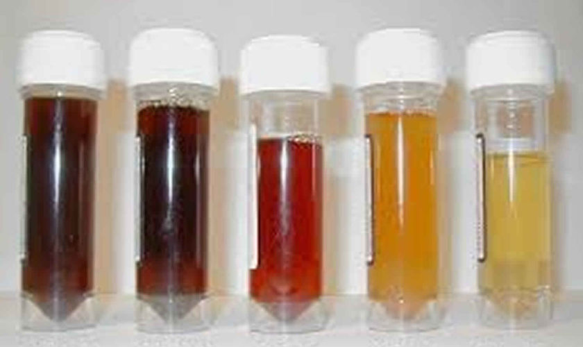
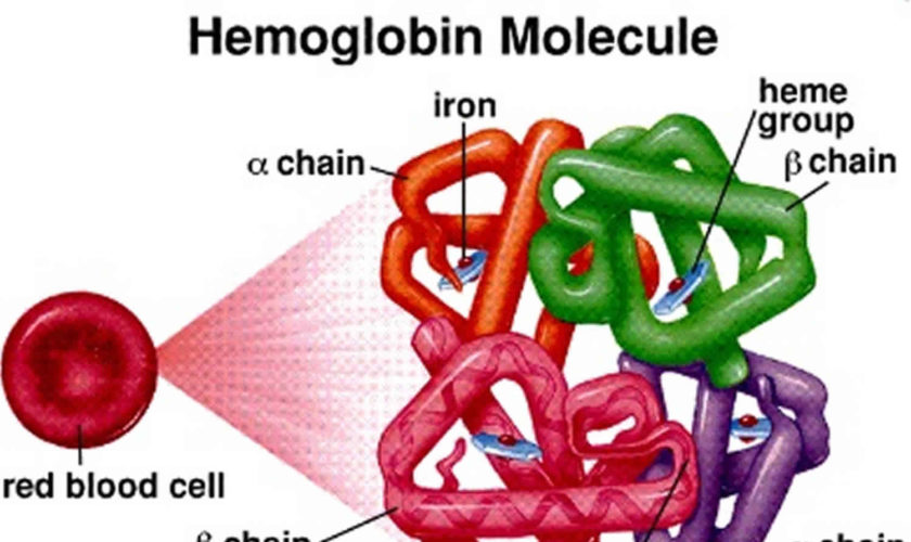
When red blood cells break apart, the hemoglobin inside is released. Hemoglobin is the red part of red blood cells that carries oxygen around the body. The release of hemoglobin causes many of the PNH symptoms. Paroxysmal means “sudden and irregular”. The term “nocturnal” refers to the belief that hemolysis is triggered by acidosis during sleep. However, this observation was later disproved. In individuals with paroxysmal nocturnal hemoglobinuria, hemolysis has been shown to occur throughout the day, but the urine concentrated overnight produces the dramatic change in color 2). Paroxysmal nocturnal hemoglobinuria (PNH) is most noticeable in the morning, upon passing urine that has accumulated in the bladder during the night 3). So, “paroxysmal nocturnal hemoglobinuria” means sudden, irregular episodes of passing dark colored urine, especially at night or in the early morning. It is important to note this can be a bit misleading, because many people with PNH do not have dark urine.

Paroxysmal nocturnal hemoglobinuria affects red blood cells (erythrocytes), which carry oxygen; white blood cells (leukocytes), which protect the body from infection; and platelets (thrombocytes), which are involved in blood clotting. This results in a deficiency of various types of blood cells and can cause signs and symptoms such as fatigue, weakness, abnormally pale skin (pallor), shortness of breath, and an increased heart rate. People with PNH may also be prone to infections and abnormal blood clotting (thrombosis) or hemorrhage, and are at increased risk of developing leukemia.

Paroxysmal nocturnal hemoglobinuria is a rare disorder, estimated to affect between 1 and 5 per million people. Experts estimate between 400 and 500 PNH cases are diagnosed in the U.S. each year 4). Paroxysmal nocturnal hemoglobinuria affects both sexes equally, and can occur at any age, although it is most often diagnosed in young adulthood. Paroxysmal nocturnal hemoglobinuria can occur at any age, but is usually diagnosed in young adulthood in people in their 30s and 40s. People with PNH have recurring episodes of symptoms due to hemolysis, which may be triggered by stresses on the body such as infections or physical exertion.

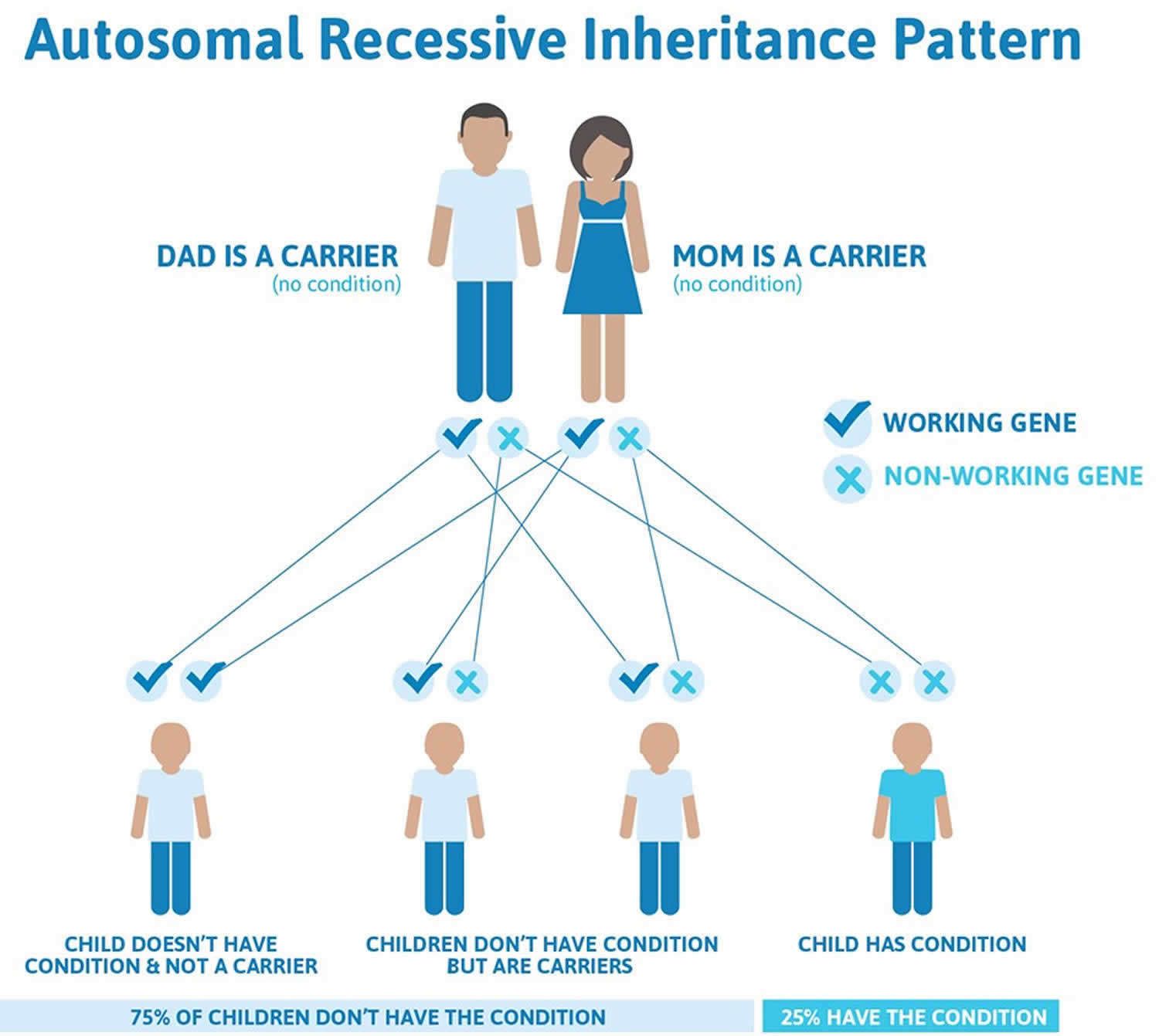
Paroxysmal nocturnal hemoglobinuria is caused by acquired, rather than inherited, mutations in the PIGA gene (located on Xp22.1); the condition is not passed down to children of affected individuals 5). Sometimes, people who have been treated for aplastic anemia may develop PNH 6). The treatment of paroxysmal nocturnal hemoglobinuria is largely based on symptoms; stem cell transplantation is typically reserved for severe cases of PNH with aplastic anemia or those whose develop leukemia 7).

What is bone marrow failure?

Bone marrow failure happens when the marrow does not produce enough red cells, white cells or platelets, or the blood cells that are produced are damaged or defective. This means the body can not supply itself with the blood it needs. PNH, along with aplastic anemia and myleodysplastic syndromes (MDS), are bone marrow failure diseases.

What happens to my blood with PNH?

Blood consists of blood cells floating in plasma. Plasma is mostly made of water. It also includes salts, proteins, hormones, minerals, vitamins and other nutrients and chemicals your body needs.

The 3 basic types of blood cells:

  1. Red blood cells (RBCs) are also called erythrocytes. They make up almost half of blood. Red blood cells are filled with the protein hemoglobin that picks up oxygen in the lungs and brings it to cells all around the body.
  2. White blood cells (WBCs) are also called leukocytes. They fight disease and infection by attacking and killing germs that get into the body. There are several kinds of white blood cells, each of which fights a different kind of germ.
  3. Platelets are also called thrombocytes. They are small pieces of cells that help blood clot and stop bleeding.

Blood cells formation

The process of making blood cells is called hematopoiesis. Blood cells are made in the bone marrow, a spongy tissue located inside certain bones. Marrow contains blood-forming stem cells that make copies of themselves to create all 3 types of blood cells. When blood cells are fully mature and functional, they leave the bone marrow and enter the bloodstream. Healthy people have enough stem cells to make all the blood cells they need.

What is the complement system in PNH?

The complement system is a group of proteins in the blood. They help support (complement) the work of white blood cells by fighting infections.

These proteins are always active at a very low level. But when bacteria, viruses and other foreign or abnormal cells get into your body, these proteins become more active. They work together to attack and destroy these abnormal cells.

Normal red blood cells have a shield of proteins. This shield protects the cells from being attacked by the complement system. The gene in charge of making this protective shield is called PIGA (phosphatidylinositol glycan class A).

Why are blood clots so common for people with PNH?

Scientists are not sure exactly why people with PNH are more likely to get blood clots. But some believe that PNH patients have abnormal platelets that are too “sticky.” This means the platelets make clots too easily.

Many people with PNH have a shortage of nitric oxide. Nitric oxide helps prevent blood clots by making it harder for platelets to stick together. Hemolysis – another symptom of PNH – can cause a shortage of nitric oxide.

How do I find out if I have a blood clot?

To diagnose a blood clot, your doctor may take pictures of your insides using:

  • CT scan (Cat Scan)
  • MRI (Magnetic Resonance Imaging)
  • Doppler scan
  • V-Q Scan (Ventilation-Perfusion Scan)

Your doctor may also order a lab test called D-dimer. It is also called Fragment D-Dimer, or Fibrin degradation fragment.

What immunizations should a PNH patient get?

Patients with PNH should receive vaccinations against certain types of bacteria to prevent infection. Ask your doctor which ones are right for you.

Seasonal flu vaccines protect against the three influenza viruses (trivalent) that research indicates will be most common during the upcoming season. Talk with your hematologist about whether you should get a flu shot. He will help you weigh the risks and benefits of getting a flu shot. Don’t forget to ask whether your family members and others in close contact with you should get a flu vaccine. This may reduce your chance of getting the flu. To read more about flu shots, read our article.

There have been a few case reports of PNH patients getting flares of hemolysis (when red blood cells are destroyed) after receiving a flu shot. Although case reports are not the same as randomized clinical trials, one case of hemolysis was severe enough to put the patient in the hospital. That is why some PNH experts recommend against having a flu shot, but each case is different. PNH patients who are receiving eculizumab may be less likely to have hemolysis after receiving a flu shot.

Can PNH patients get pregnant and have a healthy delivery?

Pregnancy is possible with PNH, but it is not a good idea. It carries serious risks for both mother and child.

A woman with PNH faces a number of risks during pregnancy:

  • Her blood may have fewer healthy cells.
  • Her bone marrow may make fewer healthy cells.
  • She is more likely to get blood clots. Most doctors place pregnant women with PNH on blood thinners to prevent clots. But warfarin (Coumadin®) cannot be used during the first trimester, since it may affect fetus development.
  • She is more likely to get preeclampsia, a dangerous condition that causes very high blood pressure and can put both mother and baby at risk.
  • She may need red blood cell transfusions more often.

A baby whose mother has PNH has a greater risk of:

  • Premature birth
  • Dying in the womb
  • Having a low birth weight
  • Having delayed growth and development

Still, about 1 out of 3 babies whose mothers have PNH do not have any of these problems.

If you do get pregnant, look for a PNH specialist and an obstetrician who specializes in high-risk births.

Is surgery safe for PNH patients?

Surgery can also be risky for people with PNH because it:

  • Makes the complement system more active, which can cause hemolysis
  • Increases the risk of getting blood clots
  • Can cause serious bleeding in people with a low platelet count
  • May require platelet transfusions before surgery.

If you do have surgery, it is a good idea to:

  • Make sure your PNH specialist talks with your surgeon
  • Take the blood thinner Heparin (Calciparine or Liquaemin) as soon as possible after surgery, as long as your platelet count is good and your doctor advises it.

Special precautions for PNH patients

Airplane travel and high altitudes

The farther you move away from sea level, the less oxygen there is. If you have anemia, flying in an airplane or visiting places at higher elevations than you’re used to may cause a shortage of oxygen. It may also cause chest pain. Before you do either of these things, it’s a good idea to:

  • Check with your doctor
  • Get a red blood cell count
  • Get treatment for your anemia (blood transfusions or growth factors)
  • If you do fly, remember to:
    • Drink plenty of water
    • Get up and walk around every hour or two if it is safe to do so

Paroxysmal nocturnal hemoglobinuria causes

Paroxysmal nocturnal hemoglobinuria or PNH is caused by a genetic change (mutation) in the PIGA gene of a single stem cell in your bone marrow. The PIGA gene provides instructions for making a protein called phosphatidylinositol glycan class A. This protein takes part in a series of steps that produce a molecule called GPI anchor. GPI anchor attaches many different proteins to the cell membrane, thereby ensuring that these proteins are available when needed at the surface of the cell.

Here are the steps that lead to PNH:

  1. The abnormal stem cell makes copies of or “clones” itself. This leads to a whole population of bone marrow stem cells that have mutant PIGA.
  2. The abnormal cells mature into red blood cells that have mutant PIGA. These are called PNH red blood cells. Doctors also call them your PNH clone.
  3. The PNH red blood cells lack the shield of proteins that protect normal red blood cells from the complement system, leaving them open to attack and destruction by the complement system proteins.

Many healthy people have a small number of PNH stem cells. In people with PNH, however, these stem cells grow fast and make lots of mature PNH red blood cells.

Some doctors believe this growth happens because people with PNH have bone marrow that is weaker than normal. This weakening may be caused by aplastic anemia or another mild and/or undiagnosed bone marrow failure disease.

If you have had aplastic anemia, you are more likely to get PNH. There are no other known factors that increase your chances of getting PNH.

Some gene mutations are acquired during a person’s lifetime and are present only in certain cells. These changes, which are called somatic mutations, are not inherited. In people with paroxysmal nocturnal hemoglobinuria, somatic mutations of the PIGA gene occur in blood-forming cells called hematopoietic stem cells, which are found mainly in the bone marrow. These mutations result in the production of abnormal blood cells. As the abnormal hematopoietic stem cells multiply, increasing numbers of abnormal blood cells are formed, alongside normal blood cells produced by normal hematopoietic stem cells.

The premature destruction of red blood cells seen in paroxysmal nocturnal hemoglobinuria is caused by a component of the immune system called complement. Complement consists of a group of proteins that work together to destroy foreign invaders such as bacteria and viruses. To protect the individual’s own cells from being destroyed, this process is tightly controlled by complement-regulating proteins. Complement-regulating proteins normally protect red blood cells from destruction by complement. In people with paroxysmal nocturnal hemoglobinuria, however, abnormal red blood cells are missing two important complement-regulating proteins that need the GPI anchor protein to attach them to the cell membrane. These red blood cells are prematurely destroyed, leading to hemolytic anemia.

Research suggests that certain abnormal white blood cells that are also part of the immune system may mistakenly attack normal blood-forming cells, in a malfunction called an autoimmune process. In addition, abnormal hematopoietic stem cells in people with paroxysmal nocturnal hemoglobinuria may be less susceptible than normal cells to a process called apoptosis, which causes cells to self-destruct when they are damaged or unneeded. These features of the disorder may increase the proportion of abnormal blood cells in the body. The proportion of abnormal blood cells affects the severity of the signs and symptoms of paroxysmal nocturnal hemoglobinuria, including the risk of hemoglobinuria and thrombosis.

Paroxysmal nocturnal hemoglobinuria inheritance pattern

Paroxysmal nocturnal hemoglobinuria is acquired, rather than inherited. Paroxysmal nocturnal hemoglobinuria results from new mutations in the PIGA gene, and generally occurs in people with no previous history of the disorder in their family. Paroxysmal nocturnal hemoglobinuria is not passed down to children of affected individuals.

Risk factors of having PNH

Having aplastic anemia is the only known risk factor for developing PNH. More than 10 out of every 100 people with aplastic anemia will develop PNH. In addition, some people with PNH will develop aplastic anemia. People with PNH can share symptoms with aplastic anemia patients, such as low blood cell counts.

On average, 2 out of 100 people with PNH go on to develop myelodysplastic syndrome (MDS).

Paroxysmal nocturnal hemoglobinuria symptoms

When your PNH red blood cells break apart, their hemoglobin is released into your plasma. Hemoglobin is the red part of red blood cells. Its carries oxygen around your body. The release of hemoglobin can cause a number of symptoms, including:

  • Dark or tea colored urine, but it does not darken in all cases
  • Low red blood cell count (anemia) which can cause you to:
    • Feel tired
    • Have headaches
    • Have trouble breathing when you exercise
    • Have an irregular heartbeat
  • Muscle spasms in certain parts of your body. This happens when the released hemoglobin binds with nitric oxide and removes it from your blood. Nitric oxide helps your muscles stay smooth and relaxed. When you have a shortage of nitric oxide, you may experience the following:
    • Mild to severe pain in your abdomen or belly area.
    • Spasms in your esophagus which is a “tube” in your throat that goes from your mouth to your stomach; the spasms can make it hard to swallow.
    • Men may have trouble getting or keeping an erection (become impotent).
  • Thrombosis is a blood clot in a vein. It is often simply called a blood clot. At least 1 out of 3 people with PNH get blood clots. The symptoms of blood clots depend on where the blood clots occur. People who are otherwise healthy and do not have PNH sometimes get blood clots in the veins of the leg. People with PNH tend to get blood clots in other parts of the body, such as in the brain or abdomen (belly area).
    • Blood clot in abdomen (belly area): You may get a blood clot in your abdomen, or belly area. That’s the area below your chest and above your hips. Some places in the abdomen where you may get a blood clot include:
      • Your spleen
      • The major vein that leaves your liver; this is called Budd-Chiari syndrome
      • Your intestine (bowel) may not get enough blood; this is called ischemia
    • Symptoms of getting a blood clot in your abdomen may include:
      • Having fluid and swelling in the belly area; this is called ascites.
      • The area where the clot is may feel warm to the touch.
      • The area where the clot is may be painful.
    • If the blood clot in your abdomen is not treated:
      • Part of your intestine may die (dead bowel)
      • Your liver may be damaged and stop working
    • Blood clot in brain: You may get a blood clot in the veins covering your brain. If this happens, symptoms may include:
      • A very bad headache.
      • Trouble speaking, seeing, or moving parts of your body.
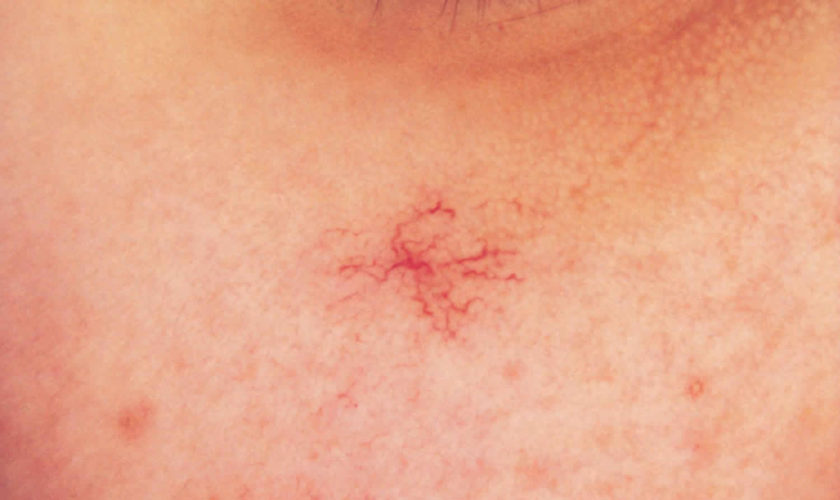
    • Blood clot in skin: You may get a blood clot in the veins of your skin. If this happens, your skin in that area may get red, puffy, warm or painful
    • Blood clot in arm or leg: You may get a blood clot in the veins of your arm or leg. If this happens that limb may get warm, puffy or painful
    • Blood clot in lung: Sometimes, a blood clot breaks off and travels to your lung. This is called a pulmonary embolism. If you have a pulmonary embolism, symptoms may include:
      • A sharp pain in your chest; it may get worse when you breathe deeply
      • Trouble breathing (shortness of breath), or you may start breathing fast
      • Suddenly feeling anxious
      • Coughing up some blood
      • Feeling dizzy; you may even faint
      • Sweating a lot

People with paroxysmal nocturnal hemoglobinuria have sudden, recurring episodes of symptoms (paroxysmal symptoms), which may be triggered by stresses on the body, such as infections or physical exertion. During these episodes, red blood cells are prematurely destroyed (hemolysis). Affected individuals may pass dark-colored urine due to the presence of hemoglobin, the oxygen-carrying protein in blood. The abnormal presence of hemoglobin in the urine is called hemoglobinuria. In many, but not all cases, hemoglobinuria is most noticeable in the morning, upon passing urine that has accumulated in the bladder during the night (nocturnal).

The premature destruction of red blood cells results in a deficiency of these cells in the blood (hemolytic anemia), which can cause signs and symptoms such as fatigue, weakness, abnormally pale skin (pallor), shortness of breath, and an increased heart rate. People with paroxysmal nocturnal hemoglobinuria may also be prone to infections due to a deficiency of white blood cells.

Abnormal platelets associated with paroxysmal nocturnal hemoglobinuria can cause problems in the blood clotting process. As a result, people with this disorder may experience abnormal blood clotting (thrombosis), especially in large abdominal veins; or, less often, episodes of severe bleeding (hemorrhage).

Individuals with paroxysmal nocturnal hemoglobinuria are at increased risk of developing cancer in blood-forming cells (leukemia).

In some cases, people who have been treated for another blood disease called aplastic anemia may develop paroxysmal nocturnal hemoglobinuria.

Low red blood cell count

PNH will mostly cause low red blood cell counts and anemia. A low red blood cell count is called anemia. If you have a low red blood cell count, you may:

  • Feel a little tired or very tired
  • Feel less alert or have trouble concentrating
  • Have a loss of appetite or lose weight
  • Have paler-than-normal skin
  • Have trouble breathing – shortness of breath
  • Have rapid heartbeat
  • Have difficulty exercising or climbing stairs

Low white blood cell counts

A low white blood cell count is called neutropenia. In general, a low white cell count lowers an aplastic anemia patient’s ability to fight bacterial infections. If you have a low white blood cell count, you may:

  • Have repeated fevers and infections
  • Get bladder infections that make it painful to urinate or make you urinate more often
  • Get lung infections that cause coughing and difficulty breathing
  • Get mouth sores
  • Get sinus infections and a stuffy nose
  • Get skin infections

A fever in an aplastic anemia patient is potentially serious. A doctor should be notified if a fever occurs.

Low platelet counts

A low platelet count is called thrombocytopenia. If you have a low platelet count, you may:

  • Bruise or bleed more easily, even from minor scrapes and bumps
  • Get heavier than normal menstrual periods
  • Get nose bleeds
  • Get tiny, flat red spots under your skin (petechiae) caused by bleeding
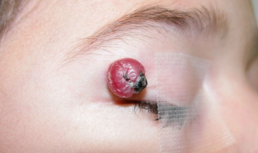
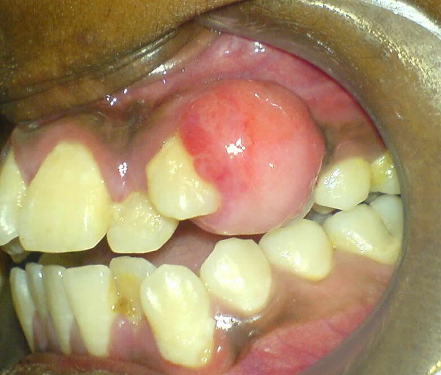
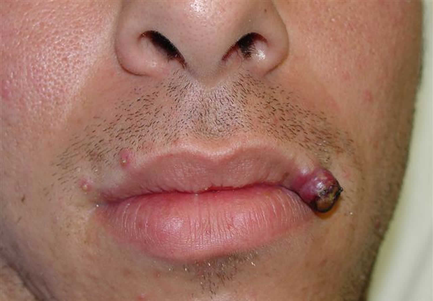
  • Have bleeding gums, especially after dental work or from brushing your teeth.

If platelet counts are not too low, there may be no obvious symptoms. In rare cases, the number of platelets can get so low that dangerous internal bleeding occurs.

Bleeding that will not stop is a medical emergency. A PNH patient needs to seek immediate medical help if they have bleeding that can’t be stopped by usual methods, such as applying pressure to the area.

Paroxysmal nocturnal hemoglobinuria diagnosis

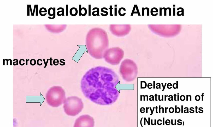
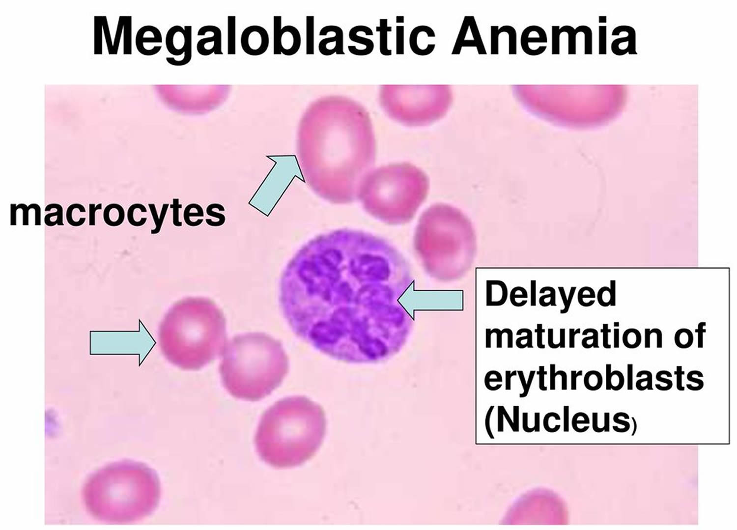
When you have anemia caused by the destruction of red blood cells, doctors call this hemolytic anemia. In addition to a complete blood cell count (CBC), the principal studies used to establish the diagnosis of paroxysmal nocturnal hemoglobinuria (PNH) are flow cytometry of peripheral blood and bone marrow analysis. Flow cytometry measures the percentage of cells that are deficient in the glycosyl phosphatidylinositol–anchored proteins (GPI-APs) and identifies discrete populations with different degrees of deficiency. Because of the missing GPI-APs, red blood cells (RBCs) and other cells in patients with PNH lack DAF (CD55) and MIRL (CD59), which regulate complement.

Hemosiderin is nearly always present in the urine sediment and can accumulate in the kidneys; this is visible on magnetic resonance images (MRI) or computed tomography (CT) scans. An elevated reticulocyte count and serum lactate dehydrogenase (LDH) level with a low serum haptoglobin level in the absence of hepatosplenomegaly are the hallmarks of intravascular hemolysis.

Bone marrow examination will differentiate classic PNH from PNH that develops in the setting of other bone marrow disorders 8). In addition, bone marrow examination will identify an erythroid and hyperplastic bone marrow during the hemolytic phase or a hypoplastic bone marrow in the aplastic phase.

Imaging studies are indicated in patients with venous thrombosis.

PNH test

There are several blood tests used to help confirm a diagnosis of PNH by looking for signs of hemolytic anemia. Specific tests include:

  • A complete blood count (CBC) to look for signs of low hemoglobin. This test uses a number of methods to measure how many of each blood cell type are in your blood sample.
  • An LDH test looks at the level of an enzyme called lactate dehydrogenase. High levels of LDH in the blood can mean that red blood cells are breaking apart (hemolysis) or that there is tissue damage in the body. It is important for patients with PNH to have LDH monitored regularly.
  • A bilirubin test measures the total amount of this substance in your blood. High levels may indicate destruction of red blood cells.
  • A reticulocyte count measures the number of young red blood cells in your blood. People who have PNH may have elevated reticulocyte counts because their bone marrow is making lots of new red blood cells.

Flow Cytometry

The gold standard for confirming the presence of PNH is a flow cytometry test. This test tells your doctor if any proteins are missing from the surface of blood cells. PNH cells are missing some or all of two proteins on their surface. These proteins are called CD55 and CD59. FLAER is a new type of flow cytometry test that is also used.

Using flow cytometry, your doctor can usually divide your blood cells into 3 types:

  • PNH I cells, or Type I cells: These cells are normal. They respond in a healthy way to the complement system.
  • PNH II cells, or Type II cells: These cells are partially sensitive to the complement system. They are missing some of the CD55 and CD59 proteins that protect them from attack.
  • PNH III cells, or Type III cells: These cells are extremely sensitive to the complement system. Of the 3 groups of cells, these break apart most easily. They are missing all the proteins that protect normal cells from attack. Most people with PNH have Type I and Type III cells, but the amount of each type of cell can vary greatly.

Other blood tests

Doctors may ask you do to several types of blood tests to help them understand your case of PNH and create a treatment plan. These include:

  • EPO level, also called erythropoietin, measures how much of this protein is being made by your kidneys. EPO is created in response to low oxygen levels in the body, typically caused by low red cell counts and anemia. EPO causes your bone marrow to make more red blood cells. A low EPO level may indicate a problem other than PNH, or it may make anemia worse in people who have PNH.
  • Iron level test, also called a ferritin test, checks the level of iron in your blood. If a shortage of iron is causing anemia, it can be easily treated with iron supplements. If you have too much iron in your body this is called iron overload. It can be caused from getting lots of red blood cell transfusions or by genetic conditions. A number of treatments exist to remove iron from your body.
  • Vitamin B12 and a folate level may be done to rule out other causes of low red cell counts. If your red blood cells have an abnormal shape, size or look, this can be caused by low levels of vitamin B12 and folate (folic acid). These abnormal looking cells don’t work right, and this can lead to anemia.

Bone marrow tests

An examination of your bone marrow is important for the diagnosis of PNH. It is usually a simple 30-minute procedure. First, the doctor uses a hollow needle to remove some bone marrow aspirate (liquid bone marrow), typically from the pelvic or breast bone. A solid piece of bone marrow is also removed for a bone marrow biopsy.

The doctor will look at your liquid bone marrow under a microscope and send a sample of your bone marrow to a lab.

A bone marrow test is done for two main reasons:

  1. To help confirm a diagnosis of PNH
  2. To understand how well or poorly your bone marrow is making blood cell

The bone marrow test shows:

  • The quantity (cellularity) of your bone marrow occupied by different cells
  • Exactly what types and amounts of cells your bone marrow is making
  • Increased, decreased, or normal levels of iron in your bone marrow
  • Chromosomal (DNA) abnormalities

Paroxysmal nocturnal hemoglobinuria treatment

According to current understanding of paroxysmal nocturnal hemoglobinuria (PNH), the ideal treatment is to replace the defective hematopoietic stem cell with a normal equivalent by stem cell transplantation; however, this is not realistic for many patients, because stem cell transplantation requires a histocompatible donor and is associated with significant morbidity and mortality 9). This form of treatment is reserved for severe cases of PNH with aplastic anemia or transformation to leukemia, both of which are life-threatening complications.

Androgens

Androgens are natural male hormones that can cause your bone marrow to make more red blood cells. This can improve anemia. Androgens are sometimes used to treat aplastic anemia and PNH.

Blood transfusions

A blood transfusion is a safe and common procedure. Most people who have a bone marrow failure disease like aplastic anemia, MDS or PNH will receive at least one blood transfusion. When you receive a blood transfusion, parts of blood from a donor are put into your bloodstream. This can help some patients with low blood counts.

Eculizumab

In 2007, eculizumab (Soliris), an anti-complement antibody targeting the CD5 complement component, was approved by the US Food and Drug Administration (FDA) and the European Medicines Evaluation Agency (EMEA) to treat PNH. It works by making your complement system less active and reduces hemolysis Soliris® is approved for the treatment of patients with PNH in nearly 50 countries worldwide.

Eculizumab alleviates the hemolysis associated with PNH and its complications, dramatically improving symptoms, improving quality of life, and eliminating complications of PNH 10). However, eculizumab does not alter the underlying defect of the disease, thus, treatment needs to continue life-long or until spontaneous remission, which occurred only in a minority of patients (12 of 80 patients in one study) before the advent of eculizumab 11).

Folic acid

Folic acid, also called folate or vitamin B-9, is found in fresh or lightly cooked green vegetables. It helps your bone marrow make normal blood cells. When your bone marrow has to make more cells, it needs a larger supply of folic acid.

Most people get enough folate in their diet. But if you have PNH, it’s a good idea to take 1 mg each day of a man-made form of folate called folic acid.

Growth factors

Growth factors are naturally occurring hormones in your body that signal your bone marrow to make more of certain types of blood cells. Man-made growth factors may be given to some people with bone marrow failure diseases to help increase red blood cell, white blood cell or platelet counts.

Iron chelation

Iron chelation therapy is the main treatment used when you have a condition called iron overload. Iron overload means you have too much iron in your body. This can be a problem for people who get lots of red blood cell transfusions.

Ravulizumab-cwvz (Ultomiris)

Ravulizumab-cwvz (Ultomiris) is a drug approved by the U.S. Food and Drug Administration (FDA) in 2018 to treat PNH. Ravulizumab-cwvz (Ultomiris) is a long-acting C5 inhibitor that works by inhibiting the C5 protein in the terminal complement cascade.

Treatment of bone marrow hypoplasia

Bone marrow hypoplasia is a serious cause of morbidity and mortality. Bone marrow hypoplasia is treated most effectively with bone marrow transplantation also called a stem cell transplant (SCT) or hematopoietic stem cell transplant (HSCT). The procedure replaces unhealthy blood-forming stem cells with healthy ones and offers some patients the possibility of a cure. But for many patients, a bone marrow transplantation is not an option due to the risks and potential long-term side effects as an “imperfect cure”.

Moroover, if there is no suitable donor available, antithymocyte globulin has been used in the treatment of aplastic anemia with considerable success.

Thromboembolism

Patients with PNH who develop acute thrombosis should immediately be started on eculizumab, if they are not already taking it, as this reduces the risk of thrombosis extension or recurrence 12). Otherwise, management of thrombotic complications follows standard principles, including using heparin emergently, then maintenance therapy with the use of an oral anticoagulant, such as warfarin. Sometimes, heparin can exacerbate the thrombotic problem, possibly by activating complement. This can be prevented using inhibitors of the cyclooxygenase system, such as aspirin, ibuprofen, and sulfinpyrazone.

Primary prophylaxis of thromboembolism for patients with PNH has been advocated. Whether this approach is safe and effective in all patients with PNH remains controversial, however.

Corticosteroids

Modulation of complement is controlled poorly by high doses of glucocorticoids. The usual adult dose of prednisone is 20-40 mg/d (0.3-0.6 mg/kg/day) given daily during hemolysis and changed to alternate days during remission. On this regimen, about 70% of adult patients experience improvement in hemoglobin levels, but long-term therapy is fraught with complications.

Investigational agents

A variety of agents that inhibit complement are under development for treatment of PNH. Novel anti-C5 agents include monoclonal antibodies and an anti-C5 small interfering RNA 13). Because clinically relevant C3-mediated extravascular hemolysis can occur in PNH 14), the anti-C3 small peptide compstatin and its derivatives are being investigated, along with inhibitors of complement Factor D or B 15).

Yuan et al 16) reported that two novel small-molecule inhibitors of Factor D, which is a component of the alternative complement pathway, show potential as oral agents for treating PNH. In the Ham test, using cells from PNH patients, the Factor D inhibitors significantly reduced complement-mediated hemolysis at concentrations as low as 0.01 μM. In an animal model, the compound ACH-4471 blocked alternative pathway activity.

PNH prognosis

The prognosis in patients with paroxysmal nocturnal hemoglobinuria (PNH) may vary from person to person, depending on the severity of symptoms and the presence of complications. You may have only mild symptoms, or you may have severe symptoms and need medicines or blood transfusions. An aplastic phase is a serious prognostic factor, because the resulting pancytopenia and thrombosis of hepatic, abdominal, and cerebral veins can have life-threatening consequences. Prophylactic anticoagulation has not been shown to be of benefit because of a lack of data from a clinical trial setting.

In many patients with paroxysmal nocturnal hemoglobinuria (PNH), the abnormal clone may eventually disappear. This usually takes at least 5 years, and often as long as 15-20 years. Reactivation of PNH in these patients has been observed with acute infections. Patients with chronic anemia alone, without thrombotic complications, can live relatively normal lives for many years.

Many people with PNH live for decades. People who develop blood clots in key parts of the body, or also have MDS (myelodysplastic syndromes) or AML (acute myeloid leukemia), may have a shorter lifespan.

The good news is that good treatments are available, and new treatments are being developed that help people with PNH live longer. You may have seen older research saying that patients with PNH live an average of 15 to 20 years, but more recent research shows that life expectancy has been steadily climbing over the past 20 years. It is even possible that PNH patients will soon live just as long as the average person of the same age.

References   [ + ]

read more

Megaloblastic anemia

megaloblastic-anemia

What is megaloblastic anemia

Megaloblastic anemia also known as macrocytic anemia, is a disorder of the bone marrow characterized by ineffective hematopoiesis, frequently manifested by reduction in the number of mature healthy red blood cells as well as the presence of unusually large, abnormal and poorly developed erythrocytes (megaloblasts), which fail to enter blood circulation due to their larger size. Megaloblastic anemia are usually caused by nutritional deficiencies (most common) of either vitamin B12 or folate (Vitamin-B9) or both, inherited disorders of DNA synthesis or following certain drug therapy 1).

There is a presence of erythroblasts in the bone marrow with delayed nuclear maturation because of defective DNA synthesis called megaloblasts 2).

Megaloblastic anemia is an uncommon problem in childhood that is most frequently associated with vitamin deficiency or gastrointestinal disease 3).

Vitamin B12 deficiency is the most common cause of megaloblastic anemia. Vitamin B12 deficiency is caused by insufficient dietary intake, as in the cases of vegetarians or malnutrition, malabsorption due to the absence of intrinsic factor caused by pernicious anemia or following gastric surgery, congenital disorders, such as transcobalamin II deficiency, or exposure to nitrous oxide.

The result of one study, conducted in Japan, indicated that the most common cause of megaloblastic anemia is pernicious anemia (61%), followed by vitamin B12 deficiency due to gastrectomy (34%), vitamin B12 deficiency due to other causes (2%), and folate deficiency (2%) 4). Vitamin B12 is contained in animal foods, and the daily intake is approximately 3‐30 μg. The daily required amount is approximately 1‐3 μg, and except for stomach or intestinal obstruction, or being a strict vegetarian, vitamin B12 deficiency is rare.

Vitamin B12 binds to intrinsic factor secreted by the gastric parietal cells, and it is absorbed in the terminal ileum. Once absorbed, vitamin B12 acts as a coenzyme in the enzymatic reaction that produces methionine from homocysteine. As a result, folic acid is converted into its active form. When vitamin B12 is deficient, active folic acid is also deficient. As a result, the intracellular reaction involving the coenzyme form of folic acid is affected. Thus, not only vitamin B12 but also folate deficiencies impair DNA synthesis. Because a large amount of vitamin B12 is stored in the liver, it takes 5‐10 years for clinical problems to manifest following decreased intake or absorption of vitamin B12 5).

The signs and symptoms induced by megaloblastic anemia due to vitamin B12 deficiency are fatigue, headache, palpitations, and dyspnea, and neurological symptoms such as dysesthesia and hypoesthesia may also be present. In severe cases, ataxia, decreased proprioception, and vibratory sensation, collectively known as subacute combined degeneration, may be present. Neurologic symptoms are not generally seen in folate deficiency. Vitamin B12 deficiency does not necessarily lead to anemia and macrocytosis. Other symptoms include Hunter’s glossitis and gray hair.

Peripheral blood smear reveals macrocytic anemias and pancytopenia, and hypersegmented neutrophils may be present in severe cases. Megaloblastic changes in erythroblasts and giant metamyelocytes are seen in bone marrow, resulting from impaired nuclear differentiation. Biochemical analysis of blood shows increased levels of indirect bilirubin and lactate dehydrogenase (LDH), and a decreased level of haptoglobin. Vitamin B12 deficiency is treated with parenteral administration of vitamin B12, and hematological levels generally return to normal within one month. For patients with a permanent decrease in the ability to absorb dietary vitamin B12, such as associated with pernicious anemia or total gastrectomy, lifelong treatment is necessary.10 During hematopoietic recovery, an iron deficiency may develop. Although it is not an established treatment, recently it has been reported that oral treatment is effective, because 1%‐5% of vitamin B12 absorption in the terminal ileum is by passive diffusion, which does not involve intrinsic factor 6).

Most, but not all, megaloblastic anemia is produced by “ineffective erythropoiesis” in the bone marrow due to either folic acid or vitamin B12 deficiency 7). In folic acid deficiency the cause frequently is inadequate dietary intake, whereas vitamin B12 deficiency is almost always conditioned by some specific type of malabsorption. Anemia with oval macrocytes, few reticulocytes, moderate leukopenia, and thrombocytopenia is typical of both. Aplastic anemia, refractory anemias with cellular marrow, preleukemia, aleukemia, and erythroleukemia may have somewhat similar blood findings but are usually recognizable from bone marrow biopsy. Decreased levels of folate or vitamin B12 are the most reliable criteria of megaloblastic anemia. With these available in advance, therapy with the appropriate vitamin can be begun at once. If serum levels are unavailable or available only in retrospect, initial treatment, especially of severe anemia, should be with both vitamins. Differentiation between folate and vitamin B12 deficiency is important but impossible by blood and bone marrow morphology alone. Thus, if serum levels are unavailable, the distinction must be made, sometimes retrospectively, on the basis of other laboratory examinations, such as gastric analysis, small-bowel x-ray films, and the Schilling test.

Megaloblastic anemia is most common in the elderly with 1 in 8000. Megaloblastic anemia can be seen in all races, but is particularly common in Nordic people. There is an association with other autoimmune diseases, particularly thyroid disease, Addison’s disease and vitilgo.

Figure 1. Megaloblastic anemia

megaloblastic anemia

Prognosis of Megaloblastic Anemia

  • Neurological changes if left untreated, can be irreversible.
  • Neuorological abnormalities only occur with very low levels of serum B12.
  • Patients present with symmetrical tingling sensation in the fingers and toes, early loss of vibration sense and propioception, and progressive weakness and ataxia.
  • Paraplegia may result.
  • Dementia and optic atrophy also occur from vitamin B12 deficiency.

Megaloblastic anemia prognosis is favorable if the cause of megaloblastosis has been identified and appropriate treatment has been instituted. However, patients are at risk for hypokalemia and anemia-related cardiac complications during therapy for cobalamin (vitamin B12) deficiency.

Folate deficiency during pregnancy can lead to neural tube defects and other developmental disorders in the fetus. However, folate in prenatal vitamins given during pregnancy has reduced these morbidities 8).

Megaloblastic anemia causes

  • Vitamin B12 deficiency;
  • Folic acid deficiency;
  • Conditions with neither B12 nor folate deficiency, e.g. orotic aciduria, where there is a defect in pyrimidine synthesis, therapy with drugs interfering with DNA synthesis and myelodysplasia.

A deficiency of folate or vitamin B12 may cause megaloblastic anemia by reducing the supply of the coenzyme methylene tetrahydrofolate.

Other congenital and acqiuired forms of megaloblastic anemia are due to interference with purine or pyrimidine causing an inhibition in DNA synthesis.

In rare cases, megaloblastic anemia is due to inherited problems:

  • Thiamine-responsive megaloblastic anemia syndrome: an autosomal recessive disease characterized by megaloblastic anemia associated with deafness and diabetes mellitus 9).
  • Inherited deficiency of intrinsic factor or the receptor in the intestines: Imerslund-Grasbeck syndrome.
  • Some infants have congenital folate malabsorption.

Folic acid is present in food such as green vegetables, fruits, meat, and liver. Daily adult needs range from 50 to 100 µg. The recommended dietary allowance is 400 µg in adults and 600 µg in a pregnant woman 10). Folic acid is meanly absorbed in the jejunum — the body stores around 5 mg of folate in the liver, enough for 3 to 4 months. Folic acid deficiency may be related to decreased intake in the case of alcoholism or malnutrition (elderly, institutions, poverty, special diets, etc.), increased demand particularly in case of pregnancy, hemolysis and hemodialysis and malabsorption (tropical sprue, celiac disease, jejunal resection, Crohn disease, etc.). In some cases, medications like anticonvulsants and anticancer agents cause megaloblastic anemia related to folate deficiency.

The primary dietary sources of cobalamin/vitamin B12 are meats, fish, eggs, and dairy products. Vegan diets are low in vitamin B12. However, not all vegans develop clinical evidence of deficiency. Vitamin B12 is first bound within the duodenum and jejunum to intrinsic factor produced by gastric parietal cells and is then absorbed in the terminal ileum. Body stores 2 to 3 mg of vitamin B12 in the liver (sufficient for 2 to 4 years).

The most frequent cause of vitamin B12 deficiency is pernicious anemia caused by autoimmune gastric atrophy and leading to intrinsic factor production reduction 11). Vitamin B12 deficiency may also develop following gastrectomy, ileal resection or ileitis of any cause. Other causes of impaired vitamin B12 absorption include Zollinger-Ellison syndrome, blind loop syndrome, fish tapeworm infestation, and pancreatic insufficiency.

Megaloblastic anemia symptoms

The most common symptom of all types of anemia is fatigue (tiredness). Fatigue occurs because your body doesn’t have enough red blood cells to carry oxygen to its various parts.

A low red blood cell count also can cause shortness of breath, dizziness, headache, coldness in your hands and feet, pale or yellowish skin, and chest pain.

A lack of red blood cells also means that your heart has to work harder to move oxygen-rich blood through your body. This can lead to irregular heartbeats called arrhythmias, heart murmur, an enlarged heart, or even heart failure.

Other signs and symptoms of megaloblastic anemia include:

  • Desire to eat ice or other non-food things (pica)
  • Diarrhea or constipation
  • Fatigue, lack of energy, or lightheadedness when standing up or with exertion
  • Loss of appetite
  • Pale skin
  • Problems concentrating
  • Shortness of breath, mostly during exercise
  • Swollen, red tongue or bleeding gums.

Signs and Symptoms of Vitamin B12 Deficiency

Vitamin B12 deficiency may lead to nerve damage. This can cause tingling and numbness in your hands and feet, muscle weakness, and loss of reflexes. You also may feel unsteady, lose your balance, and have trouble walking. Vitamin B12 deficiency can cause weakened bones and may lead to hip fractures.

Severe vitamin B12 deficiency can cause neurological problems, such as confusion, dementia, depression, and memory loss.

Other symptoms of vitamin B12 deficiency involve the digestive tract. These symptoms include nausea (feeling sick to your stomach) and vomiting, heartburn, abdominal bloating and gas, constipation or diarrhea, loss of appetite, and weight loss. An enlarged liver is another symptom.

A smooth, thick, red tongue also is a sign of vitamin B12 deficiency and pernicious anemia.

Infants who have vitamin B12 deficiency may have poor reflexes or unusual movements, such as face tremors. They may have trouble feeding due to tongue and throat problems. They also may be irritable. If vitamin B12 deficiency isn’t treated, these infants may have permanent growth problems.

If you have a low vitamin B12 level for a long time, you can have nervous system damage. Symptoms can include:

  • Confusion
  • Depression
  • Loss of balance
  • Numbness and tingling that start first in the hands and feet (from nerve damage)
  • Muscle weakness
  • Slow reflexes
  • Loss of balance
  • Unsteady walking
  • Confusion, memory loss, depression, and/or dementia in severe cases

How is Megaloblastic Anemia Diagnosed?

Primary care doctors—such as family doctors, internists, and pediatricians (doctors who treat children)—often diagnose and treat pernicious anemia. Other kinds of doctors also may be involved, including:

  • A neurologist (nervous system specialist)
  • A cardiologist (heart specialist)
  • A hematologist (blood disease specialist)
  • A gastroenterologist (digestive tract specialist)

Medical and Family Histories

Your doctor may ask about your signs and symptoms. He or she also may ask:

  • Whether you’ve had any stomach or intestinal surgeries
  • Whether you have any digestive disorders, such as Celiac disease or Crohn’s disease
  • About your diet and any medicines you take
  • Whether you have a family history of anemia or pernicious anemia
  • Whether you have a family history of autoimmune disorders (such as Addison’s disease, type 1 diabetes, Graves’ disease, or vitiligo). Research suggests a link may exist between these autoimmune disorders and pernicious anemia that’s caused by an autoimmune response.

Physical Exam

During the physical exam, your doctor may check for pale or yellowish skin and an enlarged liver. He or she may listen to your heart for rapid or irregular heartbeats or a heart murmur.

Your doctor also may check for signs of nerve damage. He or she may want to see how well your muscles, eyes, senses, and reflexes work. Your doctor may ask questions or do tests to check your mental status, coordination, and ability to walk.

The health care provider will perform investigations and blood tests.

  • A deoxyuridine suppression test can be used to rapidly determine the nature and severity of the vitamin B12 or folate deficiency in severe or complex cases of megaloblastic anemia.
  • Blood samples will show the typical features of megaloblastic anemia.
  • Serum bilirubin may be raised as a result of ineffective erythropoeisis.
  • Serum vitamin B12 can be assayed using radioisotope dilution or immunological assays.

Pernicious anemia test

  • Bone marrow examination (only needed if diagnosis is unclear)
  • Complete blood count (CBC)
  • Reticulocyte count
  • Schilling test
  • LDH level
  • Methylmalonic acid (MMA) level
  • Vitamin B12 level
  • Levels of antibodies against intrinsic factor (IF) or the cells which make intrinsic factor.

Complete Blood Count

Often, the first test used to diagnose many types of anemia is a complete blood count (CBC). This test measures many parts of your blood. For this test, a small amount of blood is drawn from a vein (usually in your arm) using a needle.

A complete blood count (CBC) checks your hemoglobin and hematocrit levels. Hemoglobin is an iron-rich protein that helps red blood cells carry oxygen from the lungs to the rest of the body. Hematocrit is a measure of how much space red blood cells take up in your blood. A low level of hemoglobin or hematocrit is a sign of anemia.

The normal range of these levels may be lower in certain racial and ethnic populations. Your doctor can explain your test results to you.

The complete blood count (CBC) also checks the number of red blood cells, white blood cells, and platelets in your blood. Abnormal results may be a sign of anemia, another blood disorder, an infection, or another condition.

Finally, the complete blood count (CBC) looks at mean corpuscular volume (MCV). Mean corpuscular volume (MCV) is a measure of the average size of your red blood cells. Mean corpuscular volume (MCV) can be a clue as to what’s causing your anemia. In pernicious anemia, the red blood cells tend to be larger than normal.

Other Blood Tests

If the complete blood count (CBC) results confirm that you have anemia, you may need other blood tests to find out what type of anemia you have.

A reticulocyte count measures the number of young red blood cells in your blood. The test shows whether your bone marrow is making red blood cells at the correct rate. People who have pernicious anemia have low reticulocyte counts.

Serum folate, iron, and iron-binding capacity tests also can help show whether you have pernicious anemia or another type of anemia.

Another common test, called the Combined Binding Luminescence Test, sometimes gives false results. Scientists are working to develop a more reliable test.

Your doctor may recommend other blood tests to check:

  • Your vitamin B12 level. A low level of vitamin B12 in the blood indicates pernicious anemia. However, a falsely normal or high value of vitamin B12 in the blood may occur if antibodies interfere with the test.
  • Your homocysteine and methylmalonic acid (MMA) levels. High levels of these substances in your body are a sign of pernicious anemia.
  • For intrinsic factor antibodies and parietal cell antibodies. These antibodies also are a sign of pernicious anemia.

Bone Marrow Tests

Bone marrow tests can show whether your bone marrow is healthy and making enough red blood cells. The two bone marrow tests are aspiration and biopsy.

For aspiration, your doctor removes a small amount of fluid bone marrow through a needle. For a biopsy, your doctor removes a small amount of bone marrow tissue through a larger needle. The samples are then examined under a microscope.

In pernicious anemia, the bone marrow cells that turn into blood cells are larger than normal.

Megaloblastic anemia treatment

  • Treatment depends on whether Vitamin B12 or folate deficiency is present.
  • Vitamin B12 deficiency is treated with hydroxycobalamin 1000ug intramuscularly to a total of 5000-6000ug over the course of 3 weeks. 1000ug is then necessary every 3 months for the rest of the patients life.
  • Folate deficiency is treated with 5mg of folic acid daily. Prophylactic folate may be given in pregnancy or in chronic hematological disorders where there is rapid cell turnover.
  • In severely ill patients, it may be necessary to treat with both folic acid an vitamin B12 while awaiting serum levels.

In cases of vitamin B12 deficiency, the treatment centers on intramuscular injections of hydroxocobalamin. Usually, patients receive 1000 ug of vitamin B12 daily in their first week of treatment. In the following month, they receive weekly and then monthly injections. Usually, reticulocytosis occurs within 3 to 5 days. By the tenth day, hemoglobin starts to increase, and a total resolution of anemia normally occurs after 2 months of treatment. Reversal of neurological changes typically takes a longer time, and some manifestations will not disappear even if treatment starts promptly. The treatment should be continued indefinitely at a dose of 1000 ug/month. In some cases, in particular, in patients with prior total gastrectomy or extensive ileal resection, preventive treatment with vitamin B12 is for life.

In the case of folate deficiency, it is essential to ascertain the absence of concomitant vitamin B12 deficiency before starting therapy. Indeed, neuropathy may worsen if large doses of folic acid are administered in the presence of a concomitant vitamin B12 deficiency.

Folic acid supplementation, 1 to 5 mg/day, is usually given orally. In patients with malabsorption, parenteral preparation is the recommendation. To prevent relapse treatment should continue for a minimum of two years.

References   [ + ]

read more

Spider angiomas

spider-angioma

What is spider angioma

A spider angioma is also called spider telangiectasia, spider nevus, arterial spider, vascular spider and naevus araneus, is composed of dilated blood vessels, and is clinically characterized by its spider-like appearance. Spider angioma is given that name because it has a central red papule (the body of the ‘spider’) from which fine red lines (smaller blood vessels) resembling the spider’s legs extend radially. An alternative explanation for the name explains that the red lines form a spider-like network.

Some of these names are Latin: ‘araneus’ for ‘spider’ and ‘angioma’ for ‘blood vessel’. ‘Nevus’ means an increase in normal or healthy tissue within the skin.

Spider angioma can develop at any age, but are more common in children. The vast majority affect healthy people, and most people have only one spider angioma or a just a few. Solitary spider angiomas are seen in 15% of young adults who usually have fewer than 3 lesions 1). Spider angiomas also can appear in other physiologic conditions like pregnancy or severe malnutrition. Multiple spider angiomas are characteristic of chronic liver disease with a specificity of 95% 2).

In one study, 38% of healthy children had at least single spider telangiectasia. They are also visible in about 60% of pregnant women. Physiological spider angiomas in younger adults usually disappear as the age advances, although in few, it may take several years to disappear completely. In women who take oral contraceptives and present with lesions, they may resolve after the patient discontinues the hormonal preparations. If spider angiomas are associated with pregnancy, they will disappear after delivery of the baby. There is no racial predilection for spider angiomas, but lesions are more apparent in light-skinned patients. Spider angiomas are more common in women than in men, and this is thought to be due to the role of steroid hormones in their formation.

The majority of spider angiomas are on the face, upper chest, back and upper arms. In children, the back of the hands are also often affected.

Spider angiomas in children may disappear after puberty, and often disappear after a woman gives birth. Untreated, spider angiomas tend to last in adults.

Spider angiomas are of cosmetic concern only and so are not usually treated. In cosmetic clinics, the central artery can be treated with an electric current (‘electrodessication’), causing it to dry up. A vascular laser such as the pulsed dye laser or KTP (potassium titanyl phosphate) laser can target the blood in the central small artery, causing it to shrink.

These treatments may hurt but do not usually need any local anesthetic. They may leave a small permanent scar like a dent in the skin, which is less common after laser treatment than after electrodessication.

About a third of spider angioma come back after treatment.

Cosmetic camouflage can be useful if there are many spider angioma which are causing cosmetic concern. Camouflage is a type of special make-up, which is matched to the color of the person’s skin and which is water resistant.

When to seek medical care
  • See your doctor if the area bleeds repeatedly or begins to grow in size or change in color.
  • If you suddenly develop many lesions, tell your physician.
  • If you have any sign or particular risk of liver disease (such as yellow skin color, swollen belly, or a history of heavy alcohol use), seek medical care.

Who gets spider telangiectasia

A solitary spider angioma is common in children and adults, affecting 10–15% of the population. Although spider angiomas can affect people of any race, spider angiomas are more easily seen in fair skin compared to skin of color. Multiple spider angiomas arise most frequently in pregnancy, in women taking a combined oral contraceptive pill, in patients with liver disease (particularly, in cirrhosis due to alcohol abuse), and in those with thyrotoxicosis.

Is spider angioma hereditary?

Spider angiomas are very common and affect at least one in ten of healthy adults and are even more common in children. Spider angiomas do not run in families.

A spider angioma is not contagious or cancerous.

Can a spider angioma be cured?

In children and some adults, spider angioma may go away on their own, which can take several years. Treatment is usually not necessary.

If spider angiomas are related to increased estrogen hormones and the levels then go back to normal (after a pregnancy or on stopping an oral contraceptive pill), the spider angioma may go away within about nine months.

A spider angioma can also completely disappear after treatment, but sometimes repeated treatments may be required. A spider angioma may come back a few months later after treatment.

What does a spider angioma look like?

Spider angiomas are often located on the face, neck, and upper chest (this has been postulated to relate to the distribution of a large vein draining the heart, the superior vena cava). Spider angiomas may also occur on the hands, arms, or other sites. Spider angiomas vary in size and number, tending to be larger and more numerous in people with severe liver disease when other cutaneous signs of liver disease may be present such as palmar erythema, leukonychia, and jaundice. A central dilated arteriole may be present without radial capillaries, and the capillaries may vary in diameter, length, and number. They can also be star-shaped.

A spider angioma has 3 features: a body, legs, and surrounding erythema 3). The body appears as a 1 to 10 mm central arteriole visible as a punctum or eminence. It is typically painless, resembles a spider’s body), and is surrounded by attenuated capillaries radiating in a spider-legged fashion, decreasing in size toward the margins.

Spider angioma lesions may briefly bleed on trauma but otherwise do not cause any symptoms or complications.

Figure 1. Spider angioma

spider angiomas

Figure 2. Spider angioma on face

Spider angioma on face

Figure 3. Spider angioma of chronic liver disease

Spider angioma of chronic liver disease

Spider angioma causes

The cause of spider angioma is not known. Spider angioma is an acquired vascular malformation. Spider angioma occurs because of the failure of a tiny smooth muscle restricting the size of an arteriole. Increased pulsating flow through the vessel (the central papule) results in the dilatation of distal vessels (the red lines).

Spider angioma may arise spontaneously or may be induced by circulating estrogen, which is increased in pregnancy, women taking combined oral contraceptive pill and in those with cirrhosis, rheumatoid arthritis, or thyrotoxicosis. Various vascular endothelial growth factors may be involved. For decades, there have been many hypotheses of possible mechanisms that lead to the arteriolar vasodilation. Important among those are direct vasodilatory effects of alcohol, substance P, hyperestrogenism, and inadequate hepatic metabolism of steroid hormones. Angiogenesis as a possible mechanism in the pathogenesis of spider nevi has been proposed due to elevated serum vascular growth factors, such as vascular endothelial growth factor (VEGF) and basic fibroblast growth factor (bFGF) levels in patients with liver cirrhosis. Sex hormone imbalance predominantly hyperestrogenism has also been implicated for the development spider angiomas. This is also suggested by the occurrence of spider angioma in individuals with a hyperestrogenic state, like pregnancy.

Prevalence of spider angiomas is highest among patients with cirrhosis, alcoholic hepatitis, and hepatopulmonary syndrome 4). Spider angiomas correspond with a higher risk of mortality among patients with the alcoholic liver disease. Spider angiomas also suggest a high likelihood of esophageal varices and are indicative of the extent of hepatic fibrosis 5). The reported prevalence of spider angiomas in cirrhosis is 33%.

What are the symptoms of a spider angioma?

Apart from its appearance, a spider angioma does not usually cause any symptoms. Bleeding from a spider angioma is unusual but may occur if picked or scratched.

The main symptom of a spider angioma is a blood vessel spot that:

  • May have a red dot in the center
  • Has reddish extensions that reach out from the center
  • Disappears when pressed on and comes back when pressure is released

Spider angiomas are characteristically found on the face, neck, upper chest, and arms in adults, corresponding to the distribution of superior vena cava. In children, lesions are common on the upper extremities. They may also be present on the backs of the hands and fingers. However, it must be emphasized that spider angiomas can also be seen in locations other than the skin, such as the mucosa of the oral cavity and gastrointestinal (GI) tract. Its characteristic appearance diagnoses spider angioma. Large spider angiomas may be pulsatile with blood flow toward the periphery secondary to the local increase in arterial blood supply. Bleeding from these lesions is unusual unless picked or scratched. Skin temperature over a spider angioma is higher than surrounding skin. The blood pressure measures 50 to 70 mm Hg in these small arterioles.

How is a spider angioma diagnosed?

A spider angioma is diagnosed by its typical appearance. Compression of the central arteriole results in the disappearance of the radial capillaries, which rapidly refill when the compression is relieved. This is best seen through a transparent object, such as a glass slide or the lens of a contact dermatoscope.

The direction of blood flow can be illustrated by applying pressure over the body of spiders with a glass slide (diascopy), leading to pallor with refilling following the release of pressure. No other angiomas show this phenomenon. Due to the varying sizes of spider angioma and for the ease of description, they can be graded from grade 1+ (readily recognizable containing a body, legs, and surrounding erythema) to grade 4+ (visible pulsations with a hand lens, and raised central punctum with many obvious “spider legs” radiating from it). Pregnant patients may present with numerous spider nevi which are harmless and usually resolve after childbirth. Patients with the chronic liver disease will typically have symptoms like jaundice, fluid retention, confusion, and on examination, shifting dullness, icterus, findings related to cause of cirrhosis, and stigmata of liver cell failure.

Most of the time, you DO NOT need tests to diagnose spider angioma. But sometimes, a skin biopsy may be needed to confirm the diagnosis. Blood tests may be done if a liver problem is suspected. As a general rule, number and size of spider angioma correlate with the severity of liver disease. In some occasions, history and exam might not point towards spider angiomas and can lead to concerns of skin malignancy, such as basal cell carcinoma. Rarely, spider angiomas can be a disguised basal cell carcinoma and may warrant a biopsy.

Spider angioma treatment

A spider angioma is harmless and does not require treatment. Patients with an underlying systemic disease like liver cirrhosis should be managed as the standard of care. However, when spider angiomas are present, these patients may already have the advanced liver disease.

A spider angioma that is unsightly can be removed by destroying the feeding central arteriole, but this may result in a small scar. Treatments to remove spider angioma include:

  • Cryotherapy
  • Electrocautery
  • Intense pulsed light
  • Vascular laser.

Rarely, fine-needle electrocautery, 585 nm pulsed, dye laser, 532 nm KTP (potassium-titanyl-phosphate) laser, or electro desiccation have been used to clear spider angioma for cosmetic concerns. The results of the procedure are generally good except for the small risk of the scar.

Surgical excision is rarely necessary and inevitably leaves a scar.

Spider angiomas can recur after treatment. Spider angiomas in healthy individuals usually disappear in a few years, in pregnancy following childbirth and those related to oral contraceptive pills after discontinuation of medication. Cirrhotic patients note the disappearance of nevi following liver transplantation.

Spider angioma prognosis

Spider angiomas can persist or disappear. In women, estrogen-induced spider angioma often disappear within 6 months after having a baby or of stopping the combined contraceptive pill. Spider angioma can also reappear after initially successful treatment.

References   [ + ]

read more

Pyogenic granulomas

Pyogenic-granuloma

What is pyogenic granuloma

Pyogenic granuloma is a common, benign (harmless) growth that often appears as a rapidly growing, bleeding bump on the skin or inside the mouth. Pyogenic granuloma is composed of blood vessels and may occur at the site of minor injury. Pyogenic granuloma carries no risk of cancer, is not contagious (cannot be spread to another person) and is not due to an infection.

When a pyogenic granuloma occurs in a pregnant woman, it is sometimes called a “pregnancy tumor” (granuloma gravidarum). Pyogenic granulomas develop in up to 5% of pregnant women.

Pyogenic granulomas occur in people of all races. Women are more frequently affected by pyogenic granulomas than men, though male and female children are equally affected.

Pyogenic granulomas are most often seen in:

  • Children and young adults
  • Pregnant women
  • Women taking oral contraceptives
  • People taking certain oral retinoid medications, including isotretinoin or acitretin (Soriatane®)
  • People taking protease inhibitors such as indinavir (Crixivan®)
  • People on chemotherapy

See your doctor if you notice any rapidly enlarging skin growth in order to establish a correct diagnosis. Because it is prone to easy bleeding, a pyogenic granuloma lesion should be covered with a bandage until you see your doctor.

Pyogenic granulomas that develop in pregnant women often resolve after delivery. Similarly, pyogenic granulomas associated with medications usually improve when the medicine is discontinued or the dosage is lowered. Depending on the size of the pyogenic granuloma and its location and symptoms, the doctor may decide that no treatment is necessary for pregnant women or for people who can safely stop or lower the dose of the medication that caused the lesion.

Although pyogenic granuloma is a benign condition, it is frequently removed due to its tendency to bleed, its tenderness, and its distressing appearance. However, untreated pyogenic granulomas may go away on their own.

  • The main problem with pyogenic granulomas is the way that they ooze and bleed so easily after minor knocks. This can be of great nuisance, but they are usually not painful.
  • People often worry that their rapid growth and bleeding mean that they are cancerous, even though they are not; however, you should always see your doctor if you have a rapidly growing skin lump.

In obvious cases of pyogenic granuloma, your physician may choose to treat it immediately after obtaining the biopsy. Such treatments include:

  • Scraping and burning (curettage and cauterization). After numbing with local anesthetic, the area is scraped with a sharp instrument (a curette) and burned with an electric needle (cautery).
  • Silver nitrate solution
  • Topical imiquimod cream (Aldara®)
  • Laser treatment
  • Freezing with liquid nitrogen (cryotherapy)
  • Surgical removal (excision)

Approximately 40% of pyogenic granulomas come back (recur) after treatment, especially those lesions located on the trunk of teenagers and young adults. Recurrent pyogenic granulomas are best treated by surgical excision.

When to seek medical care

Make an appointment with a dermatologist or another physician if any rapidly enlarging or bleeding growth develops on your skin or in the areas lining your nose or mouth (mucous membranes).

You should always go straight to your doctor if you have any marks on your skin that are growing or bleeding.

Are pyogenic granulomas inherited?

No. There doesn’t appear to be an increased risk in other family members.

Can a pyogenic granuloma be cured?

Yes, by removing it or treating it with a cream (see treatment below).

What do pyogenic granulomas look like?

As pyogenic granulomas are made up of small blood vessels, they are bright red; later they may turn a darker shade. Their surface is shiny and moist but may become crusty after they have bled.

Pyogenic granulomas stick out from the surface of the skin. Pyogenic granulomas are seldom more than 1 cm across. Some have a bumpy surface and look rather like a raspberry, while others are narrower where they come out from the skin and look as if they are on a stalk.

Figure 1. Pyogenic granuloma nose

Pyogenic granuloma nose

Figure 2. Pyogenic granuloma mouth

Pyogenic granuloma mouth

Figure 3. Pyogenic granuloma gums

Pyogenic granuloma gums

Figure 4. Pyogenic granuloma lip

Pyogenic granuloma lip

Figure 5. Pyogenic granuloma eye

Pyogenic granuloma eye

Footnote: A 30-year-old man presented with a pedunculated lesion on his right lower eyelid that had grown over a period of 3 days. Two weeks before presentation, a cyst had ruptured on the same eyelid. The new lesion started as a small lump on the bulbar conjunctiva and progressively increased in size until it protruded from the eyelid. The patient’s clinical course and a physical examination were suggestive of a pyogenic granuloma, a benign vascular lesion characterized by inflammatory cells and lobular capillary proliferation. Conjunctival pyogenic granulomas grow rapidly in the days to weeks after a conjunctival injury from surgery or trauma and can develop on the conjunctiva or external surfaces of the eyelids. The differential diagnosis includes suture granulomas, squamous papillomas, and malignant tumors, such as squamous-cell carcinoma and amelanotic melanoma. Pyogenic granulomas are often friable and prone to bleeding and can be treated with topical glucocorticoids or surgical excision. In this case, an intralesional injection of triamcinolone was administered at the time of excision to reduce the risk of recurrence. A histopathological assessment confirmed the diagnosis. On review, 3 months after excision, there was minimal scarring of the conjunctival surface and no evidence of recurrence.

[Source 1) ]

Figure 6. Pyogenic granuloma finger

Pyogenic granuloma finger

Pyogenic granuloma clinical findings

Distribution

  • Pyogenic granuloma can arise on any part of the body but the most common sites are the fingers / hands, head and upper trunk.Morphology
  • Pyogenic granuloma starts as a small red spot but quickly enlarges into a nodule
  • Initially smooth, but often becomes eroded and bleeds significantly on contact

Pyogenic granuloma causes

The exact cause of pyogenic granulomas is unknown. Most pyogenic granulomas come up for no obvious reason, but some appear to follow minor damage to the skin, such as a cut that does not heal properly or a prick from a thorn. Pyogenic granulomas can also occur after starting certain medications, such as retinoids (which are sometimes used for the treatment of acne). Pyogenic granulomas often appear following an injury on the hands, arms, or face.

Pyogenic granulomas are common in children, young adults and pregnancy, but can arise at any age. Pyogenic granuloma is sudden in onset, grows rapidly and bleeds after minimal trauma.

Pyogenic granuloma signs and symptoms

Typically, pyogenic granulomas appear as a beefy, red bump that enlarges rapidly over a few weeks. On average, pyogenic granulomas are about 5–10 mm in diameter. They may bleed easily and, in some cases, can be tender. Very rarely, more than one lesion of pyogenic granuloma may develop at the same time at the same site.

The most common locations for pyogenic granulomas include:

  • Lips, gums, and inner mouth (particularly in pregnant women)
  • Hands and fingers
  • Head and neck
  • Feet and toes
  • Upper trunk

Pyogenic granuloma possible complications

These problems may occur:

  • Bleeding from the granuloma
  • Return of the condition after treatment

Pyogenic granuloma diagnosis

Your health care provider will do a physical exam to diagnose this condition.

You may also need a skin biopsy to confirm the diagnosis.

Although the diagnosis is often straightforward the main differential diagnosis is that of an amelanotic melanoma, which tend to bleed less than pyogenic granuloma. Other features that may increase the level of suspicion include no clear history of trauma and an atypical site or age group. To this end, lesions needing treatment are best removed surgically (deep curettage and cautery, or excision) and sent for histology.

Pyogenic granuloma treatment

If the diagnosis of pyogenic granuloma is suspected, your doctor will probably want to perform a skin biopsy. The procedure involves:

  • Numbing the skin with an injectable anesthetic.
  • Sampling a small piece of skin by using a flexible razor blade, a scalpel, or a tiny cookie cutter (called a “punch biopsy”). If a punch biopsy is taken, a stitch (suture) or two may be placed and will need to be removed 6–14 days later.
  • Having the skin sample examined under the microscope by a specially trained physician (dermatopathologist).

Small pyogenic granulomas may go away suddenly. Larger bumps are treated with:

  • Surgical shaving or excision
  • Electrocautery (heat)
  • Freezing
  • A laser
  • Creams applied to the skin (may not be as effective as surgery)

A few pyogenic granulomas lose their colour and shrivel with time, but most are such a nuisance that they need to be treated before then. Freezing a pyogenic granuloma with liquid nitrogen can get rid of it but does not provide a specimen that can be checked in the laboratory. The usual treatment is to scrape pyogenic granulomas off with a sharp spoon-like instrument (a curette) after the area has been made numb by an injection of a local anaesthetic. The bleeding area left behind is then sealed with a hot point (cauterized).

A gel containing timolol or topical steroids have also been used successfully to treat pyogenic granulomas. Although the evidence for this is still limited, it is growing. This maybe especially useful in children as it avoids more invasive procedures. Other non-surgical treatments that have been used with variable success on these lesions, mostly when they are multiple or recurrent, are steroid injections, imiquimod (medication cream used to treat warts and sun damage, works by stimulating the immune system), silver nitrate, and lasers.

Pyogenic granuloma prognosis

Most pyogenic granulomas can be removed. A scar may remain after treatment. There is a high chance that the problem will come back if the whole granuloma is not destroyed during treatment. There is a risk of up to 15% of the pyogenic granuloma coming back. In these cases, the area is sometimes cut out and the wound closed with stitches.

References   [ + ]

read more

Beta thalassemia

beta thalassemia

Beta thalassemia

Beta thalassemia (β-thalassemia) also called Mediterranean anemia or erythroblastic anemia, is a blood disorder that reduces the production of hemoglobin (reduced synthesis of the hemoglobin subunit beta chains). Hemoglobin is the iron-containing protein in red blood cells that carries oxygen to cells throughout the body and low levels of hemoglobin lead to a lack of oxygen in many parts of the body. People with beta thalassemia have anemia (shortage of red blood cells), which can cause paleness, weakness, fatigue, and more serious complications. People with beta thalassemia are at an increased risk of developing abnormal blood clots.

Beta thalassemia is a fairly common blood disorder worldwide. Thousands of infants with beta thalassemia are born each year. Beta thalassemia occurs most frequently in people from Mediterranean countries, North Africa, the Middle East, India, Central Asia, and Southeast Asia.

Beta-thalassemia is characterized by microcytic hypochromic anemia, an abnormal peripheral blood smear with nucleated red blood cells, and reduced amounts of hemoglobin A (HbA) on hemoglobin analysis.

Beta thalassemia is classified into two types depending on the severity of symptoms: thalassemia major also known as Cooley’s anemia and thalassemia intermedia. Of the two types, thalassemia major is more severe.

The signs and symptoms of thalassemia major appear within the first 2 years of life. Children develop life-threatening anemia. They do not gain weight and grow at the expected rate (failure to thrive) and may develop yellowing of the skin and whites of the eyes (jaundice). Affected individuals may have an enlarged spleen, liver, and heart, and their bones may be misshapen. Some adolescents with thalassemia major experience delayed puberty. Many people with thalassemia major have such severe symptoms that they need frequent blood transfusions to replenish their red blood cell supply. Over time, an influx of iron-containing hemoglobin from chronic blood transfusions can lead to a buildup of iron in the body, resulting in liver, heart, and hormone problems.

Thalassemia intermedia is milder than thalassemia major. The signs and symptoms of thalassemia intermedia appear in early childhood or later in life. Affected individuals have mild to moderate anemia and may also have slow growth and bone abnormalities.

Can an individual with beta-thalassemia minor donate blood?

When an individual chooses to donate blood, he/she is typically examined and asked specific questions about his/her medical history (to make sure that donating blood isn’t unsafe for the individual donating or for the recipient). During this process, the individual’s hematocrit value (or hemoglobin level) is tested to make sure that the individual does not have anemia and is not likely to become anemic after donation. In order to donate blood, an individual’s hemoglobin level must be at a specific level, which is established by the U.S. Food and Drug Administration (FDA). Usually, individuals with hemoglobin levels that are too low are temporarily not permitted to donate blood. A low hematocrit level is one of the most common reason people are temporarily disqualified or “deferred” from donating blood, but some donors can actually have anemia and still be eligible to donate.

People who have beta-thalassemia minor and are interested in donating blood should speak with their healthcare provider.

My partner and I have both been diagnosed with beta-thalassemia minor. What is the likelihood of having a child without beta-thalassemia?

Thalassemia major and thalassemia intermedia are inherited in an autosomal recessive pattern, which means both copies of the HBB gene in each cell have mutations (see Figure 1 below). The parents of an individual with an autosomal recessive condition each carry one copy of the mutated gene and are referred to as carriers, but they typically do not show signs and symptoms of the condition. When two carriers have children, each child has a 25% (1 in 4) chance to be affected, a 50% (1 in 2) chance to be a carrier like each parent, and a 25% (1 in 4) chance to be unaffected and not a carrier. Sometimes, however, people (carriers) with only one HBB gene mutation in each cell develop mild anemia. These mildly affected people are said to have ‘beta-thalassemia minor’ or ‘beta-thalassemia trait’ 1).

In a small percentage of families, the HBB gene mutation is inherited in an autosomal dominant manner (see Figure 2 below). In these cases, one copy of the altered gene in each cell is sufficient to cause the signs and symptoms of beta thalassemia 2).

People with specific questions about genetic risks or genetic testing for themselves or family members should speak with a genetics professional.

Resources for locating a genetics professional in your community are available online:

Beta thalassemia types

There are three types of beta thalassemia, depending upon whether one or two beta globin genes are mutated, and the severity of symptoms 3):

  1. Beta thalassemia minor, or beta thalassemia trait, happens when one of the beta globin genes is mutated. People with this condition typically have very mild symptoms and require no treatment, but they can pass thalassemia on to their children. Usually, they are mildly anemic and their red blood cells are smaller than normal.
  2. Thalassemia major also called Cooley’s anemia – the more severe form, causing severe anemia and enlarged liver and spleen (hepatosplenomegaly). This form usually becomes apparent before 2 years of age. If not treated, it causes failure to thrive and a shortened life expectancy. Treatment involves regular transfusions and chelation therapy to reduce iron overload. Treatment allows for normal growth and development. Bone marrow transplantation or cord blood transplantation may eliminate the need for regular treatment.
  3. Thalassemia intermedia – the less severe form, becoming apparent later and causing milder anemia that does not require regular blood transfusions. People with this form are also at risk for iron overload.
  4. Dominant beta thalassemia is an extremely rare form in which individuals who have one mutated HBB gene develop certain symptoms associated with beta thalassemia. Affected individuals may develop mild to moderate anemia, jaundice, and an abnormally enlarged spleen (splenomegaly).

The signs and symptoms of thalassemia major appear within the first 2 years of life. Children develop life-threatening anemia. They do not gain weight and grow at the expected rate (failure to thrive) and may develop yellowing of the skin and whites of the eyes (jaundice). Affected individuals may have an enlarged spleen, liver, and heart, and their bones may be misshapen. Some adolescents with thalassemia major experience delayed puberty. Many people with thalassemia major have such severe symptoms that they need frequent blood transfusions to replenish their red blood cell supply. Over time, an influx of iron-containing hemoglobin from chronic blood transfusions can lead to a buildup of iron in the body, resulting in liver, heart, and hormone problems.

Thalassemia intermedia is milder than thalassemia major. The signs and symptoms of thalassemia intermedia appear in early childhood or later in life. Affected individuals have mild to moderate anemia and may also have slow growth and bone abnormalities.

Beta thalassemia causes

Mutations in the HBB gene cause beta thalassemia. The HBB gene provides instructions for making a protein called beta-globin. In extremely rare cases, a loss of genetic material (deletion) that includes the HBB gene causes beta thalassemia. Beta-globin is a component (subunit) of hemoglobin. Hemoglobin consists of four protein subunits, typically two subunits of beta-globin and two subunits of another protein called alpha-globin.

To date, more than 250 mutations that could cause beta thalassemia have been reported all over the world 4).

Some mutations in the HBB gene prevent the production of any beta-globin. The absence of beta-globin is referred to as beta-zero (B0) thalassemia. Other HBB gene mutations allow some beta-globin to be produced but in reduced amounts. A reduced amount of beta-globin is called beta-plus (B+) thalassemia. Having either B0 or B+ thalassemia does not necessarily predict disease severity, however; people with both types have been diagnosed with thalassemia major and thalassemia intermedia.

A lack of beta-globin leads to a reduced amount of functional hemoglobin. Without sufficient hemoglobin, red blood cells do not develop normally, causing a shortage of mature red blood cells. The low number of mature red blood cells leads to anemia and other associated health problems in people with beta thalassemia.

Beta thalassemia inheritance pattern

Thalassemia major and thalassemia intermedia are inherited in an autosomal recessive pattern, which means both copies of the HBB gene in each cell have mutations (see Figure 1). The parents of an individual with an autosomal recessive condition each carry one copy of the mutated gene and are referred to as carriers, but they typically do not show signs and symptoms of the condition. When two carriers have children, each child has a 25% (1 in 4) chance to be affected, a 50% (1 in 2) chance to be a carrier like each parent, and a 25% (1 in 4) chance to be unaffected and not a carrier. Sometimes, however, people (carriers) with only one HBB gene mutation in each cell develop mild anemia. These mildly affected people are said to have ‘beta-thalassemia minor’ or ‘beta-thalassemia trait’ 5).

In a small percentage of families, the HBB gene mutation is inherited in an autosomal dominant manner (Figure 2). In these cases, one copy of the altered gene in each cell is sufficient to cause the signs and symptoms of beta thalassemia 6).

Figure 1. Beta thalassemia autosomal recessive inheritance pattern

Beta thalassemia autosomal recessive inheritance pattern

Figure 2. Beta thalassemia autosomal dominant inheritance pattern

Beta thalassemia autosomal dominant inheritance pattern

People with specific questions about genetic risks or genetic testing for themselves or family members should speak with a genetics professional.

Resources for locating a genetics professional in your community are available online:

Beta thalassemia symptoms

The symptoms and severity of beta thalassemia varies greatly from one person to another. Individuals with beta thalassemia minor do not develop symptoms of the disorder but may have a mild anemia. Many individuals with beta thalassemia minor go through life never knowing they carry an altered gene for the disorder.

A beta thalassemia major diagnosis is usually made during the first two years of life and individuals require regular blood transfusions and lifelong medical care to survive. When the disorder develops later during life, a diagnosis of beta thalassemia intermedia is given; individuals may only require blood transfusions on rare, specific instances.

Beta thalassemia major (Cooley’s anemia)

Beta thalassemia major also known as Cooley’s anemia, is the most severe form of beta thalassemia. Affected infants exhibit symptoms within the first two years of life, often between 3 and 6 months after birth. The full or classic “description” of beta thalassemia major tends to primarily occur in developing countries. Most individuals will not develop the severe symptoms discussed below. Although beta thalassemia major is a chronic, lifelong illness, if individuals follow the current recommended treatments, most individuals can live happy, fulfilling lives.

Severe anemia develops and is associated with fatigue, weakness, shortness of breath, dizziness, headaches, and yellowing of the skin, mucous membranes and whites of the eyes (jaundice). Affected infants often fail to grow and gain weight as expected based upon age and gender (failure to thrive). Some infants become progressively pale (pallor). Feeding problems, diarrhea, irritability or fussiness, recurrent fevers, abnormal enlargement of the liver (hepatomegaly), and the abnormal enlargement of the spleen (splenomegaly) may also occur.

Splenomegaly may cause abdominal enlargement or swelling. Splenomegaly may be associated with an overactive spleen (hypersplenism), a condition that can develops because too many blood cells build up and are destroyed within the spleen. Hypersplenism can contribute to anemia in individuals with beta thalassemia and cause low levels of white blood cells, increasing the risk of infection, and low levels of platelets, which can lead to prolonged bleeding.

If untreated, additional complications can develop. Beta thalassemia major can cause the bone marrow, the spongy material within certain bones, to expand. Bone marrow is where most of the blood cells are produced in the body. The bone marrow expands because it is trying to compensate for chronic anemia. This abnormal expansion causes bones to become thinner, wider and brittle. Affected bones may grow abnormally (bone deformities), particularly the long bones of the arms and legs and certain bones of the face. When facial bones are affected it can result in distinctive facial features including an abnormally prominent forehead (frontal bossing), full cheek bones (prominent malar eminence), a depressed bridge of the nose, and overgrowth (hypertrophy) of the upper jaw (maxillae), exposing the upper teeth. The affected bones have an increased fracture risk, particularly the long bones of the arms and legs. Some individuals may develop ‘knock knees’ (genu valgum), a condition in which the legs bend inward so that when a person is standing the knees will touch even if the ankles and feet are not.

Even when treated, complications may develop, specifically the buildup of iron in the body (iron overload). Iron overload results from the blood transfusions required to treat individuals with beta thalassemia major. In addition, affected individuals experience greater iron absorption from the gastrointestinal tract, which contributes to iron overload (although this primarily occurs in untreated individuals). Iron overload can cause tissue damage and impaired function of affected organs such as the heart, liver and endocrine glands. Iron overload can damage the heart causing abnormal heart rhythms, inflammation of the membrane (pericardium) that lines the heart (pericarditis), enlargement of the heart and disease of the heart muscle (dilated cardiomyopathy). Heart involvement can progress to life-threatening complications such as heart failure. Liver involvement can cause scarring and inflammation of the liver (cirrhosis) and high pressure of the main liver vein (portal hypertension). Endocrine gland involvement can cause insufficiency of certain glands such as the thyroid (hypothyroidism) and, in rare cases, diabetes mellitus. Iron overload can also be associated with growth retardation and the failure or delay of sexual maturation.

Additional symptoms that may occur include masses that form because of blood cell production outside of the bone marrow (extramedullary hematopoiesis). These masses primarily form in the spleen, liver, lymph nodes, chest, and spine and can potentially cause compression of nearby structures and a variety of symptoms. Affected individuals may develop leg ulcers, an increased risk of developing blood clots within a vein (venous thrombosis) and decreased bone mineralization resulting in brittle bones that are prone to fracture (osteoporosis).

Beta thalassemia intermedia

Individuals diagnosed with beta thalassemia intermedia have a widely varied expression of the disorder. Moderately severe anemia is common and affected individuals may require periodic blood transfusions. Each individual case is unique. Common symptoms include pallor, jaundice, leg ulcers, gallstones (cholelithiasis), and abnormal enlargement of the liver and spleen. Moderate to severe skeletal malformations (as described in beta thalassemia major) may also occur.

Dominant beta thalassemia

Dominant beta thalassemia is an extremely rare form in which individuals who have one mutated HBB gene develop certain symptoms associated with beta thalassemia. Affected individuals may develop mild to moderate anemia, jaundice, and an abnormally enlarged spleen (splenomegaly).

Beta thalassemia complications

Complications associated with beta thalassemia, aside from anemia, are as follows:

  • Extramedullary hematopoiesis
  • Medical complications from long-term transfusional therapy – Iron overload and transfusion-associated infections (eg, hepatitis); iron overload cardiomyopathy accounts for the majority of deaths in thalassemia patients 7).
  • Increased risk for infections resulting from asplenia (eg, encapsulated organisms such as pneumococcus) or from iron overload (eg, Yersinia species)
  • Cholelithiasis (eg, bilirubin stones)
  • Excess iron. Kids who have beta thalassemia can end up with too much iron in their bodies, either from the disease itself or from getting repeated blood transfusions. Excess iron can cause damage to the heart, liver, and endocrine system.
  • Bone deformities and broken bones. Beta thalassemia can cause bone marrow to expand, making bones wider, thinner, and more brittle. This makes bones more likely to break and can lead to abnormal bone structure, particularly in the bones of the face and skull.
  • Enlarged spleen. The spleen helps fight off infections and filters out unwanted materials, such as dead or damaged blood cells, from the body. Beta thalassemia can cause red blood cells to die off at a faster rate, making the spleen work harder, which makes it grow larger. A large spleen can make anemia worse and may need to be removed if it gets too big.
  • Infections. Children with beta thalassemia have a higher risk of infection, especially if they’ve had their spleens removed.
  • Slower growth rates. The anemia resulting from beta thalassemia can cause children to grow more slowly and also can lead to delayed puberty.

Beta thalassemia diagnosis

In most cases, beta thalassemia is diagnosed before a child’s second birthday. Children with beta thalassemia major may have a swollen abdomen or symptoms of anemia or failure to thrive.

If the doctor suspects beta thalassemia, he or she will take a blood sample for testing. Blood tests can reveal red blood cells that are pale, varied in shape and size, and smaller than normal. They also can detect low red blood cell counts and cells with an uneven distribution of hemoglobin, which causes them to look like a bull’s-eye when seen through a microscope.

Blood tests also can measure the amount of iron in the blood. Usually the diagnosis is confirmed by a blood test called a hemoglobin electrophoresis and/or a DNA test for abnormal hemoglobin genes.

Molecular genetic testing can confirm a beta thalassemia diagnosis. Molecular genetic testing can detect mutations in the HBB gene known to cause the disorder, but is available only as a diagnostic service at specialized laboratories. Molecular genetic testing is not necessary to make a diagnosis of beta thalassemia and is generally used to identify at-risk, asymptomatic relatives, to aid prenatal diagnosis, and to attempt to predict the progression or severity of the disease in specific cases.

If both parents are carriers of the beta thalassemia disorder, doctors can conduct tests on a fetus before birth. This is done through either:

  • chorionic vilius sampling, which takes place about 11 weeks into pregnancy and involves removing a tiny piece of the placenta for testing
  • amniocentesis, which is usually done about 16 weeks into the pregnancy and involves removing a sample of the fluid that surrounds the fetus

If one parent carries a beta thalassemia gene and the other carries a different gene that also affects beta globin, such as a sickle gene, their child could have a significant blood disorder (such as a form of sickle cell disease called sickle-beta thalassemia). Therefore, people who carry beta thalassemia genes should seek genetic counseling if they’re considering having children so they can understand the risks.

Beta thalassemia treatment

Individuals with beta thalassemia major and intermedia will benefit from referral to a thalassemia treatment center. These specialized centers provide comprehensive care for individuals with beta thalassemia including the development of specific treatment plans, monitoring and follow up of affected individuals, and state-of-the-art medical care. Treatment at such a center ensures that individuals and their family members will be cared for by a professional healthcare team (physicians, nurses, physical therapists, social workers and genetic counselors) experienced in the treatment of individuals with beta thalassemia. Genetic counseling is recommended for affected individuals and their families. Psychosocial support for the entire family is essential as well.

Specific therapeutic procedures and interventions may vary, depending upon numerous factors, such as the specific type of beta thalassemia; the progression of the disease; the presence or absence of certain symptoms; severity of the disease upon diagnosis; an individual’s age and general health; and/or other elements. Decisions concerning the use of a particular drug regimen and/or other treatments should be made by physicians and other members of the health care team in consultation with the patient based upon the specifics of his or her case; a thorough discussion of the potential benefits and risks, including possible side effects and long-term effects; patient preference; and other appropriate factors.

The amount of treatment that beta thalassemia requires depends on how severe the symptoms are. For most children with beta thalassemia trait, whose only symptom may be mild anemia from time to time, no medical treatment will be necessary. It is important that individuals with beta thalassemia minor be correctly diagnosed, however, in order to avoid unnecessary treatments for similarly-appearing conditions such as iron deficiency anemia.

However, the blood counts in beta thalassemia trait look a lot like the blood counts in iron deficiency anemia, which is a very common disorder. It’s important for doctors to know when children have beta thalassemia trait so that they do not treat them with iron if it’s not needed.

Doctors also might recommend a folic acid supplement for kids with moderate cases of anemia to help boost production of new red blood cells. Supplementation with folic acid, a B vitamin, boosts the production of red blood cells in certain individuals.

Some children with moderate anemia may require an occasional blood transfusion, particularly after surgery. Those with severe cases of beta thalassemia major, on the other hand, may require regular blood transfusions their entire lives to keep them healthy. During blood transfusions, they’re given blood from donors with matching blood types. Over time, this can cause a build-up of iron in the body, so kids who receive frequent blood transfusions may have to take medications to remove excess iron from their bodies.

Some individuals may be treated by the surgical removal of the spleen (splenectomy). An abnormally enlarged spleen (splenomegaly) can cause severe pain and contribute to anemia. Splenomegaly can cause low levels of the blood cells (platelets) that allow the blood to clot. An enlarged spleen in individuals with beta thalassemia may occur due to increased destruction of red blood cells, the formation of blood cells outside of the bone marrow (extramedullary hematopoiesis), repeated blood transfusions, or iron overload. If other forms of therapy fail, removal of the spleen may be considered. Splenectomy has led to improvement in certain symptoms associated with beta thalassemia. However, this surgical procedure carries risks, which are weighed against benefits in each individual case. If a splenectomy is required, one month before the surgery pneumococcal conjugate vaccine should be given. In addition, antibiotic prophylaxis, usually penicillin 250 mg twice a day, is given the first two years after surgery and for children younger than 16 years. Because of advances in the treatment of beta thalassemia in the past several years, splenectomy is rarely necessary as a treatment for affected individuals.

Individuals with beta thalassemia major and intermedia may develop iron overload, which occurs because of two reasons. First, blood transfusions cause the accumulation of excess iron in the body. Second, beta thalassemia can cause increased absorption of dietary iron by the gastrointestinal tract. The body has no normal way to remove excess iron. In individuals who receive regular blood transfusions, iron overload primarily occurs because of treatment. Iron overload causes a variety of symptoms affecting various body organ systems. Iron overload is treated by medications that remove excess iron from the body such as deferoxamine. Deferoxamine is an iron chelator, a drug that binds to iron in the body allowing it to be dissolved in water and excreted from the body through the kidneys. Other oral iron chelators, such as deferiprone and deferasirox, have also been used to lower iron levels.

Treatment of additional complications of beta thalassemia or iron overload is symptomatic and supportive. Special attention is recommended for the early diagnosis and prompt treatment of heart (cardiac) disease potentially associated with iron overload. Cardiac disease is the main life-threatening complication in individuals with beta thalassemia.

Research into treating beta thalassemia with experimental gene therapies is ongoing. But for now, it can only be cured by a procedure called a bone marrow transplant (also called a stem cell transplant). Bone marrow, which is found inside bones, produces blood cells. In a bone marrow transplant, children are first given high doses of radiation or drugs to destroy the defective bone marrow. The bone marrow is then replaced with cells from a compatible donor, usually a healthy sibling or other relative. Bone marrow transplants carry many risks, so they usually are done only in the most severe cases of thalassemia.

If your child has beta thalassemia, support groups are available to help your family cope with the obstacles presented by the disease.

Beta thalassemia prognosis

Individuals with thalassemia minor (thalassemia trait) usually have mild, asymptomatic microcytic anemia. This state does not result in mortality or significant morbidity.

The prognosis of patients with thalassemia major is highly dependent on the patient’s adherence to long-term treatment programs, namely the hypertransfusion program and lifelong iron chelation. Allogeneic bone marrow transplantation may be curative.

Morbidity and mortality

The major causes of morbidity and mortality in beta thalassemia are anemia and iron overload. The severe anemia resulting from this disease, if untreated, can result in high-output cardiac failure; the intramedullary erythroid expansion may result in associated skeletal changes such as cortical bone thinning. The long-term increase in red-cell turnover causes hyperbilirubinemia and bilirubin-containing gallstones.

Increased iron deposition resulting from lifelong transfusions and enhanced iron absorption results in secondary iron overload. This overload causes clinical problems similar to those observed with primary hemochromatosis (eg, endocrine dysfunction, liver dysfunction, cardiac dysfunction).

A broad spectrum of neurological complications has also been reported in beta thalassemia complications, although most were subclinical. These have included the following 8):

  • Cognitive impairment
  • Abnormal findings on evoked potentials
  • Cerebrovascular disease
  • Peripheral neuropathy.

References   [ + ]

read more

Posterior urethral valves

posterior urethral valves

Posterior urethral valves

Posterior urethral valves (PUV) also referred as congenital obstructing posterior urethral membranes (COPUM), are obstructive membranes that develop in the male infants urethra (tube that drains urine from the bladder) before birth (congenital) that blocks urine from flowing freely from the bladder to the outside of the body 1). The posterior urethral valve can obstruct or block the outflow of urine through the urethra. When this occurs, the bladder, ureters and kidneys become progressively dilated, which can lead to damage. The degree that the urine is blocked will determine the severity of the urinary tract problems.

Posterior urethral valves are the most common cause of chronic kidney disease due to bladder outlet obstruction in male children 2). Posterior urethral valves are thought to develop in the early stages of fetal development.

Posterior urethral valves (PUV) affects only male infants and occurs in about 1 per 8000 to 1 per 25,000 male live births with a higher rate of occurrence in utero 3). Posterior urethral valves is usually sporadic (occurs by chance). However, some cases have been seen in twins and siblings, suggesting a genetic component 4).

In a study of referrals of boys diagnosed with suspected or confirmed posterior urethral valve in the United Kingdom and Ireland, the calculated annual incidence of posterior urethral valve was 1 in 3800 5). Overall, 35% of cases presented antenatally, 42% in infancy, and 23% late. Boys who were diagnosed antenatally had significantly higher postnatal plasma creatinine, more hydroureteronephrosis, and renal dysplasia than those diagnosed in infancy or later.

Posterior urethral valves are usually diagnosed before birth or at birth when a boy is evaluated for antenatal hydronephrosis. Before the era of antenatal ultrasonography, posterior urethral valves were discovered during evaluation of urinary tract infection (UTI), voiding dysfunction, or renal failure. Although rare, adult presentation of posterior urethral valves has been described in case reports, with symptoms ranging from obstructive voiding symptoms to postejaculatory dysuria. In the pre-ultrasonography era, a late presentation of posterior urethral valve was considered a good prognostic indicator suggestive of a lesser degree of obstruction.

Posterior urethral valve is a lifelong condition that requires continued medical management. Because of this, both the physician and family must understand the potential long-term complication of renal deterioration if bladder function is not adequately treated 6).

Patients and families need realistic expectations regarding continence. Although continence is achievable in nearly all patients, it often depends on adherence to a timed voiding schedule and intermittent catheterization.

Treatment of posterior urethral valves remains a clinical challenge, requiring long-term management from early infancy into adulthood in order to avoid progressive bladder dysfunction and deterioration of both upper and lower urinary tracts 7). Generally, treatment is coordinated best by establishing a primary pediatrician or pediatric service to coordinate further referrals. Additional pediatric subspecialty consultations often include a neonatal intensivist, a pediatric nephrologist, and a pediatric urologist.

Medical management relates to treatment of the secondary effects of the posterior urethral valves. Patients and families must also realize that medications, such as anticholinergics and suppressive antibiotics, are for controlling the symptoms of posterior urethral valve and are not curative.

The most life-threatening problem in the newborn period is the potential pulmonary hypoplasia related to renal dysfunction in utero. This may be associated with oligohydramnios. At birth, pneumothoraces may be present, thus complicating pulmonary management. Upon birth, new metabolic demands are made on the infant kidneys. Urinary stasis and elevated detrusor pressures are risk factors for urosepsis in the newborn.

Short-term goals involve treatment of pulmonary distress, immediate relief of urethral obstruction, and fluid and electrolyte management. In children who survive the pulmonary distress, the long-term issues include treatment of bladder dysfunction and renal insufficiency.

Surgical care of the patient with posterior urethral valves varies according to age, bladder status, and renal status. Procedures that may be considered include postnatal primary valve ablation, vesicostomy, cutaneous ureterostomy, augmentation cystoplasty, and continent appendicovesicostomy.

Figure 1. Normal urinary bladder anatomy of males

Normal urinary bladder anatomy of males

urinary bladder anatomy male

Figure 2. Congenital posterior urethral valves

congenital posterior urethral valves

Posterior urethral valves types

Young’s original description divided posterior urethral valves into three types, as follows 8):

  • Type 1 valves representing folds extending inferiorly from the verumontanum to the membranous urethra (~95% of posterior urethral valves). Occurs when the two mucosal folds extend anteroinferiorly from bottom of verumonatum and fuse anteriorly at lower level. A type 1 posterior urethral valve is believed to result from abnormal insertion and absorption of the most distal aspects of the Wolffian ducts during bladder development. In the healthy male, the remnants of these ducts are observed as the plicae colliculi.
  • Type 2 – Bicuspid valves as leaflets radiating from the verumontanum proximally to the bladder neck.
  • Type 3 – Valves as concentric diaphragms within the prostatic urethra, either above or below the verumontanum (~5% of posterior urethral valves). Type 3 posterior urethral valves are observed as a membrane in the posterior urethra believed to originate from incomplete canalization between the anterior and the posterior urethra.

Most pediatric urologists now regard the existence of type 2 posterior urethral valves as doubtful.

Figure 3. Congenital posterior urethral valves types

congenital posterior urethral valves types

[Source 9) ]

Posterior urethral valves causes

A posterior urethral valve is a congenital obstruction caused by a malformation of the posterior urethra. The significance of this obstruction depends on the secondary effects on the bladder, ureters, and kidneys.

During the early stages of embryogenesis, the most caudal end of the Wolffian duct is absorbed into the primitive cloaca at the site of the future verumontanum in the posterior urethra. In healthy males, the remnants of this process are the posterior urethral folds, called plicae colliculi. Histologic studies suggest that posterior urethral valves are formed at approximately 4 weeks’ gestation, as the wolffian duct fuses with the developing cloaca.

Congenital obstructing posterior urethral membrane (COPUM) was first proposed by Dewan and Goh and was later supported by histologic studies by Baskin 10). This concept proposes that instead of a true valve, a persistent oblique membrane is ruptured by initial catheter placement and, secondary to rupture, forms a valvelike configuration.

The morbidity of posterior urethral valves is not merely limited to transient urethral obstruction. The congenital obstruction of the urinary tract at a critical time in organogenesis may profoundly affect lifelong kidney, ureteral, and bladder function. In a dynamic process, bladder dysfunction may cause ongoing and progressive renal deterioration. Renal insufficiency is caused by posterior urethral valves in approximately 10-15% of children undergoing renal transplantation, and approximately one third of patients born with posterior urethral valves progress to end-stage renal disease (ESRD).

Moreover, as a result of the obstructive process, increased collagen deposition and muscle hypertrophy can significantly thicken the bladder wall. Hypertrophy and hyperplasia of the detrusor muscle and increases in connective tissue limit bladder compliance during filling. Bladder emptying then occurs at high intravesical pressures, which, in turn, can be transmitted to the ureters and up into the renal collecting system. Ultimately, patients with posterior urethral valve may be susceptible to incontinence, infection, and progressive renal damage.

As patients with posterior urethral valve age, bladder decompensation may develop, resulting in detrusor failure and increased bladder capacity. Many boys with posterior urethral valve will develop larger-than-expected bladder volumes by age 11 years, possibly as a consequence of overproduction of urine caused by tubular dysfunction and an inability to concentrate urine (nephrogenic diabetes insipidus).

Bladder function may change at puberty, resulting in high-pressure, chronic retention and necessitating the need for lifelong bladder management 11). Symptoms of bladder dysfunction may persist into adulthood in as many as one third of patients and include urinary incontinence in as many as 15% of adults with a history of posterior urethral valve 12).

Associations

Posterior urethral valves are also seen in association with other congenital abnormalities including 13):

  • chromosomal abnormalities, e.g. Down syndrome 14)
  • bowel atresia
  • craniospinal defects.

Posterior urethral valves prevention

Because posterior urethral valve is a congenital anomaly of unknown origin, it is not preventable. Urinary organogenesis occurs around week 8 of gestation, long before imaging can accurately assess anatomy. Urinary tract dilation is generally not detectable until approximately week 18 of gestation.

Subsequent renal deterioration and bladder changes can be treated and minimized with adequate follow-up care.

Posterior urethral valves symptoms

Posterior urethral valves occur in varying degrees from mild to severe. Due to increased use of prenatal imaging, posterior urethral valves may be identified before any symptoms are present. If any dilation (hydronephrosis) is identified, your baby will be monitored throughout the pregnancy and after birth. Once your baby is born, further imaging studies will confirm the diagnosis.

Children who are diagnosed later may have the following signs or symptoms that require treatment:

  • Urinary tract infection (UTI)
  • Weak urine stream
  • Difficulty with urination
  • Urinary frequency
  • New onset of urinary incontinence.

Physical examination

Most patients with posterior urethral valve have normal findings on physical examination. When present, abnormal physical findings are the result of severe renal insufficiency.

Neonates may present with severe pulmonary distress caused by lung underdevelopment lung due to oligohydramnios. An appropriate volume of amniotic fluid (produced by the kidneys) is necessary for complete and proper branching of the bronchial tree and alveoli. Physical findings can include the following:

  • Poor fetal breathing movements
  • Small chest cavity
  • Abdominal mass ( ascites)
  • Potter facies
  • Limb deformities (skin dimpling)
  • Indentation of the knees and elbows due to compression within the uterus

In older children, physical findings can include poor growth, hypertension, and lethargy. An intermittent or weak urinary stream is an unreliable sign. A large lower abdominal mass may represent a markedly distended urinary bladder.

Posterior urethral valves diagnosis

Your doctor will use voiding cystourethrogram (VCUG) to diagnose posterior urethral valves. During this test, a catheter (tube) is placed through your child’s urethra into the bladder. The tube will be used to slowly fill the bladder with a solution called contrast. While the bladder is being filled, a special machine (fluoroscopy) is used to take pictures. The radiologist looks to see if any of the contrast goes back up into the kidneys. This study is used to diagnose vesicoureteral reflux (VUR). Additional pictures are taken while your child is urinating. The radiologist will look at the urethra while urine is passing to identify the blockage (posterior urethral valves).

Your doctor will also look at your child’s kidneys and bladder with a renal bladder ultrasound. This procedure uses sound waves to outline the kidneys and bladder. It will enable us to see the degree of hydronephrosis and the shape of the bladder.

Your doctor may also order blood tests to check how well the kidneys are functioning.

Antenatal diagnosis

The widespread use of antenatal ultrasonography has enabled diagnosis of posterior urethral valves in many more individuals, with most cases of bladder outlet obstruction recognized in the second and third trimester of gestation. The diagnosis is usually made before or at birth when a boy is evaluated for antenatal hydronephrosis. Despite widespread use of antenatal ultrasound, some patients with posterior urethral valves do present later in life.

In 1989, Thomas 15) reported that 10% of patients with antenatal hydronephrosis detected by ultrasound had posterior urethral valves. In a 1993 report, Dinneen et al 16) reported the sensitivity of antenatal ultrasonography to be only 45% in detecting posterior urethral valves in 45 patients who presented when younger than 6 months. With improvements in technology, the sensitivity has increased over the last 10 years.

Patients who have posterior urethral valves that are not diagnosed on antenatal ultrasonography and who do not present with overt urinary pathology are at risk for delayed presentation of posterior urethral valves.

Delayed presentation

Indicators of possible posterior urethral valves later in childhood include the following 17):

  • Urinary tract infection (UTI)
  • Diurnal enuresis in boys older than 5 years
  • Secondary diurnal enuresis
  • Voiding pain or dysfunction
  • Abnormal urinary stream

Posterior urethral valves manifest along a spectrum of disease severity. The clinical significance of minivalves has been debated. Some studies have indicated that the significance of minor radiographic narrowing in older boys may be differentiated by means of urodynamic studies. Those with detrusor/sphincter dyssynergy may have functional or nonanatomic obstruction, and those with detrusor/sphincter synergy may have true anatomic obstruction that benefits from surgical incision 18).

Posterior urethral valves are sometimes discovered during evaluation of abdominal mass or renal failure.

Incidental diagnosis

Hydronephrosis or proteinuria found on examination for unrelated conditions may be the first sign of posterior urethral valves.

Laboratory studies

For the first 24 hours after birth, the infant’s serum chemistries are the same as the mother’s. Therefore, serum values for creatinine and blood urea nitrogen (BUN) should be obtained at least 24 hours after birth. In utero, the placenta functions as the major blood filter for the fetus, with waste passed on to the mother. Observing serial serum chemistries for several days to weeks is important to determine the true status of the newborn’s renal function.

The normal newborn kidney is still undergoing maturation at birth, and the infant’s glomerular filtration rate (GFR) continues to improve during the first several months of life. Because of renal immaturity at birth, the newborn is unable to concentrate urine and is susceptible to dehydration. This defect is exacerbated by renal dysplasia such as that found with posterior urethral valves.

As renal maturation continues, creatinine clearance normally improves. If significant renal dysplasia or damage has occurred, the serum creatinine fails to reach a normal level during the first year of life. Serum creatinine levels higher than 0.8 mg/dL during the first year of life have been demonstrated to be associated with poor long-term renal function; thus, such levels are considered a negative prognostic indicator.

Voiding cystourethrogram (VCUG)

The key to the workup of any child with antenatal hydronephrosis is voiding cystourethrography (VCUG). Voiding cystourethrogram (VCUG) is the best imaging technique for the diagnosis of posterior urethral valves. The diagnosis is best made during the micturition phase (voiding phase) in lateral or oblique views, such that the posterior urethra can be imaged adequately 19).  Cycling the bladder during the study several times improves the sensitivity of the evaluation.

The diagnosis of posterior urethral valve is indicated by visualization of the valve leaflets. Other clues to the diagnosis are a thickened trabeculated bladder, a dilated or elongated posterior urethra, and a hypertrophied bladder neck. Diverticula, cellules, vesicoureteral reflux (VUR), and reflux into the ejaculatory ducts secondary to elevated bladder and urethral pressures may also be present.

Figure 4. Voiding cystourethrography (VCUG) posterior urethral valves

vcug posterior urethral valves

Footnote: Note hypertrophied bladder neck and dilated posterior urethra proximal to valve narrowing.

Renal scintigraphy

Renal scintigraphy, though not necessary in every child, may be helpful in some cases. It should not be performed in the neonatal period, because renal immaturity does not allow for accurate estimation of renal function. If renal dysplasia is suspected, nuclear imaging can determine relative renal function. In some cases, children with a very thickened bladder wall may have secondary ureterovesical junction obstruction due to bladder hypertrophy.

Tc-dimercaptosuccinic acid (DMSA), glucoheptonate, and mercaptoacetyltriglycine (MAG-3) renal scintigraphy are cortical imaging studies that provide information about relative renal function (each kidney relative to the other) and intrarenal function (eg, photopenic areas within the kidney indicate scarring or dysplasia). Additionally, the MAG-3 renal scan with furosemide provides information about renal drainage and possible obstruction.

Cystoscopy

Cystoscopy serves both diagnostic and therapeutic functions in these infants. Appropriately-sized cystoscopes (< 8 French) are needed to avoid injury to the urethra.

Diagnostic

Confirmation with cystoscopy is required in every child in whom posterior urethral valve is suggested after VCUG. In some, the filling defect observed on VCUG may represent only external sphincter contraction during voiding; in others, the valve leaflets are confirmed.

Therapeutic (ie, transurethral incision of posterior urethral valves)

Multiple techniques are described for posterior urethral valve ablation. Disruption of the obstructing membrane by blind passage of a valve hook is now only of historic interest. Currently, valves are disrupted under direct vision by cystoscopy using an endoscopic loop, Bugbee electrocauterization, or laser fulguration. In extremely small infants (< 2 kg), a 2-French Fogarty catheter may be passed either under fluoroscopic or direct vision for valve disruption 20). This is performed in the least traumatic fashion possible to avoid secondary urethral stricture or injury to the urethral sphincter mechanism.

Vesicostomy

In some patients, the urethra may be too small for the available cystoscopic instrumentation. Fortunately, because of continued advancements in pediatric endoscopic equipment, this is an uncommon occurrence. When this situation arises, a temporary vesicostomy may be performed.

Outpatient intermittent catheterization via a sensate and dilated posterior urethra and bladder neck may not be feasible in all patients. A minivesicostomy in the subinguinal region can allow continued, intermittent passage of a catheter when the urethra is not available 21).

Posterior urethral valves treatment

Treatment for posterior urethral valves depends on the severity of the condition, your child’s age, bladder and kidney status. The surgical goal is to preserve kidney and bladder function.

  • Valve ablation: Once posterior urethral valves are identified, they need to be surgically incised. During valve ablation, the urologist will insert a cystoscope, a small device with a light and a camera lens at the end. He will use this instrument to make incisions in the valves so they collapse and no longer obstruct the urethra.
  • Vesicostomy: In a situation where your baby is too small to undergo valve ablation or when a severe obstruction is noted, a vesicostomy may be recommended. A vesicostomy provides an opening to the bladder, so that urine drains freely from the lower abdominal opening. During surgery, a small part of the bladder wall is turned inside out and sewn to the abdomen. It looks like a small slit, surrounded by pink tissue. The vesicostomy is a temporary option and can be closed in the future.

After successful bladder drainage, either by the valve ablation or vesicostomy, your child’s doctor will continue to monitor your child’s condition throughout his childhood and adolescence. Your child’s doctor will need to assess the kidney function, watch for kidney growth and see how your child does through toilet training. Some children need ultrasounds every year while others may benefit from medications and additional surgeries.

Newborn care

In newborns with posterior urethral valves, the first step in treatment is to relieve bladder outlet obstruction by placing a urethral catheter. Cystoscopic valve ablation or vesicostomy can then be performed when the child is stable. in rare cases, a urethral catheter cannot be placed, because of hypertrophy of the bladder neck. These patients require cystoscopy under anesthesia for catheter placement, suprapubic tube placement, or primary vesicostomy.

Therefore, care of the newborn depends on having adequate instrumentation (eg, pediatric cystoscopic equipment) and expertise (eg, pediatric radiologist, pediatric urologist, pediatric anesthesiologist). If these services are unavailable, place a catheter (if possible) and transfer the child to an appropriate facility.

Care of the older child

Care of the older child also requires adequate equipment and expertise. Periodic radiologic and urodynamic evaluation is important to monitor the upper urinary tract and bladder changes. These evaluations occur over an extended period of time and rarely constitute an emergency. These patients require a timely referral to a center where appropriate services are available.

Medical care

Medical management of posterior urethral valves relates to treatment of the secondary effects of the valves. Adequate care involves a team approach that includes a neonatologist, a general pediatrician, a pediatric urologist, and a pediatric nephrologist. Short-term goals involve treatment of pulmonary distress, immediate relief of urethral obstruction (placement of a 5-French feeding tube), and fluid and electrolyte management. In children who survive the pulmonary distress, the long-term issues include treatment of bladder dysfunction and renal insufficiency.

Renal insufficiency

Few patients present with bilateral renal dysplasia at birth. In the past, if patients did not die of associated pulmonary insufficiency, they died of progressive renal insufficiency. Advances in peritoneal dialysis have made it possible for some to may be treated successfully from birth. If growth is adequate, renal transplantation is often possible after the first year of life 22).

Approximately one third of patients with posterior urethral valves eventually progress to end-stage renal disease (ESRD) and will require dialysis or transplantation. Progression of ESRD is accelerated at the time of puberty as a consequence of the increased metabolic workload placed on the kidneys. Growth in these children may be significantly below the reference range for the child’s age. Adequate caloric intake and protein nutrition are essential to growth but may also accelerate the rise in serum creatinine levels.

Renal dysfunction can be accelerated by recurrent infections and elevated bladder pressures. Treatment of the lower urinary tract may influence the progression of upper urinary tract disease.

Bladder dysfunction

All male children with antenatal hydronephrosis should undergo voiding cystourethrography (VCUG) shortly after birth to exclude posterior urethral valve. While awaiting the study results, place a 5- or 8-French urethral catheter to allow for bladder drainage. If valves are confirmed, they can be incised within the first few days of life. However, the newborn urethra may be too small to accommodate available equipment. In these individuals, a vesicostomy can be performed as a temporary solution until urethral growth has been adequate to allow transurethral incision.

Secondary ureterovesical junction obstruction from bladder hypertrophy is a controversial issue. Supravesical urinary diversion procedures (eg, cutaneous ureterostomies) are reserved for patients who appear to have ureterovesical junction obstruction. This is very uncommon.

Later in childhood, severe or prolonged urethral obstruction can lead to a fibrotic, poorly compliant bladder. This occurs when the developing bladder is exposed to high pressures from bladder outlet obstruction, leading to increases in bladder collagen deposition and detrusor muscle hypertrophy and hyperplasia. These bladders manifest poor compliance, leading to elevated storage pressures. This, in turn, leads to increased risk of reflux, hydroureteronephrosis, and urinary incontinence.

Use of urodynamic testing to assess bladder compliance helps identify patients at risk. Some patients may respond to anticholinergic medication, such as oxybutynin 23). Institution of clean intermittent catheterization (CIC) may aid some patients in achieving continence by preventing the bladder from overfilling. In patients who do not gain adequate bladder capacity and safe compliance despite optimal medical management, augmentation cystoplasty may be required.

Surgical care

Surgical care of the patient with posterior urethral valve varies according to age, bladder status, and renal status. Antenatal surgery has been reported in patients diagnosed with posterior urethral valve with the goal of improving postnatal outcomes. Antenatal hydronephrosis is detectable only after renal development has occurred and urine production has started.

Improvements in antenatal ultrasonography raised hopes that earlier intervention with vesicoamniotic shunting would improve postnatal renal function 24). However, identification of those patients who may benefit from early intervention remained elusive. To date, improvement in renal function has been difficult to demonstrate. A systematic review and meta-analysis by Nassr et al 25) found that vesicoamniotic shunting appeared to confer an advantage in terms of perinatal survival but was not clearly beneficial in terms of 1- to 2-year survival and postprocedural renal function. The precise role of antenatal intervention remains to be established.

Urinary drainage

Postnatal primary valve ablation

Ideal treatment involves transurethral incision of the posterior urethral valve during the first few days of life. Current infant resectoscopes are available in 8 French and smaller sizes. The valves can be incised at the 12-, 5-, and 7-o’clock positions, with either a cold knife or an electrocautery. Some surgeons prefer to leave a catheter in place for 2-3 days after the procedure. The timing of the postoperative VCUG varies and ranges from several days to several months.

Comparison of the posterior urethral diameter with the anterior urethral diameter can provide an objective measure of valve ablation. In most patients, the posterior urethra is markedly dilated. Postincision diameter should decrease if the incision is successful. The normal posterior-to-anterior urethral ratio is approximately 2.3. Approximately two thirds of patients have successful valve ablation with one procedure, manifested by a postincision ratio of 3.1 or less 26). One third of patients require a second incision to achieve this level of posterior urethral reduction.

Because approximately one third of patients will require a second valve incision, some authors recommend routine surveillance cystoscopy 1-2 months after the initial incision to evaluate and treat any residual valvular obstruction 27).

In a study by Shirazi et al 28), factors significantly associated with a higher incidence of obstructive remnant leaflets after valve ablation for posterior urethral valve included the following:

  • Younger age at the time of surgery
  • Hyperechogenicity of renal parenchyma
  • Presence of vesicoureteral reflux (VUR)
  • Grade 4 or 5 reflux preoperatively

Vesicostomy

When urethral size precludes safe valve ablation, a communicating channel between the bladder and lower abdominal wall (ie, vesicostomy) can be created to provide bladder drainage.

Generally, an 18- to 20-French stoma is created approximately midway between the pubis and the umbilicus in the midline. Take care to bring the dome of the bladder to the skin and to limit the stomal size to prevent prolapse of bladder urothelium through the vesicostomy. Formation of too small a stoma results in stomal stenosis and inadequate bladder emptying; formation of too large a stoma allows for bladder prolapse. Vesicostomy use has decreased because most patients can be safely drained and can undergo valve ablation.

Cutaneous ureterostomies

Bilateral cutaneous ureterostomies can also be placed to provide for urinary drainage. Techniques for cutaneous ureterostomy include the following:

  • End stomal ureterostomy
  • Loop ureterostomy
  • Y-ureterostomy (in which the ureter is divided and one end is brought to the skin and the other is reanastomosed in a ureteroureterostomy)
  • Ring ureterostomy

Potential complications of cutaneous ureterostomies, all of which are rare, include the following:

  • Ureteral devascularization
  • Inadequate drainage
  • Stomal stenosis

Secondary bladder surgery

Augmentation cystoplasty

Indications for bladder augmentation include inadequately low bladder storage volumes and high bladder pressures despite anticholinergic medication and clean intermittent catheterization (CIC). The ileum is most commonly used; however, the large bowel, stomach, and ureter are also used, depending on clinical conditions and surgeon preference.

Before an augmentation procedure is undertaken, the implications of bladder augmentation should be carefully reviewed with parent and family. Augmentation should only be offered to patients willing to commit to lifelong intermittent catheterization.

Potential complications include the following:

  • Bladder rupture (~10% of patients)
  • Electrolyte disturbances, which may be worsened by the placement of intestinal mucosa in contact with urine, especially in those with a serum creatinine greater than 2 mg/dL
  • Mucus production, which can be a source of catheter blockage and may be a nidus for stone formation

The future risk of neoplasia has not yet been defined in these patients, but several cases of malignant degeneration in augmented bladders have been reported. Augmentation cytoplasty does not appear to have an adverse effect on overall renal outcome in posterior urethral valve patients who undergo kidney transplantation, though it is associated with a higher incidence of recurrent urinary tract infection (UTI) 29).

Despite these risks, augmentation can significantly improve patient lifestyle in those who have intractable incontinence as a consequence of poor compliance and bladder overactivity. By lowering intravesical pressures, the upper urinary tract may also be protected.

Continent appendicovesicostomy

Also called the Mitrofanoff technique, continent appendicovesicostomy involves placing a nonrefluxing tubular conduit for catheterization between the bladder and skin to provide an alternative channel for catheterization. In children with posterior urethral valves, institution of intermittent catheterization through a sensate urethra can be difficult. In addition, some patients may have a highly dilated proximal urethra that may not be easily catheterized. The stoma often can be hidden in the umbilicus to provide acceptable cosmesis. The appendix, ureter, and tubularized bowel can be used for formation of this channel.

Diet

Dietary restrictions depend on renal status. Avoiding the progression of renal deterioration while supporting growth requires careful regulation of protein intake, which is best managed under the care of a pediatric nephrologist.

In the absence of renal insufficiency, no modification of diet is needed.

Long-term monitoring

Posterior urethral valves represent a lifelong disorder that can have a profound effect on the entire urinary tract. Accordingly, patients need periodic long-term urologic follow-up care. The status of the kidneys determines the need for additional specialty follow-up care (eg, with a pediatric nephrologist). Medications may be necessary for years to suppress symptoms of infection or enuresis.

Relief of bladder outlet obstruction is the first step in treatment. After incision of the valves, a repeat VCUG or repeat cystoscopy 1-3 months later confirms valve resolution and urethral healing. These patients may also be at risk for subsequent urethral stricture formation; repeat these studies at any point in the future if any recurrent bladder outlet obstruction symptoms are reported.

Urodynamics

Long-term changes, which can lead to elevated intravesical pressures, may occur in the bladder of patients with posterior urethral valve. This leads to upper tract changes, urinary incontinence, and recurrent UTI. These patients may need periodic urodynamic studies to determine bladder capacity, compliance, and postvoid residual urine volumes (cystometrography).

In older children, uroflow and bladder scanning may be a less invasive way to monitor bladder dynamics. Noninvasive monitoring with voiding diary, uroflowmetry, US evaluation of residual urine, and serum creatinine measurement is acceptable, with more invasive cystometry and pressure/flow studies being reserved for those patients who manifest progressive deterioration. [33]

Upper tract changes

Patients may have baseline renal dysplasia. Elevated bladder pressures and recurrent UTI further may compromise renal function. Obtain periodic renal sonograms and serum creatinine levels. The frequency of these studies is determined by the severity of the renal and bladder dysfunction.

Urinary incontinence

Approximately one third of patients with posterior urethral valves have problems with diurnal enuresis when older than 5 years. Diurnal enuresis may be caused by the bladder changes that lead to elevated storage pressures and poor emptying. Rarely, sphincteric dysfunction secondary to valve ablation can be present. Treatment includes anticholinergic medication, clean intermittent catheterization (CIC) , and, in some patients, bladder augmentation.

Posterior urethral valves complications

Pulmonary hypoplasia secondary to intrauterine renal dysfunction and oligohydramnios is the primary cause of patient death 30). Other complications of posterior urethral valve are generally secondary to chronic bladder changes, leading to elevated detrusor pressures. This, in turn, leads to progressive renal damage, infection, and incontinence.

Renal insufficiency

Historically, of patients with adequate pulmonary function, approximately 25% died of renal insufficiency in the first year of life, 25% died later in childhood, and 50% survived to adulthood with varying degrees of renal function. Today, with the advent of better techniques in the treatment of pediatric renal insufficiency, most of these children can be expected to survive.

The goal of treatment is to preserve the maximal obtainable renal function for each patient. This entails aggressive treatment of infections and bladder dysfunction.

Certain risk factors for progression of posterior urethral valve have been identified. Elevated nadir creatinine, defined as greater than 1 mg/dL, measured during the first year of life has been identified as a risk factor for development of future renal insufficiency. Additionally, bladder dysfunction with poor compliance, elevated leak point pressures, and the need for clean intermittent catheterization (CIC) have been identified as predictive of eventual renal deterioration 31).

Vesicoureteral reflux

Vesicoureteral reflux (VUR) is commonly associated with posterior urethral valves and is present in as many as one third of patients. In most children, VUR is believed to be due to an abnormal insertion of the ureter into the bladder. When associated with posterior urethral valve, reflux is generally secondary to elevated intravesical pressures. Therefore, the treatment of VUR in patients with posterior urethral valves involves treatment of intravesical pressures using anticholinergics, timed voiding, double voiding, intermittent catheterization, and, at times, bladder augmentation.

Urinary tract infections

Recurrent urinary tract infections (UTIs) are common in patients with posterior urethral valves. Elevated intravesical pressures predispose patients to infection, possibly by altering urothelial blood flow. Additionally, patients with posterior urethral valve may have elevated postvoid residual urine volumes, leading to stasis of urine. Dilated upper urinary tracts, with or without VUR, further elevate UTI risk.

UTI management is directed at lowering bladder pressures (anticholinergic medication), lowering postvoid residual urine volume via clean intermittent catheterization (CIC) and, at times, administering prophylactic antibiotics.

Urinary incontinence

The same factors that lead to VUR and UTI also lead to urinary incontinence. Correct management of bladder function depends on adequate bladder evaluation with urodynamic studies. Lowering bladder pressure, improving bladder compliance, and minimizing postvoid residual urine volume contribute to attainment of urinary continence. In some, bladder augmentation may be needed.

Posterior urethral valves prognosis

Over the past 30 years, the prognosis of boys with posterior urethral valves has steadily improved. In the past, most children were found to have posterior urethral valves only after presenting with urosepsis or progressive renal insufficiency. Older series demonstrated mortality figures approaching 50% by late adolescence 32). Today, most individuals with posterior urethral valves are discovered when antenatal ultrasound reveals hydronephrosis. Prompt resolution of bladder obstruction, aggressive treatment of bladder dysfunction, and improved surgical techniques have lowered the neonatal mortality to less than 3% 33).

Pposterior urethral valvess are the cause of renal insufficiency in approximately 10-15% of children undergoing renal transplant, and approximately one third of patients born with posterior urethral valves progress to end stage renal disease (the last stage (stage 5) of chronic kidney disease) in their lifetimes. Early initial presentation, pneumothorax, bilateral vesicoureteral reflux (VUR), and recurrent urinary tract infections (UTIs) after valve ablation are all associated with risk for progression to end stage renal disease (ESRD) 34).

As the child grows, renal metabolic demand increases proportionately. Failure of creatinine to drop below 0.8 mg/dL in the first year of life is an indication of limited renal reserve. These patients are at risk for progression to end stage renal disease (ESRD) with somatic growth, such as occurs at puberty.

Improved dialysis and transplantation techniques have significantly improved not only mortality but also quality of life for these children. Additionally, medical and surgical management can achieve urinary continence in nearly all patients.

An interesting group of patients are those with VUR dysplasia (VURD) syndrome. In these patients, one kidney is hydronephrotic, nonfunctioning, and has high-grade vesicoureteral reflux (VUR). The high-grade vesicoureteral reflux (VUR) is thought to act as a pop-off valve, leading to reduced overall bladder pressures and preservation of contralateral renal function.

In the past, these patients were thought to have a better outcome as a result of preserved renal function in one kidney at the sacrifice of the other. Subsequent work by Narasimhan et al suggested that although short-term serum creatinine levels may be favorable, these patients may suffer long-term adverse renal function with hypertension, proteinuria, and renal failure 35). In the long run, VUR dysplasia (VURD) syndrome may not have the favorable outcome it was once thought to have.

References   [ + ]

read more

Dysplastic kidney

Multicystic-dysplastic-kidney

Multicystic dysplastic kidney

Multicystic dysplastic kidney is also called renal dysplasia or kidney dysplasia, is one of the most frequently identified congenital anomalies of the urinary tract in which the internal structures of one or both of a fetus’ kidneys do not develop normally while in the womb. During normal development, two thin tubes of muscle called ureters grow into the kidneys and branch out to form a network of tiny structures called tubules. The tubules collect urine as the fetus grows in the womb. In multicystic dysplastic kidney or kidney dysplasia, the tubules fail to branch out completely. Urine that would normally flow through the tubules has nowhere to go. Urine collects inside the affected kidney and forms fluid-filled sacs called cysts. The cysts replace normal kidney tissue and prevent the kidney from functioning. A multicystic dysplastic kidney has no function and nothing can be done to save it.

Multicystic dysplastic kidney is a common condition. Scientists estimate that multicystic dysplastic kidney affects about one in 4,000 babies 1). This estimate may be low because some people with multicystic dysplastic kidney are never diagnosed with the condition. About half of the babies diagnosed with this condition have other urinary tract defects 2).

Multicystic dysplastic kidney can be unilateral (affect one kidney) or, in rare cases, bilateral (affects both kidneys). One or both kidneys grow cysts (fluid-filled sacs) that look like a bunch of grapes. The cysts cause the unhealthy kidney to not work. Over time, most of the cysts shrink and go away. Other times, children need surgery to remove the unhealthy kidney. With unilateral multicystic dysplastic kidney, the healthy kidney grows larger to do the work of the unhealthy kidney.

Babies with severe multicystic dysplastic kidney affecting both kidneys generally do not survive birth. Those who do survive may need the following early in life:

  • blood-filtering treatments called dialysis
  • a kidney transplant

Children with multicystic dysplastic kidney in only one kidney have normal kidney function if the other kidney is unaffected. Those with mild dysplasia of both kidneys may not need dialysis or a kidney transplant for several years.

Figure 1. Multicystic dysplastic kidney ultrasound

Multicystic dysplastic kidney ultrasound

Multicystic dysplastic kidney causes

Doctors do not know what causes fetal multicystic dysplastic kidney. Sometimes, multicystic dysplastic kidney can be genetic (passed down through families) if other family members also have multicystic dysplastic kidney. Genes pass information from both parents to the child and determine the child’s traits. Sometimes, parents may pass a gene that has changed, or mutated, causing multicystic dysplastic kidney.

Genetic syndromes that affect multiple body systems can also cause multicystic dysplastic kidney. A syndrome is a group of symptoms or conditions that may seem unrelated yet are thought to have the same genetic cause. A baby with multicystic dysplastic kidney due to a genetic syndrome might also have problems of the digestive tract, nervous system, heart and blood vessels, muscles and skeleton, or other parts of the urinary tract.

A baby may also develop multicystic dysplastic kidney if his or her mother takes certain prescription medications during pregnancy, such as some used to treat seizures and high blood pressure. A mother’s use of illegal drugs, such as cocaine, during pregnancy may also cause multicystic dysplastic kidney in her unborn child.

Who is more likely to develop multicystic dysplastic kidney?

Babies who are more likely to develop multicystic dysplastic kidney include those:

  • whose parents have the genetic traits for the condition
  • with certain genetic syndromes affecting multiple body systems
  • whose mothers used certain prescription medications or illegal drugs during pregnancy

Multicystic dysplastic kidney symptoms

Many babies with multicystic dysplastic kidney in only one kidney have no signs of the condition. In some cases, the affected kidney may be enlarged at birth and may cause pain. Most cases of multicystic dysplastic kidney are detected during fetal ultrasonography and are reported as early as 15 weeks’ gestation.

Multicystic dysplastic kidney complications

The complications of multicystic dysplastic kidney can include

  • Hydronephrosis of the working kidney. A baby with kidney dysplasia in only one kidney might have other urinary tract defects. When other defects in the urinary tract block the flow of urine, the urine backs up and causes the kidneys and ureters to swell, a condition called hydronephrosis. If left untreated, hydronephrosis can damage the working kidney and reduce its ability to filter blood. Kidney damage may lead to chronic kidney disease and kidney failure.
  • Urinary tract infection (UTI). A urine blockage may increase a baby’s chance of developing a urinary tract infection (UTI). Recurring UTIs can also lead to kidney damage.
  • High blood pressure.
  • A slightly increased chance of developing kidney cancer.

Multicystic dysplastic kidney diagnosis

Health care providers may be able to diagnose kidney dysplasia during a woman’s pregnancy using a fetal ultrasound, also called a fetal sonogram. Ultrasound uses a device, called a transducer, that bounces safe, painless sound waves off organs to create an image of their structure. Fetal ultrasound is a test done during pregnancy to create images of the fetus in the womb.

Due to the increased use of prenatal imaging, many children with multicystic dysplastic kidney are diagnosed before birth. Health care providers do not always diagnose kidney dysplasia before a baby is born. After birth, health care providers often diagnose multicystic dysplastic kidney during an evaluation of the child for a urinary tract infection (UTI) or another medical condition. A health care provider uses ultrasound to diagnose kidney dysplasia after the baby is born to confirm the diagnosis. On ultrasound the kidney will appear as several cysts with no surrounding functioning kidney. In some situations a renal scan may be ordered to confirm that the kidney has no function.

Multicystic dysplastic kidney treatment

There is no treatment for multicystic dysplastic kidney. If multicystic dysplastic kidney is limited to one kidney and the baby has no signs of kidney dysplasia, no treatment may be necessary. However, the baby should have regular checkups and be monitored by the pediatric urologist (doctor who cares for problems with the urinary tract) that include:

  • checking blood pressure.
  • testing blood to measure kidney function.
  • testing urine for albumin, a protein most often found in blood. Albumin in the urine may be a sign of kidney damage.
  • performing periodic ultrasounds to monitor the damaged kidney and to make sure the functioning kidney continues to grow and remains healthy.

Most often, the multicystic dysplastic kidney will regress and disappear eventually, leaving the child with one healthy kidney.

If the multicystic dysplastic kidney is not disappearing or is growing larger, surgery may be necessary. The surgery to remove the kidney is called a nephrectomy, this surgery can be done through a minimally invasive approach either robotically or laparoscopy. The advantages are smaller incisions and a faster recovery.

Multicystic dysplastic kidney prognosis

The long-term outlook for a child with multicystic dysplastic kidney in only one kidney is generally good. A person with one working kidney, a condition called solitary kidney, can grow normally and may have few, if any, health problems.

The affected kidney may shrink as the child grows. By age 10, the affected kidney may no longer be visible on x-ray or ultrasound 3). Children and adults with only one working kidney should have regular checkups to test for high blood pressure and kidney damage. A child with urinary tract problems that lead to failure of the working kidney may eventually need dialysis or a kidney transplant.

For a child with multicystic dysplastic kidney in both kidneys

The long-term outlook for a child with multicystic dysplastic kidney in both kidneys is different from the long-term outlook for a child with one dysplastic kidney. A child with multicystic dysplastic kidney in both kidneys:

  • is more likely to develop chronic kidney disease.
  • needs close follow-up with a pediatric nephrologist—a doctor who specializes in caring for children with kidney disease. Children who live in areas that don’t have a pediatric nephrologist available can see a nephrologist who cares for both children and adults.
  • may eventually need dialysis or a kidney transplant.

References   [ + ]

read more

Prune belly syndrome

prune-belly-syndrome

What is Prune Belly syndrome

Prune belly syndrome is also known as Eagle-Barrett syndrome or Triad syndrome, is a rare birth defects characterized by partial or complete absence of the stomach (abdominal) muscles causing the skin on the abdominal area to wrinkle and appear “prune-like”, failure of both testes to descend into the scrotum (bilateral cryptorchidism), and/or urinary tract malformations 1). The urinary malformations may include abnormal widening (dilation) of the tubes that bring urine to the bladder (ureters), accumulation of urine in the ureters (hydroureter) and the kidneys (hydronephrosis), and/or backflow of urine from the bladder into the ureters (vesicoureteral reflux). Complications associated with Prune-Belly syndrome may include underdevelopment of the lungs (pulmonary hypoplasia) and/or chronic renal failure. The exact cause of Prune-Belly syndrome is not known. Prune belly syndrome is more common in males but a few female cases have been described in the medical literature.

Prune belly syndrome affects 1 per 30,000-40,000 live births 2). Approximately 3-4% of all prune belly syndrome cases occur in females. Twinning is associated with prune belly syndrome; 4% of all cases are products of twin pregnancies.

Prune belly syndrome involves these three main problems:

  1. Poor development of the abdominal muscles, causing the skin of the belly area to wrinkle like a prune
  2. Undescended testicles
  3. Urinary tract problems

Children with prune belly syndrome can present with myriad renal, ureteral, and urethral abnormalities. Obstruction and/or upper urinary tract dilatation is not unusual in these children. The site of obstruction can vary from as high as the pelviureteral junction to as low as the prostatic membranous urethra.

A lack of abdominal muscles leads to a poor cough mechanism, which, in turn, leads to increased pulmonary secretions. Weak abdominal muscles lead to constipation because of an inability to perform the Valsalva maneuver, which helps push the stool out of the rectum during defecation.

In a review of 46 patients (44 boys and 2 girls) with prune belly syndrome seen at a pediatric urology clinic, the following were the most common clinical manifestations 3):

  • Hydroureteronephrosis – 97.8%
  • Vesicoureteral reflux – 78.3%
  • Significant pulmonary insufficiency – 10.9%
  • Congenital malformations – 39.1%
  • Chronic kidney disease – 39.1%; 17.4% underwent renal transplantation

The mortality rate associated with prune belly syndrome is 20%.

Treatment options depend on the individual’s age, health, medical history, extent of disease, tolerance for certain treatments or procedures, the expected course of the disease, and the parent’s and/or guardian’s opinions and preferences. Timing of therapy may vary from patient to patient.

Early surgery is recommended to fix weak abdominal muscles, urinary tract problems, and undescended testicles.

The baby may be given antibiotics to treat or help prevent urinary tract infections.

Prune belly syndrome causes

The exact causes of prune belly syndrome are unknown. There are several theories. Prune belly syndrome affects mostly boys. Prune belly syndrome is associated with trisomy 18 (Edwards syndrome) and trisomy 21 (Down syndrome) 4). Patients with prune belly syndrome also have an increased incidence of tetralogy of Fallot and ventriculoseptal defects 5). Additionally, a mutation in the CHRM3 gene has been reported in one family with a history of prune belly syndrome. Otherwise, an underlying genetic cause has not been identified 6).

Prune belly syndrome may be caused by an abnormality in the bladder during fetal development. Accumulation of urine can distend the bladder, the ureters, and the kidney. As the bladder enlarges, it causes wasting (atrophy) of the abdominal muscles. Retention of the testes in the abdomen (cryptorchidism) may be attributed to obstruction by an unusually large bladder or to obliteration of the groin (inguinal) canals. By the time of birth, the obstruction at the bladder outlet or the urethral obstruction may have been resolved, so that no mechanical obstacle can be identified after birth.

Other researchers consider the urinary abnormalities as secondary to the incomplete development of abdominal muscles. Incomplete emptying of the bladder leading to urinary retention and infection can occur as a result. Constipation and symptoms of indigestion are additional possible complications. Since the abdominal muscles are important for respiration, deformity of the chest could be explained by their absence.

A third possibility is that the muscle deficiency and the urinary abnormalities have a common cause that has not yet been discovered. A nervous system defect that could be responsible for early malfunction of abdominal muscles may be the cause. Association with a congenital open spinal canal (spina bifida) has been identified in some children, and the presence of clubfeet is also fairly commonly associated with Prune Belly syndrome.

While in the womb, the developing baby’s abdomen swells with fluid. Often, the cause is a problem in the urinary tract. The fluid disappears after birth, leading to a wrinkled abdomen that looks like a prune. This appearance is more noticeable due to the lack of abdominal muscles.

Prune belly syndrome symptoms

The severity of symptoms in individuals with prune belly syndrome can vary greatly.

Weak abdominal muscles can cause:

  • Constipation
  • Delay in sitting and walking
  • Difficulties coughing

Urinary tract problems can cause difficulty urinating.

Common symptoms include 7):

  • Poorly developed and/or absent abdominal muscles
  • Undescended testicles in males (cryptorchidism)
  • Urinary tract problems such enlarged or blocked ureters (tube that carries urine from the bladder outside the body)
  • Enlarged bladder
  • Enlarged kidney (hydronephrosis)

Other symptoms might include 8):

  • Cardiac defects
  • Spine malformations
  • Club foot
  • Gastrointestinal anomalies

Prune Belly syndrome is characterized by partial absence of some or most abdominal muscles giving rise to a wrinkled or prune-like appearance. Often, the attachments of the muscles to the bones are present, but the muscles diminish in size and thickness over the bladder. The abdomen appears large and lax, the abdominal wall is thin and the intestinal loops can be seen through the thin abdominal wall. Skin folds may radiate from the navel or occur as transverse folds across the abdomen. A midline crease from the navel to pubic area may be present in some cases. The navel may appear as a vertical slit, or as a linear central scar, but it can also appear normal. Sometimes the navel is connected with the bladder through a canal (urachus) or a cyst. The chest is often deformed. Flaring of the rib margins or a horizontal depression under the chest (Harrison groove) can appear in many children born with Prune Belly Syndrome. Narrowing of the chest in the transverse direction (pigeon breast) may also occur.

Enlargement of the bladder is present in almost all cases. Obstruction of the neck of the bladder is the primary problem, resulting in bladder distention and urine retention. The connection between the kidney and bladder (ureter) may be abnormal; the opening between ureter and bladder may be narrowed or closed. Obstruction may also occur at the junction of the ureter and kidney. Usually, the ureters are greatly widened. Occasionally this enlargement occurs only on one side or decreases as the ureter nears the bladder. Distention of the kidney with urine (hydronephrosis), on one or both sides, may also occur. In some cases, hydronephrosis occurs on one side while the kidney is underdeveloped on the other side. Kidney cysts may also be present. The canal that carries urine from the bladder to the outside of the body (urethra) usually is unobstructed. In males, absence of an opening (atresia) in the urethra, folds acting as valves below the entrance of the semen and prostate ducts (verumontanum), compression by a pouch and overdevelopment of the prostatic urethra also have been noted in some cases.

Musculoskeletal abnormalities, especially club foot, are present in about 20% of cases, while cardiovascular abnormalities are seen in about 10% of cases.

Blood and pus in the urine (hematuria and pyuria) often signal infection. Undescended testes (cryptorchidism) and testes that may be attached to a ureter, often occur in males with Prune Belly syndrome. Abnormal fixation of the gastrointestinal tract and failure to rotate during fetal development (malrotation) have also been described in the medical literature.

Prune belly syndrome possible complications

Complications depend on the related problems. The most common are:

  • Constipation
  • Bone deformities (clubfoot, dislocated hip, missing limb, finger, or toe, funnel chest)
  • Disease of the urinary tract (may need dialysis and a kidney transplant)

Undescended testicles can lead to infertility or cancer.

Prune belly syndrome diagnosis

The diagnosis is usually obvious from birth, but care and time are required to determine the location and number of abnormalities. A full understanding of the complications will involve imaging tests such as ultrasound, X-ray, and, in order to determine the extent of involvement of the genitourinary tract, intravenous pyelogram (IVP). An IVP makes use of a dye to map the degree of involvement of the kidneys and their ducts.

The following tests may be performed on the baby after birth to diagnose the condition:

  • Blood tests
  • Intravenous pyelogram (IVP)
  • Ultrasound
  • Voiding cystourethrogram (VCUG)
  • X-ray
  • CT scan

A woman who is pregnant with a baby who has prune belly syndrome may not have enough amniotic fluid (the fluid that surrounds the fetus). This can cause the infant to have lung problems from being compressed in the womb.

An ultrasound done during pregnancy may show that the baby has a swollen bladder or enlarged kidney.

In some cases, a pregnancy ultrasound may also help determine if the baby has:

  • Heart problems
  • Abnormal bones or muscles
  • Stomach and intestinal problems
  • Underdeveloped lungs

Prune belly syndrome prognosis

Prune belly syndrome is a serious and often life-threatening problem. The prognosis associated with prune belly syndrome varies depending several factors including the severity of the underlying tract anomaly, how well the kidneys are developed, and the likelihood of renal failure. The condition can become life threatening in severely affected children; however, mild cases might be limited to undescended testicles and a small amount of abdominal wall laxity. Studies have found that 30% of individuals with prune belly syndrome require kidney transplantation in their lifetime 9).

Despite these concerns, many individuals with prune belly syndrome report having good physical and mental health as well as a good overall quality of life 10).

Prune belly syndrome life expectancy

Many infants with prune belly syndrome are either stillborn or die within the first few weeks of life. The cause of death is from severe lung or kidney problems, or from a combination of birth problems.

Some newborns survive and can develop normally. Others continue to have many medical and developmental problems. The prognosis associated with prune belly syndrome varies depending several factors including the severity of the underlying tract anomaly, how well the kidneys are developed, and the likelihood of renal failure. The condition can become life threatening in severely affected children; however, mild cases might be limited to undescended testicles and a small amount of abdominal wall laxity. Studies have found that 30% of individuals with prune belly syndrome require kidney transplantation in their lifetime 11).

Prune belly syndrome treatment

Treatment will depend upon the severity of the symptoms. Some children will require rather modest surgical procedures such as the creation of a small opening in the bladder through the abdomen (vesicostomy) that will facilitate voiding of urine, or a procedure to help the testicles descend into the scrotum (orchiopexy). More extensive surgical procedures such a. bladder reconstruction (cystoplasty), surgical widening of the urethra, and augmentation of the muscles that contract the bladder (detrusor augmentation) using a paired graft of a hip muscle (rectus femoris) have been successfully undertaken on children with prune belly syndrome. In rare cases, kidney transplantation may be necessary.

Surgical Therapy

Surgical treatment for prune belly syndrome includes repair of the abdominal wall and urinary tract abnormalities and correction of cryptorchidism. These procedures can be performed in a single comprehensive approach or in multiple steps. A review by Lopes et al concluded that comprehensive surgical treatment is feasible and has good long-term results. However, many patients require reoperations because of complications or progression of disease 12).

Undescended testis

Management of undescended testis in a patient with prune belly syndrome should be left to the experienced pediatric urologist. In some instances, patients with undescended testis do not require any urologic work and the testis can be brought down using laparoscopic techniques. The laparoscopic technique reduces the morbidity associated with intra-abdominal surgery. The author has performed reconstructive surgery in several patients using this technique, with superior results. In other cases, the testis can be brought down during open surgery for the reconstruction of the urinary tract.
Abdominal wall reconstruction

Abdominal wall reconstruction is performed in most patients with prune belly syndrome to improve respiratory function and to improve cosmesis. Several innovations in abdominal wall reconstruction have been developed. The Monfort 13):639-40.)) and Ehrlich 14) variations of the Randolph operation have improved results, with decreased morbidity and the ability to preserve the umbilicus. Furness et al have described a new technique that allows for improved results over other techniques, without opening the abdominal cavity 15). Most recently, Franco modified this technique further to obtain even better results by using laparoscopic guidance to ensure that wall tension and cosmetic results persist postoperatively 16). These innovations have significantly reduced the morbidity of abdominal wall reconstruction 17).

Placement of a percutaneous nephrostomy

Obstruction at the ureteropelvic junction (UPJ) has been observed in patients with prune belly syndrome. In some cases, diagnosing this can be difficult; however, the diagnosis can be confirmed with the placement of a percutaneous nephrostomy. This procedure can be performed under ultrasound guidance with relative ease in a dilated system and provides the opportunity to perform a renal biopsy, which could help with the later management of the urinary tract.

Placement of a percutaneous nephrostomy provides the surgeon with several options. The surgeon can confirm the diagnosis of ureteropelvic junction obstruction. The urinary tract can be decompressed in an ill child or in a child who is too small to safely undergo reconstructive surgery at the time. Finally, the technique also allows the surgeon to evaluate the renal function of the obstructed unit.

Standard pyeloplasty

If conditions are right and the child is stable, a standard pyeloplasty can be performed as another means of treating the obstructed ureteropelvic junction. The surgeon should be meticulous in the dissection of the upper ureter. The proximal ureteral blood supply should be preserved as much as possible in case ureteral tapering or reimplantation becomes necessary later.

Recent studies indicate that the more distal lower ureter is abnormal in several ways, while the more proximal portion of the ureter is more anatomically normal. Histologically, the ureter has a smooth muscle deficiency with fibrous degeneration and a poor blood supply. In addition, a decrease in nerve plexuses is reported, with irregularity in degeneration of nonmyelinated Schwann fibers.

In patients who may have supravesical obstruction and functional vesical obstruction, cutaneous pyelostomy has been recommended as the preferred means of diverting the ureter and the pelvis for several reasons. The proximal ureter is believed to less compromised with this procedure than with a high-loop ureterostomy, as had been performed previously. In addition, by performing a pyelostomy, surgeons avoid the undesirable attachment of the ureters to the abdominal wall, thereby preventing the common problem of a prolapsed ureterostomy. When ureterostomy is performed, the distal ureter is typically used because this ureter would most likely be discarded when the reconstruction is performed.

Cutaneous pyelostomy still carries the risk of resultant scarring from excessive dissection, which may be required to take down the pyelostomy at a later stage. At the time of reconstruction, ureteral tapering or reimplantation that is necessary creates a difficult situation. Recently, these problems have been overcome with the increasing use of percutaneous nephrostomy drainage and with the use of vesicostomy as a means of draining the upper tracts. Some believe that vesicostomy can decompress the upper tracts just as well as high diversion. Subsequently, since the introduction of these 2 procedures, the use of high diversion has declined significantly over the last few years, and reconstruction in these patients has become easier.
Infravesical obstruction or obstruction at the prostatic urethra

Patients with documented obstruction can be treated with several means.

Blocksom vesicostomy performed in the early newborn period is the simplest and best treatment in patients with documented obstruction. Bringing the dome of the bladder out to the skin is essential when creating the vesicostomy; this approach prevents the resultant herniation of the bladder in an improperly created vesicostomy.

Herniation of the bladder is quite prevalent in patients with prune belly syndrome because the bladder is quite large and redundant. A patent urachus can be found frequently in patients with urethral obstruction. The patent urachus is the means by which patients are able to survive; early deaths usually are observed in patients with urethral obstruction without a patent urachus. In these patients, once a vesicostomy is performed, the surgeon should try to identify the urachus and ligate it at that time.

Some patients with prune belly syndrome have posterior urethral valves, and the valves are best managed with transurethral resection. Some advocate the use of sphincterotomy, transurethral resection of the bladder neck, or internal urethrotomy to manage the functionally obstructed system. Snyder and associates believe that judicious use of urethrotomy can lower urethral resistance and improve voiding dynamics without causing incontinence 18). Opinions are mixed regarding the use of these modalities because few actual results have been seen. Passerini-Glazel et al used soft catheter dilatation to dilate the functionally obstructed urethra in patients with prune belly syndrome 19). They left soft indwelling catheters for extended periods while they gradually increased the size of the catheter until they achieved the desired caliber. Radiologic and functional results are needed.

Ureteral reimplantation in patients with megaureters, reflux, and a ureteral transplant requires an aggressive approach; as much of the abnormal distal ureter as possible should be removed and the blood supply to the proximal ureter should be preserved. Some authors advocate shortening, tapering, and reimplanting the better of the 2 ureters with a long tunnel and psoas hitch and draining the contralateral side with a transureteroureterostomy . These authors reason that the bladder may be thick and fibrous in patients with prune belly syndrome, thereby complicating placement 2 good long reimplants. Ureters are preferentially tapered over an 8F or 10F red rubber catheter and excess tissue is excised. Most recently, Starr has reimplanted the ureter using the folding cross-reimplantation. He has not reported obstruction or reflux. This technique may prevent interruption of the critical blood supply to the distal ureter; this is not possible with traditional excisions.

In patients with megacystitis, reduction cystoplasty has met with mixed reviews. Reduction cystoplasty is generally recognized to be unnecessary as a primary procedure but may be useful and is frequently performed at the time of ureteral reimplantation and tapering.

References   [ + ]

read more

Anemia in pregnancy

anemia in pregnancy

Anemia in pregnancy

Anemia is described as a reduction in the proportion of the red blood cells. Anemia is when you don’t have enough healthy red blood cells to carry oxygen to the rest of your body. Without enough oxygen, your body cannot work as well as it should, and you feel tired and run down. Anemia is not a diagnosis, but a presentation of an underlying condition. Anemia occurs in up to one third of women during the 3rd trimester 1). The most common causes of anemia in pregnancy are:

  • Iron deficiency
  • Folate deficiency

Anemia can affect anyone, but women are at greater risk for this condition. In women, iron and red blood cells are lost when bleeding occurs from very heavy or long periods (menstruation).

Anemia is common in pregnancy because a woman needs to have enough red blood cells to carry oxygen around her body and to her baby. So it’s important to prevent anemia before, during and after pregnancy. Your doctor tests you for anemia at a prenatal care visit.

Normally during pregnancy, erythroid hyperplasia of the marrow occurs, and red blood cell (RBC) mass increases. However, a disproportionate increase in plasma volume results in hemodilution (hydremia of pregnancy): hematocrit (Hct) decreases from between 38% and 45% in healthy women who are not pregnant to about 34% during late single pregnancy and to 30% during late multifetal pregnancy. Thus during pregnancy, anemia is defined as hemoglobin (Hb) < 10 g/dL (Hct < 30%). If Hb is < 11.5 g/dL at the onset of pregnancy, women may be treated prophylactically because subsequent hemodilution usually reduces hemoglobin to < 10 g/dL. Despite hemodilution, oxygen-carrying capacity remains normal throughout pregnancy. Hematocrit normally increases immediately after birth.

Insufficient iron intakes during pregnancy increase a woman’s risk of iron deficiency anemia 2). Low intakes also increase her infant’s risk of low birthweight, premature birth, low iron stores, and impaired cognitive and behavioral development.

An analysis of 1999–2006 data from the National Health and Nutrition Examination Survey (NHANES) found that 18% of pregnant women in the United States had iron deficiency 3). Rates of deficiency were 6.9% among women in the first trimester,14.3% in the second trimester, and 29.7% in the third trimester.

Randomized controlled trials have shown that iron supplementation can prevent iron deficiency anemia in pregnant women and related adverse consequences in their infants v A Cochrane review showed that daily supplementation with 9–90 mg iron reduced the risk of anemia in pregnant women at term by 70% and of iron deficiency at term by 57% 4). In the same review, use of daily iron supplements was associated with an 8.4% risk of having a low-birthweight newborn compared to 10.2% with no supplementation. In addition, mean birthweight was 31 g higher for infants whose mothers took daily iron supplements during pregnancy compared with the infants of mothers who did not take iron.

Obstetricians, in consultation with a perinatologist, should evaluate anemia in pregnant Jehovah’s Witness patients (who are likely to refuse blood transfusions) as soon as possible.

Anemia in pregnancy causes

Usually, a woman becomes anemic (has anemia) because her body isn’t getting enough iron. Iron is a mineral that helps to create red blood cells. In pregnancy, iron deficiency has been linked to an increased risk of premature birth and low birthweight Premature birth is birth before 37 weeks of pregnancy. Low birthweight is when a baby weighs less than 5 pounds, 8 ounces at birth.

Some women may have an illness that causes anemia. Diseases such as sickle cell anemia or thalassemia affect the quality and number of red blood cells the body produces. If you have a disease that causes anemia, talk with your health provider about how to treat anemia.

If women have a hereditary anemia (such as sickle cell disease, hemoglobin S-C disease, or some thalassemias), the risk of problems is increased during pregnancy. If women are at increased risk of having any of these disorders because of race, ethnic background, or family history, blood tests to check for the disorders are routinely done before delivery. Chorionic villus sampling or amniocentesis may be done to check for these disorders in the fetus.

Low iron in pregnancy

Iron deficiency is the cause of anemia during pregnancy in about 95% of cases 5). Iron is a mineral that the body needs for growth and development. Your body uses iron to make hemoglobin, a protein in red blood cells that carries oxygen from the lungs to all parts of the body, and myoglobin, a protein that provides oxygen to muscles. Your body also needs iron to make some hormones.

Iron deficiency anemia is usually caused by:

  • Not consuming enough iron in the diet (especially in adolescent girls)
  • Menstruating
  • Having had a previous pregnancy

Women normally and regularly lose iron every month during menstruation. The amount of iron lost during menstruation is about the same as the amount women normally consume each month. Thus, women cannot store much iron.

To make red blood cells in the fetus, pregnant women need twice as much iron as usual. As a result, iron deficiency commonly develops, and anemia often results.

How much iron do I need?

Before getting pregnant, women should get about 18 milligrams (mg) of iron per day. During pregnancy, the amount of iron you need jumps to 27 mg per day. Most pregnant women get this amount from eating foods that contain iron and taking prenatal vitamins that contain iron. Some women need to take iron supplements to prevent iron deficiency.

What foods provide iron?

Iron is found naturally in many foods and is added to some fortified food products. You can get recommended amounts of iron by eating a variety of foods. Foods high in iron include:

  • Lean meat, seafood, and poultry.
  • Iron-fortified breakfast cereals, breads and pastas
  • White beans, lentils, spinach, kidney beans, and peas.
  • Nuts and some dried fruits, such as raisins.
  • Eggs
  • Organ meats (liver, giblets)
  • Red meat
  • Seafood (clams, oysters, sardines)
  • Spinach and other dark leafy greens

Iron in food comes in two forms: heme iron and nonheme iron. Nonheme iron is found in plant foods and iron-fortified food products. Meat, seafood, and poultry have both heme and nonheme iron.

Your body absorbs iron from plant sources better when you eat it with meat, poultry, seafood, and foods that contain vitamin C, such as citrus fruits, strawberries, sweet peppers, grapefruit, tomatoes, and broccoli. Foods containing vitamin C can increase the amount of iron your body absorbs.

Calcium (in dairy products like milk) and coffee, tea, egg yolks, fiber and soybeans can block your body from absorbing iron. Try to avoid these when eating iron-rich foods.

Table 1. Selected Food Sources of Iron

Food Milligrams
per serving
Percent DV*
Breakfast cereals, fortified with 100% of the DV for iron, 1 serving 18 100
Oysters, eastern, cooked with moist heat, 3 ounces 8 44
White beans, canned, 1 cup 8 44
Chocolate, dark, 45%–69% cacao solids, 3 ounces 7 39
Beef liver, pan fried, 3 ounces 5 28
Lentils, boiled and drained, ½ cup 3 17
Spinach, boiled and drained, ½ cup 3 17
Tofu, firm, ½ cup 3 17
Kidney beans, canned, ½ cup 2 11
Sardines, Atlantic, canned in oil, drained solids with bone, 3 ounces 2 11
Chickpeas, boiled and drained, ½ cup 2 11
Tomatoes, canned, stewed, ½ cup 2 11
Beef, braised bottom round, trimmed to 1/8” fat, 3 ounces 2 11
Potato, baked, flesh and skin, 1 medium potato 2 11
Cashew nuts, oil roasted, 1 ounce (18 nuts) 2 11
Green peas, boiled, ½ cup 1 6
Chicken, roasted, meat and skin, 3 ounces 1 6
Rice, white, long grain, enriched, parboiled, drained, ½ cup 1 6
Bread, whole wheat, 1 slice 1 6
Bread, white, 1 slice 1 6
Raisins, seedless, ¼ cup 1 6
Spaghetti, whole wheat, cooked, 1 cup 1 6
Tuna, light, canned in water, 3 ounces 1 6
Turkey, roasted, breast meat and skin, 3 ounces 1 6
Nuts, pistachio, dry roasted, 1 ounce (49 nuts) 1 6
Broccoli, boiled and drained, ½ cup 1 6
Egg, hard boiled, 1 large 1 6
Rice, brown, long or medium grain, cooked, 1 cup 1 6
Cheese, cheddar, 1.5 ounces 0 0
Cantaloupe, diced, ½ cup 0 0
Mushrooms, white, sliced and stir-fried, ½ cup 0 0
Cheese, cottage, 2% milk fat, ½ cup 0 0
Milk, 1 cup 0 0

Footnotes: * DV = Daily Value. The U.S. Food and Drug Administration (FDA) developed DVs to help consumers compare the nutrient contents of foods and dietary supplements within the context of a total diet. The DV for iron on Nutrition Facts and Supplement Facts labels and used for the values in Table 1 is 18 mg for adults and children age 4 years and older 6). FDA requires food labels to list iron content. Foods providing 20% or more of the DV are considered to be high sources of a nutrient, but foods providing lower percentages of the DV also contribute to a healthful diet.

What kinds of iron dietary supplements are available?

Iron is available in many multivitamin-mineral supplements and in supplements that contain only iron. Iron in supplements is often in the form of ferrous sulfate, ferrous gluconate, ferric citrate, or ferric sulfate. Dietary supplements that contain iron have a statement on the label warning that they should be kept out of the reach of children. Accidental overdose of iron-containing products is a leading cause of fatal poisoning in children under 6.

Guidelines on iron supplementation during pregnancy vary, but many recommend some form of iron supplementation to prevent iron deficiency anemia:

  • The American College of Obstetricians and Gynecologists (ACOG) states that good and consistent evidence shows that iron supplementation decreases the prevalence of maternal anemia at delivery 7). However, it acknowledges that only limited or inconsistent evidence shows that iron deficiency anemia during pregnancy is associated with a higher risk of low birthweight, preterm birth, or perinatal mortality. The American College of Obstetricians and Gynecologists recommends screening all pregnant women for anemia and treating those with iron deficiency anemia (which it defines as hematocrit levels less than 33% in the first and third trimesters and less than 32% in the second trimester) with supplemental iron in addition to prenatal vitamins 8).
  • The Centers for Disease Control and Prevention (CDC) recommends that all pregnant women, at their first prenatal visit, begin taking an oral, low dose (30 mg/day) supplement of iron and be screened for iron deficiency anemia 9). Women with iron deficiency anemia (which it defines as a hemoglobin concentration less than 9 g/dL or a hematocrit level less than 27%) should be treated with an oral dose of 60-120 mg/day of iron.
  • In contrast, the U.S. Preventive Services Task Force (USPSTF) has concluded that the current evidence is insufficient to recommend for or against both screening for iron deficiency anemia in pregnant women and routinely supplementing them with iron to prevent adverse maternal health and birth outcomes 10). They note, however, that their recommendation does not apply to pregnant women who are malnourished, have symptoms of iron deficiency anemia, or those with special hematologic conditions or nutritional needs that increase iron requirements.

The Institute of Medicine notes that because the median intake of dietary iron by pregnant women is well below the Estimated Average Requirement (EAR), pregnant women need iron supplementation 11). The Dietary Guidelines for Americans advises that women who are pregnant take an iron supplement when recommended by an obstetrician or other health-care provider 12). It adds that low intakes of iron are a public health concern for pregnant women.

Note: Estimated Average Requirement (EAR) is the average daily level of intake estimated to meet the requirements of 50% of healthy individuals; usually used to assess the nutrient intakes of groups of people and to plan nutritionally adequate diets for them; can also be used to assess the nutrient intakes of individuals.

Some iron supplements may cause heartburn, constipation or nausea. Here are some tips to avoid or reduce these problems:

  • Take the supplement on an empty stomach. If it upsets your stomach, take the supplement with a small amount of food.
  • Take the supplement with orange juice or a vitamin C supplement.
  • Don’t take a supplement with dairy products (milk, cheese, yogurt), eggs, high-fiber foods (whole grain breads and cereals, raw vegetables), spinach, tea or coffee. Don’t take an iron supplement if you’re taking an antacid.

Am I getting enough iron?

Most people in the United States get enough iron. However, certain groups of people are more likely than others to have trouble getting enough iron:

  • Teen girls and women with heavy periods.
  • Pregnant women and teens.
  • Infants (especially if they are premature or low-birthweight).
  • Frequent blood donors.
  • People with cancer, gastrointestinal (GI) disorders, or heart failure.

What happens if I don’t get enough iron?

In the short term, getting too little iron does not cause obvious symptoms. The body uses its stored iron in the muscles, liver, spleen, and bone marrow. But when levels of iron stored in the body become low, iron deficiency anemia sets in. Red blood cells become smaller and contain less hemoglobin. As a result, blood carries less oxygen from the lungs throughout the body.

Symptoms of iron deficiency anemia include gastrointestinal upset, weakness, tiredness, lack of energy, and problems with concentration and memory. In addition, people with iron deficiency anemia are less able to fight off germs and infections, to work and exercise, and to control their body temperature. Infants and children with iron deficiency anemia might develop learning difficulties.

Iron deficiency is not uncommon in the United States, especially among young children, women under 50, and pregnant women. It can also occur in people who do not eat meat, poultry, or seafood; lose blood; have gastrointestinal diseases that interfere with nutrient absorption; or eat poor diets.

Folate deficiency anemia in pregnancy

Folate deficiency increases risk of neural tube defects and possibly fetal alcohol syndrome. Deficiency occurs in 0.5 to 1.5% of pregnant women; megaloblastic macrocytic anemia is present if deficiency is moderate or severe.

Rarely, severe anemia and glossitis occur.

Hemoglobinopathies in pregnancy

During pregnancy, hemoglobinopathies, particularly sickle cell disease, Hb S-C disease, and beta- and alpha-thalassemia, can worsen maternal and perinatal outcomes. Genetic screening genetic screening for some of these disorders is available.

Preexisting sickle cell disease, particularly if severe, increases risk of the following:

  • Maternal infection (most often, pneumonia, urinary tract infections [UTIs], and endometritis)
  • Pregnancy-induced hypertension
  • Heart failure
  • Pulmonary infarction
  • Fetal growth restriction
  • Preterm delivery
  • Low birth weight

Anemia almost always becomes more severe as pregnancy progresses. Sickle cell trait increases the risk of urinary tract infections but is not associated with severe pregnancy-related complications.

Hb S-C disease may first cause symptoms during pregnancy. The disease increases risk of pulmonary infarction by occasionally causing bony spicule embolization. Effects on the fetus are uncommon but, if they occur, often include fetal growth restriction.

Sickle cell–beta-thalassemia is similar to Hb S-C disease but is less common and more benign.

Alpha-thalassemia does not cause maternal morbidity, but if the fetus is homozygous, hydrops and fetal death occur during the 2nd or early 3rd trimester.

Sickle cell disease

In addition to causing symptoms of anemia, sickle cell disease increases the risk of the following during pregnancy:

  • Infections: Pneumonia, urinary tract infections, and infections of the uterus are the most common.
  • High blood pressure: About one third of pregnant women who have sickle cell disease develop high blood pressure during pregnancy.
  • Heart failure
  • Blockage of arteries in the lungs by blood clots (pulmonary embolism): This problem may be life threatening.
  • Problems in the fetus: The fetus may grow slowly or not as much as expected (small for gestational age). The fetus may be born prematurely.

A sudden, severe attack of pain, called sickle cell crisis, may occur during pregnancy as at any other time. The more severe that sickle cell disease is before pregnancy, the higher the risk of health problems for pregnant women and the fetus, and the higher the risk of death for the fetus during pregnancy. Sickle cell anemia almost always worsens as pregnancy progresses.

If given regular blood transfusions, women with sickle cell disease are less likely to have sickle cell crises, but they become more likely to reject the transfused blood. This condition, called alloimmunization, can be life threatening. Also, transfusions to pregnant women do not reduce risks for the fetus. Thus, transfusions are used only if one of the following occurs:

  • The anemia causes symptoms, heart failure, or a severe bacterial infection.
  • Serious problems, such as bleeding or an infection of the blood (sepsis), develop during labor and delivery.

If a sickle cell crisis occurs, women are treated as they would be if they were not pregnant. They are hospitalized and given fluids intravenously, oxygen, and drugs to relieve pain. If the anemia is severe, they are given a blood transfusion.

Signs and symptoms of anemia in pregnancy

Anemia takes some time to develop. In the beginning, you may not have any signs or they may be mild. Mild forms of anemia may not cause any symptoms. Fatigue, or feeling tired, is a common symptom. This is because the hemoglobin in red blood cells carries oxygen. A lack of oxygen reduces energy. It can cause your heart to work harder to pump oxygen.

Anemia can produce other symptoms, such as:

  • Fatigue (very common)
  • Dizziness
  • Headache
  • Cold hands and feet
  • Pale skin
  • Irregular heartbeat (fast, slow, or uneven heartbeat)
  • Chest pain
  • Shortness of breath
  • Brittle nails or hair loss
  • Strange food cravings (known as pica).

Because your heart has to work harder to pump more oxygen-rich blood through the body, all of these signs and symptoms can occur. Contact your doctor if you have any of these symptoms.

If anemia persists, the following may result:

  • The fetus may not receive enough oxygen, which is needed for normal growth and development, especially of the brain.
  • Pregnant women may become excessively tired and short of breath.
  • The risk of preterm labor is increased.

The bleeding that normally occurs during labor and delivery can dangerously worsen anemia in these women. Also, women with anemia are more likely to develop infections after delivery.

Anemia in pregnancy diagnosis

Anemia is usually detected when doctors do a routine complete blood count at the first examination after pregnancy is confirmed.

Diagnosis of anemia begins with complete blood count (CBC); usually, if women have anemia, subsequent testing is based on whether the mean corpuscular value (MCV) is low (< 79 fL) or high (> 100 fL):

  • For microcytic anemias (low MCV): Evaluation includes testing for iron deficiency (measuring serum ferritin) and hemoglobinopathies (using hemoglobin electrophoresis). If these tests are nondiagnostic and there is no response to empiric treatment, consultation with a hematologist is usually warranted.
  • For macrocytic anemias (high MCV): Evaluation includes serum folate and vitamin B12 levels.
  • For anemia with mixed causes: Evaluation for both types is required.

Iron deficiency anemia in pregnancy diagnosis

  • Measurement of serum iron, ferritin, and transferrin

Typically, hematocrit (Hct) is ≤ 30%, and MCV is < 79 fL. Decreased serum iron and ferritin and increased serum transferrin levels confirm the diagnosis of iron deficiency anemia.

Folate deficiency diagnosis

  • Measurement of serum folate

Folate deficiency is suspected if complete blood count (CBC) shows anemia with macrocytic indices or high red blood cell distribution width (RDW). Low serum folate levels confirm the diagnosis.

Anemia in pregnancy treatment

Treatment of anemia during pregnancy is directed at reversing the anemia and depends on the underlying cause of the anemia.

Whether blood transfusions are needed depends on whether the following occur:

  • Symptoms, such as light-headedness, weakness, and fatigue, are severe.
  • Anemia affects breathing or the heart.

Transfusion is usually indicated for any anemia if severe constitutional symptoms (eg, light-headedness, weakness, fatigue) or cardiopulmonary symptoms or signs (eg, dyspnea, tachycardia, tachypnea) are present; the decision is not based on the hematocrit.

Iron deficiency anemia in pregnancy treatment

About 95% of anemia cases during pregnancy are iron deficiency anemia. The cause is usually:

  • Inadequate dietary intake (especially in adolescent girls)
  • A previous pregnancy
  • The normal recurrent loss of iron in menstrual blood (which approximates the amount normally ingested each month and thus prevents iron stores from building up) before the woman became pregnant

Treatment

Usually ferrous sulfate 325 mg tablet taken midmorning once daily is usually effective. Higher or more frequent doses increase gastrointestinal adverse effects, especially constipation, and one dose blocks absorption of the next dose, thereby reducing percentage intake.

About 20% of pregnant women do not absorb enough supplemental oral iron; a few of them require parenteral therapy, usually iron dextran 100 mg intramuscular (IM) every other day for a total of ≥ 1000 mg over 3 weeks. Hematocrit or hemoglobin is measured weekly to determine response. If iron supplements are ineffective, concomitant folate deficiency should be suspected.

Neonates of mothers with iron deficiency anemia usually have a normal hematocrit but decreased total iron stores and a need for early dietary iron supplements.

Prevention

Although the practice is controversial, iron supplements (usually ferrous sulfate 325 mg po once/day) are usually given routinely to pregnant women to prevent depletion of body iron stores and prevent the anemia that may result from abnormal bleeding or a subsequent pregnancy.

Folate deficiency anemia in pregnancy treatment

Folate deficiency increases risk of neural tube defects and possibly fetal alcohol syndrome. Folate (folic acid) deficiency may also cause anemia during pregnancy. If folate is deficient, the risk of having a baby with a birth defect of the brain or spinal cord (neural tube defect), such as spina bifida, is increased.

Folate deficiency occurs in 0.5 to 1.5% of pregnant women; megaloblastic macrocytic anemia is present if deficiency is moderate or severe. Rarely, severe anemia and glossitis occur.

Treatment

Treatment of folate deficiency is folic acid 1 mg orally twice a day.

Severe megaloblastic anemia may warrant bone marrow examination and further treatment in a hospital.

Prevention

For prevention, all pregnant women and women who are trying to conceive are given folic acid 0.4 to 0.8 mg oral once per day. Women who have had a fetus with spina bifida should take 4 mg once/day, starting before conception.

Hemoglobinopathies in pregnancy treatment

Treatment of sickle cell disease during pregnancy is complex. Painful crises should be treated aggressively. Prophylactic exchange transfusions to keep Hb A at ≥ 60% reduce risk of hemolytic crises and pulmonary complications, but they are not routinely recommended because they increase risk of transfusion reactions, hepatitis, HIV transmission, and blood group isoimmunization. Prophylactic transfusion does not appear to decrease perinatal risk. Therapeutic transfusion is indicated for the following:

  • Symptomatic anemia
  • Heart failure
  • Severe bacterial infection
  • Severe complications of labor and delivery (eg, bleeding, sepsis)

If a sickle cell crisis occurs, women are treated as they would be if they were not pregnant. They are hospitalized and given fluids intravenously, oxygen, and drugs to relieve pain. If the anemia is severe, they are given a blood transfusion.

References   [ + ]

read more

Urethral stricture

Urethral-stricture

What is urethral stricture

Urethral stricture is an abnormal narrowing of the urethra. Urethra is the tube that carries urine out of the body from the bladder to the external environment. No formal studies of urethral stricture disease incidence have been published. Urethral stricture occur predominantly in males for the male urethra is 6-7 times longer than the female urethra (see Figure 1), allowing both infection and surgical damage to occur more frequently.

Urethral strictures that are present at birth (congenital) are rare. Urethral stricture is also rare in women.

Urethral strictures can be divided into two main types, anterior and posterior, which differ not only in their location, but also in their underlying pathogenesis 1). In a retrospective analysis of all urethral strictures that had been reconstructed at a single institution, the vast majority of urethral strictures were anterior (92.2%), with most of these occurring in the bulbar urethra (46.9%), followed by penile (30.5%), penile and bulbar (9.9%), and panurethral (4.9%) strictures 2).

Current opinion holds that urethral stricture does not occur over the short term, but is a progressive condition caused by urine leaking into tissues around the urethra. The initial damage is done through trauma and/or infection, which leads to permanent changes of the urethral lining, and long-term leakage of urine into the tissues around the urethra. The urethral stricture will progressively become more severe, and partially or sometimes totally obstruct the passage of urine to the external environment. These changes may lead to complications of urinary incontinence and kidney problems if left untreated.

Figure 1. Male and female urethra anatomy

Male and female urethra

urethra anatomy

Urethral stricture causes

Urethral stricture may be caused by swelling or scar tissue from surgery. Urethral stricture can also occur after an infection or injury. Rarely, it may be caused by pressure from a growing tumor near the urethra.

Common causes of urethral strictures include:

  • Infection
    • Sexually transmitted infection (STI) e.g., gonococcal urethritis (more common) and
    • Non-gonoccocal urethritis (less common)
    • Repeated urethritis
  • Inflammatory
    • Balanitis xerotica obliterans (BXO)
  • Injury to the pelvic area
    • Straddle injury (most common)
    • Pelvic fractures
  • Iatrogenic: procedures that place a tube into the urethra (such as a catheter or cystoscope)
    • instrumentation
    • prolonged catheterisation
    • transurethral resection of the prostate
    • open radical prostatectomy
    • urethra reconstruction (hypospadias/epispadias)
  • Congenital (uncommon), not to be confused with posterior urethral valves
  • Benign prostatic hyperplasia (BPH)

Although gonorrhea remains the most common sexually transmitted disease, urethral strictures are far less common than previously due to early treatment.

Instrumentation-related strictures usually occur in the bulbomembranous region and, less commonly, at the penoscrotal junction.

Risk factors for urethral stricture

There are a number of factors that predispose the development of urethral stricture.

Urethral stricture may occur due to infection or trauma of the urethral part of the urinary tract.

Trauma factors:

  1. Previous surgery upon the urinary tract.
  2. Presence of cancer in adjacent structures such as the prostate.
  3. Presence of a long-term urinary catheter.

Infection prediposing factors include:

  1. Other urinary tract abnormalities.
  2. Frequent participation is unprotected sexual intercourse.
  3. Poor functioning immune system.

Urethral stricture prevention

Practicing safer sex may decrease the risk of getting sexually transmitted infections (STIs) and urethral stricture.

Treating urethral stricture quickly may prevent kidney or bladder complications.

Urethral stricture symptoms

Men with symptomatic stricture disease will typically present with obstructive voiding symptoms such as straining, incomplete emptying, and a weak stream; they might also have a history of recurrent urinary tract infection (UTI), prostatitis (inflammation of the prostate), epididymitis (inflammation of the epidydimis), hematuria (blood in urine) or bladder stones.

Urethral stricture symptoms include:

  • Blood in the semen
  • Discharge from the urethra
  • Bloody or dark urine
  • Strong urge to urinate and frequent urination
  • Inability to empty bladder (urinary retention)
  • Painful urination or difficulty urinating
  • Loss of bladder control
  • Increased frequency or urgency to urinate
  • Pain in the lower abdomen and pelvic area
  • Slow urine stream (may develop suddenly or gradually) or spraying of urine
  • Swelling of the penis

Urethral stricture complications

Complications of untreated urethral strictures 3):

  • Thick-walled trabeculated bladder (85% incidence)
  • Acute retention (60% incidence)
  • Prostatitis (50% incidence)
  • Epididymo-orchitis (25% incidence)
  • Hydronephrosis (20% incidence)
  • Periurethal abscess (15% incidence)
  • Bladder or urethral stones (10% incidence)

Urethral stricture diagnosis

A physical exam may show the following:

  • Decreased urinary stream
  • Discharge from the urethra
  • Enlarged bladder
  • Enlarged or tender lymph nodes in the groin
  • Enlarged or tender prostate
  • Hardness on the under surface of the penis
  • Redness or swelling of the penis

Sometimes, the exam reveals no abnormalities.

Tests include the following:

  • Cystoscopy
  • Postvoid residual (PVR) volume
  • Retrograde urethrogram (RUG)
  • Voiding cystourethrography (VCUG)
  • Ultrasonography
  • Tests for chlamydia and gonorrhea
  • Urinalysis
  • Urinary flow rate
  • Urine culture

Retrograde urethrography (RUG) and voiding cystourethrography (VCUG) are used to determine the location, length, and severity of the urethral stricture. Typically, narrowing of the urethral lumen is evident at the site of the stricture, with dilation of the urethra proximal to the stricture. A cystoscopy can also be performed if the retrograde urethrogram and voiding cystourethrogram are inconclusive to give an idea as to the location of the stricture and the elasticity and appearance of the urethra. A cystoscopy can be performed either through the meatus (with a paediatric cystoscope or ureteroscope if necessary) or through a suprapubic cystostomy (with a flexible cystoscope) depending on the location and grade of stricture disease. Ultrasonography can be used as an adjunct to determine the length and degree of spongiofibrosis, and can influence the operative approach 4). Ultrasonography can be performed either preoperatively or intraoperatively. One advantage of intraoperative ultrasonography is that it can be performed with hydrodistension once the patient is anesthetized, enabling accurate evaluation of anterior strictures and avoiding an additional investigation during preoperative evaluation. This approach also assesses the stricture at the time of repair and, therefore, at the maximum severity of the stricture.

As urethral strictures may be caused by sexually transmitted infections (STIs), the patient may be tested for a number of STI’s that relate to the urethral stricture itself, and others which may occur in association with sexually transmitted infection. This will require blood samples to be taken from the patient. A urinary sample will also be required to test for the presence of the urinary tract infection.

Urethral stricture treatment

The most common treatment for urethral stricture is the use of dilators, to expand the narrowed segment of urethra. The urethra may be widened (dilated) during cystoscopy. Topical numbing medicine will be applied to the area before the procedure. Dilating rods are passed into the urethra to expand the narrow segment of urethra. This form of therapy provides good relief from symptoms in the short term, but the procedure will often need repeating as the stricture will commonly recur after this procedure.

You may be able to treat your stricture by learning to dilate the urethra at home.

If urethral dilation cannot correct the condition, you may need surgery. The type of surgery will depend on the location and length of the stricture. If the narrowed area is short and not near the muscles that control the exit from the bladder, the stricture may be cut or dilated.

An open urethroplasty may be done for longer strictures. This surgery involves removing the diseased area. The urethra is then rebuilt. The results vary, depending on the size and location of the stricture, the number of treatments you have had, and the surgeon’s experience.

In acute cases when you cannot pass urine, a suprapubic catheter may be placed. This is an emergency treatment. This allows the bladder to drain through the abdomen.

There are currently no drug treatments for urethral stricture.

If no other treatments work, a urinary diversion called an appendicovesicostomy (Mitrofanoff procedure) or another type of surgery may be done. This lets you drain your bladder through the wall of the abdomen using a catheter or a stoma bag.

Urethral stricture dilation

Several methods for urethral dilation exist, including dilation with a balloon, filiform and followers, urethral sounds, or self-dilation with catheters. Overall, studies have shown no difference in recurrence rates following urethral dilation versus internal urethrotomy 5). One prospective randomized controlled study of men with urethral stricture treated with filiform dilation or internal urethrotomy reported no significant difference in stricture-free rates at 3 years or in the median time to recurrence for these two approaches 6). Rates of complications and failure at the time of the procedure do not differ significantly between dilation and internal urethrotomy 7), although complications associated with urethral dilation might be more likely to occur in patients who present with urinary retention 8).

Internal urethrotomy

Direct vision internal urethrotomy is performed by making a cold-knife transurethral incision to release scar tissue, allowing the tissue to heal by secondary intention at a larger caliber and thereby increasing the size of the urethral lumen. Many studies have evaluated the benefit of placing a urethral catheter after urethrotomy, although no consensus has been reached to date on whether to leave a catheter and, if so, for what duration 9). Internal urethrotomy success rates vary widely, ranging from 8–80%, depending on patient selection, length of follow-up assessment, and methods of determining success and recurrence 10). Overall long-term success rates are estimated to be just 20–30% 11).

In general, recurrence is more likely with longer strictures; the risk of recurrence at 12 months is 40% for strictures shorter than 2 cm, 50% for strictures between 2–4 cm, and 80% for strictures longer than 4 cm 12). For each additional 1 cm of stricture, the risk of recurrence has been shown to increase by 1.22 13). Recurrence rates also vary according to stricture location; 58% of bulbar strictures will recur after urethrotomy, compared with 84% for penile strictures and 89% for membranous strictures 14). Risk of stricture recurrence is greatest at 6 months; however, if the stricture has not recurred by 1 year, the risk of recurrence is significantly decreased 15). Data from one study suggests if the stricture has not recurred within the first 3 months after a single dilation or urethrotomy, the stricture- free rate is 50–60% for up to 4 years of follow-up assessment 16). Repeat urethrotomy is known to be associated with progressively worse outcomes; in one study, the stricture-free rate for a second procedure was found to be 30–50% at 2 years, 0–40% at 4 years, and 0% at 2 years following a third procedure 17).

Overall, men with the highest success rates have strictures in the bulbar urethra that are primary strictures of <1.5 cm in length and are not associated with spongiofibrosis 18). Risk factors for recurrence include previous internal urethrotomy, strictures located within the penile or membranous urethra, strictures of >2 cm in length, multiple strictures, UTI at the time of procedure, and strictures associated with extensive periurethral spongiofibrosis 19). These data can be used to help predict which patients might be good candidates for urethrotomy or dilation.

Some urologists have suggested that self- catheterization following urethrotomy might decrease recurrence rates, reporting delayed time to recurrence and decreased rates of recurrence with self-catheterization 20). However, other studies have shown that self-dilation does not decrease recurrence rates (as it is required on a long-term basis to prevent recurrence), and that self-dilation is associated with significant long-term complications and high dropout rates 21).

The main complications following urethrotomy include recurrence, perineal hematoma, urethral haemorrhage, and extravastion of irrigation fluid into perispongiosal tissues. With deep incisions at the 10 o’clock and 2 o’clock positions, there is also a risk of entering the corpus cavernosum and creating fistulas between the corpus spongiosum and cavernosa, leading to erectile dysfunction 22). One meta-analysis of complications of cold-knife urethrotomy established an overall complication rate of 6.5%; the most common complications were erectile dysfunction (5%), urinary incontinence (4%), extravasation (3%), UTI (2%), haematuria (2%), epididymitis (0.5%), urinary retention (0.4%), and scrotal abscess (0.3%) 23). Of note, erectile dysfunction is particularly common in patients with long and dense strictures requiring extensive incision 24). In general, complications associated with internal urethrotomy are more likely to occur in men with a positive urine culture, a history of urethral trauma, multiple stricture segments, and long (>2 cm) strictures 25).

Injectable agents

Some studies have evaluated the efficacy of agents injected into the scar tissue at the time of internal urethrotomy to decrease recurrence rates. Mitomycin C has shown promise when used for both anterior urethral strictures and incision of bladder neck contractures 26). Studies evaluating the use of triamcinolone injection have shown a significant decrease in both time to recurrence and recurrence rate at 12 months without any significant increase in rates of perioperative complications 27).

Laser urethrotomy

In addition to cold-knife internal urethrotomy, studies have evaluated the use of lasers for urethrotomy 28). Many types of lasers have been utilized, including carbon dioxide, argon, potassium titanyl phosphate (KTP), neodymium- doped yttrium aluminium garnet (Nd:Yag), holmium:Yag, and excimer lasers. These lasers each use different technologies and offer differing depths of tissue penetration. As such, lasers pose distinctive risks depending on their mechanism of action, such as carbon dioxide embolus with the use of a gas cystoscope for the carbon dioxide laser, and peripheral tissue injury due to thermal necrosis with the use of the argon and Nd:Yag lasers. Overall, data seem to show equivalence in terms of both complication and success rates for these different lasers 29). Currently, there is no clear consensus on which laser or technique is best to use, but a survey conducted in 2011 showed that nearly 20% of urologists reported using laser urethrotomy to manage anterior urethral strictures 30).

A meta-analysis of complications associated with laser urethrotomy reported an overall complication rate of 12%, which compares unfavourably to the 6.5% incidence reported for cold-knife urethrotomy 31). Common complications included UTI (11% incidence), urinary retention (9% incidence), haematuria (5% incidence), dysuria (5% incidence), urinary extravasation (3% incidence), UTI (3% incidence), urinary incontinence (2% incidence), and urinary fistula (1.5% incidence).

Urethral stents

The use of urethral stents following dilation or internal urethrotomy has also been explored. Temporary stents such as the Spanner® stent (SRS Medical, USA) require exchange every 3–12 months depending on the type of stent, and are more suitable for men with posterior urethral obstruction 32). Permanent stents, such as the Urolume® (Endo Health Solutions, USA) and Memotherm® (Bard, Germany) stent, are placed into the bulbar urethra and incorporated into the wall of the urethra 33). However, use of these stents has been largely abandoned and, in some countries, these stents have been removed from the market because of limited use and high rates of complications such as perineal pain, stent migration, stent obstruction (owing to tissue hyperplasia or stone encrustation), incontinence, and infection 34).

Urethral stricture surgery

End-to-end anastomotic urethroplasty

The most commonly performed procedure is called an “anastamotic repair of the urethra.” Essentially the narrowed segment of the urethra is removed and the remaining ends attached together. This procedure will require admission to a hospital and the insertion of a catheter following the procedure to allow the urethra to heal and allow urine to pass easily, throughout the post-operative period.

An end-to-end anastomotic repair technique has traditionally been used for bulbar strictures that are <2 cm in length 35). Anastomotic urethroplasty scores highly for both objective and patient-centred subjective criteria, with most studies reporting success rates of between 90–95% 36). Anastomotic repair of the urethra has a 15-year recurrence and complication rates of 14% and 7%, respectively, in one series, and 5–10% recurrence rates in another series 37). Recurrent strictures in men who have undergone urethroplasty are generally treated with direct vision internal urethrotomy alone, resulting in good long-term success rates 38). Additionally, some clinicians have suggested that younger men might have better tissue compliance, increasing the chances of successful excision and primary re-anastomosis for fairly long strictures 39).

Urethral stricture surgery complications

Complications are rare, but include erectile and ejaculatory dysfunction, chordee, wound infections, UTIs, fistula development, neuropraxia, and incontinence. Given the dissection required by urethroplasty that inevitably injures corporal blood supply and innervation, it is unsurprising that one of the main complications is short-term erectile dysfunction. Erectile function has been shown to decrease at 3 months postoperatively but generally returns by 6 months. One year after surgical reconstruction, many studies have shown no significant difference in erectile function compared with preoperative function 40). In fact, some studies have shown improvement in ejaculation following urethroplasty 41). Data suggest that the risk of de novo dysfunction is very low, with an incidence of just 1% 42). Risk factors for the development of erectile dysfunction after repair include posterior urethral stenoses and an end-to-end anastomosis 43).

Urethral stricture prognosis

With surgical repair, the prognosis of this condition is good. Sometimes, treatment needs to be repeated to remove scar tissue.

Urethral stricture may totally block urine flow. This can cause sudden urinary retention. This condition must be treated quickly. Long-term blockage can lead to permanent bladder or kidney damage.

References   [ + ]

read more