Uncategorized

Camptodactyly

camptodactyly

Camptodactyly

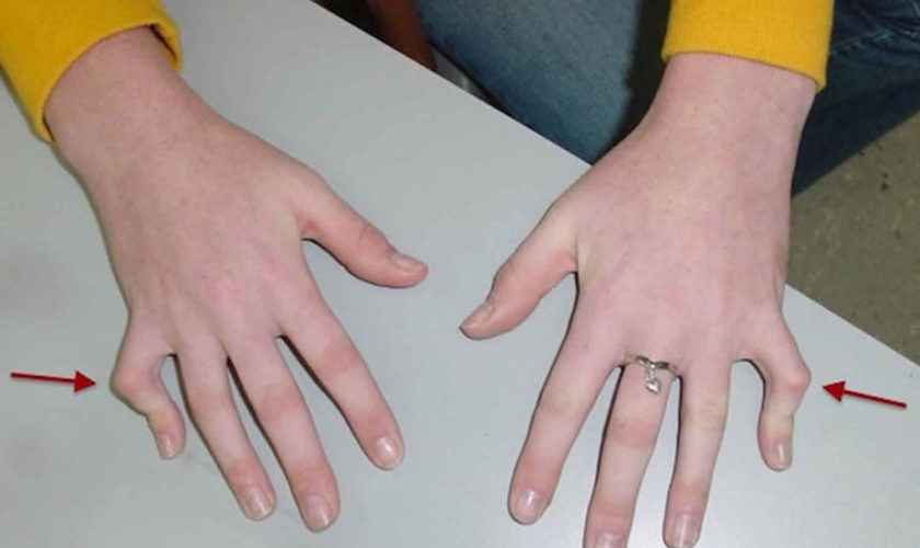
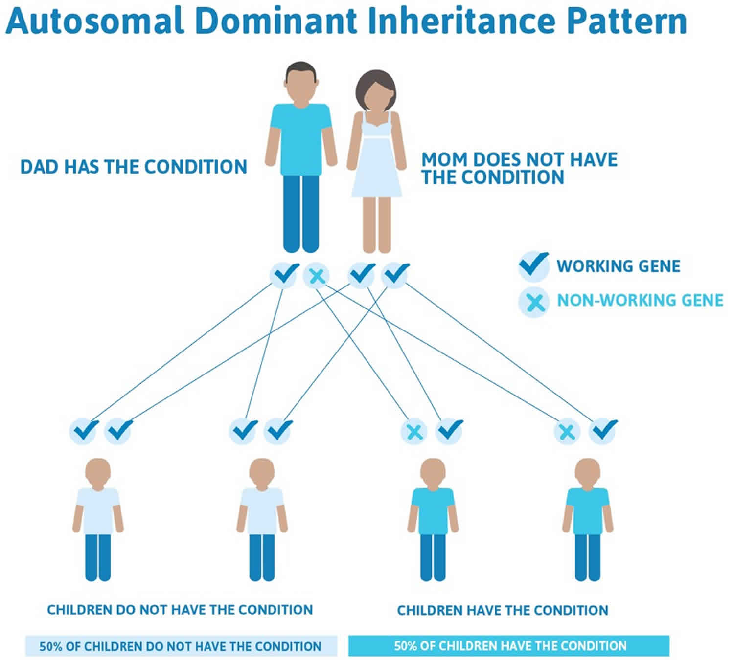
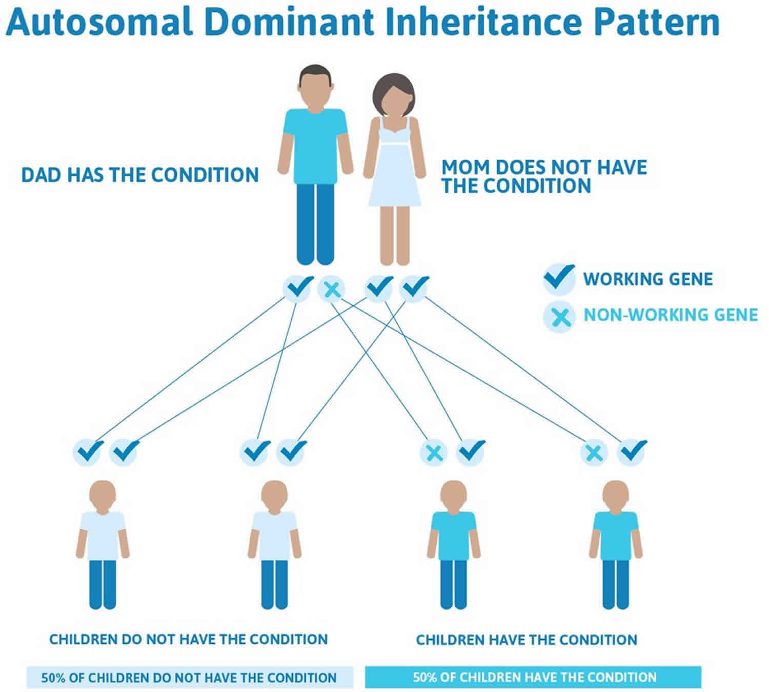
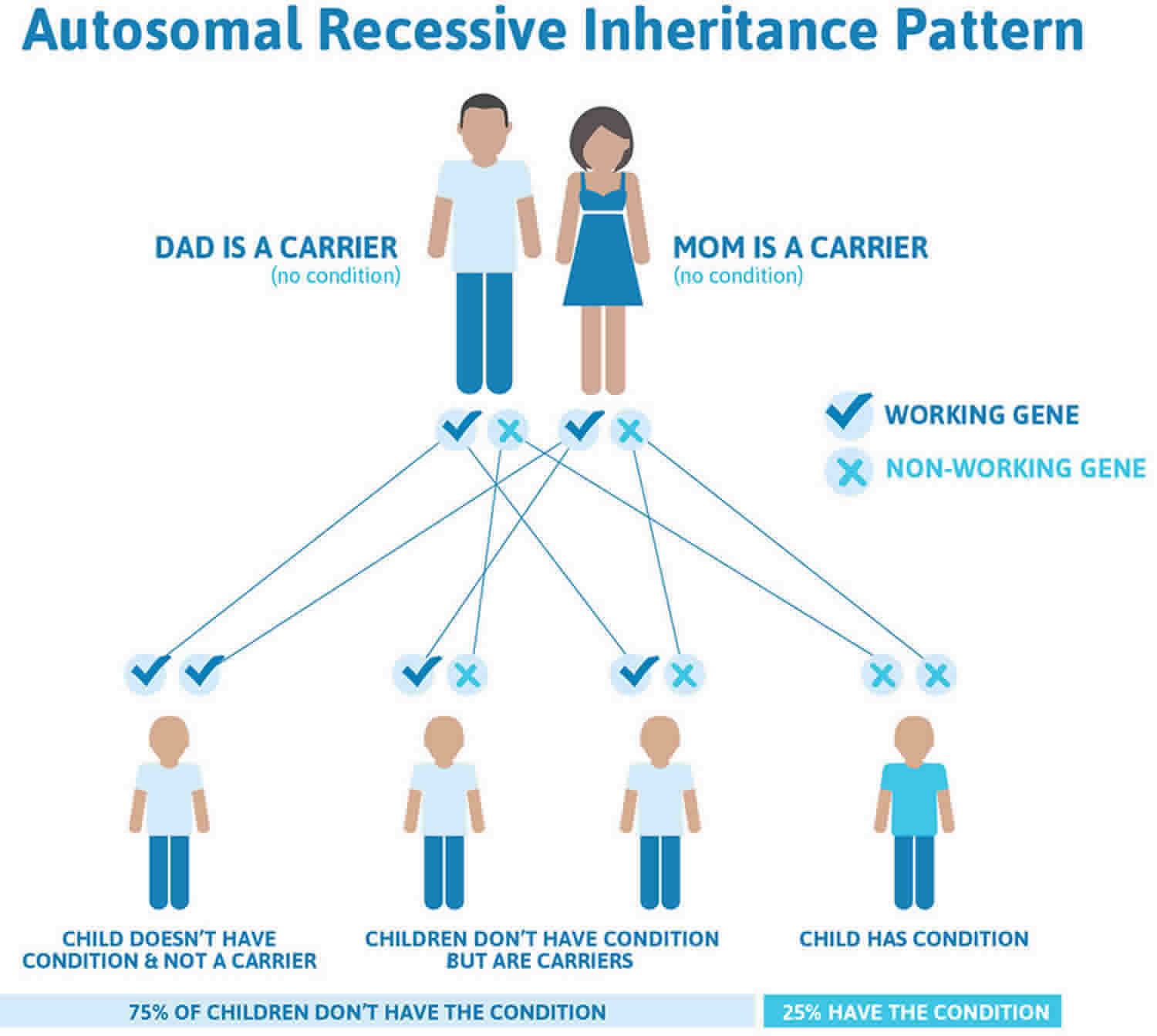
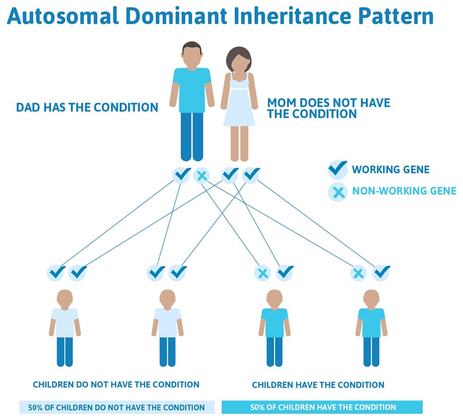
Camptodactyly is a permanent progressive flexion contracture at the proximal interphalangeal joints with skin shortening on the palmar surface of the finger and palm 1), either unilaterally or bilaterally 2). Most camptodactyly cases are limited to fifth-finger or camptodactyly pinky finger involvement; however, other fingers (sometimes seen in ring and middle fingers) may also be involved 3). The incidence of camptodactyly is approximately 1% in the general population, and involvement is frequently bilateral. Although camptodactyly inheritance is sporadic, an autosomal dominant pattern with variable penetrance and expressivity has been reported. Three types of camptodactyly have been defined: type 1 is notable in the first year of life, type 2 manifests after 10 yrs of age for reasons that remain unclear and type 3 is associated with other congenital anomalies 4).

Camptodactyly types:

  • Type I – infant onset
  • Type II – adolescent onset, more common in girls than boys
  • Type III – associated with other congenital anomalies

Camptodactyly often does not cause functional impairment, meaning that patients seek medical attention for concerns relating to cosmetic appearance or military recruitment 5). Benson et al. 6) stated that while patients who presented early with camptodactyly have an equal sex distribution, late-onset patients are mostly females.

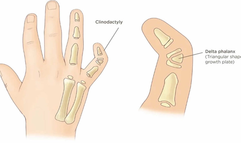
Camptodactyly can be caused by a number of different abnormal structures in your child’s finger:

  • Abnormal muscles in the hand.
  • Differences in bone shape
  • Tight skin
  • Contracted tendons and ligaments

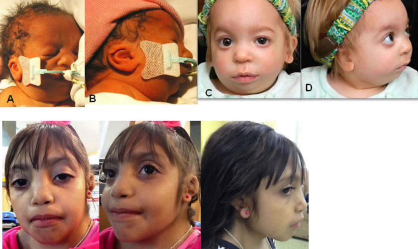
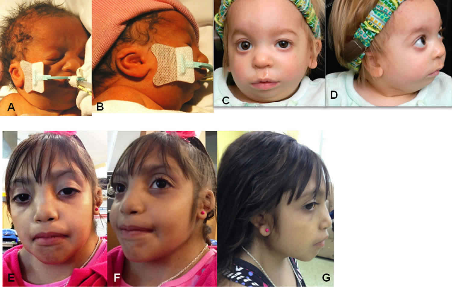
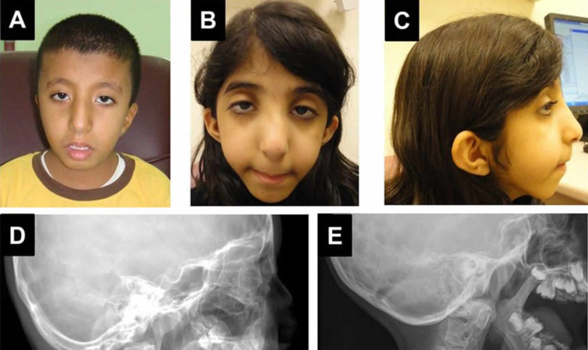
Camptodactyly may accompany other anomalies or a number of very rare syndromes. Therefore, physicians should carefully examine patients for other musculoskeletal deformities 7). Other causes of proximal interphalangeal joint flexion, such as Boutonniere deformity, Dupuytren contracture, trigger finger, and an absent extensor mechanism, must be ruled out before confirming the diagnosis. Isolated camptodactyly is not a rare condition; however, it may be overlooked if it is restricted to the fifth fingers and does not influence hand function. Camptodactyly of finger can be mistaken for other rheumatologic disorders, including rheumatoid arthritis and Dupuytren contracture. Clinical and radiological parameters are used to define the extent of the deformity and joint flexibility 8).

The diagnosis of camptodactyly at the onset is of utmost importance because the use of splints and stretching at that time may be useful. Early identification of camptodactyly will also prevent unnecessary diagnostic work-up and patient anxiety. If camptodactyly is left untreated for several years, the position of permanent fixation leads to contracture in the joint 9). Nonoperative and operative management have been proposed to treat camptodactyly, depending on its clinical severity. These diverse techniques range from splinting or stretching exercises to release of tendons, fascial bands, transfer of muscles, and tenotomy 10).

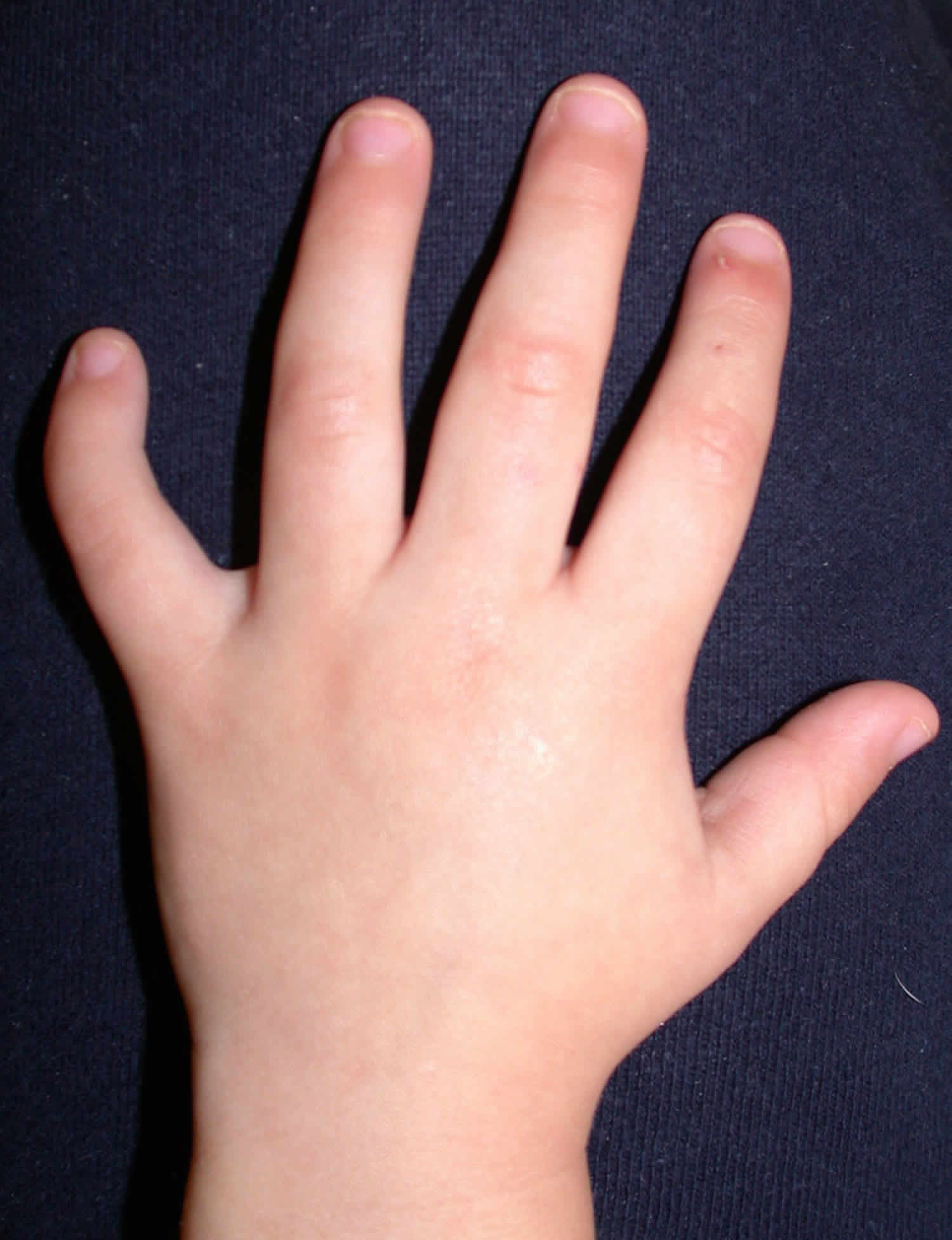
Figure 1. Camptodactyly pinky finger

Camptodactyly pinky finger

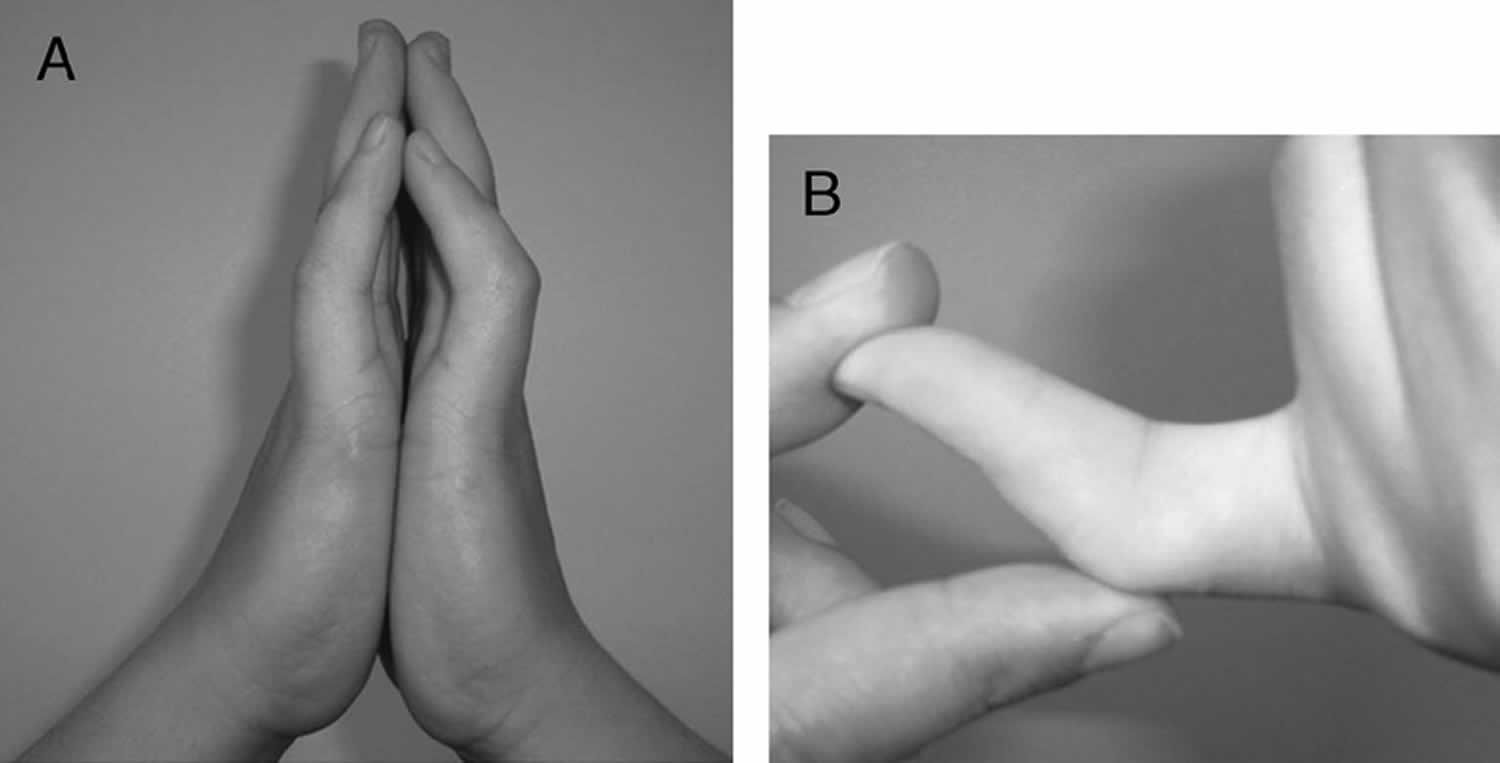
Footnote: (A) Bilateral flexion deformity of the proximal interphalangeal joints of the fifth finger. (B) The left fifth finger lacks passive extension.

[Source 11) ]

Camptodactyly causes

While the origin of camptodactyly remains unknown, it is believed to have a genetic component and may be linked to disruptions in prenatal development. Several causes have been proposed, including abnormal lumbricals; short flexor digitorum superficialis, which is often accompanied by subsequent or associated skin shortening; tight fascial bands; a deficient dorsal central slip extensor mechanism; and changes in the distal interphalangeal joint or metacarpophalangeal joint 12). Camptodactyly can have an early or late onset, and it has been proven to show an autosomal dominant pattern of inheritance 13).

Camptodactyly signs and symptoms

The main symptom of camptodactyly is a slightly flexed posture of the middle joint, where the finger cannot completely straighten. Camptodactyly is most common in the little finger, but may affect other fingers as well. Camptodactyly may worsen over time, and may often worsen during growth spurts.

In most cases, camptodactyly does not cause pain or significantly affect the function of the hand. Camptodactyly does not cause swelling, inflammation or warmth to the area.

Camptodactyly diagnosis

Camptodactyly is diagnosed by your child’s doctor after a thorough medical history and careful physical examination. X-rays are also used to confirm the diagnosis.

In addition to a physical exam and X-rays, your child may also undergo:

  • Range of motions tests to determine if the condition is affecting movement and dexterity
  • Nerve assessment tests to determine if the condition has damaged or compressed any nerves

Accurate diagnosis helps us determine the best course of treatment for your child.

Camptodactyly treatment

The treatment of camptodactyly is controversial 14). Many published studies have emphasized conservative treatment, while others have described surgical procedures. The problem with camptodactyly is that it presents in several forms, which means that there is no single model for effective treatment. Your child’s doctor will probably recommend some form of splinting and occupational hand therapy if your child’s camptodactyly is mild. Surgery may be considered if conservative measures have failed.

Nonsurgical treatment

Most cases of camptodactyly do not require surgical intervention.

If your child has a mild case of camptodactyly — less than a 30-degree bend in their finger that is not affecting hand function — nonoperative treatment will be recommended.

Nonoperative treatment may include:

  • Finger and joint stretches to extend range of motion for the affected finger or fingers
  • A splint to hold the bent finger in a straight position

Camptodactyly splinting

Camptodactyly splinting is very effective with best results achieved from early intervention.

Treatment protocol:

  • Severity and response rate of contracture guides the splinting regime/hours of required splinting. This may vary from 8-20 hours and may be adjusted throughout therapy process.
  • Ongoing splinting at night is required to consolidate and maintain the range of extension while growing.
  • At times of reduced or slower growth it may be possible to cease splinting until early signs of recurrence indicate a need to resume splinting.
  • Exercises/therapeutic play ideas to maintain range of motion and strengthen intrinsic and extrinsic extension are to be prescribed as indicated.

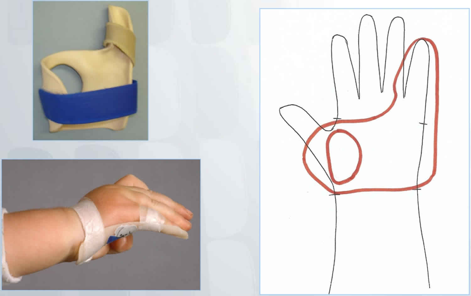
Camptodactyly splint design:

  • Hand-based to ensure that the palmar skin is lengthened and the proximal interphalangeal joint is extended while avoiding hyperextension at the metacarpophalangeal joint and distal interphalangeal joint.
  • Ensure unaffected fingers, thumb and wrist are not included in the splint and are therefore free to move.
  • Secure finger/s with either velcroor paper tape at the distal end of the proximal phalanx.
    • Tape prevents the splint from shifting and is more difficult for young children to remove than Velcro is. Adolescents may find Velcro easier to manage independently.

Figure 2. Camptodactyly splint

Camptodactyly splint

Camptodactyly surgery

If your child’s finger curvature increases rapidly, or if it progresses to the point where it interferes with hand function, your child’s doctor may recommend surgery. As there is no single cause for camptodactyly, no single operative procedure is recommended for all children. Surgery is most effective if performed while your child is still young and the bones are not fully matured.

Two common procedures are:

  • Dividing the tendon that is causing the muscle shortening
  • Transferring a tendon and/or muscle to restore balance to the hand

In rare cases, where a child’s camptodactyly is related to abnormal bones or bone structure, doctors may need to perform surgery to repair, remove or fuse a bone to optimize hand function. During this process some range of motion in the joint may be lost. After surgery, your child’s finger, hand or arm may be put in a cast, splint or sling to immobilize it as it heals.

In this study 15), operative management was planned for all cases with >60° of involvement. Further, the type of surgery was decided on the basis of the anatomical defects that were encountered on the operating table. The most commonly affected structure was the flexor digitorum superficialis, which was short and tight in 10 cases (64.29%), followed by skin shortening in two cases (14.29%) and tight flexor digitorum superficialis alone in two cases (14.29%).

Of the patients, seven (50%) underwent flexor digitorum superficialis release alone, three (21.43%) underwent flexor digitorum superficialis release and transfer to the extensors, two (14.29%) underwent flexor digitorum superficialis release and split-thickness skin grafting cover or Z-plasty, one (7.14%) had flexor digitorum superficialis release along with release of fascial bands, and one (7.14%) underwent transfer of an anomalous lumbrical insertion to the lateral band. Static splinting was administered for 2 to 3 weeks in cases of tendon transfer, after which patients were taught to engage in gradual mobilization of the proximal interphalangeal joint for 6 weeks. Night splinting was continued for a prolonged time in all cases.

On the scale (Mayo Clinic) of Siegert et al. 16), Singh et al 17) observed that irrespective of the anatomical structure affected and the surgical option used, postoperative functional integrity remained the same. However, full correction of flexion without ankylosis was not achieved in any patients. In a comparison with peer groups worldwide, Singh et al 18) found that although several procedures have been developed, simple release of the flexor digitorum superficialis, fascia, or skin mostly suffices, with no need to disturb the extensor mechanism. This, in turn, results in postoperative compromise in flexion.

Follow-up care

Follow-up care for camptodactyly will depend on the treatment needed. If your child received nonsurgical treatment, they should be monitored regularly to ensure the condition is not worsening.

If your child had surgery, they will be examined at 2 weeks and 6 weeks post-operatively, then monitored regularly. Your child’s doctor will give you specific information about a recovery program for your child and how soon they can return to daily activities.

Camptodactyly prognosis

The long-term outlook for children with camptodactyly is very good. While most children with the condition can avoid surgery, those who do need surgery generally have good outcomes.

While surgery is usually successful in partially correcting the curvature, your child will likely have some residual deformity. There is a risk for recurrence and need for future surgery.

References   [ + ]

read more

Congenital torticollis

congenital torticollis

Congenital torticollis

Congenital muscular torticollis also called congenital torticollis, is a painless form of head tilting due to a muscle of the neck, called the sternocleidomastoid muscle, being shorter on one side of the neck than the other and it is present at birth or develops soon after (see Figure 3). The tight sternocleidomastoid muscle causes the head to tilt toward the side of the neck with the shortened muscle and the head to be turned away from that side. The baby tends to look away from the tight muscle. Congenital muscular torticollis is the most common cause of a wry-neck posture in the infant. Congenital muscular torticollis is often discovered during the first 6 to 8 weeks of life, when a newborn begins to gain more control over the head and neck.

“Congenital” means a condition that is present at birth. Congenital torticollis occurs at or shortly after birth.

In newborns, torticollis can happen due to the baby’s position in the womb or after a difficult childbirth.

Some babies with congenital torticollis also have developmental dysplasia of the hip—a condition in which the head of the thighbone is not held firmly in the hip socket.

It can be upsetting to see that your baby has a tilted head or trouble turning his or her neck. But most with babies don’t feel any pain from torticollis. And the problem usually gets better with simple position changes or stretching exercises done at home.

Torticollis can also develop later in infancy and childhood and even in adults. This type of torticollis is referred to as “acquired” torticollis and may be associated with a variety of conditions that require specialized treatment. Acquired torticollis is not discussed in this post.

Congenital muscular torticollis key points

  • Torticollis is when the muscle on one side of the neck is shorter than on the other side. It causes the head to tilt to one side. The baby tends to look away from the tight muscle.
  • If your baby only looks in one direction, try to encourage them to look to the less preferred side. A physiotherapist or occupational therapist may need to prescribe specific stretches.
  • Torticollis is sometimes associated with a condition called positional plagiocephaly (flattened head syndrome), which is when the skull becomes flattened when a baby lays on their back or looks in one direction too long (see Figure 2).
  • During the baby’s first few weeks, a soft lump or bump, which is similar to a “knot” in a tense muscle, may be felt in the affected neck muscle. This lump is not painful and gradually goes away before the baby reaches 6 months of age.
  • Both of the tense muscle lump and positional plagiocephaly tend to go away as the torticollis gets better.

Figure 1. Congenital muscular torticollis

congenital muscular torticollis

congenital muscular torticollis

Figure 2. Flattened head syndrome (positional plagiocephaly)

Flattened head syndrome

Figure 3. Sternocleidomastoid muscle

sternocleidomastoid muscle

When to seek medical attention

If you feel your baby has limited neck movement, see your doctor to learn about other help available. They may refer you to a physiotherapist.

After assessing your child’s head and neck, the therapist will design a home program for your baby. You may be given exercises and other recommendations.

Should I be concerned about the lump on my baby’s neck?

No. The lump that you may be feeling is scar tissue. This is a normal result of the healing process. It is not painful to your child. With specific stretching exercises given to you by a physiotherapist, it should go away in a few months.

Why does my baby prefer to look in one direction?

A baby with torticollis may tend to look in only one direction. The shortened neck muscle causes the head to be tilted towards it. The chin turns away from it. This is why your child prefers to look away from the tight muscle.

If your baby is always on their back or prefers to looks in one direction, part of their skull may become flat. This condition is called positional plagiocephaly. Positional plagiocephaly means flattening of the skull. Torticollis and plagiocephaly are closely associated with one another.

What should I do if my child only looks in one direction?

If your baby prefers to look in one direction, you should encourage them to look to the less-preferred side until they look equally in both directions. Your baby may have a tight muscle in their neck and they may need specific stretches. You should speak to your doctor or to a physiotherapist for more information.

In the meantime, here are some things you can do:

  • During playtime, use mobiles or brightly coloured toys to encourage your baby to look in the less-preferred direction.
  • When you are holding your baby, hold them in a way to encourage them to look in the less-preferred direction.
  • If your baby’s crib is against the wall, put them at opposite ends of the crib each night. Babies prefer to look out into the room.
  • If your baby’s crib is not against a wall, move a brightly coloured crib-safe toy to encourage them to look in a different direction each night.

Congenital torticollis causes

Congenital torticollis occurs when the neck muscle, known as the sternocleidomastoid muscle, that runs up and toward the back of the infant’s neck is shortened. This tilts the head down and to one side. This is known as congenital muscular torticollis.

Scientists still don’t know exactly what causes the shortened neck muscle. Researchers think that the sternocleidomastoid muscle may have had a small tear or it may be stretched during the baby’s birth. The tear causes bleeding and swelling. And scar tissue replaces some of the muscle, making it shorter.

More recently, it has been postulated that the sternomastoid muscle shortens as a result of scarring due to a vascular disturbance in the womb. Still others think that it is due to position of the baby’s head in the womb causing fibrosis or shortening of the muscle.

Some cases of congenital torticollis are caused by a bone problem in the neck portion of the spine (cervical spine). This is known as a congenital malformation of the cervical spine.

Torticollis may also occur later in life, but this is not congenital torticollis.

Risk factors for developing congenital torticollis

Risk factors include a breech pregnancy (where the fetus is leg-down instead of face-down) and a difficulty delivery. However, most congenital torticollis occur without any apparent cause in otherwise healthy infants.

Congenital torticollis has been associated with two other birth conditions:

  • Developmental dysplasia of the hip – hip joint dislocation present at birth.
  • Metatarsus adductus – the front of the foot is bent or angled in toward the middle of the foot.

Congenital torticollis symptoms

Congenital muscular torticollis does not cause pain. Babies with torticollis will act like most other babies except when it comes to activities that involve turning. Either side can be affected although it is often more common in the right (75%). The infant keeps his or her head tilted toward the affected side and the chin rotated toward the opposite shoulder. He or she will also have difficulty turning the head from side to side and up and down.

A olive-shaped lump may be visible or felt on the affected side in the first 3 months of life. The lump may eventually disappear as a tight fibrous band is replaced over the length of the neck muscle.

A baby with congenital torticollis might:

  • tilt the head in one direction (this can be hard to notice in very young infants)
  • prefer looking at you over one shoulder instead of turning to follow you with his or her eyes
  • if breastfed, have trouble breastfeeding on one side (or prefers one breast only)
  • work hard to turn toward you and get frustrated when unable turn his or her head completely

Some babies with torticollis develop a flat head (positional plagiocephaly) on one or both sides from lying in one direction all the time. During the baby’s first few weeks, a soft lump or bump, which is similar to a “knot” in a tense muscle, may be felt in the affected neck muscle. This lump is not painful and gradually goes away before the baby reaches 6 months of age. Both of these conditions tend to go away as the torticollis gets better.

Possible results of untreated congenital torticollis

  • Plagiocephaly – This is the most common consequence of untreated Torticollis. Plagiocephaly is the mishapening of the bones on the skull, usually resulting in a large flat spot on one side of the back of the head and facial assymetries. Early diagnosis and conservative treatment can be successful in decreasing the severity of the Plagiocephaly. However, late diagnosis must be treated by a helmet or craniofacial surgery.
  • Cervical spine contractures into the preferred head rotation and sidebend – These contractures can become ossified over time, significantly impacting functional mobility and ability to interact with peers. Once a contracture is ossified, surgery is required to lengthen the muscle, followed by several sessions of physical therapy to regain full cervical spine range of motion.
  • Limited shoulder mobility – Decreased active movement into non-preferred rotation and sidebend can also result in shoulder elevation. This in turn impacts the child’s ability for upper extremity weight-bearing and reaching toward midline with hand.
  • Cervical scoliosis – Persistent head tilt in the absence of shoulder elevation can result in a lateral shift of the cervical spine, which leads to cervical scoliosis.

Congenital torticollis complications

If the problem of wry-neck is not corrected, some of the complications that will appear as the child grows include:

  • Asymmetry of skull and face – the face of the affected side may remain “flattened”. This is only reversible if the torticollis is corrected before age 1.
  • Permanent limtations in head and neck movements.

Congenital torticollis diagnosis

If you notice that your child holds his or her head tilted to one side, consult your pediatrician. He or she will discuss your child’s general health, and will ask specific questions about the torticollis symptoms.

Your baby’s doctor will perform a comprehensive physical examination to see how far your baby can turn their head and check for other conditions that can cause torticollis symptoms. Imaging tests, such as x-rays and ultrasound scans, may be taken of your child’s neck and/or hips.

There is a 20% incidence of hip dysplasia children with muscular torticollis. So your doctor will perform an ultrasound exam of the hips in the first 4 to 6 weeks of life to rule that out.

Congenital torticollis treatment

Initial treatment consists of a massage and stretching program, which is successful in most cases when started in the first 6 months of life. If your baby does have congenital muscular torticollis, your baby’s doctor might teach you neck stretching exercises to practice at home. These help loosen the tight sternocleidomastoid muscle and strengthen the weaker one on the opposite side (which is weaker due to underuse). This will help to straighten out your baby’s neck.

Sometimes, doctors suggest taking a baby to a physical therapist for more treatment.

After treatment starts, the doctor may check your baby every 2 to 4 weeks to see if the torticollis is getting better.

Most babies with torticollis get better through position changes and stretching exercises. It might take up to 6 months to go away completely, and in some cases can take a year or longer.

Stretching exercises to treat torticollis work best if started when a baby is 3–6 months old. If you find that your baby’s torticollis is not improving with stretching, talk to your doctor. Your baby may be a candidate for muscle-release surgery, a procedure that cures most cases of torticollis that don’t improve.

If your child’s head tilt is caused by something other than congenital muscular torticollis, and the X-rays show no spinal abnormality, other treatment involving rest, a special collar, traction, application of heat to the area, medication, or, rarely, surgery may be necessary. To treat Klippel-Feil syndrome, a specialist may recommend treatments ranging from physical therapy to an operation. For treating torticollis due to injury or inflammation, your doctor may recommend applying heat, as well as using massage and stretching to ease head and neck pain. Your pediatrician can refer you to a specialist for a definitive diagnosis and treatment program.

Helping your baby at home

Encourage your baby to turn the head in both directions. This helps loosen tense neck muscles and tighten the loose ones. Babies cannot hurt themselves by turning their heads on their own.

There are other things that you can do at home to help:

  • Place toys where your baby must turn his or her head to see them.
  • Carry your child so that he or she looks away from the limited side.
  • Position the crib and changing table so that your child must look away from the limited side to see you.
  • Lay your baby on his or her stomach for brief periods when awake (“tummy time”) to help strengthen the neck muscles.

Congenital torticollis exercises

The standard treatment for congenital muscular torticollis consists of an exercise program to stretch the sternocleidomastoid muscle.

Stretching exercises include turning the baby’s neck side to side so that the chin touches each shoulder, and gently tilting the head to bring the ear on the unaffected side down to the shoulder.

These exercises must be done 4 to 6 times a day. Your doctor or a physical therapist will teach you how to perform the exercises.

Here are some exercises to try:

  • When your baby wants to eat, offer the bottle or your breast in a way that encourages your baby to turn away from the favored side.
  • When putting your baby down to sleep, position them to face the wall. Since babies prefer to look out onto the room, your baby will actively turn away from the wall and this will stretch the tightened muscles of the neck. Remember — always put babies down to sleep on their back to help prevent Sudden Infant Death Syndrome (SIDS).
  • During play, draw your baby’s attention with toys and sounds to make him or her turn in both directions.

Don’t forget “Tummy Time”

Laying your baby on the stomach for brief periods while awake (known as “tummy time”) is an important exercise. It helps strengthen neck and shoulder muscles and prepares your baby for crawling.

This exercise is especially useful for a baby with torticollis and a flat head, and can help treat both problems at once. Here’s how to do it:

  • Lay your baby on your lap for tummy time. Position your baby so that his or her head is turned away from you. Then, talk or sing to your baby and encourage him or her to turn and face you. Practice this exercise for 10 to 15 minutes.

For stretching and positioning RIGHT sternomastoid torticollis

Stretching

  1. For the following stretching exercises, the parent sits with the back against the wall and knees bent.
  2. Place the child in your lap, with the child on her back and knees tucked.

Sidebending

  1. Hold the child’s RIGHT shoulder down with your LEFT hand.
  2. Place your RIGHT hand on top of the RIGHT side of the child’s head, and slowly bend her head towards her LEFT shoulder.
  3. Hold the position for 10 seconds. Repeat 15 times, 4 to 6 times a day.

Rotation

  1. Place your RIGHT forearm against the child’s LEFT shoulder, and cup the child’s head with the same hand.
  2. Use your LEFT hand to hold the child’s chin.
  3. Slowly rotate the child’s face to her RIGHT.

Hold the position for 10 seconds. Repeat 15 times, 4 to 6 times a day.

Positioning

  • Playing on stomach: When the child is on her stomach, position all toys in the crib so that the child has to turn her face to the RIGHT.

Carrying

  1. Hold the child facing away from you, in a side-lying position, with the child’s RIGHT ear resting against your RIGHT forearm.
  2. Place your LEFT arm between the child’s legs and support the child’s body.
  3. Carry the child in this position as much as possible.

Other suggestions

  1. Hold toys so that the child has to look up and out to her RIGHT.
  2. Position child in crib so that activities in the room encourage her to look RIGHT.
  3. While bottle feeding the child, position her to face RIGHT.
  4. While holding the baby across the shoulder, position her to face RIGHT.

For stretching and positioning LEFT sternomastoid torticollis


Stretching

  1. For the following stretching exercises, the parent sits with the back against the wall and knees bent.
  2. Place the child in your lap, with the child on her back and knees tucked.

Sidebending

  1. Hold the child’s LEFT shoulder down with your RIGHT hand.
  2. Place your LEFT hand on top of the LEFT side of the child’s head, and slowly bend her head towards her RIGHT shoulder.
  3. Hold the position for 10 seconds. Repeat 15 times, 4 to 6 times a day.

Rotation

  1. Place your LEFT forearm against the child’s RIGHT shoulder, and cup the child’s head with the same hand.
  2. Use your RIGHT hand to hold the child’s chin.
  3. Slowly rotate the child’s face to her LEFT.
  4. Hold the position for 10 seconds. Repeat 15 times, 4 to 6 times a day.

Positioning

  • Playing on stomach: When the child is on her stomach, position all toys in the crib so that the child has to turn her face to the LEFT.

Carrying

  1. Hold the child facing away from you, in a side-lying position, with the child’s LEFT ear resting against your LEFT forearm.
  2. Place your RIGHT arm between the child’s legs and support the child’s body.
  3. Carry the child in this position as much as possible.

Other suggestions

  1. Hold toys so that the child has to look up and out to her LEFT.
  2. Position child in crib so that activities in the room encourage her to look LEFT.
  3. While bottle feeding the child, position her to face LEFT.
  4. While holding the baby across the shoulder, position her to face LEFT.

Surgical treatment

If nonsurgical options do not correct the torticollis, your doctor may suggest surgery.

Approximately 10% of children with congenital muscular torticollis require surgery. The operation is typically scheduled once the child reaches preschool years. The procedure will lengthen the short sternocleidomastoid muscle, and may be done as an outpatient surgery, meaning your child could go home the same day.

read more

Clinodactyly

clinodactyly

Clinodactyly

Clinodactyly is a medical term that refers to a radial angulation at an interphalangeal joint in the radio-ulnar or palmar planes 1). Clinodactyly is when a finger or toe curves toward the finger or toe next to it, not up or down. Clinodactyly can happen in any finger or toe, but the most common type is when the 5th finger curves toward the ring finger. Clinodactyly may get worse with age. The estimated incidence of clinodactyly is highly variable dependent on sampling and has been reported to range between 1-18%. Clinodactyly affects about one in four children born with Down syndrome (trisomy 21).

Unless the curved finger is severe, clinodactyly may go unnoticed for years. The condition doesn’t cause pain and in most cases, does not affect hand function.

Clinodactyly is a congenital condition, meaning it is present at birth even if it is not discovered until later. Clinodactyly can be inherited or your child may be the first person in your family to have the condition. Clinodactyly can also be a symptom of an associated syndrome.

Clinodactyly is typically caused by the growth of an abnormally shaped bone in your child’s finger, which causes the finger to curve to the side. It may also be due to an irregular growth plate in one of the bones of your child’s finger.

Treatment for clinodactyly varies depends on the severity of the condition, but can include ongoing monitoring and surgery.

Figure 1. Clinodactyly pinky finger

fifth finger clinodactyly

Clinodactyly causes

Clinodactly can result from vast number pathologies ranging from congenital to acquired. There may be a growth plate on one side of the bone that causes the bone to grow longer on one side than the other. The bone doesn’t grow in the normal shape of a rectangle, but rather more like a triangle.

  • Clinodactyly might be a genetic condition (it can run in families with an autosomal recessive inheritance).
  • Clinodactyly might be a part of a syndrome (a specific group of symptoms):
    • Aneuploidic syndrome
      • Down syndrome: About 35-70% of children with Down syndrome have clinodactyly 2)
      • Klinefelter syndrome
      • trisomy 18
      • Turner syndrome
    • Non aneupliodic syndrome
      • Cornelia de Lange syndrome
      • Feingold syndrome
      • Roberts syndrome
      • Russell-Silver syndrome 2
      • Fanconi anemia
    • Non syndromic
      • macrodystrophia lipomatosa
      • brachydactyly type A3

Clinodactyly signs and symptoms

The primary symptom of clinodactyly is a finger that is abnormally curved in the middle. It may overlap with other fingers on the hand. In most cases, the condition does not cause pain, swelling or inflammation.

If your child’s clinodactyly finger includes a curve of more than 30 degrees, it may affect their hand function.

Clinodactyly diagnosis

Clinodactyly is diagnosed by examination. In most cases, clinicians will recommend an X-ray to look at the bones of the fingers/toes and to confirm the diagnosis. X-rays produce images of bones and help doctors identify the underlying structure of the hand. In the case of clinodactyly, a distinctive c-shaped bone can typically be seen in the middle bone of the affected finger.

In addition to a physical exam and X-rays, your child may also undergo:

  • Range of motions tests to determine if the condition is affecting movement and dexterity

Accurate diagnosis helps us determine the best course of treatment for your child.

Clinodactyly treatment

Most children with clinodactyly don’t need surgery to fix the condition. Surgery is usually only done if your child can’t use his/her hands normally.

Clinodactyly surgery

If your child’s clinodactyly is severe (more than 30-degree curvature) or significantly affecting her daily activities, surgery may be recommended. Surgery is most effective if performed while your child is still young and her bones are not fully matured.

If surgery is recommended:

  • The surgery will be done in the operating room under general anesthesia (the child is put to sleep).
  • Small cuts are made in the fingers to get to the bone.
  • A cut in the bone is made to straighten the bones of the fingers.
  • The fingers are put in the correct position. Small pins, wires or screws are used to hold the bone straight.
  • After surgery, the hand will be bandaged and most likely put in a cast.
  • Once the bandages are taken off, your surgeon might suggest working with a hand therapist.

Surgery for clinodactyly typically involves a phalanx-opening wedge osteotomy procedure. During this multi-step procedure, doctors will:

  • Remove a wedge-shaped portion of the curved bone in the affected finger
  • Stabilize the finger
  • Ensure the bones and soft tissues are lined up properly within the finger
  • Ensure the corrected finger lines up with the other fingers on the hand

After surgery, your child’s finger will be immobilized with a cast or splint. Doctors may also recommend a sling for your child’s arm to further protect the hand and finger as it heals.

Nonsurgical treatment

If your child’s finger is only slightly curved — and there is no problem using the fingers — clinicians will usually recommend ongoing observation to ensure the condition doesn’t worsen.

In most cases, splinting or stretching the finger is not recommended.

Follow-up care

Follow-up care for clinodactyly will depend on the treatment needed. If your child received nonsurgical treatment, they should be monitored regularly to ensure the condition does not begin to significantly affect his daily activities.

If your child had surgery, they will be examined at 2 weeks and 6 weeks post-operatively, then monitored regularly. Your child’s doctor will give you specific information about a recovery program for your child and how soon they can return to daily activities.

Clinodactyly prognosis

The long-term outlook for children with clinodactyly is very good. Many children with clinodactyly can avoid surgery. For children with more severe curvature of the finger, surgery can improve hand function and help children better manage their daily activities.

References   [ + ]

read more

Achondroplasia

achondroplasia

What is achondroplasia

Achondroplasia is a form of short-limbed dwarfism. The word achondroplasia literally means “without cartilage formation.” Cartilage is a tough but flexible tissue that makes up much of the skeleton during early development. However, in achondroplasia the problem is not in forming cartilage but in converting cartilage to bone (a process called ossification), particularly in the long bones of the arms and legs. Achondroplasia is the most common type of short-limbed dwarfism also called little people, a condition in which a person is very short (less than 4 feet 10 inches as an adult). The condition occurs in 1 in 15,000 to 40,000 newborns.

Achondroplasia is characterized by an unusually large head (macrocephaly), short upper arms (rhizomelic dwarfism), and short stature (adult height of approximately 4 feet). Achondroplasia does not typically cause impairment or deficiencies in mental abilities. If the bones that join the head and neck do not compress the brainstem or upper spinal cord (craniocervical junction compression), life expectancy is near normal.

Achondroplasia is similar to another skeletal disorder called hypochondroplasia, but the features of achondroplasia tend to be more severe.

Achondroplasia is genetic disorder caused by a change (mutation) in the fibroblast growth factor receptor 3 (FGFR3) gene. Achondroplasia occurs as a result of a spontaneous genetic mutation in approximately 80 percent of patients; in the remaining 20 percent achondroplasia is inherited from a parent.

All people with achondroplasia have short stature. The average height of an adult male with achondroplasia is 131 centimeters (4 feet, 4 inches), and the average height for adult females is 124 centimeters (4 feet, 1 inch). Characteristic features of achondroplasia include an average-size trunk, short arms and legs with particularly short upper arms and thighs, limited range of motion at the elbows, and an enlarged head (macrocephaly) with a prominent forehead. Fingers are typically short and the ring finger and middle finger may diverge, giving the hand a three-pronged (trident) appearance. People with achondroplasia are generally of normal intelligence.

Health problems commonly associated with achondroplasia include episodes in which breathing slows or stops for short periods (apnea), obesity, and recurrent ear infections. In childhood, individuals with the condition usually develop a pronounced and permanent sway of the lower back (lordosis) and bowed legs. Some affected people also develop abnormal front-to-back curvature of the spine (kyphosis) and back pain. A potentially serious complication of achondroplasia is spinal stenosis, which is a narrowing of the spinal canal that can pinch (compress) the upper part of the spinal cord. Spinal stenosis is associated with pain, tingling, and weakness in the legs that can cause difficulty with walking. Another uncommon but serious complication of achondroplasia is hydrocephalus, which is a buildup of fluid in the brain in affected children that can lead to increased head size and related brain abnormalities.

Achondroplasia can be diagnosed by characteristic clinical and radiographic findings in most affected individuals. In individuals in whom there is diagnostic uncertainty or atypical findings, identification of a heterozygous pathogenic variant in FGFR3 can establish the diagnosis.

Achondroplasia baby

People with achondroplasia can have a range of health problems, so it’s important to take your baby to see his health care provider for routine well-baby checkups.

At these checkups, your baby’s provider can compare your baby’s height, weight and head size to those of other babies with achondroplasia. This can help your baby’s provider spot and treat some problems early on.

People with achondroplasia often have these health problems:

  • Apnea. This is when a baby stops breathing for 15 to 20 seconds or more. Babies with apnea and other breathing problems may need surgery to remove the tonsils and adenoids (lymph tissue near the throat).
  • Repeat ear infections. Some babies with achondroplasia need ear tubes. These are small tubes placed in the ear that let air into the middle ear and help lower chances of ear infections. Without treatment, repeat ear infections can cause hearing loss.
  • Obesity (being very overweight). Healthy eating and being active can help your child stay at a healthy weight as she grows.
  • Compression of the upper end of the spinal cord. This is when the opening where the head and spine (backbone) connect is too small. The spinal cord gets squeezed (compressed), causing trouble with breathing. A small number of babies with achondroplasia die suddenly (often during sleep) from compression. If needed, surgery can widen the opening to ease pressure on the spinal cord.
  • Spinal stenosis. Spinal stenosis causes the spine to narrow, putting pressure on the nerves and spinal cord. This can cause low back pain, problems with urination and weakness, tingling and pain in the legs. Symptoms usually appear when a person with achondroplasia is a teen or adult. Surgery can ease pressure on the spinal cord.
  • Hydrocephalus (fluid buildup in the brain). Your baby’s provider measures your baby’s head at regular checkups to help catch hydrocephalus early. In some cases, a surgeon needs to drain the extra fluid from a baby’s brain.
  • Kyphosis (a small hump in the upper back). A baby may have kyphosis due to poor muscle tone, but it usually goes away after she starts walking. Strollers or carriers that don’t give good back support can make kyphosis worse. If your child stills has kyphosis after she starts walking, she may need a back brace or surgery to correct it.
  • Lordosis (inward curving of the lower back). This can develop after your child starts walking and can lead to waddling. Special exercises or physical therapy can help.

Achondroplasia causes

Achondroplasia results from specific changes (mutations) of a gene known as fibroblast growth factor receptor 3 (FGFR3). The FGFR3 gene provides instructions for making a protein that is involved in the development and maintenance of bone and brain tissue. Two specific mutations in the FGFR3 gene are responsible for almost all cases of achondroplasia. Researchers believe that these mutations cause the FGFR3 protein to be overly active, which interferes with skeletal development and leads to the disturbances in bone growth seen with this disorder.

Most babies (80 percent of achondroplasia cases) with achondroplasia are born to parents who don’t have the condition. This happens when there’s a random gene change in either the egg or sperm that join together and create a baby. Increased age of the father (advanced paternal age) may be a contributing factor in cases of sporadic achondroplasia.

Less commonly (20 percent of achondroplasia cases), familial cases of achondroplasia follow an autosomal dominant pattern of inheritance. Dominant genetic disorders occur when only a single copy of an abnormal gene is necessary to cause a particular disorder. The abnormal gene can be inherited from either parent or can be the result of a mutated (changed) gene in the affected individual. The risk of passing the abnormal gene from an affected parent to an offspring is 50% for each pregnancy. The risk is the same for males and females.

How is achondroplasia inherited?

Achondroplasia is inherited in an autosomal dominant pattern, which means one copy of the altered gene in each cell is sufficient to cause the disorder. Achondroplasia occurs as a result of a spontaneous genetic mutation in the FGFR3 gene in approximately 80 percent of patients; in the remaining 20 percent of people with achondroplasia have inherited an altered FGFR3 gene from one or two affected parents. Individuals who inherit two altered copies of FGFR3 gene typically have a severe form of achondroplasia that causes extreme shortening of the bones and an underdeveloped rib cage. These individuals are usually stillborn or die shortly after birth from respiratory failure.

If you or your partner has achondroplasia, you can pass it to your baby. If only one of you has the condition, there’s a 1 in 2 chance (50 percent) that your baby can have the condition.

If both you and your partner have achondroplasia, there is:

  • 1 in 2 chance (50 percent) that your baby can have achondroplasia
  • 1 in 4 chance (25 percent) that your baby won’t have achondroplasia
  • 1 in 4 chance (25 percent) that your baby has the severe kind of achondroplasia that can lead to death

If you or your partner has achondroplasia or you’re the parent of a child with achondroplasia, talk to a genetic counselor about the condition. A genetic counselor is a person who is trained to help you understand about how genes, birth defects and other medical conditions run in families, and how they can affect your health and your baby’s health.

Genetic counseling

Resources for locating a genetics professional in your community are available online:

Figure 1. Achondroplasia autosomal dominant inheritance pattern

Achondroplasia autosomal dominant inheritance pattern

Achondroplasia prevention

Genetic counseling may be helpful for prospective parents when one or both have achondroplasia. However, because achondroplasia most often develops spontaneously, prevention is not always possible.

Achondroplasia symptoms

The typical appearance of achondroplastic dwarfism can be seen at birth.

Achondroplasia baby

Infants born with achondroplasia typically have a “dome-like” (vaulted) skull, and a very broad forehead. In a small proportion there is excessive accumulation of fluid around the brain (hydrocephalus). Low muscle tone (hypotonia) in infancy is typical of achondroplasia. Acquisition of developmental motor milestones may be delayed.

A person with achondroplasia often has:

  • Short height (significantly below the average height for a person of the same age and sex). The average height of an adult male with achondroplasia is 131 centimeters (4 feet, 4 inches), and the average height for adult females is 124 centimeters (4 feet, 1 inch).
  • Short upper arms and thighs (compared to the forearms and lower legs)
  • Large head and forehead (frontal bossing) with a flat bridge of the nose (large head-to-body size)
  • Dental problems, including crowded or crooked teeth
  • Broad, flat feet, short toes and short fingers
  • Trident hand, a condition in which you have an extra space between the middle and ring fingers
  • Decreased muscle tone. Babies with weak muscle tone may have delays in meeting developmental milestones, like sitting, standing and walking.
  • Bowed legs. This is when legs curve outward between the thighs and ankles. Bowed legs can cause pain and trouble with walking. If the bowing or pain is severe, surgery can fix bowed legs.
  • Narrowing of the spinal column (spinal stenosis)
  • Spine curvatures called kyphosis and lordosis

Growth

Average adult height for men with achondroplasia is 131±5.6 cm; for women, 124±5.9 cm. Obesity is a major problem in achondroplasia 1). Excessive weight gain is manifest in early childhood. In adults, obesity can aggravate the morbidity associated with lumbar stenosis and contribute to nonspecific joint problems and possibly to early mortality from cardiovascular complications 2).

Development

In infancy, mild to moderate hypotonia is typical. Infants have difficulty in supporting their heads because of both hypotonia and large head size. That and differences in body habitus cause motor delays and unusual patterns of motor development such as snowplowing (using the head and feet to leverage movement) 3). Small joint hypermobility and short fingers can affect fine motor development and delay self-feeding 4).

Intelligence

Intelligence is normal unless hydrocephalus or other central nervous system complications occur. High-level executive function issues have been reported in some individuals 5).

Macrocephaly

Most children with achondroplasia are macrocephalic 6). Hydrocephalus requiring treatment, which probably occurs in 5% or fewer 7), may be caused by increased intracranial venous pressure because of stenosis of the jugular foramina 8). More recent literature suggests that in some individuals, foramen magnum stenosis may contribute to hydrocephalus – which is thus treatable by posterior fossa decompression or endoscopic third ventriculostomy 9).

Narrow craniocervical junction

Some infants with achondroplasia die in the first year of life from complications related to the craniocervical junction; population-based studies suggest that this excess risk of death may be as high as 7.5% without assessment and intervention 10). The risk appears to be secondary to central apnea associated with damage to respiratory control centers 11), and can be minimized by comprehensive evaluation of every infant with achondroplasia 12) and selective neurosurgical intervention 13). With such evaluation and management this risk may be decreased to as little as 0.3% 14). The best predictors of need for suboccipital decompression include lower-limb hyperreflexia or clonus, central hypopnea demonstrated by polysomnography, and reduced foramen magnum size, as determined by neuroimaging of the craniocervical junction. If computerized tomography (CT) is used, foraminal size can be compared with achondroplasia standards 15). Magnetic resonance imaging (MRI) examination provides direct visualization of the cord without radiation exposure, but there are no achondroplasia standards. T2-weighted MRI may show evidence of spinal cord abnormalities, which may guide operative decision making 16). In one study, all children undergoing surgical decompression of the craniocervical junction showed marked improvement of neurologic function 17). Quality of life indices determined up to 20 years after such surgery were comparable to quality of life indices in those for whom surgery was not indicated in childhood 18). A similar mechanism of injury can result in high cervical myelopathy (asymmetric or increased reflexes, weakness, persisting hypotonia, and poor balance) 19).

Restrictive pulmonary disease

In infancy a small subset of individuals with achondroplasia have restrictive pulmonary issues secondary to a small chest 20) and decreased compliance of the rib cage. Many infants show more rapid desaturations with minor respiratory events (e.g., physiologic periodic breathing or otherwise insignificant obstructive events). A small number have, as a consequence of these features, chronic hypoxemia 21). If a young infant has persistent tachypnea, failure to thrive, or evidence of respiratory failure, the polysomnogram obtained for other reasons in infants will show a low baseline oxygen saturation and/or desaturations associated with minimal respiratory irregularities. If such characteristics are recognized, referral to a pediatric pulmonologist is imperative. Treatment may include oxygen supplementation and, in a few, temporary tracheostomy. In virtually all instances, the need for a tracheostomy disappears as the child grows.

Sleep apnea

Obstructive sleep apnea is common in both older children and adults. It arises because of a combination of midface retrusion resulting in smaller airway size 22), hypertrophy of the lymphatic ring, airway malacia 23), and, perhaps, abnormal innervation of the airway musculature 24).

Clinical signs and symptoms of obstructive sleep apnea may include the following:

  • Difficult morning waking
  • Excessive daytime somnolence
  • Respiratory pauses during sleep
  • Loud snoring
  • Glottal stops or gasping
  • Loud sighs while sleeping
  • Poor daytime concentration
  • Irritability, fatigue, depression
  • Bedwetting

Clinical signs and symptoms of infantile sleep apnea include the following:

  • Observed apnea or exaggerated periodic breathing
  • Struggling to breathe
  • Poor feeding
  • Coughing
  • Difficulty lying flat to sleep
  • Frequent awakenings

Central sleep apnea as well as obstructive sleep apnea may be present in infants. Clinical history is a poor predictor of apnea, and polysomnography should be done 25).

Middle ear dysfunction

Middle ear dysfunction is frequently a problem 26), and if inadequately treated can result in conductive hearing loss of sufficient severity to interfere with language development. More than half of children will require pressure-equalizing tube placement 27). Overall, about 40% of individuals with achondroplasia have functionally relevant hearing loss. Expressive language development is also frequently delayed 28), although the strength of the relationship between hearing loss and expressive language issues is uncertain.

Bow legs

Bowing of the lower legs is exceedingly common in those with achondroplasia. More than 90% of untreated adults have some degree of bowing 29). “Bowing” is actually a complex deformity arising from a combination of lateral bowing, internal tibial torsion, and dynamic instability of the knee 30).

Kyphosis

Kyphosis at the thoracolumbar junction is present in 90%-95% of infants with achondroplasia 31). In about 10% it does not spontaneously resolve and can result in serious neurologic sequelae 32). Preventive strategies 33) may reduce the need for surgical intervention.

Spinal stenosis

The most common medical complaint in adulthood is symptomatic spinal stenosis involving L1-L4 34). Symptoms range from intermittent, reversible, exercise-induced claudication to severe, irreversible abnormalities of leg function and of continence 35). Claudication and stenosis can both result in sensory (numbness, pain, feelings of heaviness) and motor symptoms (weakness, tripping, limited walking endurance). Vascular claudication results from engorged blood vessels after standing and walking and is fully reversible with rest. Spinal stenosis is actual impingement of the spinal cord or nerve root by the stenotic bone of the spinal canal, and symptoms are nonreversible. Symptoms localized to a particular dermatome can result from stenosis of a particular nerve root foramina.

Other orthopedic issues

  • Joint laxity. Most joints are hypermobile in childhood. In general, this has minor consequences except for knee instability in a subset of individuals.
  • Discoid lateral meniscus. This recently recognized structural anomaly may result in chronic knee pain in some individuals 36).
  • Arthritis. Constitutive activation of FGFR-3, as in achondroplasia, may protect against development of arthritis 37).

Acanthosis nigricans

Acanthosis nigricans may be seen in about 10% of individuals with achondroplasia 38). In this population it does not reflect hyperinsulinemia or malignancy.

How do you know if your baby has achondroplasia?

Before birth, your doctor may think your baby has achondroplasia if an ultrasound shows your baby has bone problems, like shortened bones and excessive amniotic fluid surrounding the unborn infant. An ultrasound uses sound waves and a computer screen to show a picture of your baby inside the womb.

If the ultrasound shows these bone problems, your provider may recommend a prenatal test called amniocentesis (also called amnio) to confirm that your baby has achrondroplasia. In an amnio, your provider takes some amniotic fluid from around your baby in the uterus. The test checks for birth defects and genetic conditions in your baby.

Pregnant women with achondroplasia must undergo cesarean section delivery because of small pelvic size.

After birth, your baby’s provider can use X-rays, a physical exam and a blood test to check your baby for achondroplasia.

Examination of the infant after birth shows increased front-to-back head size. There may be signs of hydrocephalus (“water on the brain”).

X-rays findings can reveal achondroplasia in the newborn:

  • Short, robust tubular bones
  • Narrowing of the interpedicular distance of the caudal spine
  • Square ilia and horizontal acetabula
  • Narrow sacrosciatic notch
  • Proximal femoral radiolucency
  • Mild, generalized metaphyseal changes

However, if there is uncertainty, identification of the genetic variant of the FGFR3 gene by molecular genetic testing can be used to establish the diagnosis.

Clinical signs that may be used in the diagnosis of achondroplasia 39):

  • Disproportionate short stature
  • Macrocephaly with frontal bossing
  • Backward displacement of the midface and depressed nasal bridge
  • Shortening of the arms with redundant skin folds on limbs
  • Limitation of elbow extension
  • Shortened fingers and toes (brachydactyly)
  • Trident configuration of the hands
  • Bow legs
  • Exaggerated inward curve of the spine (lumbar lordosis)
  • Joint laxity

Achondroplasia life expectancy

People with achondroplasia seldom reach 5 feet (1.5 meters) in height. Intelligence is in the normal range.

Most babies born with achondroplasia live a normal life span, but a few may have severe bone problems that can lead to death.

Infants who receive the abnormal gene from both parents (homozygous achondroplasia) do not often live beyond a few months due respiratory insufficiency because of the small thoracic cage and neurologic deficit from cervicomedullary stenosis 40).

Increased mortality in adults with achondroplasia has been reported 41). Overall, life expectancy appeared to be decreased by about ten years 42).

Achondroplasia possible complications

Health problems that may develop include:

  • Breathing problems from a small upper airway and from pressure on the area of the brain that controls breathing
  • Lung problems from a small ribcage

Achondroplasia treatment

There is no specific treatment for achondroplasia. Related abnormalities, including spinal stenosis and spinal cord compression, should be treated when they cause problems.

Recommendations for managing children with achondroplasia are outlined by the American Academy of Pediatrics Committee on Genetics, which are designed to supplement guidelines for children with average stature.

As outlined in Pauli and Legare 43), the recommendations for the manifestations of achondroplasia include:

  • Hydrocephalus: If signs/symptoms of increased intracranial pressure arise (accelerated head growth, bulging fontanelle, vision changes, headache), referral to a neurosurgeon is required. Computerized tomography (CT) or magnetic resonance imaging (MRI) of the brain in infancy may be done to determine the presence of hydrocephalus. Ventriculoperitoneal shunting has been the standard treatment. However, endoscopic third ventriculostomy may be beneficial in some individuals 44), implying that other mechanisms, such as obstruction of fourth ventricular exit foramina from the craniocervical stenosis, may be relevant 45).
  • Craniocervical junction constriction: Predictors of the need for suboccipital decompression require evaluation by a medical professional. The best predictors of need for suboccipital decompression:
    • Lower-limb hyperreflexia or clonus
    • Central hypopnea demonstrated by polysomnography
    • Reduced foramen magnum size, determined by CT examination of the craniocervical junction and by comparison with the norms for children with
    • achondroplasia 46)
    • Evidence of spinal cord compression and/or T2 signal abnormality; more recently proposed as another factor to be considered in a decision to operate 47)
    • If there is clear indication of symptomatic compression, urgent referral to a pediatric neurosurgeon for decompression surgery should be initiated 48).
  • Obstructive sleep apnea: Can be treated with weight reduction, surgery to remove tonsils and adenoids (adenotonsillectomy), positive airway pressure, and, rarely, surgery to create an opening in the neck (tracheostomy). Improvement in disturbed sleep and some improvement in neurologic function can result from these interventions 49). In rare instances in which the obstruction is severe enough to require tracheostomy, surgical intervention to advance the midface has been used to alleviate upper airway obstruction 50).
  • Middle ear dysfunction: Ear tubes may be needed until the age of seven or eight to manage frequent middle ear infections and prevent potential hearing loss. Speech evaluation with implementation of appropriate therapies is warranted at any age if concerns arise. Routine developmental screening should be done at each well child evaluation and clinical genetics evaluation.
  • Short stature: Studies on the use of growth hormone have shown initial acceleration of growth, but with lessening effect over time and little lasting benefit 51). On average, only about 3 cm of additional adult height can be expected 52).
    • Extended limb lengthening using various techniques remains an option for some. Increases in height of up to 30-35 cm may be obtained 53). Complications are frequent and may be serious 54). Although some have advocated performing these procedures as early as ages six to eight years, many pediatricians, clinical geneticists, and ethicists have advocated postponing such surgery until the young person is able to participate in making an informed decision.
    • At least in North America, only a tiny proportion of affected individuals elect to undergo extended limb lengthening. The Medical Advisory Board of Little People of America has published a statement regarding use of extended limb lengthening.
  • Obesity: Measures to avoid obesity should begin in early childhood. Standard weight-by-height grids specific for achondroplasia should be used to monitor progress 55). It is important to note that these curves are not ideal weight-for-height curves; they were generated from thousands of data points from individuals with achondroplasia.
    • Body mass index (BMI) standards have been generated for children age 16 and under 56). BMI has not been standardized for adults with achondroplasia; comparison to average-stature BMI curves will yield misleading results 57).
  • Bow legs (varus deformity): Symptomatic bowing of the legs (varus deformity) requires referral to an orthopedist 58). However, asymptomatic bowing does not usually warrant surgical correction. Presence of progressive, symptomatic bowing should prompt referral to an orthopedist. Varus deformity alone, without symptoms, does not usually warrant surgical correction. Various interventions may be elected (e.g., guided growth using 8-plates, valgus-producing and derotational osteotomies). No controlled studies comparing outcomes of treatment options have been completed.
  • Kyphosis: Preventive measures including prohibition of unsupported sitting in the first 12-18 months of life decrease risk of developing a fixed backwards curve in the mid-spine (kyphosis). Bracing or surgery may be necessary, depending on the degree of severity of such a deformity if preventive measures are unsuccessful.
    • Kyphosis improves significantly or resolves in the majority of children upon assuming an orthograde posture and beginning to walk 59).
    • In children in whom spontaneous remission does not arise after trunk strength increases and the child begins to walk, bracing is usually sufficient to prevent persistence of the thoracolumbar kyphosis 60).
    • If a severe kyphosis persists, spinal surgery may be necessary to prevent neurologic complications 61).
  • Spinal stenosis: If signs/symptoms of spinal stenosis arise, urgent surgical referral is appropriate. Extended and wide laminectomies 62) are usually recommended. Urgency depends on level (e.g., thoracic vs lumbar) and degree of stenosis 63).
  • Immunization: All routine immunizations are necessary.
  • Adaptive needs: Environmental modifications of the home and school may be necessary to accommodate for short stature. In school these may include step stools, lowered light switches, appropriate-height toilets or other means to make them accessible, lower desks, and foot support in front of chairs. All children need to be able to independently escape the building should an emergency arise. Small hands and ligamentous laxity can make fine motor activities difficult. Appropriate adaptations include the use of smaller keyboards, weighted pens, and smoother writing surfaces. Most children should have an IEP or 504 plan. Pedal extenders for driving are almost always needed. Also needed may be workplace modification such as lower desks, smaller keyboards, step stools, and toileting access.
  • Agents/circumstances to avoid: Rear-facing car seats should be used as long as possible to avoid injury from motor vehicle accident. Avoid soft-back infant seats. Avoid activities in which there is risk of injury to the craniocervical junction, such as collision sports; use of a trampoline; diving from diving boards; vaulting in gymnastics; and hanging upside down from the knees or feet on playground equipment (due to risk of falling onto their head or neck).
  • Socialization: Patients with achondroplasia may encounter difficulties in socialization and school adjustment. Support groups such as Little People of America (https://www.lpaonline.org/) can help assist families with these issues through peer support, personal example, and social awareness programs.

References   [ + ]

read more

Ureterocele

Ureterocele

Ureterocele

Ureterocele is a cystic out-pouching of the distal ureter into the urinary bladder resulting in obstruction of urine flow, dilation of the ureter and renal pelvis and loss of renal function 1). The swelling resembles a balloon on ultrasound or during a camera examination of the bladder. Ureteroceles in two ureters draining a kidney (duplex anomalies) can be associated with urine refluxing backward to the kidney through the second adjacent ureter. This reflux is related to weakness of the flap valve from having the ureter join the bladder in an abnormal location. Ureterocele is a congenital anomaly (present at birth) that affects girls more than boys 2). Ureterocele is one of the more challenging urologic anomalies facing pediatric and adult urologists. Ureteroceles may pose a diagnostic and therapeutic dilemma, with perplexing clinical symptoms resulting from a spectrum of abnormal embryogenesis associated with anomalous development of the intravesical ureter, the kidney, or the collecting system.

An ureterocele happens when the end of ureters that enters the bladder don’t develop properly. Ureterocele is considered to be a birth defect. The ureteral end swells like a balloon that may stop flow of urine to the bladder.

Ureteroceles occur in approximately 1 in every 4000 to 1 in 12,000 children and occur most commonly in Caucasian population 3). Females are affected 4-7 times more often than males. A slight left-sided preponderance appears to exist, and approximately 10% of ureteroceles are bilateral 4). Ureteroceles are most often found in children age 2 or younger. Sometimes it is found older children or adults. In the adult population, ureteroceles also occur more frequently in females. Orthotopic ureteroceles occur in 17-35% of cases, with an incidence of ectopic ureteroceles of approximately 80% in most pediatric series. Similarly, approximately 80% of ureteroceles are associated with the upper pole moiety of a duplex system. When ectopic ureteroceles are associated with duplicated collecting systems, the upper pole moiety may be dysplastic or poorly functioning. Single-system ectopic ureteroceles are uncommon and are most often found in males.

Ureteroceles can:

  • Swell a lot, taking up most of the bladder; or swell only a small amount.
  • Be inside the bladder (intravesical) or outside the bladder, through the bladder neck and urethra (ectopic or extravesical).
  • Happen with a single ureter or a double ureter (duplex collecting system). In 90% of girls with an ureterocele, the problem is from this.
  • Happen with or without Vesicoureteral reflux (VUR) (urine flowing back to the kidneys).
  • Happen on both sides, from both kidneys (bilateral ureterocele).

Not all ureteroceles are the same:

  • Ureteroceles vary in size; some are barely seen while others can take up most of the bladder.
  • Ureteroceles can be inside the bladder (intravesical) or extend outside the bladder, through the bladder neck and urethra (ectopic or extravesical).
  • The opening of the ureterocele into the bladder can be narrow (causing some degree of obstruction), normal in size or larger in size.
  • Ureteroceles can be associated with a single system (one kidney and one ureter) or a duplex kidney (one kidney with two separate ureters).
  • Ureteroceles can be associated with vesicoureteral reflux (VUR). VUR occurs when urine in the bladder flows back into one or both ureters and often back into the kidneys.

Ureteroceles may be categorized on the basis of their relationship with the renal unit or on distal ureteral configuration and location. The following are the different types of ureteroceles classified by their association with the renal unit:

  • Single-system ureteroceles are those associated with a single kidney, collecting system, and ureter.
  • Duplex-system ureteroceles are associated with kidneys that have completely duplicated ureters.
  • Orthotopic (intravesical) ureterocele is a term used for a ureterocele contained within the bladder. An orthotopic ureterocele may prolapse into and beyond the bladder neck, but the origin of the walls of an orthotopic ureterocele are contained within the bladder. The orthotopic ureterocele usually arises from a single renal unit with one collecting system and is more commonly diagnosed in adults.
  • Ectopic (extravesical) ureterocele refers to ureteroceles with tissue that originates at the bladder neck or beyond, into the urethra. They typically arise from the upper pole moiety of a duplicated collecting system and are more common in the pediatric population.

Keep in mind that not all single-system ureteroceles assume an orthotopic position and that not all duplex collecting system ureteroceles are positioned in an ectopic location.

Another method of classifying ureterocele is based on location and configuration. Stephens proposed a classification system based on the features of the affected ureteral orifice, as follows:

  • Stenotic ureteroceles are located inside the bladder with an obstructing orifice.
  • Sphincteric ureteroceles lie distal to the internal sphincter. The ureterocele orifice may be normal or patulous, but the distal ureter leading to it becomes obstructed by the activity of the internal sphincter.
  • Sphincterostenotic ureteroceles have characteristics of both stenotic and sphincteric ureteroceles.
  • Cecoureteroceles are elongated beyond the ureterocele orifice by tunneling under the trigone and the urethra.

At present, this classification is used infrequently 5). The characterization based on the location of the orifice (intravesical vs ectopic) is more commonly used because it has therapeutic implications, especially with respect to the likelihood of the presence of vesicoureteral reflux (VUR) following transurethral puncture of the ureterocele.

Ureteroceles may be asymptomatic or may produce a wide range of clinical signs and symptoms, from recurrent cystitis to bladder outlet obstruction to renal failure. Because of the obstructive nature of ureteroceles, the activity of the affected renal unit varies from a normal, well-functioning kidney to a nonfunctioning, dysplastic renal segment or kidney. However, with proper diagnosis and treatment, the outcome remains excellent.

Management of ureterocele remains both challenging and controversial, with its wide clinical spectrum making development of a standardized approach difficult. Robotic-assisted ureteral reimplantation and heminephrectomy are gaining popularity and will continue to evolve 6). Open ureteral reimplantation, ureteropyelostomy and heminephrectomy currently remain the criterion standard for surgical management of symptomatic ureteroceles that are not successfully managed endoscopically.

Although different surgical philosophies exist in managing adult and pediatric ureteroceles, the following principles may apply:

  • Endoscopic puncture of ureteroceles should be used as a primary treatment modality in any patient with urosepsis or concurrent medical conditions that pose significant anesthesia-related risk.
  • Upper pole heminephrectomy with partial ureterectomy is reasonable in the setting of a nonfunctioning upper pole renal moiety without associated vesicoureteral reflux.
  • Ureterocelectomy and bladder reconstruction are acceptable in the setting of a ureterocele associated with significant vesicoureteral reflux in either kidney.

Figure 1. Ureterocele

Ureterocele

My baby was diagnosed with an ureterocele on a prenatal ultrasound. She seems very healthy. Is it absolutely necessary for her to undergo treatment?

To prevent kidney damage and urinary tract infections (UTIs), treatment is often recommended. Sometimes, a “watch and wait” approach is used. It is important to continue observing your child to make sure the problem either self-corrects, or is surgically corrected.

My doctor has recommended that my daughter take antibiotics because she has an ureterocele and urinary reflux. Is it safe to take antibiotics every day?

Many children and adults take a low dose of an antibiotic every day to prevent urinary tract infections (UTIs). This form of therapy has been used for over 35 years. It has proven to be relatively safe, as long as the dose is small. It is important to weigh the risk of taking the antibiotic against the risk of a serious kidney infection.

My child was diagnosed with an ureterocele and it was punctured through a small scope. Now there is reflux into the ureterocele and the lower part of the kidney. Will more surgery be necessary?

In most cases, if there is reflux up the ureter into the lower part of the kidney, the reflux should be treated. It is unlikely to disappear with time. If this is the case, removal of the ureterocele and ureteral re-implantation (recreation of the flap valve) is recommended.

Ureter anatomy

Normally, your kidneys filter and remove waste and excess water from your blood to produce urine. Urine travels from your kidneys down narrow tubes called ureters. The ureters bring urine to the bladder, where it is then stored. There is a flap valve between the ureters and the bladder to keep urine flowing in only one direction. If urine wrongly flows back to the kidneys, this is a problem called vesicoureteral reflux (VUR).

When the bladder empties, urine flows out of the body through the urethra. This is the tube that starts at the bottom of the bladder. The urethra travels to the end of the penis in boys, or out the front of the vagina in girls.

Figure 2. Ureter anatomy

Ureter anatomy

Ureter anatomy

Ureterocele location and classification

Ureteroceles are classified by location. The most common system of classification is that of the urologic division of the American Academy of Pediatrics 7). Both types are the result of cystic ectasia of the subepithelial portion of the ureter as it enters the bladder:

  • Intravesical ureterocele: occur at the normal vesicoureteric junction position
  • Extravesical ureterocele: occur ectopically low and medial, near bladder neck/urethra

They pose a challenge for diagnosis and treatment because of the wide variety of anatomical abnormalities that may exist and the non-specific symptoms that patients present with.

Figure 3. Ureterocele types

Ureterocele types

Intravesical ureterocele (~25%)

Also known as “simple” or “orthotopic” ureterocele 8). Considerably less common than the ectopic variety and is almost always confined to the adult population. There is a congenital prolapse of a dilated distal ureter into the bladder lumen. Where they do occur in children, they usually cause symptoms. Bilateral in about 30% of cases 9).

Extravesical ureterocele (~75%)

Also known as “ectopic” ureterocele 10). Almost always associated with a duplicated collecting system and the result of abnormal embryogenesis. There is an abnormality in the early development of the intravesicular ureter, the ipsilateral kidney and its collecting system 11). It is significantly more common than the simple type.

Approximately 80% of cases are unilateral and may cause obstruction to the entire renal tract because of prolapse into the bladder neck causing bladder outlet obstruction. Additionally, ureteroceles may contain calculi.

A cecoureterocele is a subtype of extravesical ureterocele which extends inferiorly to involve the urethra. Rarely, it may herniate into the urethra and present as a perineal mass 12).

Ureterocele causes

The precise embryologic cause of the ureterocele remains unknown. Theorized causes include the following:

  • Obstruction of the ureteral orifice
  • Incomplete muscular development of the intramural ureter
  • Excessive dilatation of the intramural ureter during the development of the bladder and trigone

The most commonly accepted theory behind ureterocele formation is obstruction of the ureteral orifice during embryogenesis, with incomplete dissolution of the Chwalla membrane. This is a primitive, thin membrane that separates the ureteral bud from the developing urogenital sinus. Failure of this membrane to completely perforate during development of the ureteral orifice is thought to explain the occurrence of a ureterocele.

Ureterocele symptoms

Usually there are no symptoms associated with ureterocele. Currently, most pediatric ureteroceles are found during routine prenatal screening. Adult ureteroceles may also be found incidentally during imaging studies, often obtained for complaints of unrelated symptomatology. Ureteroceles frequently do not have clinical sequelae in the adult population. However, when problems arise, presenting clinical features of ureteroceles may include the following:

  • Side, back or cyclic abdominal pain
  • Urinary tract infections (UTIs)
  • Urosepsis
  • Fever
  • Painful urination
  • Foul-smelling urine
  • Blood in the urine (hematuria)
  • Excessive urination
  • Obstructive voiding symptoms
  • Urinary retention
  • Failure to thrive
  • Ureteral calculus

Pathologic ureteroceles most often affect the pediatric population. In young infants, failure to thrive or urinary tract infection may be the first sign of a symptomatic ureterocele.

Complications of ureteroceles in both pediatric and adult populations occur because of the obstructive nature of the ureterocele and its anatomic location. Because of the distal ureteral obstruction, the ipsilateral renal moiety is often hydronephrotic or dysplastic. The degree of hydronephrosis may wax and wane depending on the amount of urine produced by the renal moiety. Cyclical expansion and decompression of the renal pelvis manifests as intermittent abdominal pain in older children and adults.

In the setting of untreated UTIs and hydronephrosis, affected older children and adults may reveal signs and symptoms of pyonephrosis or frank urosepsis. The dilated ureterocele may cause urinary stasis and is a risk factor for ureteral stone formation within the saccular cavity. When distal ureteral stones develop, they cannot pass spontaneously because of the obstructing ureterocele orifice. Presence of stones within a ureterocele is exclusive to the adult population. A prolapsing ureterocele in a female patient may cause physical obstruction of the bladder neck. Anatomic obstruction of the bladder neck by the cystic ureterocele may incite obstructive voiding symptoms or may precipitate acute urinary retention in both pediatric and adult populations. Intravesical ureterocele has also been reported to cause bladder outlet obstruction in an adult male 13).

Ureterocele complications

The main problem from ureterocele is kidney damage, and kidney infection. Urine blockage may damage the developing kidneys and reduce their ability to filter.

Reflux of urine backward to the kidney is also common, especially when there are two ureters in one kidney. This is because the ureterocele distorts the normal one-way valve between the ureter and bladder. Reflux into the opposite kidney may happen. There is also a small risk for kidney stones. In rare cases, ureterocele in girls can protrude outside the urethra and be visible as a balloon.

Ureterocele diagnosis

Often, ureteroceles can be seen during maternal ultrasounds before the birth of a child. Still, they may not be diagnosed until a child is seen for another problem, like a urinary tract infection.

Ultrasound is the first imaging test used to find ureterocele. Other imaging studies may be done to help understand what’s happening, and for treatment. For an infant or small child, the following tests may be done:

  • A voiding cystourethrogram (VCUG) may be done to see the bladder in action. This is a series of X-rays of the bladder and lower urinary tract taken with a special dye. First a catheter is inserted in the urethra to fill the bladder with a water-based dye. It is removed. Then several X-rays are taken as the patient empties the bladder. These images allow radiologists to find problems in the flow of urine through the body.
  • When a ureterocele has been found, it is also important to evaluate the kidneys for damage and evidence for blockage to urine flow across the ureterocele. A nuclear renal scan will provide ample information in this regard.
  • In cases were the relevant anatomy is not clear, an MRI test may also be done. This will allow the surgeon to better prepare for surgery (if necessary).

Ureterocele treatment

The timing and type of treatment used for fetal ureterocele are based on a few things:

  • The age and health of the patient
  • Whether or not the kidney is affected
  • Type of ureterocele
  • Whether or not vesicoureteral reflux (VUR) is present
  • Kidney function
  • Surgeon preference

Sometimes, more than one procedure is needed. Sometimes, observation (no treatment) may be recommended.

The following are treatment options:

Observation alone is rarely a good option in symptomatic ureteroceles. Antibiotic prophylaxis is started in newborns with prenatal diagnosis of ureterocele, which decreases the overall incidence of urinary tract infection (UTI). In the setting of urosepsis with ureterocele, the physician must rapidly initiate aggressive antibiotic therapy. Antibiotics should be instituted during the initial diagnostic evaluation and during surgical intervention for both pediatric and adult ureteroceles.

Indications for surgical treatment for both pediatric and adult ureteroceles depend on the site of the ureterocele, the clinical situation, associated renal anomalies, and the size of the ureterocele. Treatment of the ureterocele is indicated to relieve obstruction and to preserve renal function. Indications for surgical intervention include the following:

  • Recurrent UTI
  • Urosepsis
  • Ureteral calculi
  • Intractable pain
  • Renal compromise

Urgent decompression with endoscopic incision, followed by a definitive bladder reconstruction, is often required in cases of urosepsis or severe azotemia. Indications for intervention in the pediatric and adult population are identical. Contraindication for correction of a ureterocele is a small, asymptomatic ureterocele not causing any dilatation of the collecting system.

Goals of treatment include the following:

  • Control of infection
  • Preservation of renal function
  • Protection of ipsilateral and contralateral renal units
  • Maintenance of urinary continence
  • Elimination of obstruction and reflux

The surgical approach is selected based on the following:

  • Age of the patient
  • Size and location of ureterocele
  • Degree of renal function
  • Presence and degree of vesicoureteral reflux
  • Comorbid conditions (risk of anesthesia)

Ureterocele surgery

In the neonate or infant, transurethral puncture of the ureterocele or an upper tract approach (eg, heminephrectomy) are often the most feasible options, while excision of the ureterocele with bladder reconstruction or total reconstruction, including heminephrectomy, may be added to the therapeutic armamentarium in the older child (> 2 years). In the adult, transurethral unroofing of the ureterocele is a reasonable first-line approach, because the development of postoperative vesicoureteral reflux is less problematic than in the child.

For infants and children with an intravesical nonrefluxing ureterocele, endoscopic puncture is usually the first-line therapy because it is minimally invasive and has a high chance of providing definitive treatment 14). For those with a nonrefluxing, poorly functioning upper pole associated with an ectopic ureterocele, an upper pole heminephrectomy is a reasonable first-line therapy. Opinions and approaches vary the most in those children with ectopic ureteroceles associated with vesicoureteral reflux.

In the adult, transurethral unroofing of the ureterocele is a reasonable first-line approach, because the development of postoperative vesicoureteral reflux is less problematic than in the pediatric population.

Surgical therapy for both pediatric and adult ureteroceles may include any of the following:

  • Endoscopic puncture
  • Incision or transurethral unroofing of the ureterocele
  • Upper pole heminephrectomy
  • Excision of ureterocele and ureteral reimplantation
  • Nephroureterectomy

Transurethral puncture

With this treatment, the ureterocele is punctured and decompressed. To do this a cystoscope (a thin tube with camaera and light on the end) is used. It usually takes 15 to 30 minutes and can be done without an overnight stay in the hospital. This treatment doesn’t use a large incision. But, if the ureterocele wall is thick, it may not work. If it doesn’t work, an open operation may be needed. Also, there is a slight risk of causing an obstructive flap valve. This would make it difficult to urinate. This treatment works best when the ureterocele is within the bladder (orthotopic).

With a thick-walled ureterocele, either a larger puncture or incision, or multiple punctures may be required to establish drainage. Multiple endoscopic procedures may be required to successfully decompress an ectopic ureterocele 15).

Endoscopic puncture also allows palliative decompression in children at high risk (secondary to concurrent medical illness), so that definitive reconstruction can be delayed until an adequate healing period has occurred. Antibiotic prophylaxis should be administered postoperatively in pediatric patients until voiding cystourethrography (VCUG) can be performed to assess for vesicoureteral reflux. Ectopic ureterocele and duplicated system are associated with a significantly higher rate of secondary procedures, which is most often related to the presence of reflux. Endoscopic treatment provides definitive therapy in only 10-40% of patients with ectopic ureteroceles, compared with 80-90% of patients with a single-system intravesical ureterocele.

Transurethral unroofing

Transurethral unroofing of a ureterocele in adults reliably achieves decompression and allows effective treatment of infection and calculi in symptomatic ureteroceles. Low transverse incision of the ureterocele, as described by Monfort and colleagues 16) creates a “flap-valve” effect and minimizes the chance of subsequent vesicoureteral reflux compared with transurethral resection of the ureterocele roof. The actual incidence of reflux after endoscopic unroofing in ureteroceles in adults is unknown because a large prospective adult series is lacking. However, several case reports have alluded to the fact that the incidence of reflux appears to be proportional to the type of incision made.

Vesicoureteral reflux in adults is not routinely treated with ureteral reimplantation. Rather, these patients are monitored expectantly, and the need for reimplantation is tailored to the individual. Data on the use of bulking agents for treatment of adult vesicoureteral reflux in these situations are lacking. The potential for vesicoureteral reflux limits the use of endoscopic unroofing in children.

Upper pole nephrectomy

In some cases, the upper half of the kidney does not function from a ureterocele. If there is no urine reflux in the second ureter, the damaged part of the kidney may be removed. Often, this operation is done either through a small cut under the ribs, or laparoscopically.

Upper pole heminephrectomy and partial ureterectomy

Upper pole heminephrectomy and partial ureterectomy with ureterocele decompression involves removal of the upper pole of the kidney, as well as the affected proximal ureter to the level of iliac vessels. The remaining distal ureterocele is not excised but rather is decompressed. This is the definitive treatment in patients with an obstructed ectopic ureterocele and a dysplastic upper pole, but without associated vesicoureteral reflux. If reflux is present preoperatively, the distal ureter should be ligated. Upper pole heminephrectomy and partial ureterectomy with ureterocele decompression is a reasonable alternative for adults in whom transurethral ureterocele unroofing has failed due to technical or anatomical difficulties.

Upper pole heminephrectomy and partial ureterectomy with ureterocele decompression has been reported to cause spontaneous resolution of grade I and II vesicoureteral reflux in 60% of cases, while higher grades of reflux necessitated bladder reconstruction in 96% of cases. While upper pole heminephrectomy provides effective decompression, the risk for subsequent bladder surgery may be significant, especially if reflux is present.

Factors that may predict the likelihood of future surgical intervention include the following:

  • High-grade reflux (grades III, IV, V)
  • Complications resulting from remaining stump of upper ureter (ie, UTI, calculus)
  • Poor detrusor backing behind the remaining ureterocele

Upper pole heminephrectomy is an excellent first-line procedure for the relatively rare child with minimally functioning upper pole and no reflux. However, the patient and family should be counseled regarding the potential need for further surgical procedures.

Nephrectomy

If the entire kidney does not work because of the ureterocele, it must be removed. Nephroureterectomy is performed in patients with single-system ureterocele and a poorly functioning kidney. Usually this can be done laparoscopically. Sometimes a small incision is needed.

A retrospective review reported a 4% incidence of nephroureterectomy in children with single-system ureterocele and nonfunctioning renal unit 17). The traditional method of correcting an ectopic ureterocele in a duplex system has been to perform a total reconstruction. This involved a bladder-level operation with ureterocele excision and reimplantation of the lower pole ureter, followed by a flank incision and upper pole heminephrectomy. Since most ureteroceles typically present in young children, total reconstruction was technically challenging, and complications were common.

Removal of the ureterocele and ureteral reimplantation

If the ureterocele must be removed, then an operation is done. Ureterocele excision with ureteroneocystostomy is indicated as a primary procedure if the patient has significant vesicoureteral reflux in the lower pole moiety and/or significant contralateral vesicoureteral reflux. For this surgery, the bladder is opened, the ureterocele is removed, the floor of the bladder and bladder neck are rebuilt, and the ureteral flap valve recreated to prevent urine from flowing backward to the kidney. The operation is done with a small incision in the lower abdomen. It is a complex surgery, but it is successful 90-95% of the time.

Both ipsilateral ureters may be reimplanted within a common sheath or via ureteroureterostomy. Note that common sheath reimplantation has the distinct disadvantage of reimplanting a very dilated distal ureter into the small bladder of an infant. The decision whether to taper the ureters must be made on an individual basis. This operation is commonly delayed until the child is older (aged approximately 2 years) following endoscopic puncture as an infant. Ureteral reimplantation is not commonly performed in adults as most patients respond favorably to endoscopic unroofing of the ureterocele.

In the pediatric population, ureterocele excision and ureteral reimplantation is commonly a secondary procedure (after previous heminephrectomy or endoscopic incision of a ureterocele) because of recurrent urinary tract infections, voiding disturbance, persistent vesicoureteral reflux, or obstruction. Significant vesicoureteral reflux on initial VCUG usually indicates that lower-tract reconstruction will be necessary. Of note, if ureteral reimplant is performed as first-line treatment in the appropriately selected patient, the rate of secondary surgery is low.

Ureteropyelostomy or upper-to-lower ureteroureterostomy

Ureteropyelostomy is an operation that joins the upper pole ureter to the lower pole renal pelvis. If the upper part of the ureter works well, and there is no reflux in the lower part ureter, one option is to connect the obstructed part to the non-obstructed part of the ureter or kidney. The operation is done with a small incision in the lower abdomen. The success rate is 95%.

Ureteropyelostomy is preferred in both children and adults if the affected renal unit demonstrates significant function on nuclear renography and there is no associated vesicoureteral reflux. Alternatively, a high ureteroureterostomy may also be performed.

Antibiotics

Antibiotics are used to fight bacteria and prevent kidney infection. A child with a possible urine block or urine reflux may be given antibiotics to prevent infections until the defect is corrected.

Follow-up

Follow-up care consists of serial monitoring of renal function, periodic evaluation of voiding symptoms and bladder function, and interval radiologic studies to assess renal growth, hydroureteronephrosis, and vesicoureteral reflux.

Ureterocele prognosis

No single approach is appropriate for all patients with ureteroceles; therefore, each case must be tailored to the individual. An experienced surgeon must be armed with various surgical techniques that can be tailored to effectively treat different types of ureterocele malformations. When an appropriate operation is used to correct a specific abnormality, the outcomes remain excellent in both pediatric and adult patients.

A retrospective analysis 18) comparing the results of 12 neonates with ureterocele treated by laser-puncture to 20 neonates with ureterocele treated by electrosurgery-incision found both techniques were highly effective in relieving the obstruction. There were no significant differences regarding hospitalization, need for retreatment, and the occurrence of complications.

References   [ + ]

read more

Cyclic neutropenia

cyclic neutropenia

Cyclic neutropenia

Cyclic neutropenia is a rare disorder that causes frequent infections and other health problems in affected individuals 1). People with cyclic neutropenia have recurrent episodes of neutropenia during which there is a shortage (deficiency) of neutrophils. The episodes of neutropenia are apparent at birth or soon afterward. For most affected individuals, neutropenia recurs every 21 days and lasts about 3 to 5 days 2). Patients with cyclic neutropenia often experience an early onset of severe periodontitis and are forced to undergo tooth extraction 3).

Cyclic neutropenia is a rare condition and is estimated to occur in 1 in 1 million individuals worldwide.

Cyclic neutropenia appears to affect males and females in equal numbers. Most cases of cyclic neutropenia are thought to be present at birth (congenital); however, in some cases, the symptoms may not become obvious until childhood, adolescence, or early adulthood.

Cyclic neutropenia is a subdivision of severe chronic neutropenia. Severe chronic neutropenia is estimated to affect approximately 0.5 to 1 per million population in the United States 4).

Neutrophils are instrumental in fighting off infection by surrounding and destroying bacteria that enter the body. Neutropenia makes it more difficult for the body to fight off pathogens such as bacteria and viruses, so people with cyclic neutropenia typically develop recurrent infections of the sinuses, respiratory tract, and skin. Additionally, people with this condition often develop open sores (ulcers) in the mouth and colon, inflammation of the throat (pharyngitis) and gums (gingivitis), recurrent fever, or abdominal pain. People with cyclic neutropenia have these health problems only during episodes of neutropenia. At times when their neutrophil levels are normal, they are not at an increased risk of infection and inflammation.

Cyclic neutropenia causes

Cyclic neutropenia may be inherited or acquired. Some cases are present at birth (congenital) and appear to occur randomly for no apparent reason (sporadically). There have been reports in the medical literature in which individuals within several multigenerational families (kindreds) have an increased incidence of cyclic neutropenia. In such familial cases, the disorder may be inherited as an autosomal dominant trait.

Investigators have determined that cases of sporadic and autosomal dominant cyclic neutropenia may be caused by disruption or changes (mutations) of the ELANE gene located on the short arm (p) of chromosome 19 (19p13.3) 5).

Mutations in the ELANE gene cause cyclic neutropenia. The ELANE gene provides instructions for making a protein called neutrophil elastase, which is found in neutrophils. When the body starts an immune response to fight an infection, neutrophils release neutrophil elastase. This protein then modifies the function of certain cells and proteins to help fight the infection.

ELANE gene mutations that cause cyclic neutropenia lead to an abnormal neutrophil elastase protein that seems to retain some of its function. However, neutrophils that produce abnormal neutrophil elastase protein appear to have a shorter lifespan than normal neutrophils. The shorter neutrophil lifespan is thought to be responsible for the cyclic nature of this condition. When the affected neutrophils die early, there is a period in which there is a shortage of neutrophils because it takes time for the body to replenish its supply.

Cyclic neutropenia inheritance pattern

Cyclic neutropenia is inherited in an autosomal dominant pattern, which means one copy of the altered gene in each cell is sufficient to cause the disorder.

In cases where the autosomal dominant condition does run in the family, the chance for an affected person to have a child with the same condition is 50% regardless of whether it is a boy or a girl. These possible outcomes occur randomly. The chance remains the same in every pregnancy and is the same for boys and girls.

  • When one parent has the abnormal gene, they will pass on either their normal gene or their abnormal gene to their child. Each of their children therefore has a 50% (1 in 2) chance of inheriting the changed gene and being affected by the condition.
  • There is also a 50% (1 in 2) chance that a child will inherit the normal copy of the gene. If this happens the child will not be affected by the disorder and cannot pass it on to any of his or her children.

In most cases, an affected person inherits the mutation from one affected parent. Other cases result from new mutations in the gene and occur in people with no history of the disorder in their family. This is called a de novo mutation.

Figure 1 illustrates autosomal dominant inheritance. The example below shows what happens when dad has the condition, but the chances of having a child with the condition would be the same if mom had the condition.

Figure 1. Cyclic neutropenia autosomal dominant inheritance pattern

Cyclic neutropenia autosomal dominant inheritance pattern

People with specific questions about genetic risks or genetic testing for themselves or family members should speak with a genetics professional.

Resources for locating a genetics professional in your community are available online:

Cyclic neutropenia symptoms

The primary finding associated with cyclic neutropenia is a severe chronic decrease in certain white blood cells (neutrophils). In most cases, episodes of neutropenia recur every 21 days (cyclic) and may last for three to six days. The cycling period usually remains constant and consistent among affected individuals. In addition, abnormal levels of red blood cells that assist in clotting (platelets), immature red blood cells (reticulocytes), and other types of white blood cells (monocytes) may occur. The monocyte count invariable increases during the periods of neutropenia.

During episodes of neutropenia, affected individuals may experience fever, a general feeling of ill health (malaise), inflammation and ulceration of the mucous membranes of the mouth (stomatitis), inflammation of the throat (pharyngitis), inflammation and degeneration of the tissues that surround and support the teeth (periodontal disease), and/or loss of appetite. Peridontal disease may result in loosening of teeth and early tooth loss in young children.

Individuals with cyclic neutropenia may be abnormally susceptible to various bacterial infections that often affect the skin, digestive (gastrointestinal) tract, and respiratory system. Such bacterial infections vary in severity and, in some cases, may result in life-threatening complications.

Cyclic neutropenia diagnosis

A diagnosis of cyclic neutropenia is made based upon a detailed patient history and thorough clinical evaluation. A diagnosis may be confirmed by monitoring an individual’s neutrophil count twice or three times per week for six weeks. Individuals with cyclic neutropenia should be genetically tested for mutations in the ELANE gene.

Cyclic neutropenia treatment

Prompt, appropriate treatment of the infections associated with cyclic neutropenia is important. Such treatment may include antibiotic therapy. Careful oral and dental care is also required. In addition, individuals with cyclic neutropenia should avoid activities that may cause minor injuries.

A synthetic drug that stimulates the bone marrow’s production of neutrophils (recombinant human granulocyte-colony stimulating factor [rhG-CSF]) has been used to treat severe chronic neutropenia. One form, the orphan drug neupogen (Filgrastim), has been approved by the Food and Drug Administration for use in the treatment of severe chronic neutropenia. Studies have shown that long-term therapy can elevate the numbers of neutrophils to normal range in most individuals, thereby reducing infections and other associated symptoms. Careful evaluation prior to initiation of such therapy and ongoing observation during therapy are essential to ensure the long-term safety and effectiveness of such treatment in individuals with severe chronic neutropenia. Neupogen is manufactured by Amgen Inc.

Genetic counseling may be of benefit for individuals with inherited forms of cyclic neutropenia and their families. Other treatment is symptomatic and supportive.

Cyclic neutropenia prognosis

The prognosis of a patient with neutropenia depends on the primary cause, duration, and severity of the neutropenia. Improved broad-spectrum antibiotic agents, combined with improved supportive care, have improved the prognosis for most patients with severe neutropenia. Ultimately, patient survival depends on the recovery of adequate neutrophil numbers.

Morbidity in those with neutropenia usually involves infections during severe, prolonged episodes of neutropenia. The infections may be superficial, involving mainly the oral mucosa, gums, skin, and sinuses, or they may be systemic, with life-threatening septicemia.

Serious medical complications occur in 21% of patients with cancer and neutropenic fever. Mortality correlates with the duration and severity of the neutropenia and the time elapsed until the first dose of antibiotics is administered for neutropenic fever 6). Neutropenic fever in cancer patients typically carries an overall mortality rate of 4-30%. A study of febrile neutropenia-related hospitalizations in patients with breast cancer reported an average inhospital mortality rate during 2009-2011 of 2.6%, but a rate of 4.4% in patients 65 years of age and older. Mean length of hospital stay was 5.7 days 7).

The three identified high-risk groups among cancer patients with neutropenic fever (many of whom have received aggressive chemotherapy) are as follows:

  • Inpatients with fever while developing neutropenia
  • Outpatients requiring acute hospital care for problems beyond neutropenia and fever
  • Stable outpatients with uncontrolled cancer

However, a post-hoc analysis of the TROPIC trial in men with metastatic castration-resistant prostate cancer found that occurrence of grade ≥3 neutropenia during cabazitaxel therapy was associated with a prolonged overall survival (median 16.3 versus 14.0 months), a twice-longer progression-free survival (median 5.3 versus 2.6 months) and a higher confirmed prostate-specific antigen response ≥50% (49.8% versus 24.4%), as compared with patients who did not develop grade ≥3 neutropenia. These authors concluded that the inferior outcome in patients who failed to experience grade ≥3 neutropenia during therapy may suggest insufficient drug exposure or a limited impact on the tumor-associated immune response 8).

If agranulocytosis is untreated, the risk of dying is high. Death results from uncontrolled sepsis. If the condition can be reversed with treatment, the risk of dying is low. Antibiotic and antifungal medications can cure the infection if the absolute neutrophil count (ANC) rises. Agranulocytosis secondary to viral infections is usually self-limited, and patients with such conditions have a good prognosis.

Drug-induced agranulocytosis carries a mortality rate of 6-10%. If treated promptly and vigorously, patients with drug-induced agranulocytosis have a good prognosis.

References   [ + ]

read more

TAR syndrome

TAR-syndrome

What is TAR syndrome

TAR syndrome is also known as thrombocytopenia-absent radius syndrome, which is a rare inherited condition where children with TAR syndrome have decreased production of platelets (the cells which help the blood to clot) and are missing a bone called the radius in each forearm. This platelet deficiency (thrombocytopenia) usually appears during infancy and becomes less severe over time; in some cases the platelet levels become normal.

Thrombocytopenia prevents normal blood clotting, resulting in easy bruising and frequent nosebleeds. Potentially life-threatening episodes of severe bleeding (hemorrhages) may occur in the brain and other organs, especially during the first year of life. Hemorrhages can damage the brain and lead to intellectual disability. Affected children who survive this period and do not have damaging hemorrhages in the brain usually have a normal life expectancy and normal intellectual development.

The severity of skeletal problems in TAR syndrome varies among affected individuals. The radius, which is the bone on the thumb side of the forearm, is almost always missing in both arms. The other bone in the forearm, which is called the ulna, is sometimes underdeveloped or absent in one or both arms. TAR syndrome is unusual among similar malformations in that affected individuals have thumbs, while people with other conditions involving an absent radius typically do not. However, there may be other abnormalities of the hands, such as webbed or fused fingers (syndactyly) or curved pinky fingers (fifth finger clinodactyly). Some people with TAR syndrome also have skeletal abnormalities affecting the upper arms, legs, or hip sockets.

Other features that can occur in TAR syndrome include malformations of the heart or kidneys. Some people with this disorder have unusual facial features including a small lower jaw (micrognathia), a prominent forehead, and low-set ears. About half of affected individuals have allergic reactions to cow’s milk that may worsen the thrombocytopenia associated with this disorder.

TAR syndrome is a rare disorder, affecting fewer than 1 in 100,000 newborns.

TAR syndrome diagnosis is made by physical examination, in which the radius bones in the arms are found to be missing. Blood tests are done to assess the platelet count and for genetic analysis of chromosome 1. Affected individuals have a deletion (absence) of chromosome 1 at position 1q21.1.

TAR syndrome critical period is the first year of life. Platelet transfusions are required to prevent life threatening bleeding. For most children with TAR syndrome, platelet counts improve as they grow out of childhood. Surgery may also be required for skeletal abnormalities. Avoidance of cow’s milk to reduce the severity of gastroenteritis and to avoid exacerbations of thrombocytopenia 1). To reduce the risks of alloimmunization and infection, avoid platelet transfusion in older individuals whose platelet counts exceed a particular threshold (10/nL).

Has TAR syndrome been associated with abnormalities with white cells in the bone marrow?

In addition to problems with platelets, some individuals with TAR syndrome may, at times, make too many white cells. This is not leukemia in the sense of being a malignancy, but rather is called a leukamoid reaction – a reaction of the leukocytes or white cells during which large numbers (often exceeding 35,000 cells/mm3) of them are made 2). This most often occurs along with low platelets in infants and children who are very sick 3). Leukemoid reactions are generally short-lived 4).

The bone marrow may also make too much of a type of blood cell called the eosinophil. The eosinophil is a white blood cell that is easily identified under the microscope because of its reddish granules. It is usually associated with allergies and asthma. The reason that eosinophils are increased in some individuals with TAR syndrome is unknown 5).

Has TAR syndrome been associated with an increased risk for cancer?

While the National Cancer Institute lists leukemia (cancer of the blood and bone marrow) as a possible associated cancer, they clearly state that it is not clear whether patients with TAR syndrome are truly at increased risk for developing cancer 6).

TAR syndrome causes

Mutations in the RBM8A gene cause TAR syndrome. The RBM8A gene provides instructions for making a protein called RNA-binding motif protein 8A. This protein is believed to be involved in several important cellular functions involving the production of other proteins.

Most people with TAR syndrome have a mutation in one copy of the RBM8A gene and a deletion of genetic material from chromosome 1 that includes the other copy of the RBM8A gene in each cell. A small number of affected individuals have mutations in both copies of the RBM8A gene in each cell and do not have a deletion on chromosome 1. RBM8A gene mutations that cause TAR syndrome reduce the amount of RNA-binding motif protein 8A in cells. The deletions involved in TAR syndrome eliminate at least 200,000 DNA building blocks (200 kilobases, or 200 kb) from the long (q) arm of chromosome 1 in a region called 1q21.1. The deletion eliminates one copy of the RBM8A gene in each cell and the RNA-binding motif protein 8A that would have been produced from it.

People with either an RBM8A gene mutation and a chromosome 1 deletion or with two gene mutations have a decreased amount of RNA-binding motif protein 8A. This reduction is thought to cause problems in the development of certain tissues, but it is unknown how it causes the specific signs and symptoms of TAR syndrome. No cases have been reported in which a deletion that includes the RBM8A gene occurs on both copies of chromosome 1; studies indicate that the complete loss of RNA-binding motif protein 8A is not compatible with life.

Researchers sometimes refer to the deletion in chromosome 1 associated with TAR syndrome as the 200-kb deletion to distinguish it from another chromosomal abnormality called a 1q21.1 microdeletion. People with a 1q21.1 microdeletion are missing a different, larger DNA segment in the chromosome 1q21.1 region near the area where the 200-kb deletion occurs. The chromosomal change related to 1q21.1 microdeletion is often called the recurrent distal 1.35-Mb deletion.

TAR syndrome inheritance pattern

TAR syndrome is inherited in an autosomal recessive pattern, which means both copies of the gene in each cell are altered. In this disorder, either both copies of the RBM8A gene in each cell have mutations or, more commonly, one copy of the gene has a mutation and the other is lost as part of a deleted segment on chromosome 1. The affected individual usually inherits an RBM8A gene mutation from one parent. In about 75 percent of cases, the affected person inherits a copy of chromosome 1 with the 200-kb deletion from the other parent. In the remaining cases, the deletion occurs during the formation of reproductive cells (eggs and sperm) or in early fetal development. Although parents of an individual with TAR syndrome can carry an RBM8A gene mutation or a 200-kb deletion, they typically do not show signs and symptoms of the condition.

It is rare to see any history of autosomal recessive conditions within a family because if someone is a carrier for one of these conditions, they would have to have a child with someone who is also a carrier for the same condition. Autosomal recessive conditions are individually pretty rare, so the chance that you and your partner are carriers for the same recessive genetic condition are likely low. Even if both partners are a carrier for the same condition, there is only a 25% chance that they will both pass down the non-working copy of the gene to the baby, thus causing a genetic condition. This chance is the same with each pregnancy, no matter how many children they have with or without the condition.

  • If both partners are carriers of the same abnormal gene, they may pass on either their normal gene or their abnormal gene to their child. This occurs randomly.
  • Each child of parents who both carry the same abnormal gene therefore has a 25% (1 in 4) chance of inheriting a abnormal gene from both parents and being affected by the condition.
  • This also means that there is a 75% ( 3 in 4) chance that a child will not be affected by the condition. This chance remains the same in every pregnancy and is the same for boys or girls.
  • There is also a 50% (2 in 4) chance that the child will inherit just one copy of the abnormal gene from a parent. If this happens, then they will be healthy carriers like their parents.
  • Lastly, there is a 25% (1 in 4) chance that the child will inherit both normal copies of the gene. In this case the child will not have the condition, and will not be a carrier.

These possible outcomes occur randomly. The chance remains the same in every pregnancy and is the same for boys and girls.

Figure 1 illustrates TAR syndrome autosomal recessive inheritance. The example below shows what happens when both dad and mum is a carrier of the abnormal gene, there is only a 25% chance that they will both pass down the abnormal gene to the baby, thus causing a genetic condition.

Figure 1. TAR syndrome autosomal recessive inheritance pattern

TAR syndrome autosomal recessive inheritance pattern

People with specific questions about genetic risks or genetic testing for themselves or family members should speak with a genetics professional.

Resources for locating a genetics professional in your community are available online:

TAR syndrome symptoms

Children with TAR syndrome are almost always diagnosed at birth. Signs and symptoms include the following:

  • Bruising and bleeding as a result of the decreased platelets
  • Missing radius bone from both lower arms (although the thumbs are present)
  • Short stature and additional skeletal abnormalities, including underdevelopment of other bones in the arms and legs
  • Malformations of the heart and kidneys
  • Associated features may also include a small lower jaw (micrognathia), a prominent forehead, and low-set ears
  • About half of affected individuals experience difficulty digesting cow’s milk

TAR syndrome can potentially affect multiple systems of the body, but it is especially associated with blood (hematological) and bone (skeletal) abnormalities. The two main findings are thrombocytopenia (low levels of the platelets) and radial aplasia. A variety of additional symptoms also occur. The specific symptoms vary from patient to patient. Affected individuals will not have all of the symptoms listed below. Some symptoms improve over time and may cause little or no problems in adulthood. Most affected individuals have normal intelligence, are able live independently, and many have married and have had their own children.

Thrombocytopenia may be congenital or may develop within the first few weeks to months of life. Approximately 90 percent of affected individuals develop symptoms related to low levels of the platelets (thrombocytopenia) in the blood during the first year of life. Platelets are specialized blood cells that clump together to form clots to stop bleeding. In TAR syndrome certain specialized cells in the bone marrow known as megakaryocytes are defective or improperly developed (hypoplastic). Megakaryocytes normally develop into platelets. The normal maturation of megakaryocytes into platelets does not occur in individuals with TAR syndrome, causing the low levels of platelets, which may be referred to as (hypomegakaryocytic thrombocytopenia). The exact reason why megakaryocytes fail to develop into platelets is unknown. In one review, it was noted that thrombocytopenia developed during the first week of life in only 59% 7). In general, thrombocytopenic episodes decrease with age, with most children with TAR syndrome having normal platelet counts by school age. However, cow’s milk allergy is common, and can be associated with exacerbation of thrombocytopenia.

In individuals with TAR syndrome, the level of platelets in the blood goes up and down. Episodes of thrombocytopenia are most frequent during the first two years of life. Episodes may be preceded or triggered by certain infections, such as viral illnesses (particularly digestive [gastrointestinal] illnesses), surgery, stress, or other factors, such as intolerance to cow’s milk (see below).

Low platelet levels can result in severe bleeding episodes (hemorrhaging). Specific symptoms of thrombocytopenia include frequent nosebleeds or gastrointestinal bleeding, which can result in the vomiting of blood (hematemesis) or bloody stools. In addition, affected individuals may develop bleeding (hemorrhages) within skin (dermal) layers or layers below the mucous membranes (submucosal), resulting in easy bruising (ecchymoses) and/or the appearance of pinpoint-sized, purplish or reddish spots on the skin (petechiae). In severe cases, bleeding episodes, particularly in the brain (intracranial hemorrhaging), may lead to potentially life-threatening complications during infancy. In addition, intellectual disability has been reported in some individuals who had a history of intracranial hemorrhaging. Otherwise, intelligence in individuals with TAR syndrome is usually unaffected.

As mentioned above, thrombocytopenia typically is most severe during the first year of life. By adulthood, platelet levels may improve to almost normal ranges. Therefore, adults may have few associated symptoms; however, affected women may have unusually heavy or prolonged menstrual periods (menorrhagia).

In addition to platelets, the two other main blood cell lines (red and white cells) may also be affected. Red blood cells deliver oxygen to the body and white blood cells help in fighting off infections. Low levels of circulating red cells (anemia) may occur. Anemia is associated with fatigue, pale skin, and weakness. In some cases, affected children may have an excessive amount of white blood cells called a “leukemoid reaction”. This occurs in infants with extremely low platelet levels. There may also be enlargement of the liver and spleen (hepatosplenomegaly). In some cases, increased levels of a specific type of white blood cell called an eosinophil (eosinophilia) may also occur. The cause of eosinophilia is not known. It is often associated with allergy or asthma and may occur in children with TAR syndrome who have cow’s milk intolerance.

A variety of limb anomalies (both upper and lower limbs) occur in individuals with TAR syndrome, although upper limb involvement tends to be more severe than lower limb involvement. The characteristic finding in individuals with TAR syndrome is bilateral absence of the radius. The radius is a long thin bone that extends from the elbow to the thumb side of the wrist. The thumbs are always present in individuals with TAR syndrome, a finding that distinguishes it from other disorders involving radii. The thumbs in individuals with TAR syndrome are of near-normal size, but are somewhat wider and flatter than usual. They are also held in flexion against the palm, and tend to have limited function, particularly in terms of grasp and pinch activities 8). The hands, fingers and thumbs are almost always unaffected, although the fingers may be abnormally short.

The upper limbs may also have underdevelopment or absence of the other bone of the forearm, the ulna. Sometimes the long bone of the upper arm (humerus), which extends from the shoulder to the elbow, may be underdeveloped. In some cases, the shoulder girdle may also be underdeveloped and affected individuals may have reduced upper body strength. In severe cases, the arms may be missing and the hands may be joined to the trunk by small, irregularly-shaped bone (phocomelia). Fingers may show syndactyly, and fifth finger clinodactyly is common.

In some cases, the lower limbs may be involved. Lower limbs are affected in almost half of those with TAR syndrome; hip dislocation, coxa valga, femoral and/or tibial torsion, genu varum, and absence of the patella are common findings. The most severe limb involvement is tetraphocomelia. The severity may range from barely noticeable changes to significant malformations.

Affected individuals may exhibit abnormalities of the knees including a loose kneecap that does not slide properly within its groove (patellar subluxation) and can potentially slide completely out of the socket (dislocate), absence of the knee cap (patella) or, in rare cases, the bones of the knees may be fused together. Dislocation of the hip, in which the head of the long bone of the upper leg (femur) does not fit properly into its socket in the hip, may also occur. Additional lower limb abnormalities often occur including improper inward rotation of the long bones of the legs (femoral and tibial torsion), bowing of the legs, and abnormalities affecting the feet and toes. Lower limb abnormalities can potentially affect the ability to walk (mobility). In most cases, individuals with severe involvement of the upper limbs are more likely to have abnormalities of the lower limbs.

Cardiac anomalies affect 15%-22% 9). Approximately one third of affected infants also have structural malformations of the heart (congenital heart defects). Such cardiac defects may include an abnormal opening in the fibrous partition (septum) that divides the upper chambers of the heart (atrial septal defect) or a malformation known as tetralogy of Fallot. The latter describes a combination of heart defects, including abnormal narrowing (stenosis) of the opening between the pulmonary artery (which carries blood to the lungs) and the lower right chamber (ventricle) of the heart, an abnormal opening in the partition between the lower chambers of the heart (ventricular septal defect); displacement of the major artery that transports oxygen-rich blood to most of the body (i.e., aorta); and enlargement of the right ventricle (hypertrophy).

Gastrointestinal involvement includes cow’s milk allergy and gastroenteritis. Both tend to improve with age.

Genitourinary anomalies include renal anomalies (both structural and functional) and rarely, Mayer-Rokitansky-Kuster-Hauser syndrome (agenesis of uterus, cervix, and upper part of the vagina) 10).

Leukemoid reactions have been reported in some individuals with TAR syndrome, with white blood cell counts exceeding 35,000 cells/mm³. These leukemoid reactions are generally transient 11).

Cognitive development is usually normal in individuals with TAR syndrome.

Growth. Most have height on or below the 50th centile.

Other skeletal manifestations, including rib and cervical vertebral anomalies (e.g., cervical rib, fused cervical vertebrae), tend to be relatively rare.

In addition, cow’s milk intolerance or allergy has frequently been reported in association with TAR syndrome. In such cases, introduction of cow’s milk to the diet may precipitate thrombocytopenic, eosinophilic, and/or “leukemoid” episodes (see above). Cow’s milk intolerance can also cause a variety of gastrointestinal symptoms including nausea, vomiting, diarrhea, and failure to gain weight and grow at the expected rate (failure to thrive).

Some individuals with TAR syndrome may exhibit short stature. A variety of additional physical abnormalities have been reported to be associated with TAR syndrome including an abnormally small jaw (micrognathia), incomplete closure of the roof of the mouth (cleft palate), one or more pink or dark red irregularly shaped patches of skin (hemangiomas) on the face caused by dense collections of small blood vessels (capillaries), or minor abnormalities affecting the spine and ribs. Kidney (renal) defects may also be present, such as a malformation in which the two kidneys are abnormally joined at the base (horseshoe kidney) as well as underdevelopment (hypoplasia) and improper function of the kidneys. Some of these findings have only occurred in a few reported cases and researchers do not know whether these are coincidental occurrences or whether individuals with TAR syndrome have a greater risk of developing these manifestations.

TAR syndrome diagnosis

In most cases, the diagnosis of TAR syndrome is made at birth based upon a thorough clinical examination, identification of characteristic physical findings, and a variety of specialized tests. Such testing may include blood studies to confirm the presence of thrombocytopenia, anemia, and/or other hematologic abnormalities as well as a radiograph (X-ray) of the forearm and renal ultrasonography of the kidneys. Thrombocytopenia; usually <50 platelets/nL (normal range: 150-400 platelets/nL).

The first step in molecular genetic testing is deletion/duplication analysis for the region of chromosome band1q21 that contains the RBM8A gene. Diagnosis of TAR syndrome is confirmed if a deletion is present in an individual with bilateral absence of the radius and presence of thumbs. However, lack of identification of this deletion is not sufficient to rule out the diagnosis. Sequence analysis of the RBM8A gene should be done if no deletion is identified, or to identify the second RBM8A gene mutation for confirmation of the diagnosis.

Clinical testing and work-up

Cardiac evaluation may also be recommended to detect any heart abnormalities that may be associated with the disorder. Such evaluation may include a thorough clinical examination, during which heart and lung sounds are assessed through use of a stethoscope, and specialized tests that enable physicians to evaluate the structure and function of the heart (e.g., x-ray studies, electrocardiography [EKG], echocardiography, cardiac catheterization).

TAR syndrome treatment

The treatment of TAR syndrome is directed toward the specific symptoms that are apparent in each individual. Such treatment may require the coordinated efforts of a team of medical professionals, such as pediatricians, surgeons, physicians who diagnose and treat disorders of the skeleton, joints, muscles, and related tissues (orthopedists), specialists in the study of the blood and blood-forming tissues (hematologists), physicians who specialize in heart disease (cardiologists), and/or other health care professionals.

Physicians may recommend preventive measures to help affected infants and children avoid infection, stress, or other factors that may precipitate thrombocytopenia. In addition, experts indicate that cow’s milk should be avoided, since its introduction may precipitate thrombocytopenic, eosinophilic, or “leukemoid” episodes.

Management of the disorder may include ongoing monitoring and supportive hematologic measures as required, such as platelet transfusions or transfusions with whole blood products. In some cases, the use of certain medications or other measures may be recommended to help prevent or treat hematologic complications. As noted above, thrombocytopenia typically improves with age. Bone marrow transplantation is generally not indicated, given the transient nature of the thrombocytopenia.

In individuals with TAR syndrome, various orthopedic techniques may also be recommended, such as splints, corrective braces, and/or certain surgical measures. In some cases, the use of adaptive and/or artificial devices (prosthetics) and mobility aids, such as wheelchairs or motorized carts, may also be beneficial.

For affected individuals with congenital heart defects, treatment with certain medications, surgical intervention, and/or other measures may be necessary. The surgical procedures performed will depend upon the severity and location of the anatomical abnormalities, their associated symptoms, and other factors.

Early intervention may be important to ensure that children with TAR syndrome reach their potential. Special services that may be beneficial include special education, physical therapy, and/or other medical, social, or vocational services.

Genetic counseling is recommended for affected individuals and their families. Other treatment for this disorder is symptomatic and supportive.

Pregnancy management

Fewer than ten pregnancies have been reported in women with TAR syndrome. Almost all develop thrombocytopenia during pregnancy. In one, corticosteroids appeared to be fairly successful in treating the thrombocytopenia 12). In one pregnant woman with TAR syndrome, exacerbation of her thrombocytopenia preceded the development of preeclampsia.

Other considerations during pregnancy include potential difficulties with administration of regional anesthetics (given potential difficulties with vascular access) and difficulties accessing the airway for general anesthesia 13).

TAR syndrome prognosis

The first two years of life are the most critical in TAR syndrome 14). During this time, children frequently develop life-threatening bleeding episodes due to extremely low platelet levels (thrombocytopenia). These episodes decrease with age, and platelet counts are usually normal by the time a child goes to school 15). Many individuals with TAR syndrome are allergic to cow’s milk, which can also exacerbate the symptoms of thrombocytopenia 16). Intellectual development is usually not affected by TAR syndrome, though some individuals have intellectual disability due to complications from bleeding within the brain 17). People with TAR syndrome may be at increased risk of developing acute leukemia during childhood or adulthood 18).

TAR syndrome life expectancy

TAR syndrome affected children who survive life-threatening episodes of severe bleeding (hemorrhages) and do not have damaging hemorrhages in the brain usually have a normal life expectancy and normal intellectual development.

References   [ + ]

read more

Diamond Blackfan anemia

diamond blackfan anemia

What is Diamond Blackfan anemia

Diamond-Blackfan anemia is a rare inherited blood disorder that is characterized by a failure of the bone marrow to produce red blood cells 1). This failure causes Diamond-Blackfan anemia patients to become severely anemic. Symptoms may include a shortage of red blood cells (anemia), physical abnormalities such as small head size (microcephaly) characteristic facial features, cleft palate, cleft lip, short and webbed neck, small shoulder blades, and defects of the hands (mostly of the thumbs), as well as defects of the genitalia, urinary tract, eyes and heart. In some cases there is also short stature.

The major function of bone marrow is to produce new blood cells. In Diamond-Blackfan anemia, the bone marrow malfunctions and fails to make enough red blood cells, which carry oxygen to the body’s tissues. The resulting shortage of red blood cells (anemia) usually becomes apparent during the first year of life. Symptoms of anemia include fatigue, weakness, and an abnormally pale appearance (pallor).

The Diamond-Blackfan anemia was named for Dr. Louis K. Diamond and Dr. Kenneth D. Blackfan, the first doctors who documented cases of the disease in the 1930s.

Diamond-Blackfan anemia affects approximately 5 to 7 per million liveborn infants worldwide. There are about 25-35 new cases of Diamond-Blackfan anemia per year in the United States and Canada. Diamond-Blackfan anemia affects both boys and girls equally. It occurs in every ethnic group. Children usually appear to first be affected at 2 months of age with a range from birth to 6 years, although a few adults have been diagnosed. More than 90% of the patients present during the first year of life. The diagnosis is generally made at 12 weeks, or 3 months, of age with a range from birth to adulthood.

People with Diamond-Blackfan anemia have an increased risk of several serious complications related to their malfunctioning bone marrow. Specifically, they have a higher-than-average chance of developing myelodysplastic syndrome (MDS), which is a disorder in which immature blood cells fail to develop normally. Affected individuals also have an increased risk of developing certain cancers, including a cancer of blood-forming tissue known as acute myeloid leukemia (AML) and a type of bone cancer called osteosarcoma.

Approximately half of individuals with Diamond-Blackfan anemia have physical abnormalities. They may have an unusually small head size (microcephaly) and a low frontal hairline, along with distinctive facial features such as wide-set eyes (hypertelorism); droopy eyelids (ptosis); a broad, flat bridge of the nose; small, low-set ears; and a small lower jaw (micrognathia). Affected individuals may also have an opening in the roof of the mouth (cleft palate) with or without a split in the upper lip (cleft lip). They may have a short, webbed neck; shoulder blades which are smaller and higher than usual; and abnormalities of their hands, most commonly malformed or absent thumbs. About one-third of affected individuals have slow growth leading to short stature.

Other features of Diamond-Blackfan anemia may include eye problems such as clouding of the lens of the eyes (cataracts), increased pressure in the eyes (glaucoma), or eyes that do not look in the same direction (strabismus). Affected individuals may also have kidney abnormalities; structural defects of the heart; and, in males, the opening of the urethra on the underside of the penis (hypospadias).

The severity of Diamond-Blackfan anemia may vary, even within the same family. Increasingly, individuals with “non-classical” Diamond-Blackfan anemia have been identified. This form of the disorder typically has less severe symptoms that may include mild anemia beginning in adulthood.

Other Names for Diamond-Blackfan anemia

  • Aase-Smith syndrome II
  • Aase syndrome
  • BDA
  • BDS
  • Blackfan-Diamond disease
  • Blackfan-Diamond syndrome
  • chronic congenital agenerative anemia
  • congenital erythroid hypoplastic anemia
  • congenital hypoplastic anemia of Blackfan and Diamond
  • congenital pure red cell anemia
  • congenital pure red cell aplasia
  • DBA
  • erythrogenesis imperfecta
  • hypoplastic congenital anemia
  • inherited erythroblastopenia
  • pure hereditary red cell aplasia

Figure 1. Diamond-Blackfan anemia with mandibulofacial dystostosis

diamond blackfan anemia

Footnote: Facial photographs of a patient#1 as neonate (A,B) and at age 15 months (C,D) after mandibular distraction osteotomy, note micrognathia, low set posteriorly angulated right ear and hearing aid. Facial photographs of another patient#2 at age 8 years (E– G), note microtia, midfacial hypoplasia, and wide neck.

[Source 2) ]

Figure 2. Bone marrow anatomy

Bone marrow anatomy

Diamond-Blackfan anemia genetic changes

Diamond-Blackfan anemia is caused by mutations in several genes, some of which have been identified and some of which have not. Identified genes include but are not limited to: RPS19, RPL5, RPS10, RPL11, RPL35A, RPS7, RPS17, RPS24, RPS26 and GATA1 genes. These genes provide instructions for making several of the approximately 80 different ribosomal proteins, which are components of cellular structures called ribosomes. Ribosomes process the cell’s genetic instructions to create proteins.

Each ribosome is made up of two parts (subunits) called the large and small subunits. The RPL5, RPL11, and RPL35A genes provide instructions for making ribosomal proteins that are among those found in the large subunit. The ribosomal proteins produced from the RPS7, RPS10, RPS17, RPS19, RPS24, and RPS26 genes are among those found in the small subunit.

Figure 3. Ribosome

Ribosome

The specific functions of each ribosomal protein within these subunits are unclear. Some ribosomal proteins are involved in the assembly or stability of ribosomes. Others help carry out the ribosome’s main function of building new proteins. Studies suggest that some ribosomal proteins may have other functions, such as participating in chemical signaling pathways within the cell, regulating cell division, and controlling the self-destruction of cells (apoptosis).

Mutations in any of the genes listed above are believed to affect the stability or function of the ribosomal proteins. Studies indicate that a shortage of functioning ribosomal proteins may increase the self-destruction of blood-forming cells in the bone marrow, resulting in anemia. Abnormal regulation of cell division or inappropriate triggering of apoptosis may contribute to the other health problems that affect some people with Diamond-Blackfan anemia.

Approximately 25 percent of individuals with Diamond-Blackfan anemia have identified mutations in the RPS19 gene. About another 25 to 35 percent of individuals with this disorder have identified mutations in the RPL5, RPL11, RPL35A, RPS7, RPS10, RPS17, RPS24, or RPS26 genes. In the remaining 40 to 50 percent of cases, the cause of the condition is unknown. Researchers suspect that other genes may also be associated with Diamond-Blackfan anemia.

Different subtypes exist and are divided based on the specific gene mutated; however, they have similar features. Patients with mutations in the RPL5 gene have more serious symptoms and about 45% have cleft palate and are smaller than average size. Patients with mutations in the RPL11 gene have thumb anomalies more frequently than people with the other types. Mutations in the GATA1 gene are associated with severe anemia 3). Most cases are isolated, but about 45% of people with Diamond-Blackfan anemia inherit this condition from a parent. Inheritance is typically autosomal dominant , but can rarely be X-linked 4).

According to the mutated gene people may have some differences in their symptoms 5):

  • People who have mutation in the RPL5 gene appear to have more severe problems than people with mutations in the RPL11 and RPS19 genes.
  • People with mutations in the RPL5 gene have more chances of having cleft lip and/or cleft palate defects.
  • People with mutations in the RPL11 gene have more thumb abnormalities
  • People with mutations in the GATA1 gene may have a more severe anemia.

In about 30% of people diagnosed with Diamond-Blackfan anemia no mutation is found in any of the known DBA-linked genes 6).

Diamond-Blackfan anemia inheritance pattern

Diamond-Blackfan anemia is most commonly inherited in an autosomal dominant manner. This means that to be affected, a person only needs a change (mutation) in one copy of the mutated gene in each cell to cause the disorder. A person with Diamond-Blackfan anemia has a 50% chance with each pregnancy of passing along the mutated gene to his or her child.

In approximately 45% of affected people have inherited the mutation from a parent and about 55% have a new (de novo) mutation, where the anemia appears for the first time in the family and there are not other cases in the family. People with Diamond-Blackfan anemia may not appear to have a family history of the condition if relatives have very mild signs and symptoms 7).

In rare cases, when caused by mutations in the GATA1 and in the TSR2 gene, Diamond-Blackfan anemia can be inherited in an X-linked manner. In these cases, if a man have a mutated copy of one of these genes he will be affected; a woman who have an abnormal copy is known as “carrier” but do not have the disease. Carries have a 50% chance of transmitting the mutated copy to each of her daughters or sons in each pregnancy: All of her sons who inherit the mutated copy will have the disease and all her daughters with the mutated copy will be carriers 8).

Figure 4. Diamond-Blackfan anemia autosomal dominant inheritance pattern

Diamond-Blackfan anemia autosomal dominant inheritance pattern

People with specific questions about genetic risks or genetic testing for themselves or family members should speak with a genetics professional.

Resources for locating a genetics professional in your community are available online:

Diamond-Blackfan anemia signs and symptoms

People with Diamond-Blackfan anemia have symptoms common to all other types of anemia, including pale skin, sleepiness, rapid heartbeat, and heart murmurs. In some cases there are no obvious physical signs of Diamond-Blackfan anemia. About one-quarter of people with Diamond-Blackfan anemia have abnormal features involving the face, head, and hands, especially the thumbs. They may also have heart and kidney defects. Many children are short for their age and may start puberty later than normal.

In 90% of patients, Diamond Blackfan anemia starts before 12 months of age. It commonly presents with congenital bony malformations (50% of the cases) and growth retardation (30% of cases) 9). The median age of presentation and diagnosis is 2 months of age 10). Children usually first present with lethargy and pallor.

Patients usually present with severe macrocytic anemia and normochromic anemia along with erythroid aplasia due to congenital bone marrow failure. Typically platelet and leukocyte counts are in the normal range; however, there have been patients with low leukocyte and high platelet counts. Patients can have significantly low reticulocyte counts 11). Diamond Blackfan anemia has also associated with elevated fetal hemoglobin levels, erythropoietin, and eADA activities 12).

Diamond Blackfan anemia presents with a broad spectrum of phenotypes from mild to profound in severity. Physical abnormalities are present in 50% of cases 13). The most common congenital physical abnormalities are the thumb and upper extremity malformations, craniofacial anomalies, and short stature. The patient may also have a snub nose and wide-spaced eyes. The characteristic Diamond Blackfan anemia anomalies include a distinct facial appearance and triphalangeal thumbs 14).

PRL5 mutation has associations with cleft lip or cleft soft palate, while RPL11 largely correlates with thumb abnormalities but is also seen in cleft lip or palate cases 15).

Other physical anomalies include urogenital anomalies, atrial septal defects, ventricular septal defects 16).

Diagnostic criteria for classical Diamond Blackfan anemia 17):

  • Age of onset less than 12 months
  • Macrocytic anemia without other significant cytopenias
  • Reticulocytopenia
  • Bone marrow with normal cellularity with a lack of erythroid precursor

Major supporting criteria:

  • Gene mutation described in Diamond Blackfan anemia
  • Positive family history

Minor supporting criteria:

  • Elevated ADA activity
  • Congenital anomalies described in classical Diamond Blackfan anemia
  • Elevated HbF
  • No evidence of another inherited bone marrow failure syndrome

Diamond Blackfan anemia complications

Diamond Blackfan anemia patients are at high risk of developing a hematological complication in the first year of life.

Diamond Blackfan anemia has a high risk of developing acute myeloid leukemia (AML), myelodysplastic syndrome (MDS) and solid tumors.

Other complications like growth failure, organ failure, infection are related to iron overload due to blood transfusion, chronic steroid use, and hematopoietic stem cell transplantation (HSCT).

Diamond-Blackfan anemia diagnosis

Making a diagnosis for a genetic or rare disease can often be challenging. Healthcare professionals typically look at a person’s medical history, symptoms, physical exam, and laboratory test results in order to make a diagnosis. The following resources provide information relating to diagnosis and testing for this condition. If you have questions about getting a diagnosis, you should contact a healthcare professional.

Several tests may be used to tell if a person has Diamond-Blackfan anemia. One test your doctor can perform is called a bone marrow aspirate. This is where a needle is inserted into the bone and a small amount of bone marrow fluid is taken out and studied under a microscope. You may also have blood tests to see if there is a genetic basis for Diamond-Blackfan anemia or certain chemical abnormalities linked to Diamond-Blackfan anemia.

After meeting the clinical criteria for diagnosis, lab tests help to direct physicians towards making a correct diagnosis.

Erythropoietin levels will be elevated in Diamond Blackfan anemia due to a lack of EPO receptors in the setting of erythroid aplasia. Other biological tests may include immune phenotyping and Igg/IgA agglutinin titer 18).

For molecular diagnosis, the first step is to characterize the phenotype with a bone marrow evaluation.

Molecular tests are done to identify a heterozygous pathogenic variant in genes commonly involved in Diamond Blackfan anemia.

Three types of molecular testing include serial single-gene testing and multigene panel.

Parvovirus B19 is a common cause of bone marrow failure, making it mandatory to have parvovirus B19 serology or blood parvovirus B19 PCR done in patients where Diamond Blackfan anemia is suspected 19).

Common hematological workup includes complete blood count with differential, hemoglobin F, ADA, erythropoietin level, reticulocytes count, peripheral blood smear.

Diamond-Blackfan anemia treatment

Some people have such mild signs and symptoms that they do not require treatment.

To treat very low red blood cell counts in Diamond-Blackfan anemia patients, the two common options for treating Diamond-Blackfan anemia are corticosteroids and blood transfusions. Bone marrow/stem cell transplantation may also be considered. Some children need no specific therapy. Your doctor will recommend the best treatment for you.

In people who require treatment it may include:

  • Corticosteroids: Corticosteroid treatment is recommended in children over 1 year of age; this treatment can initially improve the red blood count in approximately 80% of people with Diamond-Blackfan anemia. Prednisone initial dose is 2 mg / kg / day given orally once a day, at morning time. After a month, if there is no improvment after a month the corticosteroids are tapered-of and suspended
  • Blood transfusions, which are given along with the corticosteroids or in people who do not get better with corticosteroids
  • Bone marrow/stem cell transplantation: It is the only curative treatment for the anemia; however, patients should continue to be followed because they are at increased risk for leukemia and cancer. Results are better for children younger than ten years of age if transplanted using an Human Leukocyte Antigen (HLA)-matched sibling.

Corticosteroids are the first-line treatment of Diamond Blackfan anemia. However, due to long term side effects of corticosteroids, patients with Diamond Blackfan anemia often require chronic blood transfusions and concurrent iron chelation therapy 20). A patient who is responsive to steroid therapy, but with intolerable side effects, will require chronic blood transfusions with goal hemoglobin of 8 g/dl and requiring blood transfusion every 35 weeks 21). Frequent monitoring of serum ferritin helps decide if iron chelation therapy is needed. Generally, iron chelation is started after 12 to 15 units of blood transfusions, if serum ferritin concentration increases to 1000 to 1500 microgram/L, or if hepatic iron concentration increases to 6 to 7 mg of the dry weight of liver tissue 22). When iron chelation is required, deferasirox and desferrioxamine are the therapeutic choices. Deferiprone is not a recommendation due to its adverse effect of neutropenia.

The mechanism of action of corticosteroids remains obscure, but it seems to have a nonspecific anti-apoptotic effect on erythroid progenitors 23). Metoclopramide can be used as a supplement to steroid therapy to decrease steroid dose and therefore, the side effects 24). Clinical trials also suggest leucine as a supplemental therapy to steroids 25).

Diamond Blackfan anemia may also receive treatment with hematopoietic stem cell transplantation (HSCT). This approach is the only treatment that cures the hematological manifestation of Diamond Blackfan anemia; the procedure proves to be risky if a matched sibling donor is not available 26). Hematopoietic stem cell transplantation has a high success rate in patients less than 10 years of age treated with an HLA-identical donor. It is indicated as an alternative to chronic blood transfusions if the patient becomes non-responsive to chronic blood transfusions or develops side effects of iron overload 27). However, hematopoietic stem cell transplantation (HSCT) also has side effects to take into consideration, including infection and graft versus host disease(GVHD) 28).

According to recent literature, gene therapy and gene editing may be potential future treatments for Diamond Blackfan anemia 29). The difference between gene therapy and allogeneic hematopoietic stem cell transplantation (HSCT) is the source of stem cells. In gene therapy, the source of stem cells in the patient’s normal hematopoietic stem cells; then a normal copy of the mutated gene can be inserted into the patient’s cells. In allogeneic hematopoietic stem cell transplantation (HSCT), normal stem cells will come from a donor 30).

Since the RPS19 gene presents in 25% of Diamond Blackfan anemia cases, gene therapy may prove beneficial in patients with the mutation; this involves the replacement of the normal copy of gene RPS19 to rid the patient of Diamond Blackfan anemia symptoms 31). For gene therapy, the viral vector system is the most common method, achieving a high success rate and safety profile 32).

About 20 to 25% of Diamond Blackfan anemia patients undergo spontaneous remission 33).

What is corticosteroid treatment?

Corticosteroids are drugs used to treat many medical conditions. One type of corticosteroid is called oral prednisone, one of the most successful treatments for children with Diamond-Blackfan anemia.

Side effects of corticosteroid treatment

Major side effects when these drugs are used in high doses for a long time include weight gain, water and salt retention, high blood pressure, muscle weakness, osteoporosis (brittle bones occasionally leading to fractures), wounds that won’t heal, headaches, growth problems, eye diseases such as cataracts and glaucoma, and the disruption of hormones that regulate normal body functions, including diabetes. Patients on these drugs should be watched carefully.

What is a blood transfusion?

In a blood transfusion, a person receives healthy red blood cells from another person. Transfusions may be needed every 3-5 weeks.

Do blood transfusions have any complications?

Sometimes patients can develop transfusion reactions with fever and rash. Medication may be given before the next transfusion to help prevent these symptoms. Red cell transfusions can also cause a build-up of extra iron in the body which can harm the heart and/or liver, cause diabetes, or slow down normal growth. The amount of iron must be regularly checked. If iron levels are too high, your doctor may recommend drugs to remove excess iron in body tissues. This process is called chelation therapy. People getting transfusions should avoid iron supplements.

What are bone marrow transplantation and peripheral blood stem cell transplantation?

Bone marrow/stem cell transplantation replaces a patient’s bone marrow/stem cells with those from a healthy, matching donor.

Bone marrow transplantation (BMT) and peripheral blood stem cell transplantation are procedures that restore stem cells that have been destroyed by high doses of chemotherapy and/or radiation therapy. There are three types of transplants:

  • In autologous transplants, patients receive their own stem cells.
  • In syngeneic transplants, patients receive stem cells from their identical twin.
  • In allogeneic transplants, patients receive stem cells from their brother, sister, or parent. A person who is not related to the patient (an unrelated donor) also may be used.

Stem cell transplantation (SCT), also known as bone marrow or cord blood or peripheral blood stem cell transplantation (depending on the donor source), is curative in Diamond-Blackfan anemia. However, the role of transplantation for patients with Diamond-Blackfan anemia remains complex and controversial. As of the last published analysis, most of the sibling transplants used chemotherapy alone as a conditioning regimen, while most of the alternative donor (mismatched family or unrelated donor) transplants used a combination of chemotherapy with radiation therapy for pre-transplant conditioning. Data from the DBAR show overall survival of 77% for allogeneic sibling stem cell transplantation (94% for allogeneic sibling stem cell transplantation age 9 years and less) and 36% for alternative donor stem cell transplantation (86% for alternative stem cell transplantation done after 2000).

What you should consider before stem cell transplantation/bone marrow transplant

Decide what your reasons are for transplant. Is it because you want it? Are you sick and tired of transfusion and chelation or steroid therapies enough that it is affecting your quality of life? Or is it because you need it? Maybe you have developed antibodies, making it impossible to find a compatible blood donor and are resistant to steroids. Maybe you have developed aplastic anemia or myelodysplastic syndrome (MDS) – which are other bone marrow failure syndromes affecting red cells, white cells and platelets. Maybe steroids do not work and you also have the hemochromatosis gene (which makes you load iron even if not transfused).

Risks vs. benefits. The benefits must outweigh the risks.

Risks:

Death may occur due to complications including: graft versus host disease (GVHD), rejection, infection.

Graft versus host disease (GVHD) – the donor cells can actually attack different parts of the recipient’s body, the body’s natural defense tries to fight the donor marrow, as it is seen as “foreign.” Skin – GVH can cause a rash, discoloration, peeling and sloughing. Gastrointestinal – can cause the gastrointestinal tract (from the mouth to the anus) to slough off causing sores and diarrhea.

Rejection – your own immune system is strong enough to reject the donor cells, this happens sometimes with “mini transplant.”

Infection – may be severe, even life threatening, if you get something as simple as a cold or virus. Even your food needs to be well cooked, no fresh fruits or vegetables, no fast food, until the immune system comes completely back to normal.

Cancer – Diamond-Blackfan anemia has a risk of cancer to begin with, even if it is a small risk. The transplant requires chemotherapy, which in itself can actually cause possible cancer in the future.

Infertility – Chemotherapy can cause the inability of the reproductive organs to work correctly.

Return of Diamond-Blackfan anemia -This can happen with a related donor who has “silent” Diamond-Blackfan anemia. That is, they have the same gene as the patient, but never knew because they never had anemia or congenital anomalies which sometimes go along with Diamond-Blackfan anemia. This is why the donor needs to be carefully screened.

Benefits:

A successful transplant eliminates the need for transfusion and steroids for treatment of anemia in the future. It does not eliminate the 50% possibility of passing it on to your children or the other risks associated with Diamond-Blackfan anemia. Diamond-Blackfan anemia is in all your genes. Transplant “fixes” the bone marrow production of red blood cells, but does NOT “cure” all aspects of Diamond-Blackfan anemia.

Are there other treatment options for Diamond-Blackfan anemia?

Other treatment options are being studied but to date none work as well as corticosteroids or transfusion therapy. The goal is to one day find a safe, reliable cure, possibly using gene therapy. But this is still many years away.

Diamond Blackfan anemia prognosis

Diamond Blackfan anemia prognosis is relatively good, but complications related to treatment may alter the patient’s quality of life 34). Severe complications as a result of treatment or the development of cancer may reduce life expectancy 35). Disease severity is determined by the quality of life and response to treatment 36).

Diamond-Blackfan anemia life expectancy

A “remission” is defined as a stable hemoglobin adequate for age, maintained for at least six months, without any corticosteroids, transfusions, or other therapy.
Approximately 20% of those affected with Diamond-Blackfan anemia have a chance of going into spontaneous remission, with 77% of these patients remitting during the first decade of life. Many of these patients have sustained remissions.

It is possible to go into and out of remission at any point of your life.

Remissions can occur following both steroid and/or transfusion therapies. Diamond-Blackfan anemia patients who are in remission are able to maintain acceptable hemoglobins without steroids and/or transfusions.

Cancer Epidemiology

The DBAR collaborated with the National Cancer Institute to confirm Diamond-Blackfan anemia as a cancer predisposition syndrome with a cumulative incidence of cancer in 22% of patients by age 46 years. A review of the literature reports cases of leukemia and solid tumors in Diamond-Blackfan anemia patients. One important feature of Diamond-Blackfan anemia -associated cancers is that they present at a younger age than these cancers are usually found. Thus careful analysis of DBA patients and their families is essential to defining the cancer risk in this population.

The following cancers have been observed in patients that were enrolled in the DBAR 37):

  • Myelodysplastic Syndrome (ages 2y, 17y, 45y, 51y)
  • Osteogenic Sarcoma (ages 4y, 13y, 22y)
  • Soft Tissue Sarcoma (age 30y)
  • Acute Myelogenous Leukemia (age 44y, 45y)
  • Breast Cancer (ages 34y, 43y)
  • Colon Cancer (ages 34y, 43y,49y)
  • Oral Squamous Cell Carcinoma (age 69y)
  • Vaginal Squamous Cell Carcinoma (age 45y)
  • Rectal Cancer (age 28y)
  • Uterine Cancer (age 64y)
  • Cervical Cancer (age 27y)
  • Testicular Cancer (age 62y)
  • Lung Cancer (age 21y)
  • Melanoma (age 50y)
  • Non Hodgkin Lymphoma (age 41y)
  • Basal Cell Cancer (age 30y).

References   [ + ]

read more

Fanconi anemia

fanconi-anemia

What is Fanconi anemia

Fanconi anemia is a rare, inherited blood disorder that leads to bone marrow failure (aplastic anemia). The disorder also is called Fanconi’s anemia. Fanconi anemia is a condition that affects many parts of the body. People with Fanconi anemia may have bone marrow failure, physical abnormalities, organ defects, and an increased risk of certain cancers.

Fanconi anemia prevents your bone marrow from making enough new blood cells for your body to work normally. Fanconi anemia also can cause your bone marrow to make many faulty blood cells. This can lead to serious health problems, such as leukemia (a type of blood cancer).

Fanconi anemia is a type of aplastic anemia. In aplastic anemia, the bone marrow stops making or doesn’t make enough of all three types of blood cells. Low levels of the three types of blood cells can harm many of the body’s organs, tissues, and systems.

With too few red blood cells (anemia), your body’s tissues won’t get enough oxygen to work well. With too few white blood cells (neutropenia), your body may have problems fighting infections. This can make you sick more often and make infections worse. With too few platelets (thrombocytopenia), your blood can’t clot normally. As a result, you may have bleeding problems.

People with Fanconi anemia may also develop myelodysplastic syndrome, a condition in which immature blood cells fail to develop normally.

Individuals with Fanconi anemia have an increased risk of developing a cancer of blood-forming cells in the bone marrow called acute myeloid leukemia (AML) or tumors of the head, neck, skin, gastrointestinal system, or genital tract. The likelihood of developing one of these cancers in people with Fanconi anemia is between 10 and 30 percent. The relative risk for acute myelogenous leukemia (AML) is increased approximately 500-fold 1). In a competing risk analysis of the combined cohorts, the cumulative incidence of AML was 13% by age 50 years, with most individuals diagnosed between ages 15 and 35 years 2).

Solid tumors may be the first manifestation of Fanconi anemia in individuals who have no birth defects and have not experienced bone marrow failure.

  • Head and neck squamous cell carcinomas (HNSCCs) are the most common solid tumor in individuals with Fanconi anemia. The incidence is 500- to 700-fold higher than in the general population. The head and neck squamous cell carcinomas in Fanconi anemia show distinct differences compared to head and neck squamous cell carcinomas seen in the general population. Head and neck squamous cell carcinomas:
    • Occur at an earlier age (20-40 years) than in the general population;
    • Are most commonly in the the oral cavity (e.g., tongue);
    • Present at an advanced stage;
    • Respond poorly to therapy.
  • Individuals with Fanconi anemia are at increased risk for second primary cancers in the skin and genitourinary tract. The pattern of second primaries resembles that observed in HPV-associated head and neck squamous cell carcinoma in the general population 3).
  • Individuals with Fanconi anemia receiving androgen treatment for bone marrow failure are also at increased risk for liver tumors.

More than half of people with Fanconi anemia have physical abnormalities. These abnormalities can involve irregular skin coloring such as unusually light-colored skin (hypopigmentation) or café-au-lait spots, which are flat patches on the skin that are darker than the surrounding area. Other possible symptoms of Fanconi anemia include malformed thumbs or forearms and other skeletal problems including short stature; malformed or absent kidneys and other defects of the urinary tract; gastrointestinal abnormalities; heart defects; eye abnormalities such as small or abnormally shaped eyes; and malformed ears and hearing loss. People with this condition may have abnormal genitalia or malformations of the reproductive system. As a result, most affected males and about half of affected females cannot have biological children (are infertile). Additional signs and symptoms can include abnormalities of the brain and spinal cord (central nervous system), including increased fluid in the center of the brain (hydrocephalus) or an unusually small head size (microcephaly).

Fanconi anemia is primarily a autosomal recessive disorder: if both parents carry a defect (mutation) in the same Fanconi anemia gene, each of their children has a 25% chance of inheriting the defective gene from both parents. When this happens, the child will have Fanconi anemia. Scientists have now discovered 21 Fanconi anemia or Fanconi anemia-like genes. These genes account for over 95% of all known Fanconi anemia patients 4). Some patients do not appear to have mutations in these 21 genes, so scientists anticipate that additional Fanconi anemia genes will be discovered in the future.

Fanconi anemia occurs equally in males and females. It is found in all ethnic groups. Research has added years to the lives of people with Fanconi anemia. Decades ago, children rarely survived to adulthood. Now, there are adults with Fanconi anemia that live into their 30s and beyond. Fanconi anemia can affect all systems of the body. Many patients eventually develop acute myeloid leukemia (AML) at a very early age. Although Fanconi anemia is a blood disorder, it also can affect many of your body’s organs, tissues, and systems. Fanconi anemia also increases the risk of some cancers and other serious health problems. Fanconi anemia patients are extremely likely to develop a variety of cancers and at a much earlier age than patients in the general population. Children who inherit Fanconi anemia are at higher risk of being born with birth defects.

Patients who have had a successful bone marrow transplant and are therefore cured of the blood problem associated with Fanconi anemia still must have regular examinations to watch for signs of cancer.

Fanconi anemia is different from Fanconi syndrome. Fanconi syndrome affects the kidneys. It’s a rare and serious condition that mostly affects children.

Children who have Fanconi syndrome pass large amounts of key nutrients and chemicals through their urine. These children may have serious health and developmental problems.

Is there any data regarding life expectancy of children who underwent a bone marrow transplant?

Allogeneic hematopoietic stem cell transplantation (HCT), a type of bone marrow transplant, has long been the primary treatment method for correcting the blood defects associated with Fanconi anemia 5). In general, it has been estimated that five-year survivors of hematopoietic stem cell transplantation may have a normal to near normal life expectancy, however Fanconi anemia is a risk factor that negatively impacts survival rates. One study estimated a 58 percent 30-years survival rate for one-year survivors of hematopoietic stem cell transplantation with Fanconi anemia 6).

Does Fanconi anemia and/or allogenic hematopoietic cell transplantation affect growth?

Yes. It is not uncommon for people with Fanconi anemia to have short stature. Also, allogenic hematopoietic cell transplantation (hematopoietic stem cell transplantation) and total body irradiation can affect final height in transplanted children, particularly when the transplant is preformed prior to the age of five. Allogeneic hematopoietic stem cell transplantation may also cause a decrease in lean body mass and a decline in body mass index after transplant 7).

What other long-term health complications are childhood survivors of allogeneic hematopoietic stem cell transplantation at risk for?

There are a number of long-term health risks associated with hematopoietic stem cell transplantation (hematopoietic stem cell transplantation). Specific risks vary depending on a variety of factors, including age, gender, type of pre-transplant therapy, donor type, reason for hematopoietic stem cell transplantation, and if the person experienced early complications (e.g., graft-versus-host disease or infections). Examples of hematopoietic stem cell transplantation related health risks include, blood cancer, solid tumors, heart disease, infection, lung toxicity, and chroinc graft-versus-host-disease.

Hematopoietic stem cell transplantation related risks appear to be higher in people with Fanconi anemia. Fanconi anemia makes the body especially sensitive to radiation and chemotherapy.

Fanconi anemia causes

Fanconi anemia is an inherited disease. The term “inherited” means that the disease is passed from parents to children through genes. At least 21 faulty genes are associated with Fanconi anemia. Proteins produced from these genes are involved in a cell process known as the FA pathway. The FA pathway is turned on (activated) when the process of making new copies of DNA, called DNA replication, is blocked due to DNA damage. The FA pathway sends certain proteins to the area of damage, which trigger DNA repair so DNA replication can continue.

The FA pathway is particularly responsive to a certain type of DNA damage known as interstrand cross-links (ICLs). ICLs occur when two DNA building blocks (nucleotides) on opposite strands of DNA are abnormally attached or linked together, which stops the process of DNA replication. ICLs can be caused by a buildup of toxic substances produced in the body or by treatment with certain cancer therapy drugs.

Eight proteins associated with Fanconi anemia group together to form a complex known as the FA core complex. The FA core complex activates two proteins, called FANCD2 and FANCI. The activation of these two proteins brings DNA repair proteins to the area of the ICL so the cross-link can be removed and DNA replication can continue.

Eighty to 90 percent of cases of Fanconi anemia are due to mutations in one of three genes, FANCA, FANCC, and FANCG. These genes provide instructions for producing components of the FA core complex. Mutations in any of the many genes associated with the FA core complex will cause the complex to be nonfunctional and disrupt the entire FA pathway. As a result, DNA damage is not repaired efficiently and ICLs build up over time. The ICLs stall DNA replication, ultimately resulting in either abnormal cell death due to an inability make new DNA molecules or uncontrolled cell growth due to a lack of DNA repair processes. Cells that divide quickly, such as bone marrow cells and cells of the developing fetus, are particularly affected. The death of these cells results in the decrease in blood cells and the physical abnormalities characteristic of Fanconi anemia. When the buildup of errors in DNA leads to uncontrolled cell growth, affected individuals can develop acute myeloid leukemia or other cancers.

Fanconi anemia inheritance pattern

Fanconi anemia is most often inherited in an autosomal recessive pattern, which means both copies of the gene in each cell have mutations. The parents of an individual with an autosomal recessive condition each carry one copy of the mutated gene, but they typically do not show signs and symptoms of the condition.

Very rarely, Fanconi anemia is inherited in an X-linked recessive pattern. The gene associated with X-linked recessive Fanconi anemia is located on the X chromosome, which is one of the two sex chromosomes. In males (who have only one X chromosome), one altered copy of the gene in each cell is sufficient to cause the condition. In females (who have two X chromosomes), a mutation would have to occur in both copies of the gene to cause the disorder. Because it is unlikely that females will have two altered copies of this gene, males are affected by X-linked recessive disorders much more frequently than females. A characteristic of X-linked inheritance is that fathers cannot pass X-linked traits to their sons.

People who have only one faulty Fanconi anemia gene are Fanconi anemia “carriers.” Carriers don’t have Fanconi anemia, but they can pass the faulty gene to their children.

If both of your parents have a faulty Fanconi anemia gene, you have:

  • A 25 percent chance of having Fanconi anemia
  • A 25 percent chance of not having Fanconi anemia
  • A 50 percent chance of being an Fanconi anemia carrier and passing the gene to any children you have

If only one of your parents has a faulty Fanconi anemia gene, you won’t have the disorder. However, you have a 50 percent chance of being an Fanconi anemia carrier and passing the gene to any children you have.

Figure 1. Fanconi anemia autosomal recessive inheritance pattern

Fanconi anemia autosomal recessive inheritance pattern

People with specific questions about genetic risks or genetic testing for themselves or family members should speak with a genetics professional.

Resources for locating a genetics professional in your community are available online:

Risk Factors for Fanconi anemia

Fanconi anemia occurs in all racial and ethnic groups and affects men and women equally.

In the United States, about 1 out of every 181 people is an Fanconi anemia carrier. This carrier rate leads to about 1 in 130,000 people being born with Fanconi anemia.

Two ethnic groups, Ashkenazi Jews and Afrikaners, are more likely than other groups to have Fanconi anemia or be Fanconi anemia carriers.

Ashkenazi Jews are people who are descended from the Jewish population of Eastern Europe. Afrikaners are White natives of South Africa who speak a language called Afrikaans. This ethnic group is descended from early Dutch, French, and German settlers.

In the United States, 1 out of 90 Ashkenazi Jews is an Fanconi anemia carrier, and 1 out of 30,000 is born with Fanconi anemia.

Major Risk Factors

Fanconi anemia is an inherited disease—that is, it’s passed from parents to children through genes. At least 21 faulty genes are associated with Fanconi anemia. Fanconi anemia occurs if both parents pass the same faulty Fanconi anemia gene to their child.

Children born into families with histories of Fanconi anemia are at risk of inheriting the disorder. Children whose mothers and fathers both have family histories of Fanconi anemia are at even greater risk. A family history of Fanconi anemia means that it’s possible that a parent carries a faulty gene associated with the disorder.

Children whose parents both carry the same faulty gene are at greatest risk of inheriting Fanconi anemia. Even if these children aren’t born with Fanconi anemia, they’re still at risk of being Fanconi anemia carriers.

Children who have only one parent who carries a faulty Fanconi anemia gene also are at risk of being carriers. However, they’re not at risk of having Fanconi anemia.

Screening and Prevention for Fanconi anemia

You can’t prevent Fanconi anemia because it’s an inherited disease. If a child gets two copies of the same faulty Fanconi anemia gene, he or she will have the disease.

If you’re at high risk for Fanconi anemia and are planning to have children, you may want to consider genetic counseling. A counselor can help you understand your risk of having a child who has Fanconi anemia. He or she also can explain the choices that are available to you.

If you’re already pregnant, genetic testing can show whether your child has Fanconi anemia.

In the United States, Ashkenazi Jews (Jews of Eastern European descent) are at higher risk for Fanconi anemia than other ethnic groups. For Ashkenazi Jews, it’s recommended that prospective parents get tested for Fanconi anemia-related gene mutations before getting pregnant.

Preventing Complications

If you or your child has Fanconi anemia, you can prevent some health problems related to the disorder. Pneumonia, hepatitis, and chicken pox can occur more often and more severely in people who have Fanconi anemia compared with those who don’t. Ask your doctor about vaccines for these conditions.

People who have Fanconi anemia also are at higher risk than other people for some cancers. These cancers include leukemia (a type of blood cancer), myelodysplastic syndrome (abnormal levels of all three types of blood cells), and liver cancer. Screening and early detection can help manage these life-threatening diseases.

Fanconi anemia symptoms

Major Signs and Symptoms

Your doctor may suspect you or your child has Fanconi anemia if you have signs and symptoms of:

  • Anemia
  • Bone marrow failure
  • Birth defects
  • Developmental or eating problems

Fanconi anemia is an inherited disorder—that is, it’s passed from parents to children through genes. If a child has Fanconi anemia, his or her brothers and sisters also should be tested for the disorder.

Anemia

The most common symptom of all types of anemia is fatigue (tiredness). Fatigue occurs because your body doesn’t have enough red blood cells to carry oxygen to its various parts. If you have anemia, you may not have the energy to do normal activities.

A low red blood cell count also can cause shortness of breath, dizziness, headaches, coldness in your hands and feet, pale skin, and chest pain.

Bone Marrow Failure

When your bone marrow fails, it can’t make enough red blood cells, white blood cells, and platelets. This can cause many problems that have various signs and symptoms.

With too few red blood cells, you can develop anemia. In Fanconi anemia, the size of your red blood cells also can be much larger than normal. This makes it harder for the cells to work well.

With too few white blood cells, you’re at risk for infections. Infections also may last longer and be more serious than normal.

With too few platelets, you may bleed and bruise easily, suffer from internal bleeding, or have petechiae. Petechiae are tiny red or purple spots on the skin. Bleeding in small blood vessels just below your skin causes these spots.

In some people who have Fanconi anemia, the bone marrow makes a lot of harmful, immature white blood cells called blasts. Blasts don’t work like normal blood cells. As they build up, they prevent the bone marrow from making enough normal blood cells.

A large number of blasts in the bone marrow can lead to a type of blood cancer called acute myeloid leukemia (AML).

Birth Defects

Many birth defects can be signs of Fanconi anemia. These include:

  • Bone or skeletal defects. Fanconi anemia can cause missing, oddly shaped, or three or more thumbs. Arm bones, hips, legs, hands, and toes may not form fully or normally. People who have Fanconi anemia may have a curved spine, a condition called scoliosis.
  • Eye and ear defects. The eyes, eyelids, and ears may not have a normal shape. Children who have Fanconi anemia also might be born deaf.
  • Skin discoloration. This includes coffee-colored areas or odd-looking patches of lighter skin.
  • Kidney problems. A child who has Fanconi anemia might be born with a missing kidney or kidneys that aren’t shaped normally.
  • Congenital heart defects. The most common congenital heart defect linked to Fanconi anemia is a ventricular septal defect (VSD). A ventricular septal defect is a hole or defect in the lower part of the wall that separates the heart’s left and right chambers.

Developmental Problems

Other signs and symptoms of Fanconi anemia are related to physical and mental development. They include:

  • Low birth weight
  • Poor appetite
  • Delayed growth
  • Below-average height
  • Small head size
  • Mental retardation or learning disabilities

Signs and Symptoms of Fanconi Anemia in Adults

Some signs and symptoms of Fanconi anemia may develop as you or your child gets older. Women who have Fanconi anemia may have some or all of the following:

  • Sex organs that are less developed than normal
  • Menstruating later than women who don’t have Fanconi anemia
  • Starting menopause earlier than women who don’t have Fanconi anemia
  • Problems getting pregnant and carrying a pregnancy to full term

Men who have Fanconi anemia may have sex organs that are less developed than normal. They also may be less fertile than men who don’t have the disease.

Fanconi anemia diagnosis

People who have Fanconi anemia are born with the disorder. They may or may not show signs or symptoms of it at birth. For this reason, Fanconi anemia isn’t always diagnosed when a person is born. In fact, most people who have the disorder are diagnosed between the ages of 2 and 15 years.

The tests used to diagnose Fanconi anemia depend on a person’s age and symptoms. In all cases, medical and family histories are an important part of diagnosing Fanconi anemia. However, because Fanconi anemia has many of the same signs and symptoms as other diseases, only genetic testing can confirm its diagnosis.

Specialists Involved

A geneticist is a doctor or scientist who studies how genes work and how diseases and traits are passed from parents to children through genes.

Geneticists do genetic testing for Fanconi anemia. They also can provide counseling about how Fanconi anemia is inherited and the types of prenatal (before birth) testing used to diagnose it.

An obstetrician may detect birth defects linked to Fanconi anemia before your child is born. An obstetrician is a doctor who specializes in providing care for pregnant women.

After your child is born, a pediatrician also can help find out whether your child has Fanconi anemia. A pediatrician is a doctor who specializes in treating children and teens.

A hematologist (blood disease specialist) also may help diagnose Fanconi anemia.

Family and Medical Histories

Fanconi anemia is an inherited disease. Some parents are aware that their family has a medical history of Fanconi anemia, even if they don’t have the disease.

Other parents, especially if they’re Fanconi anemia carriers, may not be aware of a family history of Fanconi anemia. Many parents may not know that Fanconi anemia can be passed from parents to children.

Knowing your family medical history can help your doctor diagnose whether you or your child has Fanconi anemia or another condition with similar symptoms.

If your doctor thinks that you, your siblings, or your children have Fanconi anemia, he or she may ask you detailed questions about:

  • Any personal or family history of anemia
  • Any surgeries you’ve had related to the digestive system
  • Any personal or family history of immune disorders
  • Your appetite, eating habits, and any medicines you take

If you know your family has a history of Fanconi anemia, or if your answers to your doctor’s questions suggest a possible diagnosis of Fanconi anemia, your doctor will recommend further testing.

Diagnostic Tests and Procedures

The signs and symptoms of Fanconi anemia aren’t unique to the disease. They’re also linked to many other diseases and conditions, such as aplastic anemia. For this reason, genetic testing is needed to confirm a diagnosis of Fanconi anemia. Genetic tests for Fanconi anemia include the following.

Chromosome Breakage Test

This is the most common test for Fanconi anemia. It’s available only in special laboratories (labs). It shows whether your chromosomes (long chains of genes) break more easily than normal.

Skin cells sometimes are used for the test. Usually, though, a small amount of blood is taken from a vein in your arm using a needle. A technician combines some of the blood cells with certain chemicals.

If you have Fanconi anemia, the chromosomes in your blood sample break and rearrange when mixed with the test chemicals. This doesn’t happen in the cells of people who don’t have Fanconi anemia.

Cytometric Flow Analysis

Cytometric flow analysis, is done in a lab. This test examines how chemicals affect your chromosomes as your cells grow and divide. Skin cells are used for this test.

A technician mixes the skin cells with chemicals that can cause the chromosomes in the cells to act abnormally. If you have Fanconi anemia, your cells are much more sensitive to these chemicals.

The chromosomes in your skin cells will break at a high rate during the test. This doesn’t happen in the cells of people who don’t have Fanconi anemia.

Mutation Screening

A mutation is an abnormal change in a gene or genes. Geneticists and other specialists can examine your genes, usually using a sample of your skin cells. With special equipment and lab processes, they can look for gene mutations that are linked to Fanconi anemia.

Diagnosing Different Age Groups

Before Birth (Prenatal)

If your family has a history of Fanconi anemia and you get pregnant, your doctor may want to test you or your fetus for Fanconi anemia.

Two tests can be used to diagnose Fanconi anemia in a developing fetus:

  • amniocentesis and
  • chorionic villus sampling (CVS).

Both tests are done in a doctor’s office or hospital.

Amniocentesis is done 15 to 18 weeks after a pregnant woman’s last period. A doctor uses a needle to remove a small amount of fluid from the sac around the fetus. A technician tests chromosomes (chains of genes) from the fluid sample to see whether they have faulty genes associated with Fanconi anemia.

Chorionic villus sampling is done 10 to 12 weeks after a pregnant woman’s last period. A doctor inserts a thin tube through the vagina and cervix to the placenta (the temporary organ that connects the fetus to the mother).

The doctor removes a tissue sample from the placenta using gentle suction. The tissue sample is sent to a lab to be tested for genetic defects associated with Fanconi anemia.

At Birth

Three out of four people who inherit Fanconi anemia are born with birth defects. If your baby is born with certain birth defects, your doctor may recommend genetic testing to confirm a diagnosis of Fanconi anemia.

Childhood and Later

Some people who have Fanconi anemia are not born with birth defects. Doctors may not diagnose them with the disorder until signs of bone marrow failure or cancer occur. This usually happens within the first 10 years of life.

Signs of bone marrow failure most often begin between the ages of 3 and 12 years, with 7 to 8 years as the most common ages. However, 10 percent of children who have Fanconi anemia aren’t diagnosed until after 16 years of age.

If your bone marrow is failing, you may have signs of aplastic anemia. Fanconi anemia is one type of aplastic anemia.

In aplastic anemia, your bone marrow stops making or doesn’t make enough of all three types of blood cells: red blood cells, white blood cells, and platelets.

Aplastic anemia can be inherited or acquired after birth through exposure to chemicals, radiation, or medicines.

Doctors diagnose aplastic anemia using:

  • Family and medical histories and a physical exam.
  • A complete blood count (CBC) to check the number, size, and condition of your red blood cells. The CBC also checks numbers of white blood cells and platelets.
  • A reticulocyte count. This test counts the number of new red blood cells in your blood to see whether your bone marrow is making red blood cells at the proper rate.
  • Bone marrow tests. For a bone marrow aspiration, a small amount of liquid bone marrow is removed and tested to see whether it’s making enough blood cells. For a bone marrow biopsy, a small amount of bone marrow tissue is removed and tested to see whether it’s making enough blood cells.

If you or your child is diagnosed with aplastic anemia, your doctor will want to find the cause. If your doctor suspects you have Fanconi anemia, he or she may recommend genetic testing.

Fanconi anemia treatment

At the present time, stem cell transplantation is the only long-term cure for the blood defects in Fanconi anemia. Stem cells can be taken from a donor’s marrow or peripheral blood, or can be obtained through cord blood harvested at the time of a baby’s birth. To prepare for transplant, the patient’s own bone marrow is destroyed, making space for the new, healthy stem cells to engraft. Donor stem cells can be matched or partially mismatched to the patient’s tissue type. The closer the match, the less likely that the new stem cells will recognize the patient’s cells as foreign and attack them, a complication know as graft-versus-host disease.

Doctors decide how to treat Fanconi anemia based on a person’s age and how well the person’s bone marrow is making new blood cells.

Goals of Treatment

Long-term treatments for Fanconi anemia can:

  • Cure the anemia. Damaged bone marrow cells are replaced with healthy ones that can make enough of all three types of blood cells on their own.

—Or—

  • Treat the symptoms without curing the cause. This is done using medicines and other substances that can help your body make more blood cells for a limited time.

Screening and Short-Term Treatment

Even if you or your child has Fanconi anemia, your bone marrow might still be able to make enough new blood cells. If so, your doctor might suggest frequent blood count checks so he or she can watch your condition.

Your doctor will probably want you to have bone marrow tests once a year. He or she also will screen you for any signs of cancer or tumors.

If your blood counts begin to drop sharply and stay low, your bone marrow might be failing. Your doctor may prescribe antibiotics to help your body fight infections. In the short term, he or she also may want to give you blood transfusions to increase your blood cell counts to normal levels.

However, long-term use of blood transfusions can reduce the chance that other treatments will work.

Long-Term Treatment

The four main types of long-term treatment for Fanconi anemia are:

  • Blood and marrow stem cell transplant
  • Androgen therapy
  • Synthetic growth factors
  • Gene therapy

Blood and Marrow Stem Cell Transplant

A blood and marrow stem cell transplant is the current standard treatment for patients who have Fanconi anemia that’s causing major bone marrow failure. Healthy stem cells from another person, called a donor, are used to replace the faulty cells in your bone marrow.

If you’re going to receive stem cells from another person, your doctor will want to find a donor whose stem cells match yours as closely as possible.

When the healthy stem cells come from you, the procedure is called an autologous transplant. When the stem cells come from another person, called a donor, it is an allogeneic transplant. Blood or bone marrow transplants most commonly are used to treat blood cancers or other kinds of blood diseases that decrease the number of healthy blood cells in the body. These transplants also may be used to treat other disorders.

For allogeneic transplants, your doctor will try to find a donor whose blood cells are the best match for you. Your doctor will consider using cells from your close family members, from people who are not related to you and who have registered with the National Marrow Donor Program, or from publicly stored umbilical cord blood. Although it is best to find a donor who is an exact match to you, new transplant procedures are making it possible to use donors who are not an exact match.

Blood or bone marrow transplants are usually performed in a hospital. Often, you must stay in the hospital for one to two weeks before the transplant to prepare. During this time, you will have a narrow tube placed in one of your large veins. You may be given medicine to make you sleepy for this procedure. You also will receive special medicines and possibly radiation to destroy your abnormal stem cells and to weaken your immune system so that it won’t reject the donor cells after the transplant.

On the day of the transplant, you will be awake and may get medicine to relax you during the procedure. The stem cells will be given to you through the narrow tube in your vein. The stem cells will travel through your blood to your bone marrow, where they will begin making new healthy blood cells.

After the transplant, your doctor will check your blood counts every day to see if new blood cells have started to grow in your bone marrow. Depending on the type of transplant, you may be able to leave, but stay near the hospital, or you may need to remain in the hospital for weeks or months. The length of time will depend on how your immune system is recovering and whether or not the transplanted cells stay in your body. Before you leave the hospital, the doctors will give you detailed instructions that you must follow to prevent infection and other complications. Your doctor will keep monitoring your recovery, possibly for up to one year.

Although blood or bone marrow transplant is an effective treatment for some conditions, the procedure can cause early or late complications. The required medicines and radiation can cause nausea, vomiting, diarrhea, tiredness, mouth sores, skin rashes, hair loss, or liver damage. These treatments also can weaken your immune system and increase your risk for infection. Some people may experience a serious complication called graft-versus-host disease if the donated stem cells attack the body. Other people may reject the donor stem cells after the transplant, which can be an extremely serious complication.

Stem cell transplants are most successful in younger people who:

  • Have few or no serious health problems
  • Receive stem cells from a brother or sister who is a good donor match
  • Have had few or no previous blood transfusions

During the transplant, you’ll get donated stem cells in a procedure that’s like a blood transfusion. Once the new stem cells are in your body, they travel to your bone marrow and begin making new blood cells.

A successful stem cell transplant will allow your body to make enough of all three types of blood cells.

Even if you’ve had a stem cell transplant to treat Fanconi anemia, you’re still at risk for some types of blood cancer and cancerous solid tumors. Your doctor will check your health regularly after the procedure.

Androgen Therapy

Before improvements made stem cell transplants more effective, androgen therapy was the standard treatment for people who had Fanconi anemia. Androgens are man-made male hormones that can help your body make more blood cells for long periods.

Androgens increase your red blood cell and platelet counts. They don’t work as well at raising your white blood cell count.

Unlike a stem cell transplant, androgens don’t allow your bone marrow to make enough of all three types of blood cells on its own. You may need ongoing treatment with androgens to control the effects of Fanconi anemia.

Also, over time, androgens lose their ability to help your body make more blood cells, which means you’ll need other treatments.

Androgen therapy can have serious side effects, such as liver disease. This treatment also can’t prevent you from developing leukemia (a type of blood cancer).

Synthetic Growth Factors

Your doctor may choose to treat your Fanconi anemia with growth factors. These are substances found in your body, but they also can be man-made.

Growth factors help your body make more red and white blood cells. Growth factors that help your body make more platelets still are being studied.

More research is needed on growth factor treatment for Fanconi anemia. Early results suggest that growth factors may have fewer and less serious side effects than androgens.

Gene Therapy

Researchers are looking for ways to replace faulty Fanconi anemia genes with normal, healthy genes. They hope these genes will make proteins that can repair and protect your bone marrow cells. Early results of this therapy hold promise, but more research is needed.

Surgery

Fanconi anemia can cause birth defects that affect the arms, thumbs, hips, legs, and other parts of the body. Doctors may recommend surgery to repair some defects.

For example, your child might be born with a ventricular septal defect—a hole or defect in the wall that separates the lower chambers of the heart. His or her doctor may recommend surgery to close the hole so the heart can work properly.

Children who have Fanconi anemia also may need surgery to correct digestive system problems that can harm their nutrition, growth, and survival.

One of the most common problems is an Fanconi anemia-related birth defect in which the trachea (windpipe), which carries air to the lungs, is connected to the esophagus, which carries food to the stomach.

This can cause serious breathing, swallowing, and eating problems and can lead to lung infections. Surgery is needed to separate the two organs and allow normal eating and breathing.

Living with Fanconi anemia

Improvements in blood and marrow stem cell transplants have increased the chances of living longer with Fanconi anemia. Also, researchers are studying new and promising treatments for Fanconi anemia. However, the disorder still presents serious challenges to patients and their families.

Fanconi anemia is a life-threatening illness. If you or your child is diagnosed with Fanconi anemia, you and your family members may feel shock, anger, grief, and depression. If you’re the parent or grandparent of a child who has Fanconi anemia, you may blame yourself for causing the disease.

Your doctor will want to test all of your children for Fanconi anemia if one of your children is born with the disorder. If you’re diagnosed with Fanconi anemia as an adult, your doctor may suggest testing your brothers and sisters for the disorder.

All of these things can create stress and anxiety for your entire family. Family counseling for Fanconi anemia may give you and other relatives important support, comfort, and advice.

One of the hardest issues to deal with is telling children that they have Fanconi anemia and what effect it will have on their lives.

Most Fanconi anemia support groups believe that parents need to give children information about the disorder in terms they can understand. These groups recommend answering questions honestly and directly, stressing the positive developments in treatment and survival.

If your child becomes upset or begins to act out after learning that he or she has Fanconi anemia, you may want to seek counseling.

Special Concerns and Needs

Many people who have Fanconi anemia survive to adulthood. If you have Fanconi anemia, you’ll need ongoing medical care. Your blood counts will need to be checked regularly.

Even if you have a blood and marrow stem cell transplant, you remain at risk for many cancers. You’ll need to be screened for cancer more often than people who don’t have Fanconi anemia.

If Fanconi anemia has left you with a very low platelet count, your doctor may advise you to avoid contact sports and other activities that can lead to injuries.

If your child has Fanconi anemia, he or she may have problems eating or keeping food down. Your doctor may recommend additional, special feedings to support growth and good health.

Support Groups

You or your family members may find it helpful to know about resources that can give you emotional support and helpful information about Fanconi anemia and its treatments.

Your doctor or hospital social worker may have information about counseling and support services. They also may be able to refer you to support groups that offer help with financial planning (treatment for Fanconi anemia can be costly).

Fanconi anemia prognosis

Treatment of aplastic anemia with medications, supportive use of blood products, and stem cell transplantation increases the life expectancy beyond the projected median of approximately age 30 years 8).

Cancer prevention, in particular the avoidance of smoking, and screening to identify early malignancies may reduce the mortality rate from cancer. With regard to the first serious adverse event, patients with a large number of birth defects are at higher risk of early-onset severe aplastic anemia, while those with fewer anomalies are more likely to develop leukemia or a solid tumor as young adults.

Although many patients with Fanconi anemia are short and have skeletal anomalies, intelligence is usually normal, and education and career planning should be encouraged.

Mortality and morbidity

Regarding mortality and morbidity, major adverse events for patients with Fanconi anemia are aplastic anemia (usually severe), leukemia, and solid tumors 9). The projected median survival from all causes for more than 2000 cases reported in the literature has improved in the past decade; from 1927-1999 and 2000-2009, median survivals are age 21 years and 29 years, respectively 10).

Bone marrow failure usually presents in childhood, with petechiae, bruising, and hemorrhages due to thrombocytopenia; pallor and fatigue from anemia; and infections due to neutropenia. The annual hazard rate for severe aplastic anemia reached 5% per year by age 10 years and was less than 1% per year in adults, with a cumulative incidence of 50% by age 50 years.

Leukemia usually presents primarily in teens and young adults, reaching a hazard rate of 1% per year, with a cumulative incidence of 10% by age 50 years. About one third of the cases of Fanconi anemia and leukemia in the literature did not have a prior diagnosis of Fanconi anemia, as well as a preceding phase of aplastic anemia. More than 100 cases in the literature were reported to have myelodysplastic syndrome (MDS).

The hazard rate for solid tumors rises steadily to greater than 10% per year by age 45 years, with a cumulative incidence of 25% by age 50 years, often without prior hematologic disease. As for acute myelogenous leukemia (AML), about one third of reported cases presented with a tumor and were subsequently diagnosed as Fanconi anemia.

A positive correlation between absent or abnormal radii and other congenital anomalies and bone marrow failure has been noted. The relative hazard of bone marrow failure and leukemia is higher in FANCG, compared with FANCA, and in FANCC, compared with FANCA. Patients with homozygous null mutations in FANCA have a higher risk of leukemia than those with allelic mutations, leading to an abnormal protein. Patients with biallelic mutations in BRCA2/FANCD1 have an extraordinarily high risk of acute myeloid leukemia, brain tumors (medulloblastoma), and Wilms tumors, with an approximately 95% chance of developing one of these tumors by age 5 years. Genetic background (Japanese vs Ashkenazi Jewish) and specific allelic mutations in FANCC can modulate the phenotype.

The risk of liver tumors is increased 400-fold, the risk of leukemia is about 500-fold, and head and neck cancers are increased approximately 600-fold. The risk of esophageal cancer is increased 2000-fold, and the risk of vulvar/vaginal cancer is increased 3000-fold. In competing risk analyses, the cumulative incidence of solid tumors reaches 30% by age 45 years and does not level off. Although bone marrow failure and leukemia, which may be treated or prevented by hematopoietic stem cell transplantation or gene therapy, are the concerns in treating children and adolescents, solid tumors remain the major threat to older patients with Fanconi anemia.

In a retrospective analysis of 145 patients with Fanconi anemia, 9 patients evolved to leukemia and 14 developed 18 solid tumors 11). The ratio of observed-to-expected cancers for all cancer diagnoses or for solid tumors was 40, and the ratio was 600 for leukemia. The cumulative incidence of leukemia, death from marrow failure, death from a solid tumor, and having a stem cell transplant (not necessarily a favorable outcome) was 10%, 11%, 29%, and 43%, respectively. Note that the risk of head and neck squamous cell carcinomas appeared to be higher in patients who had received a bone marrow transplantation 12).

A study by Sauter et al 13) suggested that the prevalence of oral human papillomavirus (HPV) is greater in persons with Fanconi anemia. The study found the oral HPV rate to be 11.1% in 126 patients with Fanconi anemia, versus 2.5% in 162 unaffected first-degree family members. More specifically, the oral HPV rate in sexually active persons with Fanconi anemia was 17.7%, versus 2.4% in family members, while in sexually inactive individuals with Fanconi anemia the prevalence of HPV was 8.7%, versus 2.9% in siblings 14).

A study by Sathyanarayana et al 15) suggested that in patients with Fanconi anemia, greater age is positively correlated with the incidence of chronic kidney disease.

Fanconi anemia life expectancy

People who have Fanconi anemia have a greater risk than other people for some cancers. About 10 percent of people who have Fanconi anemia develop leukemia.

People who have Fanconi anemia and survive to adulthood are much more likely than others to develop cancerous solid tumors.

The risk of solid tumors increases with age in people who have Fanconi anemia. These tumors can develop in the mouth, tongue, throat, or esophagus. The esophagus is the passage leading from the mouth to the stomach.

Women who have Fanconi anemia are at much greater risk than other women of developing tumors in the reproductive organs.

Fanconi anemia is an unpredictable disease. The average lifespan for people who have Fanconi anemia is between 20 and 30 years. The most common causes of death related to Fanconi anemia are bone marrow failure, leukemia, and solid tumors.

Advances in care and treatment have improved the chances of surviving longer with Fanconi anemia. Blood and marrow stem cell transplant is the major advance in treatment. However, even with this treatment, the risk of some cancers is greater in people who have Fanconi anemia.

References   [ + ]

read more

Klippel Feil syndrome

Klippel-Feil-syndrome

What is Klippel Feil syndrome

Klippel Feil syndrome is a rare congenital (present at birth) bone disorder characterized by the fusion of two or more spinal bones in the neck (cervical vertebrae). The vertebral fusion is present from birth. Klippel Feil syndrome is caused by a failure in the normal segmentation or division of the cervical vertebrae during the early weeks of fetal development. Three major features result from this vertebral fusion: a short neck, the resulting appearance of a low hairline at the back of the head, and a limited range of motion in the neck. Most affected people have one or two of these characteristic features. Less than half of all individuals with Klippel-Feil syndrome have all three classic features of this condition.

The most common symptoms of Klippel Feil syndrome are short neck, low hairline at the back of the head, and restricted mobility of the upper spine. The fused vertebrae can cause nerve damage and pain in the head, neck, or back. In people with Klippel-Feil syndrome, the fused vertebrae can limit the range of movement of the neck and back as well as lead to chronic headaches and muscle pain in the neck and back that range in severity. People with minimal bone involvement often have fewer problems compared to individuals with several vertebrae affected. The shortened neck can cause a slight difference in the size and shape of the right and left sides of the face (facial asymmetry). Trauma to the spine, such as a fall or car accident, can aggravate problems in the fused area. Fusion of the vertebrae can lead to nerve damage in the head, neck, or back. Over time, individuals with Klippel-Feil syndrome can develop a narrowing of the spinal canal (spinal stenosis) in the neck, which can compress and damage the spinal cord. Rarely, spinal nerve abnormalities may cause abnormal sensations or involuntary movements in people with Klippel-Feil syndrome. Affected individuals may develop a painful joint disorder called osteoarthritis around the areas of fused bone or experience painful involuntary tensing of the neck muscles (cervical dystonia). In addition to the fused cervical bones, people with this condition may have abnormalities in other vertebrae. Many people with Klippel-Feil syndrome have abnormal side-to-side curvature of the spine (scoliosis) due to malformation of the vertebrae; fusion of additional vertebrae below the neck may also occur.

People with Klippel-Feil syndrome may have a wide variety of other features in addition to their spine abnormalities. Some people with this condition have hearing difficulties, eye abnormalities, an opening in the roof of the mouth (cleft palate), genitourinary problems such as abnormal kidneys or reproductive organs, heart abnormalities, or lung defects that can cause breathing problems. Affected individuals may have other skeletal defects including arms or legs of unequal length (limb length discrepancy), which can result in misalignment of the hips or knees. Additionally, the shoulder blades may be underdeveloped so that they sit abnormally high on the back, a condition called Sprengel deformity. Rarely, structural brain abnormalities or a type of birth defect that occurs during the development of the brain and spinal cord (neural tube defect) can occur in people with Klippel-Feil syndrome.

In some cases, Klippel-Feil syndrome occurs as a feature of another disorder or syndrome, such as Wildervanck syndrome or hemifacial microsomia. In these instances, affected individuals have the signs and symptoms of both Klippel-Feil syndrome and the additional disorder.

Klippel-Feil syndrome is estimated to occur in 1 in 40,000 to 42,000 newborns worldwide. Females seem to be affected slightly more often than males.

Most cases of Klippel Feil syndrome are sporadic (happen on their own), but mutations in the GDF6 (growth differentiation factor 6) or GDF3 (growth differentiation factor 3) genes are inherited in an autosomal dominant manner; or, it may be caused by mutations in the MEOX1 (mesenchyme homeobox 1) gene and inherited in an autosomal recessive manner. These genes make proteins that are involved in bone development and segmentation of the vertebrae.

There is no cure for Klippel Feil syndrome. Treatment for Klippel-Feil Syndrome is generally symptomatic and supportive and may include surgery to relieve cervical or craniocervical instability and constriction of the spinal cord, and to correct scoliosis. Physical therapy may also be useful 1).

Is Klippel Feil syndrome inherited?

In some cases, Klippel Feil syndrome appears to occur randomly for unknown reasons (sporadically). In other cases, the condition appears to be genetic and may occur in more than one person in a family 2). Both autosomal dominant and autosomal recessive inheritance patterns have been reported, with different responsible genes 3).

When Klippel Feil syndrome is caused by mutations in the GDF6 or GDF3 genes, it is inherited in an autosomal dominant manner. This means that having a mutation in only one copy of the responsible gene is enough to cause features of the condition. When a person with an autosomal dominant condition has children, each child has a 50% (1 in 2) chance to inherit the mutated copy of the gene.

When Klippel Feil syndrome is caused by mutations in the MEOX1 gene, it is inherited in an autosomal recessive manner. This means that a person must have mutations in both copies of the responsible gene to be affected. The parents of a person with an autosomal recessive condition usually each carry one mutated copy of the gene and are referred to as carriers. Carriers are usually unaffected. When two carriers of the same autosomal recessive condition have children, each child has a 25% (1 in 4) risk to be affected, a 50% (1 in 2) chance to be an unaffected carrier like each parent, and a 25% risk to be unaffected and not be a carrier.

When Klippel Feil syndrome occurs as a feature of another condition, the inheritance pattern follows that of the other condition.

Is it possible for only one identical twin to have Klippel Feil syndrome?

It is theoretically possible for only one identical (monozygotic) twin to have Klippel Feil syndrome. While both autosomal and recessive inheritance patterns have been described, most cases of Klippel Feil syndrome are sporadic. There are various potential causes of Klippel Feil syndrome, many of which remain unknown.

An article published in 2006 described a pair of monozygotic twin girls, only one of which had Klippel Feil syndrome with no associated abnormalities. The authors suggested that Klippel Feil syndrome may result in part from a somatic mutation (a change in DNA occurring after conception), or from unidentified, environmental factors within the uterus during embryonic or fetal development 4).

Can Klippel Feil syndrome become life threatening?

The long-term outlook (prognosis) for people with Klippel Feil syndrome varies depending on the specific features and severity in each affected person. While all affected people have fusion of at least two vertebrae of the neck, additional signs and symptoms (if present) can vary greatly 5). In general, people with minimal involvement can lead normal, active lives and may have no significant restrictions or symptoms 6). People with additional abnormalities and/or severe forms of the condition may require careful and routine follow-up, but can have a good prognosis if symptoms and complications are treated early 7).

As a young adult I was diagnosed with a fusion of C4 and C5. My question is whether this condition is automatically considered Klippel Feil Syndrome?

Klippel Feil syndrome is typically diagnosed when X-rays or other imaging techniques show fusion of cervical vertebrae. X-rays of the entire spine should be performed to detect other spinal abnormalities, and additional imaging studies may be needed to assess the extent of the abnormality 8).

Klippel Feil syndrome can be associated with a wide range of other abnormalities involving many parts of the body. Therefore, other initial exams are needed to detect additional physical abnormalities or underlying conditions. These include 9):

  • examination of the chest to rule out involvement of the heart and lungs
  • examination of the chest wall to detect possible rib anomalies
  • MRI for spinal stenosis or neurological deficits
  • ultrasound of the kidneys for renal abnormalities
  • hearing evaluation due to high incidence of hearing loss
  • various lab tests to assess organ function

Additional tests or consultations with specialists may be recommended depending on the features present in each person with Klippel Feil syndrome.

Is there any relationship between Klippel Feil syndrome and low birth weight?

Klippel Feil syndrome may occur alone or in association with other birth defects or syndromes. Rarely Klippel Feil syndrome occurs in association with VACTERL association or Fetal Alcohol syndrome. VACTERL is an acronym with each letter representing the first letter of one of the more common findings seen in affected individuals: (V) = vertebral abnormalities; (A) = anal atresia; (C) = cardiac (heart) defects; (T) = tracheal anomalies including tracheoesophageal (TE) fistula; (E) = esophageal atresia; (R) = renal (kidney) and radial (thumb side of hand) abnormalities; and (L) = other limb abnormalities. Low birth weight and failure to thrive are commonly associated with these syndromes. Klippel Feil syndrome may also occur in association with other spine, kidney (and other genitourinary), hearing, heart, nerve, muscle, or skeletal defects. It has also been seen in people with Goldenhar syndrome and Mohr syndrome 10).

When Klippel Feil syndrome occurs alone, symptoms are more common in adults than in children and teens. Common symptoms in these cases include neck and arm pain, weakness, tingling, numbness, and/or loss of sensation 11).

Could Chiari malfomation type 1 be connected to Klippel Feil syndrome?

About 3-5% of people with Chiari malfomation type 1 (CM 1) also have a diagnosis of Klippel Feil syndrome. In fact medical researchers searching for the genes involved in CM1 found that their studies pointed to mutations or changes in some of the genes known to be linked to Klippel Feil syndrome 12).

Klippel Feil syndrome causes

The exact underlying causes and mechanisms of Klippel Feil syndrome are not well understood. In general, medical researchers believe Klippel Feil syndrome happens when the tissue of the embryo that normally develops into separate vertebrae does not divide correctly 13).

Isolated Klippel Feil syndrome (meaning not associated with another syndrome) can be sporadic or inherited. Although Klippel Feil syndrome may in some cases be caused by a combination of genetic and environmental factors, mutations in at least three genes have been linked to Klippel Feil syndrome: GDF6 (growth differentiation factor 6), GDF3 (growth differentiation factor 3), and MEOX1 (mesenchyme homeobox 1). These genes are involved in proper bone development. The protein produced from the GDF6 gene is necessary for the formation of bones and joints, including those in the spine. While the protein produced from the GDF3 gene is known to be involved in bone development, its exact role is unclear. The protein produced from the MEOX1 gene, called homeobox protein MOX-1, regulates the process that begins separating vertebrae from one another during early development.

GDF6 and GDF3 gene mutations that cause Klippel-Feil syndrome likely lead to reduced function of the respective proteins. MEOX1 gene mutations lead to a complete lack of homeobox protein MOX-1. Although the GDF6, GDF3, and homeobox protein MOX-1 proteins are involved in bone development, particularly formation of vertebrae, it is unclear how a shortage of one of these proteins leads to incomplete separation of the cervical vertebrae in people with Klippel-Feil syndrome.

When Klippel-Feil syndrome is a feature of another disorder, such as fetal alcohol syndrome, Goldenhar syndrome, Wildervanck syndrome or hemifacial microsomia, among others, it is caused by mutations in genes involved in the other disorder 14).

Klippel Feil syndrome inheritance pattern

In most cases, Klippel Feil syndrome appears to occur randomly for unknown reasons (sporadically). In other cases, the condition appears to be genetic and may occur in more than one person in a family. Both autosomal dominant and autosomal recessive inheritance patterns have been reported, with different responsible genes.

When Klippel Feil syndrome is caused by mutations in the GDF6 or GDF3 genes, it is inherited in an autosomal dominant manner. This means that having a mutation in only one copy of the responsible gene is enough to cause features of the condition. When a person with an autosomal dominant condition has children, each child has a 50% (1 in 2) chance to inherit the mutated copy of the gene.

When Klippel Feil syndrome is caused by mutations in the MEOX1 gene, it is inherited in an autosomal recessive manner. This means that a person must have mutations in both copies of the responsible gene to be affected. The parents of a person with an autosomal recessive condition usually each carry one mutated copy of the gene and are referred to as carriers. Carriers are usually unaffected. When two carriers of the same autosomal recessive condition have children, each child has a 25% (1 in 4) risk to be affected, a 50% (1 in 2) chance to be an unaffected carrier like each parent, and a 25% risk to be unaffected and not be a carrier.

When Klippel Feil syndrome occurs as a feature of another condition, the inheritance pattern follows that of the other condition.

Klippel Feil syndrome symptoms

Klippel Feil syndrome is characterized by the fusion of 2 or more spinal bones in the neck (cervical vertebrae). The condition is present from birth (congenital). The 3 most common features include a low posterior hairline (at the back of the head), a short neck, and limited neck range of motion. However, not all people with Klippel Feil syndrome have these features 15). Klippel Feil syndrome can also cause chronic headaches as well as pain in both the neck and the back 16).

Klippel Feil syndrome has been reported in people with a very wide variety of other conditions and abnormalities, including:

  • scoliosis (curvature of the spine)
  • cervical dystonia (painful, involuntary tensing of the neck muscles)
  • genitourinary abnormalities (those of the reproductive organs and/or urinary system, including the kidneys)
  • Sprengel deformity
  • cardiac (heart) defects such as ventricular septal defect
  • pulmonary abnormalities (relating to the lungs) and respiratory problems
  • hearing deficits
  • facial asymmetry, or other abnormalities of the head and face (such as cleft palate or hemifacial microsomia)
  • torticollis
  • central nervous system abnormalities (including Chiari malformation, spina bifida, or syringomyelia), and/or neurological symptoms
  • other skeletal abnormalities (including those of the ribs, limbs and/or fingers)
  • situs inversus
  • short stature
  • synkinesia (where movement in one hand involuntarily mimics the deliberate movement of the other hand)
  • Wildervank syndrome
  • Duane syndrome or other eye (ocular) abnormalities

In addition to the fusion of certain vertebrae, Klippel Feil syndrome can be associated with a wide variety of additional anomalies affecting many different organ systems of the body. The progression and severity of Klippel Feil syndrome can vary greatly depending upon the specific associated complications and the Class of Klippel Feil syndrome. Some cases may be mild; others may cause serious, life-long complications.

It is important to note that affected individuals will not have all of the symptoms discussed below. Affected individuals should talk to their physician and medical team about their specific case, associated symptoms and overall prognosis.

Approximately 30 percent of affected individuals have additional skeletal abnormalities, such as fusion of certain ribs or other rib defects; abnormal sideways curvature of the spine (scoliosis); or a condition known as Sprengel’s deformity. This condition is characterized by elevation and/or underdevelopment of the shoulder blade (scapula), limited movement of the arm on the affected side, and the development of a lump at the base of the neck due to elevation of the shoulder blade. Also, in some individuals with Klippel Feil syndrome, a portion of the spinal cord may be exposed due to incomplete closure of certain vertebrae (spina bifida occulta). Associated findings may include the presence of a tuft of hair or dimple over the underlying abnormality and, in some cases, leg weakness, an inability to control urination (urinary incontinence), or other findings. As mentioned above, Klippel Feil syndrome type II may be associated with incomplete development of one half of certain vertebrae (hemivertebrae) and fusion of the first vertebra of the neck (atlas) with the bone at the back of the skull (occipital bone).

Approximately 25 to 50 percent of individuals with Klippel Feil syndrome also have hearing impairment. Such hearing loss may result from impaired transmission of sound from the outer or middle ear to the inner ear (conductive hearing loss); failed transmission of sound impulses from the inner ear to the brain (sensorineural hearing loss); or both (mixed hearing loss). Various eye (ocular) abnormalities may also be associated with Klippel Feil syndrome, such as deviation of one eye toward the other (cross-eye or convergent strabismus); involuntary, rapid eye movements (nystagmus); or absence or defects of ocular tissue (colobomas). In addition, some affected individuals may have other abnormalities of the head and facial (craniofacial) area including facial asymmetry, in which one side of the face appears dissimilar from other side, with one eye higher than the other. There may also be abnormal twisting of the neck (torticollis), causing the head to be rotated into an abnormal position. According to some reports, approximately 17 percent of individuals with Klippel Feil syndrome also have incomplete closure of the roof of the mouth (cleft palate).

Klippel Feil syndrome may sometimes be associated with additional physical abnormalities. These may include structural malformations of the heart (congenital heart defects), particularly ventricular septal defects (VSDs). Ventricular septal defects are characterized by the presence of an abnormal opening in the fibrous partition (septum) that separates the two lower chambers of the heart. Some individuals may also have kidney (renal) defects, such as underdevelopment (hypoplasia) or absence (agenesis) of one or both kidneys; abnormal renal rotation or placement (ectopia); or swelling of the kidneys with urine (hydronephrosis) due to blockage or narrowing of the tubes (ureters) that carry urine to the bladder.

Some individuals with the disorder may also develop neurological complications due to associated spinal cord injury. Such injury may result from instability of cervical vertebrae. For example, unfused vertebral segments adjacent to fused cervical vertebrae may be abnormally mobile (hypermobile), making them vulnerable to increased stress, which in turn may lead to vertebral instability or degenerative changes. Associated neurological complications tend to develop between the second and third decades of life and may occur spontaneously or following minor trauma. Such complications may include pain; abnormal sensations (paresthesia), such as tingling, prickling, or burning; or involuntary muscle movements accompanying certain voluntary actions (synkinesia or mirror movements). In addition, some individuals may develop increased reflex reactions (hyperreflexia); weakness or paralysis of one side of the body (hemiplegia) or of the legs and the lower part of the body (paraplegia); or impairment of certain nerves that emerge from the brain (cranial nerve palsies).

Klippel Feil syndrome diagnosis

Klippel Feil syndrome is typically diagnosed when X-rays or other imaging techniques show fusion of cervical vertebrae. X-rays of the entire spine should be performed to detect other spinal abnormalities, and additional imaging studies may be needed to assess the extent of the abnormality 17).

Klippel Feil syndrome can be associated with a wide range of other abnormalities involving many parts of the body. Therefore, other initial exams are needed to detect additional physical abnormalities or underlying conditions. These include 18):

  • examination of the chest to rule out involvement of the heart and lungs
  • examination of the chest wall to detect possible rib anomalies
  • MRI for spinal stenosis or neurological deficits
  • ultrasound of the kidneys for renal abnormalities
  • hearing evaluation due to high incidence of hearing loss
  • various lab tests to assess organ function

Additional tests or consultations with specialists may be recommended depending on the features present in each person with Klippel Feil syndrome.

Klippel Feil syndrome life expectancy

The long-term outlook (prognosis) for people with Klippel Feil syndrome varies depending on the specific features and severity in each affected person. While all affected people have fusion of at least two vertebrae of the neck, additional signs and symptoms (if present) can vary greatly 19). In general, people with minimal involvement can lead normal, active lives and may have no significant restrictions or symptoms 20). People with additional abnormalities and/or severe forms of the condition may require careful and routine follow-up, but can have a good prognosis if symptoms and complications are treated early. Activities that can injure the neck should be avoided.

Klippel Feil syndrome treatment

Treatment is generally symptomatic and supportive. Management depends on the features and severity in each person, and can be life-long. Careful evaluation, consistent follow-up, and coordination with various specialists are needed to improve outcome and make sure that no related diagnosis is missed 21).

There are various conservative therapies available, including the use of cervical collars, braces, traction, physical therapy, non-steroidal anti-inflammatory drugs (NSAIDs), and various pain medications 22). However, for many people with Klippel Feil syndrome, symptoms are progressive due to degenerative changes that occur in the spine 23).

Surgery may be indicated for a variety of reasons, including persistent pain; neurologic deficits; cervical or craniocervical instability; constriction of the spinal cord; or to correct severe scoliosis. Some people with Klippel Feil syndrome may need surgery to repair other skeletal abnormalities, or those related to the heart, kidneys, ears, eyes, or other parts of the body 24).

Those at an increased risk for neurological complications should be regularly monitored by their health care providers and may be advised to avoid activities that could lead to trauma or injury to cervical vertebrae 25).

Because some affected individuals may have an increased risk of neurological complications, they should be regularly monitored by physicians. In addition, they should avoid activities that could lead to trauma or injury to cervical vertebrae.

In some individuals with Klippel Feil syndrome, treatment may include surgical repair of certain skeletal, auditory, ocular, cardiac, renal, or other abnormalities potentially associated with the disorder. For example, in those with cervical spinal cord compression, surgery may be conducted to correct such compression or associated vertebral instability. The surgical procedures performed will depend upon the severity of the anatomical abnormalities, their associated symptoms, and other factors.

In addition, some individuals with hearing impairment may benefit from the use of specialized hearing aids. Genetic counseling may also be of benefit for individuals with Klippel Feil syndrome and their families.

References   [ + ]

read more