Uncategorized

Thumb sucking

thumb-sucking

Thumb sucking

Thumb sucking is one of the most common habits of children 1). A habit is a behavior that your child does over and over again, almost without thinking. Thumb or finger sucking starts early in life, with 90 percent of newborns showing some form of hand sucking by two hours of age 2). Sucking is one of the most common reflexes seen in infants. It manifests when they are in womb around 29 wk of age 3). This is the first pattern of behavior observed in infant. Infants and young children may use finger, thumb, pacifiers, or other objects to feel secure and learn the outside world. This is commonly seen when the child is anxious, insecure or surrounded by strangers and in families when they are separated from their parents. Sucking habit induces sleep and hence makes infant and child calm and relaxed 4).

Hand sucking is naturally developed in 89% of infants in the second month and increases by first year of life 5). It is normal up to 2–4 years of age. Thumb sucking is normal in infants and young children and should cause no permanent problems if it is not continued past the age of 5. Likewise, it is generally harmless for infants to use pacifiers.

The prevalence of oral habit has been reported up to 88% and 30% in high school girls and boys, respectively 6). 34% of prevalence has been reported in other literature 7). It has been documented that parental education, child’s nutrition, and sucking habits are associated with each other 8). However, higher prevalence rate has been claimed with high stress level among children in recent time 9).

After second year of age, sucking habit should start decreasing and should appear only when child goes to sleep 10). When it continues in mixed and permanent dentition, it becomes a parental concern, as it is believed to affect growth of maxilla palate leading to skeletal changes, showing side effects on developing occlusion and resulting in malocclusion and constriction of skeletal structures 11).

The American Academy of Pediatric Dentistry states that most children stop thumb sucking on their own between the ages of 2 and 4. The Academy states there is no reason to be concerned until the front teeth start erupting. At this point, some problems may occur, including bite problems, or protruding front teeth. It may result in anterior open bite, increased over jet, lingual inclination of lower incisor and labial inclination of maxillary anterior, posterior crossbite, deep palate, compensatory tongue thrust, and sometimes speech defect 12). The changes in dentition depend upon duration and frequency of habit being performed. During active eruption phase of permanent teeth, children who perform sucking habit for longer duration (more than 6 hrs in a day), especially during sleep, tend to develop minor skeletal abnormalities and sever dentoalveolar changes 13).

Other problems that may occur with thumb sucking are sore thumbs, infections, and calluses on the thumb.

It is thought that pacifier use may actually be better than thumb sucking for the following reasons:

  • Pacifiers are softer and cause less damage to the teeth.
  • The plastic rim on the pacifier provides some relief of the tension placed on the teeth.
  • Pacifiers can be cleaned.

The use of pacifier may cause severe and harmful effects on dentition if used for more than 5-year-old child 14). Consult your child’s dentist if you are concerned with your child’s thumb sucking. Generally, it is not a problem for children under the age of five.

Why do habits start?

Habits can be comforting for children. Sucking is a good example. As toddlers leave behind the baby stage, habits like thumb-sucking can be a way of soothing stress or anxiety.

Sometimes habits happen because children are bored. That is, the behavior is just how children entertain themselves. For example, children are actually more likely to bite their nails while watching TV or doing nothing at all than when they’re feeling anxious.

Sometimes habits start for practical reasons but keep going when the practical reasons have gone. For example, young children with colds often pick their noses to clear them. Children who keep picking even after they’ve learned to blow their noses probably have habits.

You’re a role model for your child. If you see your child starting a habit, perhaps ask yourself whether it’s one of your own habits. For example, nail-biting might be passed on within a family.

Note: some toddlers seem to get comfort from some common but slightly unusual behavior, including body-rocking, head-rolling and head-banging. Most children stop this behaviour by the time they’re five years old.

When should children quit thumb sucking?

Until the age of five, thumb suckers shouldn’t change the shape of their jaw. Their baby teeth may be out of line a little but the greatest concern is the jaw which will require orthodontic work to treat the alignment problem.

After the age of five their jaw shape is more like to be impacted and the teeth are more likely be misaligned. It’s a good idea to try getting your child out of the habit once they have their fifth birthday.

Does thumb sucking cause orthodontic problems?

Yes, in some children, thumb and finger sucking past the age of five can cause problems such as:

  • Teeth alignment: The most obvious sign that a child sucks their thumb is changes to the front teeth. The sucked thumb or finger can cause the upper front teeth to protrude forward. The constant pressure from the hand in their mouth can also cause the lower front teeth to tip forward.
  • Jaw: While not as common as the changes to teeth, thumb sucking can change the shape of a child’s jaw. The upper jaw can narrow so it doesn’t match the bottom jaw. A cross-bite may occus because the upper and lower teeth don’t fit together. An open bite can also occur with thumb sucking. The upper teeth don’t overlap the bottom teeth when the back teeth are together. The thumb or finger has created an opening between the top and bottom front teeth and prevents them from meeting.
  • Speech: A child’s speech can be impacted by bucked teeth or an open bite. Straight front teeth help pronounce some letters. A lisp may be heard when they say the s and z sounds. Without speech therapy and orthodontic treatment later on, the lisp can remain into adulthood.
  • Face shape: The shape of your jaw influences the shape of your face. An overbite where the front teeth are pushed forward to accommodate the thumb, is likely to change the overall look and shape of a child’s face. Orthodontic treatment can reverse the face shape changes.

Factors that influence the severity of the problem

There are three main factors that determine the likelihood of thumb and finger sucking causing orthodontic problems.

  1. Duration. If the thumb sucking habit doesn’t last long or the child does not suck their thumb for long on a given day, then it’s unlikely they will do any damage to their teeth or jaw.
  2. Frequency. Some children will only suck their thumb for a short time during the day while other children will suck their thumb all day and even night while they sleep. It’s when the frequency is high that children are most at risk of doing damage.
  3. Type of sucking. Not all thumb sucking is the same. Some children just let their finger sit in their mouth but others use more force. The harder a child sucks on their finger, the more damage they can do.

Dummies vs Thumb Sucking

Parents will often wonder if they should have introduced their baby to a dummy rather than let them suck on their thumb, however a dummy isn’t much better. They can still cause jaw and teeth alignment problems if they use it long and often enough.

Research shows that there can be significant dental arch and occlusion characteristics in dummy users between 24 and 36 months compared to children who stopped using one by the age of 12 months. However, significant problems occur in long-term users after the age of five but ideally the dummy should be discarded before 3 years of age. No shape or brand of dummy is better than another at reducing teeth and jaw risks.

How to stop thumb sucking?

Childhood habits like thumb or fingers sucking are not usually something to worry about. In fact, many young kids suck their thumbs, probably as a way to cope with stress or anxiety. Thumb sucking can be soothing and comforting, and can make a child feel safe.

Most childhood habits go away on their own — especially if a parent ignores them. But if kids continue to suck their thumbs into the school-age years, it may cause shifts in their teeth and affect the way the mouth grows.

When child performs sucking habit in the first year of life, parents can move away his/her thumb smoothly and attract the child to other things.

Some tips for breaking habits

  • Gently remind your child about the habit. For example, if your child sucks on a finger, you can say, ‘Please don’t chew on your finger – it’s a bit yucky’. Remind positively rather than threaten them with punishment for thumb sucking.
  • Try to encourage your child to do something else during idle times. For example, you could encourage your child to play with a toy that has moveable parts while watching television.
  • Keep them busy. If they are doing things with their hands, particularly if they are dirty, they are less likely to put their hands in their mouth.
  • Try to find out why your child is doing the habit, and suggest an alternative.
  • Habits can come in pairs, like sucking a thumb and pulling hair. When you stop the thumb-sucking, the hair-pulling might also stop.
  • Praise will go a long way towards stopping habits. For example, you can say, ‘That’s great. I can hear your words clearly when your fingers aren’t in your mouth’.
  • If your child doesn’t seem to be outgrowing her/his thumb sucking, try some positive reinforcement. Reward her/his with an extra bedtime story or other favorite treat when she doesn’t suck her thumb. Be generous with the praise, too. It can also help eliminate anything causing her anxiety or stress. And remember, most habits in young kids are just passing phases.
  • Wearing a glove can remind a child not to put their hand in their mouth.
  • Applying nail polish can act as a reminder and encouragement not to damage their manicure by putting their thumb in their mouth.
  • A sour tasting liquid on their fingers to make them less appealing
  • For older kids, an orthodontic plate can make it less enticing to put their thumb in if the space has been taken up by the plate.

Habits in children with additional needs

Children with additional needs might have more habits than typically developing children, or habits that are more pronounced. A psychologist or other specialist experienced with additional needs can help if you’re looking for more information.

When to get help for habits

At about three years of age, thumb-sucking and finger-sucking can become a problem for children’s teeth development. If your child is still finger-sucking beyond three years, talk to your pharmacist about using other approaches, like a sticking plaster or a paint-on solution. The solution makes fingers taste yucky.

If you’re concerned that your child’s sucking is causing problems, you could see your dentist about using a palate barrier. This device makes it uncomfortable for children to suck thumbs or fingers.

If you think anxiety might be the reason behind a habit, you might need to deal with the cause of the anxiety. Talk to your doctor about getting a referral to another health professional. For example, a psychologist can teach your child some simple steps to stop the habit.

Management of sucking habit depends upon the age. Counseling by pediatric dentist and pediatrician is important. Historically, correction of thumb or finger habit revolves around direct counseling of child, encouragement to improve self-confidence by rewarding the child, appliance therapy, and, in case of more complex dental changes, orthodontic therapy along with habit breaking appliances 15). Appliance therapy involves use of either fixed or removable design in form of palatal crib or spurs. It is reminder therapy for child to make the habit unpleasant and difficult to practice. However, it causes difficulty in speech and eating and can cause iatrogenically inflicted wound and make child emotionally disturbed 16).

Tongue, palatal cribs, spurs, palatal bars, hay rakes, cage type appliances and other sharp points employ an aversive negative stimulus to cease the undesirable oral habits. They are moderately effective and may trigger unexpected behavior sometimes. However, emotional disturbances, difficulty in speech and eating, and iantrogenically self-inflicted wounds can occur with such appliances. Hay rake and cage type appliances tend to get mutilated or destroyed while eating or due to habitual sucking habit. It reminds the child as punitive therapy to cease the habit 17). In 1991, Haskell and Mink introduced Blue grass appliance, also known as habit correction roller which gained universal attention and acceptance 18). It is user friendly, nondestructive, easy to wear appliance replacing the common destructive habits. It is useful in avoiding traditional physical barriers of appliance in form of cribs and helping child with positive reinforcement. Later, similar appliance called Lingual Pearl was used as a habit breaking and for multiple clinical applications 19). Further, Baker modified blue grass appliance with multiple rollers/beads and thus expanding its use from primary to permanent dentition 20).

Figure 1. Blue grass appliance for finger sucking

Blue grass appliance for finger sucking

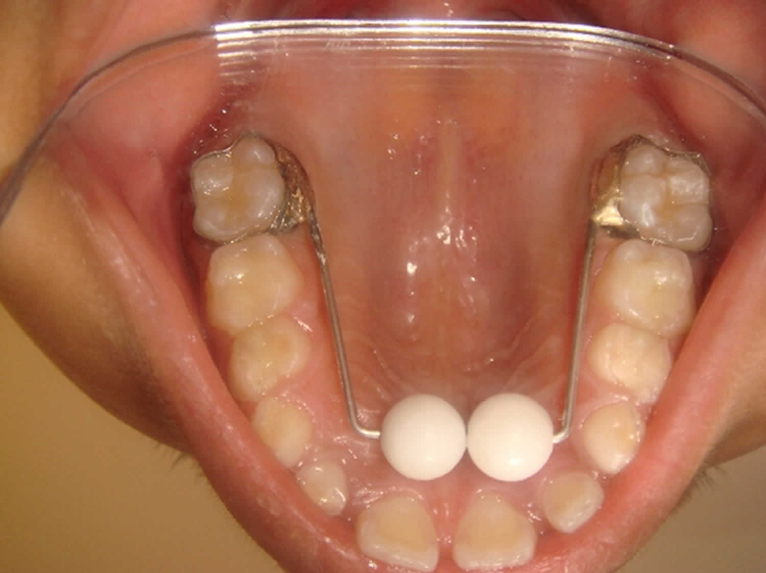
Figure 2. Palatal expander

palatal expander

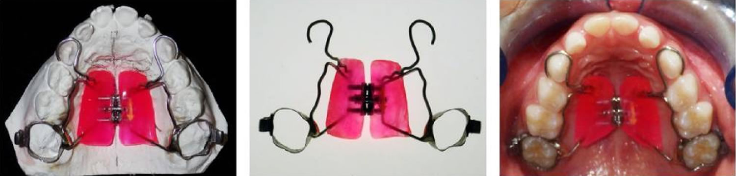
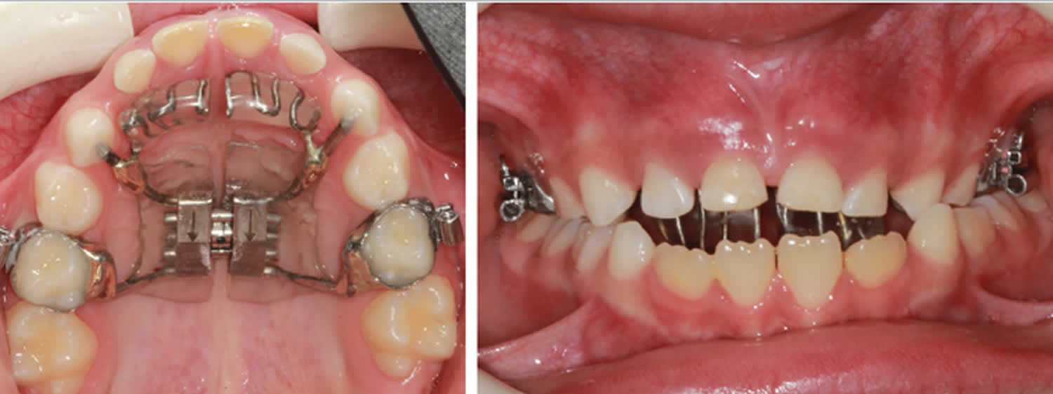
Figure 3. Haas expander with tongue crib

Haas expander with tongue crib

References   [ + ]

read more

Meningococcal infections

meningococcal infections

Meningococcal infections

Meningococcal infection refers to any illness caused by Gram-negative aerobic diplococcus bacteria called Neisseria meningitidis, also known as meningococcus. Neisseria meningitidis (meningococcus) has a typical bean or kidney shape, and is an obligate human pathogen 1). The microorganism frequently colonises the oro- or nasopharynges of even healthy individuals, but can also colonise other parts of the body 2). Meningococcal infections are often severe and can be deadly. They include infections of the lining of the brain and spinal cord (meningitis) and bloodstream infections (bacteremia or septicemia). Meningococcal disease is very serious and can be deadly in a matter of hours. Early diagnosis and treatment are very important.

The Neisseria meningitidis bacteria spread through the exchange of respiratory and throat secretions like spit (e.g., by living in close quarters, kissing). Doctors treat meningococcal disease with antibiotics, but quick medical attention is extremely important. Keeping up to date with recommended vaccines is the best defense against meningococcal disease.

Meningococcal infections causes

Bacteria called Neisseria meningitidis cause meningococcal infection. About 1 in 10 people have these bacteria in the back of their nose and throat without being ill. This is called being ‘a carrier’. Sometimes the bacteria invade the body and cause certain illnesses, which are known as meningococcal disease.

There are six serogroups (types) of Neisseria meningitidis — A, B, C, W, X, and Y — that cause most meningococcal infection worldwide. Three of these serogroups (B, C, and Y) cause most of the illness seen in the United States.

Meningococcal transmission

People spread meningococcal bacteria to other people by sharing respiratory and throat secretions (saliva or spit). Generally, it takes close (for example, coughing or kissing) or lengthy contact to spread these bacteria. Fortunately, they are not as contagious as germs that cause the common cold or the flu. People do not catch the bacteria through casual contact or by breathing air where someone with meningococcal disease has been.

Sometimes the Neisseria meningitidis bacteria spread to people who have had close or lengthy contact with a patient with meningococcal disease. Those at increased risk of getting sick include:

  • People in the same household
  • Roommates
  • Anyone with direct contact with the patient’s oral secretions, such as a boyfriend or girlfriend

Close contacts of someone with meningococcal disease should receive antibiotics to help prevent them from getting the disease. Experts call this prophylaxis. This does not mean that the contacts have the disease; it is to prevent it. Health departments investigate each case of meningococcal disease to identify all close contacts and make sure they receive prophylaxis. People who are not a close contact of a patient with meningococcal disease do not need prophylaxis.

Risk factors for meningococcal infections

Certain people are at increased risk for meningococcal disease. Some risk factors include:

  • Age: Age is an important risk factor for meningococcal pneumonia, which occurs mostly in older individuals > 50 years of age, and is the most common manifestation of meningococcal disease in those aged > 65 years, in contrast to meningococcal meningitis, which occurs predominantly in children and adolescents 3). However, more recent data suggests that the age distribution of meningococcal pneumonia is bimodal, occurring before the age of 30 years and after 60 years of age 4). Although all serogroups of the meningococcus can cause pneumonia, the less common serogroups of the pathogen are more frequently implicated as discussed below 5). Doctors more commonly diagnose meningococcal disease in infants, teens, and young adults.
  • Group settings: Infectious diseases tend to spread wherever large groups of people gather 6) Several college campuses have reported outbreaks of serogroup B meningococcal disease in recent years.
  • Travelers to the meningitis belt in sub-Saharan Africa may be at risk for meningococcal disease.
  • Certain medical conditions: Certain medical conditions and medications may weaken the immune system and increase risk of meningococcal disease.
    • Persistent complement component deficiencies: Complement component deficiencies refer to disorders of the ‘complement system,’ which helps the body fight off infections. Examples of complement component deficiencies include C3, C5-9, properdin, factor H, and factor D. These disorders are very rare and usually genetic.
    • People who take complement inhibitors such as eculizumab (Soliris®) and ravulizumab (Ultomiris™) are also at increased risk for meningococcal disease. Doctors most commonly prescribe complement inhibitors for three rare medical conditions:
    • Atypical hemolytic uremic syndrome, a blood disorder.
    • Paroxysmal nocturnal hemoglobinuria (PNH), a blood disorder
    • Generalized myasthenia gravis, a disorder that leads to muscle weakness
    • Functional or anatomic asplenia: Someone with anatomic asplenia does not have a spleen (for instance, if it was surgically removed). Someone with functional asplenia has a spleen but it doesn’t work the way that it should. People with sickle cell anemia have functional asplenia. The spleen is an important organ for fighting meningococcal infections because it helps produce antibodies and filter bacteria.
    • Human immunodeficiency virus (HIV) infection 7)

Meningococcal prevention

Keeping up to date with recommended vaccines is the best defense against meningococcal disease. Maintaining healthy habits, like getting plenty of rest and not having close contact with people who are sick, also helps.

Although rare, people can get meningococcal disease more than once. A previous infection will not offer lifelong protection from future infections. Therefore, the Centers for Disease Control and Prevention (CDC) recommends meningococcal vaccines for all preteens and teens. In certain situations, children and adults should also get meningococcal vaccines.

Vaccination

Vaccines help protect against all three serogroups (B, C, and Y) of Neisseria meningitidis bacteria most commonly seen in the United States. Like with any vaccine, meningococcal vaccines are not 100% effective. This means there is still a chance you can develop meningococcal disease after vaccination. People should know the symptoms of meningococcal disease since early recognition and quick medical attention are extremely important.

As part of the licensure process, MenACWY and MenB vaccines showed that they produce an immune response. This immune response suggests the vaccines provide protection, but data are limited on how well they work. Since meningococcal disease is uncommon, many people need to get these vaccines in order to measure their effectiveness.

Available data suggest that protection from MenACWY vaccines decreases in many teens within 5 years. Getting the 16-year-old booster dose is critical to maintaining protection when teens are most at risk for meningococcal disease. Available data on MenB vaccines suggest that protective antibodies also decrease quickly (within 1 to 2 years) after vaccination.

There are 2 types of meningococcal vaccines:

  1. Meningococcal conjugate or MenACWY vaccines
  2. Serogroup B meningococcal or MenB (recombinant) vaccines.

The CDC recommends meningococcal vaccination for all preteens and teens. In certain situations, CDC also recommends other children and adults get a meningococcal vaccine. Below is more information about which meningococcal vaccines CDC recommends for people by age.

Talk to your or your child’s clinician about what is best for your specific situation.

Infants

The Centers for Disease Control and Prevention (CDC) recommends a meningococcal conjugate (MenACWY) vaccine for children as young as 2 months old and 10 years old  if they:

  • Have a rare type of immune disorder called complement component deficiency
  • Are taking a type of medicine called a complement inhibitor (for example, Soliris® or Ultomiris®)
  • Have a damaged spleen or their spleen has been removed
  • Have HIV
  • Are traveling to or residing in countries in which the disease is common
  • Are part of a population identified to be at increased risk because of a serogroup A, C, W, or Y meningococcal disease outbreak.

Talk to your child’s clinician to find out if, and when, they will need booster shots.

CDC recommends a serogroup B meningococcal (MenB) vaccine for children 10 years or older if they:

  • Have a rare type of immune disorder called complement component deficiency
  • Are taking a type of medicine called a complement inhibitor (for example, Soliris® or Ultomiris®)
  • Have a damaged spleen or their spleen has been removed
  • Are part of a population identified to be at increased risk because of a serogroup B meningococcal disease outbreak

Preteens and teens

CDC recommends MenACWY vaccination for all 11 through 18 year olds. All 11 to 12 year olds should get a MenACWY vaccine, with a booster dose at 16 years old (since protection decreases over time). This allows teens to continue having protection during the ages when they are at highest risk. Teens may also get a serogroup B meningococcal (MenB) vaccine, preferably at 16 through 18 years old.

While any teen may choose to get a serogroup B meningococcal (MenB) vaccine, certain preteens and teens should get it if they:

  • Have a rare type of immune disorder called complement component deficiency
  • Are taking a type of medicine called a complement inhibitor (for example, Soliris® or Ultomiris®)
  • Have a damaged spleen or their spleen has been removed
  • Are part of a population identified to be at increased risk because of a serogroup B meningococcal disease outbreak

Teens and young adults

Teens and young adults (16 through 23 year olds) may also be vaccinated with a serogroup B meningococcal (MenB) vaccine, preferably at 16 through 18 years old. Healthy teens and young adults who choose to get vaccinated need two doses of the same vaccine brand.

Adults

CDC recommends a MenACWY vaccine for adults if they:

  • Have a rare type of immune disorder called complement component deficiency
  • Are taking a type of medicine called a complement inhibitor (for example, Soliris® or Ultomiris®)
  • Have a damaged spleen or their spleen has been removed
  • Have HIV
  • Are a microbiologist who is routinely exposed to Neisseria meningitidis
  • Are traveling to or residing in countries in which the disease is common
  • Are part of a population identified to be at increased risk because of a serogroup A, C, W, or Y meningococcal disease outbreak
  • Are not up to date with this vaccine and are a first-year college student living in a residence hall
  • Are a military recruit

Talk to your clinician to find out if, and when, you will need booster shots.

CDC recommends a MenB vaccine for adults if they:

  • Have a rare type of immune disorder called complement component deficiency
  • Are taking a type of medicine called a complement inhibitor (for example, Soliris® or Ultomiris®)
  • Have a damaged spleen or their spleen has been removed
  • Are a microbiologist who is routinely exposed to Neisseria meningitidis
  • Are part of a population identified to be at increased risk because of a serogroup B meningococcal disease outbreak.

Who should NOT get Meningococcal vaccines?

Because of age or health conditions, some people should not get certain vaccines or should wait before getting them. Read the guidelines below and ask your or your child’s clinician for more information.

Tell the person who is giving you or your child a meningococcal vaccine if:

  • You or your child have had a life-threatening allergic reaction or have a severe allergy.

DO NOT get a meningococcal vaccine if:

  • You have ever had a life-threatening allergic reaction after a previous dose of that meningococcal vaccine.
  • You have a severe allergy to any part of that vaccine. Your or your child’s clinician can tell you about the vaccine’s ingredients.

You are pregnant or breastfeeding.

  • Pregnant women who are at increased risk for serogroup A, C, W, or Y meningococcal disease may get MenACWY vaccines.
  • Pregnant or breastfeeding women who are at increased risk for serogroup B meningococcal disease may get MenB vaccines. However, they should talk with a clinician to decide if the benefits of getting the vaccine outweigh the risk.

You or your child are not feeling well.

  • People who have a mild illness, such as a cold, can probably get these vaccines. People who have a moderate or severe illness should probably wait until they recover. Your or your child’s clinician can advise you.

Meningococcal vaccines side effects

Most people who get a meningococcal vaccine do not have any serious problems with it. With any medicine, including vaccines, there is a chance of side effects. These are usually mild and go away on their own within a few days, but serious reactions are possible.

Mild Problems

  • MenACWY Vaccines
    • Mild problems following MenACWY vaccination can include:
      • Reactions where the shot was given
      • Redness
      • Pain
      • Fever
      • Muscle or joint pain
      • Headache
      • Feeling tired

If these problems occur, they usually last for 1 or 2 days.

  • MenB Vaccines
    • Mild problems following a MenB vaccination can include:
      • Reactions where the shot was given
      • Soreness
      • Redness
      • Swelling
      • Feeling tired
      • Headache
      • Muscle or joint pain
      • Fever or chills
      • Nausea or diarrhea

If these problems occur, they can last up to 3 to 5 days.

Problems that could happen after getting any injected vaccine

  • People sometimes faint after a medical procedure, including vaccination. Sitting or lying down for about 15 minutes can help prevent fainting, and injuries caused by a fall. Tell the clinician if you or your child feel dizzy, have vision changes, or have ringing in the ears.
  • Some people get severe pain in the shoulder and have difficulty moving the arm where the clinician gave a shot. This happens very rarely.
  • Any medicine can cause a severe allergic reaction. Such reactions from a vaccine are very rare, estimated at about 1 in a million doses. These reactions happen within a few minutes to a few hours after the vaccination.
  • As with any medicine, there is a very remote chance of a vaccine causing a serious injury or death.

Antibiotics

Close contacts of a person with meningococcal disease should receive antibiotics to prevent them from getting sick. Experts call this prophylaxis (pro-fuh-lak-sis). Examples of close contacts include:

  • People in the same household
  • Roommates
  • Anyone with direct contact with a patient’s oral secretions (saliva or spit), such as a boyfriend or girlfriend

Doctors or local health departments recommend who should get prophylaxis.

Meningococcal infection signs and symptoms

Seek medical attention immediately if you or your child develops symptoms of meningococcal infection. Symptoms of meningococcal disease can first appear as a flu-like illness and rapidly worsen. The two most common types of meningococcal infections are meningitis and septicemia (see the sections below). Both of these types of infections are very serious and can be deadly in a matter of hours.

Meningococcal infection diagnosis

Meningococcal disease can be difficult to diagnose because the signs and symptoms are often similar to those of other illnesses. If a doctor suspects meningococcal disease, they will collect samples of blood or cerebrospinal fluid (fluid near the spinal cord). Doctors then send the samples to a laboratory for testing. If Neisseria meningitidis bacteria are in the samples, laboratorians can grow (culture) the bacteria. Growing the bacteria in the laboratory allows doctors to know the specific type of bacteria that is causing the infection. Knowing this helps doctors decide which antibiotic will work best. Other tests can sometimes detect and identify the bacteria if the cultures do not.

Meningococcal infection treatment

Doctors treat meningococcal disease with a number of antibiotics. It is important that treatment start as soon as possible. If a doctor suspects meningococcal disease, they will give the patient antibiotics right away. Antibiotics help reduce the risk of dying.

This preemptive approach includes a third-generation cephalosporin antibiotics such as ceftriaxone or cefotaxime. If culture identifies the organism as penicillin-susceptible, treatment can be switched to penicillin G, although continuing third-generation cephalosporin treatment is also an option 8). For patients who have significant allergies to penicillin and other beta-lactams, chloramphenicol may be an alternative. The duration of antibiotic therapy is usually for five to seven days but can be up to twenty-one days, depending on the patient’s clinical response and culture sensitivity 9).

  • Ceftriaxone dosing is 2 g (50 mg/kg in pediatric patients older than 1 month) intravenously (IV) every 12 hours, and cefotaxime dosing is 2 g (50 mg/kg in pediatric patients older than 1 month) every 6 hours.
    • Third-generation cephalosporins are generally preferable due to high efficacy and easier dosing.
  • Penicillin dosing is 300,000 units/kg per day IV or intramuscularly (IM) with a maximum dose of 24 million units per day. Penicillin is usually given as 4 million units every four hours IV in adults and pediatric patients older than 1 month.
    • High-dose penicillin is recommended for cultures with a sensitivity of penicillin minimum inhibitory concentration 0.1 to 1.0 mcg/mL, although most clinicians will continue using third-generation cephalosporin instead.
  • Chloramphenicol dosing is 50 to 100 mg/kg per day IV with a maximum dose of 4 g per day. It is usually given in divided doses every 6 hours.
    • Serum concentrations requires monitoring due to chloramphenicol toxicity.
    • Recommended therapeutic levels include a trough of 5 to 10 mcg/mL and a peak of 10 to 20 mcg/mL.
  • Dexamethasone dosing is 0.15 mg/kg with a maximum dose of 10 mg every 6 hours.
    • Reportedly improves outcome in meningococcal meningitis
    • Ideally administered 4 hours prior to or concomitantly with antibiotics
    • Not recommended if tuberculosis meningitis is suspected
    • Not recommended if meningococcemia with shock is suspected

Patients with meningococcal infection should also be treated aggressively with supportive care, especially for sepsis or septic shock; this may include intravenous fluid resuscitation and vasopressors such as norepinephrine. Patients who show evidence of DIC may need aggressive hydration, blood transfusions, platelet replacement, and possibly even coagulation factor replacement. Researchers have proposed protein C as an adjuvant treatment, but its use is controversial and currently not commonly used 10).

Depending on how serious the infection is, people with meningococcal disease may need other treatments, including:

  • Breathing support
  • Medications to treat low blood pressure
  • Surgery to remove dead tissue
  • Wound care for parts of the body with damaged skin.

Meningococcal infection prognosis

The mortality rate of meningococcal infection can be as high as 50% in untreated patients. Early and aggressive treatment can reduce the mortality rate to approximately 10 to 14%. Early administration of antibiotics is imperative in determining a good outcome of meningococcal infection. Even with treatment, however, long-term complications can still occur in 11 to 19% of survivors. Complications of meningococcal disease include chronic pain, skin scarring, limb amputation, and neurological impairment ranging from hearing and visual impairments to motor function impairments. Hearing impairment and amputations occur in approximately 3% of cases, arthritis occurs in 10% of cases, and post-infection inflammatory syndrome occurs in 6 to 15% of cases 11).

Similar to other causes of bacterial meningitis, patients who survive meningococcal infections should follow up routinely. Hearing tests within four weeks of hospital discharge is recommended. Orthopedic follow-up and prosthetic fitting are necessary for patients who suffered limb amputations. Patients may even suffer from psychological and psychiatric complications, including post-traumatic stress disorder, depression, and behavioral abnormalities that may need to follow up with psychology and psychiatry 12).

Meningococcal infection complications

Even with antibiotic treatment, 10 to 15 in 100 people infected with meningococcal disease will die. Up to 1 in 5 survivors will have long-term disabilities, such as loss of limb(s), deafness, nervous system problems, or brain damage.

Meningococcal disease complications:

  • Chronic meningococcemia
  • Purpura fulminans
  • Disseminated intravascular coagulation
  • Acute respiratory distress syndrome
  • Superinfections
  • Pericarditis
  • Coma
  • Brain damage
  • Limb loss
  • Tenosynovitis
  • Arthritis-dermatitis syndrome
  • Urethritis
  • Chronic pain
  • Skin scarring
  • Hearing impairment
  • Visual impairment
  • Motor function impairment
  • Depression
  • Post-traumatic stress disorder
  • Behavioral abnormalities

Meningococcal meningitis

Doctors call meningitis caused by the bacteria Neisseria meningitidis meningococcal meningitis. When someone has meningococcal meningitis, the bacteria infect the lining of the brain and spinal cord and cause swelling.

Meningococcal meningitis symptoms

The most common symptoms of meningococcal meningitis include:

  • Fever
  • Headache
  • Stiff neck

There are often additional symptoms, such as:

  • Nausea
  • Vomiting
  • Photophobia (eyes being more sensitive to light)
  • Altered mental status (confusion).

Newborns and babies may not have or it may be difficult to notice the classic symptoms listed above. Instead, babies may be slow or inactive, irritable, vomiting, feeding poorly, or have a bulging in the soft spot of the skull (anterior fontanelle). In young children, doctors may also look at the child’s reflexes for signs of meningitis.

If you or your child has any of these symptoms, see a doctor right away.

Meningococcal meningitis diagnosis

Initial diagnosis of meningococcal meningitis can be made by clinical examination followed by a lumbar puncture showing a purulent spinal fluid. The bacteria can sometimes be seen in microscopic examinations of the spinal fluid. The diagnosis is supported or confirmed by growing the bacteria from specimens of spinal fluid or blood, by agglutination tests or by polymerase chain reaction (PCR). The identification of the serogroups and susceptibility testing to antibiotics are important to define control measures.

Meningococcal meningitis treatment

Meningococcal meningitis treatment requires immediate admission to a hospital. To prevent serious neurologic morbidity and death, prompt institution of antibiotic therapy is essential when the diagnosis of bacterial meningitis is suspected. Surgical interventions may be necessary for the management of complications, such as subdural effusions, empyema, and hydrocephalus.

At presentation, meningitis due to Neisseria meningitidis may be impossible to differentiate from other types of meningitis. Thus, empirical treatment with an antibiotic with effective central nervous system (CNS) penetration should be based on age and underlying disease status, since delay in treatment is associated with adverse clinical outcome.

Initial empirical therapy until the etiology is established should include dexamethasone, a third-generation cephalosporin (eg, ceftriaxone, cefotaxime), and vancomycin. Acyclovir should be considered according to the results of the initial cerebrospinal fluid (CSF) evaluation. Doxycycline should also be added during tick season in endemic areas. A 7-day course of intravenous ceftriaxone or penicillin is adequate for uncomplicated meningococcal meningitis.

If imaging studies are indicated before lumbar puncture, draw blood for culture and begin administration of empiric antibiotics. Administration of empiric antibiotics is unlikely to decrease diagnostic sensitivity if CSF is tested for bacterial antigens early in the course of the illness.

Treatment following diagnosis

Once an accurate diagnosis of meningococcal meningitis is established, appropriate changes can be made. Currently, a third-generation cephalosporin (ceftriaxone or cefotaxime) is the drug of choice for the treatment of meningococcal meningitis and septicemia. Penicillin G, ampicillin, chloramphenicol, fluoroquinolone, and aztreonam are alternatives therapies (Infectious Diseases Society of America guidelines).

The use of dexamethasone in the management of bacterial meningitis in adults remains controversial. It may be used in children, especially in those with meningitis caused by Haemophilus influenzae. In adults with suspected bacterial meningitis, especially in high-risk cases, the adjunctive use of dexamethasone may be beneficial.

Meningococcal septicemia

Meningococcal septicaemia also known as meningococcemia or meningococcal bacteremia, is a bloodstream infection caused by Neisseria meningitidis bacteria. Meningococcemia is a medical emergency. When someone has meningococcal septicemia, the bacteria enter the bloodstream and multiply, damaging the walls of the blood vessels. This causes bleeding into the skin and organs.

People with meningococcemia are often admitted to the intensive care unit (ICU) of the hospital, where they are closely monitored. They may be placed in respiratory isolation for the first 24 hours to help prevent the spread of the infection to others.

Meningococcemia treatments may include:

  • Antibiotics given through a vein immediately
  • Breathing support
  • Clotting factors or platelet replacement, if bleeding disorders develop
  • Fluids through a vein
  • Medicines to treat low blood pressure
  • Wound care for areas of skin with blood clots

In developed countries there is a mortality rate of 10% from meningococcemia and 5% for meningococcal meningitis. Mild neurological complications such as vestibular nerve damage are common but serious brain damage is uncommon.

Figure 1. Meningococcemia rash

Meningococcemia rash

Meningococcal septicemia signs and symptoms

Meningococcal septicemia symptoms may include:

  • Fever and chills
  • Fatigue (feeling tired)
  • Vomiting
  • Cold hands and feet
  • Severe aches or pain in the muscles, joints, chest, or abdomen (belly)
  • Rapid breathing
  • Diarrhea
  • In the later stages, a dark purple rash (see photos)

If you or your child has any of these symptoms, see a doctor right away.

Meningococcemia rash, which may appear as follows:

  • Small, red, flat or raised spots
  • Progression of rash to larger red patches or purple lesions (similar in appearance to large bruises)

Later symptoms may include:

  • A decline in your level of consciousness
  • Large areas of bleeding under the skin
  • Shock

In children older than 1 year, meningococcal meningitis symptoms may include:

  • Fever
  • Neck and/or back pain
  • Headache
  • Nausea and vomiting
  • Neck stiffness
  • A purple-red, splotchy rash or skin discoloration may appear as the disease progresses

In infants, meningococcal meningitis symptoms are difficult to pinpoint and may include:

  • Irritability
  • Listlessness and sleeping all the time
  • Refusing a bottle
  • Crying when picked up or being held
  • Can’t be comforted while crying
  • Bulging fontanel (soft spot on an infant’s head)
  • Behavior changes

The symptoms of meningococcal meningitis and meningococcemia may look like other conditions or medical problems. Always consult your child’s doctor for a diagnosis.

Meningococcal septicemia possible complications

Possible complications of this infection are:

  • Arthritis
  • Bleeding disorder (DIC or disseminated intravascular coagulation)
  • Gangrene due to lack of blood supply
  • Inflammation of blood vessels in the skin
  • Inflammation of the heart muscle
  • Inflammation of the heart lining
  • Shock
  • Severe damage to adrenal glands that can lead to low blood pressure (Waterhouse-Friderichsen syndrome)

Meningococcal septicemia diagnosis

Meningococcal disease can be difficult to diagnose because the signs and symptoms are often similar to those of other illnesses.

In addition to a complete medical history and physical exam, other tests may include:

If a doctor suspects meningococcal disease, they will collect samples of blood or cerebrospinal fluid (CSF) (fluid near the spinal cord).

Lumbar puncture (spinal tap). A special needle is placed into the lower back, into the spinal canal. This is the area around the spinal cord. A small amount of cerebral spinal fluid (CSF) can be removed and sent for testing to determine if there is an infection. Cerebrospinal fluid (CSF) is the fluid that bathes your brain and spinal cord. Doctors then test the samples to see if there is an infection and, if so, what germ is causing it. A Gram stain of cerebrospinal fluid (CSF) may show Gram-negative diplococci. If Neisseria meningitidis bacteria are in the samples, laboratorians can grow (culture) the bacteria. Growing the bacteria in the laboratory allows doctors to know the specific type of bacteria that is causing the infection.

An aspirate (material drawn in negative pressure from a syringe) from petechiae and meningococci can also be cultured from CSF or blood and detected by PCR (polymerase chain reaction). Knowing this helps doctors decide which antibiotic will work best. Other tests can sometimes detect and identify the bacteria if the cultures do not.

  • Blood culture
  • Culture of skin lesions or rash (not common)
  • Other blood tests

Meningococcal septicemia treatment

Prompt treatment is needed for meningococcal infections. Antibiotics (for example, penicillin) are most commonly used. If a patient has severe allergies to penicillin, other antibiotics may be used to treat the infection. Five to seven days of antibiotic therapy is usually effective. A child with meningococcal meningitis or meningococcemia will usually require IV (intravenous) antibiotics and close observation in a hospital or intensive care unit (ICU).

Other treatment for meningococcal infections is supportive (aimed at treating the symptoms present). A child with severe infection may require supplemental oxygen or be put on a ventilator to assist with breathing.

Meningococcemia treatment

  • Benzyl penicillin 2.4 g IV (intravenous) slowly should be given immediately for 7 days.
  • Meningococcal vaccine against serogroup C is available. Meningococcal vaccine that covers some, but not all, strains of meningococcus is recommended for children age 11 or 12. A booster is given at age 16. Unvaccinated college students who live in dormitories should also consider receiving this vaccine. It should be given a few weeks before they first move into the dorm. Talk to your provider about this vaccine.
  • Household, kissing or other close contacts of a case of meningococcal disease should be given oral rifampicin or ciproflaxin, (ciproflaxin should not be given to children), as prophylaxis.
  • Immunization can be offered for a group C disease.

Re-Infection

Although rare, people can get meningococcal disease more than once. A previous infection will not offer lifelong protection from future infections. Therefore, CDC recommends meningococcal vaccines for all preteens and teens. In certain situations, children and adults should also get meningococcal vaccines.

Meningococcal septicemia prognosis

Early treatment results in a good outcome. When shock develops, the outcome is less certain. Even with antibiotic treatment, 10 to 15 in 100 people infected with meningococcal disease will die 13). About 11 to 19 in 100 survivors will have long-term disabilities, such as loss of limb(s), deafness, nervous system problems, or brain damage 14).

The condition is most life threatening in those who have:

  • A severe bleeding disorder called disseminated intravascular coagulopathy (DIC)
  • Kidney failure
  • Shock

Meningococcal pneumonia

Meningococcal pneumonia occurs in between 5% and 15% of all patients with invasive meningococcal disease and is thus the second most common non-neurological organ disease caused by Neisseria meningitidis 15). Meningococcal pneumonia occurs mainly with serogroups Y, W-135 and B. Risk factors for meningococcal pneumonia have not been well characterised, but appear to include older age, smoking, people living in close contact (e.g. military recruits and students at university), preceding viral and bacterial infections, haematological malignancies, chronic respiratory conditions and various other non-communicable and primary and secondary immunodeficiency diseases.

Primary meningococcal pneumonia occurs in 5–10% of patients with meningococcal infection and is indistinguishable clinically from pneumonia caused by other common infectious pathogens. Fever, chills and pleuritic chest pain are the most common symptoms, occurring in > 50% of cases 16). Productive sputum and dyspnoea are less common 17). A rash may occur in patients with pneumonia and associated sepsis, but meningococcaemia is a rare accompaniment of pneumonia 18). Neither laboratory findings nor radiological features allow differentiation from other causes of pneumonia 19).

Diagnosis of meningococcal pneumonia may be made by the isolation of the organism in sputum, blood, or normally sterile site cultures, but is likely to underestimate the frequency of meningococcal pneumonia. If validated, PCR-based techniques may be of value for diagnosis in the future.

Patients who develop only meningococcal pneumonia with bacteremia, but without central nervous system involvement, usually recover with good outcomes and do not develop the catastrophic syndrome seen in meningococcemia 20).

Meningococcal pneumonia treatment

Prior to 1991, penicillin was the treatment of choice for meningococcal infections 21). However, with the emergence of penicillin-resistant strains in 1991 22), in the setting of the high mortality associated with meningococcal disease, the empiric treatment recommendation was changed to a third generation cephalosporin 23). Although not commonly done, should the microorganism be found to be sensitive to penicillin, therapy of meningococcal disease with high-dose penicillin may be considered. Concerningly, isolates of meningococci with decreased susceptibility to penicillin, as well as emerging resistance to other antibiotics, have been recognized in Spain, Europe, South Africa, the US, Canada and Brazil 24), with the latter study also reporting resistance of the meningococcus to ciprofloxacin. Alternative drug choices include meropenem (unavailability of a third generation cephalosporin), chloramphenicol (for severe beta-lactam allergy), aztreonam (if chloramphenicol is unavailable in the case of severe beta-lactam allergy), or a fluoroquinolone, such as moxifloxacin (currently restricted in the US, due to consideration of meningococcal fluoroquinolone resistance and lack of controlled trials in meningococcal disease) 25).

Although the optimal regimen for the treatment of meningococcal pneumonia per se has not been determined, it seems likely to be similar to that of meningococcal disease 26). In fact, it has been noted that most cases of meningococcal pneumonia received penicillin before 1991 and that most received a cephalosporin after that date 27).

Corticosteroids have been used as adjunctive therapy in patients with meningitis, but their benefit appears to be evident in pneumococcal, rather than meningococcal, meningitis. Accordingly, these agents are not usually recommended in the clinical setting of meningococcal disease 28). Although there has been emerging evidence for the benefit of adjunctive corticosteroid therapy in patients with severe community-acquired pneumonia, there are no reports on the possible benefits of corticosteroids in severe meningococcal pneumonia 29).

References   [ + ]

read more

Pilonidal cyst

pilonidal cyst

What is a pilonidal cyst

The pilonidal cyst an abnormal pocket in the skin that usually contains hair and skin debris due to a granulomatous reaction of a hair shaft penetrating the skin. Pilonidal cysts usually occur when hair punctures the skin and then becomes embedded. Hence the name pilonidal (“pilus” meaning hair and “nidal” meaning nest). A pilonidal cyst is almost always located near the tailbone at the top of the cleft of the buttocks, but may occur in the umbilicus.

Males are preferentially affected 3-4:1 females.

If a pilonidal cyst becomes infected, the resulting pilonidal abscess is often extremely painful.

  • An infected pilonidal cyst or abscess requires surgical drainage plus antibiotics. It will not heal with antibiotic medicines alone.
  • The pilonidal cyst can be drained through a small incision or removed surgically. If you continue to have infections, the pilonidal cyst can be surgically removed.
  • Depending on the type of pilonidal cyst surgery required, pilonidal cyst surgery recovery time may take 4 to 10 weeks to heal.

Pilonidal cysts most commonly occur in young men, and the problem has a tendency to recur. People who sit for prolonged periods of time, such as truck drivers, are at higher risk of developing a pilonidal cyst.

Who gets pilonidal disease?

Pilonidal disease affects both men and women usually between the ages of 20–40 years. It is 3–4 times more common in men than women. Other factors that increase the risk of pilonidal disease include:

  • Coarse, curly or crinkly hair
  • Obesity
  • Family predisposition
  • Poor hygiene
  • Prolonged sitting or buttock friction causing increased sweating
  • Repeated local injury (once known as “Jeep rider’s disease” as it hospitalized more than 80,000 US soldiers in WWII)
  • Co-existing hidradenitis suppurativa.

You may help heal the infection and prevent pilonidal cyst from happening again by:

  • keeping your buttock cleft clean and dry
  • using a mild soap and making sure all soap is removed after cleaning
  • shaving the area regularly or using a hair-removal product
  • soaking in a warm tub (sitz bath) a few times a day
  • taking pain relief medication
  • avoiding sitting for long periods.

Figure 1. Pilonidal cyst

Pilonidal cyst

Pilonidal cyst

Figure 2. Pilonidal cyst

pilonidal cyst

Figure 3. Pilonidal cyst with granuloma formation

Pilonidal cyst with granuloma formation

What is pilonidal sinus?

A pilonidal sinus is a small hole or tunnel in the skin at the top of the buttocks (natal cleft), where they divide. This is the cleft between the buttocks just below the base of the spine in the sacrococcygeal region. Pilonidal sinus disease is a chronic skin problem that it is characterized by one or more sinus tracts; these are cavities with a narrow opening on the skin surface (pilonidal sinus). In most cases, the cavity is filled with nests of hair – hence the name pilonidal (“pilus” meaning hair and “nidal” meaning nest). Hairs carry bacteria, which can cause inflammation and infection. A non-inflamed lump is known as a pilonidal cyst. If the pilonidal sinus becomes infected a pilonidal abscess may form or the pus can drain through a tunnel (sinus) out to your skin.

Pilonidal sinus doesn’t always cause symptoms and only needs to be treated if it becomes infected. An infection will cause pain and swelling.

Most people with a pilonidal sinus don’t notice it unless it becomes infected and causes symptoms. An infected pilonidal sinus is red, painful and may bleed or leak pus. These symptoms can develop quickly, often over a few days. They’re signs of pilonidal sinus infection and need treating.

See a doctor if you have a small lump at the top of your bottom (between your buttocks) that’s painful, red, bleeding or leaking pus.

Persistent, complex or recurrent pilonidal sinus disease must be treated surgically. Procedures vary from taking the roof off the sinuses to wide and deep excision (i.e. all affected areas are completely cut out). In all cases, the cavity is scrubbed and scraped out to remove hair and abnormally healing granulation tissue. Several techniques are available for wound healing and closure; these include:

  • Dressing or packing open wounds
  • Marsupialization (forming a pouch), which results in a smaller wound compared to wounds that are left open to granulate
  • Closure using skin flaps for wide excisions.

Figure 4. Pilonidal sinus

Pilonidal sinus

Pilonidal cyst complications

If a chronically infected pilonidal cyst isn’t treated properly, you may be at slightly increased risk of developing a type of skin cancer called squamous cell carcinoma (SCC).

Pilonidal cyst causes

The exact cause of pilonidal cysts isn’t clear. But most pilonidal cysts appear to be caused by loose hairs that penetrate the skin. Friction and pressure — skin rubbing against skin, tight clothing, bicycling, long periods of sitting or similar factors — force the hair down into skin. Responding to the hair as a foreign substance, the body creates a cyst around the hair.

This explanation accounts for rare cases of pilonidal cysts that occur in parts of the body other than near the tailbone. For example, barbers, dog groomers and sheep shearers have developed pilonidal cysts in the skin between fingers.

Risk factors for developing pilonidal cysts

Certain factors can make you more susceptible to developing pilonidal cysts, such as:

  • Male sex
  • Younger age (pilonidal cysts are most common in people in their 20s)
  • Obesity
  • Inactive lifestyle
  • Occupation requiring prolonged sitting
  • Excess body hair
  • Stiff or coarse hair

Pilonidal cyst prevention

To help prevent pilonidal cysts, try to:

  • Keep the area clean
  • Lose weight if needed
  • Avoid prolonged sitting

If you’ve had pilonidal cysts in the past, you might want to regularly shave the area or use hair removal products to reduce the risk of recurrence.

Pilonidal cyst symptoms

Signs and symptoms can vary from a small painless pit or dimple at the base of the spine to a large painful abscess. Most patients have progressive tenderness, particularly after prolonged periods of sitting, such as during a long drive.

When it’s infected, a pilonidal cyst becomes a swollen mass (abscess). Signs and symptoms of an infected pilonidal cyst include:

  • Pain, redness and swelling
  • Reddening of the skin
  • Small hole or holes draining fluid that may be clear, cloudy (pus) or bloody
  • If infected, the draining pus may have a foul odor
  • Fever, malaise or nausea
  • Visible or lumpy tracts 2–5 cm long in chronic or recurrent pilonidal disease.

How pilonidal cyst diagnosis made?

The clinical features of pilonidal sinus is usually straightforward. If necessary, skin biopsy can be undertaken. The histopathological features of pilonidal sinus characteristically show foreign body reaction.

Pilonidal cyst treatment

A pilonidal cyst that isn’t causing any problems doesn’t require any treatment. The patient should be advised to keep the area clean and free of hair by shaving or using a hair removal agent every 2–3 weeks. The cyst may resolve itself. Persistent and inflamed cysts (acute pilonidal abscess) are incised (cut into) and drained out to reduce inflammation and pain. Occasionally the abscess cavity may be cut out completely to remove hair nests and skin debris; this reduces the rate of recurrence to about 15%.

Persistent, complex or recurrent pilonidal sinus disease must be treated surgically. Procedures vary from taking the roof off the sinuses to wide and deep excision (i.e. all affected areas are completely cut out). In all cases, the cavity is scrubbed and scraped out to remove hair and abnormally healing granulation tissue.

Several techniques are available for wound healing and closure; these include:

  • Dressing or packing open wounds
  • Marsupialisation (forming a pouch), which results in a smaller wound compared to wounds that are left open to granulate
  • Closure using skin flaps for wide excisions.

How to get rid of a pilonidal cyst

The initial treatment for an infected pilonidal cyst is usually a procedure that can be performed in your doctor’s office. After numbing the area with an injection, your doctor makes a small incision to drain the cyst. If the cyst recurs, which often happens, you may need a more extensive surgical procedure that removes the cyst entirely.

After surgery, your doctor may choose to:

  • Leave the wound open. In this option, the surgical wound is left open and packed with dressing to allow it to heal from the inside out. This process results in a longer healing time but usually a lower risk of a recurring pilonidal cyst infection.
  • Close the wound with stitches. While the healing time is shorter with this option, there’s a greater risk of recurrence. Some surgeons make the incision to the side of the cleft of the buttocks, where healing is particularly difficult.

Wound care is extremely important after surgery. Your doctor or nurse will give you detailed instructions on how to change dressings, what to expect of a normal healing process and when to call the doctor. You may also need to shave around the surgical site to prevent hairs from entering the wound.

Pilonidal cyst removal surgery

There are several types of pilonidal cyst surgery.

Incision and drainage: This is the most common treatment for an infected pilonidal cyst. It is a simple procedure done in the doctor’s office.

  • Local anesthesia is used to numb the skin.
  • A cut is made in the cyst to drain fluid and pus. The hole is packed with gauze and left open.
  • Afterward, it can take up to 4 weeks for the cyst to heal. The gauze has to be changed often during this time.

Pilonidal cystectomy: If you keep having problems with a pilonidal cyst, it can be removed surgically. This procedure is done as an outpatient procedure, so you will not need to spend the night in the hospital.

  • You may be given medicine (general anesthesia) that keeps you asleep and pain-free. Or, you may be given medicine (regional anesthesia) that numbs you from the waist down. In rare cases, you may only be given local numbing medicine.
  • A cut is made to remove the skin with the pores and the underlying tissue with the hair follicles.
  • Depending on how much tissue is removed, the area may or may not be packed with gauze. Sometimes a tube is placed to drain fluid that collects after surgery. The tube is removed at a later time when the fluid stops draining.

It may be hard to remove the entire cyst, so there is a chance that it will come back.

  • Pilonidal cysts come back in about half of the people who have surgery the first time. Even after a second surgery, it may come back.

After the pilonidal cyst surgery:

  • You can go home after the pilonidal cyst surgery on the same day
  • The wound will be covered with a bandage
  • You will get pain medicines
  • It is very important to keep the area around the wound clean
  • Your provider will show you how to care for your wound
  • After it heals, shaving the hair in the wound area may help prevent pilonidal disease from coming back

Why is the pilonidal cyst surgery performed?

Surgery is needed to drain and remove a pilonidal cyst that does not heal.

  • Your doctor may recommend this procedure if you have pilonidal disease that is causing pain or infection.
  • A pilonidal cyst that is not causing symptoms does not need treatment.

Non-surgical treatment may be used if the area is not infected:

  • Shaving or laser removal of hair around the pilonidal cyst
  • Injection of surgical glue into the pilonidal cyst

Risks for pilonidal cyst surgery

Pilonidal cyst resection is generally safe. Ask your doctor about these complications:

  • Bleeding
  • Infection
  • Taking a long time for the area to heal
  • Having the pilonidal cyst come back

Pilonidal cysts come back in about half of the people who have surgery the first time. Even after a second surgery, it may come back.

read more

Hemifacial microsomia

hemifacial microsomia

Hemifacial microsomia

Hemifacial microsomia also called craniofacial microsomia, first and second branchial arch anomaly, branchial arch syndrome, facioauriculovertebral syndrome, oculoauriculovertebral spectrum, or lateral facial dysplasia, is a term used to describe a spectrum of congenital underdevelopment of the tissues on one side of the face that primarily affect the development of the skull (cranium) and face before birth 1). Hemifacial microsomia primarily affects the ear, mouth and jaw areas, though it may also involve the eye, cheek, neck and other parts of the skull, as well as nerves and soft tissue. In 10 to 15 percent of cases, both sides of the face are affected, often times asymmetrically.

Microsomia means abnormal smallness of body structures. Most people with hemifacial microsomia or craniofacial microsomia have differences in the size and shape of facial structures between the right and left sides of the face (facial asymmetry). In about two-thirds of cases, both sides of the face have abnormalities, which usually differ from one side to the other. Other individuals with hemifacial microsomia are affected on only one side of the face. The facial characteristics in hemifacial microsomia typically include underdevelopment of one side of the upper or lower jaw (maxillary or mandibular hypoplasia), which can cause dental problems and difficulties with feeding and speech. In cases of severe mandibular hypoplasia, breathing may also be affected.

Most children with hemifacial microsomia have facial anomalies but no other major medical issues. In some cases, babies born with hemifacial microsomia may also have other health problems such as malformed vertebrae, heart defects or abnormally shaped kidneys.

Hemifacial microsomia is typically nonprogressive, meaning that the areas of the face that are affected at birth will typically remain similarly affected throughout growth and development, neither worsening nor getting better.

Hemifacial microsomia is the second most common facial birth defect behind cleft lip and palate, affecting one in every 3,500 to 5,600 births 2). However, this range may be an underestimate because not all medical professionals agree on the criteria for diagnosis of hemifacial microsomia and because mild cases may never come to medical attention. For reasons that are unclear, hemifacial microsomia occurs about 50 percent more often in males than in females.

Hemifacial microsomia is sometimes confused with Goldenhar syndrome, a rare congenital condition. People with hemifacial microsomia and noncancerous (benign) growths in the eye called epibulbar dermoids may be said to have Goldenhar syndrome or oculoauricular dysplasia. In fact, hemifacial microsomia is just one of the distinctive characteristics of Goldenhar syndrome, which also includes spine anomalies and epibulbar dermoids or lipodermoids.

Hemifacial microsomia treatment is complex and usually requires multiple stages. The skull, eye socket and cheek bones are generally reconstructed when children are young. Timing for jaw reconstruction will vary depending on whether there are breathing or feeding problems. Ear reconstruction usually occurs in the school age years.

Children with hemifacial microsomia require long-term follow up by a multidisciplinary team. This is a complex condition and experts from different medical, surgical, and dental specialties need to work together to provide the best care for your child. Several operations may be needed as children grow.

It is very important for children with hemifacial microsomia to receive dental care from providers experienced in caring for children with craniofacial conditions.

Figure 1. Diverse manifestations of hemifacial macrosomia

Diverse manifestations of hemifacial macrosomia

Footnote: The photographs show patients with varying degrees of hemifacial microsomia. In image A, the patient presents a Grade I mandibular disorder, with slight deviation of the lower third. Image B corresponds to a Grade IIA alteration, presenting alteration of symmetry, both at the lower third and the orbits. Image C shows an alteration of the lower third, with full involvement of the right auricle, corresponding to a patient with a Grade IIB anomaly. Image D corresponds to a patient with a Grade III alteration of the left side.

Figure 2. Hemifacial microsomia baby

Hemifacial microsomia baby

Figure 3. Hemifacial microsomia teen

hemifacial microsomia

Figure 4. Hemifacial microsomia in adults

Hemifacial microsomia in adults

Footnote: Total ear reconstruction in a woman suffering from a severe hemifacial microsomia type 3 with an auricular dysplasia Weerda grade III on the left side, using a porous polyethylene framework and a temporoparietal fascia flap. (A and B) Preoperatively. (C and D) 1 year postoperatively

[Source 3) ]

Hemifacial microsomi causes

It is unclear what causes or genes are involved in hemifacial microsomia. Hemifacial microsomia results from problems in the development of structures in the embryo called the first and second pharyngeal arches (also called branchial or visceral arches). Tissue layers in the six pairs of pharyngeal arches give rise to the muscles, arteries, nerves, and cartilage of the face and neck. Specifically, the first and second pharyngeal arches develop into the lower jaw, the nerves and muscles used for chewing and facial expression, the external ear, and the bones of the middle ear. Interference with the normal development of these structures can result in the abnormalities characteristic of hemifacial microsomia.

There are several factors that can disrupt the normal development of the first and second pharyngeal arches and lead to hemifacial microsomia. Research has shown the process starts in the first trimester of pregnancy and may be caused by a vascular problem leading to poor blood supply to the fetus’ face during early development 4). The facial anomalies are not triggered by a mother’s action or diet.

Some individuals with hemifacial microsomia have chromosomal abnormalities such as deletions or duplications of genetic material; these individuals often have additional developmental problems or malformations. Occasionally, hemifacial microsomia occurs in multiple members of a family in a pattern that suggests inheritance of a causative gene mutation, but the gene or genes involved are unknown. In other families, individuals seem to inherit a predisposition to the disorder. The risk of hemifacial microsomia can also be increased by environmental factors, such as certain drugs taken by the mother during pregnancy. In most affected individuals, the cause of the disorder is unknown.

In the majority of hemifacial microsomia cases, the condition is not inherited, and happens by chance. In a small minority of cases, a child may inherit the condition from his parents. An adult with hemifacial microsomia has about a 3 percent chance or less of having a child with the same condition.

It is not well understood why certain disruptions to development affect the first and second pharyngeal arches in particular. Researchers suggest that these structures may develop together in such a way that they respond as a unit to these disruptions.

Hemifacial microsomia inheritance pattern

Hemifacial microsomia most often occurs sporadically (by chance) in a single individual in a family and is not inherited. If the condition is caused by a chromosomal abnormality, it may be inherited from one affected parent or it may result from a new abnormality in the chromosome and occur in people with no history of the disorder in their family.

In 1 to 2 percent of cases, hemifacial microsomia is inherited in an autosomal dominant pattern, which means one copy of an altered gene in each cell is sufficient to cause the disorder. In rare cases, the condition is inherited in an autosomal recessive pattern, which means both copies of a gene in each cell have mutations. The parents of an individual with an autosomal recessive condition each carry one copy of the mutated gene, but they typically do not show signs and symptoms of the condition. The gene or genes involved in hemifacial microsomia are unknown.

In some affected families, people seem to inherit an increased risk of developing hemifacial microsomia, not the condition itself. In these cases, some combination of genetic changes and environmental factors may be involved.

Hemifacial microsomi signs and symptoms

People with hemifacial microsomia usually have ear abnormalities affecting one or both ears, typically to different degrees. They may have growths of skin (skin tags) in front of the ear (preauricular tags), an underdeveloped or absent external ear (microtia or anotia), or a closed or absent ear canal; these abnormalities may lead to hearing loss. Eye problems are less common in craniofacial microsomia, but some affected individuals have an unusually small eyeball (microphthalmia) or other eye abnormalities that result in vision loss.

Abnormalities in other parts of the body, such as malformed bones of the spine (vertebrae), abnormally shaped kidneys, and heart defects, may also occur in people with craniofacial microsomia.

The following structures are involved in hemifacial microsomia:

  • Jaw and temporomandibular joint (TMJ): asymmetrical mandibular development for hypoplasia, absence of mandibular structures (condyle and ramus), absence or ankylosis of temporomandibular joint (TMJ). Recently published studies showed that hemifacial microsomia patients presented both mandibular and maxillary retrusion in comparison with the control group, along with an increase in the vertical component; these patterns were more marked in the affected side and increased according to severity 5). Mandibular height was always lower along growth, but the growth pattern was similar in both groups 6). In terms of temporomandibular joint (TMJ), it has been observed that the degree of mandible dysplasia does not correspond to the degree of disk dysplasia, which vary among individuals; while the unaffected side does not present major alterations 7)
  • Orbit: orbit dystopia (bad position), epibulbar dermoid, anophthalmia/microphthalmia, blepharoptosis, retinal or choroidal coloboma, among other less frequent anomalies 8)
  • Ears: microtia, anotia, loss of hearing, disorders of the middle ear.
  • Cranial nerves: involvement of facial nerve and, in more severe cases, of the trigeminal and hypoglossal nerves.
  • Dental: agenesis 9), dental hypoplasias 10), microdontia, and malocclusions. Delayed tooth development in hemifacial microsomia patients type IIB and III 11), with the most alterations in posterior teeth 12)
  • Maxillofacial: labio-palatal fissure 13), macrostomia, hypoplasia of the facial thirds, occlusal plane inclination (highly variable in angle), hypoplasia of masticatory muscles 14), velopharyngeal insufficiency 15)
  • Extracranial changes: primarily in kidney, lungs, heart, gastrointestinal, skeletal, and central nervous system (CNS) 16)

Symptoms of hemifacial microsomia range from severe to barely noticeable and depend greatly on the degree of deformity and how much of the face is involved. A child with a mild form of hemifacial microsomia may have a slightly smaller jaw and a skin tag in front of a normal-looking ear. In more severe forms, a child’s face may appear much smaller on one side of his face, with an abnormally shaped or absent ear.

One of the ways clinicians describe and assess the severity of hemifacial microsomia is the OMENS classification 17). The OMENS classification examines the function and appearance of each of the following, looking for characteristics commonly associated with hemifacial microsomia:

  • Orbit (eye socket): small and underdeveloped eyes with impaired vision; absent or unformed eye; growths on the eye; one eye appearing smaller than the other, but with normal vision
  • Mandible (the jaw bones): underdeveloped upper and lower jaw on one side; crooked jaw; missing, misaligned or overcrowded teeth; cleft lip and/or cleft palate; limited opening or closing of the mouth
  • Ear: small skin tags; misshapen or missing external ear; absent or abnormal development of the ear canal resulting in partial or total hearing loss
  • Nerves: ranging from mild weakness to partial or full facial paralysis
  • Soft tissues (skin, muscle, fat, tendons and ligaments): flattened forehead or cheekbone, unequal cheek fullness, asymmetrical mouth with lateral cleft.

Table 1. Prevalence of selected anomalies in hemifacial microsomia

Anomalies Prevalence 1
Principal Mandible Mandibular hypoplasia 49%-100%
Malformed glenoid fossa 24%-27%
Ear Microtia 66%-99%
Preauricular tags 34%-61%
Conductive hearing loss 50%-66%
Ocular Orbital dystopia 15%-43%
Epibulbar dermoids 4%-35%
Nerve VII nerve palsy 10%-45%
Soft tissue Masticatory muscle hypoplasia 85%-95%
Macrostomia 17%-62%
Associated
craniofacial
Velopharyngeal insufficiency 35%-55%
Palatal deviation 39%-50%
Cranial skull base abnormalities 9%-30%
Cleft lip and/or palate 15%-22%
Coloboma of the upper eyelid 4%-25%
Hypodontia/dental hypoplasia 8%-25%
Lacrimal duct atresia/stenosis 11%-14%
Frontal plagiocephaly 10%-12%
Sensorineural hearing loss 6%-16%
Preauricular sinus 6%-9%
Other Vertebral/rib 16%-60%
Cervical spine 21%-42%
Scoliosis 11%-26%
Cardiac 4%-33%
Pigmentation 13%-14%
Limb 3%-21%
Central nervous system 5%-18%
Genitourinary 4%-15%
Pulmonary 1%-15%
Gastrointestinal 2%-12%
[Source 18) ]

Hemifacial microsomi diagnosis

Hemifacial microsomia diagnosis is mainly clinical, although various complementary tests allow a better analysis of this pathology. The diagnosis of hemifacial microsomia can be made before or after birth. Some of the abnormal facial features are visible during prenatal ultrasound. The majority of children are not diagnosed with hemifacial microsomia until after birth. In this case, craniofacial experts in your hospital will evaluate your child. Experienced physicians will make the diagnosis based on your child’s appearance: the mandibular (jaw) deformity is the hallmark of hemifacial microsomia, and is classified based on the development of the jaw. In the mildest formation, the mandible is nearly normal and only slightly hypoplastic (underdeveloped), while in the most severe cases, a portion of the jaw is missing on the affected side.

Some of the facial characteristics of hemifacial microsomia mimic those seen in children with Treacher Collins syndrome; but hemifacial microsomia differences are typically one-sided or asymmetric, whereas in Treacher Collins, characteristics are similar on both sides of the face.

Diagnostic tests such as X-rays and CT scans may also be used to better examine your child’s bone and cartilage structure in order to make appropriate treatment recommendations.

Hemifacial microsomi treatment

The treatment of hemifacial microsomia varies tremendously from patient to patient and depends on the severity of the condition and long-term needs of the child. Consultation with an experienced craniofacial team is extremely important in achieving the best outcomes for your child 19).

As is the case for any patient with a complex craniofacial deformity, individual treatment varies depending upon the degree of involvement of the various structures. Your child’s individual treatment plan may vary from others similarly affected due to a variety of other factors. It is important that you see an experienced craniofacial team to manage and assess your child’s condition.

Mainly through plastic/orthognathic surgery and orthodontics, the treatment seeks to improve functionality, along with optimum facial symmetry, in order to 20):

  1. Increase the size of the affected mandibular side and its associated soft tissue.
  2. Create a joint simulating the temporomandibular joint (TMJ) in cases where it is absent.
  3. Correct secondary deformities in maxilla.
  4. Achieve functional occlusion, as well as aesthetic facial and dental appearance.
  5. Improve and horizontalize the occlusal plane.
  6. Achieve mouth opening if it is limited.

Conventional orthodontic treatment may initially include functional appliances with the use of rigid acrylic activators, which are individualized according to each case. These devices allow for expansion of affected tissue, taking advantage of patient′s physiological growth. Sometimes they can have height planes on the healthy side, allowing for vertical compensation of the affected area, always bearing in mind that facial midline should be centered with tooth midline. This can later be complemented with conventional fixed orthodontics.

Supportive treatment at birth

If your child is born with hemifacial microsomia, he may require respiratory support or a tracheostomy if the jaw is severely deficient. In most cases, your child’s airway can be managed conservatively.

Due to the presence of the jaw deformity and clefts, your child may experience feeding difficulties. He may receive supplemental feedings through a nasogastric tube to support his growth and weight gain.

If facial paralysis or eyelid abnormalities are present, eye closure may be incomplete and eye protection must be provided either via lubricants or surgical procedures.

Hemifacial microsomia surgery

As your child with hemifacial microsomia grows, he may need surgical treatment based on the severity and area affected. Not all children with hemifacial microsomia have problems in all of these areas.

Being a malformation with abundant phenotypic variation, hemifacial microsomia patients may need other surgeries, depending on the involved structures. A review of craniofacial microsomia performed by Birgfeld and Heike in 2012 21) shows a timeline as a guide in the medical and surgical management of hemifacial microsomia patients, proposed by members of the Seattle Children′s Craniofacial Center, monitoring its progress from birth to adulthood.

Below are some of the interventions your child may need:

  • Ears: Some children with abnormally-shaped or missing ears may choose to have a series of reconstructive surgeries to make the ear appear more normal. The first surgery typically occurs after age 6, when your child’s ears have almost reached adult-size. Another option is to make an artificial or prosthetic ear, which also requires several surgeries. In patients with hearing loss, the possibility of repairing atresia of the auditory canal should be evaluated.
  • Orbit and eyelid differences: It may involve surgeries of epibulbar dermoid and eyelid coloboma. In cases of changes in size or position of the orbit, the replacement is done around the age of 3 to 4 years. For children with eyelid differences, surgical procedures to reposition the lower lids and corners of the eyes may be required.
  • Soft tissue deficiencies: Children with skin, cheek and other soft tissue deficiencies may need augmentation procedures such as fat grafting or tissue transfer.
  • Facial nerve: In cases of nerve palsy, the area and degree of involvement should be evaluated; this involvement may be upper (affects the temporal and zygomatic area), lower (buccal, mandibular and cervical area), or total. In the case of oral involvement, the need for facial reanimation should be evaluated. In case of alteration of eyelid movement, some type of treatment should be considered in order to prevent corneal keratitis by exposure.
  • Cleft lip or cleft palate: Babies born with cleft lip or palate can have surgical repairs done during the child’s first year. Cleft lip repair is typically performed when your child is 3 to 6 months old, while cleft palate surgery is generally performed when your child is about a year old.
  • Lateral facial cleft: A lateral facial cleft is one of the most severe deficiencies found with hemifacial microsomia. It requires a staged reconstruction, similar to the process used in routine repair of cleft lip and palate. In this procedure, surgeons will create a ring of muscle around your child’s mouth, connecting the corners and drawing up the lateral line of the lower lip. This reconstructive procedure also helps with feeding and speaking.
  • Bony deficiencies: In mild cases of bony deficiencies, no treatment may be needed. In more severe cases, surgery may be required. Two of the most commonly performed procedures include distraction lengthening of the mandible (most common) and reconstruction of the mandible with a rib or free vascularized fibula graft (less common). For a distraction lengthening of the mandible, a surgeon cuts the mandible (jaw) in the center of its deficient region and implants a small device that allows the two bone segments to be distracted (pulled apart), creating a gap in the bone. New bone begins to form in the gap of the small jaw and the device is slowly widened until the jaw is appropriately-sized. When the jaw bone has been adjusted, it improves facial form and the way the top and bottom teeth fit together. Mandibular distraction may need to be repeated as your child grows.

Mandibular surgery

While the literature is broad in terms of techniques and surgical times, the treatment of hemifacial microsomia patients can be divided into two groups, according to the classification by Kaban-Prusansky: Grade I and Grade IIA patients are treated in the same way, while Grade IIB and Grade III patients are treated similarly among them but different to the first group 22).

Surgical management of the mandible is essential, and mandible bone distraction has several advantages over costochondral graft. These include: increase mandible vertical length, improve asymmetry of soft tissue, produce less blood loss, have better control of progress vector, and obtain a substantial improvement in the biomechanics of the lower mandible 23).

Bone distraction is based on the principle of tension-stress to allow elongation of bone and soft tissue from the controlled separation of bone segments. There are different types of distraction, which can be classified into two areas. According to location, distractors can be extraoral and intraoral; while, according to the amount of vectors with which they work, they can be univectorial or unidirectional or bi-directional (horizontal and vertical plane) or multidirectional (horizontal, vertical and transversal plane) 24).

The choice of distraction appliance depends on various factors, including patient′s age, the degree of malformation severity and the need for mobility of segments. In general, extraoral appliances require a less complex surgery, can achieve longer distraction distances, and allow handling the distraction in the three directions of space, but they leave scars on the skin, are more visible, and may be heavier, presenting patient discomfort. Intraoral devices are less visible and don′t leave facial scars, but have a limited range of motion in space, and their surgery is more complex for both insertion and removal. Re-absorbable devices have recently been developed, allowing for mandibular distraction in a single stage, and achieving progress of 15 to 30 mm 25). In anyways, the surgical simulation in a solid model can help achieve better mandible symmetry, especially in bone distraction with multivector 26).

It is important to stress the need to take into account, besides patient′s growth state, his or her treatment needs, since they vary among patients, and therefore each must be analyzed as a unique case.

Complications

A 2009 literature review 27) compared the stability and complications of bone distraction and the costochondral graft. It found out that both techniques have a similar relapse rate when performing mandibular advancement of 6 to 10 mm. While this review suggests that mandibular bone distraction is less susceptible to major complications, it also points out the it may produce minor complications that also cause patient morbidity, in addition to being affected by other factors such as a long phase of consolidation, distractor cost, patient compliance in activating the distractor and the need for a second surgery to remove the distractor.

Follow-up care

As your child with hemifacial microsomia grows into adolescence, he or she should continue to be monitored by experienced physicians who can adjust treatment plans as needed.

Because multiple body systems may be involved in hemifacial microsomia, continued monitoring for complications and any treatment as needed are important to optimal long-term outcomes.

The many specialists your child may need to see, including:

  • A plastic surgeon to manage the stages of surgical repair
  • An otolaryngologist (ear, nose and throat specialist) to monitor and treat any nose or throat issues
  • A speech therapist to address any speech problems
  • An expert from the Pediatric Feeding and Swallowing Center to address any feeding-related issues
  • A dentist and/or orthodontist to assess dental health, crowding of teeth and how well the jaw fits together
  • A psychologist or social worker to address emotional and psychological issues related to appearance differences and any other concerns
  • An orthopedic doctor if your child has cervical spine issues
  • A nephrologist if your child has any kidney abnormalities
  • A cardiologist is your child has any heart issues

During follow-up visits, diagnostic testing may be done. The goal of continued monitoring is to help spot any irregularities in growth or development and to address health issues as they develop, optimizing long-term outcomes for your child.

References   [ + ]

read more

Tibial torsion

tibial torsion

Tibial torsion

Tibial torsion is an internal or medial rotation of the shin bone (the tibia bone that is located between the knee and the ankle) relative to the upper leg bone (femur). Tibial torsion causes the child’s feet to turn inward, or have what is also known as a “pigeon-toed” appearance. Tibial torsion is typically seen among toddlers (2 to 4-year-old age group) and as the child grows taller, the tibia usually untwists and the condition usually resolves by age 8. Males and females are affected equally, and about two thirds of patients are affected bilaterally 1).

Normally, lateral rotation of the tibia increases from approximately 5º at birth to approximately 15º at maturity 2). Whereas medial tibial torsion improves with time, lateral torsion often worsens because the natural progression is toward increasing external torsion. The ability to compensate for tibial torsion depends on the amount of inversion and eversion present in the foot and on the amount of rotation possible at the hip. Internal torsion causes the foot to adduct, and the patient tries to compensate by everting the foot, externally rotating at the hip, or both. Similarly, persons with external tibial torsion invert at the foot and internally rotate at the hip 3).

The natural history of femoral torsion is to resolve by the time the patient is aged 8 to 10 years. Beyond this age, all remodeling will have occurred, and any further correction is due to a conscious modification of posture 4). However, that does not mean that the child won’t continue to have some intoeing. The child generally grows up to have legs that resemble those of the parent from whom they inherited the trait.

While occasional tripping may occur, most children learn to compensate for any rotation and have no symptoms. There is no need to restrict activities. Many studies have suggested that intoeing may even improve sports function, as intoers tend to be more effective runners and jumpers.

Bars, shoes, orthotics, and twister cables were used in the past to treat intoeing, but there is no scientific evidence that these devices have any effect on the natural tendency toward partial or complete correction by the 8 to 10 years of age.

Figure 1. Leg bones

Normal leg bones

Tibial torsion causes

Tibial torsion can occur due to the position of the baby in the uterus. Tibial torsion also has a tendency to run in families. Typically, a child’s walking style looks like that of his or her parents.

In a study by Mullaji et al 5) to determine tibial torsion norms, individuals in India were found to have less tibial torsion than Caucasians but about the same amount as the Japanese population. The differences in normal tibial torsion values are expected to be caused by the different lifestyles and postures of the different populations, such as cross-legged sitting positions 6).

Tibial torsion signs and symptoms

Tibial torsion, the most common cause of in-toeing. When the child is first learning how to walk, tibial torsion can create an intoeing appearance. As the feet toe in, the legs look like they are bowed. The bowed leg stance actually helps children achieve greater balance as they stand. Their balance is not as steady when they try to stand and walk with their feet close together or with their feet turned out. Besides the cosmetic concerns, internal tibial torsion can predispose kids to tripping and falling, mostly at the end of the day and when they are tired. Children tend to compensate for their tibial torsion by turning their feet outwards (making the feet parallel) during gait. This can give the false impression of geno varum, or bowlegs, because the patella is facing outwards during the gait and creates a “false deformity” during gait.

Parents are generally more concerned about in-toeing than the children are. Severe in-toeing can cause the child to trip or run awkwardly, and it can interfere with participation in sports. Excessive wear is seen along the lateral border of the shoe, mainly in the front half, because the child uses this as the presenting border of the foot on the heel- or foot-strike.

Tibial torsion signs and symptoms include:

  • When viewing the child standing, the foot and lower leg appear to be rotated internally. If there is an isolated problem, the kneecaps appear to be straight, thus distinguishing this condition from femoral anteversion in which the kneecaps are pointed in.
  • When walking or running the feet excessively turn in occasionally causing tripping and falling. During running the kneecaps continue to stay straight.
  • At the end of the day when fatigue sets in the in toe appear to be worse. Maybe asymmetrical (one side worse than the other).
  • May appear to be bowlegged (because the musculature in the calf is rotated towards the outside of the lower leg).
  • Could be associated with metatarsus adductus in an infant.

Tibial torsion diagnosis

Tibial torsion diagnosis is based on clinical findings, and other investigations generally are not required. Imaging studies may be helpful. However, not every child who undergoes an evaluation because of torsional issues requires any or all imaging tests.

Physical examination

Physical examination must include tests to exclude hip dysplasia, hip and ankle ranges of motion, and knee varus or valgus, which can cause apparent errors in examination.

A rotational profile consists of the following 7):

  • Foot progression angle (FPA) 8)
  • Tibial version or torsion – Thigh-foot angle (TFA), transmalleolar angle
  • Femoral anteversion (hip rotation)
  • Shape of the foot

The foot progression angle (FPA) is the angular difference between the axis of the foot and the line of progression.

Foot progression angle (FPA):

  • a rough measurement which is obtained during gait by observing the angle of the foot off of the line of progression;
  • note that severe foot deformities (club foot) which interfere with the usual measurement;
  • normal values: normal FPA is 10-15° of external rotation ;

By convention, external rotation values are positive, and internal rotation values are negative. Degrees of in-toeing are as follows:

  • Mild is –5 to –10°
  • Moderate is –10 to –15°
  • Severe is more than –15°

Tibial version or torsion is the degree of rotation of the tibia along its long axis from the knee to the ankle. It is measured with the patient prone with his or her knees flexed to 90°. It is assessed by using two measures, the thigh-foot angle (TFA) and the transmalleolar angle.

The thigh-foot angle (TFA) is measured with the patient prone and the knees flexed to 90°, with the examiner looking at the feet from above. It is the angle between the line of axis of the thigh and the line along axis of foot. A normal thigh-foot angle (TFA) is 10-15° of external rotation. By convention, external rotation values are positive, and internal rotation values are negative.

The transmalleolar axis is the axis of the line joining the two malleoli. Because the lateral malleolus is normally posterior to the medial malleolus, the transmalleolar axis is externally rotated by 15-20°, as measured with reference to the coronal plane axis. A transmalleolar axis that is externally rotated more than 20° signifies external tibial torsion, and a transmalleolar axis externally rotated less than 10° signifies internal tibial torsion.

Femoral anteversion is the axial angle between the plane of the neck of the femur and the femoral condyles. It can be clinically deduced by measuring hip rotation. Normal range of external rotation is 45-70°, and internal rotation is 10-45°. As femoral anteversion increases, internal rotation increases and external rotation decreases. These children can have as much as 90° of internal rotation and 0° of external rotation. They sit in the W position with their legs turned out (a position not attainable by normal adults), but they cannot sit cross-legged.

The shape of the foot is best assessed with the patient standing and examined from the back, or else the patient can be prone and the feet assessed by looking at the soles. Metatarsus adductus (or, uncommonly, abductus) can be seen.

Imaging studies

Plain radiographs of the hip are obtained to rule out hip dysplasia. Erect-leg full-length radiographs are important for measuring leg lengths, and anteroposterior (AP) and lateral views are important for measuring the distal femoral and proximal and distal tibial angles.

Computed tomography (CT) is the criterion standard 9). Axial sections should be obtained through the hips and femoral necks, the femoral transcondylar axis, and the transmalleolar axis 10). Fluoroscopy and biplane radiography are alternatives.

Rosskopf et al 11) conducted a study to evaluate the interchangeability and reliability of femoral and tibial torsion measurements in children using three-dimensional (3D) models based on biplanar radiography in comparison with CT measurements. They found that femoral and tibial torsion measurements in children using 3D models based on biplanar radiography were comparable with results with CT and that torsion measurements in children on biplanar radiography were as reliable as those on CT images, despite skeletal immaturity.

In children and adolescents, some prefer magnetic resonance imaging (MRI) so as to avoid exposing the patient to radiation 12). Basaran et al 13) assessed the use of MRI to measure tibial torsion in 34 limbs in 17 children (mean age, 7.3 years; range, 3-12 years) and concluded that the most useful parameters for this purpose were the anterior talus angle and the posterior malleolar angle.

Rosskopf et al also compared tibial torsion measurements in children obtained by means of MRI with those from 3D models based on low-dose biplanar radiography 14).​ They found that tibial torsion measurements differed between the two modalities but that these differences were comparable to measurement variations between CT and biplanar radiography 15).

Tibial torsion treatment

Medial tibial torsion has a benign natural history, because most cases resolve spontaneously, observation with yearly review is all that is generally needed.

Tibial torsion almost always improves without treatment, and usually before school age. Splints, special shoes, and exercise programs do not help. Surgery to re-set the bone may be done in a child who is at least 8 to 10 years old and has a severe twist that causes significant walking problems.

Surgical therapy

Osteotomy for tibial torsion is indicated if the deformity is more than three standard deviations from the mean (less than –10º or more than +35º) 16). Osteotomies (supramalleolar osteotomy) can be performed at any level 17).

Long-term monitoring

The lower extremity is immobilized in a nonweightbearing short leg cast for 4-6 weeks. The cast merely augments the initial stability achieved by using internal fixation. Once the cast is removed at 4-6 weeks after surgery, the healing is generally solid enough to allow removal of the K-wires. Immediate unprotected weightbearing is allowed.

Tibial torsion prognosis

Drexler et al 18) conducted a study to evaluate the clinical and radiographic outcomes of 12 patients (15 knees) undergoing tibial derotation osteotomy and tibial tuberosity transfer for recurrent patella subluxation associated with excessive external tibial torsion. Clinical evaluation was carried out using preoperative and postoperative Knee Society Score, Kujala Patellofemoral score, the Western Ontario and McMaster Universities Osteoarthritis Index questionnaire, the Short Form (SF)-12, and a visual analogue score (VAS) pain scale.

Significant improvement was achieved on all measures 19). Two patients had a nonunion of the tibial osteotomy site, one patient required bone grafting, and another patient required revision to total knee arthroplasty. The investigators concluded that for patients with recurrent patella subluxation secondary to excessive external tibial torsion, satisfactory outcomes in terms of pain relief and improved function can be achieved through tibial derotation osteotomy and tibial tuberosity transfer.

In a study of surgical treatment of 44 children with torsional malalignment of the tibia, Erschbamer et al 20) performed 71 percutaneous derotational osteotomies of the distal tibia. followed by application of an external fixator. On postoperative radiographs, accurate tibial derotation and pin placement were noted in all patients. In nine patients, superficial pin-tract infections developed but resolved with administration of antibiotics; in two. fractures developed after the external fixator was removed but healed in a plaster cast. The investigators found this approach to be safe, effective, and well tolerated.

References   [ + ]

read more

Rumination syndrome

rumination-syndrome

What is rumination syndrome

Rumination syndrome is a condition in which people repeatedly and unintentionally spit up (regurgitate) undigested or partially digested food from the stomach, rechew it, and then either reswallow the food or spit it out. It is a reflex response, not a conscious decision. The food hasn’t been digested, so people with rumination syndrome often report that the food tastes normal, not acidic like vomit. Rumination typically occurs every day, and at every meal, usually within 30 minutes up to 1–2 hours after most meals. Rumination syndrome is a chronic condition that typically occurs after every meal, every day. However, rumination syndrome does occur in some otherwise healthy individuals.

The precise cause of rumination syndrome is unknown, but it’s clear that rumination is a subconscious behavior, not a conscious decision. Rumination syndrome is frequently confused with bulimia nervosa, gastroesophageal reflux disease (GERD) and gastroparesis. Some people have rumination syndrome and constipation caused by a rectal evacuation disorder.

The condition has long been known to occur in infants and people with developmental disabilities, which may be related to an unvoiced desire to reject food. But it can also occur in other children, adolescents and adults. Due to a lack of good data, the exact prevalence and incidence of rumination syndrome are unknown, but rumination syndrome is thought to be relatively rare.

Patients with rumination syndrome are frequently misdiagnosed, and they often misinterpret their own symptoms, with their descriptions of their symptoms being quite different than what is actually happening. Classically, a patient with rumination syndrome presents with “recurrent vomiting.” Other patients present with “regurgitation” or a label of gastroesophageal reflux disease (GERD). Unless a detailed history is obtained, the physician will likely conclude that the patient has gastroparesis or another vomiting syndrome (e.g, an eating disorder), and he or she will prescribe diagnostic tests and treatments for vomiting that will not help the patient.

Therefore, it is important for you to explain your symptoms in a little more detail to your doctor. When patients with rumination syndrome are asked to specify what they mean by “vomiting,” they often state that food or fluid that is undigested and tastes good comes back up into their mouth, and they either spit it out or reswallow it. Patients often assume that vomiting refers to gastric contents coming up. However, in the classic presentation of rumination syndrome, these patients are not experiencing actual vomiting. Vomiting requires forceful ejection of stomach contents; when vomiting, patients cannot retain food in their mouth, as they can with rumination syndrome.

Rumination syndrome causes

The causes of rumination syndrome are unknown. The belch reflex appears to become adapted. Rumination is commonly believed to be an unconscious learned disorder (i.e., a behavioral issue) involving voluntary relaxation of the diaphragm.

Rumination syndrome can begin in childhood or adulthood. In the past, rumination syndrome was reported mainly in children with disabilities, typically mental retardation. Rumination syndrome has been largely unrecognized in adults until relatively recently when physicians began to take more careful histories. It is still a common misconception that rumination syndrome occurs only in children with mental retardation.

Rumination syndrome diagnosis

The key to diagnosing rumination syndrome is obtaining a detailed history. In the absence of a good history, there is an excellent chance that the diagnosis will be missed, because there are no routine diagnostic tests of any value for rumination syndrome.

Doctors may sometimes use other tests to rule out other causes of symptoms of rumination syndrome:

  • Esophagogastroduodenoscopy. This test allows your doctor to inspect your esophagus, stomach and the upper part of your small intestine (duodenum) to rule out any obstruction. The doctor may remove a small tissue sample (biopsy) for further study.
    Gastric emptying. This procedure lets your doctor know how long it takes food containing a marker to empty from your stomach. Another version of this test also can measure how long it takes food to travel through your small intestine and colon.
  • Single-photon emission computerized tomography (SPECT) of the stomach — lets your doctor see how your stomach functions, and is helpful in deciding whether or not to use medications to relax the stomach. A SPECT scan is a type of nuclear imaging test, which means it uses a radioactive substance and a special camera to create 3-D pictures.

An esophagogastroduodenoscopy may be performed to make sure that the patient does not have esophagitis (which may be identified in a subset of patients); likewise, 24-hour esophageal pH testing may be used to identify pathologic acid reflux (which may be identified in approximately 50% of patients, typically in the first hour after a meal, with rapid changes in pH reflecting food reswallowing). However, these tests diagnose gastroesophageal reflux disease, not rumination, and patient with rumination syndrome will not respond to antireflux therapy.

In contrast, high-resolution gastroduodenal manometry and impedance measurement are of diagnostic value in rumination syndrome and are often used to confirm the diagnosis, but it is invasive. Rumination (tall R waves) can be seen in gastric manometry tracings in approximately 40% of patients. This testing also provides an image of the disordered function for use in biofeedback. Biofeedback is part behavioral therapy for rumination syndrome. During biofeedback, the imaging can help the patient diaphragmatic breathing skills to counteract regurgitation. However, this test is not routinely administered; it is a specialized test that is available in very few centers.

Rumination syndrome treatment

Treatment depends on the exclusion of other disorders, as well as on the person’s age and cognitive ability. Gastroenterologists (digestive disease specialists) work closely with pediatricians and psychologists to treat people with rumination syndrome.

Because rumination syndrome is likely behavioral in origin, it can be unlearned, which is the most effective method for its management.

Behavior therapy

Specialists typically use habit reversal behavior therapy to treat people without developmental disabilities who have rumination syndrome. People learn to recognize when rumination occurs, and to breathe in and out with the abdominal muscles (diaphragmatic breathing) during those times. Diaphragmatic breathing prevents abdominal contractions and regurgitation.

Diaphragmatic rebreathing training teaches patients to relax their diaphragm during and after meals; because rumination syndrome cannot occur in this setting, it is eventually extinguished (unlearned). This technique is relatively easy to learn and to perform. Usually, a behavioral psychologist helps teach the technique to patients, who must then apply it at appropriate times, typically from the beginning of meals. This technique has been effective in most patients.

For people who have mental or developmental disabilities, such behavioral treatment may not be possible. Treatment may involve mild aversive training — associating rumination with negative consequences — or other behavioral techniques.

For infants, treatment usually focuses on working with parents or caregivers to change the infant’s environment and behavior.

Medication

If frequent rumination is damaging the esophagus, proton pump inhibitors may be prescribed. These medications can protect the lining of the esophagus until behavior therapy reduces the frequency and severity of regurgitation.

Some people with rumination syndrome may benefit from treatment with medication that helps relax the stomach in the period after eating.

Untreated, rumination syndrome can damage the tube between your mouth and stomach (esophagitis) and cause unhealthy weight loss in adolescent patients.

read more

Preauricular sinus

preauricular-sinus

What is preauricular sinus

Preauricular pit is also known as preauricular cyst, preauricular sinus or preauricular fissure. A preauricular ear pit is essentially a preauricular sinus tract traveling under your skin that doesn’t belong there; it’s marked by a tiny opening to the preauricular sinus tract, right in front of your ear and above your ear canal. In atypical cases, the opening appears below your ear canal, closer to the lobe. Preauricular sinus is usually asymptomatic unless it is infected.

A preauricular pit’s tract running underneath the skin can be either short or long and convoluted, with extensive branching. It’s more common for only one ear to have a preauricular pit.

Preauricular pits are congenital, meaning children are born with this malformation when ear development goes awry early in gestation. However, the malformation is not associated with hearing impairments, and only rarely associated with a genetic syndrome involving other problems. A baby born with a preauricular pit will be examined for other abnormalities to rule out these syndromes.

Developmentally, the external ear develops from six eminences on the mandibular and hyoid margin of the first external groove. Failure of the tubercles to fuse with each other or failure of some of these tubercles (hillock) to grow normally may produce a variety of external ear malformation such as congenital preauricular sinus 1).

Preauricular sinus or preauricular pit occurs with different prevalent rates among blacks and Caucasians. In different parts of the world, the prevalence varies, in the USA it is 0.1-0.9%, England is 0.9%, Taiwan is 1.6-2.5%, among Asian, it occurs in 4-6% of the population and some parts of Africa it is 4-10% 2).

The main problem with preauricular pits, if they appear in an otherwise healthy child, is that they can lead to benign cysts or infections, including small pus-filled masses known as abscesses (see Figure 3). When a child gets repeat infections, a surgeon may recommend complete removal of the pit. Otherwise, if the pit poses no chronic problems, it may be left alone.

Preauricular pits are different from preauricular tags, which are fleshy knobs of skin in front of the ears without an attached sinus tract. Tags pose only a cosmetic problem and not a risk of infection like pits do.

On the other hand, preauricular pits are less serious thanand must be differentiated from – a branchial cleft cyst. A branchial cleft cyst, which may appear as a small opening, skin tag, or dimpling on the side of the neck can become infected and drain fluid. All such malformations of the outer ear, when taken together, occur in less than 1 percent of otherwise healthy babies. They are considered a common congenital defect, even if the occurrence rate sounds low. Boys and girls are equally affected by outer ear malformations. And although these malformations don’t necessarily run in the family, when both ears are affected, a family history is more likely.

Figure 1. Preauricular pit

Preauricular pit

Figure 2. Human ear anatomy

Human ear anatomy

Figure 3. Preauricular sinus abscess

Preauricular sinus abscess

What causes preauricular sinus?

During embryogenesis, the auricle arises from the first and second branchial arches during the sixth week of gestation. Branchial arches are mesodermal structures covered by ectoderm and lined with endoderm. These arches are separated from each other by ectodermal branchial clefts externally and by endodermal pharyngeal pouches internally. The first and second branchial arches each give rise to 3 hillocks; these structures are called the hillocks of His. Three hillocks arise from the caudal border of the first branchial arch, and 3 arise from the cephalic border of the second branchial arch. These hillocks should unite during the next few weeks of embryogenesis. Preauricular sinuses are thought to occur as a result of incomplete fusion of these hillocks.

Preauricular sinuses are usually narrow, they vary in length (usually they are short), and their orifices are usually minute. They may arborize and follow a tortuous course in the immediate vicinity of the external ear. The preauricular sinuses are usually found lateral, superior, and posterior to the facial nerve and the parotid gland. In almost all cases, the duct connects to the perichondrium of the auricular cartilage. They can extend into the parotid gland.

Congenital periauricular fistulas may be seen as variations of preauricular sinuses 3).

Embryology and branchial arch development

The auricle forms during the sixth week of gestation. The first and second branchial arches give rise to a series of 6 mesenchymal proliferations known as the hillocks of His, which fuse to form the definitive auricle. The first arch gives rise to the first 3 hillocks, which form the tragus, helical crus, and the helix. The second arch gives rise to the second 3 hillocks, which form the antihelix, scapha, and the lobule.

Defective or incomplete hillock fusion during auricular development is postulated as the source of the preauricular sinus. Another theory suggests that localized folding of ectoderm during auricular development is the cause of preauricular sinus formation. The first 3 hillocks are most often linked to supernumerary hillocks, leading to preauricular tag formation.

Genetics

Correct sequential gene activation is required for normal ear and facial development. Interrupting the gene activation sequence in laboratory animals disrupts ear development.

Genetic linkage analysis studies have suggested that congenital preauricular sinus localizes to chromosome 8q11.1-q13.3 4).

The inner neurological hearing apparatus, cochlea, and auditory nerve form in conjunction with the outer ear structures during the early developmental stages. External deformities may be associated with an inner neurological deformity and, hence, suggest a possible deafness.

Associated syndromes

Syndromic expression of pits, tags, and fissures occurs at much higher frequencies in certain craniofacial dysmorphisms. Minor anomalies of the head and neck may aid the clinician in developing a presumptive diagnosis during the initial examination. Additional ear anomalies include helical fold abnormalities, asymmetry, posterior angulation, small size, absent tragus, and narrow external auditory meatus. Some syndromes with characteristic ear anomalies are as follows:

  • Branchiootorenal syndrome – Preauricular sinus
  • Beckwith-Wiedemann syndrome – Preauricular sinus with asymmetric earlobes
  • Mandibulofacial dysostosis – Auricular pits/fistulas
  • Oculoauriculovertebral dysplasia – Preauricular tags
  • Chromosome arm 11q duplication syndrome – Preauricular tags or pits
  • Chromosome arm 4p deletion syndrome – Preauricular dimples or skin tags
  • Chromosome arm 5p deletion syndrome – Preauricular tags

A study by Beleza-Meireles et al of clinical phenotypes in 51 patients with oculoauriculovertebral dysplasia found ear abnormalities in 47 (92%) of them (unilateral: 24 patients; bilateral: 23 patients) 5).

Medication

A study by Andersen et al 6) suggested that the occurrence of birth defects in the face and neck region, including preauricular cysts, may be linked to the use of propylthiouracil in early pregnancy to treat maternal hyperthyroidism. In a review of records from more than 1.6 million children born in Denmark between 1996 and 2008, the investigators concluded that in terms of having a birth defect in the head or neck region—specifically, a preauricular or branchial sinus, fistula, or cyst—children exposed to propylthiouracil had a hazard ratio of 4.92. These same children also had an hazard ratio of 2.73 for a urinary system birth defect (single renal cyst, hydronephrosis). Possible propylthiouracil-related birth defects were found in a total of 14 children, including three whose mothers were initially given methimazole/carbimazole but were switched to propylthiouracil in early pregnancy 7).

Preauricular sinus signs and symptoms

Children with a preauricular sinus or preauricular pit don’t always have the same set of symptoms. Some also have a syndrome associated with their pit. The most common symptoms of a preauricular pit by itself and in conjunction with a syndrome include:

  • A visible tiny opening in front of one or both ears.
  • An opening that appears as more of a dimpling.
  • Swelling, pain, fever, redness or pus in and around the pit, signaling an infection, such as cellulitis or an abscess. Clinical presentations of preauricular sinus abscess are usually recurrent ear discharge, pain, swelling, itching, headache and fever. Other congenital anomalies such as hearing loss or renal problem of 1.7% and 2.6% respectfully are usually associated with preauricular sinus 8). Preauricular sinus abscess is commonly mistaken for pimples (blackheads), furunculosis, chronic infection such as tuberculosis and fungal also congenital condition such as dermoids and sebaceous cysts 9).
  • A slow-growing painless lump right next to the opening, signaling a cyst. A cyst also raises the risk of infection.

Preauricular sinuses are prone to infection leading to preauricular sinus abscess, when it infected, it is mainly by Staphylococcus aureus and less commonly by Streptococcus and Proteus 10). These results in irritation, fluid drainage, edema, pain and when the sinus ostium is blocked pus accumulate leading to abscess formation. It may also be complicated by spreading to contiguous structures such as the pinna, temporomandibular joint and external auditory canal.

Associated syndromes

  • Asymmetric earlobes and an abnormally large tongue in addition to pits in front of the ears can be a sign of Beckwith-Wiedemann syndrome. This syndrome is associated with abdominal abnormalities and kidney and liver cancers.
  • Holes or pits in the side of the neck, pits and/or tags in front of the ear, hearing loss, and kidney abnormalities can all be a sign of branchio-oto-renal syndrome.

Preauricular sinus complications

A preauricular sinus is lined with skin cells and can get blocked and infected at any time. Infection can lead to abscess formation and cellulitis.3

The signs of an infected preauricular pit are redness, pain, fever, swelling, and/or yellowish, thick discharge. Infected preauricular pits need to be treated by a physician with antibiotics and sometimes incision and drainage of the pus-filled collection.

A pit can also accumulate material and become a cyst—a painless lump near the pit.

Preauricular sinus diagnosis

A preauricular pit may go unnoticed at birth. Whether you or your primary care provider first notices the tiny hole, the next step is to see an otolaryngologist (ENT doctor or ear, nose and throat specialist). An otolaryngologist can perform the proper evaluation of the pit and any associated risks. During the course of the evaluation, the otolaryngologist may:

  • Rule out various genetic syndromes that cause abnormalities of the face and head; some syndromes cause more severe abnormalities with the ear, including folded or asymmetrical ears, and hearing loss as well. Sometimes, these additional abnormalities can be very mild and hardly noticeable, but a specialist’s careful eye can recognize them.
  • Examine your child’s pits and look for signs of cysts or infection.
  • Perform imaging, such as a CT scan or MRI with contrast, in cases where the pit is in an atypical location, such as below the external auditory canal (closer to the lobe); this can be signaled by frequent swelling. Imaging is also generally recommended when pits appear with other outer-ear abnormalities.
  • Perform imaging to help the doctor differentiate cysts and abscesses
  • Perform an ultrasound of the kidneys if your child has preauricular pits and a branchial cleft cyst, to rule out branchio-oto-renal syndrome
  • Perform an audiogram if the pits are associated with other outer-ear deformities. Pits by themselves don’t usually require a hearing test. In addition, most newborns in the U.S. undergo auditory screening at birth. Confirmation of normal hearing is recommended for children with ear deformities in addition to pits.
  • Refer your child to appropriate specialists if he has organ system abnormalities or other syndromic features.

Preauricular sinus treatment

An otolaryngologist is the best type of specialist to recommend and perform treatment for a preauricular pit since treatment can vary according to a complex set of factors. In addition, if the sinus tract ultimately requires surgical removal, the tract might be lengthy and convoluted and best left to the most experienced hands. Possible treatment approaches include:

  • Leaving a pit alone if it doesn’t become infected.
  • Prescribing your child oral antibiotics if the pit shows the earliest signs of infection, including redness and swelling.
  • Performing needle aspiration on a difficult infection known as an abscess, if it fails to respond to antibiotics. The doctor may “culture,” or examine, the bacteria extracted from the pus.
  • Performing incision and drainage if the abscess fails to respond to needle aspiration.
  • Surgically removing the entire tract if the pit is prone to recurrent infections. The procedure is done under general anesthesia and may take up to an hour; it can be done in an outpatient facility. A surgeon will usually postpone surgery until after an infection and residual inflammation are cleared up. Pits behind the external auditory canal require two incisions to remove the tract completely.

Follow-up care

If your child has been treated for an infection in his preauricular pit, he will be followed closely. Repeat infections will require surgery.

If your child does have surgery to remove his pit, he can usually go to an outpatient facility for this procedure and be sent home the same day. The doctor and nurse will recommend wound care for the bandaged area, prescribe antibiotics, and schedule a follow-up appointment. Sutures will dissolve on their own. Your child will need to keep his head elevated at night for about a week and avoid showering until the bandage is removed. He can usually return to school within the week but will have to avoid strenuous activity for several weeks.

Preauricular sinus prognosis

A child with a preauricular pit by itself is usually otherwise healthy. Often, people hardly notice a pit or, if it’s close enough to the ear, may mistake it for an ear piercing. A preauricular pit can be left alone unless it poses a risk of recurrent infection or cysts. In that case, surgery can successfully remove the entire pit and your child will have no further problems associated with it.

References   [ + ]

read more

Androgen insensitivity syndrome

androgen insensitivity syndrome

Androgen insensitivity syndrome

Androgen insensitivity syndrome is a genetic condition that affects a child’s sexual development before birth and during puberty. People with androgen insensitivity syndrome are genetically male (they carry both an X and a Y chromosome), but are born with all or some of the physical traits of a female. This happens because a mutation on the X chromosome causes their body to resist androgen (male sex hormone), the hormones that produce a male appearance, they may have mostly female external sex characteristics or signs of both male and female sexual development.

There are two categories of androgen insensitivity syndrome: complete and partial androgen insensitivity syndrome:

  1. In complete androgen insensitivity syndrome, the body does not respond to androgen at all. Complete androgen insensitivity syndrome occurs when the body cannot use androgens at all. Complete androgen insensitivity syndrome occurs in as many as 2 to 5 per 100,000 births. People with complete androgen insensitivity syndrome have the external sex characteristics of females, but do not have a uterus and therefore do not menstruate and are unable to conceive a child (infertile). They are typically raised as females and have a female gender identity. Affected individuals have male internal sex organs (testes) that are undescended, which means they are abnormally located in the pelvis or abdomen. Undescended testes have a small chance of becoming cancerous later in life if they are not surgically removed. People with complete androgen insensitivity syndrome also have sparse or absent hair in the pubic area and under the arms.
  2. In partial androgen insensitivity syndrome also called Reifenstein syndrome, the body responds partially to androgen. Partial androgen insensitivity syndrome occurs at about the same rate as complete androgen insensitivity syndrome. Mild androgen insensitivity is much less common. The partial and mild forms of androgen insensitivity syndrome result when the body’s tissues are partially sensitive to the effects of androgens. People with partial androgen insensitivity can have genitalia that look typically female, genitalia that have both male and female characteristics, or genitalia that look typically male. They may be raised as males or as females and may have a male or a female gender identity. People with mild androgen insensitivity are born with male sex characteristics, but they are often infertile and tend to experience breast enlargement at puberty.

Mutations in the AR gene cause androgen insensitivity syndrome. Androgen insensitivity syndrome is an inherited condition passed down by the mother (X-linked inheritance pattern). About two-thirds of all cases of androgen insensitivity syndrome are inherited from mothers who carry an altered copy of the AR gene on one of their two X chromosomes. The remaining cases result from a new mutation that can occur in the mother’s egg cell before the child is conceived or during early fetal development. A baby’s sex is determined at the moment of conception when the mother contributes an X chromosome and the father contributes either an X or a Y chromosome. Testosterone signals an XY fetus to develop male sex organs. In androgen insensitivity syndrome, a defect on the X chromosome fully or partially blocks testosterone’s effect on the body. This prevents the fetus from responding to the male hormone, interfering with the development of the sex organs.

To prevent testicular malignancy, treatment of complete androgen insensitivity syndrome may include either removal of the testes after puberty when feminization is complete or prepubertal gonadectomy accompanied by estrogen replacement therapy. Because the risk of malignancy is low, however, removal of gonads is increasingly controversial. Additional treatment for complete androgen insensitivity syndrome may include vaginal dilatation to avoid dyspareunia. Treatment of partial androgen insensitivity syndrome in individuals with predominantly female genitalia is similar to treatment of complete androgen insensitivity syndrome, but is more likely to include prepubertal gonadectomy to help avoid increasing clitoromegaly at the time of puberty. In individuals with partial androgen insensitivity syndrome and ambiguous or predominantly male genitalia, the tendency has been for parents and health care professionals to assign sex of rearing after an expert evaluation has been completed. Those individuals with partial androgen insensitivity syndrome who are raised as males may undergo urologic surgery such as orchiopexy and hypospadias repair. Those individuals with partial androgen insensitivity syndrome who are raised as females and who undergo gonadectomy after puberty may need combined estrogen and androgen replacement therapy. Males with mild androgen insensitivity syndrome may require mammoplasty for gynecomastia. A trial of androgen pharmacotherapy may help improve virilization in infancy. It is best if the diagnosis of AIS is explained to the affected individual and family in an empathic environment, with both professional and family support.

How does androgen insensitivity syndrome affect gender identity?

There are three broad phenotypes of androgen insensitivity syndrome 1):

  1. Complete androgen insensitivity syndrome occurs when the body cannot respond at all to certain male sex hormones (called androgens). People with this form of the condition have female sex characteristics, but do not have a uterus. Without a uterus, they do not menstruate and are unable to carry a pregnancy or have their own biological child (infertile). They are typically raised as females and have a female gender identity 2).
  2. Partial androgen insensitivity syndrome occurs when the body is able to partially respond to androgens. People with partial androgen insensitivity (also called Reifenstein syndrome) can have normal female sex characteristics, both male and female sex characteristics, or normal male sex characteristics. They may be raised as males or as females, and may have a male or a female gender identity 3).
  3. Mild androgen insensitivity syndrome (MAIS) with typical male external genitalia.

Androgen insensitivity syndrome causes

Mutations in the AR gene and is inherited in an X-linked manner (passed down by the mother) cause androgen insensitivity syndrome. The AR gene provides instructions for making a protein called an androgen receptor. Androgen receptors allow cells to respond to androgens, which are hormones (such as testosterone) that direct male sexual development. Androgens and androgen receptors also have other important functions in both males and females, such as regulating hair growth and sex drive. Mutations in the AR gene prevent androgen receptors from working properly, which makes cells less responsive to androgens or prevents cells from using these hormones at all. Depending on the level of androgen insensitivity, an affected person’s sex characteristics can vary from mostly female to mostly male.

Androgen insensitivity syndrome inheritance pattern

Androgen insensitivity syndrome is inherited in an X-linked recessive pattern (passed down by the mother). A condition is considered X-linked if the mutated gene that causes the disorder is located on the X chromosome, one of the two sex chromosomes in each cell. In genetic males (who have only one X chromosome), one altered copy of the gene in each cell is sufficient to cause the condition. In genetic females (who have two X chromosomes), a mutation must be present in both copies of the gene to cause the disorder. Males are affected by X-linked recessive disorders much more frequently than females.

About two-thirds of all cases of androgen insensitivity syndrome are inherited from mothers who carry an altered copy of the AR gene on one of their two X chromosomes. The remaining cases result from a new mutation that can occur in the mother’s egg cell before the child is conceived or during early fetal development.

Figure 1. Androgen insensitivity syndrome X-linked inheritance pattern

Androgen insensitivity syndrome X-linked inheritance pattern

People with specific questions about genetic risks or genetic testing for themselves or family members should speak with a genetics professional.

Resources for locating a genetics professional in your community are available online:

Androgen insensitivity syndrome symptoms

Infants with complete androgen insensitivity syndrome appear to be female at birth, the penis and other male body parts fail to develop, but do not have a uterus, fallopian tubes or ovaries. Their testicles are hidden inside the pelvis or abdomen. At birth, the child looks like a girl. Breasts develop during puberty, but there is little or no pubic and armpit hair. Babies born with complete androgen insensitivity syndrome are typically raised as girls and have a female gender identity. In many cases, they aren’t diagnosed until adolescence or later, when they fail to menstruate or are unable to get pregnant.

Babies born with partial androgen insensitivity syndrome may have sexual characteristics that are typical of a male, a female, or both. They may have a partial closing of the outer vagina but no cervix or uterus, an enlarged clitoris and a short vagina. There may be inguinal hernia with testes that can be felt during a physical exam and normal female breasts. They may be raised as males or as females and have a male or female gender identity.

Partial androgen insensitivity syndrome can include other disorders, such as:

  • Failure of one or both testes to descend into the scrotum after birth. Testes in the abdomen or other atypical places in the body
  • Hypospadias, a condition in which the opening of the urethra is on the underside of the penis, instead of at the tip
  • Reifenstein syndrome (also known as Gilbert-Dreyfus syndrome or Lubs syndrome)

In the least severe cases, the only sign of androgen insensitivity syndrome is male infertility.

Infertile male syndrome is also considered to be part of partial androgen insensitivity syndrome.

Partial androgen insensitivity syndrome signs and symptoms

Characteristics of partial androgen sensitivity syndrome vary from person. Each person with partial androgen insensitivity syndrome is unique and may not have the same features.
Some people with partial androgen insensitivity syndrome may have more female-appearing features. For example, some can be born with female-appearing genitals but may have an enlarged clitoris (clitoromegaly) or fusion of certain areas of the labia. In addition, some individuals may be born with openings of a female-appearing urethra (duct where urine is released from the bladder to outside the body) and vagina. However, individuals with partial androgen insensitivity syndrome do not have female sex organs such as a uterus and ovaries. Some people with this condition may have undescended testes, in which one or both testicles are not able to descend completely by puberty. Because they do not have ovaries and may have issues with the development of the testes, many people with partial androgen insensitivity syndrome are infertile, because they produce no or very little sperm. Also, some individuals with partial androgen insensitivity syndrome may develop breasts (gynecomastia) during puberty.

Other people with partial androgen insensitivity syndrome may have more male-appearing features. For example, some may develop a penis. Some affected males may be born with a small penis, which is usually less than 1 cm, and may look similar to a clitoris. Those who develop a penis may be born with a feature called hypospadias, in which the opening of the penis is on the underside. As a result, boys with hypospadias may have issues urinating in certain directions. During puberty, people with partial androgen insensitivity syndrome may also develop a bifid scrotum, in which their scrotum area may separated by a groove into two parts.

Mild androgen insensitivity syndrome signs and symptoms

Mild androgen insensitivity syndrome (undervirilized male syndrome). The external genitalia of affected individuals are unambiguously male. They usually present with gynecomastia at puberty. They may have undermasculinization that includes sparse facial and body hair and small penis. Impotence may be a complaint. Spermatogenesis may or may not be impaired. In some instances, the only observed abnormality appears to be male infertility 4); therefore, mild androgen insensitivity syndrome could explain some cases of idiopathic male infertility 5).

Mild androgen insensitivity syndrome (undervirilized male syndrome) almost always runs true in families.

Androgen insensitivity syndrome diagnosis

No formal diagnostic criteria for identifying androgen insensitivity syndrome have as yet been published; large variance is seen at the molecular, biochemical, and morphologic levels due to the extreme variation in these characteristics with the various androgen insensitivity syndrome phenotypes 6).

Complete androgen insensitivity syndrome is rarely discovered during childhood when a testicle is felt as a mass in the groin or abdomen. Sometimes, a growth is felt in the abdomen or groin that turns out to be a testicle when it is explored with surgery. Most people with complete androgen insensitivity syndrome are not diagnosed until they do not get a menstrual period or they have trouble getting pregnant.

Partial androgen insensitivity syndrome is often discovered during childhood because the person may have both male and female physical traits.

Tests used to diagnose androgen insensitivity syndrome may include:

  • Blood work to check levels of testosterone, luteinizing hormone (LH), and follicle-stimulating hormone (FSH)
  • Genetic testing (karyotype) to determine the person’s genetic makeup
  • Pelvic ultrasound

Other blood tests may be done to help tell the difference between androgen insensitivity syndrome and androgen deficiency.

Suggestive findings

Androgen insensitivity syndrome should be suspected in an individual with the following clinical, family history, radiologic, and supportive laboratory findings.

Clinical features:

  • Absence of extragenital abnormalities
  • Two nondysplastic testes
  • Absent or rudimentary müllerian structures (i.e., fallopian tubes, uterus, and cervix) and the presence of a short vagina
  • Undermasculinization of the external genitalia at birth
  • Impaired spermatogenesis and/or somatic virilization (some degree of impaired virilization at puberty)

Family history of other affected individuals related to each other in a pattern consistent with X-linked inheritance. “Other affected family members” refers to:

  • Affected 46,XY individuals;
  • Manifesting heterozygous females (46,XX). About 10% of heterozygous females have asymmetric distribution and sparse or delayed growth of pubic and/or axillary hair.

Note: Absence of a family history of androgen insensitivity syndrome or suggestive features of androgen insensitivity syndrome does not preclude the diagnosis.

Radiology findings in the “predominantly male” phenotype including impaired development of the prostate and of the wolffian duct derivatives demonstrated by ultrasonography or genitourography.

Supportive laboratory findings:

  • Normal 46,XY karyotype
  • Evidence of normal or increased synthesis of testosterone (T) by the testes
  • Evidence of normal conversion of testosterone to dihydrotestosterone (DHT)
  • Evidence of normal or increased luteinizing hormone (LH) production by the pituitary gland
  • In complete androgen insensitivity syndrome, but not in partial androgen insensitivity syndrome: possible reduction in postnatal (0-3 months) surge in serum LH and serum T concentrations 7)
  • In the “predominantly male” phenotype:
    • Less than normal decline of sex hormone-binding globulin in response to a standard dose of the anabolic androgen, stanozolol 8)
    • Higher than normal levels of anti-müllerian hormone during the first year of life or after puberty has begun

Establishing the diagnosis

The diagnosis of androgen insensitivity syndrome is established in a 46,XY proband with:

  • Undermasculinization of the external genitalia, impaired spermatogenesis with otherwise normal testes, absent or rudimentary müllerian structures, evidence of normal or increased synthesis of testosterone and its normal conversion to dihydrotestosterone, and normal or increased LH production by the pituitary gland; AND/OR
  • A hemizygous pathogenic variant in AR gene identified by molecular genetic testing.

Molecular genetic testing approaches can include single-gene testing, use of a multigene panel, and more comprehensive genomic testing.

  • Single-gene testing. Sequence analysis of AR gene is performed first. Gene targeted deletion/duplication analysis to detect multiexon or whole-gene deletions or duplications may be considered if a pathogenic variant in AR is not identified by sequence analysis.
  • A multigene panel that includes AR and other genes of interest may also be considered. Note: (1) The genes included in the panel and the diagnostic sensitivity of the testing used for each gene vary by laboratory and are likely to change over time. (2) Some multigene panels may include genes not associated with androgen insensitivity syndrome; thus, clinicians need to determine which multigene panel is most likely to identify the genetic cause of the condition at the most reasonable cost while limiting identification of variants of uncertain significance and pathogenic variants in genes that do not explain the underlying phenotype. (3) In some laboratories, panel options may include a custom laboratory-designed panel and/or custom phenotype-focused exome analysis that includes genes specified by the clinician. (4) Methods used in a panel may include sequence analysis, deletion/duplication analysis, and/or other non-sequencing-based tests.
  • More comprehensive genomic testing (when available) including exome sequencing and genome sequencing may be considered if single-gene testing (and/or use of a multigene panel that includes AR) fails to confirm a diagnosis in an individual with features of androgen insensitivity syndrome. Such testing may provide or suggest a diagnosis not previously considered (e.g., mutation of a different gene or genes that results in a similar clinical presentation).

Androgen insensitivity syndrome treatment

Complete androgen insensitivity syndrome is treated with estrogen replacement therapy after puberty. Testicles that are in the wrong place may not be removed until a child finishes growing and goes through puberty. At this time, the testes may be removed because they can develop cancer, just like any undescended testicle.

Treatment for partial androgen insensitivity syndrome may include corrective surgery to match gender identity. If your child identifies as male, hormone therapy will include testosterone.

Treatment and gender assignment can be a very complex issue, and must be targeted to each individual person.

Ongoing psychological support is an important part of treatment. Parents of a newly diagnosed child may benefit from counseling as well.

Prevention of secondary manifestations

Regular weight-bearing exercises and supplemental calcium and vitamin D are recommended to optimize bone health; bisphosphonate therapy may be indicated for those with evidence of decreased bone mineral density and/or multiple fractures.

Surveillance

Periodic reevaluation for gynecomastia during puberty in individuals assigned a male sex; monitoring of bone mineral density through DEXA scanning in adults.

Androgen insensitivity syndrome prognosis

The outlook for complete androgen insensitivity syndrome is good if the testicle tissue is removed at the right time. The outlook for partial androgen insensitivity syndrome depends on the appearance of the genitals.

Children with androgen insensitivity syndrome will become infertile as adults. However, with psychological support and hormone replacement therapy, they are able to otherwise lead a normal life.

Possible complications include:

  • Infertility
  • Psychological and social issues
  • Testicular cancer.

References   [ + ]

read more

Juvenile polyposis syndrome

juvenile polyposis syndrome

Juvenile polyposis syndrome

Juvenile polyposis syndrome also called juvenile intestinal polyposis, is a disorder characterized by multiple noncancerous (benign) growths called juvenile polyps. Most people with juvenile polyposis syndrome have some polyps by the age of age 20. The term “juvenile” is usually a misnomer as it is not related to the patient age of onset of polyp but the histopathology of the polyp itself. These growths occur in the gastrointestinal tract, typically in the large intestine (colon). The number of polyps varies from only a few to hundreds, even among affected members of the same family. Most juvenile polyps are benign and symptomatic ones usually present with gastrointestinal bleeding, a shortage of red blood cells (anemia), abdominal pain, diarrhea and/or bowel obstruction depending on the polyp burden in the gastrointestinal tract. Approximately 15 percent of people with juvenile polyposis syndrome have other abnormalities, such as a twisting of the intestines (intestinal malrotation), heart or brain abnormalities, an opening in the roof of the mouth (cleft palate), extra fingers or toes (polydactyly), and abnormalities of the genitalia or urinary tract.

Juvenile polyposis syndrome occurs in approximately 1 in 100,000 to 160,000 individuals worldwide 1).

Juvenile polyposis syndrome is most frequently caused by mutations in the SMAD4 or BMPR1A genes, but together these genes accounts for only 40% of cases 2). In 60 percent of patients, the underlying cause of juvenile polyposis syndrome is unknown.

Juvenile polyposis syndrome is diagnosed when a person has any one of the following: (1) more than five juvenile polyps of the colon or rectum; (2) juvenile polyps in other parts of the gastrointestinal tract; or (3) any number of juvenile polyps and one or more affected family members. Single juvenile polyps are relatively common in children and are not characteristic of juvenile polyposis syndrome.

Three types of juvenile polyposis syndrome have been described, based on the signs and symptoms of the disorder 3). Juvenile Polyposis of Infancy is characterized by polyps that occur throughout the gastrointestinal tract during infancy 4). Juvenile polyposis of infancy is the most severe form of the disorder and is associated with the poorest outcome. Children with this type may develop a condition called protein-losing enteropathy. Juvenile polyposis of infancy results in severe diarrhea, failure to gain weight and grow at the expected rate (failure to thrive), and general wasting and weight loss (cachexia). Another type called Generalized Juvenile Polyposis is diagnosed when polyps develop throughout the gastrointestinal tract 5). In the third type, known as Juvenile Polyposis Coli (colon involved only), affected individuals develop polyps only in their colon. People with generalized juvenile polyposis and juvenile polyposis coli typically develop polyps during childhood 6).

While the polyps associated with juvenile polyposis syndrome are most often benign, they can change into a malignant cancer. It is estimated that people with juvenile polyposis syndrome have a 9 to 50 percent risk of developing a cancer of the gastrointestinal tract. The most common type of cancer seen in people with juvenile polyposis syndrome is colorectal cancer, but cancers in other parts of the digestive system have also been described, such as cancers of the stomach, upper gastrointestinal tract and pancreas. The incidence of colorectal cancer in people with juvenile polyposis syndrome is 17%-22% by the age of 35 and as high as 68% by the age of 60. The incidence of gastric cancer in those with gastric polyps is 21 percent 7).

Management of juvenile polyposis syndrome includes routine colonoscopy with removal of any polyps to reduce the risk of bleeding, intestinal obstruction, and colon cancer. When the number of polyps is large, removal of all or part of the colon or stomach may become needed. Additional screening can include upper endoscopy, complete blood count, and monitoring for symptoms such as rectal bleeding and/or anemia abdominal pain, constipation, diarrhea, or change in stool size, shape, and/or color 8).

Combined juvenile polyposis syndrome and hereditary hemorrhagic telangiectasia syndrome

There is a condition related to juvenile polyposis syndrome that is also caused by alterations in the SMAD4 gene. It is not associated with alterations in the BMPR1A gene. This condition is known as combined juvenile polyposis and hereditary hemorrhagic telangiectasia syndrome (Osler-Weber-Rendu syndrome). It is estimated that 15-22 percent of people with a genetic alteration in SMAD4 may have combined juvenile polyposis syndrome and hereditary hemorrhagic telangiectasia syndrome.

In addition to the features of juvenile polyposis syndrome (gastrointestinal bleeding, gastric and colorectal polyps), individuals with combined juvenile polyposis syndrome and hereditary hemorrhagic telangiectasia syndrome can have variable features of another condition known as hereditary hemorrhagic telangiectasia, which include:

  • Telangiectasias (small dilated blood vessels under the skin or mucous membranes)
  • Arterio-venous malformations (AVMs) (larger blood vessel abnormalities) involving the lungs, liver, brain or gastrointestinal tract
  • Epistaxis (nosebleeds)
  • Intracranial bleeding

The features of hereditary hemorrhagic telangiectasia may manifest in early childhood. The frequency of each of these hereditary hemorrhagic telangiectasia-associated features in individuals with combined juvenile polyposis syndrome/hereditary hemorrhagic telangiectasia syndrome has not been well-established; however pulmonary arterio-venous malformations appear to be more common while nosebleeds and telangiectases have not been consistently reported in individuals with combined juvenile polyposis syndrome/hereditary hemorrhagic telangiectasia syndrome.

Juvenile polyposis syndrome causes

In 60 percent of patients, the underlying cause of juvenile polyposis syndrome is unknown. In the other 40 percent of patients, juvenile polyposis syndrome develops as the result of mutations in one of two different genes, BMPR1A and SMAD4. BMPR1A is located on chromosome 10 at position q22.3-23.2 and SMAD4 is located on chromosome 18 at position q21.1. These genes provide instructions for making proteins that are involved in transmitting chemical signals from the cell membrane to the nucleus. This type of signaling pathway allows the environment outside the cell to affect how the cell produces other proteins. The BMPR1A and SMAD4 proteins work together to help regulate the activity of particular genes and the growth and division (proliferation) of cells.

The BMPR1A gene produces a protein known as a cell surface receptor. This protein and the protein encoded by the SMAD4 gene act as tumor suppressors, which means that they help to keep cells from growing and dividing too quickly. They also promote cell death.

Mutations in the BMPR1A gene or the SMAD4 gene disrupt cell signaling and interfere with their roles in regulating gene activity and cell proliferation. This lack of regulation causes cells to grow and divide in an uncontrolled way, which can lead to polyp formation.

In children with juvenile polyposis syndrome, however, each cell contains only one working copy of either BMPR1A or SMAD4. While the second copy is present, it is mutated and does not function properly.

Children with only one working copy of either BMPR1A or SMAD4 are born and develop normally, but are at an increased risk to develop both non-cancerous and cancerous growths. These growths are believed to develop because, over time, the one remaining working copy of either the BMPR1A or SMAD4 genes may become altered within one or more cells. This can lead to abnormal growth of the affected cells, increasing their chance to form tumors or cancers.

Juvenile polyposis syndrome inheritance pattern

Juvenile polyposis syndrome is inherited in an autosomal dominant pattern, which means one copy of the altered gene in each cell is sufficient to cause the disorder. A person carrying an alteration in one copy of the BMPR1A gene or in one copy of the SMAD4 gene has a 50 percent chance of passing this same alteration on to each of his or her children. Children who inherit the altered gene copy will have juvenile polyposis syndrome and be at increased risk to develop polyps and other features associated with this condition.

In approximately 75 percent of patients with juvenile polyposis syndrome, an affected person inherits an altered copy of the BMPR1A or SMAD4 gene from a parent who also has juvenile polyposis syndrome. The remaining 25 percent of cases result from new mutations in the gene and occur in people with no history of the disorder in their family. In these individuals, juvenile polyposis syndrome likely results from the development of a new mutation in one copy of either BMPR1A or SMAD4. Although these individuals will be the first ones in their family to carry the genetic change, each of their future offspring will have a 50 percent chance of inheriting the genetic mutation.

Often autosomal dominant conditions can be seen in multiple generations within the family. If one looks back through their family history they notice their mother, grandfather, aunt/uncle, etc., all had the same condition. In cases where the autosomal dominant condition does run in the family, the chance for an affected person to have a child with the same condition is 50% regardless of whether it is a boy or a girl. These possible outcomes occur randomly. The chance remains the same in every pregnancy and is the same for boys and girls.

  • When one parent has the abnormal gene, they will pass on either their normal gene or their abnormal gene to their child. Each of their children therefore has a 50% (1 in 2) chance of inheriting the changed gene and being affected by the condition.
  • There is also a 50% (1 in 2) chance that a child will inherit the normal copy of the gene. If this happens the child will not be affected by the disorder and cannot pass it on to any of his or her children.

Figure 1. Juvenile polyposis syndrome autosomal dominant inheritance pattern

Juvenile polyposis syndrome autosomal dominant inheritance pattern

People with specific questions about genetic risks or genetic testing for themselves or family members should speak with a genetics professional.

Resources for locating a genetics professional in your community are available online:

Juvenile polyposis syndrome symptoms

Children may begin to experience symptoms during early childhood. Abnormal signs and symptoms that may develop in the course of juvenile polyposis syndrome include:

  • rectal bleeding
  • diarrhea
  • anemia
  • bowel obstruction
  • visible rectal polyp

Most polyps in juvenile polyposis syndrome are non-cancerous (benign). However, polyps can change and become cancerous. Colon cancer is the most serious risk of juvenile polyposis syndrome, with up to a 50 percent chance of developing colon cancer during their lifetime.

Clinically, juvenile polyposis can present in two forms. The first is called juvenile polyposis of infancy. This is a generalized form occurring in infants with polyps in the stomach, small bowel and colon 9). The polyps vary in size from 1 to 30 mm and may be sessile or pedunculated. These infants suffer from diarrhea, hemorrhage, malnutrition and intussusception. Death usually occurs at an early age. In addition, many of these patients have congenital abnormalities, including enlarged head (macrocephaly) and generalized weak muscle tone (hypotonia) 10). Some investigators suggest that this rare form of juvenile polyposis is caused by continuous deletion of BMPR1A and PTEN genes located on chromosome 10q23.2 and 10q23.3 respectively, although others disagree 11).

In addition, generalized juvenile polyposis and juvenile polyposis coli (juvenile polyps restricted to the colorectum) have been defined 12). However, these forms appear to be variable expressions of the same disease, because patients of both forms have been reported to inherited according to a dominant mode in the same family 13). These forms may be sporadic, i.e., ‘de novo’, or inherited, and usually present later in childhood or in adult life. They are characterized by the presence of gastrointestinal juvenile polyposis and an increased risk of gastrointestinal cancer 14). A variety of extra-intestinal manifestations have been reported in these patients 15). In approximately 50% of juvenile polyposis coli or generalized juvenile polyposis syndrome cases, a heterozygous germline mutation in the SMAD4 or BMPR1A gene is identified 16). Several differences in phenotypic expression between carriers of a SMAD4 and BMPR1A mutations have been noted. SMAD4 mutations are associated with a more aggressive gastrointestinal phenotype, involving higher incidence of colonic adenomas and carcinomas and more frequent upper gastrointestinal polyps and gastric cancer than patients with a BMPR1A mutation 17). Also, the combined syndrome of juvenile polyposis syndrome and hereditary hemorrhagic telangiectasia (Osler-Weber-Rendu syndrome) is associated with germline mutations in SMAD4 18).

Juvenile polyposis syndrome diagnosis

The diagnosis of juvenile polyposis syndrome relies primarily on the presence of certain clinical findings, including hamartomatous intestinal polyps (non-cancerous tissue masses) and/or a family history of juvenile polyposis syndrome.

Juvenile polyposis syndrome is clinically diagnosed if any one of the three following findings is present 19):

  • More than five juvenile polyps in the colorectum
  • Multiple juvenile polyps throughout the gastrointestinal tract
  • Any number of juvenile polyps and a family history of juvenile polyps

In individuals with juvenile polyposis syndrome, juvenile polyps mainly involve the colon, but can also be seen in the stomach, small intestine and rectum. This type of polyp tends to retain mucous and is inflamed, which causes bleeding when the stool passes by the polyp and some of the surface cells shed.

Juvenile polyps can develop in infancy and into adulthood, but most individuals with juvenile polyposis syndrome will have polyps by the age of 20. Some individuals with juvenile polyposis syndrome may only have four or five polyps throughout their lifetime, others can have more than 100. The polyps associated with juvenile polyposis syndrome are most often benign; however, they can change into a malignant cancer.

A careful and detailed review of an individual’s medical and family history is important in diagnosing juvenile polyposis syndrome. A doctor or genetic counselor may construct a pedigree, or a multi-generation family tree, that indicates:

  • Which members of the family have developed cancer
  • The type of cancer developed
  • The age of cancer onset
  • The presence of any clinical manifestations

If the pattern of clinical features and/or cancers is suggestive of juvenile polyposis syndrome, the physician or counselor may recommend genetic testing be performed.

Genetic testing for juvenile polyposis syndrome

In order to confirm — on a molecular level — that an individual has juvenile polyposis syndrome, he or she can undergo the process of genetic testing which includes:

  • A blood or saliva sample is obtained from an affected individual.
  • DNA is isolated from the sample and the two copies of the both the BMPR1A and SMAD4 genes are evaluated using a variety of methods and compared to the normal reference sequences for BMPR1A and SMAD4.
  • If a mutation in either BMPR1A or SMAD4 is identified, the genetic counselor can next examine whether the alteration has been previously reported in other individuals with juvenile polyposis syndrome.

About 40 percent of patients diagnosed with juvenile polyposis syndrome will have a mutation in either of these two genes (20 percent will have mutations in BMPR1A and 20 percent will have mutations in SMAD4).

However, it is important to remember that not all patients with juvenile polyposis syndrome carry a detectable alteration in BMPR1A or SMAD4. There are likely to be additional, undiscovered genes that play a role in the development of juvenile polyposis syndrome for the remaining 60 percent of patients. Therefore, the failure to identify an alteration in the BMPR1A or SMAD4 genes does not exclude the diagnosis of juvenile polyposis syndrome.

BMPR1A and SMAD4 genetic test results can also provide important information for other family members. Knowing the specific alteration that is present in an individual with juvenile polyposis syndrome allows other family members to undergo testing to determine whether they also carry the alteration and could therefore develop the features of juvenile polyposis syndrome.


Juvenile polyposis syndrome treatment

Clinical management of patients with symptomatic juvenile polyposis syndrome depends primarily on the anatomy of the gastrointestinal tract involved and polyp burden. Juvenile polyposis syndrome management involves routine colonoscopy with endoscopic polypectomy to reduce the risk of cancer, bleeding, and intestinal obstruction. When a large number of polyps are present, removal of all or part of the colon or stomach may be necessary.

If left untreated, polyps can cause bleeding and anemia (lowering of the levels of red blood cells). Routine colonoscopy and removal of the polyps reduces the risk of bleeding, blockage of the intestines, and/or development of colon cancer. Individuals with a large number of polyps may require removal of all or part of the colon or stomach, based on where the polyps are located.

The following surveillance recommendations are strongly recommended for anyone with a clinical diagnosis of juvenile polyposis syndrome or who has a family history of juvenile polyposis syndrome:

  • Monitoring for rectal bleeding and/or anemia, abdominal pain, constipation, diarrhea or change in stool size, shape and/or color (any of these symptoms could warrant additional screening).
  • Complete blood counts (CBC), colonoscopy, and upper endoscopy (procedure that allows for visualization of the inside lining of one’s digestive tract) beginning in the mid-teens (age 15) for baseline screening and repeated every 3 years into adulthood. These tests should be performed before age 15 if symptoms arise earlier.
  • If only one or a few polyps are identified, the polyps should be removed. Subsequently, screening should be done annually until no additional polyps are found, at which time screening every 3 years may resume.
  • If many polyps are identified, removal of most of the colon or stomach may be necessary. Subsequently, screening should be done annually until no additional polyps are found, at which time screening every 3 years may resume.
  • Individuals with features of combined juvenile polyposis syndrome/hereditary hemorrhagic telangiectasia syndrome or with a known SMAD4 mutation can consider following hereditary hemorrhagic telangiectasia surveillance guidelines. Surveillance and treatment for features of hereditary hemorrhagic telangiectasia should be discussed with a physician with experience in the management of patients with hereditary hemorrhagic telangiectasia.

In addition to following recommended cancer surveillance guidelines, children and adults with juvenile polyposis syndrome should be encouraged to lead as healthy a lifestyle as possible, avoiding excess sun exposure and tobacco use. Patients and parents should be alert to signs of illness and pursue medical attention promptly should these occur.

There are no medicines to treat juvenile polyposis syndrome; however, COX 2 inhibitor therapy with sulindac may be tried for chemoprevention 20). Currently, nonsteroidal anti-inflammatory drugs (NSAID) chemoprevention in juvenile polyposis syndrome has not been systematically studied; however, two juvenile polyposis syndrome patients who had undergone proctocolectomy with pouch reconstruction and subsequent polypectomy from the pouch had no further polyp development in the pouch while on sulindac 21). However, the value of NSAID chemoprevention in juvenile polyposis syndrome requires further investigation.

Juvenile polyposis syndrome prognosis

Most juvenile polyps are benign; however, malignant transformation can occur. Lifetime estimated risk of developing gastrointestinal cancers in families with juvenile polyposis syndrome range from 9% to 50% 22) of individuals treated surgically and followed with surveillance. In a study by Aytac et al 23), 4/27 individuals with SMAD4 pathogenic variants and 0/8 individuals with BMPR1A pathogenic variants developed cancer. Most of the increased risk is attributed to colon cancer followed by cancers of the stomach, upper gastrointestinal tract, and pancreas. The incidence of colorectal cancer is 17%‐22% by age 35 years and approaches 68% by age 60 years 24). The median age at diagnosis is 42 years. In the study by Brosens et al 25), the relative risk for colorectal cancer was 34 in individuals with juvenile polyposis syndrome. The mean age of diagnosis of colorectal cancer was 43.9 years, with a cumulative lifetime risk of 38.7%. Therefore, due to the heightened risk of gastrointestinal malignancies in juvenile polyposis syndrome patients and families, a vigilant surveillance and robust management approach should be undertaken to mitigate the risks. The American College of Gastroenterology 26) recommends the following steps for the management and surveillance for juvenile polyposis syndrome patients:

  1. Surveillance of the gastrointestinal tract in affected or at‐risk juvenile polyposis syndrome patients should include screening for colon, stomach, and small bowel cancers.
  2. Colectomy and ileorectal anastomosis or proctocolectomy and ileal pouch anal anastomosis is indicated for polyp‐related symptoms, or when the polyps cannot be managed endoscopically.
  3. Cardiovascular examination for and evaluation for hereditary hemorrhagic telangiectasia should be considered for SMAD4 mutation carriers (conditional recommendation).

References   [ + ]

read more

Hemorrhagic disease in newborn

hemorrhagic disease in newborn

Hemorrhagic disease in newborn

Hemorrhagic disease in newborn is now called vitamin K deficiency bleeding, is a bleeding problem that occurs in some newborns during the first few days of life. Hemorrhagic disease of the newborn can separate into three categories based on the timing of the presentation. Early hemorrhagic disease in newborn presents within 24 hours after birth, classic hemorrhagic disease in newborn presents within the first week, and late hemorrhagic disease in newborn presents between one to twelve weeks of life 1). Vitamin K deficiency bleeding can cause bruising or bleeding in nearly every organ of the body. Almost half of vitamin K deficiency bleeding cases involve bleeding in the brain and brain damage.

Vitamin K refers to a group of fat-soluble compounds. There are several vitamin K-dependent proteins involved in blood clotting, bone development, and cardiovascular health. Vitamin K deficiency can contribute to significant bleeding, poor bone development, osteoporosis, and increased cardiovascular disease 2). According to the National Academy of Science Food and Nutrition Board, the dietary requirements are based on the intake of healthy adults, and the adequate intake is 120 and 90 ug/day for men and women, respectively 3).

Babies are at risk for vitamin K deficiency bleeding for the first 6 months of life. That’s because most of the vitamin K the body makes comes from the foods you eat and the healthy bacteria in your intestines. Until they start eating solid food at about 6 months of age, babies don’t have enough naturally produced vitamin K. And nursing moms don’t pass enough vitamin K in their breast milk to protect their babies from vitamin K deficiency bleeding.

Who is affected by vitamin K deficiency bleeding?

Vitamin K deficiency may result in bleeding in a very small percentage of babies. Babies at risk for developing vitamin K deficiency bleeding include the following:

  • Babies who do not receive preventive vitamin K in an injection at birth
  • Exclusively breastfed babies (breast milk contains less vitamin K than cow’s milk formula)
  • Babies whose mothers take anticonvulsants (for seizures) and anticoagulants (for clotting disorders).

How often are babies affected with vitamin K deficiency bleeding?

Since babies can be affected until they are 6 months old, healthcare providers divide vitamin K deficiency bleeding into three types; early, classical and late. The chart below helps explain these three different types.

  • Early and classical vitamin K deficiency bleeding are more common, occurring in 1 in 60 to 1 in 250 newborns, although the risk is much higher for early vitamin K deficiency bleeding among those infants whose mothers used certain medications during the pregnancy.
  • Late vitamin K deficiency bleeding is rarer, occurring in 1 in 14,000 to 1 in 25,000 infants 4).
  • Infants who do not receive a vitamin K shot at birth are 81 times more likely to develop late vitamin K deficiency bleeding than infants who do receive a vitamin K shot at birth 5).

What can I do to prevent my baby from getting vitamin K deficiency bleeding?

The good news is that vitamin K deficiency bleeding is easily prevented by giving babies a vitamin K shot into a muscle in the thigh. One shot given just after birth will protect your baby from vitamin K deficiency bleeding. In order to provide for immediate bonding and contact between the newborn and mother, giving the vitamin K shot can be delayed up to 6 hours after birth.

Why do some parents delay or refuse their newborn’s vitamin K injection?

A study in the early 1990s suggested a link between the vitamin K shot and childhood cancer. Many studies since then have found no connection between vitamin K and cancer. But that misinformation is still readily available online. As a result, some families are delaying or skipping the vitamin K shot or looking for other ways for their infants to receive vitamin K.

Since 1961, the American Academy of Pediatrics has recommended supplementing low levels of vitamin K in newborns with a single shot of vitamin K given at birth 6).

The American Academy of Pediatrics recommendations on Vitamin K 7):

  • Vitamin K should be given to all newborn infants as a single, intramuscular dose of 0.5 to 1 mg.
  • Additional research should be conducted on the efficacy, safety, and bioavailability of oral vitamin K to prevent late vitamin K deficiency bleeding.
  • Health care professionals should promote awareness among families of the risks of late vitamin K deficiency bleeding associated with inadequate vitamin K prophylaxis from current oral dosage regimens, particularly for newborns who are breastfed.

Some European countries let families choose an oral form of vitamin K. But this is far less effective than the shot at preventing bleeding, especially in the brain. Oral vitamin K is not available for newborns in the United States.

No parent enjoys the thought of their little one getting a shot. But a single injection of vitamin K can protect a baby from a serious, even deadly preventable bleeding disorder.

Why do ALL babies need a vitamin K shot, can’t I just wait to see if my baby needs it?

No, waiting to see if your baby needs a vitamin K shot may be too late. Babies can bleed into their intestines or brain where parents can’t see the bleeding to know that something is wrong. This can delay medical care and lead to serious and life-threatening consequences. All babies are born with very low levels of vitamin K because it doesn’t cross the placenta well. Breast milk contains only small amounts of vitamin K. That means that ALL newborns have low levels of vitamin K, so they need vitamin K from another source. A vitamin K shot is the best way to make sure all babies have enough vitamin K. Newborns who do not get a vitamin K shot are 81 times more likely to develop severe bleeding than those who get the shot.

Overall, what are the risks and benefits of the vitamin K shot?

The risks of the vitamin K shot are the same risks that are part of getting most any other shot. These include pain or even bruising or swelling at the place where the shot is given. A few cases of skin scarring at the site of injection have been reported. Only a single case of allergic reaction in an infant has been reported, so this is extremely rare. Reports of anaphylactoid reactions are rare but are an estimated incidence of 3/10,000 doses, and associations point to the intravenous route in more severe cases. The emulsifying agents, specifically polyoxyethylated castor oil, has been implicated as the cause of the anaphylactoid reaction in most cases 8).

Although there have been concerns about some other risks, like a risk for childhood cancer or risks because of additional ingredients, none of these risks have been proven by scientific studies.

The main benefit of the vitamin K shot is that it can protect your baby from vitamin K deficiency bleeding, a dangerous condition that can cause long-term disability or death. In addition, the diagnosis and treatment of vitamin K deficiency bleeding often includes multiple and sometimes painful procedures, such as blood draws, CT scans, blood transfusions, or anesthesia and surgery.

The American Academy of Pediatrics has recommended the Vitamin K shot since 1961, and has repeatedly stood by that recommendation because the risks of the shot don’t outweigh the risks of vitamin K deficiency bleeding, which are based on decades of evidence and decades of experience with babies who were hospitalized or died from vitamin K deficiency bleeding.

Your child’s doctor is the best person to talk to about vitamin K. Like you, your child’s doctor wants to see your children grow up safe and healthy and wants to support your efforts to make the best decisions for their health. If you have concerns about vitamin K, talk to your child’s doctor.

Can the other ingredients in the shot cause problems for my baby? Do we really know that the vitamin K shot is safe?

Yes, the vitamin K shot is safe. Vitamin K is the main ingredient in the shot. The other ingredients make the vitamin K safe to give as a shot. One ingredient keeps the vitamin K mixed in the liquid; another keeps the liquid from being too acidic. One of the ingredients is benzyl alcohol, a preservative. Benzyl alcohol is a common ingredient in many medications.

In the 1980s, doctors recognized that very premature infants who were in neonatal intensive care units might become sick from benzyl alcohol toxicity because many of the medicines and fluids needed for their intensive care contained benzyl alcohol as a preservative. Although the toxicity was only reported for very premature infants, since then doctors have tried to minimize the amount of benzyl-alcohol-containing medications they give. Clearly, the small amount of benzyl alcohol in the vitamin K shot is not enough to be dangerous, even when given in combination with other medications that also contain small amounts of this preservative.

The dose of the shot seems high. Is that too much for my baby?

No, the dose in the vitamin K shot is not too much for babies. The dose of vitamin K in the shot is high compared to the daily requirement of vitamin K. But remember babies don’t have much vitamin K when they are born and won’t have a good supply of vitamin K until they are close to six months old. This is because vitamin K does not cross the placenta and breast milk has very low levels of vitamin K.

The vitamin K shot acts in two ways to increase the vitamin K levels. First, part of the vitamin K goes into the infant’s bloodstream immediately and increases the amount of vitamin K in the blood. This provides enough vitamin K so that the infant’s levels don’t drop dangerously low in the first few days of life. Much of this vitamin K gets stored in the liver and it is used by the clotting system. Second, the rest of the vitamin K is released slowly over the next 2-3 months, providing a steady source of vitamin K until an infant has another source from his or her diet.

Doesn’t the risk of bleeding from low levels of vitamin K only last a few weeks?

No, vitamin K deficiency bleeding can happen to otherwise healthy babies up to 6 months of age. The risk isn’t limited to just the first 7 or 8 days of life and vitamin K deficiency bleeding doesn’t just happen to babies who have difficult births. In 2013, the Centers for Disease Control and Prevention (CDC) investigated 4 cases of infants with bleeding from low levels of vitamin K. All four were over 6 weeks old when the bleeding started and they had been healthy and developing normally. None of them had received a vitamin K shot at birth.

Isn’t vitamin K deficiency bleeding really rare?

Vitamin K deficiency bleeding is rare in the United States, but only because most newborns get the vitamin K shot. Over the past two decades, many countries in Europe have started programs to provide vitamin K at birth – afterward, they all saw declines in the number of cases of vitamin K deficiency bleeding to very low levels. However, in areas of the world where the vitamin K shot isn’t available, vitamin K deficiency bleeding is more common, and many cases of vitamin K deficiency bleeding have been reported from these countries

In the early 1980s in England, some hospitals started giving vitamin K only to newborns that were thought to be at higher risk for bleeding. They then noticed an increase in cases of vitamin K deficiency bleeding. This tells us that giving vitamin K to prevent bleeding is what keeps vitamin K deficiency bleeding a rare condition – when vitamin K is not given to newborns, cases of bleeding occur and vitamin K deficiency bleeding stops being rare.

What happens when babies have low levels of vitamin K and get vitamin K deficiency bleeding?

Babies without enough vitamin K cannot form clots to stop bleeding and they can bleed anywhere in their bodies. The bleeding can happen in their brains or other important organs and can happen quickly. Even though bleeding from low levels of vitamin K or vitamin K deficiency bleeding does not occur often in the United States, it is devastating when it does occur. One out of every five babies with vitamin K deficiency bleeding dies. Of the infants who have late vitamin K deficiency bleeding, about half of them have bleeding into their brains, which can lead to permanent brain damage. Others bleed in their stomach or intestines, or in other parts of the body. Many of the infants need blood transfusions, and some need surgeries.

Can I increase vitamin K in my breast milk by eating different foods or taking multivitamins or vitamin K supplements?

Doctors encourage moms to eat healthy and take multivitamins as needed. Although eating foods high in vitamin K or taking vitamin K supplements can slightly increase the levels of vitamin K in your breast milk, neither can increase levels in breast milk enough to provide all of the vitamin K an infant needs.

When infants are born, their already low levels of vitamin K fall even lower. Infants need enough vitamin K to (a) make up for their extra-low levels, (b) start storing it in the liver for future use, and (c) ensure good bone and blood health. Breast milk – even from mothers supplementing with vitamin K sources – can’t provide enough to do all of these things.

My baby is so little. What can I do to make the vitamin K shot less painful and traumatic?

Babies, just like adults, feel pain, and it is important to reduce even small amounts of discomfort. Babies feel less pain from shots if they are held and allowed to suck.You can ask to hold your baby while the vitamin K shot is given so that your baby can be comforted by you. Breastfeeding while the shot is given and immediately after can be comforting too. All of these are things parents can do to ease pain and soothe their baby.

Remember that if your baby does not get the vitamin K shot, his or her risk of developing severe bleeding is 81 times higher than if he or she got the shot. Diagnosis and treatment of vitamin K deficiency bleeding often involves many painful procedures, including repeated blood draws.

Vitamin K deficiency bleeding causes

Vitamin K is a substance that your body needs to form blood clots and to stop bleeding. You get vitamin K from the food you eat. Some vitamin K is also made by the good bacteria that live in your intestines. Babies are born with very small amounts of vitamin K stored in their bodies, which can lead to serious bleeding problems if not supplemented. A deficiency in vitamin K is the main cause of vitamin K deficiency bleeding.

All infants, regardless of sex, race, or ethnic background, are at higher risk for vitamin K deficiency bleeding until they start eating regular foods, usually at age 4-6 months, and until the normal intestinal bacteria start making vitamin K. This is because:

  • At birth, babies have very little vitamin K stored in their bodies because only small amounts pass to them through the placenta from their mothers.
  • The good bacteria that produce vitamin K are not yet present in the newborn’s intestines.
  • Breast milk contains low amounts of vitamin K, so exclusively breastfed babies don’t get enough vitamin K from the breast milk, alone 9).

Clinically significant vitamin K deficiency in adults is very rare and is usually limited to people with malabsorption disorders or those taking drugs that interfere with vitamin K metabolism 10). In healthy people consuming a varied diet, achieving a vitamin K intake low enough to alter standard clinical measures of blood coagulation is almost impossible 11).

Vitamin K deficiency in the neonatal period is also seen in Hereditary Combined Vitamin K-dependent Clotting Factors Deficiency (VKCFD). Hereditary combined vitamin K-dependent clotting factors deficiency (VKCFD) is a rare congenital bleeding disorder resulting from variably decreased levels of coagulation factors II, VII, IX and X as well as natural anticoagulants protein C, protein S and protein Z.  Hereditary combined vitamin K-dependent clotting factors deficiency (VKCFD) is extremely rare with less than 30 cases worldwide and affects males and females equally 12).

Hereditary combined vitamin K-dependent clotting factors deficiency (VKCFD) presents in the newborn period in severe cases similar to vitamin K deficiency bleeding but can present later in life in milder cases. Common presentation occurs with severe spontaneous or surgical bleeding events. History of easy bruising and mucosal bleeding is frequent, and there can be developmental and skeletal abnormalities 13).

Risk factors for vitamin K deficiency bleeding

Some things can put infants at a higher risk for developing vitamin K deficiency bleeding. Babies at greater risk include:

  • Babies who do not receive a vitamin K shot at birth. The risk is even higher if they are exclusively breastfed.
  • Babies whose mothers used certain medications, like isoniazid or medicines to treat seizures. These drugs interfere with how the body uses vitamin K.
  • Babies who have liver disease; often they cannot use the vitamin K their body stores.
  • Babies who have diarrhea, celiac disease, or cystic fibrosis often have trouble absorbing vitamins, including vitamin K, from the foods they eat.

Vitamin K deficiency bleeding prevention

Prophylaxis in newborns: 1 mg of vitamin K1 by intramuscular injection within 1 hour of birth. Alternatively, 2 mg of vitamin K1 orally at birth, at 4-6 days and at 4-6 weeks. Another alternative oral administration is 2 mg Vitamin K1 at birth and a subsequent weekly dose of 1 mg for three months. Intramuscular injection is preferable for efficacy 14).

Intramuscular injection of vitamin K is preferable in all infants due to increased efficacy over oral administration. If orally administered and the newborn vomits or regurgitates within 1 hour of dose, repeating oral dose is appropriate. Oral administration should be avoided in preterm infants, infants with cholestasis, or in infants with other intestinal maladies that may interfere with absorption. Additionally, oral administration should be avoided in infants whose mother was taking Vitamin K interfering medications such as anticonvulsants.

The American Academy of Pediatrics recommends giving every newborn baby an injection of vitamin K after delivery (a single, intramuscular dose of 0.5 to 1 milligram (mg) vitamin K1 at birth), as well as supplementing feedings with infant formulas that contain vitamin K, to prevent this potentially life-threatening disease 15).

Vitamin K deficiency bleeding signs and symptoms

Unfortunately, in the majority of cases of vitamin K deficiency bleeding, there are NO WARNING SIGNS before a life-threatening event starts. The bleeding can occur anywhere on the inside or outside of the body. When the bleeding occurs inside the body, it can be difficult to notice. Commonly, a baby with vitamin K deficiency bleeding will bleed into his or her intestines, or into the brain, which can lead to brain damage and even death. Infants who do not receive the vitamin K shot at birth can develop vitamin K deficiency bleeding at any time up to 6 months of age.

There are three types of vitamin K deficiency bleeding, based on the age of the baby when the bleeding problems start: early, classical and late (see Table 1 below).

The following are the most common signs and symptoms of vitamin K deficiency bleeding in babies. However, each baby may experience symptoms differently. Babies with vitamin K deficiency bleeding might develop any of the following signs and symptoms:

  • Blood in the baby’s bowel movements
  • Blood in the baby’s urine
  • Bleeding around the umbilical cord
  • Bruises, especially around the baby’s head and face
  • Bleeding from the nose
  • Skin color that is paler than before. For darker skinned babies, the gums may appear pale
  • After the first 3 weeks of life, the white parts of your baby’s eyes may turn yellow.
  • Stool that has blood in it, is black or dark and sticky (also called ‘tarry’), or vomiting blood
  • Irritability, seizures, excessive sleepiness, or a lot of vomiting may all be signs of bleeding in the brain.

Table 1. Three types of vitamin K deficiency bleeding

Type of vitamin K deficiency bleeding When it Occurs Characteristics
Early 0-24 hours after birth
  • Severe
  • Mainly found in infants whose mothers used certain medications  (like medicines to treat seizures or isoniazid) that interfere with how the body uses vitamin K
Classical 1-7 days after birth
  • Bruising
  • Bleeding from the umbilical cord
Late 2-12 weeks after birth is typical, but can occur up to 6 months of age in previously healthy infants
  • 30-60% of infants have bleeding within the brain
  • Tends to occur in breastfed only babies who have not received the vitamin K shot
  • Warning bleeds are rare

Vitamin K deficiency bleeding complications

Bleeding is the most significant complication because of vitamin K deficiency and is often fatal in infants. Increased fracture rates and cardiac disease may also be a complication. However, more research is required.

Vitamin K deficiency bleeding diagnosis

In addition to a complete medical history and physical examination, a diagnosis is based on signs of bleeding and laboratory tests for blood clotting times. The only clinically significant indicator of vitamin K status is prothrombin time (the time it takes for blood to clot).

Diagnostic criteria for vitamin K deficiency bleeding includes a prothrombin time (PT) greater than or equal to 4 times the normal value and one of the following:

  1. Normal or increased platelet count with normal fibrinogen and absent degradation products
  2. Prothrombin time normalization within 30 minutes after IV vitamin K administration
  3. Increased levels of PIVKA-II 16).

Prothrombin time has served as an indicator of vitamin K status because of the effect on plasma prothrombin; however, there must be approximately a 50% decrease in prothrombin before the prothrombin time becomes abnormal and is nonspecific 17). In the absence of vitamin K, there is a production of PIVKA-II and is a sensitive marker for vitamin K deficiency status. PIVKA-II has minimal variability based on other factors such as age that influence vitamin K plasma and serum concentration 18). Increased PIVKA-II levels start to become apparent in individuals consuming less than 60 mcg of vitamin K per day 19). At birth, elevated PIVKA-II levels exist in 10-50% of newborns and 70% of non-supplemented healthy infants on day of life 4 or 5 20). Direct measurement of vitamin K plasma levels show highly variable data influenced by the analytical method, nutritional and metabolic factors, and interference of lipid content. Liquid chromatography-tandem mass spectrometry is useful for determining vitamin K subtypes and concentration levels but is not readily available 21).

When hereditary combined vitamin K-dependent clotting factors deficiency (VKCFD) is suspected as the cause, a research laboratory can be employed to perform genotyping of gamma-glutamyl carboxylase and vitamin K2,3-epoxide reductase complex for confirmation 22).

Vitamin K deficiency bleeding treatment

Specific treatment for vitamin K deficiency bleeding will be determined by your baby’s doctor.

  • Vitamin K deficiency bleeding: 1 to 2 mg vitamin K1 by slow intravenous or subcutaneous infusion. Severe bleeding may require fresh frozen plasma at a dose of 10-15 mL/kg 23)
  • Vitamin K deficiency due to malabsorption: Dependent on the disease. Malabsorption requires daily administration of high doses of oral vitamin K1 0.3 to 15 mg/day. If oral dosing is ineffective, consideration should be for parenteral vitamin K1 24)
  • Hereditary combined vitamin K-dependent clotting factors deficiency (VKCFD): 10 mg vitamin K1 2-3 times per week by an oral dose by intravenous infusion. Fresh frozen plasma may be required during surgery or in cases of severe bleeding at a dose of 15-20 mL/kg. Prothrombin Complex Concentrates and recombinant Factor VII may also have utility during surgery or severe bleeding 25)
  • Vitamin K nutritional deficiency in adults: At least 120 and 90 ug/day for men and women respectively, by diet or oral supplementation to meet the National Academy of Science Food and Nutrition Board recommended intake.
  • Chronic conditions: As more research becomes available, a larger dosage of oral vitamin K1 and K2 may be beneficial. No present guidelines are available.

Blood transfusions may also be needed if bleeding is severe.

Vitamin K deficiency bleeding prognosis

Prevention in neonates reduces the incidence of vitamin K deficiency bleeding significantly. Late vitamin K deficiency bleeding has the worse prognosis, with 50% of cases presenting with intracranial hemorrhage 26). Nutritional deficiencies in adults are difficult to evaluate given confounding factors such as overall quality of diet and differences in metabolism due to comorbid conditions or genetics but are considered to have an excellent prognosis. In hereditary combined vitamin K-dependent clotting factors deficiency (VKCFD), with Vitamin K supplementation, there is a good prognosis with low impact on quality of life 27).

References   [ + ]

read more