


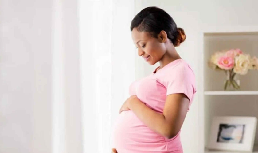
Headaches in pregnancy
Headaches are common during pregnancy and are often triggered by a change in hormones during pregnancy, but they usually improve or stop in the second and third trimester. You can take acetaminophen (paracetamol) if you need to for a mild headache, but get advice from a pharmacist or your doctor about how much to take and for how long. And make sure you follow the instructions on the packet for how much you can take. Try to take the lowest dose of paracetamol that works and for the shortest amount of time. Your doctor or pharmacist can give you more advice if the pain is ongoing and doesn’t go away with acetaminophen (paracetamol).
There are some painkillers you should NOT take while you’re pregnant. These include tablets or capsules that:
- contain added caffeine (sometimes sold with ‘extra’ on the label)
- contain codeine
- are anti-inflammatory, like ibuprofen or aspirin.
Some women may be advised to take a low dose of aspirin as a treatment if they have had miscarriages before or they are at risk of pre-eclampsia. This will be prescribed by a doctor. Aspirin should not be taken as treatment for a headache.
To help prevent more headaches:
- drink plenty of fluids
- get enough sleep
- rest and relax.
Although most pregnancy headaches are innocent, unexplained, frequent headaches later in your pregnancy could be a sign of a more serious condition called pre-eclampsia, so tell your doctor if this is the case.
If you experience frequent headaches that don’t go away with acetaminophen (paracetamol), it could be a sign of a more serious medical condition called pre-eclampsia (pregnancy induced hypertension). This usually involves an increase in the pregnant woman’s blood pressure and problems with her kidneys. There are also other serious risks for both you and your baby. Pre-eclampsia also known as gestational high blood pressure or gestational hypertension mostly occurs in the second half of pregnancy, after 20 weeks and goes away within 6 weeks of the baby’s birth.
Contact your doctor, particularly if, along with your headaches, you have a pain below your ribs, feel like you have heartburn, you suddenly swell in your face, hands or feet, or you have problems with your eyesight. This could be a sign of pre-eclampsia, a pregnancy condition that can be dangerous for you and the baby if it is not monitored and treated.
See your doctor immediately if you get any of the following symptoms as they could be symptoms of pre-eclampsia:
- a very severe headache
- a problem with vision such as blurring or flashing lights in your eyes
- severe pain just below ribs
- vomiting
- sudden swelling in your face, hands or feet
See your doctor or hospital maternity unit if you have a headache and any of the following symptoms:
- discomfort in the lowest part of your stomach (pelvis)
- back pain
- loin pain (your sides between the lower ribs and pelvis, and the lower part of the back)
- needing to wee a lot or an uncontrollable need to wee
- cloudy, foul-smelling (fishy) or bloody wee
- a raised temperature (over 99.5 °F or 37.5°C)
- feeling sick (nausea) and vomiting.
This could be a sign of a urinary tract infection (UTI). Urinary tract infections cab be treated with antibiotics that are safe to use in pregnancy.
Red Flags for headache in pregnancy 1):
- Headache that peaks in severity in less than five minutes
- New headache type versus a worsening of a previous headache
- Change in previously stable headache pattern
- Headache that changes with posture (e.g. Sstanding up)
- Headache awakening the pregnant
- Headache precipitated by physical activity or Valsalva manoeuvre (e.g. Coughing, laughing, straining)
- Thrombophilia
- Neurological symptoms or signs
- Trauma
- Fever
- Seizures
- History of malignancy
- History of HIV or active infections
- History of pituitary disorders
- Elevated blood pressure
- Recent travel at risk of infective disease
What causes headaches during pregnancy?
Many women experience headaches during pregnancy and may occur at any time during your pregnancy, but headaches tend to be most common during the first and third trimesters. If you’re pregnant, you may notice an increase in the number of headaches you have at around week 9 of your pregnancy.
As well as hormonal changes, headaches in the early stages of pregnancy can be caused by an increase in the volume of blood your body is producing.
During the first trimester, your body experiences a surge of hormones and an increase in blood volume. These two changes can cause more frequent headaches. These headaches may be further aggravated by stress, poor posture or changes in your vision.
Women who have regular migraine headaches may discover that they experience fewer migraines during pregnancy; however, some women may encounter the same number or even more migraine headaches. If you are pregnant, it is important to talk to your health care provider about any medications that you may be taking for headaches.
Headaches during the third trimester tend to be related more often to poor posture and tension from carrying extra weight. Headaches during the third trimester may also be caused by a condition called pre-eclampsia, which is high blood pressure during pregnancy.
Other causes of headaches during pregnancy can include:
- not getting enough sleep
- withdrawal from caffeine (e.g. in coffee, tea or cola drinks)
- low blood sugar
- dehydration
- feeling stressed
- poor posture, particularly as your baby gets bigger
- having depression or anxiety
Migraines and pregnancy
Migraine is a particular type of headache that mostly occurs on one side of the head – it can be either moderate or very painful. People who experience migraine can also feel sick or vomit, and be sensitive to light or sound.
Many people who have migraines may have:
- severe pain, which feels like a throbbing, pounding or pulsating pain
- nausea (feeling sick)
- pain brought on by bright lights
- an ‘aura’, such as flashing lights before the migraine starts.
In pregnancy, migraine may get worse for the first few months, but for many women it can improve in the later stages of their pregnancy when the level of the hormone estrogen stabilizes. Other women may experience no change or a decrease in the number of migraine headaches while pregnant. Some women may experience differences in migraine during different pregnancies.
If you have always had migraines, you may find that they get better during your pregnancy. But pregnancy can also change the nature of migraines, so if you have a migraine that feels different to what you’ve had before, see your doctor or hospital maternity unit.
Will a migraine hurt my baby?
Women with migraines are at slightly higher risk of developing high blood pressure and pre-eclampsia. This risk is very small and most women with migraines will not have high blood pressure.
If you have any worries at any time, talk to your doctor.
Secondary headaches
Pregnancy is a risk factor for a secondary headache disorder. Hypercoagulability, hormonal changes and anaesthesia for labour are just some of the multiple factors contributing to the high incidence of secondary headaches during pregnancy 2).
A recent study by Robbins et al 3) found 35% of secondary headaches among 140 pregnant women presenting with acute headache: hypertensive disorders of pregnancy covered 51% of these cases (about 18% of total), with preeclampsia as the major cause, followed by reversible posterior leukoencephalopathy syndrome (PRES, eclampsia), reversible cerebral vasoconstriction syndrome and acute arterial hypertension. These data place between two previous studies that reported percentages of secondary headaches ranging from 14.3% to 52.6% 4). In particular, among patients with a primary headache history, longer attack duration is the most common feature suggesting a secondary headache, reaching the statistical significance 5) or just approaching it 6).
The authors show how lack of headache history, elevated blood pressure and abnormalities at neurologic examination are the main red flags for a secondary origin of an acute headache during pregnancy 7). In the second trimester, a new onset of headache may signal the presence of a pseudotumor cerebri 8), while in case of a severe postural headache a spontaneous intracranial hypotension must be ruled out 9). In front of the well-known red flags (see the list above) brain MRI or CT scan are often required 10). Use of contrast agents such as gadolinium is not recommended, given the lack of data regarding safety to the fetus and its ability to cross the placenta and remain in the amniotic fluid 11).
Iodinated contrast should be avoided as well as it may suppress fetal thyroid function 12).
Recently the European Headache Federation published a consensus statement on technical investigation for primary headache disorders 13).
Secondary headache features may not differ from those of primary headaches; furthermore, migraine is as an independent risk factor for the development of secondary headaches (e.g., the risk of gestational hypertension increased by 1.42-fold) 14), so that recognizing these conditions in pregnant women may be a true diagnostic challenge.
Cerebral venous thrombosis, pre-eclampsia, hamorrhagic or ischemic stroke, subarachnoid haemorrhage (SAH), reversible cerebral vasoconstriction syndrome, reversible posterior leukoencephalopathy syndrome (reversible posterior leukoencephalopathy syndrome), idiopathic intracranial hypertension or pituitary apoplexy must be ruled out as soon as possible (Table 1) 15).
Table 1. Main causes of secondary headache in pregnant women
| Secondary headaches during pregnancy | |
|---|---|
| Arterial dissection | Intracranial hypotension |
| Arteriovenous malformation | Ischemic stroke |
| Brain tumors | Meningitis/encephalitis |
| Cerebral venous thrombosis (CVT) | Pituitary adenoma |
| Choriocarcinoma | Pituitary apoplexy |
| Cranial neuralgias | Pituitary meningioma |
| Dehydration | Reversible posterior leukoencephalopathy syndrome (PRES) |
| Eclampsia and pre-eclampsia | Reversible vasoconstriction syndrome (RCVS) |
| Head trauma | Sinusitis |
| Idiopathic intracranial hypertension (IIH) | Subarachnoid haemorrhage (SAH) |
| Intracranial haemorrhage (ICH) | Vasculitis |
Cerebral venous thrombosis
Headache caused by cerebral venous thrombosis has no specific characteristics: it is most often diffuse, progressive and severe, but can be unilateral and sudden (even thunderclap), or mild, and sometimes is migraine-like 16). Headache is present in 80–90% of cases of cerebral venous thrombosis and it is often associated with focal signs (neurological deficits or seizures) and/or signs of intracranial hypertension (nausea and papilledema), subacute encephalopathy or cavernous sinus syndrome, carrying a mortality rate of up to 30% 17).
Pre-eclampsia and eclampsia
Pre-eclampsia occurs in 5% of pregnancies 18): a progressive bilateral (temporal, frontal, occipital or diffuse) pulsating headache in a woman who is pregnant or in the puerperium (up to 4 weeks postpartum), often aggravated by physical activity, failing to respond to the over-the-counter remedies, may be the herald symptom of this condition, which can associate with visual changes similar to the typical visual aura. It must resolve within a week after blood pressure adjustment 19). According to the International Classification of Headache Disorders 3rd edition (ICHD-3 beta) headache should have at least two of the following three characteristics: a) bilateral location, b) pulsating quality, and c) aggravated by physical activity 20).
Ischemic stroke
Headache accompanies ischemic stroke especially within the posterior circulation, in up to one-third of cases and is usually overshadowed by focal signs and/or alterations of consciousness, which in most cases allows easy differentiation from the primary headaches. The risk of ischemic stroke in migraineurs was evaluated using the United States Nationwide Inpatient Sample from the Healthcare Cost and Utilization Project of the Agency for Healthcare Research and Quality and found to be elevated 21).
Headache has a self-limited course, and is very rarely the presenting or a prominent feature of ischemic stroke 22). It is usually of moderate intensity, and has no specific characteristics. It can be bilateral or unilateral ipsilateral to the stroke. Rarely, an acute ischemic stroke can present with an isolated sudden (even thunderclap) headache 23).
The diagnosis of headache and its causal link with ischemic stroke is easy because the headache presents both acutely and with neurological signs and because it often remits rapidly.
Subarachnoid hemorrhage
In subarachnoid hemorrhage headache is usually the prominent symptom. The pain is typically severe and sudden, peaking in seconds (thunderclap headache) or minutes, often followed by vomiting and loss of consciousness 24). Subarachnoid hemorrhage is a serious condition with mortality rate of 40–50% and with 10–20% of patients dying before arriving at hospital. The abrupt onset is the key feature and can help to distinguish from primary headaches with thunderclap features (e.g., associated with exercise or sexual activity). Subarachnoid hemorrhage presents a 20-fold increased risk in the puerperium and it gives a thunderclap headache 25).
Arterial dissection
Arterial dissection is a rare complication of pregnancy and puerperium. There have been reports of cervical carotid, vertebral and intracranial artery dissection in association with preeclampsia 26). Headache is the most frequent inaugural symptom, described as severe, unilateral (ipsilateral to the dissected vessel), with a sudden (even thunderclap) onset. Pain is persistent for days and can remain isolated or be a warning symptom preceding ischaemic infarcts.
Reversible cerebral vasoconstriction syndrome
Headache caused by reversible cerebral vasoconstriction syndrome is severe and diffuse and typically of the thunderclap type, recurring over 1–2 weeks, often triggered by sexual activity, exertion, Valsalva manoeuvres and/or emotion 27). Headache is often the only symptom of reversible cerebral vasoconstriction syndrome, but the condition can be associated with fluctuating focal neurological deficits and sometimes seizures.
Reversible cerebral vasoconstriction syndrome is commonly associated with the post-partum period, usually within a week after delivery: its severe thunderclap headache usually relapses within a few days, resolving by approximately twelve weeks after clinical onset 28). The typical differential diagnosis is cerebral vasculitis, which needs to be ruled out due to the course of the disease and different treatment options.
Posterior reversible encephalopathy syndrome
Posterior reversible encephalopathy syndrome (PRES) is a neuro-radiological clinical entity characterized by insidious onset of headache, impaired consciousness, visual changes or blindness, seizures, nausea, and vomiting, and focal neurological signs. In nearly 2/3 of patients with posterior reversible encephalopathy syndrome, headache is the most common symptom and is usually described as occipital and bilateral and dull in nature 29). Symptoms develop without prodrome, and progress over 12–48 hours.
Posterior reversible encephalopathy syndrome is often associated with hypertensive encephalopathy, preeclampsia, eclampsia, reversible cerebral vasoconstriction syndrome, renal failure, immunosuppressive therapy or chemotherapy. posterior reversible encephalopathy syndrome is more common in women and the development of this condition after delivery is unusual. The condition is usually reversible when early diagnosis is established and appropriate treatment is started without delay; symptoms generally resolve within a period of days or weeks while recovery of the MRI abnormalities takes longer 30).
Idiopathic intracranial hypertension
Usually during the first trimester, obese women can suffer from a progressive, daily headache, aggravated by Valsalva and position change, associated with papilledema and severe visual deficits, together with tinnitus or sixth-nerve palsies, defining the clinical pattern of idiopathic intracranial hypertension 31). The headache is frequently described as frontal, retro-orbital, ‘pressure like’ or explosive; migraine-like headache may also occur.
Pituitary apoplexy
Pituitary apoplexy is a rare cause of sudden and severe headache during pregnancy 32). The sudden rise of a severe headache, with nausea, vomiting, ophtalmoplegia, altered consciousness and accompanied from onset or later by visual symptoms and/or hypopituitarism must raise the clinical suspicion of a pituitary apoplexy 33). The rare clinical syndrome of pituitary apoplexy is an acute, life-threatening condition. It is important to distinguish from the other causes of thunderclap headache 34). Most cases occur as the first presentation of rapid enlargement of non-functioning pituitary macroadenomas as a result of hemorrhage and/or infarction.
What can I do to prevent headaches in pregnancy?
There are some things you can do to prevent headaches. Try to:
- figure out what causes your headache also called a headache trigger. Common headache triggers are cigarette smoke, certain foods and eye strain. Once you know your triggers, try to limit or get rid of them.
- drink at least eight glasses of fluid a day to avoid dehydration
- do something active every day
- get enough sleep every night. Rest during the day when you can.
- eat a healthy, balanced diet
- rest and relax as much as possible. You could try things like mindfulness or yoga
- try to reduce your stress. Stress is worry, strain or pressure that you feel in response to things that happen in your life. Tell your health care provider if you need help to reduce your stress.
Talk to your health care provider before you take any medicine, supplement or herbal product to relieve your headache. Some may be harmful to your baby.
Headaches in pregnancy treatment
It’s not advisable for pregnant women with migraine to use migraine medicine. For other headaches it’s also recommended that you try to treat your headache without medicine, however your health care provider may recommend acetaminophen.
You may want to try to relieve your headache with one or more of the following natural remedies:
- getting more sleep or rest and relaxation
- pregnancy yoga classes or other exercise
- practising good posture, particularly later in your pregnancy
- eating regular, well-balanced meals
- putting a warm facecloth on your eye and nose area, if it is a sinus headache
- putting a cold pack on the back of your neck, taking a bath or using a heat pack, if it is a tension headache
- neck and shoulders massage
- if you have a sinus headache, apply a warm compress around your eyes and nose
- if you have a tension headache, apply a cold compress or ice pack at the base of your neck
- maintain your blood sugar by eating smaller, more frequent meals – this may also help prevent future headaches
- get a massage – massaging your shoulders and neck is an effective way to relieve pain
- rest in a dark room and practice deep breathing
- take a warm shower or bath
- practice good posture (especially during the third trimester).
Pregnant women who experience migraine should avoid things that may trigger their migraine headaches. This may include:
- chocolate
- alcohol
- yogurt
- peanuts
- bread
- sour cream
- preserved meats
- aged cheese
- bread with fresh yeast
- monosodium glutamate (MSG)
- caffeine (withdrawal from)
- bright or flickering lights
- strong smells
- loud sounds
- computer or movie screens
- sudden or excessive exercise
- emotional triggers such as arguments or stress
If you do take medicine for your headache or migraine you should check with your doctor, pharmacist or midwife first. Acetaminophen (paracetamol) with or without codeine, is generally considered safe for pregnant women to use but you should avoid using other pain medicine such as aspirin or ibuprofen.
References [ + ]


